biomedical applications of lasers (1)
TRANSCRIPT

Seminar on
BIOMEDICAL APPLICATIONS OF LASERS

LIBS-Laser-induced breakdown spectroscopy
• (LIBS) is a type of atomic emission spectroscopy which uses a highly energetic laser pulse as the excitation source. The laser is focused to form a plasma, which atomizes and excites samples.
• In principle, LIBS can analyse any matter regardless of its physical state, be it solid, liquid or gas. Because all elements emit light of characteristic frequencies when excited to sufficiently high temperatures,
• LIBS operates by focusing the laser onto a small area at the surface of the specimen.

LIBS-Laser-induced breakdown spectroscopy

LIBS-Laser-induced breakdown spectroscopy
• A typical LIBS system consists of a Nd:YAG solid-state laser and a spectrometer with a wide spectral range and a high sensitivity, fast response rate, time gated detector. This is coupled to a computer which can rapidly process and interpret the acquired data
• The spectrometer collects electromagnetic radiation over the widest wavelength range possible, maximising the number of emission lines detected for each particular element. Spectrometer response is typically from 1100 nm (near infrared) to 170 nm (deep ultraviolet)
*Nd:YAG (neodymium-doped yttrium aluminium garnet; Nd:Y3Al5O12)
LIBS : A New Method for Heavy Metal in Biomedical Field and Applied in Plasma Medical
-Chenyu Jiang, Jinjiang Cui*, Yanbo Shi, Ningning Dong, Fan Wang, Huiming Tan
A New Method for Heavy Metal in Biomedical Field and Applied in Plasma Medical
• In this paper, the lead concentrations of samples included in glass, pig blood and so on are determinate by LIBS, based on a Nd: YAG Q-switched pulse with wavelength 1064 nm as an exciting source and nongated CCD in common spectrograph as detector.
• The energy of pulse on the surface of samples is about 100mJ and the width of pulse is 12ns.
A New Method for Heavy Metal in Biomedical Field and Applied in Plasma Medical
• There are about 40 chemical elements in the living organisms of a human body.
• These elements contain is three groups: the major group ( H, C, N, O), the trace elements group ( Na, Mg, P, S, Cl, K, Ca , Fe, Mn, Co, Zn and Ni )
• Demonstrated an all-optical technique for real-time and online analysis of elements , sample: pig blood.
• The detection limit was shown to be 63ppm in theory and 74 ppm in experiment.
A New Method for Heavy Metal in Biomedical Field and Applied in Plasma Medical
• LIBS spectra of pig blood shows the difference of K content between the pig blood and common pig blood.
• The apparatus was identical to a conventional LIBS setup, sharing most of the LIBS advantages but orders of magnitude more sensitive.
• It will be a valuable technique for elemental analysis and a rapid point-of-care (POC) medical diagnostic technology
* Laser-induced breakdown spectroscopy

Femtosecond laser
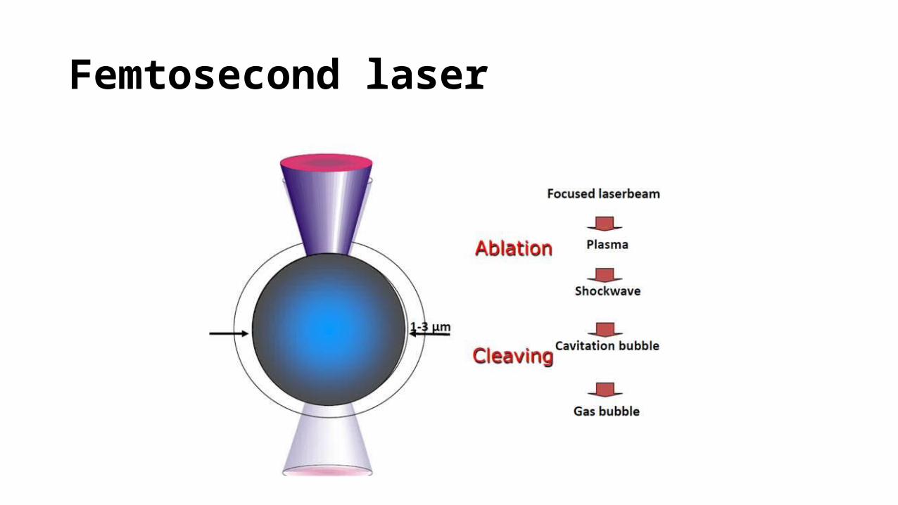
• Femtosecond (FS) laser is an infrared laser with a wavelength of 1053nm.
• FS laser like Nd: YAG laser works by producing photo disruption or photoionization of the optically transparent tissue such as the cornea.
• FS laser or Nd:YAG laser results in the generation of a rapidly expanding cloud of free electrons and ionized molecules. The acoustic shock wave so generated results in disruption of the treated tissue.
• Collateral damage with FS laser is 106 times less than with the Nd:YAG laser. This makes FS laser safe for use in corneal surgeries which require exquisite precision.
Femtosecond laser
Femtosecond laser

Biomedical material ablation by femtosecond laser double pulses through small-core hollow fibers
Yusuke Nishizawa, Seiji Takeda, and Minoru Obara Keio University, Department of Electronics and Electrical Engineering, 3-14-1, Hiyoshi,
Kohoku-ku, Yokohama, 213-8522 JAPAN

Biomedical material ablation by femtosecond laser double pulses through small-core hollow fibers
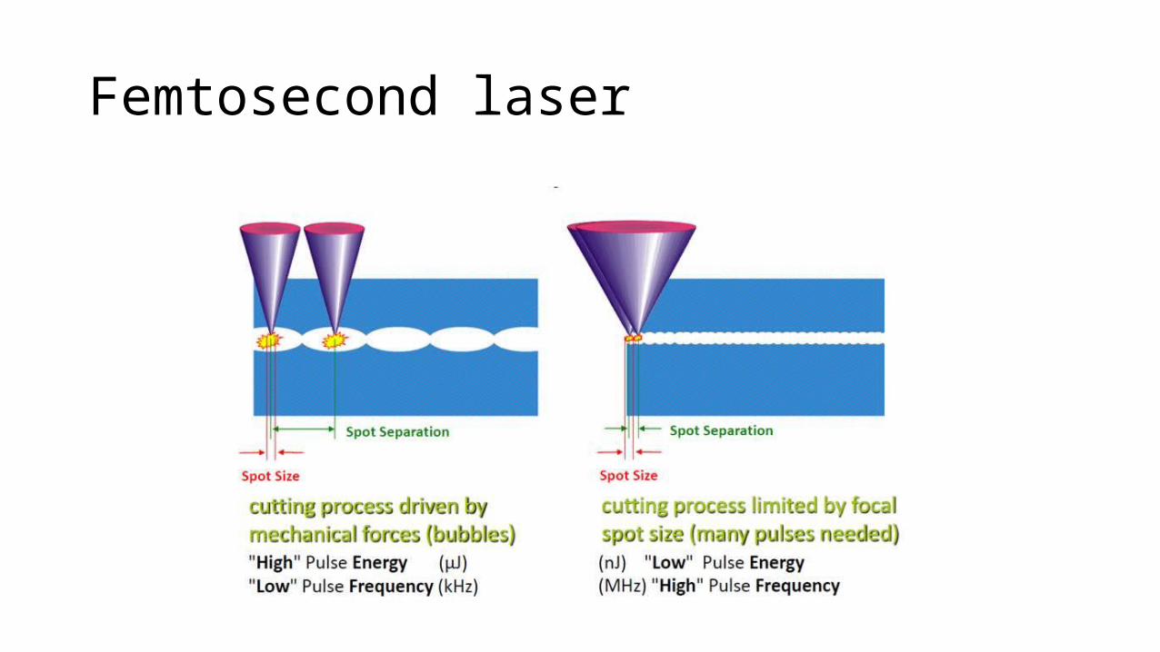
• In this paper, it demonstrate a high quality beam delivery using a flexible small-core hollow fiber and the ablation processing of hydroxyapatite (HAp) and collagen with fs laser double pulses through a bend hollow fiber. HAp is a main component of bones and teeth.
• Adopteding two new methods
1. use of a hollow fiber with a 320-µm core diameter in order to improve the output beam intensity profile by eliminating the high order modes
2. double pulse laser transmission scheme with a tailored ps-order delay time, in which the second pulse comes earlier than the ablation onset.

(3D) scanning systems
• The general image acquisition and data processing steps for obtaining a reconstruction of the exterior features of an object are:
• Calibration: The system must first undergo a calibration procedure to determine a transformation matrix H which will compensate for the projective distortion of the camera.
• Image acquisition: M views of the laser trace on the object are then captured, separated by rotations of R = 360◦ M about the vertical z-axis. This process is repeated for Nz planar cross-sections in vertical increments along the z-axis.
• Image processing: Images undergo processing in order to isolate the laser trace segments within the image, and determine a set of point coordinates to represent the trace.
(3D) scanning systems
• Data manipulation: Laser trace point coordinates are transformed using the transformation matrix H obtained from the calibration procedure. Then the segments are registered into a common coordinate system by rotating them to their respective orientation and stacking them along the vertical z-axis. Curve fitting is applied to the segments of each planar section to form entire 360◦ outlines of the external profile of the object, resulting in a 3D point cloud representing the object’s surface.
• Object reconstruction: A 3D surface mesh is applied to the point cloud in order to reconstruct the final 3D model.

A Simple, Low Cost, 3D Scanning System Using the Laser Light-
Sectioning Method Beverly D. Bradley', Adrian D.C. Chan',M. John D. Hayes2 1Systems and Computer Engineering, Carleton University 2Mechanical and Aerospace Engineering, Carleton University 1125 Colonel By Drive, Ottawa, Ontario, Canada KIS 5B6

A Simple, Low Cost, 3D Scanning System Using the Laser Light-
The 3D model acquisition pipeline have been identified in as follows: 1. planning methods for data acquisition; 2. reliable capture and robust processing of data for a larger class of
objects, environments, and objects with challenging surface properties;
3. automation of all the steps, to minimize user input; 4. real-time feedback of the acquired surface;5. improved capture and representation of surface appearance; and 6. methods for assessing global model accuracy after range scan
registration.
A Simple, Low Cost, 3D Scanning System Using the Laser Light-
• There is good reason to believe that an acceptable level of accuracy can be achieved in future iterations of the design.
• the majority of the errors are the result of manual manipulation of the device,
• Since the data are, as yet, too noisy to obtain satisfactory dimensional tolerance,
• The overall cost of the system’s components is well below the least expensive system found on the market when the price of software is considered.
Laser Scalpel
Introduction
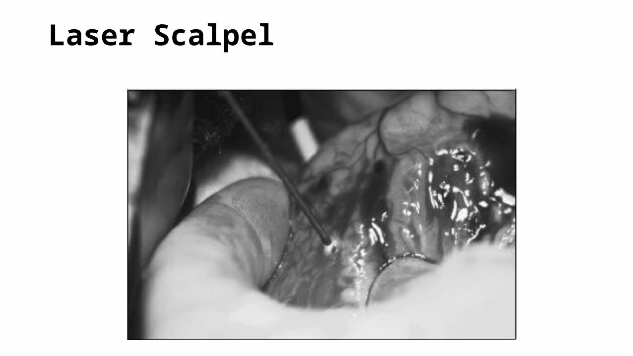
• Surgical operations were difficult to perform with convention scalpel compared to Laser scalpel.
• Focused laser beam from a carbon dioxide gas laser could cut through human tissue easily and neatly
• The surgeon could direct the beam from any angle by using a mirror mounted on a movable metal arm.
Laser Scalpel

Laser Scalpel
Advantages
• The light beam is consistent, which means that it gives off the same amount of energy from one second to next.
• Surgical laser is that the hot beam cauterizes, or seals off, the open blood vessels as it moves along.
• The cells in human tissue do not conduct heat very well, so the skin or any other tissue near the laser incision does not get very hot and is not affected by the beam. This is helpful when the doctor has to operate on a tiny area that is surrounded by healthy tissues or organs.

Laser Scalpel
Disadvantages
• It cannot totally replace the convention scalpel
• Laser beams have the capacity to do a great deal of damage when coupled with high enough energy and absorption
• It is important to guard against electric shock, as lasers require the use of high voltage.
• Laser beams can burn or destroy healthy tissue, cause injuries that are painful and sometimes permanent, and actually compound problems they are supposed to solve.

Laser Scalpel
Conclusion
• Surgical use of lasers is rapidly advancing.
• At first, lasers were considered most effective in operating on areas that are easy to reach—areas on the body's exterior, including the skin, mouth, nose, ears, and eyes
• In order to be able to direct the laser beam the doctor must be able to see inside the body.

Cleaning Arteries
Introduction
• lasers are increasingly used to clean plaque from people's arteries
• The traditional method for removing the plaque involves opening the chest and making several incisions, a long and sometimes risky operation.
• An effective alternative is to use a laser beam to burn away the plaque
• An optic fiber that has been connected to a tiny television camera can be inserted into an artery.
• The fiber-optic array is in place the laser is fired and the plaque destroyed, and then the exhaust vapors are sucked back through a tiny hollow tube that is inserted along with the optical fibers. This medical process is known as laser angioplasty

Cleaning Arteries
Advantages
• No incision is needed
• There is also little or no bleeding, and the patient can enjoy total recovery in a day or two

Cleaning Arteries
Disadvantages
• when the laser beam fires at the plaque it must be aimed very carefully because a slight miss could cut through the wall of the artery and cause serious bleeding
• small pieces of burnt debris from the destroyed plaque, enter the bloodstream, they can cause blockage in smaller blood vessels, bringing further complications
Cleaning Arteries
Conclusion
• Continuous technical advancements have considerably reduced these risks, and the number of successful laser angioplasties performed is increasing each year.

Heal and Reshape the Eyes
Introduction
• Some of the most remarkable breakthroughs for medical lasers have been in the area of ophthalmology, the study of the structure and diseases of the eye,
• The cornea in the eye lets in laser light just as well and remains unaffected by the beam.
• In advanced cases,hundreds of tiny extra blood vessels form on the retina. These block light from the surface of the membrane, resulting in partial or total blindness. The laser most often used in the treatment of this condition is powered by a medium of argon gas.
• beam is aimed through the cornea and it burns away the tangle of blood vessels covering the retina

Heal and Reshape the Eyes
Advantages
• the laser is very useful in removing extraneous blood vessels that can form on the retina—the thin, light-sensitive membrane at the back of the eyeball
• The laser can also repair a detached retina
• In case of glaucoma, the laser punches a hole in a preplanned spot and the fluid drains out through the hole
Heal and Reshape the Eyes
Conclusion
In eyes impaired with glaucoma the fluid does not drain properly, and the buildup affects vision; blindness can sometimes result. In some cases drugs can be used to treat glaucoma. If the drugs fail, however, many doctors now turn to the laser to avoid conventional surgery.
Eye Surgery
Introduction:
• The laser works like a sewing machine to repair a detached retina, the membrane that lines the interior of the eye
• The laser beam is adjusted so that it can pass harmlessly through the lens and focus on tiny spots around the damaged area of the retina.
• When it is focused, the beam has the intensity to "weld" or seal the detached area of the retina back against the wall of the eyeball.
• A technique widely known as LASIK (which stands for L aser- A ssisted I n S itu K eratomilensis is used in shaping of the eye’s cornea.
• The patient's eyeglass prescription is literally carved inside the cornea with the beam of an excimer laser [a laser device that produces pulses of ultraviolet, or UV, light].

Eye Surgery
Eye Surgery
Advantages:
• A technique widely known as LASIK (which stands for L aser- A ssisted I n S itu K eratomilensis is used in shaping of the eye’s cornea.
• The patient's eyeglass prescription is literally carved inside the cornea with the beam of an excimer laser [a laser device that produces pulses of ultraviolet, or UV, light].
• Inner portion of the cornea is exposed to the excimer laser
Eye Surgery
Disadvsntages:
The changes it makes in the cornea are permanent, and the danger of unexpected damage is ever present

Eye Surgery
Conclusion:
• Procedure has become increasingly popular each year; about a million Americans had it done in the year 2000, and about four thousand surgeons in the United States were trained to perform it.

Cosmetic
• Medical lasers are used in the removal of certain kinds of birthmarks. Port-wine stains, reddish purple skin blotches
• The medical laser is able to remove a port-wine stain for the same reason that a military laser is able to flash a message to a submerged submarine
• Both lasers take advantage of the monochromatic quality of laser light, that is, its ability to shine in one specific color. The stain is made up of thousands of tiny malformed blood vessels that have a definite reddish purple color. This color very strongly absorbs a certain shade of green light. In fact, that is why the stain looks red. It absorbs the green and other colors in white light but reflects the red back to people's eyes

Cosmetic

Cosmetic
Advantages
• To treat the stain, the doctor runs a wide low-power beam of green light across the discolored area. The mass of blood vessels in the stain absorbs the energetic laser light and becomes so hot that it is actually burned away.
• A similar method is often successful in removing tattoos
• The beam bleaches the dyes in the tattoo without burning the surrounding skin.
• laser-assists cosmetic procedure in the removal of unwanted hair.

Cosmetic
Disadvantages:
• During the process some minor scarring sometimes occurs
Assisted Dentistry
Introduction:
• People prefer laser technology to drilling cavity.
• Many dentists now employ an Nd-YAG laser (which uses a crystal for its lasing medium) instead of a drill for most cavities
Assisted Dentistry
Advantages:
• The laser treatment takes advantage of the simple fact that the material that forms in a cavity is much softer than the enamel (the hard part of a tooth). The laser is set at a power that is just strong enough to eliminate the decayed tissue but not strong enough to harm the enamel. When treating a very deep cavity bleeding sometimes occurs, and the laser beam often seals off blood vessels and stops the bleeding.
• It does not hurt. Each burst of laser light from a dental laser lasts only thirty trillionths of a second, much faster than the amount of time a nerve takes to trigger pain. In other words, the beam would have to last 100 million times longer in order to cause any discomfort. So this sort of treatment requires no anaesthetic

Assisted Dentistry
Conclusion
• There are literally hundreds of other medical uses for the laser. Still, numerous medical conditions cannot be helped by laser light. And even in those that do respond to laser treatments, a doctor may have a good reason for choosing a different method in a specific case.
• While the laser is a marvelous medical tool, it cannot cure every ill. Yet the world has seen probably only a small fraction of the laser's potential. After all, this supertool has only existed since 1960, and, considering the medical advances it already has created, the future appears promising indeed

Thank You