biologic therapy in refractory non-multiple sclerosis
TRANSCRIPT

Journal of
Clinical Medicine
Article
Biologic Therapy in Refractory Non-MultipleSclerosis Optic Neuritis Isolated or Associated toImmune-Mediated Inflammatory Diseases.A Multicenter Study
Alba Herrero-Morant 1,†, Carmen Álvarez-Reguera 1,†, José L. Martín-Varillas 2,†,Vanesa Calvo-Río 1, Alfonso Casado 1, Diana Prieto-Peña 1, Belén Atienza-Mateo 1,Olga Maiz-Alonso 3, Ana Blanco 3, Esther Vicente 4, Íñigo Rúa-Figueroa 5 ,Laura Cáceres-Martin 5, José L. García-Serrano 6, José Luis Callejas-Rubio 6,Norberto Ortego-Centeno 6, Javier Narváez 7 , Susana Romero-Yuste 8, Julio Sánchez 9,Paula Estrada 10 , Rosalía Demetrio-Pablo 1, David Martínez-López 1, Santos Castañeda 4,José L. Hernández 1,*,‡ , Miguel Á. González-Gay 1,*,‡ and Ricardo Blanco 1,*,‡
1 Rheumatology, Ophtalmology and Internal Medicine, Hospital Universitario Marqués de Valdecilla,Av. de Valdecilla, 25, 39008 Santander, Spain; [email protected] (A.H.-M.);[email protected] (C.Á.-R.); [email protected] (V.C.-R.); [email protected] (A.C.);[email protected] (D.P.-P.); [email protected] (B.A.-M.);[email protected] (R.D.-P.); [email protected] (D.M.-L.)
2 Rheumatology, Hospital Sierrallana, Barrio Ganzo, s/n, 39300 Torrelavega, Spain; [email protected] Rheumatology and Ophtalmology, Hospital de Donostia, Paseo Dr. Begiristain, 117, 20080 Donostia, Spain;
[email protected] (O.M.-A.); [email protected] (A.B.)4 Rheumatology, Hospital Universitario de La Princesa, C/Diego de León, 62, 28006 Madrid, Spain;
[email protected] (E.V.); [email protected] (S.C.)5 Rheumatology, Hospital Universitario de Gran Canaria Doctor Negrín, C/Plaza Barranco de la Ballena, s/n,
35010 Las Palmas de Gran Canaria, Spain; [email protected] (Í.R.-F.);[email protected] (L.C.-M.)
6 Internal Medicine and Ophtalmology, Hospital San Cecilio, Av. del Conocimiento, s/n, 18016 Granada, Spain;[email protected] (J.L.G.-S.); [email protected] (J.L.C.-R.); [email protected] (N.O.-C.)
7 Rheumatology, Hospital de Bellvitge, Carrer de la Feixa Llarga, s/n, 08907 L’Hospitalet de Llobregat, Spain;[email protected]
8 Rheumatology, Complejo Hospitalario Universitario de Pontevedra, Loureiro Crespo, 2, 36002 Pontevedra,Spain; [email protected]
9 Rheumatology, Hospital Universitario 12 de Octubre, Av. de Córdoba, s/n, 28041 Madrid, Spain;[email protected]
10 Rheumatology, Hospital de Sant Joan Despí Moisès Broggi, Carrer de Jacint Verdaguer, 90,08970 Sant Joan Despí, Spain; [email protected]
* Correspondence: [email protected] (J.L.H.); [email protected] (M.Á.G.-G.);[email protected] (R.B.)
† Alba Herrero-Morant, Carmen Álvarez-Reguera and José L. Martín-Varillas shared first authorship.‡ Miguel Á. González-Gay, José L. Hernández and Ricardo Blanco shared senior authorship.
Received: 21 June 2020; Accepted: 7 August 2020; Published: 11 August 2020�����������������
Abstract: We aimed to assess the efficacy of biologic therapy in refractory non-Multiple Sclerosis(MS) Optic Neuritis (ON), a condition more infrequent, chronic and severe than MS ON. This wasan open-label multicenter study of patients with non-MS ON refractory to systemic corticosteroidsand at least one conventional immunosuppressive drug. The main outcomes were Best CorrectedVisual Acuity (BCVA) and both Macular Thickness (MT) and Retinal Nerve Fiber Layer (RNFL) usingOptical Coherence Tomography (OCT). These outcome variables were assessed at baseline, 1 week,and 1, 3, 6 and 12 months after biologic therapy initiation. Remission was defined as the absence
J. Clin. Med. 2020, 9, 2608; doi:10.3390/jcm9082608 www.mdpi.com/journal/jcm

J. Clin. Med. 2020, 9, 2608 2 of 13
of ON symptoms and signs that lasted longer than 24 h, with or without an associated new lesionon magnetic resonance imaging with gadolinium contrast agents for at least 3 months. We studied19 patients (11 women/8 men; mean age, 34.8 ± 13.9 years). The underlying diseases were Bechet’sdisease (n = 5), neuromyelitis optica (n = 3), systemic lupus erythematosus (n = 2), sarcoidosis (n = 1),relapsing polychondritis (n = 1) and anti-neutrophil cytoplasmic antibody -associated vasculitis (n = 1).It was idiopathic in 6 patients. The first biologic agent used in each patient was: adalimumab (n = 6),rituximab (n = 6), infliximab (n = 5) and tocilizumab (n = 2). A second immunosuppressive drug wassimultaneously used in 11 patients: methotrexate (n = 11), azathioprine (n = 2), mycophenolate mofetil(n = 1) and hydroxychloroquine (n = 1). Improvement of the main outcomes was observed after 1 yearof therapy when compared with baseline data: mean ± SD BCVA (0.8 ± 0.3 LogMAR vs. 0.6 ± 0.3LogMAR; p = 0.03), mean ± SD RNFL (190.5 ± 175.4 µm vs. 183.4 ± 139.5 µm; p = 0.02), mean ± SDMT (270.7 ± 23.2 µm vs. 369.6 ± 137.4 µm; p = 0.03). Besides, the median (IQR) prednisone-dose wasalso reduced from 40 (10–61.5) mg/day at baseline to. 2.5 (0–5) mg/day after one year of follow-up;p = 0.001. After a mean ± SD follow-up of 35 months, 15 patients (78.9%) achieved ocular remission,and 2 (10.5%) experienced severe adverse events. Biologic therapy is effective in patients withrefractory non-MS ON.
Keywords: optic neuritis; biologic therapy; rituximab; tocilizumab; adalimumab; infliximab
1. Introduction
Optic neuritis (ON) is an acute inflammatory optic neuropathy that may be associated withdramatic visual loss and an important decrease in quality of life in absence of an adequate treatment.Multiple Sclerosis (MS) ON, the most common form of presentation, is characterized by unilateralacute retroocular pain and visual loss, more commonly observed in Caucasian women between 18 and50 years [1]. Visual acuity (VA) in patients with MS-ON usually improves within a few months evenwithout treatment [2–4]. Non-MS ON is less frequent and can be an isolated disorder or related toinfections and immune-mediated diseases such as Neuromyelitis Optica (NMO) or other systemicdiseases [5]. Non-MS ON may have atypical features such as male gender, age less than 18 or greaterthan 50 years, absence of pain and bilateral presentation [5]. In non-MS ON, a chronic progressivedisease is more common. Flare-ups are frequent, leading often to visual loss [3,6]. If not promptlytreated, the visual outcome can be devastating, causing a severe visual loss, and even with adequatetreatment, many patients may worsen over months [7–10].
Therapy has been mostly focused on MS-ON. According to the Optic Neuritis Treatment Trial [2],in patients with MS or isolated ON, intravenous (i.v.) high-dose glucocorticoids followed by oralprednisolone may accelerate the visual recovery. Nevertheless, there is not a significant improvementof VA at 6 months and 1 year compared to placebo. The most recent Cochrane Review identified sixrandomized controlled trials with a total of 750 participants. Five trials (n = 633) only analyzed MS orisolated ON. It concluded that there is still no definitive evidence that i.v. glucocorticoids improvevisual outcomes after 6 months of treatment [11].
Non-MS ON treatment has been less frequently assessed. Glucocorticoids, plasmapheresisand intravenous immunoglobulins may be effective in acute attacks, particularly in NMO [12–15].Three recent clinical trials have analyzed the use of satralizumab, eculizumab and inebilizumabin NMO [16–18]. All three have demonstrated a reduction of risk of NMO attack compared toplacebo. Conventional immunosuppressive therapies have demonstrated clinical benefits for reducingrelapses [6], but biologic agents have been rarely used. Thus, rituximab (RTX), an anti-CD20 monoclonalantibody, tocilizumab (TCZ), an IL-6 monoclonal antibody [12,19–22], and anti-TNFα therapy, especiallyadalimumab (ADA) and infliximab (IFX), have been only used in some refractory cases [23–27].
J. Clin. Med. 2020, 9, 2608 3 of 13
Taking into account all these considerations, this study aimed to assess the efficacy and safetyof biologic therapy in refractory non-MS ON, both isolated and associated with immune-mediatedinflammatory diseases.
2. Experimental Section
2.1. Design and Enrollment Criteria
We performed an observational open-label multicenter study that included 19 patients diagnosed withnon-MS ON refractory to systemic glucocorticoids and at least one conventional immunosuppressive drug.
Patients were diagnosed with non-MS ON at the Ophthalmology, Neurology and RheumatologyUnits of eleven different referral Spanish Hospitals. Since biologic therapy is an off-label indicationfor ON, written informed consent was requested and obtained from all the patients. The study wasapproved by the Clinical Research Ethics Committee (ethical approval code: 2020.010).
Diagnosis of ON was based on clinical features, ophthalmologic examination, high-definitionoptical coherence tomography (OCT), magnetic resonance imaging (MRI) and cerebrospinal fluidanalysis (CSF). The presence of subacute vision loss in adults, along with a relative afferent papillarydefect (RAPD) was required for diagnosis [2–5]. In addition, MRI findings such either T1-weightedgadolinium enhancement of the optic nerve, or T2-weighted optic nerve hyperintensity were neededfor diagnosis [28,29]. Aquaporin-4 water channels -IgG and Myelin Oligodendrocyte Glycoprotein -IgGwere assessed in all patients. Both unilateral and bilateral cases of ON were included in the diagnosis.
Inclusion criteria were as follows: (a) non-MS ON, (b) lack of response to previous treatment witha high dose of systemic glucocorticoids defined as more than 7.5 mg/day for more than 3 months and(c) to at least one conventional immunosuppressive drug at its standard doses.
MS was excluded by the McDonald’s criteria that were based on clinical, imaging and laboratoryparameters [30].
As indicated by the Spanish Biologic Treatment Administration National Recommendations,the presence of infectious diseases had to be ruled out before starting the biologic treatment. To excludelatent tuberculosis, a tuberculin skin testing (PPD) and/or an interferon assay (quantiFERON) andchest radiography were performed. In positive cases, prophylaxis with isoniazid was initiated for atleast 4 weeks before using the biologic treatment and was maintained for 9 months. The presence ofmalignancies was excluded in all patients [31–39].
According to current guidelines, avoiding the use of anti-TNFα drugs is recommended in patientswith a history or familiar occurrence of demyelinating diseases [4,40]. Besides, a relatively commonanti-TNFα Adverse Event (AE) is ON [41,42]. Thus, anti-TNFα therapy was avoided in NMO(demyelinating disease).
2.2. Outcome Variables
The outcome variables were the efficacy and the safety of biologic therapy. The main outcomesof efficacy were best-corrected visual acuity (BCVA), retinal nerve fiber layer (RNFL), and macularthickness (MT). Secondary outcomes were remission, number of relapses and sparing effect ofglucocorticoids. To determine safety, AE were evaluated.
BCVA was estimated by the logMAR chart. The optic nerve was evaluated measuring with anOCT the loss of retinal nerve fibers with RNFL analysis. The loss of retinal nerve fibers associatedwith optic atrophy in patients with optic neuropathies can easily be visualized and quantified byOCT measuring the peripapillary RNFL [43]. The RNFL thickness was measured using the optic disccube protocol of the Fourier Cirrus HD-OCT (Carl Zeiss Meditec Inc., Dublin, CA, USA) software,version 6.0. This protocol generates a cube of data through a 6-mm square grid by acquiring a series of200 horizontal scan lines, each composed of 200 A-scans. The RNFL thickness at each pixel is measuredand an RNFL thickness map is generated. RNFL thickness quantification is a good measure of axonal

J. Clin. Med. 2020, 9, 2608 4 of 13
integrity associated with VA. It can predict the degree of visual recovery in acute cases of non-MSON [44,45].
Similarly, macular edema has been related to non-MS ON prognosis [46]. The macular cube512 × 128 scan was used to obtain MT; this protocol performs 512 horizontal A-scans and 128 verticalB-scan lines within a 6 × 6 mm cube of acquired signal data centered on the fovea. It has been assessedevaluating six areas of the macular cube (superior, superonasal, inferonasal, inferior, inferotemporaland superotemporal sectors) [46]. Besides, MRI was performed to determine optic nerve inflammatorychanges and to rule out structural lesions or other causes of ON [47–50].
Remission was defined as the absence of ON symptoms and signs that lasted longer than 24 h, withor without an associated new lesion on MRI with gadolinium contrast agents for at least 3 months [51,52].The remission status was classified as complete when there was full recovery of visual outcomes, partialrecovery when there was incomplete recovery and no remission when there was no improvement atall [53]. Relapses were defined as new ON symptoms and signs that lasted longer than 24 h, with orwithout an associated new lesion on MRI with gadolinium contrast agents [52]. AE related to biologictreatment were evaluated and recorded at follow-up.
2.3. Data Collection and Statistical Analysis
These outcome variables were recorded in each center according to a follow-up protocol agreedbeforehand. Information was stored in a computerized database, and to minimize entry error, all datawere double-checked.
Results are expressed as mean ± standard deviation (SD) for the variables with a normaldistribution or as median and interquartile range (25th–75th interquartile range (IQR)) for thosenot normally distributed. Continuous variables were compared with the 2-tailed Student t-test orthe Mann–Whitney U-test, as appropriate. The chi-square test or the Fisher exact test was used forthe dichotomous variables. The outcome variables were assessed and compared between baseline(at biologic therapy initiation), 1 week, 1 month, 3 months, 6 months and 1 year separately in eachoutcome, and Wilcoxon signed-rank test was used to assess continuous variables.
3. Results
3.1. Demographic and Clinical Features at Baseline
We studied 19 patients (11 women/ 8 men) with non-MS ON refractory to systemic glucocorticoidsand at least one conventional immunosuppressive drug. The mean age was 34.8 ± 13.9 years.The underlying diseases were idiopathic ON (n = 6), Bechet’s disease (n = 5), NMO (n = 3),systemic lupus erythematosus (SLE) (n = 2), sarcoidosis (n = 1), relapsing polychondritis (n = 1),and myeloperoxidase-anti-neutrophil cytoplasmic antibody-associated vasculitis (n = 1). Non-MS ONwas unilateral (n = 10) and bilateral (n = 9). The main demographic and clinical data are summarizedin Table 1.
Before biologic therapy initiation, all patients had received oral glucocorticoids (mean Maximumprednisone dose, 53.7 ± 17.7 mg/day). In 16 cases, i.v. pulses of Methylprednisolone (MP) (mean ± SDdose 3.3 ± 1.5 g) were used before oral glucocorticoids. The conventional immunosuppressive drugspreviously used and doses were azathioprine (AZA) (n = 8; 100–250 mg/p.o./day), methotrexate (MTX)(n = 7; 15–25 mg/s.c. or p.o./week), mycophenolate mofetil (MMF) (n = 5; 760–2000 mg/p.o./day),cyclophosphamide (CPM) (n = 4; 9 mg/kg/i.v./weekly), hydroxychloroquine (HCQ) (n = 2;200–400 mg/p.o./day), cyclosporine A (CyA) (n = 2; 250–300 mg/p.o./day) and leflunomide (LFN) (n = 1;20 mg/p.o/day). (Figure 1).
J. Clin. Med. 2020, 9, 2608 5 of 13
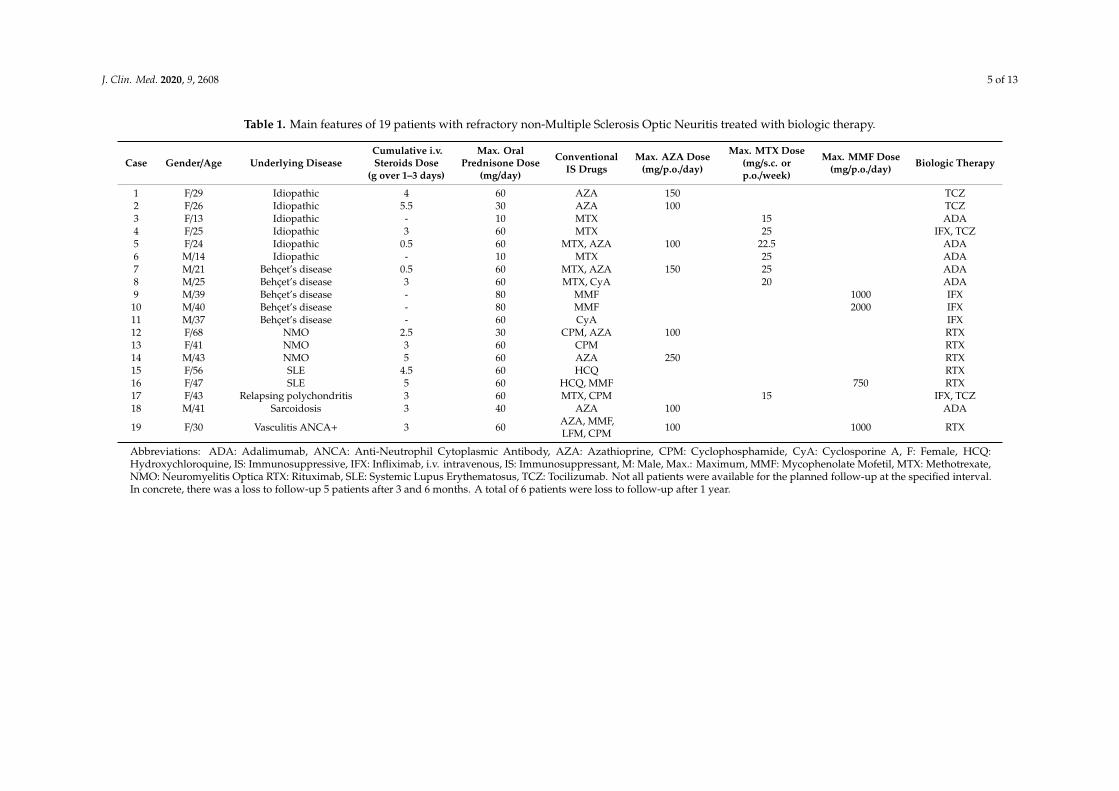
Table 1. Main features of 19 patients with refractory non-Multiple Sclerosis Optic Neuritis treated with biologic therapy.
Case Gender/Age Underlying DiseaseCumulative i.v.Steroids Dose
(g over 1–3 days)
Max. OralPrednisone Dose
(mg/day)
ConventionalIS Drugs
Max. AZA Dose(mg/p.o./day)
Max. MTX Dose(mg/s.c. orp.o./week)
Max. MMF Dose(mg/p.o./day) Biologic Therapy
1 F/29 Idiopathic 4 60 AZA 150 TCZ2 F/26 Idiopathic 5.5 30 AZA 100 TCZ3 F/13 Idiopathic - 10 MTX 15 ADA4 F/25 Idiopathic 3 60 MTX 25 IFX, TCZ5 F/24 Idiopathic 0.5 60 MTX, AZA 100 22.5 ADA6 M/14 Idiopathic - 10 MTX 25 ADA7 M/21 Behçet’s disease 0.5 60 MTX, AZA 150 25 ADA8 M/25 Behçet’s disease 3 60 MTX, CyA 20 ADA9 M/39 Behçet’s disease - 80 MMF 1000 IFX10 M/40 Behçet’s disease - 80 MMF 2000 IFX11 M/37 Behçet’s disease - 60 CyA IFX12 F/68 NMO 2.5 30 CPM, AZA 100 RTX13 F/41 NMO 3 60 CPM RTX14 M/43 NMO 5 60 AZA 250 RTX15 F/56 SLE 4.5 60 HCQ RTX16 F/47 SLE 5 60 HCQ, MMF 750 RTX17 F/43 Relapsing polychondritis 3 60 MTX, CPM 15 IFX, TCZ18 M/41 Sarcoidosis 3 40 AZA 100 ADA
19 F/30 Vasculitis ANCA+ 3 60 AZA, MMF,LFM, CPM 100 1000 RTX
Abbreviations: ADA: Adalimumab, ANCA: Anti-Neutrophil Cytoplasmic Antibody, AZA: Azathioprine, CPM: Cyclophosphamide, CyA: Cyclosporine A, F: Female, HCQ:Hydroxychloroquine, IS: Immunosuppressive, IFX: Infliximab, i.v. intravenous, IS: Immunosuppressant, M: Male, Max.: Maximum, MMF: Mycophenolate Mofetil, MTX: Methotrexate,NMO: Neuromyelitis Optica RTX: Rituximab, SLE: Systemic Lupus Erythematosus, TCZ: Tocilizumab. Not all patients were available for the planned follow-up at the specified interval.In concrete, there was a loss to follow-up 5 patients after 3 and 6 months. A total of 6 patients were loss to follow-up after 1 year.

J. Clin. Med. 2020, 9, 2608 6 of 13
J. Clin. Med. 2020, 9, x FOR PEER REVIEW 6 of 13
Before biologic therapy initiation, all patients had received oral glucocorticoids (mean Maximum prednisone dose, 53.7 ± 17.7 mg/day). In 16 cases, i.v. pulses of Methylprednisolone (MP) (mean ± SD dose 3.3 ± 1.5 g) were used before oral glucocorticoids. The conventional immunosuppressive drugs previously used and doses were azathioprine (AZA) (n = 8; 100–250 mg/p.o./day), methotrexate (MTX) (n = 7; 15–25 mg/s.c. or p.o./week), mycophenolate mofetil (MMF) (n = 5; 760–2000 mg/p.o./day), cyclophosphamide (CPM) (n = 4; 9 mg/kg/i.v./weekly), hydroxychloroquine (HCQ) (n = 2; 200–400 mg/p.o./day), cyclosporine A (CyA) (n = 2; 250–300 mg/p.o./day) and leflunomide (LFN) (n = 1; 20 mg/p.o/day). (Figure 1).
Figure 1. Flow-chart of biologic therapy in refractory non-Multiple Sclerosis optic neuritis. Abbreviations: ADA: Adalimumab, IFX: Infliximab; IS: Immunosuppressive, RTX: Rituximab; TCZ: Tocilizumab.
At biologic therapy initiation, MRI with gadolinium contrast agents was performed in all patients with the following results: normal (n = 8), ocular enhancement (chiasma (n = 1), choroidal bilateral (n = 1), periorbital bilateral (n = 1)), spinal cord enhancement (transverse myelitis (n = 2); C2-D3 (n = 1); C1-T1 (n = 1); D5-D7 (n = 1)), and cerebral enhancement (frontal subcortical bilateral (n = 1), supratentorial (n = 1)). CSF fluid analysis was only performed in 3 patients obtaining normal results in all of them.
3.2. Biologic Therapy and Efficacy
Biologic agents used were RTX (n = 6; two i.v. doses of 1 g/every 2 weeks and then every 6 months), ADA (n = 6; 40 mg/sc/1-2 week), IFX (n = 5; 5 mg/kg/i.v. at 0, 2 and 6 weeks and then every 8 weeks) and TCZ (n = 4; 2 as first and 2 as second biologic therapy; 8 mg/kg/i.v. 2–4 weeks). A second immunosuppressive drug was used simultaneously in 11 patients: MTX (n = 11), AZA (n = 2), MMF (n = 1) and HCQ (n = 1). In addition, all patients received oral prednisone (median (IQR) maximum dose at baseline of 40 (10–61.5) mg/day).
Thus, after one year of biologic therapy, mean ± SD BCVA improved from 0.63 ± 0.34 logMAR to 0.84 ± 0.29 logMAR (p= 0.03). Similarly, a significant improvement in optic nerve inflammation was observed since mean ± SD RNFL OCT increased from 183.4 ± 139.6 to 190.5 ± 175.4 μm (p = 0.02). Mean ± SD MT decreased from 369.6 ± 137.4 to 270.7 ± 23.2 μm (p = 0.03) (Figure 2). A 10.5% (7.1% of eyes) of patients experience a loss in BCVA.
After a mean ± SD follow-up of 35.3 ± 25.1 months, 15 patients (78.9%) achieved ocular remission [51,52]. They were on ADA (n = 6, 83.3%), RTX (n = 6, 66.6%), IFX (n = 5, 60%) and TCZ (n = 4, 100%). Only one relapse was described, at 3 months of RTX therapy in one patient.
Figure 1. Flow-chart of biologic therapy in refractory non-Multiple Sclerosis optic neuritis. Abbreviations:ADA: Adalimumab, IFX: Infliximab; IS: Immunosuppressive, RTX: Rituximab; TCZ: Tocilizumab.
At biologic therapy initiation, MRI with gadolinium contrast agents was performed in all patientswith the following results: normal (n = 8), ocular enhancement (chiasma (n = 1), choroidal bilateral(n = 1), periorbital bilateral (n = 1)), spinal cord enhancement (transverse myelitis (n = 2); C2-D3(n = 1); C1-T1 (n = 1); D5-D7 (n = 1)), and cerebral enhancement (frontal subcortical bilateral (n = 1),supratentorial (n = 1)). CSF fluid analysis was only performed in 3 patients obtaining normal results inall of them.
3.2. Biologic Therapy and Efficacy
Biologic agents used were RTX (n = 6; two i.v. doses of 1 g/every 2 weeks and then every 6 months),ADA (n = 6; 40 mg/sc/1-2 week), IFX (n = 5; 5 mg/kg/i.v. at 0, 2 and 6 weeks and then every 8 weeks)and TCZ (n = 4; 2 as first and 2 as second biologic therapy; 8 mg/kg/i.v. 2–4 weeks). A secondimmunosuppressive drug was used simultaneously in 11 patients: MTX (n = 11), AZA (n = 2), MMF(n = 1) and HCQ (n = 1). In addition, all patients received oral prednisone (median (IQR) maximumdose at baseline of 40 (10–61.5) mg/day).
Thus, after one year of biologic therapy, mean ± SD BCVA improved from 0.63 ± 0.34 logMARto 0.84 ± 0.29 logMAR (p = 0.03). Similarly, a significant improvement in optic nerve inflammationwas observed since mean ± SD RNFL OCT increased from 183.4 ± 139.6 to 190.5 ± 175.4 µm (p = 0.02).Mean ± SD MT decreased from 369.6 ± 137.4 to 270.7 ± 23.2 µm (p = 0.03) (Figure 2). A 10.5% (7.1% ofeyes) of patients experience a loss in BCVA.
After a mean±SD follow-up of 35.3±25.1 months, 15 patients (78.9%) achieved ocular remission [51,52].They were on ADA (n = 6, 83.3%), RTX (n = 6, 66.6%), IFX (n = 5, 60%) and TCZ (n = 4, 100%). Only onerelapse was described, at 3 months of RTX therapy in one patient.
Likewise, a decrease in the median (IQR) prednisone dose was also achieved (40 (10–61.5) mg/dayat baseline vs. 2.5 (0–5) mg/day at one year of follow-up; p = 0.001) (Figure 3). Before the initiationof biological therapy, all patients required an oral prednisone dose of more than 7.5 mg/day after3 months. After 3 months of biological therapy, 43.7% of patients required an oral prednisone dose ofmore than 7.5 mg/day. The biological therapies that required high doses of corticosteroids for morethan three months were IFX (n = 2), ADA (n = 1), RTX (n = 1).

J. Clin. Med. 2020, 9, 2608 7 of 13
J. Clin. Med. 2020, 9, x FOR PEER REVIEW 7 of 13
(a)
(b) Figure 2. Improvement in (a) Best Corrected Visual Acuity (BCVA) and (b) Optical Coherence Tomography (OCT) measurements. Abbreviations: BCVA: Best Corrected Visual Acuity; MT: Macular Thickness, OCT: Optical Coherence Tomography; RNFL: Retinal Nerve Fiber Layer. * p < 0.05 compared with basal data.
Likewise, a decrease in the median (IQR) prednisone dose was also achieved (40 (10–61.5) mg/day at baseline vs. 2.5 (0–5) mg/day at one year of follow-up; p = 0.001) (Figure 3). Before the initiation of biological therapy, all patients required an oral prednisone dose of more than 7.5 mg/day after 3 months. After 3 months of biological therapy, 43.7% of patients required an oral prednisone dose of more than 7.5 mg/day. The biological therapies that required high doses of corticosteroids for more than three months were IFX (n = 2), ADA (n = 1), RTX (n=1).
0
100
200
300
400
500
600
BASAL 1 WEEK 1 MONTH 3 MONTH 6 MONTH 1 YEAR
(μ)
MT
RNFL
*
*
* *
*
*
*
Figure 2. Improvement in (a) Best Corrected Visual Acuity (BCVA) and (b) Optical CoherenceTomography (OCT) measurements. Abbreviations: BCVA: Best Corrected Visual Acuity; MT: MacularThickness, OCT: Optical Coherence Tomography; RNFL: Retinal Nerve Fiber Layer. * p < 0.05 comparedwith basal data.J. Clin. Med. 2020, 9, x FOR PEER REVIEW 8 of 13
Figure 3. Glucocorticoid sparing effect of biologic therapy in refractory non-Multiple Sclerosis Optic Neuritis. * p < 0.05 compared with basal data.
Anti-TNFα drugs (ADA, IFX, n = 11) were compared with non-anti-TNFα (RTX, TCZ, n = 10) agents (Table 2). The underlying diseases were different in both groups. In Bechet’s Disease and sarcoidosis, anti-TNFα were used more frequently, and in NMO and SLE, non-anti-TNFα agents were the most frequently prescribed. At one-month, a greater improvement in BCVA was observed with anti-TNFα drugs (p = 0.048) while a greater change from baseline in RNFL OCT was observed with non-anti-TNFα drugs (p = 0.007). In any case, at one-year of biologic therapy, improvement in BCVA and RNFL OCT was similar in both groups.
Table 2. Comparison of patients treated with anti-TNFα and with non-Anti-TNFα.
Anti-TNFα Non-Anti-TNFα n 11 8
Sex, male/female 7M/4F 1M/7F Mean age, (SD) 29.3 (11.0) 42.5 (14.5)
Underlying disease, (n)
Behçet’s disease (5) Idiopathic (4)
Relapsing polychondritis (1) Sarcoidosis (1)
NMO (3) Idiopathic (2)
SLE (2) Vasculitis ANCA+ (1)
Conventional IS, (n)
MTX (7) AZA (3) MMF (2) CyA (1) CPM (1)
AZA (5) CPM (3) MMF (2) HCQ (2)
LFM (1)
Second biologic therapy, (n) TCZ (2) - Mean follow up in months, (SD) 32.6 (20.1) 38.6 (31.4)
Remission, n (%) 8 (72.7) 5 (62.5) Abbreviations: ADA: Adalimumab, ANCA: Anti-Neutrophil Cytoplasmic Antibody, AZA: Azathioprine, CPM: Cyclophosphamide, CyA: Cyclosporine A, F: Female, HCQ: Hydroxychloroquine, IFX: Infliximab, IS: Immunosuppressive drug, M: Male, MMF: Mycophenolate Mofetil, MTX: Methotrexate, NMO: Neuromyelitis Optica RTX: Rituximab, SLE: Systemic Lupus Erythematosus, TCZ: Tocilizumab.
0
10
20
30
40
50
60
70
BASAL 1 WEEK 1 MONTH 3 MONTHS 6 MONTHS 1 YEAR
(mg)
*
*
*
*
Figure 3. Glucocorticoid sparing effect of biologic therapy in refractory non-Multiple Sclerosis OpticNeuritis. * p < 0.05 compared with basal data.

J. Clin. Med. 2020, 9, 2608 8 of 13
Anti-TNFα drugs (ADA, IFX, n = 11) were compared with non-anti-TNFα (RTX, TCZ, n = 10)agents (Table 2). The underlying diseases were different in both groups. In Bechet’s Disease andsarcoidosis, anti-TNFα were used more frequently, and in NMO and SLE, non-anti-TNFα agents werethe most frequently prescribed. At one-month, a greater improvement in BCVA was observed withanti-TNFα drugs (p = 0.048) while a greater change from baseline in RNFL OCT was observed withnon-anti-TNFα drugs (p = 0.007). In any case, at one-year of biologic therapy, improvement in BCVAand RNFL OCT was similar in both groups.
Table 2. Comparison of patients treated with anti-TNFα and with non-Anti-TNFα.
Anti-TNFα Non-Anti-TNFα
n 11 8Sex, male/female 7M/4F 1M/7FMean age, (SD) 29.3 (11.0) 42.5 (14.5)
Underlying disease, (n)
Behçet’s disease (5)Idiopathic (4)
Relapsing polychondritis (1)Sarcoidosis (1)
NMO (3)Idiopathic (2)
SLE (2)Vasculitis ANCA+ (1)
Conventional IS, (n)
MTX (7)AZA (3)MMF (2)CyA (1)CPM (1)
AZA (5)CPM (3)MMF (2)HCQ (2)LFM (1)
Second biologic therapy, (n) TCZ (2) -Mean follow up in months, (SD) 32.6 (20.1) 38.6 (31.4)
Remission, n (%) 8 (72.7) 5 (62.5)
Abbreviations: ADA: Adalimumab, ANCA: Anti-Neutrophil Cytoplasmic Antibody, AZA: Azathioprine,CPM: Cyclophosphamide, CyA: Cyclosporine A, F: Female, HCQ: Hydroxychloroquine, IFX: Infliximab, IS:Immunosuppressive drug, M: Male, MMF: Mycophenolate Mofetil, MTX: Methotrexate, NMO: NeuromyelitisOptica RTX: Rituximab, SLE: Systemic Lupus Erythematosus, TCZ: Tocilizumab.
3.3. Safety of Biologic Therapy
Severe AEs were observed in 2 patients (10.5%), a 30-year-old woman who suffered a severeinfusion-related reaction to RTX and a 28-year-old man who had severe nausea and vomiting whileon ADA.
IFX was withdrawn in 2 patients (10.5%) due to ongoing active neuritis. Both developed anti-drugantibodies and tachyphylaxis after 36 and 5 months of treatment. They were switched to TCZ achievingcomplete remission.
4. Discussion
In this study, biologic therapy with both anti-TNFα (ADA, IFX) and non-anti-TNFα (RTX, TCZ)drugs was useful in patients with non-MS ON refractory to systemic glucocorticoids and at least oneconventional immunosuppressive drug.
About 20% of patients with non-MS ON are refractory to conventional immunosuppressivedrugs [54]. Some studies have analyzed the efficacy of biologic therapy in these cases achievingcomplete remission in a large number of patients. The use of RTX in NMO has been well-established.Recently, a metanalysis that included 26 studies and 577 participants analyzing the effectiveness of RTXin NMO was conducted [20]. In this study, 62.9% participants reached complete remission. Similarly,in a clinical trial conducted with 7 patients with NMO refractory to treatment, a monthly injectionof TCZ was given to all patients. Complete remission was reached in 71.4% of patients. There is

J. Clin. Med. 2020, 9, 2608 9 of 13
less information on anti-TNFα effectiveness. A recent study analyzed the use of IFX in 11 patientswith ocular Behçet’s disease. Six patients had ON. A total of 5 patients achieved partial remission,and 1 patient achieved complete remission after a mean ± SD follow-up of 12.3 ± 5.7 years [25]. Twosingle cases of neurosarcoidosis refractory to treatment achieved partial remission with the use ofIFX [55,56]. Recently, other biologic therapies such as satralizumab, eculizumab and inebilizumabhave been tested in NMO in three recent clinical trials with promising results [16–18]. These newclinical trials underline the necessity of new treatment options for non-MS ON and the importance ofbiological therapy in management of this disease.
In our study, 15 out of 19 patients were relapse-free, and the treatment response rate was 78.9%.The study showed the efficacy of biologic therapy on refractory non-MS ON and compared the efficacyand safety of four different biologic agents. Patients with idiopathic ON were treated with eitheranti-TNFα or non-anti TNFα drugs. In contrast, patients with an underlying disease were treated withanti-TNFα or non-anti TNFα agents, based on the underlying disease’s latest treatment guidelines andevidence-based clinical information [57–62]. BCVA and RNFL OCT results were different in patientstreated with non-anti-TNFα compared to patients treated with anti-TNFα agents in early stages of thestudy. The reason is unknown. However, it seems that initially a release of large amounts of TNF-α candecrease visual acuity while a high CD19+B cell response and immunoglobulin synthesis could reduceRFNL thickness [63,64]. This initial difference in pathogenesis could maybe explain the disparity ofthese results.
Biologic therapy was well tolerated in our series. However, two severe AEs related to the use ofRTX were observed. Severe AE rate in other studies has been similar to ours: TCZ (1–8%) [65], ADA(5%) [66], IFX (4%) [67], RTX (1–4%) [68]. The slightly higher AEs rate in this study may relate to thesmaller sample sizes than in other studies.
The present study has several limitations which affect the generalization of the results. First,the study population was only from Spain. Second, there is an important lack of data in practicallyevery variable. Third, the variability in time of follow-up makes results hard to compare and correlate.A larger scale study should be performed to identify more subtle associations.
In conclusion, the present study suggests that biologic therapy may be effective in patientswith non-MS ON refractory to systemic glucocorticoids and conventional immunosuppressive drugs.Further controlled prospective studies with a larger sample are needed to confirm our results.However, patient recruitment might be difficult, as non-MS ON refractory to systemic glucocorticoidsand immunosuppressive drugs are a heterogeneous and rare condition. The use of a multicenterinternational randomized clinical trial would maybe help to overcome not only this challenge but alsothe challenge that only certain biologics are currently licensed for certain diseases depending on thegeographical area, and it would help decrease the cost of these drugs. In the meantime, clinical seriesare certainly helpful to improve our understanding and management of this disorder.
Author Contributions: Investigation, A.H.-M., C.Á.-R., D.P.-P., B.A.-M., O.M.-A., A.B., E.V., Í.R.-F., L.C.-M.,J.L.G.-S., J.L.C.-R., N.O.-C., J.N., S.R.-Y., J.S., P.E., R.D.-P., D.M.-L., S.C. and J.L.H.; Supervision, M.Á.G.-G. and R.B.;Writing—original draft, A.H.-M. and C.Á.-R.; Writing—review and editing, J.L.M.-V., V.C.-R., A.C., M.Á.G.-G. andR.B. All authors have read and agreed to the published version of the manuscript.
Funding: This research received no external funding.
Conflicts of Interest: Disclosures that might be interpreted as constituting of possible conflict(s) of interest forthe study: Dr. José L. Martín-Varillas received grants/research supports from AbbVie, Pfizer, Lilly, Janssen andCelgene; Dr. Vanesa Calvo-Río received grants/research supports from MSD and Roche and had consultationfees/participation in company-sponsored speaker’s bureau from Abbott, Lilly, Celgene, Grünenthal and UCBPharma; Dr. Ana Blanco had consultation fees/participation in company-sponsored speaker’s bureau fromAbbVie. Dr. Javier Narváez had consultation fees/participation in the company-sponsored speaker’s bureau fromBristol-Myers Squibb. Professor Miguel A. González-Gay received grants/research supports from AbbVie, MSD,Jansen and Roche and had consultation fees/participation in company-sponsored speaker’s bureau from AbbVie,Pfizer, Roche, Sanofi, Lilly, Celgene and MSD. Dr. Ricardo Blanco received grants/research supports from AbbVie,MSD and Roche and had consultation fees/participation in company-sponsored speaker’s bureau from AbbVie,Pfizer, Roche, Bristol-Myers, Janssen, Novartis, Sanofi, Lilly and MSD. The following authors did not declare

J. Clin. Med. 2020, 9, 2608 10 of 13
financial disclosures: Alba Herrero-Morant, Carmen Álvarez-Reguera, Alfonso Casado, Diana Prieto-Peña, BelénAtienza-Mateo, Olga Maiz-Alonso, Esther Vicente, Iñigo Rúa-Figueroa, Laura Cáceres-Martin, José L. García-Serrano,José Luis Callejas-Rubio, Norberto Ortego-Centeno, Susana Romero-Yuste, Julio Sánchez, Paula Estrada, RosalíaDemetrio-Pablo, David Martínez-López, Santos Castañeda and José L. Hernández.
References
1. Burton, E.V.; Greenberg, B.M.; Frohman, E.M. Optic neuritis: A mechanistic view. Pathophysiology 2011, 18,81–92. [CrossRef] [PubMed]
2. Beck, R.W. The Optic Neuritis Treatment Trial. Arch. Ophthalmol. 1988, 106, 1051–1053. [CrossRef] [PubMed]3. Beck, R.W.; Gal, R.L. Treatment of acute optic neuritis: A summary of findings from the optic neuritis
treatment trial. Arch. Ophthalmol. 2008, 126, 994–995. [CrossRef] [PubMed]4. Seror, R.; Richez, C.; Sordet, C.; Rist, S.; Gossec, L.; Direz, G.; Houvenagel, E.; Berthelot, J.-M.; Pagnoux, C.;
Dernis, E.; et al. Pattern of demyelination occurring during anti-TNF-α therapy: A French national survey.Rheumatology 2019, 21, 868–874. [CrossRef] [PubMed]
5. Gaier, E.D.; Boudreault, K.; Rizzo, J.F.; Falardeau, J.; Cestari, D.M. Atypical Optic Neuritis. Curr. Neurol.Neurosci. Rep. 2015, 15, 76. [CrossRef] [PubMed]
6. Stellmann, J.P.; Krumbholz, M.; Friede, T.; Gahlen, A.; Borisow, N.; Fischer, K.; Hellwig, K.; Pache, F.;Ruprecht, K.; Havla, J.; et al. Immunotherapies in neuromyelitis optica spectrum disorder: Efficacy andpredictors of response. J. Neurol. Neurosurg. Psychiatry 2017, 88, 639–647. [CrossRef]
7. Warren, F.A. Atypical optic neuritis. J. Neuro Ophthalmol. 2014, 34, e12–e13. [CrossRef]8. Boudreault, K.; Durand, M.L.; Rizzo, J.F. Investigation-directed approach to inflammatory optic neuropathies.
Semin. Ophthalmol. 2016, 31, 117–130. [CrossRef]9. Kale, N. Optic neuritis as an early sign of multiple sclerosis. Eye Brain 2016, 8, 195–202. [CrossRef]10. Malik, A.; Ahmed, M.; Golnik, K. Treatment options for atypical optic neuritis. Indian J. Ophthalmol. 2014, 62,
982–984.11. Gal, R.L.; Vedula, S.S.; Beck, R. Corticosteroids for treating optic neuritis. Cochrane Database Syst. Rev. 2015.
[CrossRef]12. Toosy, A.T.; Mason, D.F.; Miller, D.H. Optic neuritis. Lancet Neurol. 2014, 13, 83–99. [CrossRef]13. Jeong, I.H.; Park, B.; Kim, S.-H.; Hyun, J.-W.; Joo, J.; Kim, H.J. Comparative analysis of treatment outcomes in
patients with neuromyelitis optica spectrum disorder using multifaceted endpoints. Mult. Scler. J. 2015, 22,329–339. [CrossRef] [PubMed]
14. Trebst, C.; Jarius, S.; Berthele, A.; Paul, F.; Schippling, S.; Wildemann, B.; Borisow, N.; Kleiter, I.; Aktas, O.;Kümpfel, T. Update on the diagnosis and treatment of neuromyelitis optica: Recommendations of theNeuromyelitis Optica Study Group (NEMOS). J. Neurol. 2014, 261, 1–16. [CrossRef] [PubMed]
15. Kimbrough, D.J.; Fujihara, K.; Jacob, A.; Lana-Peixoto, M.A.; Isabel Leite, M.; Levy, M.; Marignier, R.;Nakashima, I.; Palace, J.; De Seze, J.; et al. Treatment of neuromyelitis optica: Review and recommendations.Mult. Scler. Relat. Disord. 2012, 1, 180–187. [CrossRef] [PubMed]
16. Yamamura, T.; Kleiter, I.; Fujihara, K.; Palace, J.; Greenberg, B.; Zakrzewska-Pniewska, B.; Patti, F.; Tsai, C.-P.;Saiz, A.; Yamazaki, H.; et al. Trial of Satralizumab in Neuromyelitis Optica Spectrum Disorder. N. Engl.J. Med. 2019, 381, 2114–2124. [CrossRef]
17. Pittock, S.J.; Berthele, A.; Fujihara, K.; Kim, H.J.; Levy, M.; Palace, J.; Nakashima, I.; Terzi, M.; Totolyan, N.;Viswanathan, S.; et al. Eculizumab in Aquaporin-4–Positive Neuromyelitis Optica Spectrum Disorder.N. Engl. J. Med. 2019, 381, 614–625. [CrossRef]
18. Cree, B.A.C.; Bennett, J.L.; Kim, H.J.; Weinshenker, B.G.; Pittock, S.J.; Wingerchuk, D.M.; Fujihara, K.; Paul, F.;Cutter, G.R.; Marignier, R.; et al. Inebilizumab for the treatment of neuromyelitis optica spectrum disorder(N-MOmentum): A double-blind, randomised placebo-controlled phase 2/3 trial. Lancet 2019, 394, 1352–1363.[CrossRef]
19. Hayward-Koennecke, H.; Reindl, M.; Martin, R.; Schippling, S. Tocilizumab treatment in severe recurrentanti-MOG-associated optic neuritis. Neurology 2019, 92, 765–767. [CrossRef]
20. Gao, F.; Chai, B.; Gu, C.; Wu, R.; Dong, T.; Yao, Y.; Zhang, Y. Effectiveness of rituximab in neuromyelitisoptica: A meta-analysis. BMC Neurol. 2019, 19, 36. [CrossRef]

J. Clin. Med. 2020, 9, 2608 11 of 13
21. Araki, M.; Matsuoka, T.; Miyamoto, K.; Kusunoki, S.; Okamoto, T.; Murata, M.; Miyake, S.; Aranami, T.;Yamamura, T. Efficacy of the anti-IL-6 receptor antibody tocilizumab in neuromyelitis optica. Neurology 2014,82, 1302–1306. [CrossRef] [PubMed]
22. Albassam, F.; Longoni, G.; Yea, C.; Wilbur, C.; Grover, S.A.; Yeh, E.A. Rituximab in children with myelinoligodendrocyte glycoprotein antibody and relapsing neuroinflammatory disease. Dev. Med. Child Neurol.2020, 62, 390–395. [CrossRef] [PubMed]
23. Baughman, R.; Weiss, K.L.; Golnik, K.C.; BaughmanWeiss, K.L.; Golnik, K.C.; Weiss, K.L.; Golnik, K.C.Neuro-ophthalmic sarcoidosis. Eye Brain 2012, 4, 13. [CrossRef] [PubMed]
24. Bradshaw, M.J.; Pawate, S.; Sparks, J.A. Neurosarcoidosis BT—Neurorheumatology: A Comprehenisve Guide toImmune Mediated Disorders of the Nervous System; Cho, T.A., Bhattacharyya, S., Helfgott, S., Eds.; SpringerInternational Publishing: Cham, Switzerland, 2019; pp. 73–85.
25. Sfikakis, P.P.; Arida, A.; Ladas, D.S.; Markomichelakis, N. Induction of ocular Behçet’s disease remissionafter short-term treatment with infliximab: A case series of 11 patients with a follow-up from 4 to 16 years.Clin. Exp. Rheumatol. 2019, 37, 137–141.
26. Desbois, A.C.; Addimanda, O.; Bertrand, A.; Deroux, A.; Pérard, L.; Depaz, R.; Hachulla, E.; Lambert, M.;Launay, D.; Subran, B.; et al. Efficacy of Anti-TNFα in Severe and Refractory Neuro-Behcet Disease:An Observational Study. Medicine 2016, 95, e3550. [CrossRef]
27. Hatemi, G.; Seyahi, E.; Fresko, I.; Talarico, R.; Hamuryudan, V. One year in review 2018: Behçet’s syndrome.Clin. Exp. Rheumatol. 2018, 36, S13–S27.
28. Abel, A.; McClelland, C.; Lee, M.S. Critical review: Typical and atypical optic neuritis. Surv. Ophthalmol.2019, 64, 770–779. [CrossRef]
29. Kupersmith, M.J.; Alban, T.; Zeiffer, B.; Lefton, D. Contrast-enhanced MRI in acute optic neuritis: Relationshipto visual performance. Brain 2002, 125, 812–822. [CrossRef]
30. Thompson, A.J.; Banwell, B.L.; Barkhof, F.; Carroll, W.M.; Coetzee, T.; Comi, G.; Correale, J.; Fazekas, F.;Filippi, M.; Freedman, M.S.; et al. Diagnosis of multiple sclerosis: 2017 revisions of the McDonald criteria.Lancet Neurol. 2018, 17, 162–173. [CrossRef]
31. Riancho-Zarrabeitia, L.; Calvo-Río, V.; Blanco, R.; Mesquida, M.; Adan, A.M.; Herreras, J.M.; Aparicio, Á.;Peiteado-Lopez, D.; Cordero-Coma, M.; García Serrano, J.L.; et al. Anti-TNF-α therapy in refractory uveitisassociated with sarcoidosis: Multicenter study of 17 patients. Semin. Arthritis Rheum. 2015, 45, 361–368.[CrossRef]
32. Riancho-Zarrabeitia, L.; Delgado-Alvarado, M.; Riancho, J.; Oterino, A.; Sedano, M.J.; Rueda-Gotor, J.;Pérez-Martín, I.; González-Vela, M.C.; Berciano, J.; González-Gay, M.A.; et al. Anti-TNF-α therapy in themanagement of severe neurosarcoidosis: A report of five cases from a single centre and literature review.Clin. Exp. Rheumatol. 2014, 32, 275–284.
33. Martín-Varillas, J.L.; Calvo-Río, V.; Beltrán, E.; Sánchez-Bursón, J.; Mesquida, M.; Adán, A.; Hernandez, M.V.;Garfella, M.H.; Pascual, E.V.; Martínez-Costa, L.; et al. Successful Optimization of Adalimumab Therapy inRefractory Uveitis Due to Behçet’s Disease. Ophthalmology 2018, 125, 1444–1451. [CrossRef]
34. Atienza-Mateo, B.; Martín-Varillas, J.L.; Calvo-Río, V.; Demetrio-Pablo, R.; Beltrán, E.; Sánchez-Bursón, J.;Mesquida, M.; Adan, A.; Hernández, M.V.; Hernández-Garfella, M.; et al. Comparative study of infliximabversus adalimumab in refractory uveitis due to Behçet disease, National multicenter study of 177 cases.Arthritis Rheumatol. 2019, 71, 2081–2089. [CrossRef]
35. Calvo-Río, V.; Blanco, R.; Santos-Gómez, M.; Rubio-Romero, E.; Cordero-Coma, M.; Gallego-Flores, A.; Veroz, R.;Torre, I.; Hernández, F.F.; Atanes, A.; et al. Golimumab in refractory uveitis related to spondyloarthritis.Multicenter study of 15 patients. Semin. Arthritis Rheum. 2016, 46, 95–101. [CrossRef] [PubMed]
36. Calderón-Goercke, M.; Loricera, J.; Aldasoro, V.; Castañeda, S.; Villa, I.; Humbría, A.; Moriano, C.;Romero-Yuste, S.; Narváez, J.; Gómez-Arango, C.; et al. Tocilizumab in giant cell arteritis. Observational,open-label multicenter study of 134 patients in clinical practice. Semin. Arthritis Rheum. 2019, 49, 126–135.[CrossRef] [PubMed]
37. Calvo-Río, V.; de la Hera, D.; Blanco, R.; Beltrán-Catalán, E.; Loricera, J.; Cañal, J.; Ventosa, J.; Cifrián, J.M.;Ortiz-Sanjuán, F.; Rueda-Gotor, J.; et al. Golimumab in uveitis previously treated with other anti-TNF-alphadrugs: A retrospective study of three cases from a single centre and literature review. Clin. Exp. Rheumatol.2014, 32, 864–868.

J. Clin. Med. 2020, 9, 2608 12 of 13
38. Fernández-Díaz, C.; Loricera, J.; Castañeda, S.; López-Mejías, R.; Ojeda-García, C.; Olivé, A.;Rodríguez-Muguruza, S.; Carreira, P.E.; Pérez-Sandoval, T.; Retuerto, M.; et al. Abatacept in patients withrheumatoid arthritis and interstitial lung disease: A national multicenter study of 63 patients. Semin. ArthritisRheum. 2018, 48, 22–27. [CrossRef] [PubMed]
39. Calvo-Río, V.; de la Hera, D.; Beltrán-Catalán, E.; Blanco, R.; Hernández, M.; Martínez-Costa, L.; Loricera, J.;Cañal, J.; Ventosa, J.; Ortiz-Sanjuán, F.; et al. Tocilizumab in uveitis refractory to other biologic drugs: A studyof 3 cases and a literature review. Clin. Exp. Rheumatol. 2014, 32, S54–S57. [PubMed]
40. Kemanetzoglou, E.; Andreadou, E. CNS Demyelination with TNF-α Blockers. Curr. Neurol. Neurosci. Rep.2017, 17, 36. [CrossRef]
41. Alexandre, B.; Vandermeeren, Y.; Dewit, O.; Moreels, T.; de Boer, N.; Dhar, A.; Ziady, C.; Shitrit, A.B.G.;Steinwurz, F.; Jojic, N.; et al. Optic neuritis associated or not with TNF antagonists in patients withinflammatory bowel disease. J. Crohn’s Colitis 2016, 10, 541–548. [CrossRef]
42. Kim, A.; Saffra, N. A case report of adalimumab-associated optic neuritis. J. Ophthalmic Inflamm. Infect. 2012,2, 145–147. [CrossRef] [PubMed]
43. Lamirel, C.; Newman, N.J.; Biousse, V. Optical coherence tomography (OCT) in optic neuritis and multiplesclerosis. Rev. Neurol. 2010, 166, 978–986. [CrossRef]
44. Helmut, W.; Martin, S. Diagnostik und Therapie der Optikusneuritis. Dtsch. Arztebl. Int. 2015, 112, 616–626.45. Outteryck, O.; Zephir, H.; Defoort, S.; Bouyon, M.; Debruyne, P.; Bouacha, I.; Ferriby, D.; Lacour, A.;
Labalette, P.; de Seze, J.; et al. Optical Coherence Tomography in Clinically Isolated Syndrome: No Evidenceof Subclinical Retinal Axonal Loss. Arch. Neurol. 2009, 66, 1373–1377. [CrossRef] [PubMed]
46. Virgili, G.; Menchini, F.; Casazza, G.; Hogg, R.; Das, R.R.; Wang, X.; Michelessi, M. Optical coherencetomography (OCT) for detection of macular oedema in patients with diabetic retinopathy. Cochrane DatabaseSyst. Rev. 2015. [CrossRef] [PubMed]
47. Igarashi, H.; Sakai, F.; Okada, J.; Tazaki, Y.; Kan, S. Magnetic resonance imaging of the brain in patients withmigraine. Cephalalgia 1991, 11, 69–74. [CrossRef] [PubMed]
48. Frederiksen, J.L.; Larsson, H.B.W.; Olesen, J. Correlation of magnetic resonance imaging and CSF findings inpatients with acute monosymptomatic optic neuritis. Acta Neurol. Scand. 1992, 86, 317–322. [CrossRef]
49. Hickman, S.J.; Toosy, A.T.; Jones, S.J.; Altmann, D.R.; Miszkiel, K.A.; MacManus, D.G.; Barker, G.J.; Plant, G.T.;Thompson, A.J.; Miller, D.H. A serial MRI study following optic nerve mean area in acute optic neuritis.Brain 2004, 127, 2498–2505. [CrossRef]
50. Simon, J.H.; O’Connor, P.; Fleming, P.; Gray, T.; Jacobs, L.; Miller, C.; Munschauer, F.; Kinkel, R.P.; Bolibrush, D.;Cohen, J.; et al. MRI predictors of early conversion to clinically definite MS in the CHAMPS placebo group.Neurology 2002, 59, 998–1005.
51. Oliveira, L.M.; Apóstolos-Pereira, S.L.; Pitombeira, M.S.; Bruel Torretta, P.H.; Callegaro, D.; Sato, D.K.Persistent MOG-IgG positivity is a predictor of recurrence in MOG-IgG-associated optic neuritis, encephalitisand myelitis. Mult. Scler. J. 2018, 25, 1907–1914. [CrossRef]
52. Mealy, M.A.; Wingerchuk, D.M.; Palace, J.; Greenberg, B.M.; Levy, M. Comparison of Relapse andTreatment Failure Rates Among Patients With Neuromyelitis Optica: Multicenter Study of TreatmentEfficacy. JAMA Neurol. 2014, 71, 324–330. [CrossRef] [PubMed]
53. Kleiter, I.; Gahlen, A.; Borisow, N.; Fischer, K.; Wernecke, K.-D.; Wegner, B.; Hellwig, K.; Pache, F.; Ruprecht, K.;Havla, J.; et al. Neuromyelitis optica: Evaluation of 871 attacks and 1153 treatment courses. Ann. Neurol.2016, 79, 206–216. [CrossRef]
54. Myers, T.D.; Smith, J.R.; Wertheim, M.S.; Egan, R.A.; Shults, W.T.; Rosenbaum, J.T. Use of corticosteroidsparing systemic immunosuppression for treatment of corticosteroid dependent optic neuritis not associatedwith demyelinating disease. Br. J. Ophthalmol. 2004, 88, 673–680. [CrossRef] [PubMed]
55. Pereira, J.; Anderson, N.E.; McAuley, D.; Bergin, P.; Kilfoyle, D.; Fink, J. Medically refractory neurosarcoidosistreated with infliximab. Intern. Med. J. 2011, 41, 354–357. [CrossRef] [PubMed]
56. Kobylecki, C.; Shaunak, S. Refractory neurosarcoidosis responsive to infliximab. Pract. Neurol. 2007, 7, 112.57. Sellner, J.; Boggild, M.; Clanet, M.; Hintzen, R.Q.; Illes, Z.; Montalban, X.; Du Pasquier, R.A.; Polman, C.H.;
Sorensen, P.S.; Hemmer, B. EFNS guidelines on diagnosis and management of neuromyelitis optica. Eur. J.Neurol. 2010, 17, 1019–1032. [CrossRef]

J. Clin. Med. 2020, 9, 2608 13 of 13
58. Yates, M.; Watts, R.A.; Bajema, I.M.; Cid, M.C.; Crestani, B.; Hauser, T.; Hellmich, B.; Holle, J.U.; Laudien, M.;Little, M.A.; et al. EULAR/ERA-EDTA recommendations for the management of ANCA-associated vasculitis.Ann. Rheum. Dis. 2016, 75, 1583–1594. [CrossRef]
59. Kingdon, J.; Roscamp, J.; Sangle, S.; D’Cruz, D. Relapsing polychondritis: A clinical review for rheumatologists.Rheumatology 2018, 57, 1525–1532. [CrossRef]
60. Ungprasert, P.; Ryu, J.H.; Matteson, E.L. Clinical Manifestations, Diagnosis, and Treatment of Sarcoidosis.Mayo Clin. Proc. Innov. Qual. Outcomes 2019, 3, 358–375. [CrossRef]
61. Hatemi, G.; Christensen, R.; Bang, D.; Bodaghi, B.; Celik, A.F.; Fortune, F.; Gaudric, J.; Gul, A.; Kötter, I.;Leccese, P.; et al. 2018 Update of the EULAR recommendations for the management of Behcet’s syndrome.Ann. Rheum. Dis. 2018, 77, 808–818. [CrossRef]
62. Fanouriakis, A.; Kostopoulou, M.; Alunno, A.; Aringer, M.; Bajema, I.; Boletis, J.N.; Cervera, R.; Doria, A.;Gordon, C.; Govoni, M.; et al. 2019 Update of the EULAR recommendations for the management of systemiclupus erythematosus. Ann. Rheum. Dis. 2019, 78, 736–745. [CrossRef] [PubMed]
63. Durrani, K.; Kempen, J.H.; Ying, G.-S.; Kacmaz, R.O.; Artornsombudh, P.; Rosenbaum, J.T.; Suhler, E.B.;Thorne, J.E.; Jabs, D.A.; Levy-Clarke, G.A.; et al. Adalimumab for Ocular Inflammation. Ocul. Immunol.Inflamm. 2017, 25, 405–412. [CrossRef]
64. Knier, B.; Leppenetier, G.; Wetzlmair, C.; Aly, L.; Hoshi, M.-M.; Pernpeintner, V.; Biberacher, V.; Berthele, A.;Mühlau, M.; Zimmer, C.; et al. Association of Retinal Architecture, Intrathecal Immunity, and Clinical Coursein Multiple Sclerosis. JAMA Neurol. 2017, 74, 847–856. [CrossRef] [PubMed]
65. Oldfield, V.; Dhillon, S.; Plosker, G.L. Tocilizumab. Drugs 2009, 69, 609–632. [CrossRef]66. Plosker, G.L.; Lyseng-Williamson, K.A. Adalimumab. BioDrugs 2007, 21, 125–132. [CrossRef] [PubMed]67. Vultaggio, A.; Matucci, A.; Parronchi, P.; Rossi, O.; Palandri, F.; Romagnani, S.; Maggi, E. Safety and
Tolerability of Infliximab Therapy: Suggestions and Criticisms Based on Wide Clinical Experience. Int. J.Immunopathol. Pharmacol. 2008, 21, 367–374. [CrossRef]
68. Kimby, E. Tolerability and safety of rituximab (MabThera®). Cancer Treat. Rev. 2005, 31, 456–473. [CrossRef][PubMed]
© 2020 by the authors. Licensee MDPI, Basel, Switzerland. This article is an open accessarticle distributed under the terms and conditions of the Creative Commons Attribution(CC BY) license (http://creativecommons.org/licenses/by/4.0/).