biogenesis of iron-sulfur cluster proteins in plastidsrydberg.biology.colostate.edu/epsmitslab/pilon...
TRANSCRIPT
1
Biogenesis of Iron-Sulfur Cluster Proteins in Plastids
Marinus Pilon*, Salah E. Abdel-Ghany, Douglas Van Hoewyk, Hong Ye,and Elizabeth A.H. Pilon-Smits
Biology DepartmentColorado State UniversityFort Collins, Colorado 80523
*Contact informationemail: [email protected]: 1-970-491-0803fax: 1-970-491-0649
Acknowledgement:The work in the authors’ laboratory is supported by USDA-NRI grant # 2003-35318-13758
2
SummaryIron-sulfur (Fe-S) clusters are cofactors of proteins that perform a number of biologicalroles, including electron transfer, redox and non-redox catalysis, regulation of geneexpression, and as sensors within all living organisms, prokaryotes and eukaryotes. Theseclusters are thought to be among the oldest structures found in biological cells. Inchloroplasts, Fe-S clusters play a key role in photosynthetic electron transport as well asnitrogen and sulfur assimilation. The capacity of the Fe atom in Fe-S clusters toreversibly take up an electron provides the required electron carrier capacity in thesepathways. Iron and sulfur limitation both affect plant primary production and growth. Ithas long been known that iron deficiency leads to defects in photosynthesis and bleachingin young leaves, phenomena which are closely linked to a defect in chloroplasticphotosystem-I (PSI) accumulation, a major Fe-S containing protein complex in plants.Although the functional importance of Fe-S cluster proteins is evident and isolatedchloroplasts have been shown to be able to synthesize their own Fe-S clusters, still muchis to be learned about the biosynthesis of iron-sulfur proteins in plastids. The recentdiscovery of a NifS-like protein in plastids has hinted to the existence of an assemblymachinery related to bacterial Fe-S assembly systems. This papers aims to summarizewhat we presently know about the assembly of Fe-S clusters in plants with an emphasison green plastids.
3
Introduction: Iron-sulfur clusters, functions, evolution and formationIron-sulfur (Fe-S) clusters are cofactors of proteins that perform a variety of
biological roles (1). Many iron-sulfur clusters are redox active due to the capacity of Feto reversibly take up an electron. This property is used, for instance, in components of thephotosynthetic electron transport chain and in the respiratory electron transport chain ofmitochondria and bacteria. Next to these electron transport roles in energy transducingsystems, Fe-S proteins can have redox roles in enzymes, for instance those involved innitrogen and sulfur reduction. Fe-S clusters in enzymes can also have a catalytic roleother than redox activity, e.g. in aconitase. Finally some Fe-S proteins function in theregulation of gene expression, and as sensors for oxygen and Fe-status within all livingorganisms, prokaryotes and eukaryotes (1).
The Fe in Fe-S clusters is mostly found as Fe3+ with the possibility for specific Featoms to be reduced to Fe2+. Iron-sulfur clusters contain S as sulfide (S2-) (1). In Fe-Sproteins, the inorganic and acid labile S of the cluster is typically bonded with iron and itis iron that is chelated directly to the protein side chain residues. With these general rulesfor the architecture, many types of cluster are possible in biological systems. Iron-sulfurclusters can differ in the number of Fe and S atoms and the way in which they arechelated to a protein, properties which in turn can affect cluster redox potential andbiological function. Furthermore, some clusters contain additional metal ions such as Niand Mo. The most common types of clusters are 4Fe-4S clusters and the 2Fe-2S clusters.Typically, Fe-S clusters are chelated to the protein by cysteines, but other residues maycontribute to chelation of the cluster. For instance, the 2Fe-2S cluster of ferredoxin-typeproteins is chelated by four cysteines, with the thiol S of the protein bonding to the Featoms while in the Rieske-type proteins the 2Fe-2S cluster is coordinated to two proteincysteines and two histidines (2).
The Fe-S clusters are thought to be among the oldest structures found inbiological cells (3). Indeed, Fe and S may have been abundantly available in theenvironment in which life first evolved and the conditions at that time probably favoredspontaneous Fe-S cluster formation. This availability as well as the utility of Fe-S clustersin catalysis and electron transfer may have contributed to an early “addiction” of life toFe, particularly as Fe-S (3). Somewhat ironically, oxygenic photosynthesis, madepossible by the use of Fe in electron transport systems, greatly reduced the availability ofFe due to reactivity of oxygen to Fe and the insolubility of the resulting iron oxides.Thus, present-day organisms are in fierce competition for limited available iron, despitethe abundance of Fe in the earth’s outer crust. Sulfur, perhaps available abundantly inreduced form in the early atmosphere, may have gradually become oxidized and is nowavailable to plants mostly as sulfate.
Although 2Fe-2S and 4Fe-4S clusters have been assembled in vitro in somemodel proteins with ferrous iron and sulfide, it is now clear that the process is notspontaneous in vivo and iron-sulfur assembly proteins have been shown to be required forthe biological formation of these (for reviews see; 4, 5). We aim to describe what ispresently known about the iron sulfur cluster assembly pathways in plants, particularly inchloroplasts.

4
Fe-S proteins in plastidsIron-sulfur clusters are essential components for photosynthesis, the process unique
for plants and algae that drives life on earth. The Fe in Fe-S clusters plays a pivotal rolein electron transfer from water to NADPH, which is used to reduce CO2 to form sugars,(6) as well as for N and S reduction and assimilation e.g. by nitrite reductase and sulfitereductase (7, 8).
The photosynthetic electron transport chain contains 3 major complexes,photosystem-II (PSII), the cytochrome b6/f complex and PSI. In addition the NDHcomplex, which is similar to the mitochondrial NADH reductase, functions in cyclicelectron transport (9). Iron is present in all of these complexes, and as such Fe is the mostimportant redox-active metal ion for photosynthetic electron transport, both quantitativelyand qualitatively (10). Iron in PS-II is present in heme and non-heme iron. In thecytochrome b6/f complex Fe is present as heme and as 2Fe-2S Rieske type clusters. PSIcontains three 4Fe-4S clusters (11) and the electron carrier ferredoxin (Fd) contains one2Fe-2S cluster (6). Some other plastidic Fe-S proteins are Tic55, a Rieske-type protein ofthe plastid protein import machinery (2Fe-2S), ferredoxin-thioredoxin reductase (4Fe-4S), sulfite reductase (4Fe-4S), nitrite reductase (4Fe-4S) and glutamate synthase (3Fe-4S) (2, 12).
The biogenesis of Fe-S proteinsIn eukaryotes, an Fe-S assembly machinery is present in the mitochondria. Work in
yeast suggested that Fe-S cluster formation is the only essential function of mitochondria.For a review see (4). Furthermore, cytosolic Fe-S clusters depend on the mitochondrialIsc machinery involving homologues of the genes encoded by the nif/isc clusters ofbacteria and an ABC-type transporter in the mitochondrial inner membrane, which mayserve to export intermediates in Fe-S assembly (13). A similar mitochondrial machinerymay be present in plants (14). Mutations in the Arabidopsis Starik gene encoding amitochondrial protein that is a functional homologue of the yeast ABC exporter requiredfor cytosolic Fe-S cluster formation produce plants with severe growth anddevelopmental phenotypes (Kushnir et al). More recently, there is also evidence for acytosolic Fe-S cluster formation machinery in yeast (15)
Most chloroplast proteins are nuclear-encoded and synthesized with a cleavable N-terminal transit sequence required for translocation into the organelle (16). Nuclear-encoded chloroplast metallo-proteins like ferredoxin (Fd) acquire their cofactors afterimport into the organelle (17, 18, 19, 20). Indeed, chloroplasts appear to have their ownFe-S biosynthetic machinery: Fe-S cluster assembly in Fd was observed in isolatedchloroplasts with cysteine as the sulfur donor, a reaction that further requires light orATP and NADPH (21, 22). FeS cluster assembly into radiolabeled freshly importedferredoxin precursor obtained by in vitro translation was demonstrated using isolatedintact chloroplasts (17). The reaction proceeds in the absence of cytosol (19). Together,these experiments indicate the presence of an Fe-S cluster formation machinery inchloroplasts.
Following import into the organelle, the maturation of Fe-S proteins depends on anumber of processes, described in more detail in the following sections. Firstly, iron mustbe taken up and mobilized. Secondly, sulfur must be taken up, reduced and assimilated.Finally, Fe-S clusters must be assembled from available components and inserted into
5
apo-proteins. Since cysteine was identified as a source for Fe-S formation in chloroplasts(21, 22), a protein with cysteine desulfurase activity is likely involved in this process.CpNifS, the first characterized NifS like protein from plants, is the only plastid proteinwith this activity that has been identified (23, 24) and a CpNifS-dependent machinerylikely is responsible for plastid Fe-S cluster formation.
Iron uptake and storage in plantsA first step required for Fe-S formation is the uptake of iron. Although Fe is
abundant in the earth's crust, it is mainly present as insoluble iron-oxide in the soil whichis not bioavailable. As a consequence, Fe is one of the 3 most limiting nutrients to plantgrowth (10). Plants have developed two strategies to take up iron (for reviews see 25,26). Grasses secrete phyto-siderophores which complex iron to make it soluble andavailable for uptake in the root by a specialized transporter (27). Other plants likeArabidopsis thaliana use a ferric reductase (28) to reduce Fe(III) to Fe(II) which is moresoluble and can be taken up by the IRT transporter at the root surface (29, 30). Plants alsomake Fe more bioavailable by pumping protons into their rhizosphere using ATPases;these protons can replace Fe and other cations at negatively charged groups on the soilsurface.
Still much is to be learned about how iron is distributed throughout the plant andinside plant cells. Fe may be chelated by nicotianamine or organic acids during longdistance transport. Iron import in the mesophyll cells may involve the activity of a ferricreductase and the action of metal transporter of the NRAMP or YSL families but theexact mechanism is not yet clear (25). It is estimated that up to 90% of the iron in greentissues is in chloroplasts. Fe(II) transport activity has been identified for chloroplastenvelopes (31), however, the molecular machinery involved is not yet identified. In leafchloroplasts much of the Fe is used in photosynthesis, particularly in PSI, while theremaining or excess Fe is chelated and stored by the chloroplast protein ferritin (32). TheArabidopis genome encodes 4 different plastid ferritins, which are differentiallyexpressed. It is very likely that the Fe used for Fe-S clusters is recruited from ferritin-bound stores.
S assimilation and reduction.Sulfur (S) is an essential macronutrient for plants, and present at 0.1-1% of plant
dry weight depending on the plant family and soil type (10). Sulfur is generally lesslimiting for plant growth than other macronutrients such as N or P, but neverthelesspositive responses to S fertilization have been reported from many areas in the worldincluding most agricultural areas. Sulfur deficiency manifests itself as chlorosis ofyounger leaves and stunted growth (10). The role of S in molecules is very diverse; this isbecause S can exist in multiple oxidation states (+6, +4, 0, -2) with different chemicalproperties (33). Next to its role in Fe-S clusters, sulfur is an essential element for plantprimary metabolism as a structural component of proteins and lipids, antioxidants,regulatory molecules, metal-binding molecules and cofactors/coenzymes.
The flow of S in plants can be summarized as follows. Most S is taken up assulfate, which is first activated and then reduced to sulfite and finally sulfide, which issubsequently incorporated into cysteine. The main form of S in soils and thus the formtaken up by plants is sulfate. This is the most oxidized form of S (valence state +6), and
6
the predominant bioavailable form in most soils. The form of S present in biomolecules ismostly reduced S, although S also occurs in its oxidized form in sulfolipids and varioussulfated compounds (for a review see 34). Cysteine is the first organic form of S aftersulfate reduction.
The assimilation of sulfate into cysteine takes place mainly in the chloroplast. Onits way from the soil to the chloroplast, sulfate enters the plant via group 1 high-affinitysulfate transporters in the plasma membrane (35, 36, 37). Translocation of sulfate to theshoot via the xylem appears to be facilitated by sulfate transporters from groups 4, 3 and2 in Arabidopsis roots, involved in vacuolar efflux and xylem loading (38, 39). Sulfate istaken up from the xylem into leaf mesophyll cells, perhaps by the combined action ofgroup 2 and 3 sulfate transporters (40, 41). From the cytosol, sulfate is transported to thechloroplasts. There may be a H+-sulfate cotransporter in the chloroplast envelope, but sofar none has been identified.
Sulfate is activated by reaction with ATP to form adenosine-5-phosphosulfate(APS). This reaction is catalyzed by ATP sulfurylase. The predominant isoform of thisenzyme is located in the plastids, but there is also a minor cytosolic form; the twoisoforms are regulated differently (42). The further reduction of sulfite to sulfide ismediated by sulfite reductase, a plastidic enzyme (43). The six electrons needed for thisstep are thought to come from ferredoxin (Fd). Sulfide is incorporated into cysteine(Cys) by coupling to O-acetylserine (OAS). This reaction is mediated by the enzymeOAS thiol lyase, also called cysteine synthase; the OAS needed for this reaction isproduced by serine acetyltransferase (SAT). Since only plastidic forms of APS reductaseand sulfite reductase have been found, reduction of sulfate to sulfide is thought to occurexclusively in plastids. Because of the higher reducing power in the photosyntheticchloroplasts, most of sulfate reduction probably happens in chloroplasts, although non-green plastids also perform sulfate reduction. After formation, Cys is rapidly converted toother compounds in the chloroplast or other compartments. Therefore, the Cysconcentration in the cell is quite low (mM range).
Much of Cys is incorporated into proteins, either in the plastids or in the cytosol.Cysteine residues in proteins often serve an important role in protein structure andfunction. The structural importance is due to the capacity of two Cys thiol groups to forma disulfide bond, which can contribute to protein tertiary and quaternary structure. Inintracellular proteins, thiols are mostly in a reduced state. The reducing power of thesethiol groups can be used to reduce other cell components. For instance, in chloroplaststhe redox capability of Cys in thioredoxin is crucial for the regulation of photosyntheticenzymes (12). The thiol group of Cys also has metal-binding properties and isresponsible for the metal-binding capacity of many metal-binding proteins including Fe-Sclusters but also other proteins such as metallothioneins (44) and metal transporters suchas P-type ATPases of which there are eight in Arabidopsis (45).
Cysteine holds a central position in S metabolism and is used for the biosynthesisof a variety of other reduced S compounds including methionine, S-adenosylmethionine(SAM), glutathione (GSH) and phytochelatins (PCs), the coenzymes thiamine, biotin,lipoic acid and Coenzyme-A, the Molybdenum cofactor and Fe-S clusters. About 2% ofthe organic reduced S in the plant is present in the form of non-protein thiols, and around90% of this fraction is glutathione (g-Glu-Cys-Gly , GSH) Glutathione is synthesizedenzymatically in both the plastids and the cytosol (67).

7
Cysteine can be converted to alanine and sulfide by Cys desulfurases (CysD).These are NifS-like proteins, i.e. related in structure to the NifS protein from Azotobactervinelandii (46). In Arabidopsis, one NifS-like enzyme has been reported in plastids (23,24), while a second form may be present in mitochondria (14). CysD enzymes functionto provide reduced S for the production of Fe-S clusters (see below) as well as severalcoenzymes (47).
Microbial Iron-Sulfur cluster biosynthetic machineriesThe study of Fe-S assembly has progressed most rapidly in microbial systems and
because these studies provided very useful insights into the Fe-S machinery in plastidswe provide a brief overview here. Iron-sulfur cluster assembly in microbes can de dividedinto 3 steps: mobilization of S from cysteine and Fe from cellular stores, cluster assemblyand finally insertion in apo-proteins (5).
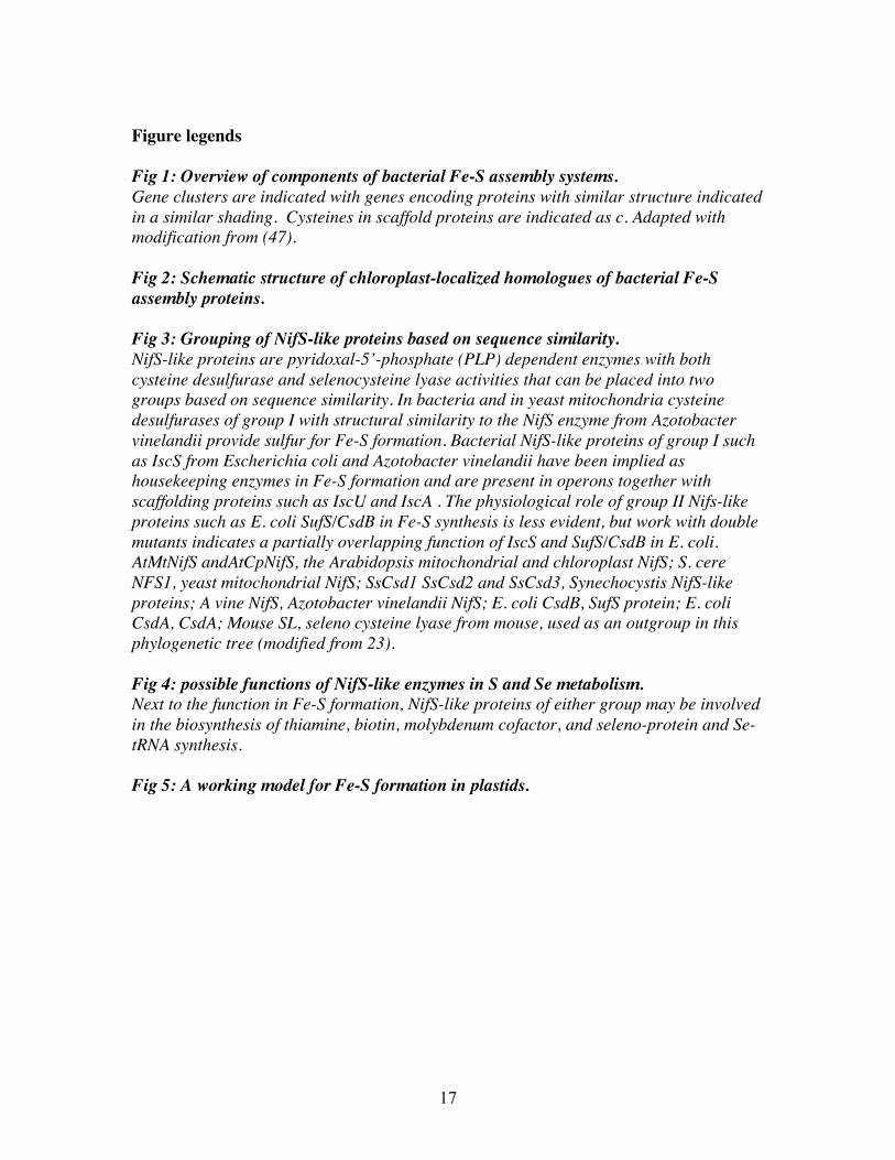
The first Fe-S assembly machinery studied was the nif system of Azotobactervinelandii, which is responsible for the formation of Fe-S clusters for nitrogenase,required under nitrogen fixation conditions (46). The A. vinelandii nif gene clusterincludes a cysteine desulfurase (CysD) encoding gene, NifS, as well as the other genesnifU, nifA, NifV and cysE, all thought to be involved in Fe-S cluster formation. NifS-likeproteins are pyridoxal 5’ phosphate (PLP)-dependent, enzymes that produce elementalsulfur or selenium from (seleno)cysteine, leaving alanine (48; for a review on cysteinedesulfurases see 47). A second NifS-like protein occurs in A. vinelandii, IscS, which hasa housekeeping function in the formation of other cellular Fe-S proteins (49). IscS ispresent in a gene cluster that contains paralogs of some of the nif genes (iscU, similar tothe N-terminus of nifU, and iscA); thus the nif and isc clusters have a similarorganization (49). The NifU- and NifA-like proteins are thought to serve a scaffoldfunction for the Fe-S cluster during its synthesis and before its transfer to the targetprotein and conserved cysteines play a pivotal role in this process (50, 51). The Isc genecluster also includes an Hsp70 and Hsp40 and a ferredoxin type protein. Homologues ofthe nif/isc genes have been discovered in several other bacteria including E. coli (49) andare also present in the mitochondria of eukaryotes (4). Next to IscU- and IscA- typeproteins the mitochondria have an Nfu protein, which is similar to the C-terminus ofNifU, (4). In yeast mitochondria the Hsp70/40 machinery is required for the utilization ofFe-S clusters assembled on IscU (52).
In E. coli and Erwinia chrysanthemi, a third gene cluster involved in Fe-S clusterformation is the Suf operon, which also includes a NifS-like cysteine desulfurase calledSufS/CsdB in E. coli. (53). A major function of the Suf operon may be in protecting thecell from oxidative stress and iron starvation (54, 55). Figure 1 summarizes the structuresof the 3 gene clusters implied in Fe-S formation in bacteria. A comparison of thesequences of NifS-like protins from various organisms reveals two classes of theseproteins (48). The Isc-type cysteine desulfurases fall into class I, whereas the Suf operonencoded NifS-like protein (SufS/CsdB) falls into class II, more related to enzymesimplied in selenium metabolism. (see figure 2). Besides a NifS-like protein the Sufoperon contains SufA, SufB, SufC, SufD and SufE. SufA is related to NifA and IscA andmay have a scaffold function (56), while SufE was shown to activate SufS (57, 58). SufCis a non-intrinsic cytosolic member of the ABC domain transporter super-family. It forms

8
a complex with Suf B and D but the biochemical role of this complex is not yet clear (54,58).
Chloroplasts are thought to be derived from a cyanobacterial ancestor. Even thoughstill much is to be learned about the assembly of Fe-S clusters in cyanobacteria, it is ofinterest to know that the genome of non-nitrogen fixing cyanobacteria, which are perhapsthe most close to plant plastids, encode homologues of IscS and Nfu (C-terminus ofNifU) as well as homologues of the E. coli Suf operon including SufS, but proteinscorresponding to IscU (or the N-terminus of NifU) are absent.
The Fe-S assembly machinery in plastids
CpNifSSince cysteine was the sulfur source for Fe-S formation in ferredoxin a plastidic
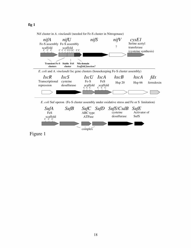
NifS-like protein or a similar enzyme should be involved in Fe-S formation inchloroplasts (20). Two genes encoding NifS-like proteins have been identified in theArabidopsis genome. One of the encoded proteins is present in mitochondria (14) and theother one, called CpNifS, is located in plastids (23, 24). The discovery of a NifS-likeprotein in plastids has prompted database searches for possible NifS-dependent proteinfactors that may function in Fe-S cluster assembly in chloroplasts. Putative Fe-Sassembly factors with chloroplast transit sequences are indeed encoded in theArabidopsis genome (see table 1 and figure 2). CpNifS is most similar to a cyanobacterialNifS-like protein, and among the E.coli homologs is most similar to SufS/CsdB a groupII NifS-like protein (see figure 3). CpNifS was found to be able to use both Cys andSeCys as substrates, with a 300-fold lower cysteine desulfurase activity compared to itsselenocysteine lyase activity (23).
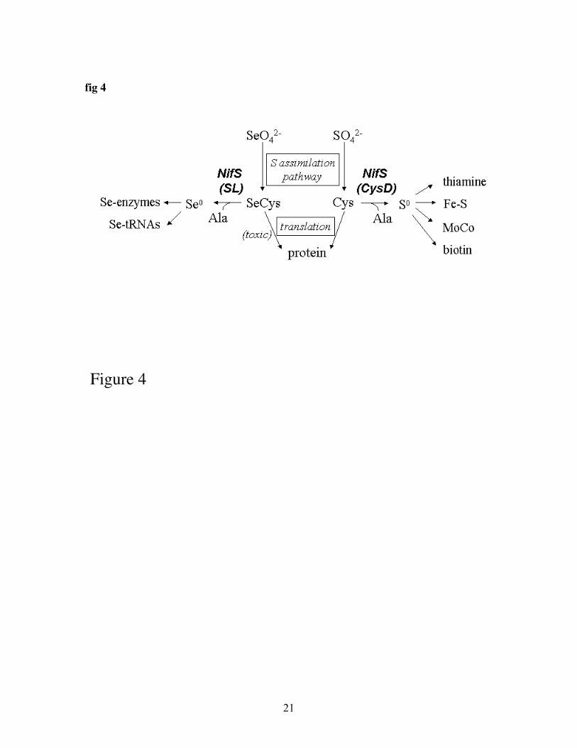
In microbes, NifS-like proteins have also been implied to function in aspects of Smetabolism other than Fe-S cluster formation, namely the biosynthesis of biotin,thiamine, and molybdenum cofactor, MoCo (47). NifS-like proteins may play similarroles in plants. In bacteria and mammals, essential Se metabolism also involves NifS-like proteins, which are needed for the incorporation of Se into selenoproteins and seleno-tRNAs (47). A summary of the various possible roles of NifS-like proteins in S and Semetabolism is given in figure 4.
It has now been shown that Se is an essential element for bacteria and animals - arequirement not yet shown for plants. On the other hand, higher Se concentrations aretoxic to all organisms. Thus organisms must prevent Se toxicity and at the same timemany organisms need Se for their metabolism; NifS-like proteins may play a role in bothaspects. Indeed, Arabidopsis plants that over-express CpNifS show increased tolerance toselenate. Furthermore, transcript profiling experiments in Arabidopsis showed that agroup of genes which are up-regulated in S deficiency are also up-regulated by selenatetreatment but this up-regulation is less pronounced in plants that over-express CpNifS(Van Hoewyk et al., unpublished data). Together these results suggest that CpNifS canhelp reduce Se stress by avoiding Se-induced S deficiency.
The role of CpNifS in Fe-S formation was first addressed directly by Ye et al. (59).To test whether CpNifS is involved in Fe-S cluster formation for photosynthetic proteins,an in vitro reconstitution assay was developed for ferredoxin. In this assay, apo-fd isreconstituted to the holo-form by acquiring an Fe-S cluster, which was synthesized in
9
vitro from cysteine sulfur and a ferrous iron salt. Holo-fd was separated from apo-fd andother proteins and quantified by HPLC using an ion exchange column. Purified CpNifSwas active by itself in stimulating holo-fd formation in this assay. The amount ofreconstituted ferredoxin was dependent on the CpNifS concentration. It was calculatedthat under the assay conditions 16 molecules of apo-Fd were reconstituted per CpNifSmonomer. Thus CpNifS has a catalytic role in iron-sulfur cluster formation in ferredoxinin vitro. The activity requires an intact PLP-cofactor, and CpNifS protein with a mutationof the conserved active site cysteine (Cys418-Ser) is inactive, indicating that ferredoxinreconstitution involves the cysteine desulfurase activity of CpNifS.
Stromal proteins at 300 micrograms/ml showed activity comparable to 10micrograms/ml CpNifS. Based on a quantification by means of Western blotting wecalculated that CpNifS constitutes 0.06 + 0.02% of total stromal protein. Thus theapparent reconstitution activity of stroma was 50-80 times more than that of pure CpNifSprotein and stromal components activate CpNifS. To investigate whether Fe-S clusterreconstitution activity of stroma was dependent on CpNifS, an affinity column was usedto deplete stroma of CpNifS, the removal of which was confirmed by immunoblot. Boththe original stroma and the antibody-treated stroma were examined for ferredoxinreconstitution activity. The activity of the antibody-treated stroma was decreased tobackground levels, suggesting that the reconstitution activity of stroma was entirelydependent on CpNifS. Importantly, adding back pure CpNifS to depleted stroma to itsoriginal concentration restored the reconstitution activity. Stroma that had been treatedwith pre-immune serum did not lose its Fe-S reconstitution activity.
To investigate whether CpNifS may be complexed to other stromal proteins, agelfiltration experiment was performed using a high resolution column, and the elution ofCpNifS followed using immunoblotting. Purified CpNifS eluted from the column in asingle peak with a retention time expected for the dimer, as was found before (23Interestingly, the CpNifS present in stroma eluted in two peaks approximately 90%eluted as a CpNifS dimer of 86 kDa, as was observed earlier using pure CpNifS. Anadditional, smaller amount of CpNifS eluted at a high Mw of ~600 kDa. This resultindicates that CpNifS interacts with other proteins in vivo and may form a transientcomplex with them (59).
CpNfu as a possible Fe-S assembly scaffold protein in plastids.Database searches (TAIR: www.arabidopsis.org) indicate that plastids do not have a IscUhomolog, or a protein similar to the N-terminal domain of NifU, but several otherpotential members of a plastid Fe-S cluster formation machinery were be identified (seetable 1 for a listing). The 3 CpNFU genes (CpNfu 1-3) encode chloroplast proteins thatare differentially expressed but closely related in sequence to each other and similar tocyanobacterial Nfu and the C-terminus of NifU (60, 61). The domain structure of CpNfuproteins is of interest. The three chloroplast Nfu proteins have a domain with highsimilarity to cyanobacterial Nfu including the conserved cysteines that is implied intransient cluster binding. In addition, a second Nfu –like domain which lacks the cysteineresidues is present at the C-terminus of the CpNfu proteins. CpNfu2 forms a transientcluster (60) that can be passed on to apo-ferredoxin in vitro (60, 61). Insertion mutants inone of the CpNfu genes (CpNfu2, At5g49940) have a dwarf phenotype and are deficientin some but not all plastid Fe-S proteins (61, 62). In the mutant lines the accumulation of
10
both 2Fe-S and 4Fe-S proteins (PSI and sulfite reductase) is diminished and theorganization of PSI is affected. Interestingly though, the KO-is viable and Fe-S proteinlevels were only diminished in vitro. Furthermore, the Rieske type 2Fe-2S of the B/Fcomplex and the 3Fe-4S cluster of glutamate synthase were not affected (Yabe et al;Touraine et al., 2004). It is possible that those clusters would require the action of any ofthe other two CpNfu gene products. However, the expression levels and sequencesimilarities of these genes may suggest that a different scaffold may be required for thesesubstrate proteins. Since CpNfu proteins can carry a transient Fe-S cluster that can betransferred to Fd, the observed effect on Fe-S assembly in the CpNfu2 mutant is likely adirect one. However in view of the observed effect of the CpNfu mutation on two typesof clusters, it would be of interest to verify the mRNA expression levels of Fd and PSIencoding genes, to rule out indirect effects of the mutations. So far a direct link betweenCpNifS and CpNfu has not been established at the biochemical level, but this may onlybe a matter of time.
CpIscA as an alternative scaffoldAnother potential candidate for interaction with CpNifS is CpSufA (Abdel-Ghany, Ye,Pilon-Smits and Pilon, unpublished). A T-DNA insertion line for this gene was obtained.Thus far only plants that are heterozygous for the insertion were found. A preliminaryanalysis indicates that when these heterozygotes are sown on media with sucrose, 1/4 ofthe seedlings show a visible growth phenotype. Thus, the homozygous CpSufA knockoutmay be lethal in plants grown on soil, perhaps due to impaired photosynthesis. However,further analyses will be required before firm conclusions on the in vivo role of CpSufAcan be made. CpSufA was shown to be plastidic by GFP-fusion studies. We have purifiedCpSufA and studied its effects on CpNifS-dependent reconstitution of Fd in vitro. Pre-incubation of pure CpNifS and pure CpSufA in the presence of cysteine and a ferrousiron salt was shown to give a 2x stimulation of apo-Fd reconstitution compared toCpNifS alone. Gel filtration experiments indicated purified CpSufA is a tetramer.However, upon incubation with CpNifS purified CpSufA acquires a transient Fe-S clusteras indicated by the absorption spectrum of CpSufA and direct measurement of Fe and itbecomes a dimer. The cluster in dimeric CpSufA can subsequently be transferred to apo-Fd to form holo-Fd. Thus CpSufA can function as an assembly scaffold for Fe-S clusters.
Other Suf-type system components and Hcf101Other potential candidate proteins that may assist CpNifS in Fe-S cluster formation inplastids are the Arabidopsis homologs of SufA, B, C, D and E and Hcf101 protein (tableI). The putative SufB and SufC homologs are confirmed to be in the chloroplast andmutants have phenotypes that indicate a role in development (63, 64). Expressionprofiling indicated that the potential SufB gene is regulated by Fe-deficiency inArabidopsis (65) but so far a link of CpSufBCD or E with CpNifS or Fe-S clusters hasnot been made in plants. Bacterial SufE protein is required to stimulate the lowendogenous cysteine desulfurase activity of SufS/CsdB. Our laboratory has localized theplant SufE protein to the chloroplast and we subsequently labeled the protein CpSufE.CpSufE is expressed in all major tissues, like CpNIFS, and it is feasible that CpSufE andCpNifS interact. Preliminary experiments in our lab indicate that CpSufE can indeedstimulate the cysteine desulfurase activity of CpNifS (Ye et al., unpublished). It will be
11
interesting to see what the exact physiological role of the CpSufE is. The function of thebacterial SufB,C,D proteins is still unclear; they appear to form a complex and may beinvolved in providing ferrous iron, or in transferring the Fe-S cluster from the scaffoldprotein to the target protein. In view of the function of the bacterial Suf operon inprotection from oxidative stress, Suf homologs should make suitable members of theplastidic Fe-S cluster machinery, since the chloroplast is an oxygenic compartment due toits photosynthetic oxygen production.
Another interesting putative component of the plastid Fe-S machinery is HCF101.HCF101 (high chlorophyll fluorescence 101) encodes a NifH-related P-loop ATPase thatseems to be required for 4Fe-4S but not 2Fe-2S in chloroplasts (66). The mechanism ofaction of the protein is so far not clear.
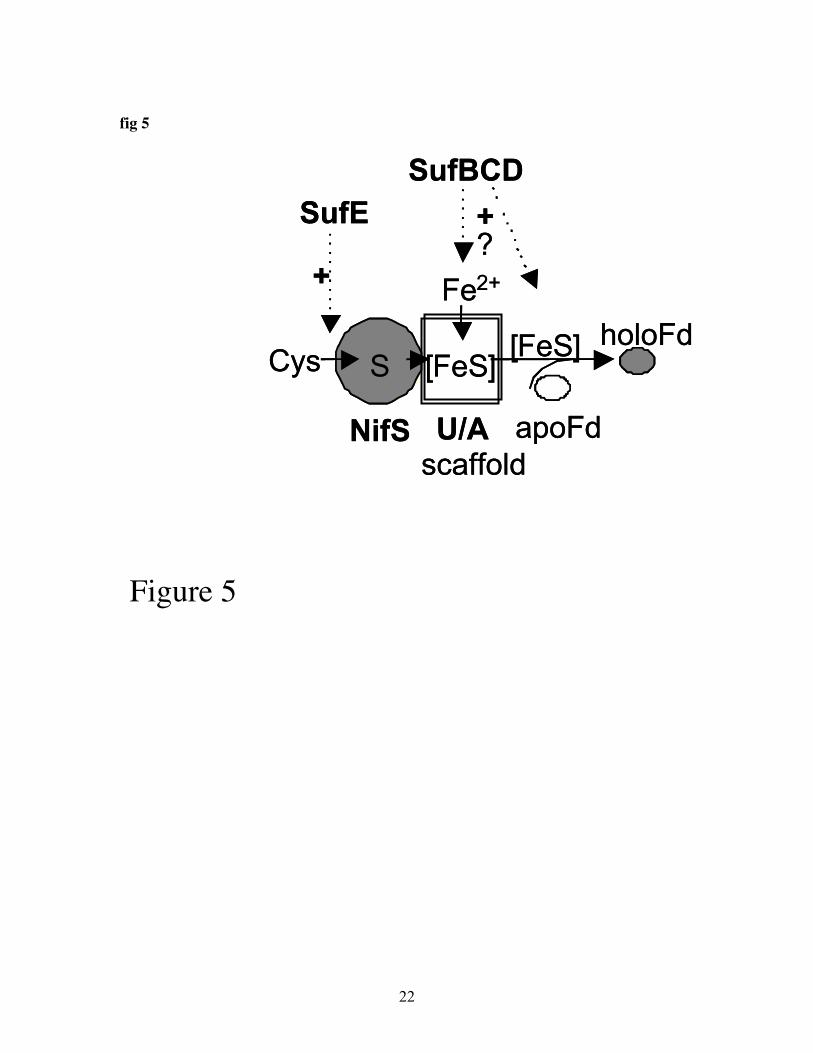
Future prospectsThe mechanisms of Fe-S assembly in plastids are complex and we are far from a
complete understanding of this fascinating process. Figure 5 shows a working model forthe Fe-S cluster formation machinery in plastids that includes components that have beencharacterized biochemically. The exact role of scaffold proteins in the biosynthesis ofspecific Fe-S proteins requires the development of sophisticated in vitro systems that canmeasure not only 2Fe-2S insertion in ferredoxin type proteins, but also insertion inRieske type proteins, 4Fe-4S and 3Fe-3S proteins. This requires novel model proteins andassays that take into account the observed need for NADPH and ATP in plastid Fe-Sassembly. Furthermore, the analysis of double mutants will help reveal possiblefunctional overlap. To assess whether effects of mutations on the accumulation ofproteins is a direct effect, expression at the mRNA level should be studied.Thus far very little is know about the molecular details of Fe uptake in plastids.Furthermore, the regulation of Fe-storage and recruitment for Fe-S assembly is unclear.How do plants coordinate the need for Fe in photosynthesis with S metabolism and Feuptake and mobilization? This question is not trivial since both free Fe and S areconsidered toxic. The newly available genetic and genomics tools will help reveal novelelements of the Fe-S biosynthetic machinery and the regulation of the machinery as awhole in response to developmental cues, the need for photosynthesis, and nutrient status.
12
References cited
1 Beinert, H., Holm, R.H., and Munck, E. (1997) Science 277, 653-659.
2 Imsande, J. (1999) Plant Physiol. Biochem. 37, 87-97.
3 Vorburger Mielcsarek, E., and Bertch McGrayne, S. (2000) Iron, nature’s universalelement. Rutgers University press, pp. 1-204. New Brunswick, NJ.
4 Lill, R., and Kispal, G .(2000) Trends Biochem. Sci. 25, 352-356.
5 Frazzon, J., Fick, J.R., and Dean, D.R. (2002) Biochem. Soc. Trans. 30, 680-685.
6 Raven, J.A., Evans, M.C., and Korb, R.E. (1999) Photosynthesis Res. 60, 111-149.
7 Lancaster, J.R., Vega, J.M., Kamin, H., Orme-Johnson, N.R., Orme-Johnson, W.H.,Krueger, W.H., and Siegel, L.M. (1979) J. Biol. Chem. 254, 1268-1272.
8 Krueger, R.J., and Siegel, L.M. (1982) Biochemistry 21: 2892-2904.
9 Munekage, Y., Hashimoto, M., Miyake, C., Tomizawa, K., Endo, T., Tasaka, M., andShikanai, T. (2004) Nature 429, 579-82.
10 Marschner, H. (1995) Mineral nutrition of higher plants, pp. 3-884. Acad. Press,London.
11 Chitnis, P.R. (2001) Annu. Rev. Plant Physiol. Plant Mol. Biol. 52, 593-626.
12 Buchanan, B.B., Gruissem, W.G., and Jones, R.L. (2000) Biochemistry andmolecular biology of plants, pp. 2-1367. Amer. Soc. Plant Biol., Rockville, MD.
13 Kispal, G., Csere, P., Prohl, C., and Lill, R. (1999) EMBO J. 18, 3981-3989.
14 Kushnir, S., Babiychuk, E., Storozhenko, S., Davey, M.W., Papenbrock, J., DeRycke, R., Engler, G., Stephan, U.W., Lange, H., Kispal, G., Lill, R., and Van Montagu,M. (2001) Plant Cell 13, 89-100.
15 Balk, J., Pierik, A.J., Netz, D.J., Muhlenhoff ,U., and Lill, R (2004) EMBO J. 23,2105-2115.
16 Keegstra, K., and Cline, K. (1999) Plant Cell 11, 557-570.
17 Li, H-M., Theg, S.M., Bauerle, C.M., and Keegstra, K. (1990) Proc. Natl. Acad. Sci.U.S.A. 8, 6748-6752.

13
18 Pilon, M., de Kruijff, B., and Weisbeek, P.J. (1992) J. Biol. Chem. 267: 2548-2556.
19 Pilon, M., America, T., van 't Hof, R., de Kruijff, B. and Weisbeek, P. (1995) In:Advances in molecular and cell biology (Rothman SS Ed.) Membrane protein transport.JAI Press, Greenwich. Vol. 4, pp. 229-255.
20 Merchant, S., and Dreyfuss B.W. (1998) Annu .Rev. Plant Physiol. Plant Mol. Biol.49, 25-51.
21 Takahashi, Y., Mitsui, A., Hase, T., and Matsubara, H. (1986) Proc. Natl. Acad. Sci.U.S.A. 83, 2434-2437.
22 Takahashi, Y., Mitsui, A., and Matsubara, H. (1990) Plant Physiol. 95, 97-103.
23 Pilon-Smits, E.A.H., Garifullina, G., Abdel-Ghany, S., Kato, S., Mihara, H., Hale,K.L., Burkhead, J., Esaki, N., Kurihara, T., and Pilon, M. (2002) Plant Physiol. 130,1309-1318.
24 Leon, S., Touraine, B., Briat, J-F., and Lobreaux, S. (2002) Biochem. J. 366, 557-564.
25 Curie, C., and Briat, J.F. (2003) Annu. Rev. Plant Physiol. Plant Mol. Biol. 54, 183-206.
26 Hell, R., and Stephan, U.W. (2003) Planta 216, 541-551.
27 Curie, C., Panaviene, Z., Loulergue, C., Dellaporta, S.L., Briat, J.F., and Walker,E.L. (2001) Nature 409, 346-349.
28 Robinson, N.J., Procterm, C.M., Connolly, E.L., and Guerinot M.L . (1999) Nature397, 694-697.
29 Connolly, E.L., Fett, J.P., and Guerinot, M.L. (2002) Plant Cell 14, 1347-1357.
30 Vert, G., Grotz, N., Dedaldechamp, F., Gaymard, F., Guerinot, M.L., Briat, J.F., andCurie, C. (2002) Plant Cell 14, 1223-1233.
31 Shingles, R., North, M., and McCarty, R.E. (2002) Plant Physiol. 128, 1022-1030.
32 Petit, J-M., Briat, J-F., and Lobreaux, S. (2001) Biochem. J. 359: 575-582.
33 Beinert, H. (2000). Eur. J. Biochem. 267, 5657-5664.
34 Leustek, T., Martin, M.N., Bick, J-A., and Davies, J.P. (2000) Annu. Rev. PlantPhysiol. Plant Mol. Biol. 5, 141-165.
14
35 Smith, F.W., Ealing, P.M., Hawkesford, M.J., and Clarkson, D.T. (1995) Proc. Natl.Acad. Sci. U.S.A. 92, 9373-9377.
36 Shibagaki, N., Rose, A., McDermott, J., Fujiwara, T., Hayashi, H., Yoneyama, T.,and Davies, J.P. (2002) Plant J. 29, 475-486.
37 Yoshimoto, N., Takahashi, H., Smith, F.W., Yamaya, T., and Saito, K. (2002) Plant J.29, 465-473.
38 Takahashi, H., Yamazaki, M., Sasakura, N., Watanabe, A., Leustek, T., de AlmeidaEngler, J., van Montagu, M., and Saito, K. (1997) Proc. Natl. Acad. Sci. U.S.A. 94, 1102-11107.
39 Takahashi, H., Watanabe-Takahashi, A., Smith, F.W., Blake-Kalff, M., Hawkesford,M.J., and Saito, K. (2000) Plant J. 23, 171-182.
40 Takahashi, H., Sasakura, N., Kimura, A., Watanabe, A., and Saito, K. (1999) PlantPhysiol. 121, 686.
41 Grossman, A.R., and Takahashi, H. (2001) Annu. Rev. Plant Physiol. Plant Mol. Biol.52, 163-210.
42 Rotte, C., and Leustek, T. (2000) Plant Physiol. 124, 715-724.
43 Bork, C., Schwenn, J.D., and Hell, R. (1998) Gene 212, 147—153.
44 Zhou, J., and Goldsbrough, P.B. (1994) Plant Cell 6: 875—884.
45 Axelsen, K.B., and Palmgren, M.G. (2001) Plant Physiol. 126, 696—706.
46 Zheng, L., White, R.H., Cash, V.L., Jack, R.F., and Dean, D.R. (1993) Proc. Natl.Acad. Sci. U.S.A. 90, 2754-2758.
47 Mihara, H., and Esaki, N. (2002) Appl. Microbiol. Biotechnol. 60, 12-23.
48 Mihara, H., Kurihara, T., Yoshimura, T., Soda, K., and Esaki, N. (1997) J. Biol.Chem. 272, 22417-22424.
49 Zheng, L., Cash, V.L., Flint, D.H., and Dean, D.R .(1998) J. Biol. Chem. 273, 13264-13272.
50 Agar, J.N., Krebs, C., Frazzon, J., Huynh, B.H., Dean, D.R., and Johnson, M.K.(2000) Biochemistry 39, 7856-7862.
51 Krebs, C., Agar, J.N., Smith, A.D., Frazzon, J., Dean, D.R., Huynh, B.H., andJohnson M.K. (2001) 40, 14069-14080.
15
52 Muhlenhoff, U., Gerber, J., Richhardt, N., and Lill, R. (2003) EMBO J. 22, 4815-4825.
53 Takahashi, Y., and Tokumoto, U. (2002) J. Biol. Chem. 277, 28380 – 28383.
54 Nachin, L., Loiseau, L., Expert, D., Barras, F. (2003) EMBO J. 22, 427-437.
55 Outten, F.W., Djaman, O., and Storz, G. (2004) Mol. Microbiol. 52, 861-872.
56 Ollagnier-de Choudens, S., Nachin, L., Sanakis, Y., Loiseau, L., Barras, F., andFontecave, M. (2003) J. Biol. Chem. 278, 17993-18001.
57 Loiseau, L., Ollagnier-de-Choudens, S., Nachin, L., Fontecave, M., and Barras, F.(2003) J. Biol. Chem. 278, 38352 – 38359.
58 Outten, F.W., Wood, M.J., Munoz, F.M., and Storz, G. (2003) J. Biol. Chem. 278,45713-45719.
59 Ye, H., Garifullina, G.F., Abdel-Ghany, S., Zhang, L., Pilon-Smits, E.A.H., andPilon, M. (2004) Planta, in press.
60 Leon, S., Touraine, B., Ribot, C., Briat, J.F., and Lobreaux, S. (2003) Biochem. J.371: 823-830.
61 Yabe, T., Morimoto, K., Kikuchi, S., Nishio, K., Terashima, I., and Nakai, M. (2004)Plant Cell 16, 993-1007.
62 Touraine, B., Boutin, J-P., Marion Poll, A., Briat, J-F., Peltier, G., and Lobreaux S.(2004) Plant J., in press.
63 Moller, G.M., Kunkel, T., and Chua, N-H. (2001) Genes and Development 15, 90-103.
64 Xu, X.M., and Moller, S.G. (2004) Proc. Natl. Acad. Sci. U.S.A. 101, 9143-9148.
65 Wintz, H., Fox. T., Wu, Y.Y., Feng, V., Chen, W., Chang, H.S., Zhu, T., and Vulpe,C. (2003) J. Biol. Chem. 278, 47644-47653.
66 Lezhneva, L., Amann, K., and Meurer, J. (2004) Plant J. 37, 174-85.
67 Noctor, G., Arisi, A.C.M., Jouanin, L., and Foyer, C.H. (1998) Plant Physiol. 118,471—482.
16
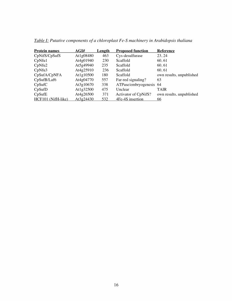
Table I: Putative components of a chloroplast Fe-S machinery in Arabidopsis thaliana
Protein names AGI# Length Proposed function ReferenceCpNifS/CpSufS At1g08480 463 Cys-desulfurase 23, 24CpNfu1 At4g01940 230 Scaffold 60, 61CpNfu2 At5g49940 235 Scaffold 60, 61CpNfu3 At4g25910 236 Scaffold 60, 61CpSufA/CpNFA At1g10500 180 Scaffold own results, unpublishedCpSufB/Laf6 At4g04770 557 Far-red signaling? 63CpSufC At3g10670 338 ATPase/embryogenesis 64CpSufD At1g32500 475 Unclear TAIRCpSufE At4g26500 371 Activator of CpNifS? own results, unpublishedHCF101 (NifH-like) At3g24430 532 4Fe-4S insertion 66
17
Figure legends
Fig 1: Overview of components of bacterial Fe-S assembly systems.Gene clusters are indicated with genes encoding proteins with similar structure indicatedin a similar shading. Cysteines in scaffold proteins are indicated as c. Adapted withmodification from (47).
Fig 2: Schematic structure of chloroplast-localized homologues of bacterial Fe-Sassembly proteins.
Fig 3: Grouping of NifS-like proteins based on sequence similarity.NifS-like proteins are pyridoxal-5’-phosphate (PLP) dependent enzymes with bothcysteine desulfurase and selenocysteine lyase activities that can be placed into twogroups based on sequence similarity. In bacteria and in yeast mitochondria cysteinedesulfurases of group I with structural similarity to the NifS enzyme from Azotobactervinelandii provide sulfur for Fe-S formation. Bacterial NifS-like proteins of group I suchas IscS from Escherichia coli and Azotobacter vinelandii have been implied ashousekeeping enzymes in Fe-S formation and are present in operons together withscaffolding proteins such as IscU and IscA . The physiological role of group II Nifs-likeproteins such as E. coli SufS/CsdB in Fe-S synthesis is less evident, but work with doublemutants indicates a partially overlapping function of IscS and SufS/CsdB in E. coli.AtMtNifS andAtCpNifS, the Arabidopsis mitochondrial and chloroplast NifS; S. cereNFS1, yeast mitochondrial NifS; SsCsd1 SsCsd2 and SsCsd3, Synechocystis NifS-likeproteins; A vine NifS, Azotobacter vinelandii NifS; E. coli CsdB, SufS protein; E. coliCsdA, CsdA; Mouse SL, seleno cysteine lyase from mouse, used as an outgroup in thisphylogenetic tree (modified from 23).
Fig 4: possible functions of NifS-like enzymes in S and Se metabolism.Next to the function in Fe-S formation, NifS-like proteins of either group may be involvedin the biosynthesis of thiamine, biotin, molybdenum cofactor, and seleno-protein and Se-tRNA synthesis.
Fig 5: A working model for Fe-S formation in plastids.
18
fig 1
nifU cysE1nifS nifVNif cluster in A. vinelandii (needed for Fe-S cluster in Nitrogenase)
Fe-S assembly scaffold
Fe-S assembly scaffold
cysteinedesulfurase
?Serine acetyl transferase(cysteine synthesis)
nifA
Transient Fe-S clusters
Nfu domainScaffold function?
Stable FeS cluster
C C C C C C C CCCCC
C C C C C C
IscSIscRTranscriptionalrepression
IscU Fe-S scaffold
IscA FeS scaffold
hscB hscA fdxHsp 20 Hsp 66 ferredoxin
E. coli and A. vinelandii Isc gene clusters (housekeeping Fe-S cluster assembly)
E. coli Suf operon (Fe-S cluster assembly under oxidative stress and Fe or S limitation)
C C C
SufA FeS scaffold
cysteinedesulfurase
SufS/CsdB SufEActivator of SufS
SufB SufC SufDABC type ATPase
complexFigure 1
19
fig 2
Putative components of the plastid Fe-S assembly system
CpSufA / CpIscA: scaffold for Fe-S
CpSufB = laf6: chlorophyll synthesis, FR response CpSufC?: ABC transporter like proteins w/o transmembrane domains
CpSufD? forms complex with SufB and SufC?
CpSufE; stimulates cysteine desulfurase of CpNifS
CpNFU (C-terminus of NifU scaffold, almost twice): 3 genes
CpNifSp: SufS/CsdB-like cysteine desulfurase/selenocysteine lyaseC C C
C C
Figure 2

20
fig 3
21
fig 4
Figure 4
22
fig 5
NifS U/Ascaffold
Cys S [FeS]
Fe2+
holoFd[FeS]
apoFd
+
SufESufBCD
+?
NifS U/Ascaffold
Cys S [FeS]
Fe2+
holoFd[FeS]
apoFd
+
SufESufBCD
+?
Figure 5