pyrroloquinoline quinone biogenesis: demonstration … · consensus sequence that is unique to a...
TRANSCRIPT

pubs.acs.org/BiochemistryPublished on Web 09/11/2009r 2009 American Chemical Society
Biochemistry 2009, 48, 10151–10161 10151
DOI: 10.1021/bi900918b
Pyrroloquinoline Quinone Biogenesis: Demonstration That PqqE from Klebsiellapneumoniae Is a Radical S-Adenosyl-L-methionine Enzyme†
StephenR.Wecksler,‡ Stefan Stoll, )HaTran,‡ Olafur T.Magnusson,‡,^ Shu-paoWu,‡,@DavidKing,§ R. David Britt, ) andJudith P. Klinman*,‡
‡Department of Chemistry and Department of Molecular and Cell Biology, University of California, Berkeley, California 94720,§Howard Hughes Medical Institute Mass Spectrometry Laboratory, Department of Molecular and Cell Biology, University ofCalifornia, Berkeley, California 94720, and )Department of Chemistry, University of California, Davis, California 95616.
^Present address: Decode Genetics, Sturlagata 8, IS-101 Reykjavik, Iceland. @Present address: Department ofApplied Chemistry, National Chiao Tung University, 1001 Hsueh Rd., Hsinchu, Taiwan, Republic of China.
Received June 1, 2009; Revised Manuscript Received September 10, 2009
ABSTRACT: Biogenesis of pyrroloquinoline quinone (PQQ) inKlebsiella pneumoniae requires the expression ofsix genes (pqqA-F ). One of these genes (pqqE ) encodes a 43 kDa protein (PqqE) that plays a role in the initialsteps in PQQ formation [Veletrop, J. S., et al. (1995) J. Bacteriol. 177, 5088-5098]. PqqE contains two highlyconserved cysteine motifs at the N- and C-termini, with the N-terminal motif comprised of a CX3CX2Cconsensus sequence that is unique to a family of proteins known as radical S-adenosyl-L-methionine (SAM)enzymes [Sofia, H. J., et al. (2001) Nucleic Acids Res. 29, 1097-1106]. PqqE from K. pneumoniae was clonedinto Escherichia coli and expressed as the native protein and with an N-terminal His6 tag. Anaerobicexpression and purification of the His6-tagged PqqE results in an enzyme with a brownish-red hue indicativeof Fe-S cluster formation. Spectroscopic and physical analyses indicate that PqqE contains a mixture ofFe-S clusters, with the predominant form of the enzyme containing two [4Fe-4S] clusters. PqqE isolatedanaerobically yields an active enzyme capable of cleaving SAM to methionine and 50-deoxyadenosine in anuncoupled reaction (kobs=0.011( 0.001min-1). In this reaction, the 50-deoxyadenosyl radical either abstractsa hydrogen atom from a solvent accessible position in the enzyme or obtains a proton and electron frombuffer. The putative PQQ substrate PqqA has not yet been shown to be modified by PqqE, implying thatPqqA must be modified before becoming the substrate for PqqE and/or that another protein in thebiosynthetic pathway is critical for the initial steps in PQQ biogenesis.
The broad spectrum of chemical reactions catalyzed byenzymes frequently requires functional groups that are unavail-able from the side chains of the 20 naturally occurring aminoacids. In particular, there is a lack of electrophilic functionalgroups necessitating the involvement of exogenous metal ions ororganic cofactors. The quino-cofactor family of enzymes con-tains intrinsic electrophilic centers that are derived fromnaturallyoccurring amino acids, thereby expanding the scope of aminoacid side chains available within active sites of folded proteins.These cofactors arise via two fundamentally different pathways;in the first case, the pathway involves the addition of a redoxactive copper ion to a folded precursor protein followed by the“self-processing” of a tyrosine side chain to trihydroxyphenyla-lanine quinone (TPQ) or the cross-linked lysyl tyrosine quinone(LTQ) (1). The second class of cofactors is derived from active sitetryptophans anddepends on an exogenous family of gene productsto produce the active site tryptophanyl tryptophan quinone (TTQ)and cysteinyl tryptophan quinone (CTQ). Finally, there is theprokaryotic cofactor pyrroloquinoline quinone (PQQ), which is
formed and then excised from a peptide to generate a “standalone” cofactor. The CTQ-containing amine dehydrogenase andthe PQQ biosynthetic operons contain uncharacterized enzymeswith a conserved Cys-X-X-X-Cys-X-X-Cys motif thatis specific to members of the radical S-adenosyl-L-methionine(SAM) enzyme family (2, 3). In this report, we document the firstevidence of an active radical SAM enzyme in the biogenesispathway leading to an amino acid-derived quino-cofactor.
PQQ is a noncovalently bound cofactor dominantly utilized byalcohol and sugar dehydrogenases localized in the periplasm ofGram-negative bacteria (4-6). The electrons obtained uponreduction of PQQ to PQQH2 are subsequently transferred toan electron transport chain responsible for ATP production (7).Certain bacteria, such as Escherichia coli, are unable to them-selves produce PQQ, and PQQ has been designated as aprokaryotic vitamin in such cases (7). The role of PQQ inmammals is much more controversial. PQQ has been found inhigh concentrations in breast milk, shown to be an essentialnutrient for proper growth and development in mice, and evensuggested to be a novel B vitamin (8-10). These observationshave stoked considerable interest in its physiological function andhave highlighted the importance of understanding the biosyn-thetic pathway in vivo.
Insights into the PQQ biosynthetic pathway have emergedpiecemeal over the past two decades. A major breakthrough was
†This work was supported by research grants from the NationalInstitutes of Health (GM039296 to J.P.K., GM073789 to R.D.B., andF32GM080795 to S.R.W.) and the Howard Hughes Medical Institute(to D.K.).*To whom correspondence should be addressed. Telephone: (510)
642-2668. Fax: (510) 643-6232. E-mail: [email protected].

10152 Biochemistry, Vol. 48, No. 42, 2009 Wecksler et al.
the demonstration that PQQ is formed from the fusion ofglutamate and tyrosine (Scheme 1) (11, 12). This observationparalleled the discovery of genes involved in PQQ biogenesis(13-15). InKlebsiella pneumoniae, there are six genes (designatedpqqA-F) located in the pqq operon (15). pqqA encodes a23-residue peptide (PqqA) with a strictly conserved glutamateand tyrosine that is believed to be the substrate for PQQbiogenesis (15). PqqC catalyzes the final step in PQQ biogenesis,which is the eight-electron oxidation and ring cyclization of 3a-(2-amino-2-carboxyethyl)-4,5-dioxo-4,5,6,7,8,9-hexahydroquino-line-7,9-dicarboxylic acid (AHQQ) (16, 17). Although no defini-tive functions for PqqB and PqqF have been experimentallydemonstrated, sequence alignments suggest that both proteinsmay function as proteases. PqqD is a small 10 kDa protein thathas no homology to other proteins in the ProteinData Bank, andPqqE contains a conserved cysteine motif that is present inproteins known as radical SAM enzymes (18).
The most detailed mechanistic information in the biosyntheticpathway describes the final step for PQQ formation catalyzed byPqqC (19), with little being known about the preceding chemicalsteps in biogenesis. An in vivo study of the genes fromK. pneumoniae suggests that PqqA, PqqD, and PqqE are criticalfor the first steps in PQQbiogenesis (16).We nowpresent the firstin vitro characterizationof one of these proteins, the radical SAMenzyme PqqE. As described herein, PqqE is most homologous tothe radical SAM enzymeMoaA (18% identical and 38% similartoMoaA fromMycobacterium tuberculosis) (20).MoaA,which isinvolved in molybdenum cofactor biosynthesis, possesses two[4Fe-4S] clusters, one at its N-terminus and one at itsC-terminus (21). The Fe-S cluster at the N-terminal domainprovides a site for SAM binding and activation, whereas thesecond cluster at the C-terminus appears to play a role inanchoring and positioning of the 50-GTP substrate. Spectro-scopic and physical measurements of PqqE confirm two [4Fe-4S]clusters that display spectroscopic properties nearly identical tothose found in other radical SAM enzymes. Importantly, thesePqqE preparations are shown to reductively cleave SAM tomethionine and 50-deoxyadenosyl radical in an uncoupled reac-tion, and that the enzyme is capable of multiple turnovers. Thesestudies raise many provocative questions regarding the couplingof a radical SAM enzyme to substrate oxidation and open up, for
the first time, the full characterization of radical SAMenzymes inquino-cofactor biogenesis.
EXPERIMENTAL PROCEDURES
Materials. All chemicals and reagents were purchased fromSigma-Aldrich, Mallinckrodt, Acros, or Fisher. Chemicals andreagents were purchased at the highest purity available and usedwithout further purification. pET24b, pET28b, and BugBusterwere purchased from Novagen. E. coli BL21(DE3) and XL-1Blue competent cells were purchased from Stratagene. Allrestriction enzymes (NdeI andBamHI) used for cloning reactionswere purchased from New England BioLabs; calf intestinalalkaline phosphatase was from Invitrogen, and high-fidelityPFUpolymerase andT7DNA ligasewere fromRoche. BradfordAssay reagents were purchased from Bio-Rad. Bovine serumalbumin was from Pierce. Ni2+-nitrilotriacetic acid (Ni-NTA)agarose resin was from Qiagen, and Q-Sepharose and SephacrylS-300 were from Sigma.
Plasmids pPH149 and pPH151 containing the E. coli IscSUA-HscBA-Fd and E. coli suf ABCDSE genes, respectively, weregenerously donated by P. Hanzelmann from the Institute forStructural Biology at the University of W€urzburg (W€urzburg,Germany). Plasmid pBCP-165 containing the pqq operon fromK. pneumoniae was originally made in the laboratory of P. W.Postma (University of Amsterdam, Amsterdam, The Nether-lands) and donated by R. Rucker from University of California,Davis. A clone containing the sam2 gene from E. coli [TB1(pUC18:sam2)] was generously donated by C. Roessner fromthe laboratory of I. Scott (Texas A&M University, CollegeStation, TX).GeneralMethods.All inert atmosphere work was conducted
in an 855-AC-controlled atmosphere chamber from Plas-Labs,Inc. (Lansing, MI). For inert atmosphere work, we made allbuffer solutions anaerobic by purging solutions with argon for 1mL/min. Reagents were either prepared as anaerobic buffers orbrought into the box in crystalline powder form and thenreconstituted with anaerobic buffer. DNA sequencing was per-formed at the DNA sequencing facility at the University ofCalifornia (Berkeley, CA). N-Terminal sequencing was con-ducted at the Stanford PAN facility. Iron and sulfide analyseswere conducted using methods described by Beinert (22, 23).Protein concentrations were calculated using the Bradford assay.UV-vis spectra were recorded on either a Hewlett-Packard 8452diode array spectrophotometer or a Cary 50 Bio spectrophot-ometer. UV-visible measurements were performed in long stemquartz cuvettes (Starna) sealed with a rubber septum. PCRs wereconducted on a PTC-200 Peltier thermal cycler (MJ Research).
Cloning of pqqE from pBCP-165. pBCP-165 was originallygenerated in the laboratory of P. W. Postma (15). The genesequence for the pqq operon from K. pneumoniae has beendeposited in the NCBI database and used as a reference forcomparing DNA and protein sequencing information. The openreading frame for pqqE starts at base 3023 in the operon;however, this start codon is not the typical ATG codon foundfor most bacterial genes. Position 3023 in the deposited sequenceis listed as a guanine base and was mutated to an adenosine togenerate the ATG start site when appropriate primers weredesigned for the cloning of pqqE.
pqqE was cloned into the NdeI and BamHI restriction sites ofpET28b (or pET24b) using standard cloning techniques. Thefollowing primers (obtained from Operon) were used to clone
Scheme 1: Formation of PQQ from the Fusion of Glutamateand Tyrosine via the Intermediacy of AHQQ

Article Biochemistry, Vol. 48, No. 42, 2009 10153
pqqE out of pBCP-165: 50-CGCATTACATATGAGCCAGAG-TAAACCCACCGTCAATCCG-30 and 50-CGCGGATCCT-TACAGGTCCCGGGTTTGGTAGATCAGCTG-30. Under-lined bases show engineered restriction sites for NdeI andBamHI, respectively. The plasmids (named pET28b-pqqE andpET24b-pqqE) were isolated and sequenced using the T7 pro-moter and terminator primers, respectively.
After isolation, pET28b-pqqE was cotransformed into E. coliBL21(DE3) competent cells with either pPH149 or pPH151. Thebacteria were plated onto LB agar plates containing bothkanamycin (50 μg/mL) and chl (50 μg/mL) and grown at 37 �Covernight. The following day, several colonies were picked andgrown in liquid LB medium at 37 �C containing both antibioticsat 50 μg/mL. After several hours, the bacteria had reached logphase and were quickly mixed with 50% glycerol, immediatelyfrozen in liquid nitrogen, and stored at -80 �C until further use.Expression andPurification of PqqE. (i)Developmental
Work. Initial efforts aimed at the expression and purification ofPqqE were focused on either aerobic growth of the transformedE. coli cells, followed by purification of enzyme from inclusionbodies and chemical reconstitution, or expression of PqqE inE. coli grown anaerobically and subsequent isolation of theenzyme from soluble supernatants under aerobic conditions.These methods, which proved to be unsatisfactory for variousreasons, are summarized in the Supporting Information.
(ii)N-Terminal His6 Tag-Containing PqqE. E. coliBL21-(DE3) cells containing pET28b-pqqE and pPH151 were grownand induced under identical anaerobic growth conditions asdescribed in the Supporting Information for anaerobic growthof E. coli BL21(DE3) containing pET24b-pqqE with the excep-tion that the medium also contained 50 μg/mL chl.
For the purification of His6 tag-containing PqqE, all stepswere performed under strictly anaerobic conditions in an inertatmosphere glovebox. Cell pellets (cell paste yields were approxi-mately 4 g/10 L of LB medium) were brought into the gloveboxand lysed for 30 min with Bugbuster in 50 mM Tris (pH 7.9),1 mM DTT, 300 mM NaCl, 10 mM imidazole, and 5 μL ofbenzonase nuclease. The lysates were loaded into anaerobiccentrifuge tubes (Beckman) and cleared via centrifugation at15000g and 4 �C for 20 min. The lysates were then brought backinto the glovebox and the reddish brown supernatants loadedonto a column containing 50 mL of Ni-NTA resin equilibratedwith the same buffer (column size, 12 in. long and 2.5 in. wide).The bound protein was then washed with 100 mL of loadingbuffer followed by two, 100 mL stepwise gradient washescontaining the same buffer but with 25 mM imidazole in thefirst and 50 mM imidazole in the second. PqqE was then elutedoff the column with an increase in the imidazole concentration to200 mM. The brownish red fractions were pooled and concen-trated to approximately 5mLusing a 30kDaAmiconmembrane.
After concentration, the imidazole was removed using a gelfiltration PD-10 column equilibrated with 50 mM Tris (pH 7.9),1mMDTT, and 300mMNaCl. The protein was collected off thePD-10 column and concentrated to approximately 10 mg/mLwith a 30 kDaAmiconmembrane. Aliquots (200 μL)were loadedinto glass vials (Agilent) equipped with rubber caps, brought outof the glovebox, and immediately frozen in liquid nitrogen. Theprotein was stored at -80 �C until further use.
(iii) Preparation of S-Adenosyl-L-methionine. The enzy-matic synthesis, purification, and characterization of S-adenosyl-L-methionine (SAM) largely followed literature protocols (24-26).Details can be found in the Supporting Information.
(iv) Determination of Enzyme Activity for PqqE usingLC-MS. LC-MSwas performed on anAgilent LC 1100 seriesinstrument equipped with an HP LC/MSD electrospray ionsource and quadrupole mass spectrometer. Reversed-phaseHPLC was performed on a Jupiter Proteo 4 μm, 90 A, 250 mm�4.60 mm C18 column (Phenomenex), and the following elutionconditions were used for identifying the enzymatic products ofthe radical SAM reaction in PqqE: flow rate of 0.4 mL/min,0.05% formic acid, 1.0% acetonitrile in water for 10 min,followed by a linear gradient from 1 to 30% acetonitrile over30min and ending with a linear gradient increase from 30 to 90%acetonitrile over the next 30 min. The quadrupole mass spectro-meter was operated in the positive ion mode using the scanfunction from m/z 100 to 1000 (fragmentor, 70; gain, 1.0;threshold, 150; step size, 0.1). The elution profiles were recordedat 215 and 260 nm (10 nm bandwidth and referenced at 350 nm).
Samples for LC-MS were generated as follows. A stocksolution of PqqE (∼125 μM) in (pH 7.9) 50 mM Tris, 1 mMDTT, 300 mM NaCl, and 20% glycerol was prepared anaerobi-cally and incubated with a 10-fold excess of sodium dithionite(DT) for 10min. Aliquots (100 μL) of reduced PqqEwere dilutedwith an equal volume of buffer, and the reaction was initiated bya 10-fold addition of SAM (600 μM). The reaction mixture wasleft for 2 h and then the reaction quenched by the addition of neatformic acid to give a final concentration of 5% (v/v). The sampleswere pelleted via centrifugation and brought out of theglovebox, and 80 μL volumes were injected onto the LC-MSinstrument.
(v) Quantification of 50-Deoxyadenosine Formation.High-performance liquid chromatography (HPLC) was con-ducted on a Beckman system equipped with a diode arraydetector and operated by 32 Karat version 8.0. The softwarewas also used for data collection and analysis. Reversed-phaseHPLC was performed on a Jupiter Proteo 4 μm, 90 A, 250 mm�4.60 mmC18 column. The following elution conditions were usedfor quantifying the time-dependent formation of 50-deoxyadeno-sine: flow rate of 1mL/min, 0.05% formic acid, 1.0% acetonitrilein water for 30min, followed by a linear gradient from 1 to 100%acetonitrile over 10 min. Spectra were recorded at 215 and260 nm. Under these conditions, SAM and methionine elute inthe dead time of the instrument (∼5.0 min) and 50-deoxyadeno-sine elutes at 23 min. The concentration of 50-deoxyadenosinewas determined via integration of the area under the peak andcomparison to a linear calibration curve from a series ofstandards at known concentrations.
All assays were performed in an anaerobic chamber. For atypical assay, PqqE was prepared as a stock solution at 39 μM in50 mMTris (pH 7.9), 1 mMDTT, 300 mMNaCl, 20% glycerol,and 1 mM dithionite. The stock solution was left to incubate atroom temperature for approximately 10 min, at which pointSAMwas added to the reaction mixture for a final concentrationof 420 μM. The samples were left to incubate for various lengthsof time and the reactions quenched with neat formic acid to afinal concentration of 5%. The samples were pelleted viacentrifugation and 100 μL aliquots injected into the HPLCinstrument using the procedure outlined above.
(vi) EPR Characterization. Preparation of all samples wasconducted in an anaerobic glovebox. In brief, a 1.5 mL stocksolution of PqqEwas prepared with 169 μMPqqE in 50mMTris(pH 7.9), 1 mM DTT, 300 mM NaCl, and 20% glycerol. Thesamples were cleared of precipitate at 14000 rpm for 10 min,diluted to a final concentration of 115 μM, loaded intoEPR tubes

10154 Biochemistry, Vol. 48, No. 42, 2009 Wecksler et al.
equipped with rubber septa, brought out of the glovebox, andimmediately frozen in liquid nitrogen. To prepare the reducedPqqE sample, the stock solution was mixed with a 10-fold excessof sodium dithionite (1.15 mM) and left to incubate for approxi-mately 10 min before being loaded into the EPR tubes. Thereduced samples that contained SAMwere prepared in a manneridentical to that of the reduced PqqE sample, except a 10-foldexcess of SAM (1.15 mM) was added after the 10 min incubationwith dithionite. The total incubation time for the reduced PqqEwith SAM was approximately 5 min.
EPR experiments were conducted at the CalEPR center at theUniversity of California, Davis, using a Bruker ECS106 X-bandspectrometer equipped with a TE102 cavity (ER4102ST) resona-ting at ∼9.5 GHz and an Oxford ESR900 helium cryostat withan ITC503 temperature controller. The field modulation was1.0 mT at 100 kHz. All spectra were background-corrected usinga control without PqqE and SAM. Spin quantitationwas achieved under nonsaturating conditions (40 K, 1 mW) withthe double integrals of the EPR spectra using a series ofCu2+-EDTA solutions at concentrations between 22 and 177μM for calibration.
(vii) Synthesis and Purification of PqqA. PqqA (WKKP-AFIDLRLGLEVTLYISNR) was synthesized on an ABI 431synthesizer via standard Fmoc chemistry on a solid-phase sup-port. The peptide was cleaved from the PEG resin under argonusing reagent K (trifluoroacetic acid, phenol, water, thioanisole,and ethanedithiol), filtered through a fritted glass funnel, andprecipitated into a bath of ice-cold diethyl ether. The precipitatedpeptide was centrifuged at 15000g for 20 min at 4 �C and thenwashed several times with cold ether. The pellet was vacuum-dried overnight and then reconstituted with 50% acetonitrile inwater and 0.1% trifluoroacetic acid. The reconstituted peptidewas lyophilized to dryness and then stored at-20 �C until it waspurified.
The crude peptidewas purified via reversed-phaseHPLCusinga semipreparative Luna 5 μm, 100 A, 250 mm�10.00 mm C18
column (Phenomenex). The following elution conditions wereused for the purification of PqqA: flow rate of 2 mL/min, 0.05%formic acid, 1.0% acetonitrile in water for 5 min, followed by alinear gradient from 1 to 15%acetonitrile over 5min, followed bya linear gradient from 15 to 65% acetonitrile over 50 min, andfinishing with a wash from 65 to 100% acetonitrile over 10 min.Spectra were recorded at 215 and 280 nm. Under these condi-tions, PqqA elutes at approximately 38% acetonitrile. Thefractions were analyzed via mass spectrometry and appropriatefractions pooled and lyophilized to dryness. The final yield forpurified peptide was approximately 60%. A detailed descriptionof the methods and instrumentation used for obtaining high-resolution mass spectra of the purified peptide and the peptidesubject to reaction conditions is given in the Supporting Infor-mation.MALDI-TOFMass SpectrometryAssayUsed ToChar-
acterize Enzyme-Induced Modifications of PqqA. PurifiedPqqA was brought into the glovebox and anaerobicallyreconstituted with 50 mM Tris (pH 7.9) and 300 mM NaCl.After purification, the peptide had extremely poor solubility inwater or buffer, and therefore, the concentration of the solublepeptide in solution was determined via an estimated extinctioncoefficient at 280 nm of 6970 M-1 cm-1 (estimation based on alinear combination of known extinction coefficients for aromaticresidues). Saturated solutions of PqqA gave a final concentrationof ∼200 μM.
All assays were performed in an anaerobic chamber. For atypical assay, PqqE was prepared as a stock solution at 90 μM in50 mMTris (pH 7.9), 1 mMDTT, 300 mMNaCl, 20% glycerol,and 1 mM dithionite. The stock solution was left to incubate atroom temperature for approximately 10 min, at which point50 μL aliquots were taken out and added to an equal volume ofbuffer containing a 10-fold excess of SAM(450μM). The sampleswere then mixed with stoichiometric amounts of PqqA (45 μM),left to incubate for various lengths of time, and quenched withneat formic acid to a final concentration of 5% (v/v). Thereaction mixtures were pelleted via centrifugation, and approxi-mately 40 μL of the sample solution was loaded onto a C4 zip tip(Millipore) and washed several times with water. The peptide/proteinmixturewas then eluted onto theMALDIplatewith 80%acetonitrile, 0.05% formic acid, and ∼1 mM sinapinic acid.
MALDI-TOF mass spectrometry was performed on a Voya-ger-DE Pro instrument (Applied Biosystems). The samples wereanalyzed in positive ion reflectron mode (accelerating voltage,20000 V; grid, 75; guide wire, 0; delay time, 130 ns; number ofshots per spectrum, 100; mass range, 500-5000; low mass gate,500). Calibration curves were generated using the mass spectro-metry standards (Sigma), ProteoMass ACTH fragment 18-39(2464.1989), and insulin-oxidized B chain (3494.6513).
RESULTS
Initial Efforts Aimed at Aerobic Expression of PqqE.Other investigators have reported the successful expression ofradical SAM enzymes under aerobic conditions (27). However,when E. coli cells containing the pqqE expression vector [pET24b(see Experimental Procedures)] were grown and induced underaerobic conditions, PqqE was found solely in insoluble inclusionbodies. A purification scheme consisting of protein refoldingfollowed by anion exchange and gel filtration resulted in anenzyme thatwas>95%pure [according to SDS-PAGEanalysis(data not shown)]. These preparations of PqqE were purged withargon, brought into an inert atmosphere glovebox, and anaero-bically reconstituted in the presence of DTTwith a 10-fold excessof Fe2+/Fe3+ and S2- ions. After the protein had been desaltedthrough a PD-10 column, the enzyme exhibited a red-brown hueindicative of Fe-S cluster formation.
The UV-vis spectrum of reconstituted PqqE had absorbancemaxima at 390 and 420 nm and labile iron and sulfide contents of8.5 ( 0.8 and 6.7 ( 0.6 mol/monomer of protein, respectively.The EPR spectrum (data not shown) of reconstituted PqqEappeared to be consistent with a [4Fe-4S]+ cluster (g=2.05 and1.94); we had expected this form of the enzyme to be EPR silentbecause of the typical diamagnetic [4Fe-4S]2+ oxidation statefound in other radical SAM enzymes (28, 29). Reduction of theenzyme with a 10-fold excess of dithionite had little effect on theEPR or UV-vis spectrum, even in the presence of SAM. Theanaerobic reconstitution of apo-PqqE with iron and sulfide ionswas performed in the presence of 1 mM DTT, and we hadoriginally suspected that the redox potential of the cluster(s) mighthave permitted reduction to the [4Fe-4S]+ state by the excessdithiothreitol (ε�=-0.33V at pH7). This would be consistent withthe fact that no change in the spectrum is observed upon additionof a 10-fold excess of dithionite. It is clear from the spectral shapeand g values that this species is not a [3Fe-4S]+ cluster (30), butwhether the species is a [4Fe-4S]+ or [4Fe-4S]3+ cluster is stillunknown. Regardless, these spectral characteristics are not con-sistent with those found for other radical SAM enzymes and, thus,dictated a new approach to obtaining active enzyme.
Article Biochemistry, Vol. 48, No. 42, 2009 10155
A soluble expression system was developed for PqqE bygrowing and inducing the cells under anaerobic conditions,eliminating the need to refold the protein. However, aerobicpurification of these preparations resulted in physical andspectroscopic properties (after anaerobic chemical recons-titution) similar to that of refolded PqqE. Further, despiteextensive efforts, these forms of the protein failed to exhibitactivity toward SAM cleavage. Therefore, it seemed mandatoryto use a strictly anaerobic expression and purification protocol,which has been shown to enhance the activity for other radicalSAM enzymes (27, 28, 31).Cloning, Expression, and Anaerobic Purification of
N-Terminal His6 Tag-Containing PqqE. In the context ofmaintaining strictly anaerobic conditions during cell growth andpurification, a His6 tag was appended to the N-terminus of PqqE(see Experimental Procedures). Additionally, E. coli BL21(DE3)cells were cotransfected with vectors expressing the E. coli sufABCDSE (pPH151) genes as well as pqqE, with the goal offacilitating Fe-S cluster repair and assembly (32, 33). In apreviously published report, pPH151 was cotransformed with agene encoding the radical SAM protein MOCS1A into E. coliBL21(DE3) cells (27), resulting in a 1.7-2.2-fold increase in theamount of solubleMOCS1A; a similar 2-fold increase for solublePqqE was obtained in our case.
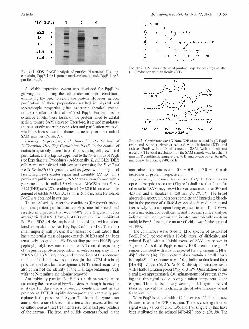
The use of strictly anaerobic conditions (for growth, induc-tion, and protein purification, see Experimental Procedures)resulted in a protein that was >90% pure (Figure 1) at anaverage yield of 0.5-1.5 mg/L of LB medium. The mobility ofPqqE on SDS gel electrophoresis is consistent with a calcu-lated molecular mass for His6-PqqE of 44.9 kDa. There is asmall impurity still present after anaerobic purification thathas a molecular mass of approximately 30 kDa and has beententatively assigned to a FK506 binding protein (FKBP)-typepeptidyl-prolyl cis-trans isomerase. N-Terminal sequencingof the purified protein preparation unveiled an impurity with aMKVAKDLVVS sequence, and comparison of this sequenceto that of other known sequences (in the NCBI database)provided the basis for the assignment. N-Terminal sequencingalso confirmed the identity of the His6 tag-containing PqqEwith the N-terminus methionine removed.
Anaerobically purified PqqE has a dark brown-red colorindicating the presence of Fe-S clusters. Although the enzymeis stable for days under anaerobic conditions and in thepresence of DTT, it rapidly decomposes and eventually pre-cipitates in the presence of oxygen. This form of enzyme is notamenable to anaerobic reconstitution with an excess of ferrousor sulfide ions as these treatments resulted in fast precipitationof the enzyme. The iron and sulfide contents found in the
anaerobic preparations are 10.4 ( 0.9 and 7.0 ( 1.0 mol/monomer of protein, respectively.Spectroscopic Characterization of PqqE. PqqE has an
optical absorption spectrum (Figure 2) similar to that found forother radical SAM enzymes with absorbance maxima at 390 and420 nm and a shoulder at 550 nm (27, 28, 31). The broadabsorption spectrum undergoes complete and immediate bleach-ing in the presence of a 10-fold excess of sodium dithionite andthen slowly re-forms upon being exposed to air. The UV-visspectrum, extinction coefficients, and iron and sulfide analysesindicate that PqqE grown and isolated anaerobically containsmultiple Fe-S clusters, the nature of which was further probedvia EPR.
The continuous wave X-band EPR spectra of as-isolatedPqqE, PqqE reduced with a 10-fold excess of dithionite, andreduced PqqE with a 10-fold excess of SAM are shown inFigure 3. As-isolated PqqE is nearly EPR silent in the g=2region, consistent with what is expected for a diamagnetic [4Fe-4S]2+ cluster (30). The spectrum does contain a small nearlyisotropic S=1/2 resonance at g=2.01, similar to that found for a[3Fe-4S]+ cluster (20, 21). At 40 K, this signal saturates easilywith a half-saturation power (P1/2) of 3 mW.Quantitation of thissignal gives approximately 0.01 spin/monomer of protein, show-ing that this signal is due to only a minor component of theenzyme. There is also a very weak g = 4.3 signal observed(data not shown) that is characteristic of adventitiously boundferric ions (30).
When PqqE is reduced with a 10-fold excess of dithionite, newfeatures arise in the EPR spectrum. There is a strong rhombicsignal with g values of 2.06, 1.96, and 1.91 (Figure 3) that havebeen attributed to the reduced [4Fe-4S]+ species (29, 30). The
FIGURE 1: SDS-PAGE analysis of purified N-terminal His6 tag-containing PqqE: lane 1, protein markers; lane 2, crude PqqE; lane 3,purified PqqE.
FIGURE 2: UV-vis spectrum of purified PqqE before (;) and after( 3 3 3 ) reduction with dithionite (DT).
FIGURE 3: ContinuouswaveX-bandEPRof as-isolatedPqqE,PqqE(with and without glycerol) reduced with dithionite (DT), andreduced PqqE with a 10-fold excess of SAM (with and withoutglycerol). The total incubation for the SAM sample was less than 5min. EPR conditions: temperature, 40K;microwave power, 6.3mW;microwave frequency, 9.480 GHz.

10156 Biochemistry, Vol. 48, No. 42, 2009 Wecksler et al.
shape and position of these signals are nearly identical to thoseobserved in the radical SAM enzyme pyruvate-formate lyase-activating enzyme (PFL-AE) (34) and very similar to those foundfor MOCS1A (27). Spin quantitation gives∼0.17 spin/monomerof protein. Unlike the oxidized enzyme, this species does notsaturate at 40 K (P1/2>200 mW).
Interestingly, there is only a moderate change in the EPRspectrum upon addition of SAM (Figure 3). Also, the spinquantitiation shows that the number of spins is unchanged. Thisbehavior is very different from that found for PFL-AE, where ithas been demonstrated that the presence of SAM significantlychanges the EPR spectrum of the reduced [4Fe-4S]+ cluster (34).There has also been a report on anaerobic ribonucleotidereductase that shows a change of the axial g=1.96 signal to amore rhombic one in the presence of SAM (29). The effect ofSAM on the EPR spectrum of the reduced [4Fe-4S]+ cluster wasfurther probed by measuring EPR samples without glycerol.Glycerol is known to be able to coordinate to the [4Fe-4S]+
cluster, inducing spectroscopic changes similar to that for SAMcoordination (29). When glycerol was omitted from the samples,the EPR spectrum of the reduced enzyme looked nearly identicalto that which contained glycerol (Figure 3). However, thereduced enzyme with SAM (without glycerol) looked slightlydifferent, with the g=2.00 feature more pronounced than whenboth SAM and glycerol were present in the sample. There werealso small changes in the signal shape at g=1.96.Demonstration of Enzymatic Activity for PqqE. PqqE
was incubated anaerobically at room temperature with a 10-foldexcess of dithionite and SAM and left for 2 h. The reaction wasthen quenched with formic acid and the mixture analyzed byLC-MS. A second reaction (without PqqE) was run underidentical conditions as a control reaction. In the control reaction(Figure 4A), the major species elutes immediately after thesolvent front at approximately 8 min and has been assigned toS-adenosylmethionine. Standards of SAM elute at an identicaltime and give the expected mass-to-charge ratio for the singlycharged positive ion at m/z 399.5 (C15H23N6O5S
+). Two otherpeaks are observed in the control reactions at approximately10min (m/z 136) and 33.5min (m/z 298.5), which are attributableto the singly charged ions of adenine (C5H5N5 + H+) andmethylthioadenosine (C11H14N5O3S
+), respectively. These com-pounds are typical thermal decomposition products of SAM andhave been observed by others (35).
The same peaks are also observed in the reaction mix-ture containing PqqE, but two new features emerge from the
chromatogram. The peak eluting at 8 min (assigned to SAM) hasdecreased in intensity, and there is a new species that elutes at 28min(see Figure 4B). This ion has an m/z of 252.4 (C10H13N5O3 +H+) and is due to 50-deoxyadenosine, as standards of50-deoxyadenosine elute at an identical time and give an identicalmass-to-charge ratio and isotope pattern. The other product ofthe radical SAM reaction is methionine. Although methionineabsorbs weakly at 260 nm, it can be observed when the LC-MSspectrum ismonitored at 215 nm. In this case (data not shown), alarge peak was observed that eluted at approximately 12min andhad an m/z of 150.2. This peak elutes at a time identical to thoseof standards ofmethionine andgives the expectedmass-to-chargeratio and isotope pattern for the singly charged, protonatedspecies (C5H11NO2S + H+).PqqE Is Capable of Undergoing Multiple Turnovers.
Anaerobic incubation of PqqE with dithionite and SAM resultsin the production of methionine and 50-deoxyadenosine. Theamount of 50-deoxyadenosine formed varied with time, andexperiments were performed to determine the pseudo-first-orderrate constant for the reaction. In these experiments, PqqE(39 μM) was reacted with an approximate 10-fold excess of bothsodium dithionite (420 μM) and SAM (420 μM) at 23 �C forvarious amounts of time, and then the reaction was quenched bythe addition of formic acid and the mixture analyzed via RP-HPLC (protocols outlined in Experimental Procedures).
The time-dependent formation of 50-deoxyadenosine is shownin Figure 5. The reaction is linear over 45 min, during which timePqqE undergoes approximately one turnover. The ratemeasuredfor the linear portion of this reaction was 0.417( 0.019 μM/min.When the enzyme concentration was decreased 2-fold (19 μM),the rate of 50-deoxyadenosinewasmeasured at 0.236( 0.0011μM/min. These experiments clearly showan enzyme dependence of 50-deoxyadenosine formation and give a pseudo-first-order rateconstant for the uncoupled reaction (kobs) of 0.011( 0.001min-1.The amount of 50-deoxyadenosine formation was also found tobe dependent on dithionite concentration, with multiple turn-overs observed only when in excess of PqqE (i.e., greater thanstoichiometric amounts).Incorporation ofDeuterium into 50-Deoxyadenosine dur-
ing Turnover. PqqE was exchanged into D2O by gel filtrationover a PD-10 column, and reactions were initiated and analyzedin a procedure identical to that described above. The chromato-grams from the reactions in D2O are nearly identical to thoseshown in Figure 4B. Peaks are observed at 8, 12, and 28 min,corresponding to SAM, methionine, and 50-deoxyadenosine,respectively. Although the retention time for the products isidentical to those observed in H2O, the mass-to-charge ratio andisotope patterns are significantly different. The mass spectrum of50-deoxyadenosine formed from the radical SAM reaction of
FIGURE 4: LC-MS analyses of the reaction products from in vitroactivation of PqqE with dithionite and SAM. (A) LC-MS elutionprofile (monitored at 260 nm) of a control without PqqE. (B)LC-MS elution profile of an anaerobic reaction mixture containingPqqE, SAM, and dithionite.
FIGURE 5: Time-dependent formation of 50-deoxyadenosine fromthe radical SAM reaction of PqqE measured via HPLC. The blacklines shown are single exponentials fitted to the data.
Article Biochemistry, Vol. 48, No. 42, 2009 10157
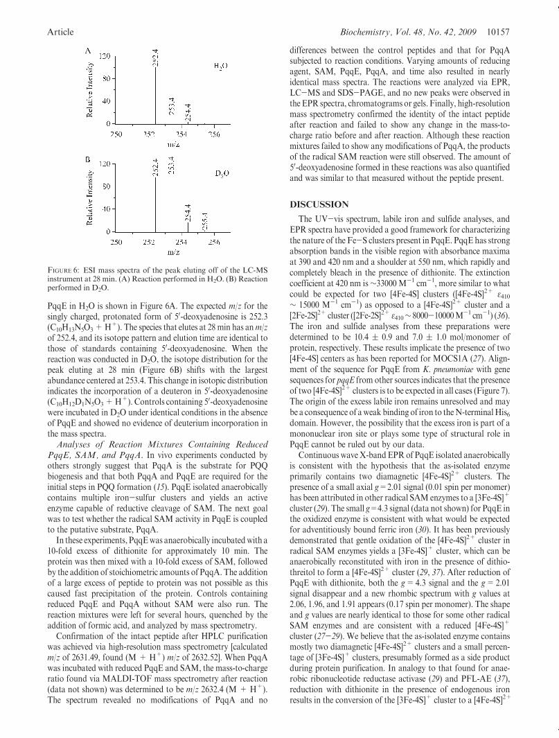
PqqE in H2O is shown in Figure 6A. The expected m/z for thesingly charged, protonated form of 50-deoxyadenosine is 252.3(C10H13N5O3+H+). The species that elutes at 28min has anm/zof 252.4, and its isotope pattern and elution time are identical tothose of standards containing 50-deoxyadenosine. When thereaction was conducted in D2O, the isotope distribution for thepeak eluting at 28 min (Figure 6B) shifts with the largestabundance centered at 253.4. This change in isotopic distributionindicates the incorporation of a deuteron in 50-deoxyadenosine(C10H12D1N5O3+H+). Controls containing 50-deoxyadenosinewere incubated in D2O under identical conditions in the absenceof PqqE and showed no evidence of deuterium incorporation inthe mass spectra.Analyses of Reaction Mixtures Containing Reduced
PqqE, SAM, and PqqA. In vivo experiments conducted byothers strongly suggest that PqqA is the substrate for PQQbiogenesis and that both PqqA and PqqE are required for theinitial steps in PQQ formation (15). PqqE isolated anaerobicallycontains multiple iron-sulfur clusters and yields an activeenzyme capable of reductive cleavage of SAM. The next goalwas to test whether the radical SAM activity in PqqE is coupledto the putative substrate, PqqA.
In these experiments, PqqEwas anaerobically incubatedwith a10-fold excess of dithionite for approximately 10 min. Theprotein was then mixed with a 10-fold excess of SAM, followedby the addition of stoichiometric amounts of PqqA. The additionof a large excess of peptide to protein was not possible as thiscaused fast precipitation of the protein. Controls containingreduced PqqE and PqqA without SAM were also run. Thereaction mixtures were left for several hours, quenched by theaddition of formic acid, and analyzed by mass spectrometry.
Confirmation of the intact peptide after HPLC purificationwas achieved via high-resolution mass spectrometry [calculatedm/z of 2631.49, found (M + H+) m/z of 2632.52]. When PqqAwas incubated with reduced PqqE and SAM, the mass-to-chargeratio found via MALDI-TOF mass spectrometry after reaction(data not shown) was determined to be m/z 2632.4 (M + H+).The spectrum revealed no modifications of PqqA and no
differences between the control peptides and that for PqqAsubjected to reaction conditions. Varying amounts of reducingagent, SAM, PqqE, PqqA, and time also resulted in nearlyidentical mass spectra. The reactions were analyzed via EPR,LC-MS and SDS-PAGE, and no new peaks were observed inthe EPR spectra, chromatograms or gels. Finally, high-resolutionmass spectrometry confirmed the identity of the intact peptideafter reaction and failed to show any change in the mass-to-charge ratio before and after reaction. Although these reactionmixtures failed to show any modifications of PqqA, the productsof the radical SAM reaction were still observed. The amount of50-deoxyadenosine formed in these reactions was also quantifiedand was similar to that measured without the peptide present.
DISCUSSION
The UV-vis spectrum, labile iron and sulfide analyses, andEPR spectra have provided a good framework for characterizingthe nature of the Fe-S clusters present in PqqE. PqqE has strongabsorption bands in the visible region with absorbance maximaat 390 and 420 nm and a shoulder at 550 nm, which rapidly andcompletely bleach in the presence of dithionite. The extinctioncoefficient at 420 nm is∼33000 M-1 cm-1, more similar to whatcould be expected for two [4Fe-4S] clusters ([4Fe-4S]2+ ε410∼ 15000 M-1 cm-1) as opposed to a [4Fe-4S]2+ cluster and a[2Fe-2S]2+ cluster ([2Fe-2S]2+ ε410∼ 8000-10000M-1 cm-1) (36).The iron and sulfide analyses from these preparations weredetermined to be 10.4 ( 0.9 and 7.0 ( 1.0 mol/monomer ofprotein, respectively. These results implicate the presence of two[4Fe-4S] centers as has been reported for MOCS1A (27). Align-ment of the sequence for PqqE from K. pneumoniae with genesequences for pqqE from other sources indicates that the presenceof two [4Fe-4S]2+ clusters is to be expected in all cases (Figure 7).The origin of the excess labile iron remains unresolved and maybe a consequence of aweak binding of iron to theN-terminalHis6domain. However, the possibility that the excess iron is part of amononuclear iron site or plays some type of structural role inPqqE cannot be ruled out by our data.
Continuous wave X-bandEPRof PqqE isolated anaerobicallyis consistent with the hypothesis that the as-isolated enzymeprimarily contains two diamagnetic [4Fe-4S]2+ clusters. Thepresence of a small axial g=2.01 signal (0.01 spin per monomer)has been attributed in other radical SAM enzymes to a [3Fe-4S]+
cluster (29). The small g=4.3 signal (data not shown) for PqqE inthe oxidized enzyme is consistent with what would be expectedfor adventitiously bound ferric iron (30). It has been previouslydemonstrated that gentle oxidation of the [4Fe-4S]2+ cluster inradical SAM enzymes yields a [3Fe-4S]+ cluster, which can beanaerobically reconstituted with iron in the presence of dithio-threitol to form a [4Fe-4S]2+ cluster (29, 37). After reduction ofPqqE with dithionite, both the g=4.3 signal and the g=2.01signal disappear and a new rhombic spectrum with g values at2.06, 1.96, and 1.91 appears (0.17 spin per monomer). The shapeand g values are nearly identical to those for some other radicalSAM enzymes and are consistent with a reduced [4Fe-4S]+
cluster (27-29). We believe that the as-isolated enzyme containsmostly two diamagnetic [4Fe-4S]2+ clusters and a small percen-tage of [3Fe-4S]+ clusters, presumably formed as a side productduring protein purification. In analogy to that found for anae-robic ribonucleotide reductase activase (29) and PFL-AE (37),reduction with dithionite in the presence of endogenous ironresults in the conversion of the [3Fe-4S]+ cluster to a [4Fe-4S]2+
FIGURE 6: ESI mass spectra of the peak eluting off of the LC-MSinstrument at 28 min. (A) Reaction performed in H2O. (B) Reactionperformed in D2O.

10158 Biochemistry, Vol. 48, No. 42, 2009 Wecksler et al.
cluster, followed by further reduction to a [4Fe-4S]+ cluster.Although we propose that the loss of the g=4.3 and 2.01 spectralchanges upon reduction is due to the conversion of smallamounts of the [3Fe-4S]+ cluster to the final [4Fe-4S]+ cluster,we cannot rule out the conversion of some unspecified para-magnetic species to a diamagnetic form.
EPR has also been used as a valuable tool to probe SAMcoordination to the reduced cluster (25). The binding of SAMwas investigated in the absence and presence of glycerol,as glycerol has been previously shown to bind to the reduced[4Fe-4S]+ cluster and induce spectral changes similar to that ofthe binding of SAM (29). When glycerol was omitted, there aresmall perturbations in the EPR spectrum in the presence of SAMwith the g=2.00 feature becoming more pronounced and the g=1.96 feature changing shape. This indicates that the g=2.00 signalmight be part of a rhombic minority species with g values of 2.00,
∼1.95, and 1.92. When SAM was added to reduced PqqE in thepresence of glycerol, only small perturbations in the EPRspectrum were observed with the shape and g values nearlyidentical to those without SAM (see Figure 3). These resultssuggest that either SAM is not coordinated to the [4Fe-4S]+
cluster or it is coordinated only weakly. Whether this species ischemically relevant is unclear.
The spectroscopic and physical measurements described aboveindicate that anaerobically purified PqqE contains multipleFe-S clusters, with the predominant form of the isolated enzymecontaining two [4Fe-4S]2+ clusters. The EPR and UV-visspectra suggested that at least one of the clusters would be redoxactive with characteristics similar to those found for other radicalSAM enzymes. The role of the second cluster is still unclear, butin analogy to that forMOCS1A, it may play a role in positioningthe substrate for H-atom abstraction.
FIGURE 7: Conserved residues (asterisks denote cysteines proposed to bind [4Fe-4S] clusters) in the N- and C-terminal domains for PqqE fromK. pneumoniae, Methylibium petroleiphilum PM1, Colwellia psychrerythraea 34H, Methylococcus capsulatus Bath, Nitrosococcus oceani ATCC19707,Pseudomonas fluorescens Pf-5, Pseudomonas putidaGB-1,Pseudomonas syringae pv.Phaseolicola 1448A, and Exiguobacterium sp. AT1b.

Article Biochemistry, Vol. 48, No. 42, 2009 10159
When PqqE was anaerobically incubated with an excess ofSAM and dithionite, methionine and 50-deoxyadenosine wereformed as products of the enzymatic reaction (Figure 4). Thecleavage of SAM to methionine and 50-deoxyadenosyl radical isenergetically unfavorable (bond dissociation energy for the C-Sbond of 60 kcal/mol) and is possible only via enzymatic activa-tion (Scheme 2) (38, 39). This radical is highly reactive andgenerally initiates the next step in catalysis via abstraction of aH-atom from a substrate. We have thoroughly looked for thepresence of stable organic radicals in PqqE reaction mixtures viaEPR without success. Therefore, the production of methionineand 50-deoxyadenosine is concluded to derive from an “un-coupled reaction”, whereby the generation of the 50-deoxyade-nosyl radical secures a proton and electron frombuffer sources toform 50-deoxyadenosine. When the reaction was performed inD2O and the reaction mixtures were analyzed via LC-MS, anincrease of one mass unit in the mass-to-charge ratio of50-deoxyadenosine (Figure 6) indicates that solvent-derived deu-terium has been incorporated into the product.
The quenching of the 50-deoxyadenosyl radical to form50-deoxyadenosine could occur through two different mechan-isms: (1) abstraction of a H-atom from the protein followed byfast outer-sphere electron transfer to the protein-derived radicalor (2) reduction of the 50-deoxyadenosyl radical by dithionite tothe corresponding carbanion followed by rapid abstraction of aproton from the solvent. Although no organic radicals wereobserved in the EPR, the first mechanism cannot be ruled outbecause outer-sphere electron transfer to the protein-derivedradical may be so fast that it is undetectable via EPR underour assay conditions; further, abstraction of a H-atom from asolvent exchangeable position on PqqE would lead to the samedeuterated product as direct transfer of a proton from solvent.The “uncoupled” production of 50-deoxyadenosine in severalother radical SAM enzymes has been observed previously(40, 41), but to the best of our knowledge, this is the first exampleof an uncoupled radical SAM reaction that leads to the incor-poration of a solvent deuterium into 50-deoxyadenosine. Thismay be suggestive of a solvent-accessible amino acid residue inthe active site of PqqE that is not shared with other radical SAMenzymes. At this juncture, we are not sure how the radical isquenched, but the aggregate experiments show that a 50-deox-yadenosyl intermediate is formed from reductive cleavage ofSAM by PqqE and are consistent with the proposed mechanismfor radical SAM enzymes (38, 39). The fact that deuteriumincorporation is only approximately 50% can be attributed toincomplete buffer exchange in PqqE (see ExperimentalProcedures) and dilution of D2O with protiated glycerol addedat a mole ratio of 8% to reaction mixtures.
The amount of 50-deoxyadenosine formed from the uncoupledradical SAM reaction in PqqE varied with time, undergoing
multiple turnovers over the course of several hours. Furthermore,decreasing the enzyme concentration 2-fold caused the rate of50-deoxyadenosine formation to decrease by a factor of 2, clearlyindicating the dependence of 50-deoxyadenosine formation onenzyme. The rate for the production of 50-deoxyadenosine waslinear for approximately 45 min, and during this time period,kobs=0.011 ( 0.001 min-1. Although this rate constant seemsquite slow for an enzymatic reaction, the uncoupled rate is onlyapproximately 6 times slower than single-turnover rate constantsof biotin synthase (0.07min-1) (42), 10 times slower than that forlipoyl synthase (0.175 min-1) (43), and ∼30 times slower thanthat for AtsB (0.38 min-1) (31). This rate is only∼7 times slowerwhen compared to the rate-limiting step for TPQ biogenesis(kbio=0.08 min-1) (44), indicating that the turnover rate is atleast comparable with those found for the formation of otherquinone cofactors.
We have proceeded on the premise that PqqE catalyzes thefirst step in PQQ biogenesis, thought to be the linking of theamino acid side chains of glutamate and tyrosine in PqqA.Previous in vivo labeling experiments demonstrated PQQ wasformed from the fusion of glutamate and tyrosine, also providingevidence for how these amino acids are linked together (seeScheme 1) (11, 12). A carbon-carbon bond is formed betweenC9 fromglutamate andC9a from tyrosine. It seems likely that theradical SAM enzyme could catalyze this step since the largeactivation energy needed to break the aromaticity of the tyrosinering could be overcome by formation of a glutamate radicalgenerated via H-atom abstraction by the 50-deoxyadenosylradical.
We tested PqqA as a substrate for PqqE by incubating theenzyme with varying amounts of SAM, dithionite, and PqqA.For these reactions, the enzyme needed to be stabilized withglycerol and PqqA slowly titrated into the reaction mixtures dueto significant protein precipitation problems. Reactions were alsoattempted with a small amount of ethanol or denaturant tocircumvent potential peptide aggregation. The reaction mixtureswere left for various periods of time and then analyzed viaMALDI-TOFmass spectrometry, LC-MS, SDS-PAGE, EPR,and high-resolution mass spectrometry. Under all conditions, nomodifications of the peptide or (radical) intermediates wereobserved, even though the uncoupled reaction products(methionine and 50-deoxyadenosine) were still detected. Further-more, there were no new peaks observed in the EPR, LC-MStraces, or bands in the SDS-PAGE analyses. The reactionmixtures were also exposed to molecular oxygen after anaerobicincubation, on the premise that O2 may play a role in trappingradical intermediates and driving the reaction toward products.However, these experiments failed to show any new products ormodifications of PqqA. Experiments performed with glutamateand tyrosine as potential substrates for PqqE also failed to showany new products.
The fact that no modifications of PqqA have been observedmay be due to a lack of sensitivity of detection (for instance, ifonly a very small amount of product were formed), or perhapsa reversible cleavage of SAM with an equilibrium that favorsthe reactants. Experiments that aim to test these hypothesesusing spin-trapping agents and also utilizing radiolabeled[2,3,50-3H]SAM to look for tritium scrambling into the peptideare under way.
Alternatively, other proteins in the PQQ biosynthetic pathwaymay be necessary for the initial steps in PQQ biogenesis. Onepossible candidate is PqqD, a gene product with no known
Scheme 2: PqqE Catalyzes the Reductive Cleavage of SAMToForm Methionine and a 50-Deoxyadenosyl Radical

10160 Biochemistry, Vol. 48, No. 42, 2009 Wecksler et al.
homology to any annotated protein. PqqA may also need to bemodified by another gene product in the pqq operon prior toreaction with PqqE. One important question that we would liketo resolve is that of which chemical step happens first, thecross-linking of PqqA or the hydroxylation of the conservedtyrosine. Veletrop et al. have demonstrated that molecularoxygen is critical for PQQ biogenesis (15), and it is reasonableto suggest that the hydroxylation of the tyrosine in PqqA occursvia activation of molecular oxygen.We had initially thought thatPqqE may be responsible for both of these steps; however, wehave yet to observe this type of unprecedented radical SAMchemistry.
It has been recently demonstrated in TTQ biogenesis thatmutation of critical residues in the mauG gene results in theformation of a stable intermediate that does not have thetryptophan-tryptophan cross-link or the quinone moiety butdoes contain a monohydroxylated tryptophan (45-47). Incuba-tion of the monohydroxylated intermediate with active MauGand either hydrogen peroxide or oxygen with reducing equiva-lents results in tryptophan-tryptophan cross-linking and con-comitant formation of the quinone (48-50). In analogy to TTQbiosynthesis, we suspect that hydroxylation of the tyrosine mayoccur before cross-linking of PqqA, after which PqqE catalyzesthe cross-linking via a radical mechanism. Ongoing experimentsin this laboratory are aimed at oxidatively modifying the con-served tyrosine in PqqA, with the goal of assessing whether suchderivations of PqqA are capable of entering into reaction withPqqE.
ACKNOWLEDGMENT
We thank Prof. Robert Rucker for the donation of plasmidpBCP-165 containing the pqq operon, Dr. Petra Hanzelmann forthe donation of plasmids pPH149 and pPH151 containing theE. coli IscSUA-HscBA-Fd and E. coli suf ABCDSE genes,respectively, and Dr. Charles Roessner for a clone containingthe sam2 gene from E. coli [TB1(pUC18:sam2)]. We also thankDr. Junko Yano for help with preliminary EPR measurements,Dr. Tony Iavarone for obtaining high-resolution mass spectra ofpeptides, and Ms. Mae Tulfo for help preparing the manuscript.
SUPPORTING INFORMATION AVAILABLE
Methods for the aerobic and anaerobic growth and inductionof E. coli BL21(DE3) cells harboring the pET24b-pqqE plasmid,aerobic purification of PqqE, anaerobic reconstitution of PqqEwith iron and sulfide ions, synthesis and purification of SAM,and a detailed description of the high-resolution mass spectro-metry experiments. This material is available free of charge viathe Internet at http://pubs.acs.org.
REFERENCES
1. Klinman, J. P. (2001) Howmany ways to craft a cofactor? Proc. Natl.Acad. Sci. U.S.A. 98, 14766–14768.
2. Ono,K., Okajima, T., Tani,M.,Kuroda, S., Sun,D., Davidson, V. L.,and Tanizawa, K. (2006) Involvement of a putative [Fe-S]-cluster-binding protein in the biogenesis of quinohemoprotein aminedehydrogenase. J. Biol. Chem. 281, 13672–13684.
3. Meulenberg, J. J. M., Sellink, E., Riegman, N. H., van Kleef, M., andPostma, P. W. (1992) Nucleotide sequence and structure of theKlebsiella pneumoniae pqq operon. Mol. Gen. Genet. 232, 284–294.
4. Anthony, C., and Zatman, L. J. (1967) The microbial oxidation ofmethanol: The prosthetic group of alcohol dehydrogenase of Pseudo-monas sp. M27; a new oxidoreductase prosthetic group. Biochem. J.104, 953–959.
5. Salisbury, S.A., Forrest,H. S., Cruse,W. B. T., andKennar, O. (1979)A novel coenzyme from bacterial primary alcohol dehydrogenases.Nature 280, 843–844.
6. Duine, J. A., Frank, J., and van Zeeland, J. K. (1979) Glucosedehydrogenase from Acinetobacter calcoacetcus. FEBS Lett. 108,443–446.
7. Goodwin, P. M., and Anthony, C. (1998) The Biochemistry, Physiol-ogy andGenetics of PQQand PQQ-containingEnzymes. InAdvancesin Microbial Physiology (Poole, R. K., Ed.) Vol. 40, pp 1-80, Elsevier,Amsterdam.
8. Mitchell, A. E., Jones, A. D., Mercer, R. S., and Rucker, R. B. (1999)Characterization of pyrroloquinoline quinone amino acid derivativesby electrospray ionization mass spectrometry and detection in humanmilk. Anal. Biochem. 269, 317–325.
9. Killgore, J., Smidt, C., Duich, L., Romero-Chapman, N., Tinker, D.,Reiser, K., Melko, M., Hyde, D., and Rucker, R. B. (1989) Nutri-tional importance of pyrroloquinoline quinone. Science 245, 850–852.
10. Kasahara, T., and Kato, T. (2003) A new redox-cofactor vitamin formammals. Nature 422, 832.
11. Houck, D. R., Hanners, J. L., and Unkefer, C. J. (1988) Biosynthesisof pyrroloquinoline quinone. 1. Identification of a biosynthetic pre-cursor using 13C labeling and NMR spectroscopy. J. Am. Chem. Soc.110, 6920–6921.
12. Houck, D. R., Hanners, J. L., and Unkefer, C. J. (1991) Biosynthesisof pyrroloquinoline quinone. 2. Biosynthetic assembly from glutamateand tyrosine. J. Am. Chem. Soc. 113, 3162–3166.
13. Toyama,H., Chistoserdova, L., and Lidstrom,M. E. (1997) Sequenceanalysis of pqq genes required for biosynthesis of pyrroloquinolinequinone inMethylobacterium extorquensAM1 and the purification ofa biosynthetic intermediate. Microbiology 143, 595–602.
14. Goosen,N.,Horsman,H. P. A.,Huinen, R.G.M., and Putte, V.D. P.(1989) Acinetobacter calcoaceticus genes involved in biosynthesis ofthe coenzyme pyrrolo-quinoline-quinone: Nucleotide sequence andexpression in Escherichia coli K-12. J. Bacteriol. 171, 447–455.
15. Veletrop, J. S., Sellink, E., Meulenberg, J. J. M., Bulder, D. S., andPostma, P. W. (1995) Synthesis of pyrroloquinoline quinone in vivoand in vitro and detection of an intermediate in the biosyntheticpathway. J. Bacteriol. 177, 5088–5098.
16. Magnusson, O. T., Toyama,H., Saeki, M., Schwarzenbacher, R., andKlinman, J. P. (2004) The structure of a biosynthetic intermediate ofpyrroloquinoline quinone (PQQ) and elucidation of the final step ofPQQ biogenesis. J. Am. Chem. Soc. 126, 5342–5343.
17. Toyama, H., Fukumoto, J., Saeki, M., Matsushita, K., Adachi, O.,and Lidstrom, M. E. (2002) PqqC/D, which converts a biosyntheticintermediate to pyrroloquinoline quinone. Biochem. Biophys. Res.Commun. 299, 268–272.
18. Sofia, H. J., Chen, G., Hetzler, B. G., Reyes-Spindola, J. F., andMiller, N. E. (2001) Radical SAM, a novel protein superfamily linkingunresolved steps in familiar biosynthetic pathways with radicalmechanisms: Functional characterization using new analysisand information visualization methods. Nucleic Acids Res. 29,1097–1106.
19. Magnusson, O. T., Toyama, H., Saeki, M., Rojas, A., Reed, J. C.,Liddington, R. C., Klinman, J. P., and Schwarzenbacher, R. (2004)Quinone biogenesis: Structure and mechanism of PqqC, the finalcatalyst in the production of pyrroloquinoline quinone. Proc. Natl.Acad. Sci. U.S.A. 101, 7913–7918.
20. Menendez, C., Igloi, G., Henninger, H., and Brandsch, R. (1995) ApAO-1-encodedmolybdopterin cofactor gene (moaA) ofArthrobacternicotinovorans: Characterization and site-directed mutagenesis of theencoded protein. Arch. Microbiol. 164, 142–151.
21. Hanzelmann, P., and Schindelin, H. (2006) Binding of 50-GTP to theC-terminal FeS cluster of the radical S-adenosylmethionine enzymeMoaA provides insights into its mechanism. Proc. Natl. Acad. Sci. U.S.A. 103, 6829–6834.
22. Beinert, H. (1978) Micro methods for the quantitative determinationof iron and copper in biological material.Methods Enzymol. 54, 435–445.
23. Beinert, H. (1983) Semi-micro methods for analysis of labile sulfideand of labile sulfide and sulfane sulfur in unusually stable iron-sulfurproteins. Anal. Biochem. 131, 373–378.
24. Park, J., Tai, J., Roessner, C. A., and Scott, A. I. (1996) Enzymaticsynthesis of S-adenosyl-L-methionine on the preparative scale.Bioorg.Med. Chem. 4, 2179–2185.
25. Walsby, C. J., Hong, W., Broderick, W. E., Cheek, J., Ortillo, D.,Broderick, J. B., and Hoffman, B. M. (2002) Electron-nuclear doubleresonance spectroscopic evidence that S-adenosylmethionine binds incontact with the catalytically active [4Fe-4S]+ cluster of pyruvateformate-lyase activating enzyme. J. Am. Chem. Soc. 124, 3143–3151.

Article Biochemistry, Vol. 48, No. 42, 2009 10161
26. Shapiro, S. K., and Ehniger, D. J. (1966) Methods for analysis andpreparation of adenosylmethionine and adenosylhomocysteine.Anal.Biochem. 15, 323–333.
27. Hanzelmann, P., Hern�andez, H. L., Menzel, C., Garcia-Serres, R.,Huynh, B. H., Johnson, M. K., Mendel, R. R., and Schindelin, H.(2004) Characterization of MOCS1A, and oxygen-sensitive iron-sulfur protein involved in human molybdenum cofactor biosynthesis.J. Biol. Chem. 279, 34721–34732.
28. Hern�andez, H. L., Pierrel, F., Elleingand, E., Garcıa-Serres, R.,Huynh, B. H., Johnson, M. K., Fontecave, M., and Atta, M. (2007)MiaB, a bifunctional radical-S-adenosylmethionine enzyme involvedin thiolation andmethylation of tRNA, contains two essential [4Fe-4S]clusters. Biochemistry 46, 5140–5147.
29. Lim, A., and Grasland, A. (2000) Electron paramagnetic resonanceevidence for a novel interconversion of [3Fe-4S]+ and [4Fe-4S]+
clusters with endogenous iron and sulfide in anaerobic ribonucleotidereductase activase in vitro. J. Biol. Chem. 275, 12367–12373.
30. Beinert, H., Kennedy, M. C., and Stout, C. D. (1996) Aconitase asiron-sulfur protein, enzyme, and iron-regulatory protein. Chem. Rev.96, 2335–2373.
31. Grove, T. L., Lee, K.-H., St. Clair, J., Krebs, C., and Booker, S. J.(2008) In vitro characterization ofAtsB, a radical SAM formylglycine-generating enzyme that contains three [4Fe-4S] clusters. Biochemistry47, 7523–7538.
32. Takahashi, Y., and Tokumoto, U. (2002) A third bacterial system forthe assembly of iron-sulfur clusters with homologs in archaea andplastids. J. Biol. Chem. 277, 28380–28383.
33. Nachin, L., Loiseau, L., Expert, D., and Barras, F. (2003) SufC: Anunorthodox cytoplasmic ABC/ATPase required for [Fe-S] biogenesisunder oxidative stress. EMBO 22, 427–437.
34. Walsby, C. J., Ortillo, D., Yang, J., Nnyepi, M. R., Broderick, W. E.,Hoffman, B. M., and Broderick, J. B. (2005) Spectroscopicapproaches to elucidating novel iron-sulfur chemistry in the “radi-cal-SAM” protein superfamily. Inorg. Chem. 44, 727–741.
35. Hoffman, J. L. (1986) Chromatographic analysis of the chiral andcovalent instability of S-adenosyl-L-methionine. Biochemistry 25,4444–4449.
36. Dailey, H. A., Finnegan, M. G., and Johnson, M. K. (1994) Humanferrochelatase is an iron-sulfur protein. Biochemistry 33, 403–407.
37. Krebs, C., Broderick, W. E., Henshaw, T. F., Broderick, J. B., andHuynh, B. H. (2002) Coordination of adenosylmethionine to a uniqueiron site of the [4Fe-4S] cluster of pyruvate formate lyase-activatingenzyme: A M€ossbauer spectroscopic study. J. Am. Chem. Soc. 124,912–913.
38. Frey, P. A. (2001) Radical mechanisms of enzyme catalysis. Annu.Rev. Biochem. 70, 121–148.
39. Frey, P. A., and Magnusson, O. T. (2003) S-Adenosylmethionine:A wolf in sheep’s clothing, or a rich man’s adenosylcobalamin?Chem.Rev. 103, 2129–2148.
40. Ollagnier, S., Sanakis, Y., Hewiston, K. S., Roach, P., andMunck, E.(2002) Reductive of S-adenosylmethionine by biotin synthase fromEscherichia coli. J. Biol. Chem. 277, 13449–13454.
41. Farrar, C. E., and Jarrett, J. T. (2009) Protein residues that controlthe reaction trajectory is S-Adenosylmethionine radical enzymes:Mutagenesis of asparagine 153 and aspartate 155 in Escherichia colibiotin synthase. Biochemistry 48, 2448–2458.
42. Ugulava, N. B., Gibney, B. R., and Jarrett, J. T. (2000) Iron-sulfurcluster interconversions in biotin synthase: Dissociation and reasso-ciation of iron during conversion of [2Fe-2S] to [4Fe-4S] clusters.Biochemistry 39, 5206–5214.
43. Cicchillo, R. M., Iwig, D. F., Jones, A. D., Nesbitt, N. M., Baleanu-Gogonea, C., Souder, M. G., Tu, L., and Booker, S. J. (2004) Lipoylsynthase requires two equivalents of S-adenosyl-L-methionine tosynthesize one equivalent of lipoic acid. Biochemistry 43, 6378–6386.
44. Schwartz, B., Dove, J. E., and Klinman, J. P. (2000) Kinetic analysisof oxygen utilization during cofactor biogenesis in a copper-contain-ing amine oxidase from yeast. Biochemistry 39, 3699–3707.
45. Pearson, A. R., Jones, L. H., Higgins, L., Ashcroft, A. E., Wilmot,C. M., and Davidson, V. L. (2003) Understanding quinone cofactorbiogenesis in methylamine dehydrogenase through novel cofactorgeneration. Biochemistry 42, 3224–3230.
46. Pearson, A. R., De La Mora-Rey, T., Graichen, M. E., Wang, Y.,Jones, L. H., Marimanikkupam, S., Agger, S. A., Grimsrud, P. A.,Davidson, V. L., and Wilmot, C. M. (2004) Further insights intoquinone cofactor biogenesis: Probing the role of mauG in methyla-mine dehydrogenase tryptophan tryptophylquinone formation.Biochemistry 43, 5494–5502.
47. Pearson, A. R., Marimanikkuppam, S., Li, X., Davidson, V. L., andWilmot, C. M. (2006) Isotope labeling studies reveal the order ofoxygen incorporation into the tryptophan tryptophylquinone cofactorof methylamine dehydrogenase. J. Am. Chem. Soc. 128, 12416–12417.
48. Wang, Y., Graichen, M. E., Liu, A., Pearson, A. R., Wilmot, C. M.,and Davidson, V. L. (2003) MauG, a novel diheme protein requiredfor tryptophan tryptophylquinone biogenesis. Biochemistry 42, 7318–7325.
49. Wang, Y., Li, X., Jones, L. H., Pearson, A. R., Wilmot, C. M., andDavidson, V. L. (2005) MauG-dependent in vitro biosynthesis oftryptophan tryptophylquinone in methylamine dehydrogenase.J. Am. Chem. Soc. 127, 8258–8259.
50. Li, X., Jones, L. H., Pearson, A. R., Wilmot, C. M., and Davidson,V. L. (2006)Mechanistic possibilities inMauG-dependent tryptophantryptophylquinone biosynthesis. Biochemistry 45, 13276–13283.