bioconjugation strategies for microtoroidal optical - mdpi.com
TRANSCRIPT
Sensors 2010, 10, 9317-9336; doi:10.3390/s101009317
sensors ISSN 1424-8220
www.mdpi.com/journal/sensors
Article
Bioconjugation Strategies for Microtoroidal Optical Resonators
Heather K. Hunt 1, Carol Soteropulos 2 and Andrea M. Armani 1,3,*
1 Mork Family Department of Chemical Engineering and Materials Science, University of Southern
California, Los Angeles, CA 90089, USA; E-Mail: [email protected] 2 Department of Biomedical Engineering, University of Southern California, Los Angeles, CA
90089, USA; E-Mail: [email protected] 3 Ming Hsieh Department of Electrical Engineering-Electrophysics, University of Southern
California, Los Angeles, CA 90089, USA
* Author to whom correspondence should be addressed; E-Mail: [email protected];
Tel.: +1-213-740-4428.
Received: 14 September 2010; in revised form: 8 October 2010 / Accepted: 14 October 2010 /
Published: 18 October 2010
Abstract: The development of label-free biosensors with high sensitivity and specificity is
of significant interest for medical diagnostics and environmental monitoring, where rapid
and real-time detection of antigens, bacteria, viruses, etc., is necessary. Optical resonant
devices, which have very high sensitivity resulting from their low optical loss, are uniquely
suited to sensing applications. However, previous research efforts in this area have focused
on the development of the sensor itself. While device sensitivity is an important feature of
a sensor, specificity is an equally, if not more, important performance parameter.
Therefore, it is crucial to develop a covalent surface functionalization process, which also
maintains the device’s sensing capabilities or optical qualities. Here, we demonstrate a
facile method to impart specificity to optical microcavities, without adversely impacting
their optical performance. In this approach, we selectively functionalize the surface of the
silica microtoroids with biotin, using amine-terminated silane coupling agents as linkers.
The surface chemistry of these devices is demonstrated using X-ray photoelectron
spectroscopy, and fluorescent and optical microscopy. The quality factors of the surface
functionalized devices are also characterized to determine the impact of the chemistry
methods on the device sensitivity. The resulting devices show uniform surface coverage,
with no microstructural damage. This work represents one of the first examples of
non-physisorption-based bioconjugation of microtoroidal optical resonators.
OPEN ACCESS
Sensors 2010, 10
9318
Keywords: bioconjugation; optical resonators; sensors; high quality factor
1. Introduction
The development of biosensors with high sensitivity and specificity is of significant interest to
scientific communities, such as medical diagnostics and environmental monitoring, where rapid and
real-time detection of antigens, bacteria, viruses, etc., is necessary. A prime example for this need is
the development of biosensors for detection in complex environments, such as whole blood or serum.
Common, high sensitivity methods for detection in such complex environments include
immunoassays, such as the ELISA assays and fluorescent immunoassays, which require the presence
of a label moiety for detection [1-5]. However, these traditional, labeled sensors detect the presence of
the label or probe rather than the molecule of interest, and require foreknowledge of the presence of
the target. On the other hand, label-free sensors, such as electrical sensors [6,7], mechanical cantilever
sensors [8-10], and optical sensors, such as optical waveguides [11-13], surface plasmon waveguides
and resonators [14-19], and ring resonators [20], can detect the molecule of interest, but often have
difficulty discriminating between targeted and non-targeted species in complex environments, such as
serum and whole blood. Therefore, currently, traditional labeled biosensors have a significant
advantage over label-free biosensors in many “real-world” applications.
This limitation of label-free biosensors can be eradicated by the addition of a component that adds
specificity to the device. While the majority of previous research efforts on label-free biosensors have
focused on the development of the sensor itself [6,10,21-30], specificity is an equally if not more
important feature of any sensing platform, especially for detection in complex environments [31,32].
Label-free sensor performance can be improved by requiring the addition of a component, such as a
probe molecule, that allows the sensor to selectively identify the target molecule [33,34]. In many sensing
methods, this has been accomplished by surface immobilization of probe molecules via physical
adsorption, self-assembly, or covalent attachment [35]. Of these techniques, covalent attachment provides
the most stable immobilization towards changing environmental conditions, such as temperature,
humidity, and pH. To date, the majority of research using optical sensors based on waveguides and
surface plasmon resonance, have been paired through physisorption with enzymes [14,36],
peptides [22,37,38], antibodies (or antibody fragments) [1,6,14,39,40], aptamers [41,42], and
receptors [16,31,43-45] as environmental probes. However, recently, there has been increased interest
in developing more robust surface functionalization protocols for optical devices, such as whispering
gallery mode resonant cavities [46].
Although initially designed for telecommunications, these whispering gallery mode microcavities
have demonstrated unique capabilities in the biosensing arena primarily due to their very low optical
loss, corresponding to very high sensitivities in biodetection [26,29,30,47-59]. Whispering gallery
mode optical resonators efficiently confine light at specific resonant frequencies within the resonator
periphery (Figure 1a). In these devices, the optical field is not completely confined to the resonator,
but instead extends or evanesces into the surrounding environment, and interacts with its surroundings,
thus enabling the detection and sensing capabilities of the resonators (Figure 1b). The primary gauge
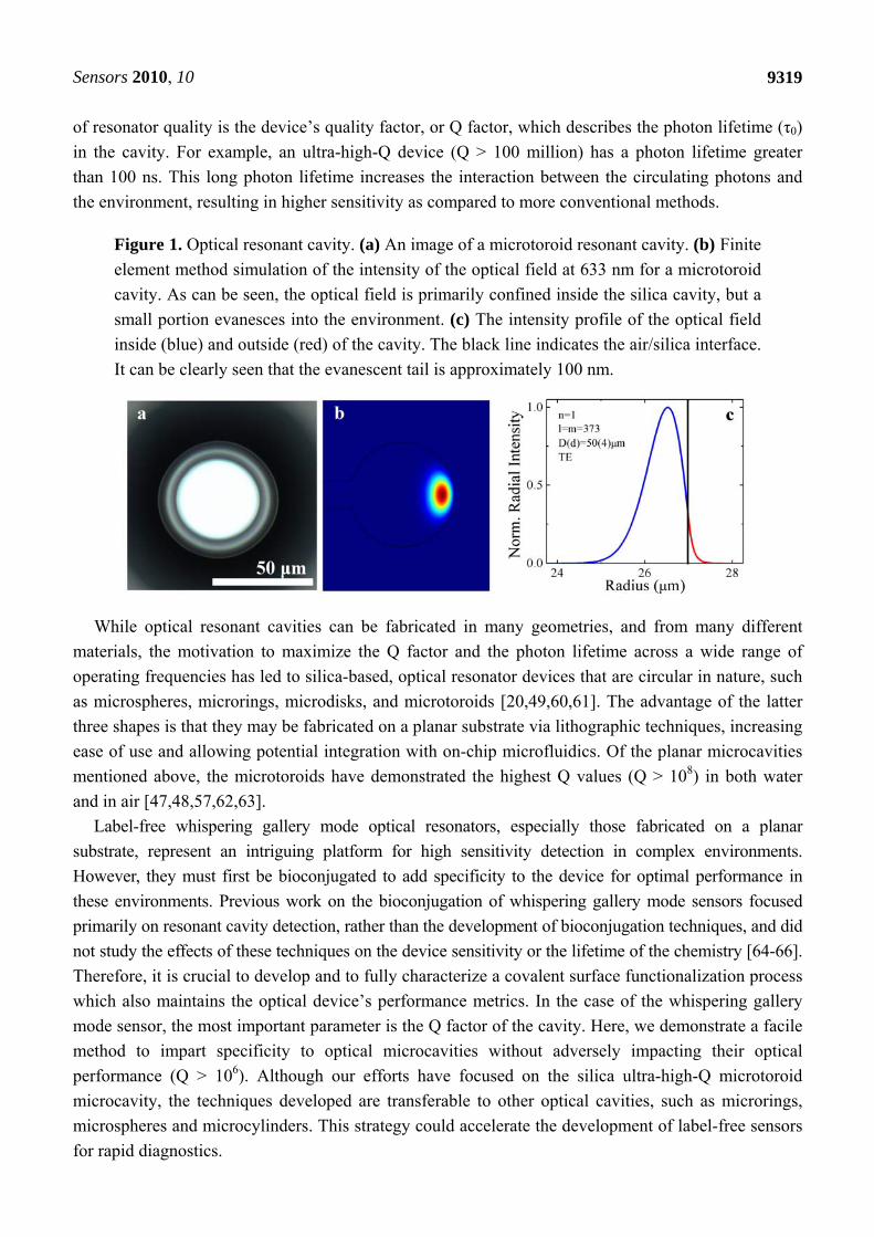
Sensors 2010, 10
9319
of resonator quality is the device’s quality factor, or Q factor, which describes the photon lifetime (τ0)
in the cavity. For example, an ultra-high-Q device (Q > 100 million) has a photon lifetime greater
than 100 ns. This long photon lifetime increases the interaction between the circulating photons and
the environment, resulting in higher sensitivity as compared to more conventional methods.
Figure 1. Optical resonant cavity. (a) An image of a microtoroid resonant cavity. (b) Finite
element method simulation of the intensity of the optical field at 633 nm for a microtoroid
cavity. As can be seen, the optical field is primarily confined inside the silica cavity, but a
small portion evanesces into the environment. (c) The intensity profile of the optical field
inside (blue) and outside (red) of the cavity. The black line indicates the air/silica interface.
It can be clearly seen that the evanescent tail is approximately 100 nm.
While optical resonant cavities can be fabricated in many geometries, and from many different
materials, the motivation to maximize the Q factor and the photon lifetime across a wide range of
operating frequencies has led to silica-based, optical resonator devices that are circular in nature, such
as microspheres, microrings, microdisks, and microtoroids [20,49,60,61]. The advantage of the latter
three shapes is that they may be fabricated on a planar substrate via lithographic techniques, increasing
ease of use and allowing potential integration with on-chip microfluidics. Of the planar microcavities
mentioned above, the microtoroids have demonstrated the highest Q values (Q > 108) in both water
and in air [47,48,57,62,63].
Label-free whispering gallery mode optical resonators, especially those fabricated on a planar
substrate, represent an intriguing platform for high sensitivity detection in complex environments.
However, they must first be bioconjugated to add specificity to the device for optimal performance in
these environments. Previous work on the bioconjugation of whispering gallery mode sensors focused
primarily on resonant cavity detection, rather than the development of bioconjugation techniques, and did
not study the effects of these techniques on the device sensitivity or the lifetime of the chemistry [64-66].
Therefore, it is crucial to develop and to fully characterize a covalent surface functionalization process
which also maintains the optical device’s performance metrics. In the case of the whispering gallery
mode sensor, the most important parameter is the Q factor of the cavity. Here, we demonstrate a facile
method to impart specificity to optical microcavities without adversely impacting their optical
performance (Q > 106). Although our efforts have focused on the silica ultra-high-Q microtoroid
microcavity, the techniques developed are transferable to other optical cavities, such as microrings,
microspheres and microcylinders. This strategy could accelerate the development of label-free sensors
for rapid diagnostics.
Sensors 2010, 10
9320
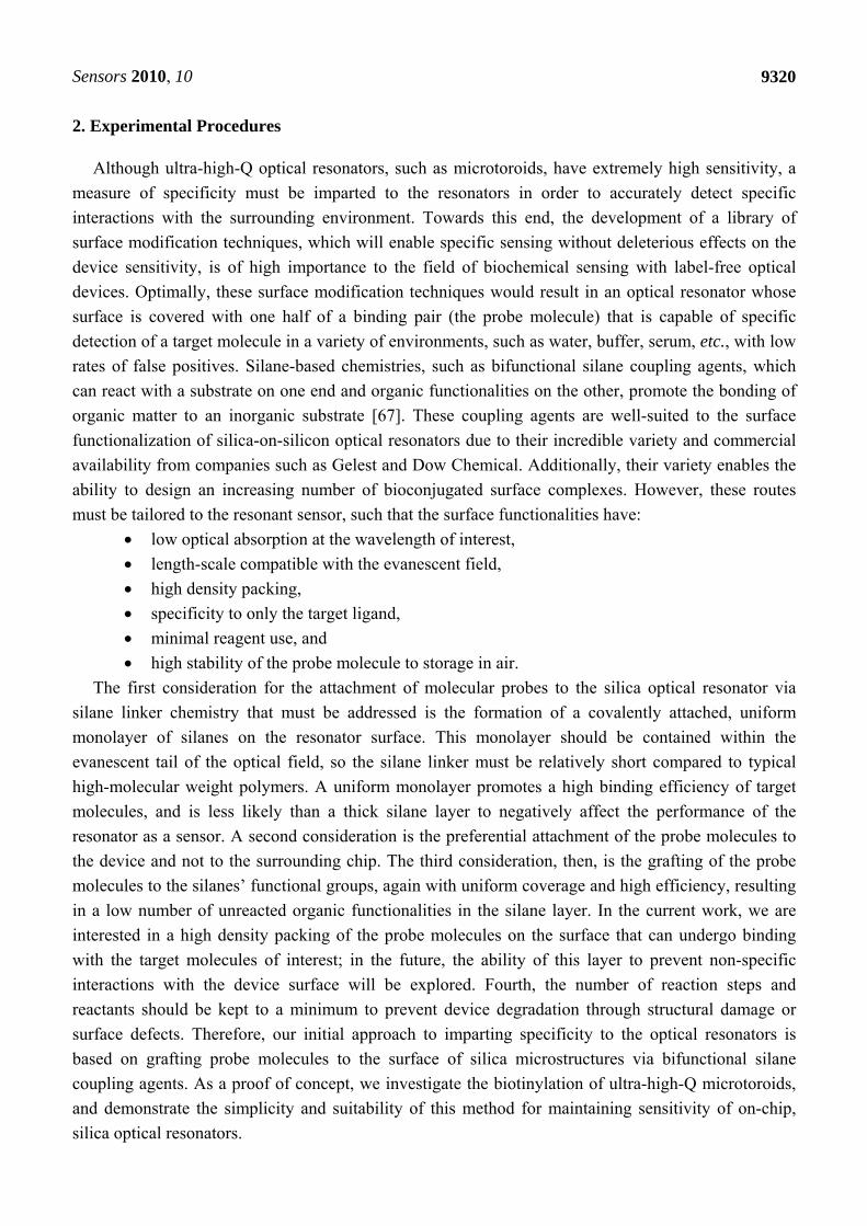
2. Experimental Procedures
Although ultra-high-Q optical resonators, such as microtoroids, have extremely high sensitivity, a
measure of specificity must be imparted to the resonators in order to accurately detect specific
interactions with the surrounding environment. Towards this end, the development of a library of
surface modification techniques, which will enable specific sensing without deleterious effects on the
device sensitivity, is of high importance to the field of biochemical sensing with label-free optical
devices. Optimally, these surface modification techniques would result in an optical resonator whose
surface is covered with one half of a binding pair (the probe molecule) that is capable of specific
detection of a target molecule in a variety of environments, such as water, buffer, serum, etc., with low
rates of false positives. Silane-based chemistries, such as bifunctional silane coupling agents, which
can react with a substrate on one end and organic functionalities on the other, promote the bonding of
organic matter to an inorganic substrate [67]. These coupling agents are well-suited to the surface
functionalization of silica-on-silicon optical resonators due to their incredible variety and commercial
availability from companies such as Gelest and Dow Chemical. Additionally, their variety enables the
ability to design an increasing number of bioconjugated surface complexes. However, these routes
must be tailored to the resonant sensor, such that the surface functionalities have:
low optical absorption at the wavelength of interest,
length-scale compatible with the evanescent field,
high density packing,
specificity to only the target ligand,
minimal reagent use, and
high stability of the probe molecule to storage in air.
The first consideration for the attachment of molecular probes to the silica optical resonator via
silane linker chemistry that must be addressed is the formation of a covalently attached, uniform
monolayer of silanes on the resonator surface. This monolayer should be contained within the
evanescent tail of the optical field, so the silane linker must be relatively short compared to typical
high-molecular weight polymers. A uniform monolayer promotes a high binding efficiency of target
molecules, and is less likely than a thick silane layer to negatively affect the performance of the
resonator as a sensor. A second consideration is the preferential attachment of the probe molecules to
the device and not to the surrounding chip. The third consideration, then, is the grafting of the probe
molecules to the silanes’ functional groups, again with uniform coverage and high efficiency, resulting
in a low number of unreacted organic functionalities in the silane layer. In the current work, we are
interested in a high density packing of the probe molecules on the surface that can undergo binding
with the target molecules of interest; in the future, the ability of this layer to prevent non-specific
interactions with the device surface will be explored. Fourth, the number of reaction steps and
reactants should be kept to a minimum to prevent device degradation through structural damage or
surface defects. Therefore, our initial approach to imparting specificity to the optical resonators is
based on grafting probe molecules to the surface of silica microstructures via bifunctional silane
coupling agents. As a proof of concept, we investigate the biotinylation of ultra-high-Q microtoroids,
and demonstrate the simplicity and suitability of this method for maintaining sensitivity of on-chip,
silica optical resonators.
Sensors 2010, 10
9321
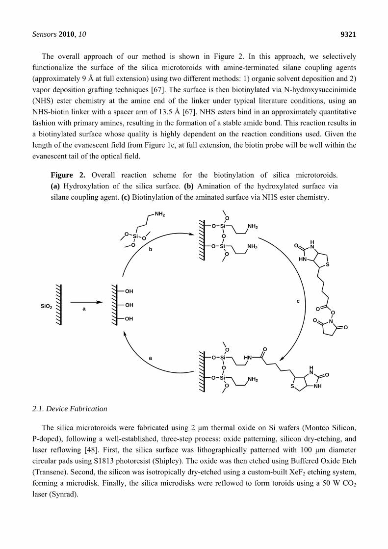
The overall approach of our method is shown in Figure 2. In this approach, we selectively
functionalize the surface of the silica microtoroids with amine-terminated silane coupling agents
(approximately 9 Å at full extension) using two different methods: 1) organic solvent deposition and 2)
vapor deposition grafting techniques [67]. The surface is then biotinylated via N-hydroxysuccinimide
(NHS) ester chemistry at the amine end of the linker under typical literature conditions, using an
NHS-biotin linker with a spacer arm of 13.5 Å [67]. NHS esters bind in an approximately quantitative
fashion with primary amines, resulting in the formation of a stable amide bond. This reaction results in
a biotinylated surface whose quality is highly dependent on the reaction conditions used. Given the
length of the evanescent field from Figure 1c, at full extension, the biotin probe will be well within the
evanescent tail of the optical field.
Figure 2. Overall reaction scheme for the biotinylation of silica microtoroids.
(a) Hydroxylation of the silica surface. (b) Amination of the hydroxylated surface via
silane coupling agent. (c) Biotinylation of the aminated surface via NHS ester chemistry.
SiO2
OH
OH
OH
SiO
OO
NH2
O
O
Si
O
Si
O
O
NH2
NH2
NOO
OO
S
HN
HN
O
O
O
Si
O
Si
O
O
HN
NH2
O
S NH
HN O
a
b
c
a
2.1. Device Fabrication
The silica microtoroids were fabricated using 2 μm thermal oxide on Si wafers (Montco Silicon,
P-doped), following a well-established, three-step process: oxide patterning, silicon dry-etching, and
laser reflowing [48]. First, the silica surface was lithographically patterned with 100 μm diameter
circular pads using S1813 photoresist (Shipley). The oxide was then etched using Buffered Oxide Etch
(Transene). Second, the silicon was isotropically dry-etched using a custom-built XeF2 etching system,
forming a microdisk. Finally, the silica microdisks were reflowed to form toroids using a 50 W CO2
laser (Synrad).
Sensors 2010, 10
9322
2.2. Device Functionalization Protocols
All anhydrous solvents used during device functionalization were stored under UHP Ar. All chemicals
and other solvents for the following reactions were used as received from their respective suppliers. The
silica microtoroids fabricated following the above procedures were functionalized via three primary
reaction steps: surface hydroxylation, amination, and finally, biotinylation. First, the silica surface was
terminated with hydroxyl groups via typical literature procedures to promote covalent attachment of the
silane coupling agent, using either O2 plasma treatment (120 W, 200 mTorr, 2 minutes) or a 70:30 H2SO4
(fuming, Aldrich): H2O2 (30 wt %, Aldrich) piranha etch for 45 minutes [68-70]. Following the
piranha etch, the samples were cleaned with DDI H2O, and air-dried between 25–60 °C
for 30 minutes–24 hours to completely remove the water from the sample surface. Second, aminated
silica surfaces were prepared by grafting silane coupling agents to the surface using two different
methods: organic solvent deposition or chemical vapor deposition [41,67,71,72]. In the first method,
a 2 mM solution of 3-aminopropyltrimethoxysilane (APTMS, Aldrich) was prepared in anhydrous
toluene in a glove bag under a UHP Ar environment. The sample was then placed in the solution, and
reacted at room temperature on an incubating rocker (VWR) for 10 minutes–4 hours to form a
monolayer of silane on the surface. The sample was then removed from the reaction solution, and
washed with toluene, ethanol, and then water for 5 minutes each on the incubating rocker. The sample
was again air-dried between 25–60 °C for 30 minutes–24 hours to completely remove the water from
the sample surface. In the second method (chemical vapor deposition), the hydroxylated sample was
exposed to APTMS vapor in a vacuum desiccator under aspirator vacuum for 15 minutes–4 hours.
Lastly, the aminated samples were then biotinylated via reaction in a 10 mM solution of NHS-Biotin
(Pierce) in dimethylsulfoxide (DMSO, anhydrous, Aldrich) at room temperature for 30 minutes, on an
incubating rocker. The surface was then washed with water and then acetone, as above, to remove any
physically absorbed biotin from the surface, and air-dried at room temperature for 30 minutes.
Amine-terminated samples were fluorescently labeled using fluoroscein-5-isothiocyanate (FITC,
Pierce), which forms a stable thiourea bond with the primary amines present on the surface in a 1:1
ratio. The FITC reaction solution was prepared in a darkened room by dissolving 1 mg FITC in 1 mL
DMSO (anhydrous), and diluting it via addition of 50 μL of the solution to 1 mL of 0.1 M sodium
carbonate buffer at pH 9.0 (VWR). The samples were gently inserted into this reaction solution in
covered vials, placed in an ice bath, and reacted for at least 8 hours in an incubating rocker. Physically
adsorbed FITC was removed via rinses with DMSO. The samples were then air-dried at 25–60 °C
for 30 minutes–24 hours. Biotin-terminated samples were fluorescently labeled with Texas Red
fluorescent dye conjugated to avidin protein (Invitrogen). Biotin-avidin binding was accomplished by
reacting the biotinylated samples in a covered, 10 μg/mL solution of Texas Red-avidin protein
conjugate in phosphate buffered saline (PBS, VWR) for 30 minutes at room temperature in an
incubating rocker. The sample was removed from the solution and washed with PBS to remove
physically-adsorbed reactant.

Sensors 2010, 10
9323
2.3. Device Characterization Protocols
The as-fabricated and surface-modified microtoroid devices were characterized qualitatively using a
Nikon H550S optical microscope equipped with a Nikon digital camera, and NIS-Elements imaging
software. The surface chemistry of these devices was explored via X-ray photoelectron spectroscopy
(XPS, M-Probe ESCA) using a 1,487 eV Al Kα source, and a survey scan from 0 to 1,000 binding eV
to identify all chemical species on the surface. Reaction efficacy and quality was monitored using
fluorescent imaging with a Nikon ECLIPSE LV100D-U fluorescence microscope equipped with a
Nikon digital camera at each reaction step. Intensity measurements were obtained using NIS-Elements
imaging software and Texas Red and FITC excitation and emission filters, where appropriate. Lastly,
ellipsometric measurements were taken using a Gaertner L166C ellipsometer to determine the film
thickness of the initial silane layer. For the XPS and ellipsometry techniques, control samples
consisting of surface-modified, non-patterned, silica-on-silicon wafers were used in place of the
patterned samples.
The devices were characterized quantitatively by microcavity analysis at each reaction step to
determine the impact of the surface functionalization methods on the device sensitivity and to evaluate
the bioconjugation conditions best suited to ensuring the devices’ performance. The resonator quality
factor was measured using a tapered optical fiber waveguide to couple power into the devices from a
narrow linewidth, CW tunable diode laser centered at 635 nm (New Focus) (Figure 3b) [73-75]. The
F-SV optical fiber (Newport) was tapered to ~500 nm waist diameter by heating with an oxyhydric
torch while stretching the fiber with a two-axis stage controller (Sigma Koi). During testing, the device
was placed on a 3-axis nanopositioning stage (Optosigma), and the device was monitored using
side- and top-view cameras simultaneously. Coupling into the resonator results in the excitation of the
whispering gallery modes of the microcavity (Figure 3c). The resonance linewidth data was recorded
using a digitizer/oscilloscope card which was directly integrated into the computer for automated data
recording (NI, PCI-5114). The laser scan speed and scan direction was optimized to ensure that neither
distorted the resonance lineshape. The quality factor of the microtoroid resonator was determined by
calculating the resonance linewidth (full width at half-maximum) from the recorded spectra taken in
the undercoupled regime [48,60,63,75-77].
Figure 3. (a) Scanning electron micrograph of as-fabricated microtoroid. (b) Side view
optical micrograph of microtoroid during device characterization. The tapered fiber can be
clearly seen. (c) PovRay rendering of the microtoroid resonator and tapered optical fiber.
The optical field is primarily confined inside the silica cavity, but a small portion
evanesces into the environment.

Sensors 2010, 10
9324
3. Results and Discussion
Four different routes to biotinylation were explored for their suitability towards silica microtoroid
resonators, based on combinations of hydroxylation (piranha etch, O2 plasma etch) and amination
(organic solvent deposition, chemical vapor deposition) conditions. As previously mentioned, one of
the key requirements for adding specificity included minimizing reagent use, the number of reaction
steps needed, and harsh environmental conditions, which should minimize surface and structural
damage. Therefore, the exploration of these four routes enabled the selection of a facile and efficient
technique that can be applied to silica optical resonators without severely impacting device sensitivity.
3.1. Analysis of the Surface Functionalization
Our exploration of these specific combinations grew out of the requirements of grafting
organosilanes to inorganic surfaces, such as silica, alumina, titania, etc. Typically, silane coupling
agents (R(CH2)nSiX3, where R is an organic functional group and X is a hydrolyzable leaving group)
undergo hydrolysis to labilize the leaving groups and form a reactive silanol intermediate species. This
silanol species can couple to surface hydroxyl groups via condensation reactions and form stable
siloxane bonds. However, the condensation reaction that couples the silane to the surface requires a
high density of hydroxyl groups to form a uniform surface coverage [35,67,71,78]. Typically, the –OH
surface density is increased by an oxidative piranha treatment, which leaves the surface extremely
hydrophilic. This treatment, however, may be too harsh in terms of temperature and acid strength with
regard to microstructured optical devices, and may lead to difficulties in preserving the structural and
surface integrity of the devices. An alternative is the use of O2 plasma, which is typically used in
fabrication processes to clean photoresist from structured surfaces. O2 plasma etching is simple,
environmentally benign, minimizes device handling, and does not require post-treatment to remove
excess water from the surface. On the other hand, O2 plasma treatment may not yield as high of a
surface hydroxyl density, leading to low surface coverage of the organosilane. Therefore, it is
important to identify which route is most compatible with our devices.
After hydroxylation, the silane coupling agent can be grafted to the surface. Here, we used APTMS
as our silane coupling agent due to its short organic tether between the silica head and the terminal
amine, which should ensure that the final biotin probe is well within the evanescent tail of the
functioning device. Additionally, the amine portion should not cause additional material losses due to
absorption at the wavelength of interest. Surface grafting of APTMS may be accomplished via reaction
in aqueous or organic solvent, or through vapor phase deposition. Typically, alkoxysilanes (where X is
an alkoxy group) are used to functionalize the surface of microstructured devices, since they minimize
the formation of a silica polymer network on the surface and help achieve monolayer coverage, due to
the lower hydrolyzability of the alkoxy leaving groups [67]. This lower hydrolyzability can lead to the
necessity of hydrolyzing the alkoxysilane in water first, as they are unreactive to surface hydroxyls at
ambient conditions in their native form. Unfortunately, aqueous solvent deposition causes the same
multi-layer deposition problem as working with non-alkoxysilanes. Fortunately, chloro- and
methoxysilanes, such as APTMS, do not need pre-hydrolysis, and can be coupled to the substrate in a
uniform monolayer through siloxane bonding via anhydrous organic solvent deposition or vapor
deposition [67]. In both deposition cases, since water is not present in the system, the
Sensors 2010, 10
9325
three-dimensional silica polymer network does not form. Either route may be appropriate for
microstructured optical devices, but vapor deposition typically requires less chemical use,
post-treatment, and handling than organic solvent deposition. Here, we investigate both routes, along
with the hydroxylation alternatives, to determine their impact on the devices.
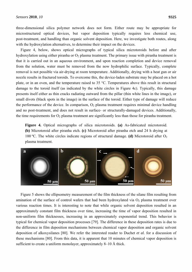
Figure 4, below, shows optical micrographs of typical silica microtoroids before and after
hydroxylation using either piranha or O2 plasma treatment. The primary issue with piranha treatment is
that it is carried out in an aqueous environment, and upon reaction completion and device removal
from the solution, water must be removed from the now hydrophilic surface. Typically, complete
removal is not possible via air-drying at room temperature. Additionally, drying with a heat gun or air
nozzle results in fractured toroids. To overcome this, the device-laden substrate may be placed on a hot
plate, or in an oven, and the temperature raised to 35 °C. Temperatures above this result in structural
damage to the toroid itself (as indicated by the white circles in Figure 4c). Typically, this damage
presents itself either as thin cracks radiating outward from the pillar (thin white lines in the image), or
small divots (black spots in the image) in the surface of the toroid. Either type of damage will reduce
the performance of the device. In comparison, O2 plasma treatment requires minimal device handling
and no post-treatment, and does not result in surface- or structurally-damaged devices. Additionally,
the time requirements for O2 plasma treatment are significantly less than those for piranha treatment.
Figure 4. Optical micrographs of silica microtoroids. (a) As-fabricated microtoroid.
(b) Microtoroid after piranha etch. (c) Microtoroid after piranha etch and 24 h drying at
100 ºC. The white circles indicate regions of structural damage. (d) Microtoroid after O2
plasma treatment.
Figure 5 shows the ellipsometry measurement of the film thickness of the silane film resulting from
amination of the surface of control wafers that had been hydroxylated via O2 plasma treatment over
various reaction times. It is interesting to note that while organic solvent deposition resulted in an
approximately constant film thickness over time, increasing the time of vapor deposition resulted in
non-uniform film thicknesses, increasing in an approximately exponential trend. This behavior is
typical for chemical vapor deposition processes [79]. The difference in these deposition rates is due to
the difference in film deposition mechanisms between chemical vapor deposition and organic solvent
deposition of alkoxysilanes [80]. We refer the interested reader to Duchet et al. for a discussion of
these mechanisms [80]. From this data, it is apparent that 10 minutes of chemical vapor deposition is
sufficient to create a uniform monolayer, approximately 8–10 Å thick.

Sensors 2010, 10
9326
Figure 5. Ellipsometry data showing film thicknesses after various reaction times for
organic solvent deposition and vapor deposition.
Figure 6 shows fluorescent micrographs of FITC- and Texas Red-labeled, aminated and
biotinylated (respectively) toroids obtained using the four combinations of hydroxylation/amination
procedures. The combination of O2 plasma treatment and vapor deposition results in the most uniform
surface coverage of the toroids. Interestingly, in each case, amination occurred only on the silica toroid
itself, and not on the silicon wafer. This was interesting to us, as the process of hydroxylating the
surface of the device and the substrate should lead to the ability to aminate both surfaces (the silica of
the device as well as the silicon of the substrate). As an exploration of why this preferential amination
appeared only on the silica, we hydroxylated (using O2 plasma) and aminated three different sets of
planar substrates: a bare silicon wafer, a bare silicon wafer subjected to XeF2 etching using the same
parameters as used to make the devices, and a 2 μm SiO2 on silica wafer. The aminated samples were
subjected to FITC labeling, and the average intensities of the resulting surfaces were measured,
normalized by the background intensity of each sample. Interestingly, the average intensity (a.u.) of
the FITC labeled surfaces showed that XeF2 etching seems to reduce the overall amination of the
silicon surface (intensity of the bare wafer: 1161, intensity of the bare wafer subjected to XeF2 etching
prior to functionalization: 364, and intensity of the thermal oxide wafer: 924). Clearly, the exposure to
the XeF2 is inducing the improved functionalization of the device surface. However, at this point, the
precise mechanism is unclear.
The combination of piranha treatment and organic solvent deposition typically led to the poorest
results, with severe surface clumping, indicating either a non-uniform coverage by the silane coupling
agent, or the presence of water drops remaining on the surface after hydroxylation. Clumping of the
silane layer during organic solvent deposition could be minimized by carrying out the reaction on a
rocker; however, it was never entirely prevented, as shown by Figure 6c, which shows the O2 plasma
treatment/organic solvent deposition combination. Note that the image was deliberately overexposed
to help show the detail of surface clumping and surface damage from organic solvent deposition.
The results from the series of experiments are summarized in Table 1. Overall, the difficulties
involved with proper drying of the piranha-treated samples and with clumping of the organic solvent
deposition samples indicated that these methods were inappropriate for further use. Note that, due to

Sensors 2010, 10
9327
its poor performance, the piranha treatment/organic solvent deposition combination was not pursued to
the point of biotinylation. Additionally, several biotinylated samples were labeled with FITC to
determine the extent of the NHS ester reaction with surface amine groups; fluorescent microscopy did
not yield samples with intensities above the substrate background (no fluorescent labeling occurred),
indicating that the density of amines remaining after biotinylation was too low to be detected by
fluorescent imaging techniques. This implies that the reaction biotinylated all the amine groups present
or that steric hindrance prevented access of the FITC to the remaining amines on the surface.
Regardless, the inaccessibility of the amine groups for reaction with FITC, a relatively small molecule
compared to antibodies and proteins, indicates that the presence of the amine groups should lead to
minimal activity towards non-targeted species in the environment.
Figure 6. Fluorescent micrographs of microtoroids after labeling. (a) FITC-labeled,
aminated microtoroid after piranha treatment, followed by organic solvent deposition.
(b) Texas Red-labeled, biotinylated microtoroid after piranha treatment, followed by vapor
deposition. (c) Texas Red-labeled, biotinylated microtoroid after O2 plasma treatment,
followed by organic solvent deposition. (d) Texas Red-labeled, biotinylated microtoroid
after O2 plasma treatment, followed by vapor deposition.
Table 1. Summary of the effects of hydroxylation and amination surface functionalization
techniques on microtoroid surfaces.
O2 Plasma Etch Piranha Etch Organic Solvent
Deposition Uneven coating of amine layer No structural damage
Water droplets on surface Structural damage from high temperatures used to remove accumulated surface water Clumping of amine layer
Vapor Deposition No structural damage No water droplets on surface Uniform coating of amine layer No clumping visible
Water droplets on surface Structural damage from high temperatures used to remove accumulated surface water Clumping present to a lesser degree than organic solvent deposition

Sensors 2010, 10
9328
Although fluorescent labeling provides confirmation of the presence and uniformity of the surface
species, it is an indirect method to do so. An additional confirmation may be obtained using X-ray
photoelectron spectroscopy (XPS), which directly probes the surface composition through irradiation
of the sample with X-rays, resulting in the ejection of electrons from the top ~10 nm of the sample.
Simultaneously measuring the kinetic energy and number of electrons ejected allows us to quantify the
elemental source of these electrons. Figure 7 show the resulting surface composition at each reaction
step obtained through XPS data collection. The C (1s) signal that appears in the spectra is from
primarily adventitious carbon in the ultra-high vacuum chamber, so direct comparison in terms of
intensity of these peaks between runs is inappropriate, as the signal from the sample is masked by
varying amounts of carbon in the chamber. Additionally, it is difficult to directly compare the
intensities of the peaks between runs, since slight differences in spectrometer environment can lead to
significant differences in peak intensities between runs (for instance, the Si (2p) and Si (2s) signals in
each spectra); hence the designation of arbitrary units for the intensity data. Overall, the XPS data
shown provide an additional confirmation of the success of each reaction step via the introduction of
nitrogen (N) and sulfur (S) peaks into the spectra after amination and biotinylation, respectively. The
combination of fluorescence imaging and XPS verify both the surface chemistry and surface quality of
the samples.
Figure 7. Chemical composition of control surfaces after each reaction step (using O2
plasma etching and vapor deposition conditions).
3.2. Stability of the Surface Chemistry
To determine the stability of the biotinylated devices for long-term storage in ambient conditions,
seven chips, each containing 1–5 toroids, were biotinylated at the same time (day 1), then stored for
various time-frames, bound with an avidin-Texas Red complex, and subjected to fluorescent
microscopy. The intensity due to the binding reaction of biotin and avidin, and how it changes over
storage time, can be used as a measure of the degradation or instability of the biotin probe with respect
to avidin over time. Figure 8 shows the average device intensity, normalized by toroid surface area,
over approximately 1.5 months of storage. It is important to note that long-term storage does not have
Sensors 2010, 10
9329
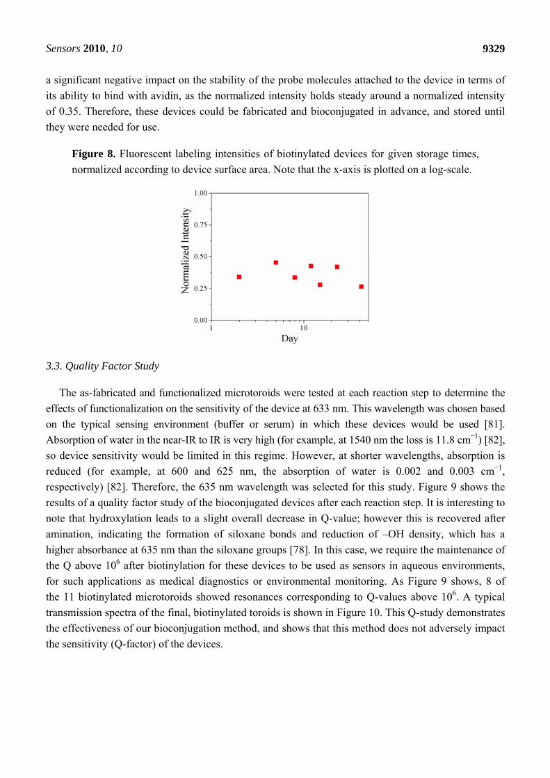
a significant negative impact on the stability of the probe molecules attached to the device in terms of
its ability to bind with avidin, as the normalized intensity holds steady around a normalized intensity
of 0.35. Therefore, these devices could be fabricated and bioconjugated in advance, and stored until
they were needed for use.
Figure 8. Fluorescent labeling intensities of biotinylated devices for given storage times,
normalized according to device surface area. Note that the x-axis is plotted on a log-scale.
3.3. Quality Factor Study
The as-fabricated and functionalized microtoroids were tested at each reaction step to determine the
effects of functionalization on the sensitivity of the device at 633 nm. This wavelength was chosen based
on the typical sensing environment (buffer or serum) in which these devices would be used [81].
Absorption of water in the near-IR to IR is very high (for example, at 1540 nm the loss is 11.8 cm−1) [82],
so device sensitivity would be limited in this regime. However, at shorter wavelengths, absorption is
reduced (for example, at 600 and 625 nm, the absorption of water is 0.002 and 0.003 cm−1,
respectively) [82]. Therefore, the 635 nm wavelength was selected for this study. Figure 9 shows the
results of a quality factor study of the bioconjugated devices after each reaction step. It is interesting to
note that hydroxylation leads to a slight overall decrease in Q-value; however this is recovered after
amination, indicating the formation of siloxane bonds and reduction of –OH density, which has a
higher absorbance at 635 nm than the siloxane groups [78]. In this case, we require the maintenance of
the Q above 106 after biotinylation for these devices to be used as sensors in aqueous environments,
for such applications as medical diagnostics or environmental monitoring. As Figure 9 shows, 8 of
the 11 biotinylated microtoroids showed resonances corresponding to Q-values above 106. A typical
transmission spectra of the final, biotinylated toroids is shown in Figure 10. This Q-study demonstrates
the effectiveness of our bioconjugation method, and shows that this method does not adversely impact
the sensitivity (Q-factor) of the devices.

Sensors 2010, 10
9330
Figure 9. Effect of surface functionalization on the quality factor of the microtoroids at
each reaction step.
Figure 10. Transmission spectra of biotinylated toroid, showing a single, high-Q resonance
(data connected by a solid black line) with the corresponding Lorentz fit (solid red line)
4. Conclusion and Future Outlook
Silica microtoroidal optical resonators were successfully conjugated with biotin, without surface or
structural damage, and without a significant, negative impact on the overall sensitivity of the devices.
The most successful bioconjugation protocol examined was the covalent attachment of biotin to the
silica surface through a silane coupling agent via O2 plasma treatment, followed by vapor deposition of
the silane. This strategy is a simple method for imparting specificity to highly sensitive optical devices,
and could easily be extended to other sensor structures due to its flexibility and low impact on the
device. This method opens the possibility for a reagentless “recycling” of functionalized devices by the
removal of the organic surface coverage via O2 plasma etching after the sensor has been used. The
covalent nature of attachment presents a significant improvement in terms of environmental stability
compared with other methods of device functionalization, such as physical adsorption. The method
described here results in devices whose probe molecules are stable for long-term storage. Additionally,
this technique allows for the controlled presentation of the probe to the surrounding environment, and

Sensors 2010, 10
9331
avoids background binding on the substrate due to the non-biological nature of the reaction strategy
and the functionalization of the toroid surface only. Lastly, this method improves the performance of
optical devices used as sensors by increasing the specificity to a target of interest.
This work represents one of the first examples non-physisorption-based bioconjugation of optical
microtoroid resonators that can be used for the label-free detection of biomolecules. The primary
advantage of a device that has been functionalized using the methods described above is that a wide
array of biomolecules can be studied, since the functionalization process does not limit the study to a
specific ligand-substrate pair, or even a specific type of molecule. For example, the protocols
developed here are easily extended to a wide variety of probe molecule/target molecule pairings by
using the high affinity of biotin for (strept)avidin-modified probes molecules, following the
Avidin-Biotin Complexation (ABC) technique [83], allowing the easy interchange of the terminal
functional group probes on the surface of the devices. Lastly, the protocols demonstrated here will result
in new methods to produce highly sensitive and highly specific sensor devices that can be used for other
applications, such as rapid medical diagnostics and environmental monitoring [1,31,32,84-86].
Acknowledgements
The authors would like to acknowledge Heather McCaig (Nate E. Lewis research group, MMRC,
California Institute of Technology) for her assistance with the XPS and the ellipsometer
measurements, and for useful discussions regarding surface polymerization techniques. Research was
carried out in part at the Molecular Materials Research Center (MMRC) of the Beckman Institute of
the California Institute of Technology. The authors would also like to thank the National Science
Foundation [0852581] and the Office of Naval Research Young Investigator Program [N00014-09-1-
0898] for funding this work.
References and Notes
1. Luppa, P.B.; Sokoll, L.J.; Chan, D.W. Immunosensors––Principles and applications to clinical
chemistry. Clin. Chim. Acta 2001, 314, 1-26.
2. Ambrose, W.P.; Goodwin, P.M.; Jett, J.H.; van Orden, A.; Werner, J. H.; Keller, R. A. Single
molecule fluorescence spectroscopy at ambient temperature. Chem. Rev. 1999, 99, 2929-2956.
3. Funatsu, T.; Harada, Y.; Higuchi, H.; Tokunaga, M.; Saito, K.; Ishii, Y.; Vale, R.D.; Yanagida, T.,
Imaging and nano-manipulation of single biomolecules. Biophys. Chem. 1997, 68, 63-72.
4. Wormke, S.; Mackowski, S.; Brotosudarmo, T.H.P.; Jung, C.; Zumbusch, A.; Ehrl, M.; Scheer,
H.; HofMann, E.; Hiller, R.G.; Brauchle, C. Monitoring fluorescence of individual chromophores
in peridininchlorophyll-protein complex using single molecule spectroscopy. Biochim. Biophys.
Acta-Bioenerg. 2007, 1767, 956-964.
5. Xie, S.N. Single-molecule approach to enzymology. Single Mol. 2001, 2, 229-236.
6. Zhang, J.K.; Dong, S.M.; Lu, J.H.; Turner, A.P.F.; Fan, Q.J.; Jia, S.R.; Yang, H.J.; Qiao, C.S.;
Zhou, H.; He, G.W. A label free electrochemical nanobiosensor study. Anal. Lett. 2009, 42,
2905-2913.
7. Mao, S.; Lu, G.H.; Yu, K.H.; Chen, J.H. Specific biosensing using carbon nanotubes
functionalized with gold nanoparticle-antibody conjugates. Carbon 2010, 48, 479-486.

Sensors 2010, 10
9332
8. Campbell, G.A.; Mutharasan, R. Detection and quantification of proteins using self-excited
PZT-glass millimeter-sized cantilever. Biosens. Bioelectron. 2005, 21, 597-607.
9. Campbell, G. A.; Mutharasan, R., Detection of pathogen Escherichia coli O157 : H7 using self-
excited PZT-glass microcantilevers. Biosens. Bioelectron. 2005, 21, 462-473.
10. Datar, R.; Kim, S.; Jeon, S.; Hesketh, P.; Manalis, S.; Boisen, A.; Thundat, T., Cantilever Sensors:
Nanomechanical Tools for Diagnostics. MRS Bull. 2009, 34, 449-454.
11. Horvath, R.; Pedersen, H. C.; Skivesen, N.; Selmeczi, D.; Larsen, N. B., Optical waveguide
sensor for on-line monitoring of bacteria. Optics Lett. 2003, 28, 1233-1235.
12. Dumais, P.; Callender, C.L.; Noad, J.P.; Ledderhof, C.J., Silica-on-silicon optical sensor based on
integrated waveguides and microchannels. IEEE Photonic. Techn. Lett. 2005, 17, 441-443.
13. Andras, S.; Adanyi, N.; Szekacs, I.; Majer-Baranyi, K.; Istvan, S. Optical waveguide light-mode
spectroscopy immunosensors for environmental monitoring. Appl. Opt. 2009, 48, B151-B158.
14. Shankaran, D.R.; Gobi, K.V.A.; Miura, N. Recent advancements in surface plasmon resonance
immunosensors for detection of small molecules of biomedical, food and environmental interest.
Sens. Actuat. B-Chem. 2007, 121, 158-177.
15. Sundberg, F.; Karlsson, R. Rapid detection and characterization of immune responses using
label-free biacore immunoassays. Immunology 2007, 120, 46-47.
16. Kumbhat, S.; Shankaran, D.R.; Kim, S.J.; Gobi, K.V.; Joshi, V.; Miura, N. Surface plasmon
resonance biosensor for dopamine using D3 dopamine receptor as a biorecognition molecule.
Biosens. Bioelectron. 2007, 23, 421-427.
17. Nagel, T.; Ehrentreich-Forster, E.; Singh, M.; Schmitt, K.; Brandenburg, A.; Berka, A.; Bier, F.F.
Direct detection of tuberculosis infection in blood serum using three optical label-free approaches.
Sens. Actuat. B-Chem. 2008, 129, 934-940.
18. Tetz, K.A.; Pang, L.; Fainman, Y. High-resolution surface plasmon resonance sensor based on
linewidth-optimized nanohole array transmittance. Opt. Lett. 2006, 31, 1528-1530.
19. Chen, H.M.; Pang, L.; Kher, A.; Fainman, Y. Three-dimensional composite metallodielectric
nanostructure for enhanced surface plasmon resonance sensing. Appl. Phys. Lett. 2009, 94,
073177.
20. Li, H.; Fan, X. Characterization of sensing capability of optofluidic ring resonator biosensors.
Appl. Phys. Lett. 2010, 97, 011105-3.
21. Alvarez, S.D.; Li, C.P.; Chiang, C.E.; Schuller, I.K.; Sailor, M.J. A label-free porous alumina
interferometric immunosensor. ACS Nano 2009, 3, 3301-3307.
22. Bolduc, O.R.; Clouthier, C.M.; Pelletier, J.N.; Masson, J.F. Peptide self-assembled monolayers
for label-free and unamplified surface plasmon resonance biosensing in crude cell lysate. Anal.
Chem. 2009, 81, 6779-6788.
23. Densmore, A.; Vachon, M.; Xu, D. X.; Janz, S.; Ma, R.; Li, Y. H.; Lopinski, G.; Delâge, A.;
Lapointe, J.; Luebbert, C.C.; Liu, Q.Y.; Cheben, P.; Schmid, J.H. Silicon photonic wire biosensor
array for multiplexed real-time and label-free molecular detection. Opt. Lett. 2009, 34,
3598-3600.
24. Fan, X.D.; White, I.M.; Shopoua, S.I.; Zhu, H.Y.; Suter, J.D.; Sun, Y.Z. Sensitive optical
biosensors for unlabeled targets: A review. Anal. Chim. Acta 2008, 620, 8-26.
Sensors 2010, 10
9333
25. Gao, Z.; Agarwal, A.; Trigg, A.D.; Singh, N.; Fang, C.; Tung, C.-H.; Fan, Y.; Buddharaju, K.D.;
Kong, J. Silicon nanowire arrays for label-free detection of DNA. Anal. Chem. 2007, 79,
3291-3297.
26. Hanumegowda, N.M.; White, I.M.; Oveys, H.; Fan, X.D. Label-free protease sensors based on
optical microsphere resonators. Sens. Lett. 2005, 3, 315-319.
27. Lee, P.H. Label-free optical biosensor: A tool for G protein-coupled receptors pharmacology
profiling and inverse agonists identification. J Recept Sig Transd 2009, 29, 146-153.
28. Syahir, A.; Mihara, H.; Kajikawa, K. A new optical label-free biosensing platform based on a
metal−insulator−metal structure. Langmuir 2010, 26, 6053–6057.
29. Washburn, A.L.; Gunn, L.C.; Bailey, R.C. Label-Free quantitation of a cancer biomarker in
complex media using silicon photonic microring resonators. Anal. Chem. 2009, 81, 9499-9506.
30. Washburn, A.L.; Luchansky, M.S.; Bowman, A.L.; Bailey, R.C. Quantitative, label-free detection
of five protein biomarkers using multiplexed arrays of silicon photonic microring resonators.
Anal. Chem. 2010, 82, 69-72.
31. Subrahmanyam, S.; Piletsky, S. A.; Turner, A. P. F., Application of natural receptors in sensors
and assays. Anal. Chem. 2002, 74, 3942-3951.
32. Hunt, H.K.; Armani, A.M. Label-free biological and chemical sensors. Nanoscale 2010, 2,
1544-1559.
33. Hock, B. Antibodies for immunosensors––A review. Anal. Chim. Acta 1997, 347, 177-186.
34. Feuz, L.; Jonsson, P.; Jonsson, M.P.; Hook, F. Improving the limit of detection of nanoscale
sensors by directed binding to high-sensitivity areas. ACS Nano 2010, 4, 2167-2177.
35. Kalia, J.; Raines, R.T. Advances in bioconjugation. Curr. Org. Chem. 2010, 14, 138-147.
36. Amine, A.; Mohammadi, H.; Bourais, I.; Palleschi, G. Enzyme inhibition-based biosensors for
food safety and environmental monitoring. Biosens. Bioelectron. 2006, 21, 1405-1423.
37. Hegnerova, K.; Bockova, M.; Vaisocherova, H.; Kristofikova, Z.; Ricny, J.; Ripova, D.; Homola,
J. Surface plasmon resonance biosensors for detection of Alzheimer disease biomarker. Sens.
Actuat. B-Chem. 2009, 139, 69-73.
38. Zanoli, L.; D'Agata, R.; Spoto, G. Surface plasmon-based optical detection of DNA by peptide
nucleic acids. Minerva Biotecnol. 2008, 20, 165-174.
39. Gobi, K.V.; Iwasaka, H.; Miura, N. Self-assembled PEG monolayer based SPR immunosensor for
label-free detection of insulin. Biosens. Bioelectron. 2007, 22, 1382-1389.
40. Gobi, K.V.; Kim, S.J.; Tanaka, H.; Shoyama, Y.; Miura, N. Novel surface plasmon resonance
(SPR) immunosensor based on monomolecular layer of physically-adsorbed ovalbumin conjugate
for detection of 2,4-dichlorophenoxyacetic acid and atomic force microscopy study. Sens. Actuat.
B-Chem. 2007, 123, 583-593.
41. Kim, K.S.; Lee, H.-S.; Yang, J.-A.; Jo, M.-H.; Hahn, S.K. The fabrication, characterization and
application of aptamer-functionalized Si-nanowire FET biosensors. Nanotechnology 2009,
doi: 10.1088/0957-4484/20/23/235501.
42. Lee, M.; Walt, D.R. A fiber-optic microarray biosensor using aptamers as receptors. Anal.
Biochem. 2000, 282, 142-146.
43. Lee, S.H.; Ko, H.J.; Park, T.H. Real-time monitoring of odorant-induced cellular reactions using
surface plasmon resonance. Biosens. Bioelectron. 2009, 25, 55-60.

Sensors 2010, 10
9334
44. Dover, J.E.; Hwang, G.M.; Mullen, E.H.; Prorok, B.C.; Suh, S.J. Recent advances in peptide
probe-based biosensors for detection of infectious agents. J. Microbiol. Meth. 2009, 78, 10-19.
45. Kumbhat, S.; Shankaran, D.R.; Kim, S.J.; Gobi, K.V.; Joshi, V.; Miura, N. A novel
receptor-based surface-plasmon-resonance affinity biosensor for highly sensitive and selective
detection of dopamine. Chem. Lett. 2006, 35, 678-679.
46. Byeon, J.-Y.; Limpoco, F.T.; Bailey, R.C. Efficient bioconjugation of protein capture agenst to
biosensor surfaces using aniline-catalyzed hydrazone ligation. Langmuir 2010, 26, 15430-15435.
47. Zhang, X.; Choi, H.-S.; Armani, A.M. Ultimate quality factor of silica microtoroid resonant
cavities. Appl. Phys. Lett. 2010, 96, 153304.
48. Armani, D.K.; Kippenberg, T.J.; Spillane, S.M.; Vahala, K.J. Ultra-high-Q toroid microcavity on
a chip. Nature 2003, 421, 925-928.
49. Gorodetsky, M.L.; Savchenkov, A.A.; Ilchenko, V.S. Ultimate Q of optical microsphere
resonators. Opt. Lett. 1996, 21, 453-455.
50. Grudinin, I.S.; Ilchenko, V.S.; Maleki, L. Ultrahigh optical Q factors of crystalline resonators in
the linear regime. Phys. Rev. A 2006, 74, doi: 10.1103/PhysRevA.74.063806.
51. Savchenkov, A.A.; Matsko, A.B.; Ilchenko, V.S.; Maleki, L. Optical resonators with ten million
finesse. Opt. Express 2007, 15, 6768-6773.
52. Vernooy, D.W.; Ilchenko, V.S.; Mabuchi, H.; Streed, E.W.; Kimble, H.J. High-Q measurements
of fused-silica microspheres in the near infrared. Opt. Lett. 1998, 23, 247-249.
53. Zhu, J.; Ozdemir, S.K.; Xiao, Y.-F.; Li, L.; He, L.; Chen, D.-R.; Yang, L. On-chip single
nanoparticle detection and sizing by mode splitting in an ultrahigh-Q microresonator. Nat.
Photon. 2009, 4, 46-49.
54. Arnold, S.; Khoshsima, M.; Teraoka, I.; Holler, S.; Vollmer, F. Shift of whispering-gallery modes
in microspheres by protein adsorption. Opt. Lett. 2003, 28, 272-274.
55. Boyd, R.W.; Heebner, J.E. Sensitive disk resonator photonic biosensor. Appl. Opt. 2001, 40,
5742-5747.
56. Blair, S.; Chen, Y. Resonant-enhanced evanescent-wave fluorescence biosensing with cylindrical
optical cavities. Appl. Opt. 2001, 40, 570-582.
57. Armani, A.M.; Kulkarni, R.P.; Fraser, S.E.; Flagan, R.C.; Vahala, K.J. Label-free,
single-molecule detection with optical microcavities. Science 2007, 317, 783-787.
58. Zhu, H.Y.; Dale, P.S.; Caldwell, C.W.; Fan, X.D. Rapid and label-free detection of breast cancer
biomarker CA15-3 in clinical human serum samples with optofluidic ring resonator sensors. Anal.
Chem. 2009, 81, 9858-9865.
59. Vollmer, F.; Arnold, S.; Keng, D. Single virus detection from the reactive shift of a whispering
gallery mode. Proc. Nat. Acad. Sci. USA 2008, 105, doi: 10.1073/pnas.0808988106.
60. Kippenberg, T.J.; Spillane, S.M.; Armani, D.K.; Vahala, K.J. Fabrication and coupling to planar
high-Q silica disk microcavities. Appl. Phys. Lett. 2003, 83, 797-799.
61. Zhang, X.; Choi, H.S.; Armani, A.M. Ultimate quality factor of silica microtoroid resonant
cavities. Appl. Phys. Lett. 2010, 96, 153304-153307
62. Armani, A.M.; Vahala, K.J. Biological and chemical detection using ultra-high-Q toroidal
microresonators. Biophys. J. 2007, 94, 29a-29a.
Sensors 2010, 10
9335
63. Armani, A.M.; Armani, D.K.; Min, B.; Vahala, K.J.; Spillane, S.M. Ultra-high-Q microcavity
operation in H2O and D2O. Appl. Phys. Lett. 2005, doi: 10.1063/1.2099529.
64. Zhu, H.; White, I.M.; Suter, J.D.; Fan, X. Phage-based label-free biomolecule detection in an
opto-fluidic ring resonator. Biosens. Bioelectron. 2008, 24, 461-466.
65. Zhu, H.; White, I.M.; Suter, J.D.; Zourob, M.; Fan, X. Opto-fluidic micro-ring resonator for
sensitive label-free viral detection. The Analyst 2008, 133, 356-360.
66. Arnold, S.; Ramjit, R.; Keng, D.; Kolchenko, V.; Teraoka, I. MicroParticle photophysics
illuminates viral bio-sensing. Faraday Discuss. 2008, 137, 65-83.
67. Hermanson, G.T. Bioconjugate Techniques, 2nd ed.; Academic Press: London, UK, 2008.
68. Ljungberg, K.; Jansson, U.; Bengtsson, S.; Soderbarg, A. Modification of silicon surfaces with
H2SO4:H2O2:HF and HNO3:HF for wafer bonding applications. J. Electrochem. Soc. 1996, 143,
1709-1714.
69. Chandekar, A.; Sengupta, S.K.; Whitten, J.E. Thermal stability of thiol and silane monolayers: A
comparative study. Appl. Surf. Sci. 2010, 256, 2742-2749.
70. Donskoi, A.V.; Dresvin, S.V.; Orlova, M.A.; Osovskii, B.B.; Khait, O.D.; Paushkin, E.V. Plasma
polishing of surface of wares made of silicate glass of any composition. Glass Ceram. 1976, 33,
162-165.
71. Brzoska, J.B.; Benazouz, I.; Rondelez, F. Silanization of Solid Substrates––A Step toward
Reproducibility. Langmuir 1994, 10, 4367-4373.
72. Lin, Y.B.; Tsui, T.Y.; Vlassak, J.J. Octamethylcyclotetrasiloxane-based, low-permittivity
organosilicate coatings––Composition, structure, and polarizability. J. Electrochem. Soc. 2006,
153, F144-F152.
73. Cai, M.; Vahala, K. Highly efficient hybrid fiber taper coupled microsphere laser. Opt. Lett. 2001,
26, 884-886.
74. Spillane, S.M.; Kippenberg, T.J.; Painter, O.J.; Vahala, K.J. Ideality in a fiber-taper-coupled
microresonator system for application to cavity quantum electrodynamics. Phys. Rev. Lett. 2003,
91, doi: 10.1103/PhysRevLett.91.043902.
75. Armani, A.M.; Srinivasan, A.; Vahala, K.J. Soft lithographic fabrication of high Q polymer
microcavity arrays. Nano Lett. 2007, 7, 1823-1826.
76. Little, B.E.; Laine, J.P.; Haus, H.A. Analytic theory of coupling from tapered fibers and
half-blocks into microsphere resonators. J. Lightwave Technol. 1999, 17, 704-715.
77. Yariv, A. Universal relations for coupling of optical power between microresonators and
dielectric waveguides Electron. Lett. 2000, 36, 999.
78. Arkles, B.; Larson, G. Silicon Compounds: Silanes and Silicones––A Survey of Properties and
Chemistry, 2nd ed.; Gelest, Inc.: Morrisville, PA, USA, 2008.
79. Hampdensmith, M.J.; Kodas, T.T. Chemical-vapor-deposition of metals. 1. An overview of CVD
processes Chem. Vapor Depos. 1995, 1, 8-23.
80. Duchet, J.; Chabert, B.; Chapel, J.P.; Gérard, J.F.; Chovelon, J.M.; Jaffrezic-Renault, N. Influence
of the Deposition process on the structure of grafted alkylsilane layers. Langmuir 1997, 13,
2271-2278.
81. Armani, A.M.; Armani, D.K.; Min, B.; Vahala, K.J.; Spillane, S.M. Ultra-high-Q microcavity
operation in H2O and D2O. Appl. Phys. Lett. 2005, doi: 10.1063/1.2099529.

Sensors 2010, 10
9336
82. Hale, G.M.; Querry, M.R. Optical-constants of water in 200-Nm to 200-Mum Wavelength region.
Appl. Opt. 1973, 12, 555-563.
83. Bratthauer, G.L. The Avidin-Biotin Complex (ABC) method. In Immunocytochemical Methods
and Protocols; Javois, L.C., Ed.; Humana Press: Totowa, NJ, USA 1995.
84. Manz, A.; Graber, N.; Widmer, H.M. Miniaturized total chemical analysis systems: A novel
concept for chemical sensing. Sens. Actuator. B-Chem. 1990, 1, 244-248.
85. McDonagh, C.; Burke, C.S.; MacCraith, B.D. Optical chemical sensors. Chem. Rev. 2008, 108,
400-422.
86. Scognamiglio, V.; Pezzotti, G.; Pezzotti, I.; Cano, J.; Buonasera, K.; Giannini, D.; Giardi, M.
Biosensors for effective environmental and agrifood protection and commercialization: From
research to market. Microchim. Acta 2010, 170, 215-225.
© 2010 by the authors; licensee MDPI, Basel, Switzerland. This article is an open access article
distributed under the terms and conditions of the Creative Commons Attribution license
(http://creativecommons.org/licenses/by/3.0/).