small ruminant lentiviruses: genetic variability - mdpi.com
TRANSCRIPT

Viruses 2013, 5, 1175-1207; doi:10.3390/v5041175
viruses ISSN 1999-4915
www.mdpi.com/journal/viruses
Review
Small Ruminant Lentiviruses: Genetic Variability,
Tropism and Diagnosis
Hugo Ramírez 1,
*, Ramsés Reina 2, Beatriz Amorena
2, Damián de Andrés
2,† and
Humberto A. Martínez 1
1 Laboratory of Virology, Genetics and Molecular Biology, FES-Cuautitlán, UNAM C-4 Veterinary,
Cuautitlán Izcalli, State of Mexico 54714, Mexico; E-Mail: [email protected] 2 Institute of Agrobiotechnology, CSIC-UPNA-Government of Navarra, Ctra. Mutilva Baja s/n,
Navarra 31192, Spain; E-Mails: [email protected] (R.R.); [email protected] (B.A.);
[email protected] (D.A.)
† Current address: Game and Fish Research Centre, University of Córdoba,
Ctra Nacional IV-a Km 396, Córdoba 14071, Spain.
* Author to whom correspondence should be addressed; E-Mail: [email protected];
Tel.: +52-55-5623-1920; Fax: +52-55-5870-5671.
Received: 20 February 2013; in revised form: 9 April 2013 / Accepted: 12 April 2013 /
Published: 23 April 2013
Abstract: Small ruminant lentiviruses (SRLV) cause a multisystemic chronic disease
affecting animal production and welfare. SRLV infections are spread across the world with
the exception of Iceland. Success in controlling SRLV spread depends largely on the use of
appropriate diagnostic tools, but the existence of a high genetic/antigenic variability among
these viruses, the fluctuant levels of antibody against them and the low viral loads found in
infected individuals hamper the diagnostic efficacy. SRLV have a marked in vivo tropism
towards the monocyte/macrophage lineage and attempts have been made to identify the
genome regions involved in tropism, with two main candidates, the LTR and env gene,
since LTR contains primer binding sites for viral replication and the env-encoded protein
(SU ENV), which mediates the binding of the virus to the host’s cell and has hypervariable
regions to escape the humoral immune response. Once inside the host cell, innate immunity
may interfere with SRLV replication, but the virus develops counteraction mechanisms to
escape, multiply and survive, creating a quasi-species and undergoing compartmentalization
events. So far, the mechanisms of organ tropism involved in the development of different
disease forms (neurological, arthritic, pulmonary and mammary) are unknown, but
OPEN ACCESS

Viruses 2013, 5 1176
different alternatives are proposed. This is an overview of the current state of knowledge
on SRLV genetic variability and its implications in tropism as well as in the development
of alternative diagnostic assays.
Keywords: SRLV; CAEV; VMV; genetic variability; tropism; diagnosis
1. Introduction
Small ruminant lentiviruses (SRLV), which include caprine arthritis encephalitis virus (CAEV) and
visna/maedi virus (VMV), also called ovine progressive pneumonia virus (OPPV), cause a progressive
multisystemic chronic disease clinically characterized by wasting (visna); breathing difficulty (maedi)
associated with pneumonia; encephalitis; arthritis and/or mastitis that affect considerably animal
welfare and production. The economic effect of SRLV, often underestimated and still under study,
depends on factors related to environment, breed, individual susceptibility, production system, farming
practices and age of culling [1,2]. The productive impact is partly due to the premature removal of
diseased animals and the consequent increase in the replacement rate [3]. In addition, SRLV infected
sheep have shown in some studies decreased fertility and number of lambs per birth, as well as decreased
birth weight and decreased weight gain from birth to weaning in progeny of seropositives [4,5].
However, there are studies not claiming any impact of this infection in prolificacy and weight at birth
or weaning [6–8]. Differences in disease status of the animals and in severity of mammary gland
lesions might explain the discrepancies. Animals with advanced disease present a significantly reduced
body weight at slaughter, and their carcass may not be qualified for consumption [9]. Furthermore,
production losses in adult females may result from decreased milk production accompanied by loss of
lambs in the first week of life or a weight shortfall at weaning. SRLV infections represent a serious
threat to production in small ruminants especially in intensive milk production systems, because SRLV
infection occurs primarily in these systems [2]. The impact of SRLV-induced mastitis on production is
also a controversial issue. Some studies do not detect differences in the quantity and quality of milk
from infected and uninfected goats [10–12]. Other reports show a reduction in milk production (9%
in goats of the Murciano-Grenadine breed) without altering milk quality [13]. Finally, there are
reports showing a 15% decrease in milk production, associated with low quality by reducing the fat
content [14,15]. In dairy sheep, a mean annual decline in milk production and milk fat percentage of
3.2 and 2%, respectively, has been observed [16]. In addition, SRLV infection in the small ruminants
may negatively affect the quality of the milk, and it appears to trigger an increased number of somatic
cells [15,17].
The intake of infected colostrum and milk by the offspring constitutes a major route of SRLV
transmission, but the virus is also transmitted via the respiratory route, especially upon close contact,
particularly under intensive housing or grazing conditions [2,18–22]. Both routes of transmission are
not mutually exclusive, and the most probable scenario is that, after ingestion of infected colostrum,
lambs are further exposed to the virus when raised together with infected adult animals (horizontal
transmission). The horizontal transmission route has been widely accepted as responsible for SRLV
spread between different geographic regions through programs involving export and exchange of goats

Viruses 2013, 5 1177
and sheep [2]. In fact, the presence of visna/maedi was first described after importing Iceland 20
Karakul sheep from Halle, Germany in 1933 to improve Icelandic local breeds [23], and there are
different documented examples that depict SRLV spread through the import of animals from Germany
to Greece, Sweden to Finland, Denmark to Norway, Holland to France, Denmark to Scotland, Scotland
to Canada, France to England, England to Hungary, Switzerland to USA, and USA to Mexico [2,24–27].
In the search for SRLV ancestors, recent findings reveal that, together with humans, domestic animals
including goats or sheep have travelled from the Fertile Crescent to different countries through the
Mediterranean Sea. Viruses infecting those animals could have been the original ancestors of
SRLV [28–30].
2. Viral Genetic Variability Sources
As observed in other lentiviruses, the SRLV proviral genome consists in two identical positive-
sense single-stranded RNA subunits (8.4–9.2 kb). Both contain structural (gag, pol and env) and
regulatory (vpr-like, vif and rev) genes flanked by non-coding long terminal repeat regions
(LTRs) [28]. Despite the high evolution rate of lentiviruses, many elements in the lentiviral genome
are conserved over time. One of the most conserved regions of the lentiviral genome is the RNAtlys
primer binding site (PBS-GAACAGGGACUUGAA), where the host lysine transfer RNA hybridizes
to the viral RNA genome, serving as a primer for reverse transcription. The polypurine tract, Rev
responsive element (RRE) and other elements involved in replication and packaging of the viral
genome are also conserved at different degrees among lentiviruses [31]. The gag and pol genes and
some regions of the env gene are relatively conserved, but others, such as those encoding Env surface
protein sites able to bind antibodies, are highly variable. Genetic diversity displayed as viral quasi-species
is one of the hallmarks of retroviral infection. The concept of viral quasi-species was first proposed by
Manfred Eigen [32] and is defined as a set of viruses found in an infected individual [33]. Under
certain circumstances of selective pressure such as that exerted by the immune system, the frequency
of genetic forms in the viral population can shift. An “archive” of earlier forms of the virus is retained
in proviral DNA and these forms may re-emerge. The extent of genetic diversity within a quasi-species
depends on a complex set of factors, including high viral turnover, high mutation rates, retroviral
recombination and selection by the host immune system until the limits of genetic and phenotypic
constraints to variation [33–36].
2.1. Mutation
Mutations are the substrate for natural selection and underpin the ability of lentiviruses to evade the
immune system. Like in other retroviruses, most SRLV mutations are introduced at the reverse
transcription stage of the viral life cycle. The most prominent source of variation is attributed to the
reverse transcriptase (RT) enzyme itself, which due to the lack of a proofreading capability leads to a
high error rate (0.2–2 mutations per genome per cycle) [33,37]. This extremely low fidelity could
explain the extremely high levels of genetic variation observed in vivo.
However, the genetic heterogeneity observed in vivo cannot be fully attributed to the low fidelity of
RT. The minority of subpopulations in the mutant spectrum of the quasi-species viral variants that
were dominant in vivo early in the evolutionary lineage of a virus can also influence the subsequent

Viruses 2013, 5 1178
evolution of the quasi-species population [35]. In addition, early investigations into the mutation rate
of human immunodeficiency virus (HIV) uncovered hypermutated retroviral genomes, where up to
40% of all available guanine bases are substituted by adenines [38]. It is now appreciated that this type
of hypermutation is the result of cytosine deamination by members of the APOBEC family of nucleic
acid editing enzymes [39,40]. APOBEC proteins are packaged into lentiviral virions and associate with
the reverse transcription complex in the target cell, where they deaminate cytosine residues to uracyl in
the single-stranded DNA minus strand, leading to G-to-A mutation in the plus strand. The cytosine
deamination does not occur randomly, since APOBEC family members have distinct dinucleotide
preferences. Furthermore, terminally differentiated cell types, such as macrophages, have imbalanced
intracellular dNTP pools, with an excess of dUTP (uracyl) [41]. Uracyl is a natural base in RNA, but is
not normally found in DNA. However, it can be incorporated into DNA due to the inability of RT to
distinguish between dTTP and dUTP. Consistent with an important role for uracyl in the retroviral life
cycle, many macrophage-tropic non-primate lentiviruses (such as SRLV) encode a deoxyuridine
5'-triphosphate nucleotidohydrolyase (dUTPase), which catalyzes the conversion of dUTP to dUMP,
maintaining a low dUTP:dTTP ratio that ultimately prevents the misincorporation of dUTP by RT [42,43].
Inactivation of the dUTPase in CAEV and feline immunodeficiency virus (FIV) leads to an increase in
the mutation rate with the accumulation of guanine to adenine mutations. Both, dUTPase and vpr-like
deletions appear to be implicated in the RT fidelity [44]. The dUTPase defective recombinant viruses
have a less efficient replication in macrophages and fibroblast-like cells. This may confer an advantage
to the host in vivo leading to a decreased viral replication and pathogenesis, since dUTPase is
apparently required to eventually develop lesions such as those involved in bilateral carpal
arthritis [42,44]. The effect of dUTPase defect has been recently proposed also in infections with a
field isolate (genotype E1)—whose genome naturally lacks the dUTPase encoding region and the
vpr-like gene—which do not appear to ever reach clinical stages, including carpal arthritis [45].
Another genotype E variant (E2), also lacking dUTPase, however, showed certain pathogenic features
in vitro and in vivo. Strikingly, neither E1 nor E2 genome exhibit high mutation rates [46]. Overall,
these findings, together with recent reports describing alternative dUTPase functions may re-issue the
study on the role of dUTPase in lentiviral replication [47]. Additional mechanisms altering
dUTP:dTTP ratio in the cell, the docking of cellular DNA glycosylates, or the counteraction of host
apolipoprotein (APOBEC3) by viral vif [48] may also result in a major source of viral heterogeneity
and define the infection outcome [39].
2.2. Recombination
Mutation alone is unlikely to explain the adaptive flexibility of lentiviruses. Recombination may
occur frequently in the viral genome, as shown in the env gene of VMV strain 1514 in vitro and
in vivo [49,50]. This mechanism of genetic diversification can efficiently shuffle mutations within a
quasi-species; can rapidly assemble beneficial genetic combinations that would be difficult to generate
by mutation alone; and can also effectively remove deleterious mutations. In contrast with the slow
and steady changes caused by mutation, recombination is a much more powerful evolutionary force.
First, recombination facilitates the repair of viral genomes. This can be due to physical repair at
genome or breaks accumulating deleterious mutations via a copy choice mechanism [36]. In the

Viruses 2013, 5 1179
absence of recombination, organisms tend to accumulate deleterious mutations that reduce viral fitness
in each replication cycle. Multiple recombination events take place, with estimates generally falling
between three and nine recombination events per genome per replication cycle [51].
Efficient recombination in retrovirus arises as a result of the co-infection of two viruses in the same
cell and co-packaging of two copies of the RNA genome into each virion [52]. When a cell becomes
co-infected by two or more different viruses, the corresponding RNA genomes can become co-packaged
into the viral progeny. During subsequent reverse transcription, the viral reverse transcriptase readily
switches between these two templates [53,54], which leads to the production of recombinant cDNA. If
these templates are identical, then template switching will be genetically neutral and recombination
will not be detected. Conversely, if these two genomes are non-identical, template switching will lead
to viral recombination and progeny virus will be genetically distinct from the parental strains [36]. In
natural infections, SRLV recombinations between different genetic groups (CAEV (B)-VMV (A)) [55]
and between genetic variants of the same group (B1 CAEV) in goats have been identified [25].
Recombinant viruses may contain recombinant env genes involved in SRLV tropism, so that they may
exhibit a modified range of targets (cell, tissue, and host species).
3. Phylogeny
The existence of high genetic variability among SRLV has given rise to numerous studies on the
phylogenetic relationships among sequences obtained in different countries [56]. The classification of
viral genotypes into groups and subtypes proposed in the last decade [27] in studies involving two long
segments of the SRLV genome (gag-pol segment 1.8 kb; and pol segment 1.2 kb) is widely accepted at
present. Accordingly, SRLV are classified into five groups (A–E). Following HIV classification
criteria [57], genotype groups differ by 25% to 37% in their nucleotide sequences. However, genotypes
A, B and E may further be distributed into different subtypes, differing in 15% to 27% of their
sequence. Genotype D has been only described in Swiss and Spanish sheep, and only regarding pol
sequences. However, there are no other studies confirming the existence of this genotype. Rather,
phylogenetic analysis on additional (gag) sequences of the same (group D) isolates, classify these
sequences with genotype A, suggesting that genotype D is in fact genotype A, exhibiting divergence in
pol gene.
Group A has so far 15 recognized subtypes, A1–A15, group B has three subtypes, B1–B3 and group
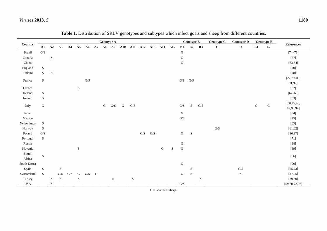
E has only two subtypes, E1 and E2 (Table 1). Although SRLV infection in small ruminants is widely
distributed in all continents [27,58], little information is available on the genetic variants circulating in
different geographic regions (Table 1). SRLV complete genomes have been sequenced and are
available in the GenBank derived from goat (CAEV-CO [59,60], 1GA [61,62], Gansu [63],
Shanxi [64], FESC-752 [25], Seui [46] Roccaverano [45] and A4 [27] viruses) and sheep (Fonni [30],
Volterra [30], 496 [65], SA-OMVV [66], KV1514 [67], KV1772 [68], LV1 [69], EV1 [70],
P1OLV [71], 85/34 [72] and 697 [73] viruses) (Figure 1). In addition, partial sequences have been
published in Brazil [74–76], Canada [77], Finland [78], France [27,79–81], Greece [82], Ireland [83],
Japan [84], Netherlands [85], Poland [86,87], Russia [88], Slovenia [89], South Korea [90] and
Turkey [29,30].
Viruses 2013, 5 1180
Table 1. Distribution of SRLV genotypes and subtypes which infect goats and sheep from different countries.
Country Genotype A Genotype B Genotype C Genotype D Genotype E
References A1 A2 A3 A4 A5 A6 A7 A8 A9 A10 A11 A12 A13 A14 A15 B1 B2 B3 C D E1 E2
Brazil G/S G [74–76]
Canada S G [77]
China G [63,64]
England S [70]
Finland S S [78]
France S G/S G/S G/S [27,79–81,
91,92]
Greece S [82]
Iceland S [67–69]
Ireland G [83]
Italy G G G/S G G/S G/S S G/S G G [30,45,46,
89,93,94]
Japan G [84]
Mexico G/S [25]
Netherlands S [85]
Norway S G/S [61,62]
Poland G/S G/S G/S G S [86,87]
Portugal S [71]
Russia G [88]
Slovenia S G S G [89]
South
Africa S [66]
South Korea G [90]
Spain S S S G/S [65,73]
Switzerland S G/S G/S G G/S G G S S [27,95]
Turkey S S S S S S [29,30]
USA S G/S [59,60,72,96]
G = Goat; S = Sheep.

Viruses 2013, 5 1181
Figure 1. Phylogenetic tree involving SRLV complete sequences obtained from GenBank
(accession numbers in bold and underlined) that include the name of isolate and country
origin. Country abbreviations: CHI - China (Gansu AY900630 and Shanxi GU120138);
ENG - England (EV1 S51392); ICE - Iceland (KV1514 M60610, LV1 M10608 and
KV1772 L06906); ITA - Italy (Roccaverano EU293537, Seui GQ381130, Fonni
JF502416 and Volterra JF502417); MEX - Mexico (FESC-752 HM210570); NOR -
Norway (1GA AF322109); POR - Portugal (P1OLV AF479638); SOA - South Africa
(SAOMVV M34193); SPA - Spain (496 FJ195346 and 697 HQ848062); SWI -
Switzerland (A4 AY445885); USA - United States of America (CAEVCo M33677 and
85/34 AY101611, U64439). The SRLV genotypes and subtypes are indicated.
According to the most recent phylogenetic information, mostly based on the gag gene, the types B,
C and D and only nine of the 15 group subtypes A (A1, A3, A4, A5, A6, A9, A11, A12 and A13)
infect both sheep and goats. Other groups and/or subtypes have only been described in one of the
ruminant species: sheep (A2 and A15), or goats (A7, A8, A10 [93], A14, E1 and E2). However, as
more information is generated, SRLV species restrictions are likely to dwindle (Table 1).

Viruses 2013, 5 1182
4. Tropism
SRLV tropism is linked to both, host genetics and viral genome heterogeneity, and can be studied
according to differences in the targets addressed regarding: (a) host species (goats or sheep, or both);
(b) tissue, differing according to the form of disease (mastitis, arthritis, encephalitis and/or pneumonia);
and (c) cell type.
4.1. Host Species Restriction
Studies from several countries have confirmed that CAEV and VMV, originally established as
specific pathogens in goats and sheep respectively, often cross the species barrier infecting the new
host, persisting in it and spreading across the new host population. The first evidence of this
transgression was obtained by experimentally infecting sheep and goats with CAEV or VMV [97–102].
Subsequently, evidence has been obtained from phylogenetic analysis of sequences derived from
French sheep infected by viruses closer to CAEV (group B2) than to VMV (group A) [79–81,91,92].
On the other hand, molecular epidemiology studies have demonstrated direct virus passage from sheep
infected with VMV-like strains (A4) to goats [95]. In line with this, the genotype B1 had been
considered strictly caprine until the description of B1 infected sheep [103]. Thus, even if some
genotypes might have been originally assigned to a single host species, the host species spectrum may
be wider in nature.
Cell receptor usage explains, at least in part, different patterns in SRLV host restriction. As
indicated above, the high variability of the ENV protein may be due to its interaction with the immune
system and the generation of viral escape mutants [49,104]. Thus, changes in env may affect the ability
of the virus to bind the putative cell receptor(s) and co-receptor(s). Differences in receptor usage have
been observed between VMV and CAEV strains [105]. Also, differences in permissivity have been
found in vitro between cell lines of heterologous origin (chicken, hamster, human, monkey and quail),
which are permissive to SRLV infection and Chinese ovary hamster cells non-permissive to the
infection [106,107]. Specific amino acid residues of the viral SU protein (see below) have been linked
to cell receptor recognition [108]. Some of these residues may also be related to the presence of
arthritis [104].
Another explanation for the host-specific viral restriction lies in the individual genetic background.
Besides adaptive immunological correlates of protection according to immunization studies [109], cell
molecules of the innate immunity may be involved in delimiting host restriction. Among these,
APOBEC and TRIM5 are the most widely studied, as they act in different species directly by mutating
viral genome and interacting with viral capsids, respectively, showing species specificity [110,111].
However, the virus has developed mechanism to counteract proteins of different host species. VMV
studies have shown that the viral Vif protein can neutralize a broad number of A3Z3 proteins
(APOBEC) irrespective of the species of origin (sheep, humans, macaques, cows and cats) [112].
4.2. Viral Genetics by Organ and Tissue
Amino acid motifs conserved at specific viral protein positions involved in the viral entry
(“signature patterns”) [113] have been found in individuals presenting the same form of disease in

Viruses 2013, 5 1183
HIV [114] and FIV [115] infections. Signature patterns that define organ tropism (disease form) have
not been found so far in SRLV infections [116,117]. At the single individual level infected with a
lentiviral quasi-species, each particular virus variant may be confined to one particular compartment,
organ or tissue within the individual. In HIV infections it is known that these viral populations change
to adapt to local media within the individual through genetic drift (founder principle) or selection
pressure. However, when the flow of genes between viral subpopulations within the individual is
significantly restricted, then each subpopulation can become genetically distinct. This phenomenon
named compartmentalization [115,118,119] may be derived from micro-evolution and/or the presence
of various but phylogenetically related genotypes [120–122]. High viral mutation rate in vivo can
quickly enhance the genetic distance between subpopulations. Also, differences in selective pressures
imposed by the immune system can result in divergent evolution of the virus, affecting cell tropism,
phenotypic characteristics and/or pathogenesis, as shown in HIV infections [123–126].
Regarding the particular viral genetic region involved in tropism, LTR and env have been the most
widely studied in lentiviral infections. LTR, being a non-coding region responsible for docking
important cellular transcription factors, is essential for viral replication and may affect viral phenotype
in cell culture [127,128]. This LTR function is achieved through LTR transcription factor binding sites
within the U3 region, some of which are related to cell tropism [129]. Tissue tropism has been
evaluated ex vivo by studying SRLV LTR sequences in different tissue samples but, again, no
signature patterns have been found so far [130]. Alternatively, in other lentiviruses, genomic regions
such as hypervariable regions of the env gene involved in compartmentalization have been found
implicated in tropism. This is the case of the V3 hypervariable region of env in HIV infections, being
determinant of cell tropism and replication efficiency as it relates to the fusion and virus adsorption to
the cell in macrophage-tropic strains [113]. In SRLV genomes, five variable (V1 to V5) and four
conserved (C1 to C4) regions in the envelope protein have been identified [108,131]. The SRLV
env hypervariable region (V4) is structurally and functionally analogous to the V3 region of
HIV [108,132,133]. To colonize different organs (lung, mammary gland, brain, joints) SRLV can
undergo variations in the V4 region of the env gene during early infection giving rise to different viral
subpopulations, as shown in other lentiviruses [132]. A study on SRLV compartmentalization in the
mammary gland of goats and sheep showed the presence of different viral sequences in the V4 region
of the env gene compared to blood-derived cells and colostrum. It was proposed that the mammary
gland was colonized with a “founder virus” which possibly represented the most common variant
circulating (dominant) in the blood at the time of infection. Alternatively, it was proposed that the
initial mechanism could be totally different to compartmentalization and would reflect a selection
pressure caused by a particular cellular tropism or related to the immune system action exerted against
the virus in this compartment [116].
In another study, SRLV genotype A sequences from sheep suffering from neurological disease
(visna) showed a clear tissue compartmentalization in the central nervous system (CNS) and other
organs such as lung and mammary gland, related to horizontal and vertical transmission of SRLV
infection, respectively. Bayesian approach inferences have suggested that proviruses from alveolar
macrophages and peripheral blood mononuclear cells (PBMC) represent the most probable common
ancestors (infecting viruses) in the animal. Likely, PBMC become infected after the intake of virus or
infected cells/particles through respiratory and/or mammary secretions, including colostrum/milk.

Viruses 2013, 5 1184
Neuroinvasion in the visna outbreak involved microevolution after initial infection with SRLV [117].
Of note, the findings on SRLV diversification, adaptation and/or compartmentalization in the brain
may be independent of those observed in lymphoid tissues or other organs, where the virus is exposed
to more abundant immune pressures affecting viral replication [134].
4.3 Cellular Tropism
The members of the lentivirus genus differ in cellular tropism and disease development, being
distributed into two groups. One group includes HIV, FIV and simian immunodeficiency virus (SIV),
all of which replicate in macrophages and lymphocytes causing acquired immunodeficiency syndrome
and specific-organ disease affecting lungs, CNS and gastro-intestinal tract. The other group includes
equine infectious anemia virus (EIAV) and SRLV, both of which replicate primarily in macrophages
(lymphocytes are not infected) and cause a disease affecting specific organs, mainly lung, mammary
gland, CNS and/or joints [127,135].
In the VMV epidemic in Iceland, the most common clinical signs corresponded to the maedi
(respiratory form) and the visna (nervous form) was only reported in herds with high prevalence of
maedi for several years. This was originally attributed to the cell tropism of the virus rather than the
genetic variation of sheep, because there was only one sheep breed in Iceland [136,137]. Subsequently,
it was shown that the cellular tropism of VMV strains from sheep with visna appeared to be different
from those isolated from sheep with maedi. The virus isolated from brain cells replicated more rapidly
in sheep choroid plexus compared to isolates from lung and this difference was related to differences
in env and LTR regions [138].
In vivo SRLV have a marked tropism for cells of the monocyte/macrophage lineage and
dendritic cells [139,140], the virus infects monocytes, so these cells are permissive for viral entry and
genome integration (provirus), but the infection remains latent until the cell differentiates into
macrophage [139,141,142]. This maturation enhances the expression of the transcription factors c-Fos
and c-Jun, which bind to the AP-1 and AP-4 promoter binding sites of LTR triggering transcription,
and consequently viral replication and productive infection [143]. Thus, SRLV replicate in mature
macrophages or related tissues rather than in circulating monocytes, the latter acting as a “Trojan
horse” [144]
SRLV are highly variable genetically and antigenically and also show phenotypic differences
in vitro. Specifically, isolates may differ from each other in the ability to selectively and productively
infect particular cell types and also in the capacity to induce a cytopathic effect. Accordingly, SRLV
may be classified phenotypically as rapid/high or slow/low. The rapid/high strains replicate rapidly,
inducing the formation of syncytia, cell lysis, and reaching high titers. In contrast, the slow/low viruses
grow slowly and at a low titer. Frequently, sheep isolates belong to the first type; whereas isolates from
goats show a slow/low phenotype. However, SRLV strains that show an intermediate phenotype
between the most extreme phenotypes of VMV-like and CAEV-like viruses have been isolated from
both sheep and goats [145,146].
Because SRLV show in vitro high viral production in permissive cells such as macrophages, these
cells are used routinely for isolating the virus, even though they are terminally differentiated [147].
Fibro-epithelial synovial membrane cells and choroid plexus cells derived from goats or sheep are also

Viruses 2013, 5 1185
routinely used for in vitro production of SRLV [96,148]. Other cell types permissive to SRLV
infection are from lung explants [149], skin fibroblasts [140], spleen and corneal cells [150,151],
testicle cells [152], goat endothelium [153,154], mammary gland epithelial cells (TIGMEC cell
line) [155,156], granulose cells [157], oviduct cells [158], microglial cells [159,160], tubular epithelial
cells [161], hepatocytes [162], cardiac myocytes [162] and the third eyelid cells [163]. However, the
restricted viral antigen expression and the low number of infected cells found in some of these cells
should be taken into account before interpreting the biological relevance of these cells in SRLV
persistence [164]. Experiments employing pseudotyped vectors (vaccinia virus or murine leukemia
virus-based) with SRLV ENV indicate that ENV-based interaction with cells renders them permissive
to virus entry. This is the case of cell lines Hela, BHK-21 and others from different origins (human or
other species) [106,107], suggesting that these cells have a receptor for ENV and that the virus enters
the host cell by fusion of the membrane receptor to viral ENV SU protein gp135 [165]. However, this
permissiveness at the entry step [166] may not be associated with a capability of productive infection
or to a possible natural susceptibility. Although in previous SRLV studies restriction had been
attributed to the lack of functional receptors [167], post-entry restriction factors such as those
interacting with Gag [110] rather than Env proteins may have been responsible for the lack of
productive infection.
Different attempts have been made to identify and characterize SRLV receptor(s). Candidate SRLV
receptor molecules include: a membrane-associated proteoglycan substituted with one 30 kDa chain (s)
chondroitin sulfate glycosaminoglycan [168]; molecules of the major histocompatibility complex class
II (MHC class II), as preincubation with MHC class II antigens inhibits VMV infection (even though
antibodies specific to antigens of MHC class II do not inhibit infection) [169]; CD4 and CXCR4
molecules, which have been proposed as optional auxiliary components for VMV receptor (or receptor
complex) that facilitates membrane fusion events mediated by VMV [166,170]; and a complex
comprising three membrane proteins of 15, 30 and 45 kDa, identified as VMV binding proteins [171].
However, none of these molecules has been established as the main/essential receptor. Analysis of
somatic hybrid cell lines (CHO cell panel containing different mouse chromosomes) permissive to
VMV entry has shown that the gene for cellular receptor is in the murine chromosome 2 or 4 (although
involvement of chromosomes 6 and X is not excluded). On the other hand, it also showed using hybrid
cell lines [172] that the VMV receptor gene maps to sheep chromosome 3p (OAR-3p) and a region of
human chromosome 2 (HAS-2p25 > q13) has retained synteny with sheep chromosome 3p. These
regions do not include any gene of known receptor or
co-receptor of lentiviruses, which would indicate that SRLV receptor uses a different molecule to
infect human cells. Apparently, SRLV may use different cell receptors likely depending the cell it
encounters [105]. Recently, the mannose receptor (MR, codified in mice by chromosome 2) has been
described in sheep and identified as a potential SRLV receptor. Accordingly, three cell phenotypes
have been proposed in small ruminants regarding the expression and usage of MR as SRLV receptor:
cells using this molecule as the main receptor (synovial membrane cells), cells having this and at least
another main SRLV receptor(s) (macrophages) and cells lacking MR and using other SRLV
receptor(s) instead (skin fibroblasts) [173]. Interestingly, the presence of this receptor has been
associated with the evolution of disease, as its expression increases in target organs exhibiting the most
severe lesions in SRLV clinical infections [174].

Viruses 2013, 5 1186
5. In Vitro Diagnosis
Different laboratories have applied direct diagnostic methods such as viral isolation, a very
laborious method whose performance can be hampered by the lack of permissive cell lines and limited
viral production in cultured cells [96]. Isolation of virus attempted from leukocytes of infected animals
often fail, indicating that a negative result is not always reliable [175]. Although this method is not
suitable for large-scale studies, it can be performed by co-cultivation (commonly of blood monocyte
derived macrophages with fibro-epithelial cells), whereby the detection of cytopathic effect or reverse
transcriptase activity indicate the presence of virus [65]. Similarly, immunohistochemical methods,
allowing the detection of viral protein antigens by specific antibodies in histological samples, smears
or cytospin preparations, are useful in research and confirmatory studies, but are not commonly
applied in differential routine diagnosis on live animals, because of the cost, low availability of
reagents and samples and limited sensitivity of some of these techniques [175]. Also, in situ
hybridization methods have been employed for histologic studies, but this laborious procedure is only
used for research purposes [176,177]. Although PCR-based diagnosis is the most common of the
molecular techniques applied in SRLV diagnosis in vitro (see below), indirect methods such as
serological diagnosis to detect antibodies to SRLV are overall the most commonly applied approach to
detect infection. However, there are problems inherent to serological diagnosis. These include the high
genetic/antigenic variability existing in laboratory and field strains, the presence of maternal antibodies,
the relatively long lag period between infection and antibody production (serological gap) and the
intermittent seroconversion (by fluctuating titers throughout the animal’s life). Antibody production
has been detected by techniques such as agar gel immunodiffusion (AGID), the enzyme-linked
immunosorbent assay (ELISA), radioimmunoprecipitation (RIPA) and Western blot (WB) [178–180].
Both RIPA and WB are mainly used for confirmation [181]. AGID is highly specific but relatively
insensitive [178] and often linked to subjective interpretation, inapplicability for the determination of
antibodies in milk and lack of automation, motivating its replacement by ELISA methods of relatively
low cost and easy implementation and interpretation [2,179,181].
5.1. ELISA Tests
In spite of the antigenic variability, cross-reacting antibodies between VMV-like and CAEV-like
antigens have been described [182] and have enabled for years the control of SRLV-induced disease.
Sheep and goats remain infected for life, but many seropositive animals may never show clinical signs
of SRLV infection. Antibodies do not protect against the disease and are mainly indicators of infection.
The virus escapes from the immune attack even in the presence of neutralizing antibodies, as proviral
DNA integrates into host cell genome and viral mutations take place during viral replication [183]. On
the other hand, a seronegative animal cannot be strictly considered free of infection, either because
antibody titers are below detection level before seroconversion, or because fluctuation in antibody
levels occurs throughout the animal’s life. Newborns infected at birth have maternal antibodies
(through colostrum/milk intake) for at least two or three months. Thereafter, they are usually
seronegative until they seroconvert between six and twelve months of age. Performing diagnostic tests
immediately before pregnancy does not necessarily ensure that mothers will not seroconvert after

Viruses 2013, 5 1187
parturition. Thus, infected seronegative animals constitute a potential source of infection through
vertical and horizontal transmission [179,181].
To date, no single technique or test can be proposed as “gold standard” to determine the infection
status of the animal [175]. Competitive ELISA methods (cELISA CAEV of VMRD Inc. Pullman,
WA) [184,185] using monoclonal antibodies to viral envelope protein (ENV-SU, gp135) epitopes have
been developed [186] and indirect ELISAs have been frequently applied [187,188], although few of
them have been compared internationally for both sheep and goats [189–191]. Currently available
indirect standard ELISAs are used in different formats and designs. Examples are those using either
whole virus antigens such as the AG-CHEKIT (CAEV/MVV kit, IDEXX Switzerland AG, Liebefeld,
Bern, Switzerland) [192] or recombinant proteins plus a peptide, specifically, the GAG p25
recombinant protein and a TM peptide derived from genotype A, as used in assays Elitest-MVV
(HYPHEN Biomed, Neuville-sur-Oise, France) [193] and Pourquier (ELISA Maedi-Visna/CAEV
serum verification Institut Pourquier, Montpellier, France). But in some cases, monostrain assays may
not cover the whole SRLV antigenic spectrum and fail to detect genotype E [188], B2 [194] and A4
infections [195]. Besides the viral p25 protein included in the test, p14 and p17 proteins also present
immunodominant epitopes and could be used for differential VMV/CAEV diagnosis [196,197]. Gag
proteins (p25) have been used in early diagnosis and detection of cross-reacting antibodies for more
than 20 years [179]. However, new genotypes have emerged [30,45], broadening diagnostic
possibilities, leading to the development of new standard indirect ELISA tests, such as the one based
on a mixture of gag and env peptides of three different SRLV genotypes (A, B and E) (IN3 diagnostic.
Eradikit® SRLV indirect ELISA for Small Ruminant Lentivirus).
The ENV TM protein has an added value in diagnosis of long-term infections such as those present
in diseased animals [198] and therefore is included in most subunit assays. SU peptides have also been
widely used in serological diagnosis and, due to high variability of SU proteins, these peptides are
being useful to serotype and perform strain-specific diagnosis [198]. Synthetic peptide ELISAs [199]
have been evaluated by our group, outlining their utility in field sample diagnosis of animals from
neurological [73] and arthritic [65] outbreaks.
Antibody detection technology is certainly a valuable tool for determining the infection status of a
herd/flock, although in some areas with limited resources this technology may be somewhat expensive
if performed at the individual level. Therefore, strategies for detecting antibodies and infection in the
flock, involving the use of bulk milk [200–202] or pooled serum [203] and semen samples [191,204]
are particularly useful in these areas.
5.2. Polymerase Chain Reaction (PCR)
Molecular techniques of use in SRLV diagnosis include the heteroduplex mobility assay
(HMA) [80], which may help to a genotypic characterization of circulating strains within the flock, a
loop-mediated isothermal amplification technique [64] and polymerase chain reaction (PCR)
procedures, the most commonly used to directly assess the presence of viral nucleic acid. Conventional
PCR for diagnostic purposes has been more extensively studied than the real-time PCR (rtPCR). The
introduction of rtPCR represents a breakthrough in molecular diagnosis [205,206]. The PCR and the
early detection of amplicons have been combined in this technique [206,207]. The rtPCR has been

Viruses 2013, 5 1188
useful for detection and quantification of viral nucleic acids in different cells or tissues, although its
use in SRLV routinary diagnosis is not widespread [207–209]. Primers designed in almost all SRLV
genetic regions [205] have yielded reliable and fast results. The rtPCR diagnostic approach presents a
decreased risk of cross-contamination and has a high sensitivity using SYBR Green I or TaqMan-
based methods [207]. Furthermore, the use of oligonucleotide probes helps to increase rtPCR
specificity [205]. However, this technique may work only on a restricted number of strains and its use
in field diagnosis may present difficulties due to variation in the primer binding site.
The first conventional PCR protocols designed to detect SRLV were published in the early
1990s [210,211]. PCR was considered initially a successful tool in SRLV diagnosis, due to its ability
to directly detect viral nucleic acids, either in the infected cell as a provirus (DNA) or in exudates that
contain free virus particles (RNA; reverse transcription-PCR) [212]. In addition to conventional PCR,
variants of this technique have appeared involving two (or more) amplification rounds
(nested-PCR and seminested-PCR) in order to increase the sensitivity [213–217]. Different studies
have shown the utility of conventional PCR to detect SRLV infections in different animal samples,
namely PBMC [214,218–220], peripheral blood leukocytes (PBL) [213,221], milk or mammary
secretions [212,214,218–220,222], semen [223,224], synovial fluid [218] and other tissues [214,222,225].
The efficiency of PCR depends mainly on the specificity of the primers designed, the choice of the
amplified target viral region, and the sensitivity of the technique [35,127,226]. The low viral load
existing in infected animals, the low proportion of permissive cells in some tissues (there is one
infected monocyte in 104–10
5 PBMC) [206] and the high genetic heterogeneity may hamper PCR
diagnosis of SRLV [2,226]. Furthermore, the cell type may affect PCR performance. However, PCR
has the advantage over serological methods of detecting infection in animals with colostral antibodies.
In milk samples from sheep and goats (small ruminants) a seminested-PCR method based on the pol
region (pol-seminested-PCR) has yielded positive results in all the infected small ruminants tested (the
seropositives determined previously by a serological test and a seronegative animal) [212]. Similar
PCR sensitivities have also been found in PBMC, milk cells and synovial fluid [218]. Here, however,
are also studies reporting a decreased sensitivity when using milk samples (23.6%) relative to blood
samples (33.3%), using a gag-PCR [219] or a LTR-PCR procedure [222]. Target sequences for PCR
primers design are widespread throughout the SRLV genome including LTR, gag, pol and env
regions [164,212,222,224] and lead to different sensitivity and specificity values. In PCRs amplifying
segments of the SRLV gag and pol regions, the gag-PCR was more sensitive than pol-PCR [211].
However, the LTR-PCR may be more sensitive than PCRs based on gag and pol regions [213,222,226].
In a study that examined four pairs of primers designed in LTR, gag (capsid and matrix) and env
regions using DNA from cells infected with virus from various geographical areas, PCR primers
targeting the LTR region, gag (capsid), env and gag (matrix) amplified 100%, 62.5%, 25% and 12.5% of
the samples, respectively [227]. This is in contrast with another study showing that a PCR based on env
sequences could be more efficient at detecting infected animals (93.3%) compared to pol-PCR which
only detected 60% of animals infected. Despite its presumed high variability, the env region can be
suitable for diagnosis by PCR (Table 2) [224].
In SRLV infections, PCR-positive reactions are often found amongst seronegative animals (false
negatives) (Table 2) [219,228], which later seroconvert, thereby demonstrating a lack of sensitivity of
the serological methods compared to PCR under particular conditions [195].

Viruses 2013, 5 1189
Table 2. Different PCRs techniques, target DNA samples, regions of the viral genome used for PCR design and sensitivity of the PCR method
for detection of infection by SRLV.
PCR type DNA or cDNA source Primers location into
the viral genome Sensitivity test References
cPCR Cell culture gag and pol pol-PCR was more sensitive than gag-PCR [210]
snPCR PBL pol and LTR LTR-PCR was more sensitive than pol-PCR [212]
nPCR PBMC gag and pol gag-PCR was more sensitive than pol-PCR [213]
snPCR PBMC gag gag-PCR was less sensitive than AGID [214]
snPCR Cell culture and PBMC pol High sensitivity, by using degenerate primers [215]
nPCR PBMC gag gag-PCR was more sensitive than AGID in seronegative animals [216]
nPCR PBL and blood gag and LTR gag-PCR was more sensitive than LTR-PCR [220]
cPCR Semen env and pol env-PCR was more sensitive than gag-PCR [222]
cPCR PBMC, milk cells and tissues LTR LTR-PCR had a sensitivity of 98% with regard to AGID and ELISA [223]
cPCR Cell culture LTR, gag and env LTR-PCR was more sensitive than gag-PCR and env-PCR [226]
cPCR PBMC gag gag-PCR was less sensitive than AGID [228]
cPCR Cell culture and PBMC gag gag-PCR was more sensitive than the ELISA and WB [229]
nPCR PBL gag gag-PCR increases its sensitivity when used along with hybridization [230]
cPCR: conventional PCR; snPCR: seminested PCR; nPCR: nested PCR; PBMC: peripheral blood mononuclear cells; PBL: peripheral blood leukocytes; AGID: agar gel
immunodiffusion; ELISA: enzyme-linked immunoassay; WB: western blot.
Viruses 2013, 5 1190
Studies based on gag-PCR diagnosis reveal that this method may detect infection in 88.3% of sheep
and goats that are seropositive by AGID and in 11% of seronegatives (67% of animals eventually
seroconverted) [229]. In line with this, a gag-seminested-PCR procedure allows the detection of 70%
of the AGID positive sheep and 11% of seronegative animals [215]. Comparative studies on the
sensitivity of the PCR with regard to serological tests (ELISA and AGID) have shown that LTR-PCR
is less sensitive than the ELISA and AGID. However, PCR showed a specificity of 100% (like AGID),
whereas ELISA was less specific (59%) [230]. LTR-PCR specificity is maintained (100%) in different
tissues, but sensitivity values change according to the tissue under study [83.5% (peripheral blood
leukocytes), 66.7% (milk cells) and 88% (tissues)], being less sensitive than the ELISA and AGID [222].
Since PCR assays may fail to detect virus when virus load is below the assay threshold, PCR
sensitivity may increase upon co-cultivation of PBMC from infected animals with permissive
fibroblasts in vitro [225,231,232] or by using probes in the Southern blot [214,219] or in situ
hybridization [228,233] techniques. The use of strain-specific and sequence-degenerated primers could
also improve sensitivity, as shown by Elthair and collaborators [216] using a pol-seminested-PCR on
AGID-seropositive animals infected with two viral subtypes (genotypes A and B). The percentage of
pol-PCR positives in this study was 62.2% and 30.5% for types A and B, respectively. In addition, the
pol-PCR detected 38.2% of positive animals among AGID seronegatives [216].
Although different studies indicate that in general the PCR methods tend to be less sensitive than
serological tests (particularly ELISA tests) (Table 2) [213,220], PCR testing can detect infected
animals prior to seroconversion [213,219,228,234,235]. According to SRLV pathogenesis, provirus
should be present prior to the antibody response to the virus during initial infection of adult animals,
but this is not always detected. In studies comparing PCR with serological test (AGID and ELISA)
diagnosis, agreement (positive concordance) ranged from 70% to 94.7%, whereas disagreement
(negative concordance) ranged from 87.5% to 100% [181,218,219,222,224,229]. This suggests that a
combination of serology and PCR might be optimal for detecting the infectious status of a
population [217,230,236]. Furthermore, PCR products can be systematically sequenced [27,94], which
may allow a better design of primers for new PCRs with a wide range of recognition. The development
of a single PCR to detect all SRLV may still be utopian, although attempts to cover a large spectrum of
genomes by including different sets of primers in the assay have been made [27].
6. Concluding Remarks
The basic mechanisms of mutation and recombination are complex biological processes, but they
may help to understand the true role of viral diversity in SRLV pathogenesis. Virus and host genetic
and microenvironmental factors involved in cell, tissue/organ and host tropism of these viruses are
pivotal issues in current investigations. Genetic and antigenic variations of the virus represent
challenges in SRLV diagnosis. The success in avoiding SRLV infection spread depends largely on
early detection and culling of infected animals in the heard/flock. Detection of specific antibodies in
serum or milk by ELISA, typing the circulating strains by local strain-based ELISAs, HMA or
PCR-sequencing techniques, and use of PCR for confirmatory purposes are solid technological
approaches of practical use for reducing the risk of misdiagnosis in different areas. Finally, diagnostic

Viruses 2013, 5 1191
methods suitable to both sheep and goats should be implemented in joint programs for these species
for successful SRLV control and eradication strategies.
Acknowledgments
The authors would like to thank Karen Janett Becerra for help in writing of the manuscript.
Conflict of Interest
The authors declare no conflict of interest.
References
1. Christodoulopoulos, G. Maedi-Visna: Clinical review and short reference on the disease status in
Mediterranean countries. Small Rum. Res. 2006, 62, 47–53.
2. Peterhans, E.; Greenland, T.; Badiola, J.; Harkiss, G.; Bertoni, G.; Amorena, B.; Eliaszewicz, M.;
Juste, R.A.; Krassnig, R.; Lafont, J.P.; et al. Routes of transmission and consequences of small
ruminant lentiviruses (SRLVs) infection and eradication schemes. Vet. Res. 2004, 35, 257–274.
3. Anderson, B.C.; Bulgin, M.S.; Adams, S.; Duelke, B. Firm udder in periparturient ewes with
lymphocytic accumulations, retrovirus infection, and milk unavailable at the teat. J. Am. Vet. Med.
Assoc. 1985, 186, 391–393.
4. Keen, J.E.; Hungerford, L.L.; Littledike, E.T.; Wittum, T.E.; Kwang, J. Effect of ewe ovine
lentivirus infection on ewe and lamb productivity. Prev. Vet. Med. 1997, 30, 155–169.
5. Pekelder, J.J.; Veenink, G.J.; Akkermans, J.P.; van Eldik, P.; Elving, L.; Houwers, D.J. Ovine
lentivirus induced indurative lymphocytic mastitis and its effect on the growth of lambs.
Vet. Rec. 1994, 134, 348–350.
6. Dungu, B.; Vorster, J.; Bath, G.F.; Verwoerd, D.W. The effect of a natural maedi-visna virus
infection on the productivity of South African sheep. Onderstepoort. J. Vet. Res. 2000, 67, 87–96.
7. Legrottaglie, R.; Martini, M.; Barsotti, G.; Agrimi, P. The effects of ovine lentivirus infection on some
productive aspects in a Sardinian sheep flock from Italy. Vet. Res. Commun. 1999, 23, 123–131.
8. Snowder, G.D.; Gates, N.L.; Glimp, H.A.; Gorham, J.R. Prevalence and effect of subclinical
ovine progressive pneumonia virus infection on ewe wool and lamb production. J. Am. Vet. Med.
Assoc. 1990, 197, 475–479.
9. Arsenault, J.; Dubreuil, P.; Girard, C.; Simard, C.; Belanger, D. Maedi-visna impact on
productivity in Quebec sheep flocks (Canada). Prev. Vet. Med. 2003, 59, 125–137.
10. Leitner, G.; Krifucks, O.; Weisblit, L.; Lavi, Y.; Bernstein, S.; Merin, U. The effect of caprine
arthritis encephalitis virus infection on production in goats. Vet. J. 2010, 183, 328–331.
11. Nord, K.; Adnoy, T. Effects of infection by caprine arthritis-encephalitis virus on milk production
of goats. J. Dairy Sci. 1997, 80, 2391–2397.
12. Turin, L.; Pisoni, G.; Giannino, M.L.; Antonini, M.; Rosati, S.; Ruffo, G.; Moroni, P. Correlation
between milk parameters in CAEV seropositive and negative primiparous goats during an
eradication program in Italian farm. Small Rum. Res. 2005, 57, 73–79.

Viruses 2013, 5 1192
13. Martínez, N.B.; Peris, R.C.; Roche, J.M.L.; Caballero, G.C. Efectos del virus de la artritis-
encefalitis caprina sobre la producción y composición de la leche en cabras murciano-granadinas.
PR: Pequeños Rumiantes 2002, 3, 26–30.
14. Ryan, D.P.; Greenwood, P.L.; Nicholls, P.J. Effect of caprine arthritis-encephalitis virus infection
on milk cell count and N-acetyl-beta-glucosaminidase activity in dairy goats. J. Dairy Res. 1993,
60, 299–306.
15. Martinez-Navalon, B.; Peris, C.; Gomez, E.A.; Peris, B.; Roche, M.L.; Caballero, C.; Goyena, E.;
Berriatua, E. Quantitative estimation of the impact of caprine arthritis encephalitis virus infection
on milk production by dairy goats. Vet. J. 2013, doi:10.1016/j.tvjl.2012.12.020.
16. Christodoulopoulos, G. Milk Production and Milk Fat Content in Commercial Karagouniko Breed
Flocks Infected by Maedi Disease in Greece. In Proceedings of the Sixth International Sheep
Veterinary Congress, Hersonissos Crete, Greece, 17–21 June 2005; pp. 344–345.
17. Leitner, G.; Silanikove, N.; Merin, U. Estimate of milk and curd yield loss of sheep and goats
with intrammamary infection and its relation to somatic cell count. Small Rum. Res. 2008, 74,
221–225.
18. Broughton-Neiswanger, L.E.; White, S.N.; Knowles, D.P.; Mousel, M.R.; Lewis, G.S.;
Herndon, D.R.; Herrmann-Hoesing, L.M. Non-maternal transmission is the major mode of ovine
lentivirus transmission in a ewe flock: A molecular epidemiology study. Infect. Genet. Evol. 2010,
10, 998–1007.
19. Leginagoikoa, I.; Daltabuit-Test, M.; Alvarez, V.; Arranz, J.; Juste, R.A.; Amorena, B.;
de Andres, D.; Lujan, L.L.; Badiola, J.J.; Berriatua, E. Horizontal Maedi-Visna virus (MVV)
infection in adult dairy-sheep raised under varying MVV-infection pressures investigated by
ELISA and PCR. Res. Vet. Sci. 2006, 80, 235–241.
20. Leginagoikoa, I.; Minguijon, E.; Juste, R.A.; Barandika, J.; Amorena, B.; de Andres, D.;
Badiola, J.J.; Lujan, L.; Berriatua, E. Effects of housing on the incidence of visna/maedi virus
infection in sheep flocks. Res. Vet. Sci. 2010, 88, 415–421.
21. Alvarez, V.; Arranz, J.; Daltabuit-Test, M.; Leginagoikoa, I.; Juste, R.A.; Amorena, B.;
de Andres, D.; Lujan, L.L.; Badiola, J.J.; Berriatua, E. Relative contribution of colostrum from
Maedi-Visna virus (MVV) infected ewes to MVV-seroprevalence in lambs. Res. Vet. Sci. 2005,
78, 237–243.
22. Leginagoikoa, I.; Juste, R.A.; Barandika, J.; Amorena, B.; De Andres, D.; Lujan, L.; Badiola, J.;
Berriatua, E. Extensive rearing hinders Maedi-Visna Virus (MVV) infection in sheep. Vet. Res.
2006, 37, 767–778.
23. Sigurdsson, B.; Grimsson, H.; Palsson, P.A. Maedi, a chronic, progressive infection of sheep’s
lungs. J. Infect. Dis. 1952, 90, 233–241.
24. Blacklaws, B.A.; Berriatua, E.; Torsteinsdottir, S.; Watt, N.J.; de Andres, D.; Klein, D.;
Harkiss, G.D. Transmission of small ruminant lentiviruses. Vet. Microbiol. 2004, 101, 199–208.
25. Ramirez, H.; Glaria, I.; de Andres, X.; Martinez, H.A.; Hernandez, M.M.; Reina, R.; Iraizoz, E.;
Crespo, H.; Berriatua, E.; Vazquez, J.; et al. Recombinant small ruminant lentivirus subtype B1 in
goats and sheep of imported breeds in Mexico. Vet. J. 2011, 190, 169–172.

Viruses 2013, 5 1193
26. Straub, O.C. Maedi-Visna virus infection in sheep. History and present knowledge. Comp.
Immunol. Microbiol. Infect. Dis. 2004, 27, 1–5.
27. Shah, C.; Boni, J.; Huder, J.B.; Vogt, H.R.; Muhlherr, J.; Zanoni, R.; Miserez, R.; Lutz, H.;
Schupbach, J. Phylogenetic analysis and reclassification of caprine and ovine lentiviruses based
on 104 new isolates: Evidence for regular sheep-to-goat transmission and worldwide propagation
through livestock trade. Virology 2004, 319, 12–26.
28. Gifford, R.J. Viral evolution in deep time: Lentiviruses and mammals. Trends Genet. 2012, 28,
89–100.
29. Muz, D.; Oguzoglu, T.C.; Rosati, S.; Reina, R.; Bertolotti, L.; Burgu, I. First molecular
characterization of visna/maedi viruses from naturally infected sheep in Turkey. Arch. Virol. 2012,
doi:10.1007/s00705-012-1518-1.
30. Bertolotti, L.; Mazzei, M.; Puggioni, G.; Carrozza, M.L.; Dei Giudici, S.; Muz, D.; Juganaru, M.;
Patta, C.; Tolari, F.; Rosati, S. Characterization of new small ruminant lentivirus subtype B3
suggests animal trade within the Mediterranean Basin. J. Gen. Virol. 2011, 92, 1923–1929.
31. Foley, B.T. An Overview of the Molecular Phylogeny of Lentiviruses. In HIV Sequence
Compedium; Kuiken, C., Foley, B., Freed, E., Hahn, B., Korber, B., Marx, P.A., McCutchan, F.,
Mellors, J.W., Mullins, J.I., Sodroski, J., Wolinksy, S., Eds.; Theoretical Biology and Biophysics
Group, Los Alamos National Laboratory: Los Alamos, NM, USA, 2000; pp. 35–43.
32. Eigen, M. Selforganization of matter and the evolution of biological macromolecules.
Naturwissenschaften 1971, 58, 465–523.
33. Ojosnegros, S.; Perales, C.; Mas, A.; Domingo, E. Quasispecies as a matter of fact: Viruses and
beyond. Virus Res. 2011, 162, 203–215.
34. Korber, B.; Gaschen, B.; Yusim, K.; Thakallapally, R.; Kesmir, C.; Detours, V. Evolutionary and
immunological implications of contemporary HIV-1 variation. Br. Med. Bull. 2001, 58, 19–42.
35. Pasick, J. Maedi-visna virus and caprine arthritis-encephalitis virus: Distinct species or
quasispecies and its implications for laboratory diagnosis. Can. J. Vet. Res. 1998, 62, 241–244.
36. Smyth, R.P.; Davenport, M.P.; Mak, J. The origin of genetic diversity in HIV-1. Virus Res. 2012,
169, 415–429.
37. Roberts, J.D.; Bebenek, K.; Kunkel, T.A. The accuracy of reverse transcriptase from HIV-1.
Science 1988, 242, 1171–1173.
38. Vartanian, J.P.; Meyerhans, A.; Asjo, B.; Wain-Hobson, S. Selection, recombination, and G----A
hypermutation of human immunodeficiency virus type 1 genomes. J. Virol. 1991, 65, 1779–1788.
39. Bishop, K.N.; Holmes, R.K.; Sheehy, A.M.; Davidson, N.O.; Cho, S.J.; Malim, M.H. Cytidine
deamination of retroviral DNA by diverse APOBEC proteins. Curr. Biol. 2004, 14, 1392–1396.
40. Dang, Y.; Wang, X.; Esselman, W.J.; Zheng, Y.H. Identification of APOBEC3DE as another
antiretroviral factor from the human APOBEC family. J. Virol. 2006, 80, 10522–10533.
41. Terai, C.; Carson, D.A. Pyrimidine nucleotide and nucleic acid synthesis in human monocytes and
macrophages. Exp. Cell. Res. 1991, 193, 375–381.
42. Turelli, P.; Guiguen, F.; Mornex, J.F.; Vigne, R.; Querat, G. dUTPase-minus caprine arthritis-
encephalitis virus is attenuated for pathogenesis and accumulates G-to-A substitutions. J. Virol.
1997, 71, 4522–4530.

Viruses 2013, 5 1194
43. Wain-Hobson, S.; Sonigo, P.; Guyader, M.; Gazit, A.; Henry, M. Erratic G→A hypermutation within
a complete caprine arthritis-encephalitis virus (CAEV) provirus. Virology 1995, 209, 297–303.
44. Juganaru, M.; Reina, R.; Bertolotti, L.; Stella, M.C.; Profiti, M.; Armentano, M.; Bollo, E.;
Amorena, B.; Rosati, S. In vitro properties of small ruminant lentivirus genotype E. Virology
2011, 410, 88–95.
45. Reina, R.; Grego, E.; Bertolotti, L.; De Meneghi, D.; Rosati, S. Genome analysis of small-
ruminant lentivirus genotype E: A caprine lentivirus with natural deletions of the dUTPase
subunit, vpr-like accessory gene, and 70-base-pair repeat of the U3 region. J. Virol. 2009, 83,
1152–1155.
46. Reina, R.; Bertolotti, L.; Dei Giudici, S.; Puggioni, G.; Ponti, N.; Profiti, M.; Patta, C.; Rosati, S.
Small ruminant lentivirus genotype E is widespread in Sarda goat. Vet. Microbiol. 2010, 144, 24–31.
47. Tormo-Mas, M.A.; Mir, I.; Shrestha, A.; Tallent, S.M.; Campoy, S.; Lasa, I.; Barbe, J.;
Novick, R.P.; Christie, G.E.; Penades, J.R. Moonlighting bacteriophage proteins derepress
staphylococcal pathogenicity islands. Nature 2012, 465, 779–782.
48. Sheehy, A.M.; Gaddis, N.C.; Choi, J.D.; Malim, M.H. Isolation of a human gene that inhibits
HIV-1 infection and is suppressed by the viral Vif protein. Nature 2002, 418, 646–650.
49. Andresdottir, V.; Skraban, R.; Matthiasdottir, S.; Lutley, R.; Agnarsdottir, G.; Thorsteinsdottir, H.
Selection of antigenic variants in maedi-visna virus infection. J. Gen. Virol. 2002, 83, 2543–2551.
50. Andresdottir, V. Evidence for recombination in the envelope gene of maedi-visna virus. Virus
Genes 2003, 27, 5–9.
51. Jetzt, A.E.; Yu, H.; Klarmann, G.J.; Ron, Y.; Preston, B.D.; Dougherty, J.P. High rate of
recombination throughout the human immunodeficiency virus type 1 genome. J. Virol. 2000, 74,
1234–1240.
52. Jolly, P.E.; Narayan, O. Evidence for interference, coinfections, and intertypic virus enhancement of
infection by ovine-caprine lentiviruses. J. Virol. 1989, 63, 4682–4688.
53. Hu, W.S.; Temin, H.M. Retroviral recombination and reverse transcription. Science 1990, 250,
1227–1233.
54. Hu, W.S.; Temin, H.M. Genetic consequences of packaging two RNA genomes in one retroviral
particle: Pseudodiploidy and high rate of genetic recombination. Proc. Natl. Acad. Sci. USA 1990,
87, 1556–1560.
55. Pisoni, G.; Bertoni, G.; Puricelli, M.; Maccalli, M.; Moroni, P. Demonstration of coinfection with
and recombination by caprine arthritis-encephalitis virus and maedi-visna virus in naturally
infected goats. J. Virol. 2007, 81, 4948–4955.
56. Zanoni, R.G. Phylogenetic analysis of small ruminant lentiviruses. J. Gen. Virol. 1998, 79,
1951–1961.
57. Peeters, M.; Esu-Williams, E.; Vergne, L.; Montavon, C.; Mulanga-Kabeya, C.; Harry, T.;
Ibironke, A.; Lesage, D.; Patrel, D.; Delaporte, E. Predominance of subtype A and G HIV type 1
in Nigeria, with geographical differences in their distribution. AIDS Res. Hum. Retroviruses 2000,
16, 315–325.
58. Adams, D.S.; Oliver, R.E.; Ameghino, E.; DeMartini, J.C.; Verwoerd, D.W.; Houwers, D.J.;
Waghela, S.; Gorham, J.R.; Hyllseth, B.; Dawson, M.; et al. Global survey of serological
evidence of caprine arthritis-encephalitis virus infection. Vet. Rec. 1984, 115, 493–495.

Viruses 2013, 5 1195
59. Narayan, O.; Clements, J.E.; Strandberg, J.D.; Cork, L.C.; Griffin, D.E. Biological characterization
of the virus causing leukoencephalitis and arthritis in goats. J. Gen. Virol. 1980, 50, 69–79.
60. Saltarelli, M.; Querat, G.; Konings, D.A.; Vigne, R.; Clements, J.E. Nucleotide sequence and
transcriptional analysis of molecular clones of CAEV which generate infectious virus. Virology
1990, 179, 347–364.
61. Gjerset, B.; Jonassen, C.M.; Rimstad, E. Natural transmission and comparative analysis of small
ruminant lentiviruses in the Norwegian sheep and goat populations. Virus Res. 2007, 125, 153–161.
62. Gjerset, B.; Storset, A.K.; Rimstad, E. Genetic diversity of small-ruminant lentiviruses:
Characterization of Norwegian isolates of Caprine arthritis encephalitis virus. J. Gen. Virol. 2006,
87, 573–580.
63. Qu, J.; Guo, W.; Zhao, L.; Sheng, R.; Xiang, W. The gene cloning and sequence analysis of the
whole genome of Caprine Arthritis Encephalitis virus (CAEV) GANSU strain. Chin. J. Virol.
2005, 21, 389–392.
64. Huang, J.; Sun, Y.; Liu, Y.; Xiao, H.; Zhuang, S. Development of a loop-mediated isothermal
amplification method for rapid detection of caprine arthritis-encephalitis virus proviral DNA.
Arch. Virol. 2012, 157, 1463–1469.
65. Glaria, I.; Reina, R.; Crespo, H.; de Andres, X.; Ramirez, H.; Biescas, E.; Perez, M.M.;
Badiola, J.; Lujan, L.; Amorena, B.; et al. Phylogenetic analysis of SRLV sequences from an
arthritic sheep outbreak demonstrates the introduction of CAEV-like viruses among Spanish
sheep. Vet. Microbiol. 2009, 138, 156–162.
66. Querat, G.; Audoly, G.; Sonigo, P.; Vigne, R. Nucleotide sequence analysis of SA-OMVV, a
visna-related ovine lentivirus: Phylogenetic history of lentiviruses. Virology 1990, 175, 434–447.
67. Andresson, O.S.; Elser, J.E.; Tobin, G.J.; Greenwood, J.D.; Gonda, M.A.; Georgsson, G.;
Andresdottir, V.; Benediktsdottir, E.; Carlsdottir, H.M.; Mantyla, E.O. Nucleotide sequence and
biological properties of a pathogenic proviral molecular clone of neurovirulent visna virus.
Virology 1993, 193, 89–105.
68. Sonigo, P.; Alizon, M.; Staskus, K.; Klatzmann, D.; Cole, S.; Danos, O.; Retzel, E.; Tiollais, P.;
Haase, A.; Wain-Hobson, S. Nucleotide sequence of the visna lentivirus: Relationship to the
AIDS virus. Cell 1985, 42, 369–382.
69. Staskus, K.A.; Retzel, E.F.; Lewis, E.D.; Silsby, J.L.; St Cyr, S.; Rank, J.M.; Wietgrefe, S.W.;
Haase, A.T.; Cook, R.; Fast, D.; et al. Isolation of replication-competent molecular clones of
visna virus. Virology 1991, 181, 228–240.
70. Sargan, D.R.; Bennet, I.D.; Cousens, C.; Roy, D.J.; Blacklaws, B.A.; Dalziel, R.G.; Watt, N.J.;
McConnell, I. Nucleotide sequence of EV1, a British isolate of maedi-visna virus. J. Gen. Virol.
1991, 72, 1893–1903.
71. Fevereiro, M.T.; Barros, S.S. Caracterização biológica e molecular de um lentivírus de ovino
isolado em Portugal. RPCV 2004, 99, 27–39.
72. Karr, B.M.; Chebloune, Y.; Leung, K.; Narayan, O. Genetic characterization of two
phenotypically distinct North American ovine lentiviruses and their possible origin from caprine
arthritis-encephalitis virus. Virology 1996, 225, 1–10.

Viruses 2013, 5 1196
73. Glaria, I.; Reina, R.; Ramirez, H.; de Andres, X.; Crespo, H.; Jauregui, P.; Salazar, E.; Lujan, L.;
Perez, M.M.; Benavides, J.; et al. Visna/Maedi virus genetic characterization and serological
diagnosis of infection in sheep from a neurological outbreak. Vet. Microbiol. 2012, 155, 137–146.
74. Castro, R.S.; Greenland, T.; Leite, R.C.; Gouveia, A.; Mornex, J.F.; Cordier, G. Conserved
sequence motifs involving the tat reading frame of Brazilian caprine lentiviruses indicate
affiliations to both caprine arthritis-encephalitis virus and visna-maedi virus. J. Gen. Virol. 1999,
80, 1583–1589.
75. Feitosaa, L.A.L.V.; de Silva Teixeira, M.F.; Rizaldo, P.R.; Silva da Cunhac, R.M.;
Santos Limad, J.P.M.; Andriolib, A.; Medeiros D.T.V.; Pessoa de Meloa, V.S.;
Nunes Pinheiroa, D.C.S. Phylogenetic analysis of small ruminant lentiviruses from Northern
Brazil. Small Rum. Res. 2010, 94, 205–209.
76. Ravazzolo, A.P.; Reischak, D.; Peterhans, E.; Zanoni, R. Phylogenetic analysis of small ruminant
lentiviruses from Southern Brazil. Virus Res. 2001, 79, 117–123.
77. L'Homme, Y.; Ouardani, M.; Levesque, V.; Bertoni, G.; Simard, C.; Pisoni, G. Molecular
characterization and phylogenetic analysis of small ruminant lentiviruses isolated from Canadian
sheep and goats. Virol. J. 2011, 8, doi:10.1186/1743-422X-8-271.
78. Laamanen, I.; Jakava-Viljanen, M.; Sihvonen, L. Genetic characterization of maedi-visna virus
(MVV) detected in Finland. Vet. Microbiol. 2007, 122, 357–365.
79. Leroux, C.; Chastang, J.; Greenland, T.; Mornex, J.F. Genomic heterogeneity of small ruminant
lentiviruses: Existence of heterogeneous populations in sheep and of the same lentiviral genotypes
in sheep and goats. Arch. Virol. 1997, 142, 1125–1137.
80. Germain, K.; Valas, S. Distribution and heterogeneity of small ruminant lentivirus envelope
subtypes in naturally infected French sheep. Virus Res. 2006, 120, 156–162.
81. Valas, S.; Benoit, C.; Guionaud, C.; Perrin, G.; Mamoun, R.Z. North American and French
caprine arthritis-encephalitis viruses emerge from ovine maedi-visna viruses. Virology 1997, 237,
307–318.
82. Angelopoulou, K.; Karanikolaou, K.; Papanastasopoulou, M.; Koumpati-Artopiou, M.;
Vlemmas, I.; Papadopoulos, O.; Koptopoulos, G. First partial characterisation of small ruminant
lentiviruses from Greece. Vet. Microbiol. 2005, 109, 1–9.
83. Rolland, M.; Mooney, J.; Valas, S.; Perrin, G.; Mamoun, R.Z. Characterisation of an Irish caprine
lentivirus strain—SRLV phylogeny revisited. Virus Res. 2002, 85, 29–39.
84. Konishi, M.; Tsuduku, S.; Haritani, M.; Murakami, K.; Tsuboi, T.; Kobayashi, C.;
Yoshikawa, K.; Kimura, K.M.; Sentsui, H. An epidemic of caprine arthritis encephalitis in Japan:
Isolation of the virus. J. Vet. Med. Sci. 2004, 66, 911–917.
85. Zanoni, R.G.; Nauta, I.M.; Pauli, U.; Peterhans, E. Expression in Escherichia coli and sequencing
of the coding region for the capsid protein of Dutch maedi-visna virus strain ZZV 1050:
Application of recombinant protein in enzyme-linked immunosorbent assay for the detection of
caprine and ovine lentiviruses. J. Clin. Microbiol. 1991, 29, 1290–1294.
86. Kuzmak, J.; Rola, M.; Gallay, K.; Chebloune, Y. Molecular characterization of lentiviruses from
goats from Poland based on gag gene sequence analysis. Comp. Immunol. Microbiol. Infect. Dis.
2007, 30, 211–223.

Viruses 2013, 5 1197
87. Olech, M.; Rachid, A.; Croise, B.; Kuzmak, J.; Valas, S. Genetic and antigenic characterization of
small ruminant lentiviruses circulating in Poland. Virus Res. 2011, 163, 528–536.
88. Baryshnikova, E.I.; Malogolovkin, A.S.; Kolbasova, O.L.; Tsybanov, S. Comparative characteristics
of the biological properties of small ruminant lentiviruses. Vopr. Virusol. 2011, 56, 42–45.
89. Kuhar, U.; Barlic-Maganja, D.; Grom, J. Phylogenetic analysis of small ruminant lentiviruses
detected in Slovenia. Vet. Microbiol. 2012, doi:10.1016/j.vetmic.2012.08.024.
90. Park, J.E.; Son, S.Y.; Shin, H.J. Sequence comparison on gag gene of caprine arthritis encephalitis
virus from Korea. Virus Genes 2010, 41, 99–101.
91. Leroux, C.; Vuillermoz, S.; Mornex, J.F.; Greenland, T. Genomic heterogeneity in the pol region
of ovine lentiviruses obtained from bronchoalveolar cells of infected sheep from France. J. Gen.
Virol. 1995, 76, 1533–1537.
92. Leroux, C.; Greenland, T.; Mornex, J.F. Molecular characterization of field isolates of lentiviruses
of small ruminants. AIDS Res. Hum. Retroviruses 1996, 12, 427–429.
93. Pisoni, G.; Bertoni, G.; Manarolla, G.; Vogt, H.R.; Scaccabarozzi, L.; Locatelli, C.; Moroni, P.
Genetic analysis of small ruminant lentiviruses following lactogenic transmission. Virology 2010,
407, 91–99.
94. Grego, E.; Bertolotti, L.; Quasso, A.; Profiti, M.; Lacerenza, D.; Muz, D.; Rosati, S. Genetic
characterization of small ruminant lentivirus in Italian mixed flocks: Evidence for a novel
genotype circulating in a local goat population. J. Gen. Virol. 2007, 88, 3423–3427.
95. Shah, C.; Huder, J.B.; Boni, J.; Schonmann, M.; Muhlherr, J.; Lutz, H.; Schupbach, J. Direct
evidence for natural transmission of small-ruminant lentiviruses of subtype A4 from goats to
sheep and vice versa. J. Virol. 2004, 78, 7518–7522.
96. Chebloune, Y.; Sheffer, D.; Karr, B.M.; Stephens, E.; Narayan, O. Restrictive type of replication
of ovine/caprine lentiviruses in ovine fibroblast cell cultures. Virology 1996, 222, 21–30.
97. Banks, K.L.; Adams, D.S.; McGuire, T.C.; Carlson, J. Experimental infection of sheep by caprine
arthritis-encephalitis virus and goats by progressive pneumonia virus. Am. J. Vet. Res. 1983, 44,
2307–2311.
98. Dickson, J.; Ellis, T. Experimental caprine retrovirus infection in sheep. Vet. Rec. 1989, 125,
doi:10.1136/vr.125.26-27.649.
99. Oliver, R.; Cathcart, A.; McNiven, R.; Poole, W.; Robati, G. Transmission of caprine arthritis
encephalitis virus to sheep. NZ Vet. J. 1984, 32, 199–200.
100. Oliver, R.; Cathcart, A.; McNiven, R.; Poole, W.; Robati, G. Infection of lambs with caprine
arthritis encephalitis virus by feeding milk from infected goats. Vet. Rec. 1985, 116, 83.
101. Oliver, R.E.; McNiven, R.A.; Julian, A.F.; Poole, W.S. Experimental infection of sheep and goats
with caprine arthritis-encephalitis virus. NZ Vet. J. 1982, 30, 158–159.
102. Smith, V.W.; Dickson, J.; Coackley, W.; Carman, H. Response of merino sheep to inoculation
with a caprine retrovirus. Vet. Rec. 1985, 117, 61–63.
103. Pisoni, G.; Quasso, A.; Moroni, P. Phylogenetic analysis of small-ruminant lentivirus subtype B1
in mixed flocks: Evidence for natural transmission from goats to sheep. Virology 2005, 339,
147–152.

Viruses 2013, 5 1198
104. Cheevers, W.P.; Knowles, D.P., Jr.; Norton, L.K. Neutralization-resistant antigenic variants of
caprine arthritis-encephalitis lentivirus associated with progressive arthritis. J. Infect. Dis. 1991,
164, 679–685.
105. Hotzel, I.; Cheevers, W. Differential receptor usage of small ruminant lentiviruses in ovine and
caprine cells: Host range but not cytopathic phenotype is determined by receptor usage. Virology
2002, 301, 21–31.
106. Bruett, L.; Clements, J.E. Functional murine leukemia virus vectors pseudotyped with the visna
virus envelope show expanded visna virus cell tropism. J. Virol. 2001, 75, 11464–11473.
107. Hotzel, I.; Cheevers, W.P. Host range of small-ruminant lentivirus cytopathic variants determined
with a selectable caprine arthritis-encephalitis virus pseudotype system. J. Virol. 2001, 75, 7384–7391.
108. Valas, S.; Benoit, C.; Baudry, C.; Perrin, G.; Mamoun, R.Z. Variability and immunogenicity of
caprine arthritis-encephalitis virus surface glycoprotein. J. Virol. 2000, 74, 6178–6185.
109. Patel, J.R.; Heldens, J.G.; Bakonyi, T.; Rusvai, M. Important mammalian veterinary viral
immunodiseases and their control. Vaccine 2012, 30, 1767–1781.
110. Bieniasz, P.D. An overview of intracellular interactions between immunodeficiency viruses and
their hosts. AIDS 2012, 26, 1243–1254.
111. Jauregui, P.; Crespo, H.; Glaria, I.; Lujan, L.; Contreras, A.; Rosati, S.; de Andres, D.;
Amorena, B.; Towers, G.J.; Reina, R. Ovine TRIM5alpha can restrict visna/maedi virus. J. Virol.
2012, 86, 9504–9509.
112. LaRue, R.S.; Lengyel, J.; Jonsson, S.R.; Andresdottir, V.; Harris, R.S. Lentiviral Vif degrades the
APOBEC3Z3/APOBEC3H protein of its mammalian host and is capable of cross-species activity.
J. Virol. 2010, 84, 8193–8201.
113. Korber, B.T.; Kunstman, K.J.; Patterson, B.K.; Furtado, M.; Mcevilly, M.M.; Levy, R.;
Wolinsky, S.M. Genetic differences between blood- and brain-derived viral sequences from
Human Immunodeficiency Virus Type-1 infected patients: Evidence of conserved elements in the
V3 region of the Envelope protein of brain-derived sequences. J. Virol. 1994, 68, 7467–7481.
114. Keys, B.; Karis, J.; Fadeel, B.; Valentin, A.; Norkrans, G.; Haqberg, L.; Chiodi, F. V3 sequences
of paired HIV-1 isolates from blood and cerebrospinal fluid cluster according to host and show
variation related to the clinical stage of disease. Virology 1993, 196, 475–483.
115. Liu, P.; Hudson, L.C.; Tompkins, M.B.; Vahlenkamp, T.W.; Meeker, R.B. Compartmentalization
and evolution of feline immunodeficiency virus between the central nervous system and periphery
following intracerebroventricular or systemic inoculation J. Neurovirol. 2006, 12, 307–321.
116. Pisoni, G.; Moroni, P.; Turin, L.; Bertoni, G. Compartmentalization of small ruminant lentivirus
between blood and colostrum in infected goats. Virology 2007, 369, 119–130.
117. Ramirez, H.; Reina, R.; Bertolotti, L.; Cenoz, A.; Hernandez, M.M.; San Roman, B.; Glaria, I.;
de Andres, X.; Crespo, H.; Jauregui, P.; et al. Study of compartmentalization in the visna clinical
form of small ruminant lentivirus infection in sheep. BMC Vet. Res. 2012, 8, e8.
118. Chen, M.F.; Westmoreland, S.; Ryzhova, E.V.; Martín-García, J.; Soldan, S.S.; Lackner, A.;
González-Scarano, F. Simian immunodeficiency virus envelope compartmentalizes in brain
regions independent of neuropathology. J. Neurovirol. 2006, 12, 73–89.

Viruses 2013, 5 1199
119. Zárate, S.; Kosakovsky, P.S.L.; Shapshak, P.; Frost, S.D.W. Comparative study of methods for
detecting sequence compartmentalization in human immunodeficiency virus type 1. J. Virol.
2007, 81, 6643–6651.
120. Abbate, I.; Cappiello, G.; Longo, R.; Ursitti, A.; Spano, A.; Calcaterra, S.; Dianzani, F.;
Antinori, A.; Capobianchi, M.R. Cell membrane proteins and quasispecies compartmentalization
of CSF and plasma HIV-1 from AIDS patients with neurological disorders. Infect. Genet. Evol.
2005, 5, 247–253.
121. Burkala, E.; He, J.; West, J.; Wood, C.; Petito, C. Compartmentalization of HIV-1 in the central
nervous system: Role of the choroid plexus. AIDS 2005, 19, 675–684.
122. Philpott, S.; Burger, H.; Tsoukas, C.; Foley, B.; Anastos, K.; Kitchen, C.; Weiser, B. Human
immunodeficiency virus type 1 genomic RNA sequences in the female genital tract and blood:
Compartmentalization and intrapatient recombination. J. Virol. 2005, 79, 353–363.
123. Saag, M.S.; Hahn, B.H.; Gibbons, J.; Li, Y. Extensive variation of human immunodeficiency
virus type-1 in vivo. Nature 1988, 334, 440–444.
124. Shankarappa, R., Margolick, J.B.; Gange, S.J.; Rodrigo, A.G.; Upchurch, D.; Farzadegan, H.;
Gupta, P.; Rinaldo, C.R.; Learn, G.H.; He, X.; et al. Consistent viral evolutionary changes
associated with the progression of human immunodeficiency virus type 1 infection. J. Virol. 1999,
73, 10489–10502.
125. Si-Mohamed, A.; Kazatchkine, M.D.; Heard, I.; Goujon, C.; Prazuck, T.; Aymard, G.; Cessot, G.;
Kuo, Y.H.; Bernard, M.C.; Diquet, B.; et al. Selection of drug-resistant variants in the female
genital tract of human immunodeficiency virus type 1-infected women receiving antiretroviral
therapy. J. Infect. Dis. 2000, 182, 112–122.
126. Smit, T.; Brew, B.; Tourtellotte, W.; Morgello, S.; Gelman, B.; Saksena., N. Independent
evolution of human immunodeficiency virus (HIV) drug resistance mutations in diverse areas of
the brain in HIV-infected patients, with and without dementia, on antiretroviral treatment.
J. Virol. 2004, 78, 10133–10148.
127. Pepin, M.; Vitu, C.; Russo, P.; Mornex, J.F.; Peterhans, E. Maedi-visna virus infection in sheep:
A review. Vet. Res. 1998, 29, 341–367.
128. Barros, S.C.; Andresdottir, V.; Fevereiro, M. Cellular specificity and replication rate of Maedi
Visna virus in vitro can be controlled by LTR sequences. Arch. Virol. 2005, 150, 201–213.
129. Oskarsson, T.; Hreggvidsdottir, H.S.; Agnarsdottir, G.; Matthiasdottir, S.; Ogmundsdottir, M.H.;
Jonsson, S.R.; Georgsson, G.; Ingvarsson, S.; Andresson, O.S.; Andresdottir, V. Duplicated
sequence motif in the long terminal repeat of maedi-visna virus extends cell tropism and is
associated with neurovirulence. J. Virol. 2007, 81, 4052–4057.
130. Murphy, B.; McElliott, V.; Vapniarsky, N.; Oliver, A.; Rowe, J. Tissue tropism and promoter
sequence variation in caprine arthritis encephalitis virus infected goats. Virus Res. 2010, 151,
177–184.
131. Bertoni, G.; Hertig, C.; Zahno, M.L.; Vogt, H.R.; Dufour, S.; Cordano, P.; Peterhans, E.;
Cheevers, W.P.; Sonigo, P.; Pancino, G. B-cell epitopes of the envelope glycoprotein of caprine
arthritis-encephalitis virus and antibody response in infected goats. J. Gen. Virol. 2000, 81,
2929–2940.

Viruses 2013, 5 1200
132. Hotzel, I.; Cheevers, W.P. Sequence similarity between the envelope surface unit (SU)
glycoproteins of primate and small ruminant lentiviruses. Virus Res. 2000, 69, 47–54.
133. Mwaengo, D.M.; Grant, R.F.; DeMartini, J.C.; Carlson, J.O. Envelope glycoprotein nucleotide
sequence and genetic characterization of North American ovine lentiviruses. Virology 1997, 238,
135–144.
134. Blacklaws, B.; Bird, P.; McConnell, I. Early events in infection of lymphoid tissue by a lentivirus,
maedi-visna. Trends Microbiol. 1995, 3, 434–440.
135. Leroux, C.; Mornex, J.F. Retroviral infections in sheep and the associated diseases. Small Rum.
Res. 2008, 76, 68–76.
136. Palsson, P.A. Maedi and visna in sheep. Front. Biol. 1976, 44, 17–43.
137. Torsteinsdottir, S.; Agnarsdottir, G.; Matthiasdottir, S.; Rafnar, B.; Andresdottir, V.;
Andresson, O.S.; Staskus, K.; Petursson, G.; Palsson, P.A.; Georgsson, G. In vivo and in vitro
infection with two different molecular clones of visna virus. Virology 1997, 229, 370–380.
138. Andresdottir, V.; Tang, X.; Agnarsdottir, G.; Andresson, O.S.; Georgsson, G.; Skraban, R.;
Torsteinsdottir, S.; Rafnar, B.; Benediktsdottir, E.; Matthiasdottir, S.; et al. Biological and genetic
differences between lung- and brain-derived isolates of maedi-visna virus. Virus Genes 1998, 16,
281–293.
139. Narayan, O.; Wolinsky, J.S.; Clements, J.E.; Strandberg, J.D.; Griffin, D.E.; Cork, L.C. Slow
virus replication: The role of macrophages in the persistence and expression of visna viruses of
sheep and goats. J. Gen. Virol. 1982, 59, 345–356.
140. Ryan, S.; Tiley, L.; McConnell, I.; Blacklaws, B. Infection of dendritic cells by the Maedi-Visna
lentivirus. J. Virol. 2000, 74, 10096–10103.
141. Gendelman, H.E.; Narayan, O.; Kennedy-Stoskopf, S.; Kennedy, P.G.; Ghotbi, Z.; Clements, J.E.;
Stanley, J.; Pezeshkpour, G. Tropism of sheep lentiviruses for monocytes: Susceptibility to
infection and virus gene expression increase during maturation of monocytes to macrophages.
J. Virol. 1986, 58, 67–74.
142. Guiguen, F.; Asso, J. Inhibition of CAEV expression in goats experimentally and naturally
infected. Dev. Biol. Stand. 1990, 72, 235–239.
143. Narayan, O.; Kennedy-Stoskopf, S.; Sheffer, D.; Griffin, D.E.; Clements, J.E. Activation of
caprine arthritis-encephalitis virus expression during maturation of monocytes to macrophages.
Infect. Immun. 1983, 41, 67–73.
144. Peluso, R.; Haase, A.T.; Stowring, L.; Edwards, M.; Ventura, P. A Trojan horse mechanism for
the spread of visna virus in monocytes. Nucleic Acids Res. 1988, 16, 4116–4173.
145. Barros, S.C.; Ramos, F.; Duarte, M.; Fagulha, T.; Cruz, B.; Fevereiro, M. Genomic characterization
of a slow/low maedi visna virus. Virus Genes 2004, 29, 199–210.
146. Querat, G.; Barban, V.; Sauze, N.; Filippi, P.; Vigne, R.; Russo, P.; Vitu, C. Highly lytic and
persistent lentiviruses naturally present in sheep with progressive pneumonia are genetically
distinct. J. Virol. 1984, 52, 672–679.
147. Singh, D.K.; Chebloune, Y.; Mselli-Lakhal, L.; Karr, B.M.; Narayan, O. Ovine lentivirus-infected
macrophages mediate productive infection in cell types that are not susceptible to infection with
cell-free virus. J. Gen. Virol. 1999, 80, 1437–1444.

Viruses 2013, 5 1201
148. Sihvonen, L., Veijalainen, P. Kinetics of maedi virus production in sheep choroid plexus cells.
Vet. Microbiol. 1981, 6, 1–8.
149. Sigurdardottir, B.; Thormar, H. Isolation of a viral agent from the lungs of sheep affected with
maedi. J. Infect. Dis. 1964, 114, 55–60.
150. Belov, L.; Whalley, J.M. Virus-specific polypeptides of caprine arthritis-encephalitis virus
recognized by monoclonal antibodies to virion proteins p24 and p14. J. Gen. Virol. 1988, 69,
1097–1103.
151. Kirkland, P.D.; Batty, E.M. Caprine arthritis-encephalitis virus: An efficient method for the large
scale production of serological antigens. J. Virol. Methods 1987, 16, 323–326.
152. Dahlberg, J.E.; Gaskin, J.M.; Perk, K. Morphological and immunological comparison of caprine
arthritis encephalitis and ovine progressive pneumonia viruses. J. Virol. 1981, 39, 914–919.
153. Lechat, E.; Milhau, N.; Brun, P.; Bellaton, C.; Greenland, T.; Mornex, J.F.; Le Jan, C. Goat
endothelial cells may be infected in vitro by transmigration of caprine arthritis-encephalitis
virus-infected leucocytes. Vet. Immunol. Immunopathol. 2005, 104, 257–263.
154. Milhau, N.; Bellaton, C.; Balleydier, S.; Gaonach, M.; Le Jan, C. In vitro infection of aortic
endothelial cells by caprine arthritis encephalitis virus enhances in vitro transmigration of
peripheral blood leukocytes and modulates their phenotypic expression. Vet. Res. 2003, 34,
273–284.
155. Mselli-Lakhal, L.; Guiguen, F.; Fornazero, C.; Favier, C.; Durand, J.; Grezel, D.; Moussa, A.;
Mornex, J.F.; Chebloune, Y. Immortalized goat milk epithelial cell lines replicate CAEV at high
level. Vet. Res. 2001, 32, 429–440.
156. Bolea, R.; Monleon, E.; Carrasco, L.; Vargas, A.; de Andres, D.; Amorena, B.; Badiola, J.J.;
Lujan, L. Maedi-visna virus infection of ovine mammary epithelial cells. Vet. Res. 2006, 37, 133–144.
157. Lamara, A.; Fieni, F.; Mselli-Lakhal, L.; Tainturier, D.; Chebloune, Y. Efficient replication of
caprine arthritis-encephalitis virus in goat granulosa cells. Virus Res. 2001, 79, 165–172.
158. Lamara, A.; Fieni, F.; Mselli-Lakhal, L.; Tainturier, D.; Chebloune, Y. Epithelial cells from goat
oviduct are highly permissive for productive infection with caprine arthritis-encephalitis virus
(CAEV). Virus Res. 2002, 87, 69–77.
159. Baszler, T.V.; Harwood, W.G.; Lester, K.L.; Davis, W.C.; Knowles, D.P. Characterization of
caprine microglial cells and in vitro infection with caprine arthritis-encephalitis lentivirus.
Lab. Invest. 1994, 70, 933–943.
160. Adebayo, I.A.; Olaleye, O.D.; Awoniyi, T.A. Affinity (tropism) of caprine arthritis encephalitis
virus for brain cells. Afr. J. Med. Med. Sci. 2008, 39, 89–93.
161. Angelopoulou, K.; Brellou, G.D.; Vlemmas, I. Detection of maedi-visna virus in the kidneys of
naturally infected sheep. J. Comp. Pathol. 2006, 134, 329–335.
162. Brellou, G.D.; Angelopoulou, K.; Poutahidis, T.; Vlemmas, I. Detection of maedi-visna virus in
the liver and heart of naturally infected sheep. J. Comp. Pathol. 2007, 136, 27–35.
163. Capucchio, M.T.; Sanna, E.; Sanna, M.P.; Farigu, S.; Minelli, R.; Guarda, F. Maedi-visna virus
detection in ovine third eyelids. J. Comp. Pathol. 2003, 129, 37–43.
164. Carrozza, M.L.; Mazzei, M.; Bandecchi, P.; Arispici, M.; Tolari, F. In situ PCR-associated
immunohistochemistry identifies cell types harbouring the Maedi-Visna virus genome in tissue
sections of sheep infected naturally. J. Virol. Methods 2003, 107, 121–127.

Viruses 2013, 5 1202
165. Sanchez, A.B.; Rodriguez, D.; Garzon, A.; Amorena, B.; Esteban, M.; Rodriguez, J.R.
Visna/maedi virus Env protein expressed by a vaccinia virus recombinant induces cell-to-cell
fusion in cells of different origins in the apparent absence of Env cleavage: Role of glycosylation
and of proteoglycans. Arch. Virol. 2002, 147, 2377–2392.
166. Lyall, J.W.; Solanky, N.; Tiley, L.S. Restricted species tropism of maedi-visna virus strain EV-1
is not due to limited receptor distribution. J. Gen. Virol. 2000, 81, 2919–2927.
167. Mselli-Lakhal, L.; Favier, C.; Leung, K.; Guiguen, F.; Grezel, D.; Miossec, P.; Mornex, J.F.;
Narayan, O.; Querat, G.; Chebloune, Y. Lack of functional receptors is the only barrier that
prevents caprine arthritis-encephalitis virus from infecting human cells. J. Virol. 2000, 74,
8343–8348.
168. Bruett, L.; Barber, S.A.; Clements, J.E. Characterization of a membrane-associated protein
implicated in visna virus binding and infection. Virology 2000, 271, 132–141.
169. Dalziel, R.G.; Hopkins, J.; Watt, N.J.; Dutia, B.M.; Clarke, H.A.; McConnell, I. Identification of a
putative cellular receptor for the lentivirus visna virus. J. Gen. Virol. 1991, 72, 1905–1911.
170. Hovden, A.O.; Sommerfelt, M.A. The influence of CD4 and CXCR4 on maedi-visna virus-
induced syncytium formation. APMIS 2002, 110, 697–708.
171. Crane, S.E.; Buzy, J.; Clements, J.E. Identification of cell membrane proteins that bind visna
virus. J. Virol. 1991, 65, 6137–6143.
172. Hotzel, I.; Cheevers, W.P. A maedi-visna virus strain K1514 receptor gene is located in sheep
chromosome 3p and the syntenic region of human chromosome 2. J. Gen. Virol. 2002, 83,
1759–1764.
173. Crespo, H.; Reina, R.; Glaria, I.; Ramírez, H.; de Andrés, X.; Jáuregui, P.; Luján, L.;
Martinez-Pomares, L.; Amorena, B.; de Andrés, D.F. Identification of the ovine mannose receptor
and its possible role in Visna/Maedi virus infection. BMC Vet. Res. 2011, 42, e28.
174. Crespo, H.; Jáuregui, P.; Glaria, I.; Sanjose, L.; Polledo, L.; García-Marin, J.F.; Luján, L.;
de Andrés, D.; Amorena, B.; Reina, R. Mannose receptor may be involved in small ruminant
lentivirus pathogenesis. BMC Vet. Res. 2012, 43, e43.
175. Reina, R.; Berriatua, E.; Lujan, L.; Juste, R.; Sanchez, A.; de Andrés, D.; Amorena, B. Prevention
strategies against small ruminant lentiviruses: An update. Vet. J. 2009, 182, 31–37.
176. Biescas, E.; Preziuso, S.; Bulgin, M.; DeMartini, J.C. Ovine lentivirus-associated leucomyelitis in
naturally infected North American sheep. J. Comp. Pathol. 2005, 132, 107–116.
177. Storset, A.K.; Teig, A.; Rimstad, E. Detection of caprine arthritis-encephalitis virus RNA in
macrophages by in situ hybridization using fluorescein-labelled single-stranded RNA probes.
Vet. Microbiol. 1996, 52, 25–35.
178. Knowles, D.P., Jr.; Evermann, J.F.; Shropshire, C.; VanderSchalie, J.; Bradway, D.; Gezon, H.M.;
Cheevers, W.P. Evaluation of agar gel immunodiffusion serology using caprine and ovine
lentiviral antigens for detection of antibody to caprine arthritis-encephalitis virus. J. Clin.
Microbiol. 1994, 32, 243–245.
179. De Andres, D.; Klein, D.; Watt, N.J.; Berriatua, E.; Torsteinsdottir, S.; Blacklaws, B.A.;
Harkiss, G.D. Diagnostic tests for small ruminant lentiviruses. Vet. Microbiol. 2005, 107, 49–62.

Viruses 2013, 5 1203
180. Knowles, D.P., Jr., Laboratory diagnostic tests for retrovirus infections of small ruminants.
Vet. Clin. North. Am. Food Anim. Pract. 1997, 13, 1–11.
181. Herrmann-Hoesing, L.M. Diagnostic assays used to control small ruminant lentiviruses. J. Vet.
Diagn. Invest. 2010, 22, 843–855.
182. Gogolewski, R.P.; Adams, D.S.; McGuire, T.C.; Banks, K.L.; Cheevers, W.P. Antigenic cross-
reactivity between caprine arthritis-encephalitis, visna and progressive pneumonia viruses
involves all virion-associated proteins and glycoproteins. J. Gen. Virol. 1985, 66, 1233–1240.
183. Cheevers, W.P.; Cordery-Cotter, R.; McGuire, T.C.; DeMartini, J.C. Neutralizing antibody
responses and evolution of antigenic variants in monozygotic twin lambs infected with
phenotypically distinct ovine lentiviruses. Virology 1999, 258, 382–388.
184. Herrmann, L.M.; Cheevers, W.P.; Marshall, K.L.; McGuire, T.C.; Hutton, M.M.; Lewis, G.S.;
Knowles, D.P. Detection of serum antibodies to ovine progressive pneumonia virus in sheep by
using a caprine arthritis-encephalitis virus competitive-inhibition enzyme-linked immunosorbent
assay. Clin. Diagn. Lab. Immunol. 2003, 10, 862–865.
185. Herrmann, L.M.; Cheevers, W.P.; McGuire, T.C.; Adams, D.S.; Hutton, M.M.; Gavin, W.G.;
Knowles, D.P. Competitive-inhibition enzyme-linked immunosorbent assay for detection of
serum antibodies to caprine arthritis-encephalitis virus: Diagnostic tool for successful eradication.
Clin. Diagn. Lab. Immunol. 2003, 10, 267–271.
186. Herrmann-Hoesing, L.M.; Broughton-Neiswanger, L.E.; Gouine, K.C.; White, S.N.; Mousel, M.R.;
Lewis, G.S.; Marshall, K.L.; Knowles, D.P. Evaluation of a caprine arthritis-encephalitis
virus/maedi-visna virus indirect enzyme-linked immunosorbent assay in the serological diagnosis
of ovine progressive pneumonia virus in U.S. sheep. Clin. Vaccine Immunol. 2010, 17, 307–310.
187. Carrozza, M.L.; Mazzei, M.; Lacerenza, D.; Del Chiaro, L.; Giammarioli, M.; Marini, C.;
Rutili, D.; Rosati, S.; Tolari, F. Seroconversion against SU5 derived synthetic peptides in sheep
experimentally infected with different SRLV genotypes. Vet. Microbiol. 2009, 137, 369–374.
188. Reina, R.; Grego, E.; Profiti, M.; Glaria, I.; Robino, P.; Quasso, A.; Amorena, B.; Rosati, S.
Development of specific diagnostic test for small ruminant lentivirus genotype E. Vet. Microbiol.
2009, 138, 251–257.
189. Brinkhof, J.; van Maanen, C. Evaluation of five enzyme-linked immunosorbent assays and
an agar gel immunodiffusion test for detection of antibodies to small ruminant lentiviruses.
Clin. Vaccine Immunol. 2007, 14, 1210–1214.
190. Toft, N.; Akerstedt, J.; Tharaldsen, J.; Hopp, P. Evaluation of three serological tests for diagnosis
of Maedi-Visna virus infection using latent class analysis. Vet. Microbiol. 2007, 120, 77–86.
191. Ramirez, H.; Roman, B.S.; Glaria, I.; Reina, R.; Hernandez, M.M.; de Andres, X.; Crespo, H.;
Hichou, B.; Cianca, S.; Goni, C.; et al. Antibody-based diagnosis of small ruminant lentivirus
infection in seminal fluid. Theriogenology 2009, 72, 1085–1096.
192. Zanoni, R.G.; Vogt, H.R.; Pohl, B.; Bottcher, J.; Bommeli, W.; Peterhans, E. An ELISA based on
whole virus for the detection of antibodies to small-ruminant lentiviruses. Zentralbl.
Veterinarmed. 1994, 41, 662–669.
193. Saman, E.; Van Eynde, G.; Lujan, L.; Extramiana, B.; Harkiss, G.; Tolari, F.; Gonzalez, L.;
Amorena, B.; Watt, N.; Badiola, J. A new sensitive serological assay for detection of lentivirus
infections in small ruminants. Clin. Diagn. Lab. Immunol. 1999, 6, 734–740.

Viruses 2013, 5 1204
194. Lacerenza, D.; Giammarioli, M.; Grego, E.; Marini, C.; Profiti, M.; Rutili, D.; Rosati, S. Antibody
response in sheep experimentally infected with different small ruminant lentivirus genotypes.
Vet. Immunol. Immunopathol. 2006, 112, 264–271.
195. Cardinaux, L.; Zahno, M.L.; Deubelbeiss, M.; Zanoni, R.; Vogt, H.R.; Bertoni, G. Virological and
phylogenetic characterization of attenuated small ruminant lentivirus isolates eluding efficient
serological detection. Vet. Microbiol. 2013, 162, 572–581.
196. Grego, E.; Bertolotti, L.; Carrozza, M.L.; Profiti, M.; Mazzei, M.; Tolari, F.; Rosati, S. Genetic
and antigenic characterization of the matrix protein of two genetically distinct ovine lentiviruses.
Vet. Microbiol. 2005, 106, 179–185.
197. Grego, E.; Profiti, M.; Giammarioli, M.; Giannino, L.; Rutili, D.; Woodall, C.; Rosati, S. Genetic
heterogeneity of small ruminant lentiviruses involves immunodominant epitope of capsid antigen
and affects sensitivity of single-strain-based immunoassay. Clin. Diagn. Lab. Immunol. 2002, 9,
828–832.
198. Mordasini, F.; Vogt, H.R.; Zahno, M.L.; Maeschli, A.; Nenci, C.; Zanoni, R.; Peterhans, E.;
Bertoni, G. Analysis of the antibody response to an immunodominant epitope of the envelope
glycoprotein of a lentivirus and its diagnostic potential. J. Clin. Microbiol. 2006, 44, 981–991.
199. De Andrés, X.; Ramírez, H.; Bertolotti, L.; San Román, B.; Glaria, I.; Crespo, H.; Jáuregui, P.;
Minguijón, E.; Juste, R.; Leginagoikoa, I.; et al. An insight into a combination of ELISA
strategies to diagnose Small Ruminant Lentivirus infections. Vet. Immunol. Immunopathol. 2013,
152, 277–288.
200. Brinkhof, J.M.; Houwers, D.J.; Moll, L.; Dercksen, D.; van Maanen, C. Diagnostic performance
of ELISA and PCR in identifying SRLV-infected sheep and goats using serum, plasma and milk
samples and in early detection of infection in dairy flocks through bulk milk testing. Vet. Microbiol.
2010, 142, 193–198.
201. Motha, M.X.; Ralston, J.C. Evaluation of ELISA for detection of antibodies to CAEV in milk.
Vet. Microbiol. 1994, 38, 359–367.
202. Plaza, M.; Sánchez, A.; Corrales, J.C.; De la Fe, C.; Contreras, A. Caprine arthritis encephalitis
virus diagnosed by ELISA in lactating goats using milk samples. Small Rum. Res. 2009, 81,
189–192.
203. Brinkhof, J.M.A.; Houwers, D.J.; van Maanen, C. Development of a sample pooling strategy
for the serodiagnosis of small ruminant lentiviral infections using the ELITEST-MVV ELISA.
Small Rum. Res. 2007, 70, 194–199.
204. Reina, R.; Glaria, I.; Cianca, S.; Crespo, H.; Andres, X.D.; Goni, C.; Lasarte, J.M.; Lujan, L.;
Amorena, B.; de Andres, D.F. Use of small ruminant lentivirus-infected rams for artificial
insemination. Vet. J. 2010, 189, 106–107.
205. Gudmundsson, B.; Bjarnadottir, H.; Kristjansdottir, S.; Jonsson, J.J. Quantitative assays for
maedi-visna virus genetic sequences and mRNA’s based on RT-PCR with real-time FRET
measurements. Virology 2003, 307, 135–142.
206. Zhang, Z.; Watt, N.J.; Hopkins, J.; Harkiss, G.; Woodall, C.J. Quantitative analysis of
maedi-visna virus DNA load in peripheral blood monocytes and alveolar macrophages. J. Virol.
Methods 2000, 86, 13–20.

Viruses 2013, 5 1205
207. Brinkhof, J.M.; van Maanen, C.; Wigger, R.; Peterson, K.; Houwers, D.J. Specific detection of
small ruminant lentiviral nucleic acid sequences located in the proviral long terminal repeat and
leader-gag regions using real-time polymerase chain reaction. J. Virol. Methods 2008, 147,
338–344.
208. Carrozza, M.L.; Mazzei, M.; Bandecchi, P.; Fraisier, C.; Perez, M.; Suzan-Monti, M.; de Andres, D.;
Amorena, B.; Rosati, S.; Andresdottir, V.; et al. Development and comparison of strain specific
gag and pol real-time PCR assays for the detection of Visna/maedi virus. J. Virol. Methods 2010,
165, 161–167.
209. Herrmann-Hoesing, L.M.; White, S.N.; Lewis, G.S.; Mousel, M.R.; Knowles, D.P. Development
and validation of an ovine progressive pneumonia virus quantitative PCR. Clin. Vaccine Immunol.
2007, 14, 1274–1278.
210. Haase, A.T.; Retzel, E.F.; Staskus, K.A. Amplification and detection of lentiviral DNA inside
cells. Proc. Natl. Acad. Sci. USA 1990, 87, 4971–4975.
211. Zanoni, R.; Pauli, U.; Peterhans, E. Detection of caprine arthritis-encephalitis- and maedi-visna
viruses using the polymerase chain reaction. Experientia 1990, 46, 316–319.
212. Leroux, C.; Lerondelle, C.; Chastang, J.; Mornex, J.F. RT-PCR detection of lentiviruses in milk or
mammary secretions of sheep or goats from infected flocks. Vet. Res. 1997, 28, 115–121.
213. Alvarez, V.; Daltabuit-Test, M.; Arranz, J.; Leginagoikoa, I.; Juste, R.A.; Amorena, B.;
de Andres, D.; Lujan, L.; Badiola, J.J.; Berriatua, E. PCR detection of colostrum-associated
Maedi-Visna virus (MVV) infection and relationship with ELISA-antibody status in lambs.
Res. Vet. Sci. 2006, 80, 226–234.
214. Barlough, J.; East, N.; Rowe, J.D.; Van Hoosear, K.; DeRock, E.; Bigornia, L.; Rimstad, E.
Double-nested polymerase chain reaction for detection of caprine arthritis-encephalitis virus
proviral DNA in blood, milk, and tissues of infected goats. J. Virol. Methods 1994, 50, 101–113.
215. Celer, V., Jr.; Celer, V.; Nejedla, E.; Bertoni, G.; Peterhans, E.; Zanoni, R.G. The detection of
proviral DNA by semi-nested polymerase chain reaction and phylogenetic analysis of Czech
Maedi-Visna isolates based on gag gene sequences. J. Vet. Med. B Infect. Dis. Vet. Public Health
2000, 47, 203–215.
216. Eltahir, Y.M.; Dovas, C.I.; Papanastassopoulou, M.; Koumbati, M.; Giadinis, N.;
Verghese-Nikolakaki, S.; Koptopoulos, G. Development of a semi-nested PCR using degenerate
primers for the generic detection of small ruminant lentivirus proviral DNA. J. Virol. Methods
2006, 135, 240–246.
217. Modolo, J.R.; Stachissini, A.V.M.; Padovani, C.R.; Araujo Júnior, J.P.; Castro, R.S.;
Ravazzolo, A.P.; Leite, B.L.S. PCR associated with agar gel immunodiffusion assay improve
caprine arthritis-encephalitis (CAEV) control. Small Rum. Res. 2009, 81, 18–20.
218. Reddy, P.G.; Sapp, W.J.; Heneine, W. Detection of caprine arthritis-encephalitis virus by
polymerase chain reaction. J. Clin. Microbiol. 1993, 31, 3042–3043.
219. Rimstad, E.; East, N.E.; Torten, M.; Higgins, J.; DeRock, E.; Pedersen, N.C. Delayed
seroconversion following naturally acquired caprine arthritis-encephalitis virus infection in goats.
Am. J. Vet. Res. 1993, 54, 1858–1862.

Viruses 2013, 5 1206
220. Zanoni, R.G.; Cordano, P.; Nauta, I.M.; Peterhans, E. PCR for the detection of lentiviruses from
small ruminants. Schweiz Arch. Tierheilkd. 1996, 138, 93–98.
221. Leginagoikoa, I.; Minguijon, E.; Berriatua, E.; Juste, R.A. Improvements in the detection of small
ruminant lentivirus infection in the blood of sheep by PCR. J. Virol. Methods 2009, 156, 145–149.
222. Extramiana, A.B.; González, L.; Cortabarría, N.; García, M.; Juste, R.A. Evaluation of a PCR
technique for the detection of Maedi-Visna proviral DNA in blood, milk and tissue samples of
naturally infected sheep. Small Rum. Res. 2002, 44, 109–118.
223. Ali Al Ahmad, M.Z.; Fieni, F.; Pellerin, J.L.; Guiguen, F.; Cherel, Y.; Chatagnon, G.; Bouzar, A.B.;
Chebloune, Y. Detection of viral genomes of caprine arthritis-encephalitis virus (CAEV) in semen
and in genital tract tissues of male goat. Theriogenology 2008, 69, 473–480.
224. Travassos, C.; Benoit, C.; Valas, S.; da Silva, A.; Perrin, G. Caprine artritis-encephalitis virus in
semen of naturally infected bucks. Small Rum. Res. 1999, 32, 101–106.
225. Brodie, S.J.; Pearson, L.D.; Snowder, G.D.; DeMartini, J.C. Host-virus interaction as defined by
amplification of viral DNA and serology in lentivirus-infected sheep. Arch. Virol. 1993, 130,
413–428.
226. Zanoni, R.; Nauta, I.M.; Kuhnert, P.; Pauli, U.; Pohl, B.; Peterhans, E. Genomic heterogeneity of
small ruminant lentiviruses detected by PCR. Vet. Microbiol. 1992, 33, 241–351.
227. Rosati, S.; Kwang, J.; Keen, J.E. Genome analysis of North American small ruminant lentiviruses
by polymerase chain reaction and restriction enzyme analysis. J. Vet. Diagn. Invest. 1995, 7, 437–443.
228. Johnson, L.K.; Meyer, A.L.; Zink, M.C. Detection of ovine lentivirus in seronegative sheep by in
situ hybridization, PCR, and cocultivation with susceptible cells. Clin. Immunol. Immunopathol.
1992, 65, 254–260.
229. Wagter, L.H.; Jansen, A.; Bleumink-Pluym, N.M.; Lenstra, J.A.; Houwers, D.J. PCR detection of
lentiviral GAG segment DNA in the white blood cells of sheep and goats. Vet. Res. Commun.
1998, 22, 355–362.
230. Karanikolaou, K.; Angelopoulou, K.; Papanastasopoulou, M.; Koumpati-Artopiou, M.;
Papadopoulos, O.; Koptopoulos, G. Detection of small ruminant lentiviruses by PCR and serology
tests in field samples of animals from Greece. Small Rum. Res. 2005, 58, 181–187.
231. Brodie, S.J.; Marcom, K.A.; Pearson, L.D.; Anderson, B.C.; de la Concha-Bermejillo, A.;
Ellis, J.A.; DeMartini, J.C. Effects of virus load in the pathogenesis of lentivirus-induced
lymphoid interstitial pneumonia. J. Infect. Dis. 1992, 166, 531–541.
232. Clavijo, A.; Thorsen, J. Application of polymerase chain reaction for the diagnosis of caprine
arthritis-encephalitis. Small Rum. Res. 1996, 22, 69–77.
233. Kuzmak, J.; Kedziora, A.; Rola, M.; Kozaczynska, B.; Chebloune, Y.; Gallay, K. Evaluation of
PCR and PCR/hybridation method for the detection of caprine arthritis-encephalitis virus
infection in goats. Bull. Vet. Inst. Pulawy 2003, 47, 293–300.
234. Herrmann-Hoesing, L.M.; Palmer, G.H.; Knowles, D.P. Evidence of proviral clearance following
postpartum transmission of an ovine lentivirus. Virology 2007, 362, 226–234.
235. Barquero, N.; Arjona, A.; Domenech, A.; Toural, C.; de las Heras, A.; Fernandez-Garayzabal, J.F.;
Ruiz-Santa Quiteria, J.A.; Gomez-Lucia, E. Diagnostic performance of PCR and ELISA on blood
and milk samples and serological survey for small ruminant lentiviruses in central Spain. Vet. Rec.
2011, 168, doi:10.1136/vr.c4951.

Viruses 2013, 5 1207
236. Brinkhof, J.M.; Moll, L.; van Maanen, C.; Houwers, D.J. Use of serology and polymerase chain
reaction for the rapid eradication of small ruminant lentivirus infections from a sheep flock:
A case report. Res. Vet. Sci. 2010, 88, 41–43.
© 2013 by the authors; licensee MDPI, Basel, Switzerland. This article is an open access article
distributed under the terms and conditions of the Creative Commons Attribution license
(http://creativecommons.org/licenses/by/3.0/).