biochemical characterization and n-terminomics analysis of
TRANSCRIPT
BIOCHEMICAL CHARACTERIZATION AND N-TERMINOMICS ANALYSIS OF LEUKOLYSIN, THE MEMBRANE-TYPE 6 MATRIX METALLOPROTEASE (MMP25):
CHEMOKINE AND VIMENTIN CLEAVAGES ENHANCE CELL MIGRATION AND MACROPHAGE PHAGOCYTIC ACTIVITIES
Amanda E. Starr1, 2, Caroline L. Bellac1, 3, Antoine Dufour1, 3, Verena Goebeler1, 3 and Christopher M. Overall1, 2, 3
From the Centre for Blood Research1, Department of Biochemistry & Molecular Biology2, Department of Oral Biological & Medical Sciences3, University of British Columbia, Canada
Running title: Proteomics of inflammatory substrates of neutrophil MT6-MMP Address correspondence to: Christopher M. Overall, University of British Columbia, Centre for Blood Research, 4.401 Life Sciences Institute, 2350 Health Sciences Mall, Vancouver, BC,
Canada V6T 1Z3. Email: [email protected]. Tel: +1-604-822-2958. Fax: +1-604-822-7742. Background: Neutrophil-specific membrane type 6-matrix metalloproteinase (MT6-MMP)/leukolysin has 7 known substrates. Results: We identified 72 new MT6-MMP substrates by proteomics and family-wide chemokine screens. Cell membrane-bound vimentin chemoattracts macrophages, while MT6-MMP-cleaved vimentin is an “eat-me” signal greatly increasing phagocytosis. Conclusions: MT6-MMP substrates indicate a role for clearance of apoptotic neutrophils. Significance: MT6-MMP cleaves many bioactive proteins important in innate immunity. The neutrophil-specific protease membrane-type 6-matrix metalloproteinase (MT6-MMP) / MMP-25/leukolysin is implicated in multiple sclerosis and cancer, yet remains poorly characterized. To characterize the biological roles of MT6-MMP it is critical to identify its substrates, for which only seven are currently known. Here, we biochemically characterized MT6-MMP, profiled its TIMP inhibitory spectrum, performed degradomics analyses, and screened 26 chemokines for cleavage using MALDI-TOF mass spectrometry. MT6-MMP processes 7 each of the CXC and CC chemokine subfamilies. Notably, cleavage of the neutrophil chemoattractant CXCL5 activates the chemokine thereby increasing its agonist activity, indicating a feed-forward mechanism for neutrophil recruitment. Likewise, cleavage also activated CCL15 and CCL23 to increase monocyte recruitment. Utilizing the proteomics approach Proteomic Identification of Cleavage site Specificity (PICS), we identified 286 peptidic cleavage sites spanning from P6 to P6’, from which an unusual glutamate preference in P1 was identified. The degradomics screen Terminal Amine Isotopic Labeling of Substrates (TAILS), which enriches for neo-N-terminal peptides of cleaved substrates, was used to identify 58 new native substrates in fibroblast
secretomes after incubation with MT6-MMP. Vimentin, cystatin C, galectin-1, IGFBP-7 and SPARC were among those substrates we biochemically confirmed. An extracellular “moonlighting” form of vimentin is chemoattractant for THP-1 cells, but MT6-MMP cleavage abolishes monocyte recruitment. Unexpectedly, the MT6-MMP-cleaved vimentin potently stimulated phagocytosis, which was not a property of the full-length protein. Hence, MT6-MMP regulates neutrophil and monocyte chemotaxis and by generating “eat-me” signals upon vimentin cleavage, potentially increases phagocytic removal of neutrophils to resolve inflammation.
As a critical cell of the innate immune system neutrophils store components that are required for transendothelial migration, antibacterial and proinflammatory activities (1). In inflammation, neutrophils release proteases including neutrophil-specific membrane-type 6-MMP (MT6-MMP; MMP25; leukolysin) and MMP8, and antimicrobial peptides, they phagocytose pathogens and release soluble mediators, including proinflammatory cytokines and chemokines to propagate the inflammatory response (2). Short-lived, neutrophils die by apoptosis in a MMP8 regulated process (3). Upon display of “eat-me” proteins on the
http://www.jbc.org/cgi/doi/10.1074/jbc.M111.314179The latest version is at JBC Papers in Press. Published on February 24, 2012 as Manuscript M111.314179
Copyright 2012 by The American Society for Biochemistry and Molecular Biology, Inc.
by guest on January 30, 2018http://w
ww
.jbc.org/D
ownloaded from
MMP
Proteomics of inflammatory substrates of neutrophil MT6-MMP 2
apoptotic cell membrane, phagocytosis by macrophages is promoted (4,5) as a key event prior to tissue remodeling and resolution of inflammation. However, the mechanisms that target neutrophils for phagocytic clearance are not well understood nor is it clear how eat-me signals are generated from normal proteins.
Cellular recruitment of neutrophils and monocytes is dependent in part upon specific chemoattracting cytokines, termed chemokines, which are produced and released from resident and recruited cells. Of the two main chemokine subfamilies, CXC chemokines primarily recruit neutrophils whereas CC chemokines are more important in the recruitment of monocytes. Proteolysis of chemokine termini results in significant functional changes (6) with MMP cleavage (7) having importance in modifying cell recruitment and inflammation in vivo (6).
The neutrophil chemoattractants human CXCL8 and CXCL5, and murine CXCL5/LIX are potently activated by stromal MMPs and neutrophil-specific MMP8 (8-10), whereas human CXCL1, 2 and 3 are inactivated by MMP1, 9 and macrophage-specific MMP12 (8). This leukocyte-MMP directed regulation of neutrophil and monocyte chemokines led us to address the role of the poorly understood neutrophil-specific cell membrane MT6-MMP in processing chemokines, for which just seven substrates, mainly extracellular matrix proteins, have been identified in the past 13 years since cloning (11,12).
MT6-MMP is membrane-associated through a GPI-anchor and contains a furin cleavage sequence for intracellular activation in the golgi (11,12). Enzymatic activity of MT6-MMP is regulated by the abundant serum protein clusterin (13) and by the tissue inhibitors of metalloproteinases (TIMPs) 1, 2, and 3 (14-16); notably the role of TIMP4 is unknown, which is frequently associated with vascular tissue. MT6-MMP is localized primarily in neutrophil gelatinase granules, but is also found in specific granules, secretory vesicles, and in lipid rafts on the plasma membrane of resting cells (15,17). Stimulation of neutrophils by CXCL8 and interferon-gamma induces MT6-MMP release, whereas stimulation and induction of apoptosis by PMA relocates MT6-MMP to the neutrophil surface (15,17) suggesting that the enzyme
functions differently at multiple stages of the inflammatory process. MT6-MMP function is implicated in development and disease by increased expression (18), but its few known substrates are limited to the usual ones tested for MMP activity: type IV collagen, gelatin, fibronectin, fibrin, alpha-1 proteinase inhibitor, urokinase plasminogen activator receptor, and myelin basic protein (14,19-21), so revealing little in distinguishing it from other MMPs in its potential in vivo roles.
Identification of a protease’s substrate repertoire—the substrate degradome (22)—is critical to deciphering the biological role of proteases. We recently developed a proteomics approach termed terminal amine isotopic labeling of substrates (TAILS) to specifically enrich for the new N-termini (termed neo-N-termini) of cleaved substrates from a protease-treated proteome (23). The use of isobaric mass tags for relative and absolute quantification (iTRAQ) enables highly controlled experiments by multiplex mass spectrometry analyses (24). TAILS has enabled identification of many new substrates for proteases (23-25).
To explore the biological roles of MT6-MMP, we expressed and purified a soluble form of MT6-MMP. First, we evaluated the ability of MT6-MMP to cleave both neutrophil and monocyte chemoattractants in a hypothesis-directed approach. Using human lung fibroblast secretome as a relevant proteome that might be encountered by migrating neutrophils, we then applied TAILS to identify MT6-MMP substrates in a hypothesis-generating proteomics screen. In total, 72 substrates were identified and in biochemically confirming 19 new substrates, the results of this research provide insight into the role of MT6-MMP in the potentiation and resolution of inflammation.
EXPERIMENTAL PROCEDURES Proteins—Recombinant human MMP1, 2, 3,
8, 9, 12, 13, and soluble (s) MMP14, and recombinant murine TIMP1, 2 and 4 were expressed and purified from mammalian systems (26); Recombinant MMP7 (U.S. Biochemical Corporation), human vimentin and galectin (R&D systems), and DQ gelatin (Molecular Probes) were purchased. Chemokines and the small molecule MMP inhibitor marimastat were
by guest on January 30, 2018http://w
ww
.jbc.org/D
ownloaded from
MMP
Proteomics of inflammatory substrates of neutrophil MT6-MMP 3
chemically synthesized, purified and validated for activity as described (27,28). Insulin-like growth factor binding protein (IGFBP)-7 protein and antibody, and cystatin C were kindly provided by Drs Kaoru Miyazaki (Yokohama City University, Japan) and Magnus Abrahamson (University of Lund, Lund, Sweden), respectively.
Recombinant human MT6-MMP protein expression and purification—We expressed and purified different soluble forms of MT6-MMP (Fig 1A) that are catalytically active upon purification (sMT6-MMP), that could be activated by 1 mM APMA (sMT6-MMPΔF) since the furin site had been replaced, or remain catalytically inactive (sMT6-MMP(E234A)), all with a FLAG tag. To do so, a HindIII site was introduced at the 5’ end of full-length MT6-MMP cDNA (kindly provided by Dr D. Pei, Guangzhou Institutes of Biomedicine and Health, China) using the forward primer 5’CCGAAGCTTATGCGGCTGCGGCTCCGG-3’. A FLAG tag, EcoRI site and stop sequence were exchanged with GPI-anchor region residues starting after Gly514 to create sMT6-MMP using the reverse primer 5’-CGGGAATTCCTACTTGTCATCGTCGTCCTTGTAGTCACCAGAGCTCGGGGCGGG-3’. After PCR (35 cycles: 95 °C 30 sec, 60 °C 30 sec, 72 °C 60 sec) the gel-purified product was digested with HindIII and EcoRI, gel purified and A-tailed, and then ligated into pGEM-T.
The furin activation site of sMT6-MMP was mutated at 103RRRRR107 to 103GAGAG107 resulting in sMT6-MMPΔFurin (sMT6-MMPΔF) using the primers 5’-GGGGCTGGT C GGTGCCGGTGCCGTTACGCTGTCAG-3’ and 3’-CCCCGACCAGCCACGGCCACGGC CAATGCGAGACTC-5’ (loop-in region indicated in bold) in 18 PCR cycles (95 °C 30 sec, 55 °C 60 sec, 68 °C 13.5 min). Separately, a catalytic inactive mutant (Glu234Ala) was generated using the forward 5’-GGCT GTCCATG CGTTTGGCCACGCC-5’ and reverse 3’-CCGACAGGTACGCAAACCG GTGCGG-5’ primers producing sMT6-MMP(E234A). All PCR products were confirmed by DNA sequencing.
After ligation into pGW1GH vector (generously provided by J.M. Clements, British Biotech Pharmaceuticals, Oxford, UK), the
plasmids were electroporated into CHO-K1 cells. Clonal selection was with mycophenolic acid in DMEM supplemented with 10% cosmic calf serum, HT supplement (Invitrogen) and xanthine. Positive expression in clones was screened for in 24-h conditioned media by Western blots using the M2-αFLAG primary antibody (Sigma).
We devised a protein purification procedure for MT6-MMP, which was similar for each construct. After stable-transfectant CHO-K1 cells reached 90% confluency in MPA-containing media the cells were washed with PBS three times and the media was replaced with CHO-SFM. Conditioned media was then collected every 24 h for 5-10 days, clarified by centrifugation at 1500 xg for 10 min and 0.2 µm filtered. A green-Sepharose column (Sigma) in water was used as a first purification step; following washing, proteins were eluted with a 0.5-1.5 M NaCl gradient. After dialysis against Tris-buffered saline (pH 7.4), samples were loaded onto a αFLAG-agarose column and eluted with 0.1 mM αFLAG-peptide (Sigma). Protein purity was confirmed by silver-stained 15% SDS-PAGE and by Western blotting using both M2αFLAG and rabbit αMT6-MMP (ab39031, Abcam). Proteins were quantified by Bradford assay, and activity of both sMT6 and sMT6-MMPΔF quantified by active-site titration using recombinant TIMP1 and a FRET quenched fluorescent (QF) methylocoumarin (Mca) and 2,4,dinitrophenyl (Dpa) substrate, QF24 (Mca-Pro-Leu-Gly-Leu-Dpa-Ala-Arg-NH2), with excitation/emission 320/405 nm.
Kinetic evaluation of fluorescent substrates—Enzyme kinetics of sMT6-MMP, sMT6-MMPΔF and sMT6-MMP(E234A) were determined using QF24 and QF35 (Mca-Pro-Leu-Ala-Nva-Dpa-Ala-Arg-NH2), and activity quantified relative to the fluorescence of a standard curve of Mca. Increasing amounts of active site-titrated enzyme were added to substrate and the reaction kinetics evaluated at excitation/emission of 320/405 nm at 37 °C on a Polarstar Optima 96-well fluorimeter (BMG). The initial velocity (Vi) was calculated by (ΔRFU/t) x [Mca])/RFUMCA where RFU is the relative fluorescent units due to enzyme activity, and t is time. This value was entered into the equation kcat/Km=Vi/([E][S]) to solve for kcat/Km.
by guest on January 30, 2018http://w
ww
.jbc.org/D
ownloaded from
MMP
Proteomics of inflammatory substrates of neutrophil MT6-MMP 4
Peptide-based active site evaluation of
MT6-MMP by proteomics—Proteomic identification of cleavage site specificity (PICS) (29,30) was performed using 200 µg of a proteome-derived database searchable peptide library (prepared from K562 cell (ATCC #CCL-243) lysate by tryptic digestion followed by trypsin inactivation and blocking of all primary amines by dimethylation). After incubation with active sMT6-MMPΔF (0.8 µg; corresponding to a 1:250 w/w ratio in a total reaction volume < 400 µl) in 50 mM HEPES, 10 mM CaCl2, 200 mM NaCl, pH 7.8 for 16 h at 37 °C, the PICS assay was halted by heat inactivation (70 °C, 30 min). Cleaved prime-side peptide products were biotinylated at the newly generated primary amine with 0.5 mM sulfosuccinimidyl 2-(biotinamido)-ethyl-1,3-dithiopropionate (Pierce) for 2 h, 22 °C. Biotin-labeled peptides were then bound to streptavidin Sepharose (GE Healthcare), equilibrated in 50 mM HEPES, 150 mM NaCl, pH 7.4 for 16 h, 22 °C. Unbound peptides were removed by buffer washing under centrifugation (1,000 xg, 1 min) in Spin Columns (Pierce). Bound peptides were eluted with 40 mM DTT in 50 mM HEPES. Impurities were removed with C18 Sep-Pak cartridges, eluting with 80% acetonitrile and the volume decreased under vacuum centrifugation. Cleaved peptides were analyzed by LC-MS/MS on a QStar XL Hybrid ESI mass spectrometer (Applied Biosystems). Wiff files were searched with both Mascot and X! Tandem and the union of the peptides analyzed by WebPICS (http://clipserve.clip.ubc.ca/pics/) (31) with IceLogos defining specificity then generated (http://iomics.ugent.be /icelogoserver/main.html) (32).
Inhibition of MT6-MMP—Enzyme was incubated in the presence of increasing concentrations of recombinant murine TIMP1, TIMP2, TIMP4 or marimastat for 2 h at 37 °C, before the addition of QF24. The kinetics of the reactions were assessed at 37 °C on a Polarstar Optima 96-well fluorimeter. Data were imported and Ki values calculated in Prism (GraphPad).
Cleavage assays—In vitro native substrate cleavage assays, performed at enzyme to substrate ratios of 1:10 w/w, of chemokines, vimentin, IGFBP-7, cystatin C, galectin-1 and
Secreted Protein, Acidic and Rich in Cysteine (SPARC) by sMT6-MMPΔF were performed in a 10 µl reaction containing 50 mM HEPES, 200 mM NaCl, 5 mM CaCl2, pH 7.4 for 16 h at 37 °C. Chemokine cleavage assay products were analyzed by matrix-assisted laser desorption/ionization time-of-flight mass spectrometry (MALDI-TOF MS) on a Voyager-DE STR (Applied Biosystems) using sinapinic acid matrix (33), and confirmed by silver-stained 15% Tris-Tricine SDS-PAGE. Chemokine cleavage was defined to be positive when the mass spectrometry spectra showed a cleavage product with >20% ion intensity of the full-length chemokine. Cystatin C processing was confirmed by silver-stained 15% Tris-Tricine SDS-PAGE. Vimentin, IGFBP-7, galectin-1, and SPARC cleavages were confirmed by silver-stained 15% or 7.5% Tris-glycine SDS-PAGE. Gelatin and casein processing were evaluated by zymography of gels polymerized with 0.2 mg/ml of the proteins. MMP processing of 25 µg/ml DQ gelatin was evaluated by an increase in fluorescence at excitation/emission of 495/515 nm on a Polarstar Optima 96-well fluorimeter.
HFL-1 secretome preparation—Human fetal lung fibroblast-1 (HFL-1) cells (obtained from Dr. C Roberts, University of British Columbia, Vancouver, Canada) were grown to 90% confluency in DMEM containing 10% fetal calf serum. Cells were washed with PBS x3 and then with serum-free media x2, before adding fresh serum-free DMEM. After 16 h, conditioned media was clarified by centrifugation at 1500 xg for 10 min and 0.2 µm filtered. Conditioned media was concentrated, and buffer exchanged to 50 mM HEPES by ultracentrifugation using 3-kDa-cutoff membranes (Amicon). The protein concentration was measured by Bradford assay (Bio-Rad).
Terminal amine isotopic labeling of substrates (TAILS) of MT6-MMP—For proteomic screening to discover MT6-MMP native substrates, we used TAILS (23). Active sMT6-MMPΔF or sMT6-MMP(E234A), as control, were added to concentrated, serum-free HFL-1 secretome at a 1:250 (w/w) ratio in 50 mM HEPES, 150 mM NaCl and 5 mM CaCl2 in a 541 µl volume, and incubated for 16 h, 37 °C. To enrich for full-length protein amino-terminal and neo-N-termini peptides, the resulting
by guest on January 30, 2018http://w
ww
.jbc.org/D
ownloaded from

MMP
Proteomics of inflammatory substrates of neutrophil MT6-MMP 5
cleavage products were subjected to TAILS as a 2-plex iTRAQ experiment (23,24). Briefly, GuHCl and HEPES were added to the HFL-1 cleavage assay products to a final concentration of 2.5 M and 250 mM, respectively, to denature proteins. Cysteines were reduced with 1 mM tris(2-carboxyethyl)phosphine at 65 °C and alkylated using 5 mM iodoacetamide. Whole protein iTRAQ labeling from the sMT6-MMPΔF or sMT6-MMP(E234A) digested proteomes was achieved in 50% DMSO with iTRAQ labels 114 and 115, respectively. After 30 min at 25 °C, the reaction was quenched with 100 mM ammonium bicarbonate. Labeled samples were combined and precipitated with 9 volumes of acetone/methanol (8:1 v/v) at -20 °C. The protein precipitate was pelleted by centrifugation at 2,500 xg for 30 min, 4 °C, washed with methanol, and resuspended in 50 mM HEPES for digestion with TrypsinGold (Promega) at a 1:100 enzyme:protein ratio for 16 h, 37 °C. Complete digestion was confirmed by silver-stained 15% SDS-PAGE. To enrich for amino-termini, peptides were incubated under acidic and reductive conditions at 37 °C for 16 h with a dendritic polyglycerol aldehyde polymer (www.flintbox.com/public/project/1948); internal and carboxyl-terminal peptides react to the aldehyde derivatized polymer. Amino-terminally blocked peptides, due to iTRAQ labeling or natural acetylation or cyclization, were recovered from the unbound fraction by ultracentrifugation using 10,000-kDa-cutoff membranes (Amicon) as described in full (34).
The enriched N-terminome samples were fractionated by SCX-HPLC using a polysulfoethyl A 100 x 4.6 mm, 5 µM, 300 column (PolyLC Inc) on a 1,200 series HPLC (Agilent Technologies). Bound peptides were washed for 15 min in 10 mM potassium phosphate and 25% acetonitrile, pH 2.7. Peptides were eluted over a 22 min gradient to 0.3 M NaCl, then 6 min to 0.4 NaCl and 2 min to reach 1 M NaCl. 16 fractions, collected every 1.5 min, were concentrated under vacuum and desalted using C18 OMIX tips and combined based on HPLC relative peak height. Peptide samples (8 fractions) were analyzed by nanospray LC-MS/MS using a C18 column interfaced with a QStar XL mass spectrometer.
TAILS data analysis—Acquired MS2 scans
were searched by both Mascot (version 2.2.2, Matrix Science) and X! Tandem (2007.07.01 release) against the human International Protein Index protein database (v.3.69). Search parameters were: Semi-Arg-C cleavage specificity with up to 2 missed cleavages; fixed modifications of cysteine carbamidomethylation and lysine iTRAQ; variable modifications of amino-terminal iTRAQ, amino-terminal acetylation, methionine oxidation; peptide tolerance and MS/MS tolerance at 0.4 Da; scoring scheme was ESI-QUAD-TOF. Search results were statistically modeled using Peptide Prophet and iProphet of the TransProteomic Pipeline (35) (TPP) (TPPv.4.3, rev 0, Build 200902191420) and Libra for quantification of iTRAQ reporter ion intensities. Each data set included only the peptides with an iProphet probability error rate ≤0.05. Using in-house software, CLIPPER (27), data sets were converted to a common format using ClipperConvert. Mascot and X! Tandem lists were combined for each experiment and analyzed as a single experiment or in tandem by CLIPPER. The data was normalized using a correction factor obtained by analysis of the average log2(ratio) natural amino-termini peptides. Candidate substrates were those having a ratio ≥2 standard deviations from the mean. The highest confidence substrates were proteins identified by the same peptide found in two biological replicate experiments. High confidence substrates were identified by multiple spectra of multiple forms of the peptide in one experiment, whereas candidate substrates requiring biochemical validation were identified by peptides identified by one unambiguous spectra.
Transwell migration assays—The chemotactic potential of full-length and MMP-cleaved vimentin and chemokines for THP-1 cells (ATCC) was evaluated by Transwell migration (33). The relative number of cells migrating to the lower chamber was determined by CyQuant reagent (Invitrogen) and fluorescence evaluated by excitation/emission at 485/538 nm. The chemotactic index was calculated by the ratio of the relative fluorescence of samples from cells migrating in response to stimuli compared with the media control. Experiments were carried out in ≥
by guest on January 30, 2018http://w
ww
.jbc.org/D
ownloaded from

MMP
Proteomics of inflammatory substrates of neutrophil MT6-MMP 6
quadruplicate and repeated twice. Statistical significance of cleaved versus full-length vimentin was evaluated by t-test.
Phagocytosis assay—Fluoresbrite® microparticles (Polysciences, Inc., Warrington, PA) were incubated with test stimuli in the presence of normal human serum for 30 min, 37 °C, and then washed twice with Hank’s balanced saline solution (HBSS). THP-1 cells were differentiated with 200 ng/mL PMA for 24 h. Cells were combined with the coated microparticles for 1 h. Phagocytosis was stopped by the addition of ice-cold HBSS and the cells washed free of coated microparticles twice with HBSS, trypsinized, and washed. Then phagocytosis of the coated microparticles was analysed by FACS on a BD FACSCanto II with BD FACSDiva software (BD biosciences, San Jose, CA). Statistical significance of cleaved vs. full-length vimentin was evaluated by t-test. RESULTS
Expression of recombinant sMT6-MMP, sMT6-MMPΔF and sMT6-MMP(E234A)—On SDS-PAGE analysis the main recombinant sMT6-MMP band electrophoreses at the expected position with some minor bands resulting from autoactivation (Fig 1B) which, as expected, were absent in the preparation of the inactive sMT6-MMP(E234A). The amino acid sequence 108YALSG at the start of the 57-kDa (+DTT) sMT6-MMP(E234A) (Fig 1B) is the correct start of the catalytic domain indicating that furin cleavage and removal of the prodomain occurs normally. The lower band copurifying with sMT6-MMP(E234A) in equimolar concentrations was identified by western blot to be TIMP2 (Fig 1B), consistent with previous studies which identified TIMP2 complexed with mature MT6-MMP (16).
We also generated sMT6-MMPΔF that has the furin activation site deleted so as to have a form that can be activated by 1 mM APMA and for increased stability during purification and storage. sMT6-MMPΔF migrated as an ~57 kDa doublet (-DTT), likely to represent glycosylation variants (36), and at ~37 kDa and 30 kDa when reduced (Fig 1B). In the absence of the furin cleavage site, the prodomain was still removed intracellularly at 100G↓L (Fig 1A). Under reducing conditions, the bands at ~30 kDa have
the N-terminal sequence 348LVSPR352, representing a cleavage within the hemopexin domain (Fig 1B, C) as observed previously (13). Thus the disulfide bonded (C317–Cys508) hemopexin domain of sMT6-MMPΔF is cleaved, with the C-terminal fragment being retained to the catalytic domain by the disulfide bond (Fig 1C). The lack of hemopexin domain cleavage in sMT6-MMP(E234A) suggests that this is an autocatalytic event.
Conventional substrate and peptide activities—Using the delta furin form of the enzyme we found that the gelatinolytic activity of sMT6-MMP is virtually absent as shown by zymography (Fig 2A) and by using DQ-gelatin substrate (Fig 2B). Similar weak activity in casein zymograms was also found (Fig 2C).
Using quenched fluorescent substrates the kcat/Km of sMT6-MMPΔF was calculated to be 171 M-1s-1 for QF24 and 36 M-1s-1 for QF35, both considerably lower than for sMT1-MMP (results not shown). As expected, sMT6-MMP(E234A) had no detectable activity (not shown). Cleavage rates of these FRET peptides by sMT6-MMPΔF were less than that of sMT6-MMP (Fig 2D), possibly due to the extra 7 amino acid residues at the N-terminus. This may lead to an absence of a stabilizing potential salt bridge between the N-terminal primary amine and the carboxylate group of the conserved 232Asp in helix C as shown for MMP-8 (37). The sMT6-MMPΔF protein showed the expected improved stability and activity following storage at -80C, as compared with sMT6-MMP, and so was used in most experiments.
Proteomic identification of cleavage site specificity—The low activity of MT6-MMP against the conventional substrates commonly used to profile MMPs indicated these were neither optimal nor natural substrates. So, using a K562 cell proteome tryptic library we employed PICS, which enables the simultaneous detection of prime and nonprime side cleavage residues by mass spectrometry (29,30). In total 286 cleavage sites were identified from P6 to P6’ that showed a strong preference for Leu at P1’ and Val at P2’, and preferences for Pro/Val at P3 and Ala/Glu at P1 (Fig 2E). After adjusting for the natural amino acid abundance, the IceLogo of the PICS data confirmed this, and also revealed preferences for Asn in P1 and P2 (Fig
by guest on January 30, 2018http://w
ww
.jbc.org/D
ownloaded from

MMP
Proteomics of inflammatory substrates of neutrophil MT6-MMP 7
2F). Notably, MT6-MMP has a preference for glutamate at P1, an unusual preference for MMPs. Thus, the reduced activity of MT6-MMP for standard MMP QF substrates is consistent with the different preferences observed by PICS analysis; specifically, the preference for Gly at P1 by several MMPs that is not preferred for MT6-MMP, but is present in QF24.
TIMP inhibition—English and colleagues found TIMP2 and TIMP3 to be much stronger inhibitors than TIMP1 of a MT6-MMP form consisting of the catalytic domain alone (14). However, other groups observe similar inhibition by TIMP1 and TIMP2 of both the catalytic domain and sMT6-MMP forms (13,16). We found that TIMP1 and the previously untested inhibitor TIMP4 were more potent inhibitors of sMT6-MMPΔF than TIMP2, and all were stronger than the small molecule inhibitor marimastat (Fig 2G).
Chemokine processing by MT6-MMP—To evaluate the role of the neutrophil-specific MT6-MMP in modulating inflammatory cell recruitment, we evaluated its cleavage specificity for 26 chemokines. Substrate selectivity was observed in that cleavage sites were identified in 7 of each of the subfamilies of CXC and CC chemokines (Fig 3A, 3B) whereas 12 chemokines were not processed by MT6-MMP. Cleavage of human, but not murine (m) CXCL2 resulted in a product lacking the four amino-termini residues (Fig 3C) as is common for many MMP-cleaved chemokines (38). Notably the N-terminal sequence of murine CXCL2, AVVASELR, differs significantly from human. Cleavage was also not observed in the related chemokines CXCL1 and CXCL3, which have Ser-Val residues in place of Pro-Leu at P3-P2 of CXCL2, consistent with PICS data (Fig 2E, F). Both human and murine CXCL5 were processed by MT6-MMP at the amino-terminus. While mCXCL5(10-92) had not been previously identified, the products hCXCL5(8-78) and mCXCL5(5-92) were previously shown to have increased agonist activity (9,10,39,40).
CXCL6 was cleaved to a product that lacks the first 28 residues; cleavage by MMPs beyond the conserved cysteine residues is rarely observed outside of degradation, yet this product was observed at all enzyme to chemokine ratios and was the only product also found with 8 other
MMPs (results not shown). The MT6-MMP C-terminal truncation product CXCL9(1-90) was previously observed following MMP7, 9 and 12 activity (10,41). Processing of CXCL12α by MT6-MMP showed results consistent with those of other MMPs, removing four amino-terminal residues to result in a loss of CXCL12 activity on CXCR4, a switch in receptors from CXCR4 to CXCR3, and increased neurotoxicity (42,43).
Seven CC chemokines were processed by MT6-MMP (Fig 3B). The removal of 4 amino-terminal residues from CCL2, CCL7, and CCL13 by MT6-MMP is consistent with the previous findings of processing by multiple MMPs; these products are potent receptor antagonists (7,8,38). CCL4 (1-69) was cleaved to the product CCL4 (7-69), which is known to reduce dimerization (44). CCL15 and CCL23 were cleaved at multiple sites by MT6-MMP in their extended amino-termini, which activates these chemokines (45). Truncation of the first four residues of CCL16 has not previously been reported for any MMP.
TAILS proteomics analysis to identify MT6-MMP cellular substrates—Secretomes from HFL-1 cells were incubated either with sMT6-MMPΔF (and labeled with iTRAQ 114), or the catalytically inactive sMT6-MMP(E234A) as control (and labeled with iTRAQ 115) to identify substrates for MT6-MMP. Samples from two biological replicates (experiment 1 and 2 (Exp1 and Exp2, respectively)) were processed by TAILS to purify their N-terminomes (Fig 4A). From Exp1, 312 and 224 unique peptides at >95% confidence were identified by Mascot and X! Tandem, respectively, after statistical modeling using the TPP. The union yielded 407 unique and high confidence N-terminal and neo-N-terminal peptides (Fig 4B). From Exp 2, Mascot and X! Tandem searches identified 224 and 166 unique peptides with >95% confidence, respectively for a combined total of 300 unique identifications (Fig 4B). 89 peptides were common to both experiments (written as Exp1XExp2) (Fig 4C; Fig S1, Supplemental Table I). Separate CLIPPER analysis, which has extremely stringent requirements for the quality of the spectra to peptide assignment, identified an additional 219 and 112 peptides in the Exp1 and Exp2 data sets that were not common to Exp1XExp2 (Fig 4C; Supplemental Tables II,
by guest on January 30, 2018http://w
ww
.jbc.org/D
ownloaded from
MMP
Proteomics of inflammatory substrates of neutrophil MT6-MMP 8
III). As expected, the abundance ratios of amino-
terminal semi-tryptic peptides derived from the natural amino-terminus of full-length proteins (± amino-terminal methionine or ± signal peptide) are unchanged and equally distributed in both sMT6-MMPΔF- and sMT6-MMP(E234A)-treated secretomes (Supplementary Fig 1). The distribution of unblocked (now labeled by iTRAQ) and naturally acetylated termini were similar, ~50% (Supplementary Fig 2)—as found previously by TAILS of secretomes (23,24).
MT6-MMP substrate cleavage results in an iTRAQ ratio >1 (114(sMT6-MMPΔF) / 115(sMT6-MMP(E234A)) whereas a ratio <1 indicates cleavage within and loss of the amino-terminus. For increased confidence in the substrates identified, the value of the cutoff ratio for each experiment was set at ≥2 standard deviations from the mean. Ultimately, 171 unique peptides from 58 proteins met all criteria for inclusion. Proteins that were identified in biological replicate experiments were considered to be the highest confidence candidate substrates of MT6-MMP (Table I), those identified by ≥2 different spectra were high confidence substrates (Table II), whereas if identified by 1 form of a peptide, these were considered as candidate substrates of MT6-MMP (Table III) requiring biochemical validation. Notably, where partially overlapping sequences from multiple cleavages are observed, the site of MT6-MMP processing may be obscured by subsequent aminopeptidase activity, as is a limitation of proteomics experiments that evaluate a proteome in the native state.
Validation of TAILS candidate substrates—In total, 58 substrates were identified by TAILS and we biochemically confirmed five of these, namely cystatin C, IGFBP7, galectin-1, vimentin and SPARC. By SDS-PAGE, the shift of cystatin C following MT6-MMP processing is evident and complete (Fig 5A). From the TAILS data, cystatin C cleavage occurs at the same site, 34R↓L, as for MMP2, which we showed reduces its cathepsin inhibitory activity (46). MT6-MMP cleavage of IGFBP-7 was also confirmed (Fig 5B). IGFBP-7 was previously identified as an MMP-2 substrate by iTRAQ-degradomics (47), but was not biochemically confirmed. Related to IGFBP-7, IGFBP-1, 2, 3, 4, and 6 are known
MMP substrates (24,46,48). Since galectin-1 is known to be a substrate
of MMP2 and 14 (46,48) we considered it likely that it was also a substrate for MT6-MMP even though it was identified by only one high confidence peptide (Table III). This was the case with SDS-PAGE analysis of incubations of MT6-MMP and this candidate confirming cleavage (Fig 5C). SPARC was previously identified as a substrate of MMP2, 3, 7, 9 and 13 (48), and we now confirm the same for MT6-MMP (Fig 5D). Unlike the other substrates validated in vitro, the one peptide identified for SPARC was only present in the control digests, that is, the experimental iTRAQ ratio decreased thereby indicating degradation. Indeed, our analyses revealed complete loss of the full-length SPARC band confirming this rather than cleavage to specific fragments. These analyses again confirm that TAILS is highly predictive for substrate identification even from single peptides. Thus, we expect that the majority, if not all, of the candidates are bona fide substrates.
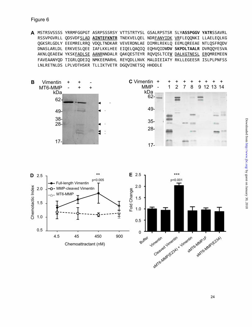
Vimentin functions as a chemoattractant and upon MMP cleavage as an eat-me signal for monocytes—We identified vimentin as one of the highest confidence substrates of MT6-MMP (Table I) by 10 different peptides shown on the vimentin sequence in Figure 6A, three of which were common to both experiments. We characterized this substrate further. By in vitro cleavage assay, we confirmed that vimentin is cleaved not only by MT6-MMP (Fig 6B) but also by seven additional MMPs (Fig 6C). Interestingly, vimentin is displayed on the surface of apoptotic neutrophils (49) and so we reasoned it may act as an “eat-me” signal (4).
To explore this potential role of vimentin and MT6-MMP in neutrophil function, we showed in Transwell migration assays that full-length vimentin is a chemoattractant for THP-1 monocytic cells whereas vimentin cleaved by MT6-MMP (Fig 6D) or MMP12 (not shown) loses chemoattractant activity. The peak chemotactic response was at 450 nM, which is comparable with that of other intracellular proteins having chemoattractant activity (50). In contrast, phagocytosis by differentiated THP-1 cells of microparticles coated with cleaved vimentin cut by MT6-MMP (Fig 6E) or MMP12 (not shown) was increased more than 2-fold.
by guest on January 30, 2018http://w
ww
.jbc.org/D
ownloaded from
MMP
Proteomics of inflammatory substrates of neutrophil MT6-MMP 9
Notably, full-length vimentin showed no activity over controls. Hence, macrophage chemoattraction and phagocytosis of apoptotic neutrophils may be important functions of cell-surface displayed and then neutrophil MT6-MMP-cleaved vimentin. DISCUSSION With just seven substrates reported in the past 13 years since the discovery of MT6-MMP, its function has remained enigmatic. Given the neutrophil-specific expression, we hypothesized that this cell surface MMP would be involved in the proteolytic regulation of specific inflammatory molecules involved in innate immune processes. For the first time, we found that MT6-MMP is inhibited by the predominantly vascular expressed TIMP4 and by using chemokine family-wide screens and proteomics analyses we profiled the active site specificity of MT6-MMP. In identifying 286 cleavage sites by PICS the active site substrate recognition cleft was mapped to extend from P3 to P6’. With 72 new substrates for MT6-MMP discovered, including 14 chemokines that recruit neutrophils and monocytes, the substrate degradome of this MMP has been greatly expanded. Notably, the precise cleavage of the CC and CXC chemokines by MT6-MMP leads to their inactivation or activation consistent with a role in the regulation of innate immune cellular responses. Further, several of the 58 new substrates identified by TAILS are known to contribute to inflammation through actions on leukocyte migration. Indeed, with only 20% of the new substrates identified as being extracellular matrix proteins, rather than being primarily an extracellular matrix degrader, MT6-MMP appears to be mainly involved in bioactive molecule cleavage. Notably, vimentin on the cell surface is a chemoattractant for monocytes and we found that MMP-cleaved vimentin potently increases phagocytosis, suggesting that this contributes to marking apoptotic neutrophils for phagocytic clearance by macrophages.
Neutrophils are amongst the first cells recruited in inflammation. In this early phase of innate immunity, the predominant neutrophil chemoattractant CXCL8 is produced by macrophages, monocytes and neutrophils, and is activated particularly by neutrophil-specific
MMP8 in a feed-forward mechanism (9) and also by MMP9, 12, 13, and 14 (8-10,26), but strangely, not by MT6-MMP. Similarly, a murine functional orthologue mCXCL5/LIX is activated by MMP8 and to a lesser extent by MMP9 (9,10), which only poorly compensates for the lack of MMP8 in the Mmp8 -/- mouse (9). Following neutrophil activation by CXCL8 and interferon-gamma, PMNs express MT6-MMP on the surface and shed the enzyme in an active, soluble form (15,17) that corresponds to the sMT6-MMP construct. We identified that MT6-MMP processes and activates the neutrophil chemoattractants CXCL2 and human and murine CXCL5 to the products CXCL2(5-73), CXCL5(8-78) and mCXCL5(5-92, 10-92), which we and others previously showed have enhanced agonist and chemotactic potential over their full-length counterparts (9,10,39,40,51). These results suggest a discrete role for MT6-MMP in promoting different phase of neutrophil migration. By acting in a positive feedback manner MT6-MMP activation of human and murine CXCL5 and human CXCL2 has potential to amplify the initial neutrophil recruitment driven by MMP-8 activation of CXCL8. Pleiotropic effects are also likely since the MT6-MMP cleavage of CXCL6, which was also observed for MMP1, 2, 3, 7, 8, 9, 12, 13 and 14 (not shown), is predicted to inactivate the chemokine by destabilization of the protein core as truncation occurs C-terminal to both partners of the two disulfide bridges at CXC.
In addition to neutrophil recruitment, MT6-MMP can promote the recruitment of monocytes. Once recruited to tissue, monocyte receptor expression changes from CCR1high (binding CCL15 and CCL23) to CCR2high (CCL2). Both CCL15 and CCL23 are produced by monocytes and macrophages, but are relatively weak CCR1 receptor agonists in their full-length forms. Processing of CCL15 and CCL23 by MT6-MMP results in amino-termini-truncated forms that have recently been shown to exhibit increased agonist activity with increased chemotactic potential (45). This suggests a role for neutrophils, through MT6-MMP, in a positive feed-forward mechanism to progress the inflammatory response through increased recruitment of monocytes expressing CCR1. In contrast, CXCL12 and the chemokines CCL2,
by guest on January 30, 2018http://w
ww
.jbc.org/D
ownloaded from
MMP
Proteomics of inflammatory substrates of neutrophil MT6-MMP 10
CCL7, and CCL13 lose agonist activity or become receptor antagonists following amino-termini truncation (7,8,38,42) as observed by MT6-MMP proteolysis. Overall, this differential processing of chemokines by MT6-MMP may then halt recruited cells—signaling that they have reached the target site, or terminate continued recruitment as one aspect in the dampening of the inflammatory response.
Neutrophil apoptosis is a final critical event in the innate immune response, important for chemotaxis and activation of macrophages (4,5,52,53), during which there is also enhanced cell surface expression of MT6-MMP (15). One of the highest confidence substrates we identified was vimentin, which recent studies have shown has a bona fide extracellular role as a “moonlighting” protein (54). Vimentin is actively secreted by macrophages following TNF-α stimulation (55) and is displayed on the surface of apoptotic neutrophils (49). We found that upon loss of chemoattractant activity, MMP-cleaved vimentin potently promotes phagocytosis by differentiated THP-1 cells. Hence, vimentin displayed on the cell surface of apoptotic neutrophils, and when cleaved by MT6-MMP, may function as an “eat-me” signal for macrophage phagocytosis.
A number of cleaved proteins identified by TAILS are components of cell proliferation and collagen production pathways, and so may be involved in the early phase of wound healing. These include PCPE, peptidyl-prolyl cis-trans isomerase A, and insulin-like growth factor binding protein (IGFBP)-5 and -7, the latter being also biochemically confirmed as a novel substrate. IGFBPs are critical regulators of IGF, sequestering and inhibiting the activity of approximately 99% of circulating IGF (56,57); cleavage and inactivation of IGFBPs promote IGF activity. Unbound IGF binds surface expressed IGF receptor to promote cell growth
(58,59). Hence, the specific cleavage of IGFBP-5 and 7, shown here for the first time by any MMP, suggests a role for MT6-MMP in unmasking IGF that may promote healing.
Finally, a number of ECM-associated proteins were identified as substrates including fibrillin-1, extracellular matrix protein 1, syndecan-4, and types I, II, IV and VI collagens, with galectin-1 and SPARC being also biochemically validated as new MT6-MMP substrates. The latter two are matricellular proteins with multifunctional roles including proinflammatory functions and wound healing (60-62). This highlights the usefulness of unbiased proteomics screens using TAILS to identify protease substrates from even one prime side cleaved peptide.
Thus, through hypothesis-directed and hypothesis-generating approaches, we have discovered a large number of new MT6-MMP substrates that sheds light on the in vivo role of this enzyme and neutrophil function. Notably, the two PMN-specific proteases, MMP8 and MT6-MMP, act in a temporally distinct manner on discrete neutrophil CXC chemokines. This implicates MT6-MMP in the later recruitment of neutrophils through the activation of CXCL2 and CXCL5, and in the progression of the acute inflammatory response toward monocyte recruitment by activation of CCL15 and CCL23. Interstitial roles for MT6-MMP are also suggested wherein monocyte chemoattractants, including vimentin, are cleaved halting haptotaxis, and to then stimulate phagocytosis of apoptotic neutrophils. In view of the biological activities of these new MT6-MMP substrates and cleavage products, MT6-MMP can potentially contribute to the complex regulation needed in inflammation and wound healing that is being explored in ongoing studies.
REFERENCES 1. Borregaard, N., and Cowland, J. B. (1997) Blood 89(10), 3503-3521 2. Serhan, C. N., and Savill, J. (2005) Nat Immunol 6(12), 1191-1197 3. Cox, J. H., Starr, A. E., Kapplehoff, R., Yan, R., Roberts, C. R., and Overall, C. M. (2010)
Arthritis Rheum 62 4. Guzik, K., and Potempa, J. (2008) Biochimie 90(2), 405-415 5. Kennedy, A. D., and DeLeo, F. R. (2009) Immunol Res 43(1-3), 25-61 6. Mortier, A., Gouwy, M., Van Damme, J., and Proost, P. (2010) Exp Cell Res 317(5), 642-654
by guest on January 30, 2018http://w
ww
.jbc.org/D
ownloaded from

MMP
Proteomics of inflammatory substrates of neutrophil MT6-MMP 11
7. McQuibban, G. A., Gong, J. H., Tam, E. M., McCulloch, C. A., Clark-Lewis, I., and Overall, C. M. (2000) Science 289(5482), 1202-1206
8. Dean, R. A., Cox, J. H., Bellac, C. L., Doucet, A., Starr, A. E., and Overall, C. M. (2008) Blood 112(8), 3455-3464
9. Tester, A. M., Cox, J. H., Connor, A. R., Starr, A. E., Dean, R. A., Puente, X. S., Lopez-Otin, C., and Overall, C. M. (2007) PLoS One 2(3), e312
10. Van Den Steen, P. E., Wuyts, A., Husson, S. J., Proost, P., Van Damme, J., and Opdenakker, G. (2003) Eur J Biochem 270(18), 3739-3749
11. Kojima, S., Itoh, Y., Matsumoto, S., Masuho, Y., and Seiki, M. (2000) FEBS Lett 480(2-3), 142-146
12. Pei, D. (1999) Cell Res 9(4), 291-303 13. Matsuda, A., Itoh, Y., Koshikawa, N., Akizawa, T., Yana, I., and Seiki, M. (2003) J Biol Chem
278(38), 36350-36357 14. English, W. R., Velasco, G., Stracke, J. O., Knauper, V., and Murphy, G. (2001) FEBS Lett
491(1-2), 137-142 15. Fortin, C. F., Sohail, A., Sun, Q., McDonald, P. P., Fridman, R., and Fulop, T. (2010) Int Immunol
22(8), 637-649 16. Radichev, I. A., Remacle, A. G., Shiryaev, S. A., Purves, A. N., Johnson, S. L., Pellecchia, M.,
and Strongin, A. Y. (2010) J Biol Chem 285(21), 16076-16086 17. Kang, T., Yi, J., Guo, A., Wang, X., Overall, C. M., Jiang, W., Elde, R., Borregaard, N., and Pei,
D. (2001) J Biol Chem 276(24), 21960-21968 18. Sohail, A., Sun, Q., Zhao, H., Bernardo, M. M., Cho, J. A., and Fridman, R. (2008) Cancer
Metastasis Rev 27(2), 289-302 19. Andolfo, A., English, W. R., Resnati, M., Murphy, G., Blasi, F., and Sidenius, N. (2002) Thromb
Haemost 88(2), 298-306 20. Nie, J., and Pei, D. (2004) Exp Cell Res 296(2), 145-150 21. Shiryaev, S. A., Savinov, A. Y., Cieplak, P., Ratnikov, B. I., Motamedchaboki, K., Smith, J. W.,
and Strongin, A. Y. (2009) PLoS One 4(3), e4952 22. Lopez-Otin, C., and Overall, C. M. (2002) Nat Rev Mol Cell Biol 3(7), 509-519 23. Kleifeld, O., Doucet, A., auf dem Keller, U., Prudova, A., Schilling, O., Kainthan, R. K., Starr, A.
E., Foster, L. J., Kizhakkedathu, J. N., and Overall, C. M. (2010) Nat Biotechnol 28(3), 281-288 24. Prudova, A., auf dem Keller, U., Butler, G. S., and Overall, C. M. (2010) Mol Cell Proteomics
9(5), 894-911 25. Becker-Pauly, C., Barre, O., Schilling, O., Auf dem Keller, U., Ohler, A., Broder, C., Schutte, A.,
Kappelhoff, R., Stocker, W., and Overall, C. M. (2011) Mol Cell Proteomics 10(9), M111 009233 26. Butler, G. S., Tam, E. M., and Overall, C. M. (2004) J Biol Chem 279(15), 15615-15620 27. auf dem Keller, U., Prudova, A., Gioia, M., Butler, G. S., and Overall, C. M. (2010) Mol Cell
Proteomics 9(5), 912-927 28. Clark-Lewis, I., Vo, L., Owen, P., and Anderson, J. (1997) Methods Enzymol 287, 233-250 29. Schilling, O., Huesgen, P. F., Barre, O., Auf dem Keller, U., and Overall, C. M. (2011) Nat Protoc
6(1), 111-120 30. Schilling, O., and Overall, C. M. (2008) Nat Biotechnol 26(6), 685-694 31. Schilling, O., auf dem Keller, U., and Overall, C. M. (2011) Biol Chem 392(11), 1031-1037 32. Colaert, N., Helsens, K., Martens, L., Vandekerckhove, J., and Gevaert, K. (2009) Nat Methods
6(11), 786-787 33. Starr, A. E., and Overall, C. M. (2009) Methods Enzymol 461, 281-307 34. Kleifeld, O., Doucet, A., Prudova, A., auf dem Keller, U., Gioia, M., Kizhakkedathu, J. N., and
Overall, C. M. (2011) Nat Protoc 6(10), 1578-1611 35. Pedrioli, P. G. (2010) Methods Mol Biol 604, 213-238 36. Zhao, H., Sohail, A., Sun, Q., Shi, Q., Kim, S., Mobashery, S., and Fridman, R. (2008) J Biol
Chem 283(50), 35023-35032
by guest on January 30, 2018http://w
ww
.jbc.org/D
ownloaded from
MMP
Proteomics of inflammatory substrates of neutrophil MT6-MMP 12
37. Reinemer, P., Grams, F., Huber, R., Kleine, T., Schnierer, S., Piper, M., Tschesche, H., and Bode, W. (1994) FEBS Lett 338(2), 227-233
38. McQuibban, G. A., Gong, J. H., Wong, J. P., Wallace, J. L., Clark-Lewis, I., and Overall, C. M. (2002) Blood 100(4), 1160-1167
39. Nufer, O., Corbett, M., and Walz, A. (1999) Biochemistry 38(2), 636-642 40. Wuyts, A., D'Haese, A., Cremers, V., Menten, P., Lenaerts, J. P., De Loof, A., Heremans, H.,
Proost, P., and Van Damme, J. (1999) J Immunol 163(11), 6155-6163 41. Cox, J. H., Dean, R. A., Roberts, C. R., and Overall, C. M. (2008) J Biol Chem 283(28), 19389-
19399 42. McQuibban, G. A., Butler, G. S., Gong, J. H., Bendall, L., Power, C., Clark-Lewis, I., and
Overall, C. M. (2001) J Biol Chem 276(47), 43503-43508 43. Zhang, K., McQuibban, G. A., Silva, C., Butler, G. S., Johnston, J. B., Holden, J., Clark-Lewis, I.,
Overall, C. M., and Power, C. (2003) Nat Neurosci 6(10), 1064-1071 44. Laurence, J. S., LiWang, A. C., and LiWang, P. J. (1998) Biochemistry 37(26), 9346-9354 45. Starr, A. E., Dufour, A., Maier, J., and Overall, C. M. (2011) J Biol Chem 46. Dean, R. A., and Overall, C. M. (2007) Mol Cell Proteomics 6(4), 611-623 47. Dean, R. A., Butler, G. S., Hamma-Kourbali, Y., Delbe, J., Brigstock, D. R., Courty, J., and
Overall, C. M. (2007) Mol Cell Biol 27(24), 8454-8465 48. Butler, G. S., and Overall, C. M. (2009) Biochemistry 48(46), 10830-10845 49. Moisan, E., and Girard, D. (2006) J Leukoc Biol 79(3), 489-498 50. Oppenheim, J. J., Dong, H. F., Plotz, P., Caspi, R. R., Dykstra, M., Pierce, S., Martin, R., Carlos,
C., Finn, O., Koul, O., and Howard, O. M. (2005) J Leukoc Biol 77(6), 854-861 51. King, A. G., Johanson, K., Frey, C. L., DeMarsh, P. L., White, J. R., McDevitt, P., McNulty, D.,
Balcarek, J., Jonak, Z. L., Bhatnagar, P. K., and Pelus, L. M. (2000) J Immunol 164(7), 3774-3782 52. Guzik, K., Bzowska, M., Smagur, J., Krupa, O., Sieprawska, M., Travis, J., and Potempa, J.
(2007) Cell Death Differ 14(1), 171-182 53. Parente, L., and Solito, E. (2004) Inflamm Res 53(4), 125-132 54. Butler, G. S., and Overall, C. M. (2009) Nat Rev Drug Discov 8(12), 935-948 55. Mor-Vaknin, N., Punturieri, A., Sitwala, K., and Markovitz, D. M. (2003) Nat Cell Biol 5(1), 59-
63 56. Firth, S. M., and Baxter, R. C. (2002) Endocr Rev 23(6), 824-854 57. Pollak, M. (2008) Nat Rev Cancer 8(12), 915-928 58. Clemmons, D. R., Sleevi, M., Allan, G., and Sommer, A. (2007) J Clin Endocrinol Metab 92(7),
2652-2658 59. Liu, X. J., Malkowski, M., Guo, Y., Erickson, G. F., Shimasaki, S., and Ling, N. (1993)
Endocrinology 132(3), 1176-1183 60. Almkvist, J., and Karlsson, A. (2004) Glycoconj J 19(7-9), 575-581 61. Chlenski, A., and Cohn, S. L. (2010) Semin Cell Dev Biol 21(1), 55-65 62. Malik, R. K., Ghurye, R. R., Lawrence-Watt, D. J., and Stewart, H. J. (2009) Glycobiology
19(12), 1402-1407 FOOTNOTES
A.E.S was supported by the Natural Sciences and Engineering Research Council of Canada, the Michael Smith Foundation for Health Research, and the CIHR Strategic Training Program STP-53877. C.M.O. is a Canada Research Chair in Metalloproteinase Proteomics and Systems Biology, and this research was supported by a Canadian Institutes of Health Research grant and an Infrastructure Grant from the Michael Smith Research Foundation (University of British Columbia Centre for Blood Research) and by the British Columbia Proteomics Network.
The abbreviations used in this paper are: HFL, human fetal lung fibroblast; IGFBP, insulin-
like growth factor binding protein; iTRAQ, isobaric tags for relative and absolute quantitation;
by guest on January 30, 2018http://w
ww
.jbc.org/D
ownloaded from
MMP
Proteomics of inflammatory substrates of neutrophil MT6-MMP 13
MMP, matrix metalloproteinase; MT6-MMP, membrane-type 6 MMP; PICS, proteomic identification of cleavage site specificity; SPARC, secreted protein, acidic and rich in cysteine; TAILS, terminal amine isotopic labeling of substrates; TPP, TransProteomic Pipeline.
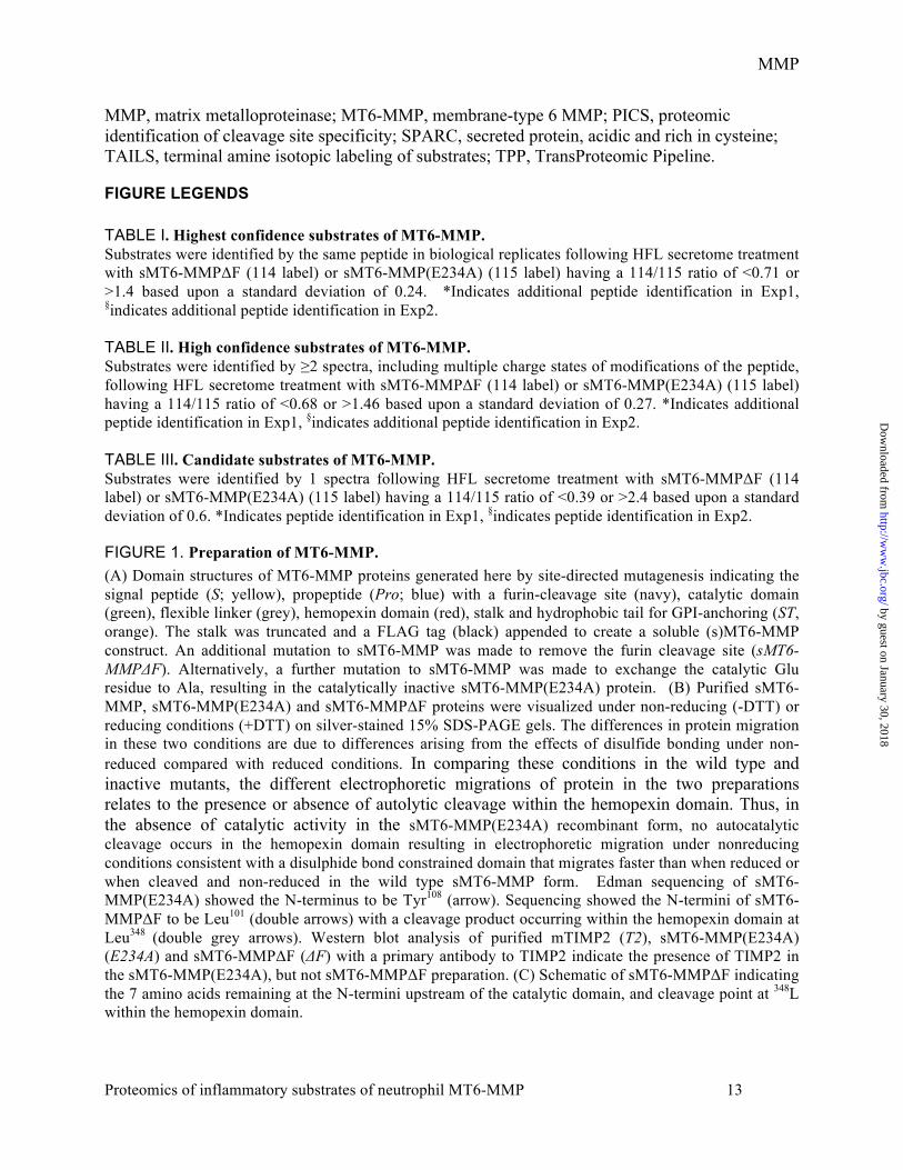
FIGURE LEGENDS TABLE I. Highest confidence substrates of MT6-MMP. Substrates were identified by the same peptide in biological replicates following HFL secretome treatment with sMT6-MMPΔF (114 label) or sMT6-MMP(E234A) (115 label) having a 114/115 ratio of <0.71 or >1.4 based upon a standard deviation of 0.24. *Indicates additional peptide identification in Exp1, §indicates additional peptide identification in Exp2. TABLE II. High confidence substrates of MT6-MMP. Substrates were identified by ≥2 spectra, including multiple charge states of modifications of the peptide, following HFL secretome treatment with sMT6-MMPΔF (114 label) or sMT6-MMP(E234A) (115 label) having a 114/115 ratio of <0.68 or >1.46 based upon a standard deviation of 0.27. *Indicates additional peptide identification in Exp1, §indicates additional peptide identification in Exp2. TABLE III. Candidate substrates of MT6-MMP. Substrates were identified by 1 spectra following HFL secretome treatment with sMT6-MMPΔF (114 label) or sMT6-MMP(E234A) (115 label) having a 114/115 ratio of <0.39 or >2.4 based upon a standard deviation of 0.6. *Indicates peptide identification in Exp1, §indicates peptide identification in Exp2.
FIGURE 1. Preparation of MT6-MMP. (A) Domain structures of MT6-MMP proteins generated here by site-directed mutagenesis indicating the signal peptide (S; yellow), propeptide (Pro; blue) with a furin-cleavage site (navy), catalytic domain (green), flexible linker (grey), hemopexin domain (red), stalk and hydrophobic tail for GPI-anchoring (ST, orange). The stalk was truncated and a FLAG tag (black) appended to create a soluble (s)MT6-MMP construct. An additional mutation to sMT6-MMP was made to remove the furin cleavage site (sMT6-MMPΔF). Alternatively, a further mutation to sMT6-MMP was made to exchange the catalytic Glu residue to Ala, resulting in the catalytically inactive sMT6-MMP(E234A) protein. (B) Purified sMT6-MMP, sMT6-MMP(E234A) and sMT6-MMPΔF proteins were visualized under non-reducing (-DTT) or reducing conditions (+DTT) on silver-stained 15% SDS-PAGE gels. The differences in protein migration in these two conditions are due to differences arising from the effects of disulfide bonding under non-reduced compared with reduced conditions. In comparing these conditions in the wild type and inactive mutants, the different electrophoretic migrations of protein in the two preparations relates to the presence or absence of autolytic cleavage within the hemopexin domain. Thus, in the absence of catalytic activity in the sMT6-MMP(E234A) recombinant form, no autocatalytic cleavage occurs in the hemopexin domain resulting in electrophoretic migration under nonreducing conditions consistent with a disulphide bond constrained domain that migrates faster than when reduced or when cleaved and non-reduced in the wild type sMT6-MMP form. Edman sequencing of sMT6-MMP(E234A) showed the N-terminus to be Tyr108 (arrow). Sequencing showed the N-termini of sMT6-MMPΔF to be Leu101 (double arrows) with a cleavage product occurring within the hemopexin domain at Leu348 (double grey arrows). Western blot analysis of purified mTIMP2 (T2), sMT6-MMP(E234A) (E234A) and sMT6-MMPΔF (ΔF) with a primary antibody to TIMP2 indicate the presence of TIMP2 in the sMT6-MMP(E234A), but not sMT6-MMPΔF preparation. (C) Schematic of sMT6-MMPΔF indicating the 7 amino acids remaining at the N-termini upstream of the catalytic domain, and cleavage point at 348L within the hemopexin domain.
by guest on January 30, 2018http://w
ww
.jbc.org/D
ownloaded from

MMP
Proteomics of inflammatory substrates of neutrophil MT6-MMP 14
FIGURE 2. Characterization of MT6-MMP protein. (A) Gelatinase activity of sMT6-MMPΔF was evaluated by gelatin zymography. 10 ng of MMP-9 resulted in a clear band (arrow) whereas 100 and 1,000 ng of sMT6-MMPΔF results in only a slight clearance (arrowhead), indicating weak gelatinolytic activity (n=2). (B) Quenched fluorescein-conjugated gelatin processing by sMT6-MMPΔF compared with MMP-2 processing was monitored at excitation/emission of 485/530 (n=2). (C) Casein processing by sMT6-MMPΔF was evaluated by zymography. 10 ng of MMP-7 resulted in a clear band (arrow) whereas 100 and 1,000 ng of sMT6-MMPΔF results in only a slight clearance (arrowhead), indicating that casein is not a good substrate for MT6-MMP (n=2). (D) The rate of cleavage of QF24 substrate by increasing concentrations of MMPs were compared by measurement of the increase in fluorescence at excitation/emission of 320/405 (n=2). (E) Heat map of the amino acid occurrence at positions P6 to P6’ of the 286 peptides analyzed and identified by MS/MS following PICS analysis of sMT6-MMPΔF cleavage of a tryptic library. (F) IceLogo analysis of the PICS data, adjusting for relative abundance of the amino acids in the homo sapien proteome, indicating the relative occurrence of amino acids at positions P6 to P6’ from the 286 peptides analyzed and identified. (G) Inhibition of 3 nM sMT6-MMPΔF processing of 1.0 µM QF24 by TIMP1, -2, and -4, and by marimastat was evaluated by measurement of fluorescence at excitation/emission of 320/405 nm and the Ki calculated in Prism (GraphPad) (n=3).
FIGURE 3. MT6-MMP selectively processes both CXC and CC chemokines. Human or mouse (m) chemokines (1 µg) were incubated at 37 ºC for 16 h with recombinant sMT6-MMPΔF in a 10 µl reaction at an enzyme to substrate ratio of 1:10 (w/w). Cleavage assay products of (A) CXCL or (B) CCL chemokines were visualized on silver-stained 15% Tris-Tricine gels. Cleavages of the chemokines listed were not detectable by MALDI-TOF following incubation at a 1:20 molar ratio with sMT6-MMPΔF at 37 ºC for 16 h (n>2). (C) Cleavage products were assigned by MALDI-TOF mass spectrometry by comparison of measured with predicted mass to charge ratios (m/z) with +1 charge ionization ([M + H]+). Deconvoluted cleavage sites are indicated by an arrow in the corresponding sequence.
FIGURE 4. Peptide and substrate identification by TAILS analysis. (A) Schematic overview of the method of the TAILS analysis of conditioned medium from cells treated with sMT6-MMPΔF or sMT6-MMP(E234A). (B) Four-way Venn diagram of unique peptide identifications, with ≥95% confidence, by Mascot and X !Tandem in experiments (Exp)1 and Exp2. (C) Two-way Venn diagram of unique peptides that have reached the experimental cutoff for that dataset identified in Exp1, Exp2, or in both experiments.
FIGURE 5. MT6-MMP cleaves cystatin C, IGFBP7, galectin-1 and SPARC. Candidate substrates (1 µg) (A) cystatin C, (B) IGFBP7, (C) galectin-1, and (D) SPARC were incubated in a 10 µl reaction at an enzyme to substrate of 1:10 (w/w) with sMT6-MMPΔF (MT6) for 16 h at 37°C. Cleavage assay products were visualized under reducing conditions by silver-stained SDS-PAGE. Arrow indicates full-length protein; broken arrow indicates cleavage product in the presence of MT6-MMP. FIGURE 6. MMP-processed vimentin has increased chemotactic and phagocytic potential for THP-1 cells. (A) Protein sequence of vimentin. Underlined or bold text indicates peptides identified with increased or decreased iTRAQ-ratio by TAILS analysis respectively. (B) Vimentin (1 µg) was incubated with recombinant sMT6-MMPΔF at an enzyme to substrate ratio of 1:10 (w/w) in a 10 µl reaction volume at 37 ºC for 16 h. Cleavage assay products were visualized by silver-stained 15% SDS-PAGE. Arrow indicates full-length vimentin; broken arrow indicates cleavage product in the presence of MT6-MMP (n=2). Comparable preparations were used for migration and phagocytosis assays. (C) Vimentin was incubated in the presence of MMP1, 2, 7, 8, 9, 12, 13, and 14 for 16 h at 37°C. Cleavage assay products were visualized by silver-stained 15% SDS-PAGE. (D) Transwell migration of THP-1 cells for 90 min
by guest on January 30, 2018http://w
ww
.jbc.org/D
ownloaded from

MMP
Proteomics of inflammatory substrates of neutrophil MT6-MMP 15
towards full-length or sMT6-MMPΔF-cleaved vimentin or an equivalent concentration of sMT6-MMPΔF through a 5 µm pore-sized filter. Migrated cells were quantified by CyQUANT assay and displayed as chemotactic index, defined as the ratio of cells migrating in response to stimulus compared with buffer control. Results shown are the mean +/- SEM of 2 replicates of experiments completed in quadruplicate. (E) Vimentin was incubated in the presence of sMT6-MMPΔF (Cleaved vimentin), sMT6-MMP(E234A) or buffer to allow for processing prior to incubation and coating of Fluoresbrite® microbeads. Fluorescence of THP-1 cells was evaluated by FACS following incubation of cells with Fluoresbrite® microbeads, previously coated with full-length or MMP-processed vimentin. Increased fluorescence indicates increased phagocytosis of coated microparticles. Results shown are the mean +/- SD of 3 replicates of experiments completed in duplicate. *, Statistical significance of cleaved versus full-length vimentin was evaluated by t-test as indicated.
by guest on January 30, 2018http://w
ww
.jbc.org/D
ownloaded from

MMP
Proteomics of inflammatory substrates of neutrophil MT6-MMP 16
Table 1
Protein Ratio 114/115
Peptide
Actin 0.64 22 GFAGDDAPR 2.22 31 AVFPSIVGR 11.14 107 LTEAPLNPKANR 4.41 300 VLSGGTTMYPGIADR 4.53 8 LVVDNGSGMCKAGFAGDDAPR* 26.19 45 VMVGMGQKDSYVGDEAQSKR§ 21.62 54 SYVGDEAQSKR§ Collagen type I α-1 chain 10.29 1001 GPPGLAGPPGESGR 31.73 977 GPSGEPGKQGPSGASGER§ 10.98 1004 GLAGPPGESGR§ 36.17 1005 LAGPPGESGR§ 42.57 1006 AGPPGESGR§ 12.67 1050 APGAPGPVGPAGKSGDR§ 12.27 1052 GAPGPVGPAGKSGDR§ 0.36 1075 AGPVGPVGAR§ 6.55 1237 SLSQQIENIR§ 23.76 1239 SQQIENIR§ Collagen type I α-2 chain 1.42 868 GAPGILGLPGSR 2.33 333 VGAAGATGAR 4.68 1141 SLNNQIETLLTPEGSR 17.94 113 FQGPAGEPGEPGQTGPAGAR§ 14.84 115 GPAGEPGEPGQTGPAGAR§ 0.65 121 GEPGQTGPAGAR* 20.15 331 GPVGAAGATGAR§ 48.93 370 GPPGPSGEEGKR§ 1.48 385 GEAGSAGPPGPPGLR* 43.99 559 GPSGPAGEVGKPGER§ 7.94 784 GMTGFPGAAGR§ 23.30 808 GPPGPAGKEGLR§ 3.10 868 GAPGILGLPGSR§ 24.51 967 GPVGPAGKHGNR§ 38.25 1103 VSGGGYDFGYDGDFYR§ 10.94 1109 DFGYDGDFYR§ 14.89 1110 FGYDGDFYR§ 29.73 1111 GYDGDFYR§ 27.86 1112 YDGDFYR§ 20.83 1144 NQIETLLTPEGSR§ 39.22 1142 LNNQIETLLTPEGSR§ 25.98 1143 NNQIETLLTPEGSR§ 42.37 1146 IETLLTPEGSR§ 22.07 1149 LLTPEGSR§ Cystatin C 5.54 35 LVGGPMDASVEEEGVRR 26.71 35 LVGGPMDASVEEEGVR 26.35 36 VGGPMDASVEEEGVR§ 7.48 37 GGPMDASVEEEGVR§
by guest on January 30, 2018http://w
ww
.jbc.org/D
ownloaded from

MMP
Proteomics of inflammatory substrates of neutrophil MT6-MMP 17
Protein Ratio 114/115
Peptide
31.22 42 ASVEEEGVR§ IGFBP-5 0.63 164 KFVGGAENTAHPR 2.97 213 AVYLPNCDR 0.18 165 FVGGAENTAHPR§ 0.29 166 VGGAENTAHPR* IGFBP-7 1.46 98 AGAAAGGPGVSGVCVCKSR 2.42 98 AGAAAGGPGVSGVCVCKSR§ 6.72 99 GAAAGGPGVSGVCVCKSR§ 32.03 100 AAAGGPGVSGVCVCKSR§ MT6-MMP 8.38 318 EGNFDAIANIR 36.59 29 VSLGVDWLTR* 8.67 254 FYQGPVGDPDKYR* 4.41 255 YQGPVGDPDKYR* 23.62 359 FWEGLPAQVR§ 8.73 386 SGPQFWVFQDR* 17.25 473 GDTYFFKGAHYWR* Transgelin 0.66 2 ANKGPSYGMSR Tropomyosin α-4 chain 3.56 133 LVILEGELER 0.68 16 ALQQQADEAEDR* 19.62 115 AKHIAEEADR§ 15.24 134 VILEGELER§ Vimentin 0.58 55 SSPGGVYATR 2.05 87 SLADAINTEFKNTR 1.55 88 LADAINTEFKNTR 0.30 54 ASSPGGVYATR§ 0.65 91 AINTEFKNTR* 35.77 114 FANYIDKVR§ 0.39 258 VDVSKPDLTAALR§ 8.83 295 FADLSEAANR§ 2.69 296 ADLSEAANR§ 4.21 330 VDALKGTNESLER§ YIPF3 0.71 321 DIPAMLPAAR 26S protease regulatory subunit 8 1.82 2 ALDGPEQMELEEGKAGSGLR
by guest on January 30, 2018http://w
ww
.jbc.org/D
ownloaded from

MMP
Proteomics of inflammatory substrates of neutrophil MT6-MMP 18
Table II
Protein Ratio 114/115
Peptide No. of Spectra
Α-actinin-1 24.56 13 MQPEEDWDR§ 2 Collagen type II α-1 chain 43.76 576 GPAGPAGER§ 2 8.59 825 GATGFPGAAGR§ 2 Collagen type IV α-2 chain 6.84 76 GLQGFPGLQGR§ 1
20.66 461 FPGLPGSPGAR§ 1 32.24 651 GPAGTPGQIDCDTDVKR§ 1
5.81 655 TPGQIDCDTDVKR§ 1 Collagen type VI α-1 chain 1.71 443 GDPGEAGPQGDQGR* 1
16.85 497 GPPGDPGLMGER§ 2 7.81 500 GDPGLMGER§ 1
Collagen type VI α-3 chain 2.96 3106 LTETDICKLPKDEGTCR§ 1 Dickkopf-related protein 3 33.89 131 TVITSVGDEEGR§ 2
15.46 54 VEELMEDTQHKLR§ 1 51.66 252 ITWELEPDGALDR§ 1
Extracellular matrix protein 1 0.15 20 ASEGGFTATGQR§ 3 Fibrillin-1 0.37 25 ADANLEAGNVKETR§ 2 Latent-transforming growth factor beta-binding protein 2 1.48 1729 FEGLQAEECGILNGCENGR* 1 5.36 249 SSAAGEGTLAR§ 1 MMP-1 4.98 328 LEAAYEFADR* 2
7.30 364 IYSSFGFPR* 1 Peptidyl-prolyl cis-trans isomerase A 7.88 9 DIAVDGEPLGR§ 3
8.52 8 FDIAVDGEPLGR§ 1 Procollagen C-endopeptidase enhancer 1 37.52 309 SPSAPDAPTCPKQCR§ 2 Protein-lysine 6-oxidase 0.61 59 SLGSQYQPQR* 2
4.75 57 LLSLGSQYQPQR* 2 0.39 22 APPAAGQQQPPR§ 1 2.12 58 LSLGSQYQPQR* 1
Serpin H1 0.23 139 SVSFADDFVR* 2 Sulfhydryl oxidase 1 42.01 565 AMGALELESR§ 7 32.69 566 MGALELESR§ 2 Stromal cell derived factor 4 12.27 84 GKDLGGFDEDAEPR§ 1 7.94 88 GGFDEDAEPR§ 1 Syndecan-4 7.00 24 TEVIDPQDLLEGR§ 2
7.49 26 VIDPQDLLEGR§ 1 Tropomyosin 3 6.84 170 LVIIEGDLER§ 1
38.06 171 VIIEGDLER§ 2 Tropomyosin beta chain 41.62 22 AEQAEADKKQAEDR§ 2
48.86 24 QAEADKKQAEDR§ 1 17.45 74 AEKKATDAEADVASLNR§ 1 23.20 151 AKHIAEDSDR§ 1
by guest on January 30, 2018http://w
ww
.jbc.org/D
ownloaded from

MMP
Proteomics of inflammatory substrates of neutrophil MT6-MMP 19
Table III Protein Ratio
114/115 Peptide
α2-macroglobulin 8.24 708 YESDVMGR* Calumenin precursor 10.54 53 LGAEEAKTFDQLTPEESKER§ Cellular nucleic acid-binding protein 50.29 45 FVSSSLPDICYR§ Dihydrolipoyllysine-residue succinyltransferase 0.66 51 DDLVTVKTPAFAESVTEGDVR* Elongation factor 1-delta 2.58 60 SLAGSSGPGASSGTSGDHGELVVR§ Fibulin-1 36.94 170 EQEDPYLNDR§ Filamin-C 4.27 2304 FTVGPLGEGGAHKVR§ Galectin-1 3.22 35 LGKDSNNLCLHFNPR§ Galectin-3-binding protein 9.49 447 FQAPSDYR§ Gelsolin precursor 1.47 404 GLGLSYLSSHIANVER* Glyceraldehyde-3-phosphate dehydrogenase 6.84 4 VKVGVNGFGR* Nestin 5.54 1397 LLDPAAWDR* Polymerase I and transcript release factor 11.70 344 VGADDDEGGAER§ Protein disulfide-isomerase A6 precursor 0.45 72 LYSSSDDVIELTPSNFNR* Protein-lysine 6-oxidase 0.25 56 SLLSLGSQYQPQR§ Sodium-dependent phosphate transport protein 51.66 319 ISSVLQANLR§ SPARC 0.63 156 LDSELTEFPLR* Spondin-2 25.78 251 FIPPAPVLPSR§ Sulfhydryl oxidase 1 precursor 51.66 34 ALYSPSDPLTLLQADTVR§ Transcription elongation factor SPT4 0.65 2 ALETVPKDLR* Tubulin α-4A chain 6.46 92 LITGKEDAANNYAR* VEGF C 1.52 112 AHYNTEILKSIDNEWR* V-type proton ATPase subunit G 1 2.03 2 ASQSQGIQQLLQAEKR* WD repeat-containing protein 1 6.84 8 VFASLPQVER§ 40S ribosomal protein SA 4.33 90 FAAATGATPIAGR§ 5,6-dihydroxyindole-2-carboxylic acid oxidase 21.06 425 FPLENAPIGHNR§ 60S ribosomal protein L26-like 1 0.57 1 MKFNPFVTSDR*
by guest on January 30, 2018http://w
ww
.jbc.org/D
ownloaded from

1 21 107 280 316 508 562 S Pro Catalytic Hemopexin Link ST
A
sMT6-MMP 512 DYKDDDDK523
Furin
HEFGHA
Figure 1 C
S
S
348
108 101
66- 45- 35-
25-
kDa FL
AG
B
69- 49-
38-
28-
kDa
sMT6-MMP
DTT - + - + sMT6-MMP∆F
101LVGAG
348LVSPR
sMT6-MMP(E234A)
- +
108YALSG
MT6-MMP
T2 E234A ∆F
αTIMP2
19
69- 49-
38-
28-
kDa 69- 49-
38-
28-
kDa
SSG
YALSG LVGAG
LVSPR
101
sMT6-MMPΔF
GAGAG 108
HEFGHA
YALSG LV
230
sMT6-MMP(E234A)
A FGHA238
HAFGHA
VAVH
by guest on January 30, 2018http://w
ww
.jbc.org/D
ownloaded from

Figure 2
MMP-9 (ng) sMT6-MMP∆F (ng)
10 1000 -
- - 100 A
Time (min)
B
RFU
4
3
2
1 0 15 30 45 60 75 90
MMP-2 (5 pmol)
sMT6-MMP∆F (50 pmol) sMT6-MMP∆F (5 pmol)
MMP-7 (ng) 10 100 1000 sMT6-MMP∆F (ng)
- -
69
35
25
- C
RFU
TIMP1 (nM)
Marimastat (M) TIMP4 (nM)
TIMP2 (nM)
KI = 0.1687 nM KI=0.526 nM
KI <0.1 nM KI=19.36 nM
G
RFU
sMT1-MMP
sMT6-MMP
sMT6-MMP∆F R
FU
D
20
E P1 P2 P3 P4 P5 P6 P6’ P5’ P4’ P3’ P2’ P1’ R
elative occurrence in %
A C D E G H I K
F
L M N P R S T V
Q
W Y
45
40
35
30
25
20
15
10
Am
ino
Aci
d
F
50
25
-25
-50 P va
lue
= 0.
05 %
diff
eren
ce
P1’ P2’ P3’ P4’ P5’ P6’ P1 P2 P3 P4 P6 P5
Time (min)
by guest on January 30, 2018http://w
ww
.jbc.org/D
ownloaded from

Chemokine
m/z [M+H]+
Sequence Predicted Measured
CXCL2 (1-73) 7893 7893 1 10 APLA↓TELRCQC… CXCL2 (5-73) 7541 7523
CXCL5 (1-78) 8407 8398 1 10 AGPAAAV↓LRELRCVCL… CXCL5 (8-78) 7869 7865
mCXCL5 (1-92) 9853 9843 1 10 APSS↓VIAAT↓ELRCVC… mCXCL5 (5-92) 9511 9501
mCXCL5 (10-92)
9056 9050
CXCL6 (1-77) 8317 8310 1 10 20 30 GPSVSAVLTELRCTCLRVTLRVNPKTIGK↓LQVFP… CXCL6 (29-77) 5309 5301
CXCL9 (1-103) 11721 11728 1 10 90 100 TPVVRKGRCSC….LLLVLK↓VRKSQRSQKKTT CXCL9 (1-90) 10136 10140
CXCL12α (1-67) 7835 7832 1 10 KPVS↓LSYRCPC… CXCL12α (5-67) 7424 7421
CXCL12β (1-72) 8526 8522 1 10 KPVS↓LSYRCPC… CXCL12β (5-72) 8116 8114
C
CCL2 (1-76) 8685 8667 1 10 QPDA↓INAPVTCC… CCL2 (5-76) 8275 8257
CCL4 (1-69) 7819 7822 1 10 APMGSD↓PPTACC… CCL4 (7-69) 7260 7261
CCL7 (1-76) 8957 8956 1 10 QRVG↓INTSTTCC… CCL7 (5-76) 8576 8556
CCL13 (1-75) 8581 8583 1 10 70 QPDA↓LNVPSTCC…QNYMKHLGRK↓AHT CCL13 (1-72) 8239 8240
CCL13 (5-72) 7845 7844 CCL15 (1-92) 10157 10154 1 10 20 30
QFINDAETELMMSKLPLENPVVLN↓SF↓HFAADCC… CCL15 (25-92) 7447 7447 CCL15 (27-92)
7212 7194
CCL16 (1-97) 11202 11186 1 10 QPKV↓PEWVNTPSTCC… CCL16 (5-97) 10750 10741
CCL23 (1-99) 11367 11367 1 10 20 30 RVTKDAETEF↓MMSKLPLENP↓VLLDR↓FHATSADCC… CCL23 (11-99) 10191 10187
CCL23 (21-99) 9049 9046 CCL23 (26-99) 8452 8449
Figure 3
- + CCL2 CCL7 - + - +
14 -
6 -
B - + - +
CCL15 - +
CCL16 CCL23 CCL4 CCL13 - +
A CXCL12α hCXCL5 mCXCL5 - + - + - + - +
14 -
6 -
- CXCL2 - +
CXCL9 - +
CXCL6 + sMT6-MMP∆F
CXCL12β
21
sMT6-MMP∆F
CXCL1 CXCL3 CXCL8 CXCL10 CXCL11 CCL1 CCL3 CCL11 CCL14 CCL17
Not Cleaved mCXCL1 mCXCL2
by guest on January 30, 2018http://w
ww
.jbc.org/D
ownloaded from

Prot
eom
e P
repa
ratio
n iT
RA
Q-T
AIL
S B
ioifo
mat
ics
and
Ana
lysi
s
Cells + sMT6-MMP(E234A)
Collect conditioned medium
iTRAQ label 114
Combine
Precipitate
In solution tryptic digest
N-terminal enrichment (Polymer pullout)
SCX Fractionation
LC MS/MS
Mascot Search X!Tandem Search
TransProteomic Pipeline
Ratio and statistical analysis by CLIPPER
iTRAQ label 115
Figure 4
22
A Cells + sMT6-MMPΔF
Collect conditioned medium
B
C
219 89 112
Exp2 Exp1
Exp1-Mascot Exp2-Mascot
Exp2-X!Tandem Exp1-X!Tandem
Numbers of unique high confidence peptides identified
Numbers of unique high confidence peptides identified
by guest on January 30, 2018http://w
ww
.jbc.org/D
ownloaded from

Figure 5
A
49-
14-
6-
28-
sMT6-MMP∆F - Cystatin C
+ + - + +
kDa
B sMT6-MMP∆F -
IGFBP7 + +
- + +
22-
kDa
36- 50- 64-
C
23
D
sMT6-MMP(E234A) sMT6-MMP∆F
SPARC + -
++ +
kDa
28-
38-
49-
17-
+ - - +
+ -
- - -
-
sMT6-MMP∆F - Galectin-1
+ + - + +
kDa
6-
17-
28-
3-
38-
by guest on January 30, 2018http://w
ww
.jbc.org/D
ownloaded from
MSTRSVSSSS YRRMFGGPGT ASRPSSSRSY VTTSTRTYSL GSALRPSTSR SLYASSPGGV YATRSSAVRL RSSVPGVRLL QDSVDFSLAD AINTEFKNTR TNEKVELQEL NDRFANYIDK VRFLEQQNKI LLAELEQLKG QGKSRLGDLY EEEMRELRRQ VDQLTNDKAR VEVERDNLAE DIMRLREKLQ EEMLQREEAE NTLQSFRQDV DNASLARLDL ERKVESLQEE IAFLKKLHEE EIQELQAQIQ EQHVQIDVDV SKPDLTAALR DVRQQYESVA AKNLQEAEEW YKSKFADLSE AANRNNDALR QAKQESTEYR RQVQSLTCEV DALKGTNESL ERQMREMEEN FAVEAANYQD TIGRLQDEIQ NMKEEMARHL REYQDLLNVK MALDIEIATY RKLLEGEESR ISLPLPNFSS LNLRETNLDS LPLVDTHSKR TLLIKTVETR DGQVINETSQ HHDDLE
A
Vimentin + MT6-MMP
+ + + -
-
49-
28-
17-
38-
kDa 62-
B
Figure 6
4.5 45 450 900
Chemoattractant (nM)
Full-length Vimentin MMP-cleaved Vimentin
D
24
0.5
1.0
1.5
0
*** p<0.001
2.0
2.5
Fold
Cha
nge
C MMP - 1 2 7 8 9 12 13 14
+ + + + + + + + +
49-
35- 28-
17-
kDa
Vimentin
62-
E ** p<0.005
0.5
1.0
1.5
2.0
2.5
Che
mot
actic
Inde
x
MT6-MMP
by guest on January 30, 2018http://w
ww
.jbc.org/D
ownloaded from

OverallAmanda E. Starr, Caroline L. Bellac, Antoine Dufour, Verena Goebeler and Christopher M.
cleavages enhance cell migration and macrophase phagocytic activitiesmembrane-type 6 matrix metalloprotease (MMP25): chemokine and vimentin
Biochemical characterization and N-terminomics analysis of leukolysin, the
published online February 24, 2012J. Biol. Chem.
10.1074/jbc.M111.314179Access the most updated version of this article at doi:
Alerts:
When a correction for this article is posted•
When this article is cited•
to choose from all of JBC's e-mail alertsClick here
Supplemental material:
http://www.jbc.org/content/suppl/2012/02/24/M111.314179.DC1
by guest on January 30, 2018http://w
ww
.jbc.org/D
ownloaded from