biochemical and functional characterization of
TRANSCRIPT
Accepted Manuscript
Biochemical and functional characterization of glycosaminoglycans releasedfrom degranulating rat peritoneal mast cells: Insights into the physiological role ofendogenous heparin
Rebecca Lever, Amir Smailbegovic, Yanira Riffo-Vasquez, Elaine Gray, JohnHogwood, Stephen M. Francis, Neville V. Richardson, Clive P. Page, Barbara Mulloy
PII: S1094-5539(16)30146-8
DOI: 10.1016/j.pupt.2016.11.002
Reference: YPUPT 1568
To appear in: Pulmonary Pharmacology & Therapeutics
Received Date: 8 June 2016
Revised Date: 1 October 2016
Accepted Date: 1 November 2016
Please cite this article as: Lever R, Smailbegovic A, Riffo-Vasquez Y, Gray E, Hogwood J, Francis SM,Richardson NV, Page CP, Mulloy B, Biochemical and functional characterization of glycosaminoglycansreleased from degranulating rat peritoneal mast cells: Insights into the physiological role of endogenousheparin, Pulmonary Pharmacology & Therapeutics (2016), doi: 10.1016/j.pupt.2016.11.002.
This is a PDF file of an unedited manuscript that has been accepted for publication. As a service toour customers we are providing this early version of the manuscript. The manuscript will undergocopyediting, typesetting, and review of the resulting proof before it is published in its final form. Pleasenote that during the production process errors may be discovered which could affect the content, and alllegal disclaimers that apply to the journal pertain.
MANUSCRIP
T
ACCEPTED
ACCEPTED MANUSCRIPT Characterization of heparin released from mast cells
1
Biochemical and functional characterization of glycosaminoglycans released from degranulating rat peritoneal mast cells: Insights into the physiological role of endogenous heparin
1Rebecca Lever, 1,2Amir Smailbegovic, 2Yanira Riffo-Vasquez, 2,3Elaine Gray, 3John Hogwood,
4Stephen M. Francis, 4Neville V. Richardson, 2Clive P. Page, and 2,3Barbara Mulloy
1UCL School of Pharmacy, Brunswick Square, London, WC1N 1AX, UK
2Sackler Institute of Pulmonary Pharmacology, Institute of Pharmaceutical Science, King's College London, London, SE1 9NH, UK
3National Institute for Biological Standards and Control, Potters Bar, Hertfordshire, UK
4School of Chemistry, University of St. Andrews, St. Andrews, KY16 9ST, UK
*Running title: Characterization of heparin released from mast cells
To whom correspondence should be addressed: Rebecca Lever, UCL School of Pharmacy Keywords: Heparin, mast cells, dermatan sulphate, glycosaminoglycan
MANUSCRIP
T
ACCEPTED
ACCEPTED MANUSCRIPT Characterization of heparin released from mast cells
2
ABSTRACT The properties of commercially prepared heparin as an anticoagulant and antithrombotic agent in
medicine are better understood than is the physiological role of heparin in its native form, where it is
uniquely found in the secretory granules of mast cells. In the present study we have isolated and
characterised the glycosaminoglycans (GAGs) released from degranulating rat peritoneal mast cells.
Analysis of the GAGs by NMR spectroscopy showed the presence of both heparin and the
galactosaminoglycan dermatan sulphate; heparinase digestion profiles and measurements of anticoagulant
activity were consistent with this finding. The rat peritoneal mast cell GAGs significantly inhibited
accumulation of leukocytes in the rat peritoneal cavity in response to IL-1β (p<0.05, n=6/group), and
inhibited adhesion and diapedesis of leukocytes in the inflamed rat cremasteric microcirculation in
response to LPS (p<0.001, n=4/group). FTIR spectra of human umbilical vein endothelial cells
(HUVECs) were altered by treatment of the cells with heparin degrading enzymes, and restored by the
addition of exogenous heparin. In conclusion, we have shown that rat peritoneal mast cells contain a
mixture of GAGs that possess anticoagulant and anti-inflammatory properties.

MANUSCRIP
T
ACCEPTED
ACCEPTED MANUSCRIPT Characterization of heparin released from mast cells
3
INTRODUCTION
The glycosaminoglycan (GAG) heparin was discovered a century ago and, as an anticoagulant drug, ranks
as one of the most commonly used agents in modern medicine (1). Whilst much is now known about the
nature of commercially prepared pharmaceutical heparin, both in its unfractionated and low-molecular
weight forms, with respect to structure, biological activity and clinical effects (2-4), the physiological role
of endogenous heparin is considerably less well understood. It has long been known, however, that
heparin possesses additional effects that are both separate to, and separable from, its well-characterized
effects on blood coagulation, many of which involve modulation of aspects of immune or inflammatory
cell function (5,6). In contrast to the closely related GAG heparan sulphate, the ubiquitous expression of
which alone goes some way towards explaining its pivotal role in normal physiology (7,8), mammalian
heparin is produced exclusively by mast cells. In this respect, heparin has been suggested to be primarily
important for the storage of histamine and certain pro-inflammatory granule proteins within the mast cell
(9,10). However, it would seem unlikely that a potent anticoagulant molecule having a broad range of
biological activities (11) should be biosynthesized solely for this purpose, and indeed solely within a cell
type found outside the vasculature. The localization of mast cells close to vessels of the microcirculation
though, as well as their more recent description in pathological tissue sites including tumors and
atheromatous plaques (12,13), suggests that endogenous heparin may be important in regulation of
pathophysiological responses, as well as in normal physiology. It has been suggested that heparin,
released from activated mast cells, may be involved in physiological regulation of inflammation (14)
through the binding and neutralization of cytotoxic and pro-inflammatory proteins, thus limiting the
extent of the inflammatory response and potential tissue damage and remodelling as part of homeostasis.
Many of the non-anticoagulant actions of heparin are mediated through interactions with proteins
such as chemokines and growth factors, which often depend upon the binding of heparan sulphate for full
activity (15-17). Whilst the structural basis of the anticoagulant activity of heparin is well understood
(reviewed in 11), the exact structural requirements for the majority of the anti-inflammatory effects of
heparin remain to be fully determined. The ability of heparin to interact with a wide variety of proteins
can vary from strongly sequence specific, such as the binding of antithrombin, to relatively non-specific,
in part due to the size and polyanionic nature of the molecule (18,19). In this regard, it is important to
consider that commercially-available heparin, which is usually extracted from porcine intestinal mucosa,
is standardized only for its anticoagulant activity, which depends heavily on the presence of the high
affinity antithrombin-binding pentasaccharide (18). Therefore, any biological activity confined to other
polysaccharide sequences contained within the heparin structure may not necessarily correlate with the
total amount of material present in the resultant heterogenous mixture, and may even be fractionated out
by the current techniques for preparing heparin commercially as an anticoagulant. A greater
MANUSCRIP
T
ACCEPTED
ACCEPTED MANUSCRIPT Characterization of heparin released from mast cells
4
understanding of the nature of the GAGs present in mast cells may elucidate the physiological role(s) of
endogenous heparin and potentially facilitate the design of drugs to mimic specific biological effects of
heparin other than anticoagulant activities.
In the present study, therefore, we have sought to examine the nature of GAGs released from
peritoneal mast cells of the rat as a product of their degranulation.
EXPERIMENTAL PROCEDURES
Animals- Male Sprague-Dawley rats (200-250 g; Harlan, UK) were housed in an animal unit on a 12:12 h
light:dark cycle, with access to standard laboratory chow and water ad libitum, for at least seven days
prior to experimentation. All experiments were performed in accordance with local Ethical and UK
Home Office approval and guidelines.
Isolation of rat mast cell GAGs (RMCG)- Rats were euthanized by CO2 exposure and their peritoneal
cavities immediately lavaged with 20 mL normal saline containing 0.05 mM EDTA. Recovered cells
were washed in modified HBSS (Ca2+/Mg2+ free) at 250 x g for 2 minutes, followed by density-dependent
centrifugation to separate mast cells from mononuclear cells. Mast cell pellets were re-suspended in 5
mL buffer (PBS containing 0.1 mg mL-1 HSA and 5.6 mM glucose) and incubated for 10 minutes at 37o C
prior to addition of a further 5 mL buffer containing 5 µg mL-1 compound 48/80 and incubation for a
further 20 minutes at 37o C. Gross cellular material was removed by centrifugation at 150 x g for 10
minutes and discarded. Supernatants were then transferred to 1 mL micro-centrifuge tubes and
centrifuged at 10,000 rpm for 15 minutes to sediment intact granules. Supernatants (A) were collected
and transferred to a refrigerator and pelleted granules were re-suspended in 1 mL 2 M NaCl by vortexing,
then incubated at room temperature for 30 minutes to facilitate the release of granule contents. The
suspension was again centrifuged at 10,000 rpm for 15 minutes, supernatants (B) were collected and
dialysed overnight against five changes of 1.5 L dH2O and pellets were discarded. Supernatants A and B
were added to poly-L-lysine agarose (Sigma-Aldrich) packed into 5 ml columns (6 mL per columns, pre-
washed with 3 x 3 mL 2 M NaCl), which were capped and placed on a roller for 60 minutes at room
temperature. Unbound contents were washed with 3 x 3 mL dH2O and bound contents eluted with 4 x 3
mL of 1.5 M NaCl. Eluents derived from supernatants A and B were combined, dialysed overnight
against 5 changes of 1.5 L dH2O and freeze-dried. Average RMCG yield was 0.26 mg per 106 cells, with
0.8 – 1.2 x 10⁷ cells retrieved per cavity estimated by weight of material.
Heparinase digestion- 1 mg mL-1 solutions of the RMCG, unfractionated heparin (5th International
Standard; NIBSC) and heparan sulphate (HS1 as previously described (20)) were prepared, respectively,
and each solution treated with 10 µL heparinase I (approximately 0.02 IU) from F. heparinum (EC:
MANUSCRIP
T
ACCEPTED
ACCEPTED MANUSCRIPT Characterization of heparin released from mast cells
5
4.2.2.7) (a kind gift of Leo Pharma, Ballerup, Denmark). Absorbance at 234 nm was monitored for 60
minutes (heparin and heparan sulphate) or 120 minutes (mast cell material).
Molecular weight distribution- The molecular weight distributions for RMCG and the USP Heparin
Sodium Identification Reference Standard (USP, Rockville, MD, USA) were determined by size
exclusion chromatography/gel permeation chromatography (SEC/GPC), as described in (21). Briefly,
samples were taken up to a concentration of 5 mg mL-1 in 0.1 M ammonium acetate containing 2 mg mL-1
alpha-cyclodextrin as a flowrate marker. Duplicate chromatography runs were performed on a column
system consisting of TSK SWXL guard column, TSK G4000 SWXL and TSL G3000 SWXL columns in
series, with 0.1 M ammonium acetate as the mobile phase at 0.6 mL min-1 and refractive index detection.
The peak molecular weight Mp, weight average molecular weight Mw, number average molecular weight
Mn and polydispersity were calculated using Cirrus software (Agilent, Santa Clara, CA, USA)
Anticoagulant activity- Assessment was carried out using two plasma based assays (activated partial
thromboplastin time, APTT), using sheep plasma (First Link, UK in accordance with the European
Pharmacopoeia (01/2008:20705) or human plasma (NBTS, UK). Purified reagent assays were also carried
out to investigate antithrombin dependent inhibition of factor Xa and factor IIa activity (USP34 NF26)
and heparin cofactor II (HCII) dependent inhibition of thrombin. Two heparin preparations, bovine
mucosa and porcine mucosa, from the NIBSC panel were included as comparators. All assays used the 6th
International Standard for Unfractionated Heparin (07/328, NIBSC, UK) as the standard with data
analysis carried out using the parallel line bioassay model (Combistats, EDQM).
NMR spectroscopy- RMCG (~5mg) was dissolved in 99.8% D2O and transferred to a 5mm NMR tube.
One dimensional 1H and two dimensional TOCSY, and NOESY spectra, were recorded at 500 MHz, 60
ºC, using a Varian Unity 500 NMR spectrometer, with pulse sequences supplied by the manufacturer.
Chemical shifts are reported relative to deuterated trimethylsilylpropionic acid sodium salt (TSP-d4)
(Sigma-Aldrich Ltd. UK) at 0 ppm.
Effects in in vivo models of inflammation- For peritoneal inflammatory cell recruitment experiments, rats
(as before) were injected i.p. with the RMCG, or an equal volume of vehicle (200 µL saline), 30 min prior
to the administration of 20 ng rat recombinant interleukin-1β (Sigma-Aldrich Ltd, UK) or vehicle (200 µL
saline). Animals were euthanized 2 h later and peritoneal cavities lavaged immediately with 20 mL saline.
Total cells in lavage fluids were counted and differential cell counts were obtained from cytospin
preparations, stained using the DiffQuick system (Gamidor, UK). For intravital microscopy of the
cremaster muscle, rats were administered RMCG or saline i.v. immediately prior to s.c injection of 25 µg
LPS to the scrotal sac. Four hours later, animals were anaesthetized with urethane (2 mg kg-1 i.p.).
Cremaster muscles were exteriorized following midline incision and carefully exposed over a transparent
viewing area of a heated microscope stage, maintained at 37o C, and constantly superfused with Tyrode-
MANUSCRIP
T
ACCEPTED
ACCEPTED MANUSCRIPT Characterization of heparin released from mast cells
6
HEPES buffer. Unbranched, post-capillary venules of 30-50 µm diameter (≥ 5 per animal) were viewed
under a Zeiss Axioskop 2 FS microscope, fitted with a x 40 water-immersion lens and a x 10 eye piece.
Digital images were captured using an ORCA flash digital camera (Hamamatsu, Japan) attached to an
Axio-Workstation computer and images were viewed and recorded for subsequent off-line analysis using
IHC acquisition software (Hamamatsu, Japan). Leukocyte rolling flux was quantified as the number of
rolling cells passing a fixed point on the venular wall per 30 s and adherent leukocytes were considered
those cells that were stationary for at least 30 seconds within a given 100 µm vessel wall segment.
Migrated cells were classed as all cells present within a 100 µm2 area of the surrounding extravascular
tissue. FTIR spectroscopy of the endothelial glycocalyx- Human umbilical vein endothelial cells
(HUVECs; TCS Cellworks Ltd., U.K.) were cultured to confluency in 6-well tissue culture plates
(Corning Costar Ltd., U.K.) at 37 oC, 5% CO2, in medium (MCDB 131) supplemented with fetal bovine
serum (2% v/v), hydrocortisone (1 ng mL-1), gentamicin (50 µg mL-1), amphotericin-B (50 ng mL-1) and
human epidermal growth factor (10 ng mL-1). Cultures were washed three times with phosphate buffered
saline, to remove culture medium, and some wells were incubated with a combination of the enzymes
heparinases I, II and III (Sigma-Aldrich Ltd., U.K.; 60 minutes at room temperature, each at 0.5 IU mL-1).
Following heparinase treatment, monolayers were washed and some of these wells subsequently received
unfractionated heparin (500 IU mL-1 Multiparin®, CP Pharmaceuticals Ltd., Wrexham, U.K. – Multiparin
is a porcine intestinal mucosal heparin; 20 minutes at room temperature) and were washed again. Cells
were removed from the plates using a rubber policeman, blotted onto the FTIR crystal and gently dried
under nitrogen to remove excess buffer. ATR-FTIR spectra were taken using a 6021 Galaxy Series
spectrometer (Mattson Instruments Ltd., U.K.), set at 50 scans per run.
RESULTS
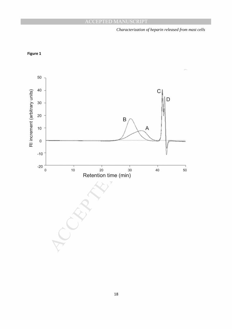
Molecular weight distribution- RMCG was found to be of lower average molecular weight (peak, number
and weight averages) than typical porcine unfractionated heparin, but with greater dispersity (Table 1).
Moreover, the chromatogram for RMCG exhibits a non-symmetrical peak, which may indicate the
presence of more than one distinct population of molecules within the material, in contrast to the
symmetrical peak for the heparin standard (Figure 1).
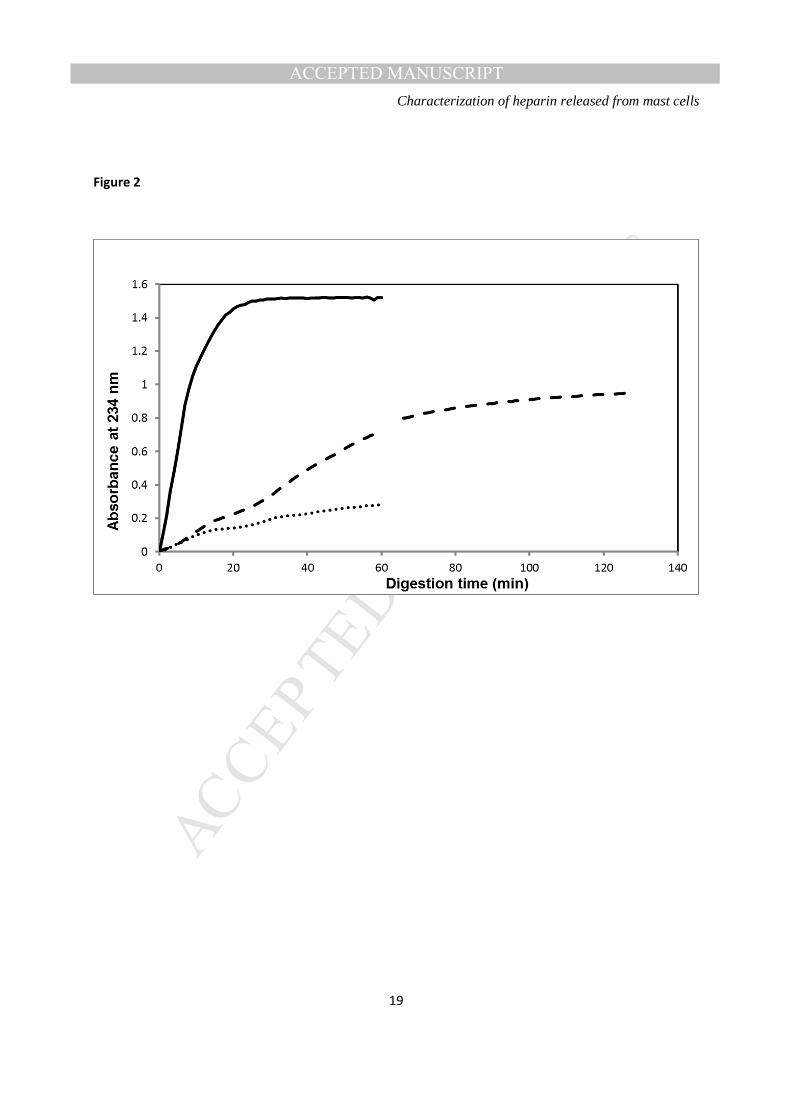
Susceptibility to heparinase digestion- Figure 2 shows reaction progress curves for the digestion of equal
concentrations of heparin standard, RMCG and heparan sulphate, respectively, by a fixed concentration of
heparinase I, as measured by the change in absorbance at 234 nm. RMCG was less susceptible to the
actions of this enzyme than standard heparin, both in terms of initial rate and plateau, but more
susceptible than heparan sulphate, suggesting the material to be comprised substantially, but not
MANUSCRIP
T
ACCEPTED
ACCEPTED MANUSCRIPT Characterization of heparin released from mast cells
7
exclusively, of heparin chains. After 60 minutes exposure to the enzyme, unsaturated uronic acid
generation from the RMCG was approximately half of that from the heparin standard at the same time
point, and 2.5 times that from heparan sulphate. However, when the reaction time was extended to two
hours, allowing it to plateau, total generation from the RMCG was increased to approximately 60% of
that obtained from the heparin standard and more than three times that from heparan sulphate.
Anticoagulant activity- The RMCG was found to give a valid potency estimation against the porcine
unfractionated heparin standard (07/328), although the specific activity of RMCG is lower than porcine
and bovine heparin (Table 1) . The APTT assay using sheep plasma was found to give the highest specific
activity of 78 IU mg-1 (Table 1). The APTT assay using human plasma and the HCII based assay gave
similar specific activity, 61 IU mg-1, whilst the two antithrombin dependent assays gave lower activity,
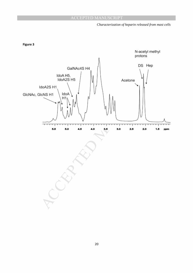
anti-Xa 55 IU mg-1 and anti-IIa 46 IU mg-1. The ratio of anti-Xa to anti-IIa activity was 1.2. 1H-NMR spectra of the RMCG- display signals (Figure 3) are consistent with the RMCG material being
predominantly heparin (22,23), but interestingly containing a substantial proportion, possibly as much as
40-50%, of dermatan sulphate (24). Specifically, in spite of the rather broad resonances from this sample,
the TOCSY spectrum of RMCG contains spin systems consistent with the presence of the IdoA residue
(2-O-sulfo-α-L-iduronic acid; I2S) and glucosamine residues (α-D-N-sulfoglucosamine-6-O-sulphate;
GlcNS,6S) that together make up the major trisulphated disaccharide repeat unit of heparin (Table 2). In
addition, resonances were apparent in the 1H spectrum that correspond to the presence of the
galactosamine (β-D-N-acetylgalactosamine-4-O-sulphate; GalNac4S) and non-sulphated IdoA residue (α-
L-iduronic acid; I) in the major disaccharide repeat unit of dermatan sulphate (Table 2). Comparisons
with literature values shown in Table 2 confirm that the signals attributable to GalNAc4S are
characteristic of those in dermatan sulphate (24) rather than chondroitin-4-sulphate (28), and the signals
attributable to IdoA are characteristic of those in dermatan sulphate rather than in heparin.
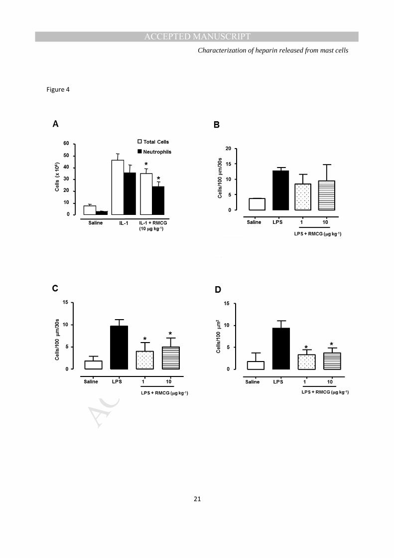
Anti-inflammatory activity- was measured in vivo in the rat in two models of inflammation. Pre-treatment
with RMCG inhibited accumulation of leukocytes in the peritoneal cavity in response to the cytokine IL-
1β, when both agents were administered i.p. (Figure 4A). In addition, systemic pre-treatment with
RMCG inhibited the firm adhesion and diapedesis of leukocytes in the cremasteric microcirculation,
without significantly affecting the number of rolling cells (Figure 4B-D).
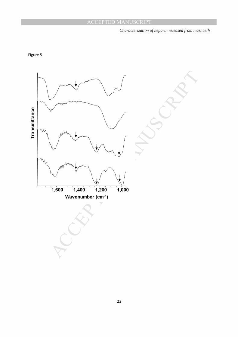
FTIR spectroscopy of the endothelial glycocalyx- FTIR spectroscopy of HUVECs yielded spectra
indicating the presence of sulphated functional groups on the cell surface. Differences were observed in
the spectra of untreated endothelial cells when compared with those from enzymatically-treated cells
(Figure 5), in that peaks in the window associated with the presence of sulphate groups were abolished
following treatment with heparinase enzymes, suggesting degradation of heparan sulphate on the
endothelial surface. Interestingly, addition of exogenous heparin to enzymatically treated HUVECs led to
MANUSCRIP
T
ACCEPTED
ACCEPTED MANUSCRIPT Characterization of heparin released from mast cells
8
restoration of the sulphate peak in the spectra of these cells, with introduction of additional peaks found in
the spectrum of heparin itself, suggesting the binding of heparin to the cell surface of the endothelium.
DISCUSSION
A specific physiological role for heparin, over and above that associated with its general characteristics as
a GAG, has yet to be defined. Heparin is sometimes considered to be a specialized form of heparan
sulphate and, indeed, many of the important protein-binding characteristics of heparan sulphate are
known to be shared, and often exceeded, by heparin. In the case of the anticoagulant actions of heparin,
this increased potency is known to be due in large part to the relatively frequent occurrence of the
antithrombin-binding pentasaccharide sequence that is expressed more rarely in heparan sulphate chains,
whereas for other examples of GAG-protein interactions, that are less specific in terms of the GAG
structure involved, the significantly greater sulphation density of heparin appears to be the key feature.
In the present study, we have isolated GAGs from degranulating rat peritoneal mast cells in good
yield using affinity chromatography on poly-L-lysine agarose. This technique has the advantage of
reducing to a minimum any co-purification of mast cell granule proteins with the GAGs. We have found
that endogenous heparin released by degranulating rat peritoneal mast cells is characterised by a relatively
low molecular weight profile by comparison with a porcine mucosal heparin reference standard. Free
GAGs released from mast cells on degranulation have been depolymerised from their original
macromolecular form by mast cell heparanase (25). The molecular weight distribution of the resulting
GAG mixture is characteristic of the source tissue and species; for example, porcine mucosal and bovine
lung heparins have consistently different molecular weight profiles (26). Using NMR spectroscopy we
have also observed the presence of dermatan sulphate; signals from both IdoA and GalNAc are present,
with chemical shift values characteristic of dermatan sulphate (Table 2), clearly distinguishable from
those of unsulphated IdoA in heparin sequences (27) and GalNAc in chondroitin sulphate A (28). The
presence of dermatan sulphate in rat peritoneal mast cells has been suggested by comparison of the
disaccharide products of digestion with chondroitinase ABC (to which DS is sensitive) with
chondroitinase AC (to which DS is not sensitive)(29). Here we confirm the presence of DS by direct
spectroscopic observation of the GAG mixture, without degradation or separation of its components.
Minor proportions of chondroitin-4-sulphate (CSA), and traces of chondroitin-4,6-sulphate (CSE) found
by Akiyama et al. may be present, but are not visible in our NMR spectra (29). The presence of both
MANUSCRIP
T
ACCEPTED
ACCEPTED MANUSCRIPT Characterization of heparin released from mast cells
9
heparin and dermatan sulphate chains presumably gives rise to the skewed molecular weight distribution
of the rat GAGs (Fig. 1); the mode of depolymerisation of mast cell DS is not currently known to us.
There have been a few previous attempts to look at the release of endogenous rat heparin (30,31), the first
of which (30) characterized the whole serglycin proteoglycan rather than the GAG. Wang and Kovanen
have also previously reported the release of endogenous heparin from rat mast cells, and studied the
ability of this material to inhibit the proliferation of aortic vascular smooth muscle cells, suggesting that
the endogenous heparin may be more potent in this regard than an exogenous heparin preparation (31).
We have demonstrated in this study that rat peritoneal mast cell GAGs, consisting of a mixture of heparin
and DS, possess significant anti-inflammatory activity in a number of assays in addition to a clear ability
to act as an anti-coagulant. The GAG content of mast cells and of other granule-containing cells such as
basophils has not been accurately determined thus far, although it has been proposed that in rodents there
are two types of mast cell referred to as connective tissue (containing heparin) and mucosal (containing
chondroitin sulphate), but only based on histochemical staining (32,33). It has been previously suggested
that heparin is found in mast cell granules whereas chondroitin sulphate is found in basophils (34).
However, in the present study, the mast cell granule contents we have examined contain both heparin and
dermatan sulphate (chondroitin sulphate B), suggesting that both GAGs may occur in the same granule
(though it is possible that our peritoneal mast cell preparation includes two separate cell populations).
The time course profile of heparinase I digestion of the RMCG indicates that roughly 60% of its
constituent material is susceptible to the enzyme, which is specific for the highly sulphated sequences
typical of heparin rather than the low-sulphated HS sample. The anticoagulant profile for the RMCG is
also consistent with the presence of both heparin and DS. DS exclusively potentiates heparin cofactor II
inhibition of thrombin, whereas heparin potentiates both antithrombin and HCII dependent inhibition of
thrombin. Since measurable activity was found using antithrombin dependent assays, it can be concluded
that RMCG contains the prerequisite pentasaccharide sequence for potentiation of antithrombin. A ratio
between the antithrombin anti-Xa and anti-IIa of 1.2 highlights the overall lower molecular weight of
RMCG in comparison to heparin. Since expression of anti-IIa activity requires a longer chain length than
anti-Xa activity, a higher ratio suggests the presence of a higher proportion of lower molecular weight
material. This observation fits in with the relatively lower molecular weight of the RMCG by comparison
with porcine heparin. Overall the anticoagulant potency of the RMCG is lower than both bovine and
porcine heparins, which may be due to the presence of a substantial proportion of DS, though relatively
low molecular weight rat heparin may also have inherently lower activity.
Nonetheless as has been reported for exogenous heparin (for example 35,36), the endogenous heparin we
have isolated and characterised was able to inhibit the infiltration of neutrophils into the rat peritoneal
cavity suggestive of anti-inflammatory activity. Furthermore, using intra-vital microscopy we have also
MANUSCRIP
T
ACCEPTED
ACCEPTED MANUSCRIPT Characterization of heparin released from mast cells
10
shown that endogenous heparin was able to inhibit the adhesion and transmigration of leukocytes across
the vascular endothelium. These findings extend previous observations that exogenous heparin can inhibit
leukocyte infiltration into a number of tissues, both experimentally (35,37) and clinically (38). Our results
support the hypothesis (14) that the release of endogenous heparin in an inflammatory response may serve
to act as a homeostatic braking mechanism to limit cell infiltration into tissues and by virtue of the well
known ability of heparin to neutralise the actions of various cationic pro-inflammatory mediators, may
also serve to limit the effects of the mediators released from infiltrating inflammatory cells.
It has recently been demonstrated that the loss of the glycocalyx from the endothelium lining blood
vessels following exposure to heparanase released by inflammatory cells may play an important role in
the subsequent trafficking of inflammatory cells into tissues (39), and indeed we have recently reported
that recombinant human heparanase is pro-inflammatory and can elicit leukocyte infiltration into tissues
(33). Furthermore, we have also recently reported that heparanase inhibitors are able to inhibit neutrophil
recruitment into lung tissue following certain types of inflammatory insult (40). It is therefore of interest
that by use of FTIR spectroscopy, we have observed the loss of sulphation on the surface of vascular
endothelial cells exposed to heparinase and that we could restore this barrier by adding back exogenous
heparin. Given the proximity of mast cells to blood vessels it thus remains plausible that the release of
endogenous heparin serves as a “top up” mechanism to help preserve the integrity of the glycocalyx
during normal defence mechanisms further supporting the suggestion that endogenous heparin may play
an important physiological role in regulating the innate inflammatory response (14). It is of interest
therefore that patients with allergy, where there is regular mast cell degranulation in response to
sensitising allergens, have been reported to have increased levels of “circulating heparin like material”
(41) which may account for the observations that atopic patients can have a mild haemostatic defect (42),
and even have evidence of less calcification of their arteries (43). However, more recent observations
have suggested that there may a loss of heparin in the circulation of patients with asthma (44) and that
some patients can have a hyper-coaguable state (45).
It is clear from heparin preparations used in medicine that heparin composition varies between species
and tissue of origin (46). Heparin from bovine lung differs markedly in the degree of sulphation from
porcine intestinal mucosal heparin (22), and both are different from bovine intestinal mucosal heparin
(47). We can speculate that these differences in structure and in relative content of heparin vs,
chondroitin/dermatan sulphate may have functional significance in the context of each particular tissue
and species. Clearly, further research is needed to clarify these structure activity relationships of
endogenous GAGs.
In conclusion, we have isolated and partially characterised the GAGs released from degranulating rat
peritoneal mast cells and demonstrated the presence of both heparin and DS. Whilst it remains to be
MANUSCRIP
T
ACCEPTED
ACCEPTED MANUSCRIPT Characterization of heparin released from mast cells
11
determined what the physiological role of this endogenous material is, we have provided evidence that
this material exhibits anti-coagulant activity comparable to heparin obtained from other species and
shows anti-inflammatory activity.
ACKNOWLEDGEMENTS
We acknowledge the support of the Wellcome Trust for a grant to RL, CPP and NVR to support some of
this work.
REFERENCES
1. T.W. Barrowcliffe. History of heparin. Handb. Exp. Pharmacol. 207 (2012) 3-22.
2. E. Gray, B. Mulloy, T.W. Barrowcliffe. Heparin and low-molecular-weight heparin. Thromb. Haemost. 99 (2008) 807-18.
3. B. Casu, A. Naggi, G. Torri. Re-visiting the structure of heparin. Carbohydr. Res. 403 (2015) 60-8.
4. P. Gresele, C. Busti, G. Paganelli. Heparin in the prophylaxis and treatment of venous thromboembolism and other thrombotic diseases. Handb. Exp. Pharmacol. 207 (2012)179-209.
5. L.B. Jaques. Heparins--anionic polyelectrolyte drugs. Pharmacol. Rev. 31 (1979) 99-166.
6. R. Lever, C.P. Page. Non-anticoagulant effects of heparin: an overview. Handb. Exp. Pharmacol. 207 (2012) 281-305.
7. D. Xu, J.D. Esko. Demystifying heparan sulfate-protein interactions. Annu. Rev. Biochem. 83 (2014) 129-157.
8. J. Turnbull, A. Powell, S. Guimond. Heparan sulfate: decoding a dynamic multifunctional cell regulator. Trends Cell Biol. 11 (2001) 75-82.
9. D.E. Humphries, G.W. Wong, D.S. Friend, M.F. Gurish, W.T. Qiu, C. Huang, A.H. Sharpe, R.L. Stevens. Heparin is essential for the storage of specific granule proteases in mast cells. Nature 400 (1999) 769-772.
10. E. Forsberg, G. Pejler, M. Ringvall, C. Lunderius, B. Tomasini-Johansson, M. Kusche-Gullberg, I. Eriksson, J. Ledin, L. Hellman, L. Kjellén. Abnormal mast cells in mice deficient in a heparin-synthesizing enzyme. Nature 400 (1999) 773-776.
MANUSCRIP
T
ACCEPTED
ACCEPTED MANUSCRIPT Characterization of heparin released from mast cells
12
11. B. Mulloy, J. Hogwood, E. Gray, R. Lever, C.P. Page. Pharmacology of heparin and related drugs. Pharmacol Rev. 67 (2015) 1-66.
12. M. Samoszuk, E. Kanakubo, J.K. Chan. Degranulating mast cells in fibrotic regions of human tumors and evidence that mast cell heparin interferes with the growth of tumor cells through a mechanism involving fibroblasts. BMC Cancer 5 (2005) 121.
13. P.T. Kovanen. Mast cells in atherogenesis: actions and reactions. Curr. Atheroscler. Rep. 11 (2009) 214-219.
14. C.P. Page. One explanation of the asthma paradox: inhibition of natural anti-inflammatory mechanism by beta 2-agonists. Lancet 337 (1991) 717-720.
15. F. Bono, P. Rigon, I. Lamarche, P. Savi, V. Salel, J-M. Herbert. Heparin inhibits the binding of basic fibroblast growth factor to cultured human aortic smooth-muscle cells. Biochem. J. 326 (1997) 661-668.
16. T.M. Handel, Z. Johnson, S.E. Crown, E.K. Lau, A.E. Proudfoot. Regulation of protein function by glycosaminoglycans - as exemplified by chemokines. Annu. Rev. Biochem. 74 (2005) 385-410.
17. A.D. Luster. Chemokines - chemotactic cytokines that mediate inflammation. N. Engl. J. Med. 338 (1989) 436-445.
18. E. Gray, J. Hogwood, B. Mulloy. The anticoagulant and antithrombotic mechanisms of heparin. Handb. Exp. Pharmacol. 207 (2012) 43-61.
19. B. Mulloy, D.T. Crane, A.F. Drake, D.B. Davies. The interaction between heparin and polylysine: a circular dichroism and molecular modelling study. Braz. J. Med. Biol. Res. 29 (1996) 721-729.
20 B. Casu, E.A. Johnson, M. Mantovani, B. Mulloy, P. Oreste, R. Pescador, G. Prino, G. Torri, G. Zoppetti. Correlation between structure, fat-clearing and anticoagulant properties of heparins and heparan sulphate. Arzneimittel-Forschung Drug Res. 33 (1983) 135-142.
21. B. Mulloy, E. Gray, T.W. Barrowcliffe. Characterisation of Unfractionated Heparin Samples: Comparison of samples from the last 50 years. Thromb. Haemostas. 84 (2000) 1052-1056.
22. B. Mulloy, E.A. Johnson. Assignment of the 1H NMR spectra of heparin and heparan sulphate. Carbohydr. Res. 170 (1987) 151-165.
23. B. Mulloy, M.J. Forster, C. Jones, A.F. Drake, E.A. Johnson, D.B. Davies. The effect of variation of substitution on the solution conformation of heparin: a spectroscopic and molecular modelling study. Carbohydr. Res. 55 (1994) 1-26.
24. P.N. Sanderson, T.N. Huckerby, I.A. Nieduszynski. Chondroitinase ABC digestion of dermatan sulphate. N.m.r. spectroscopic characterization of the oligo- and poly-saccharides. Biochem. J. 257 (1989) 347–354.
MANUSCRIP
T
ACCEPTED
ACCEPTED MANUSCRIPT Characterization of heparin released from mast cells
13
25. F. Gong, P. Jemth, M.L. Escobar Galvis, I. Vlodavsky, A. Horner, U. Lindhal, J.P. Li. Processing of macromolecular heparin by heparanase. J. Biol. Chem. 278: (2003) 35152-35158
26 B. Mulloy, E. Gray, T.W. Barrowcliffe. Characterization of unfractionated heparin: comparison of materials from the last 50 years. Thromb. Haemostas.84: (2000) 1052-1056
27. E.A. Yates, F. Santini, M. Guerrini, A. Naggi, G. Torri, B. Casu,. 1H and 13 C NMR spectral assignments of the major sequence of twelve systematically modified heparin derivatives. Carbohydr. Res 294: (1996) 15-27.
28. D. Welti, D.A. Rees, E.J. Welsh. Solution confirmation of glycosaminoglycans: assignment of 300-MHz 1H magnetic resonance spectra of chondroitin 4-sulphate, chondroitin 6-sulphate and hyaluronate, and investigation of an alkali-induced conformation change. Eur. J. Biochem 94 (1979) 505-514.
29. H. Akiyama, S. Shidawara, A. Mada, H. Toyoda, T. Toida, T. Imanari. Chemiluminescence high performance liquid chromatography for the determination of hyaluronic acid, chondroitin sulphate and dermatan sulphate. J. Chromatogr. 579: (1992) 203-207
30. R.W. Yurt, R.W. Leid, K.F. Austen, J.E. Silbert. Native heparin from rat peritoneal mast cells J. Biol. Chem. 252 (1977) 518-521.
31. Y. Wang, P.T. Kovanen. Heparin proteoglycans released from rat serosal mast cells inhibit proliferation of rat aortic smooth muscle cells in vitro. Circ Res 84 (1999) 74-83.
32 L. Enerback, S.O.Kolset, M. Kusche, A. Hjerped, U.Lindahl. Glycosaminoglycans in rat mucosal mast cells Biochem. J. 227 (1985) 661-668.
33. R.L. Stevens, M.E. Rothenberg, F. Levi-Schaffer, K.F. Austen. Ontogeny of in vitro-differentiated mouse mast cells. Fed. Proc. 46 (1987) 1915-1919.
34. E. Crivellato, D. Ribatti. The mast cell: an evolutionary perspective Biol. Rev. Camb. Philos. Soc. 85 (2010) 347-360.
35 R. Lever, A. Smailbegovic, C.P. Page. Locally available heparin modulates inflammatory cell recruitment in a manner independent of anticoagulant activity. Eur J Pharmacol. 630 (2010) 137-44.
36 J.G. Wan, J.S. Mu, H.S. Zhu, J.G. Geng. N-desulfated non-anticoagulant heparin inhibits leukocyte adhesion and transmigration in vitro and attenuates acute peritonitis and ischemia and reperfusion injury in vivo. Inflamm Res. 51 (2002) 435-43.
37 E.A.M. Seeds, C.P. Page. Heparin inhibits allergen induced eosinophil infiltration into guinea pig lung via a mechanism unrelated to its anticoagulant activity. Pulm. Pharmacol. Ther. 14 (2001) 111-119.

MANUSCRIP
T
ACCEPTED
ACCEPTED MANUSCRIPT Characterization of heparin released from mast cells
14
38 C. Vancheri, C. Mastruzzo, F. Armato, V. Tomaselli, S. Magri, M.P. Pistorio, M. LAMicela, L. D’Amico, N. Crimi. Intranasal heparin reduces eosinophil recruitment after nasal allergen challenge in patients with allergic rhinitis. J Allergy Clin Immunol 108 (2001) 703-708.
39 E.P. Schmidt, Y. Yang, W.J. Janssen, A. Gandjeva, M.J. Perez, L. Barthel, R.L. Zemans, J.C. Bowman, D.E. Koyanagi, Z.X. Yunt. The pulmonary endothelial glycocalyx regulates neutrophil adhesion and lung injury during experimental sepsis. Nat Med. 18 (2012) 1217-1223.
40 A. Morris, B. Wang, I. Waern, V. Radhakrishnan, C.P. Page, E. Schmidt, S. Wernesson, J-P. Li, D. Spina. The role of heparanase in pulmonary cell recruitment in response to an allergic but not non-allergic stimulus. PLOS ONE 2015;10: e0127032.
41 E.C. Lasser, R.A. Simon, S.G. Lyon, A.E. Hamblin, R. Stein. Heparin-like anticoagulants in asthma. Allergy. 42 (1987) 619-625.
42 A. Szczeklik, M. Schmitz-Schumann, M. Krzanowski, C. Virchow. Delayed generation of thrombin in clotting blood of atopic patients with hayfever and asthma. Clin Exp Allergy. 21(4) (1991) 411-5.
43 E.C. Lasser, C. Berry, K. Kortman. Diminished atherosclerotic arterial calcifications in asthma. A possible role for elevated endogenous heparin-like material. Allergy. 42 (1987) 549-52.
44 H. Davids, A. Ahmed, A. Oberholster, C. van der Westhuizen, M. Mer, I. Havlik. Endogenous heparin levels in the controlled asthmatic patient. S Afr Med J 100 (2010) 3070-308.
45 J.D. de Boer, C.J. Majoor, C. van’t Veer, E.H. Bel, T. van der Poll. Asthma and coagulation. Blood 119 (2012) 3236-3244.
46 P. Bianchini, L. Liverani, G. Mascellani, B. Parma. Heterogeneity of unfractionated heparins studied in connection with species, source, and production processes. Semin. Thromb. Hemost. 23 (1997) 3-10.
47 A. Naggi, C. Gardini, G. Pedrinola, L. Mauri, E. Urso, A. Alekseeva, B. Casu, G. Cassinelli, M. Guerrini, M. Iacomini, V. Baigorria, G. Torri. Structural peculiarity and antithrombin binding region profile of mucosal bovine and porcine heparins. J. Pharm. Biomed. Anal. 118 (2015) 52-63.
MANUSCRIP
T
ACCEPTED
ACCEPTED MANUSCRIPT Characterization of heparin released from mast cells
15
TABLE 1
A: Peak molecular weight Mp, number average molecular weight Mn, weight average molecular weight Mw and polydispersity Mw/Mn for rat peritoneal mast cell GAGs (RMCG) and for a heparin identity standard, the USP Heparin Sodium Identity Reference Standard (USP ID RS). Results are the means of duplicate determinations.
B: Anticoagulant potencies of RMCG, a bovine and porcine heparin sample estimated against 6th International Standard for Unfractionated Heparin using a parallel line analysis model. Values are calculated using multiple concentrations of unknown samples against the standard to give IU/mg and 95% confidence limits range for the estimated value.
A Mp Mn Mw Polydispersity (Mw/Mn)
RMCG 7333 7471 12027 1.611
USP ID RS
15346 12718 15856 1.247
B Potency in IU/mg (95% Confidence Limits)
Sheep Plasma APTT Assay
Human Plasma APTT Assay
anti-Xa assay anti-IIa assay HCII assay
RMCG 78 76 – 81
61 57 – 64
55 54 – 56
46 44 – 47
61 54 – 68
Bovine Heparin
108 (103 – 113)
89 (85 – 93)
94 (91 – 97)
87 (80 – 94)
120 (112 – 129)
Porcine Heparin
200 (194 – 206)
185 (175 – 195)
197 (186 – 208)
192 (175 – 211)
199 (186 – 214)
TABLE 2
Assignment of the 1H NMR spectrum of RMCG: chemical shifts in ppm at 500 MHz, 60 ºC, in D2O relative to TSP at 0 ppm.
Heparin Dermatan sulphate GlcNS,6S IdoA2S GalNAc4S IdoA
This study
Ref. 22
This study
Ref. 22
This study
Ref. 24 Ref 28 This study
Ref. 24 Ref 27
H1 5.39 5.39 5.21 5.24 4.65-
4.7 4.7 4.60 4.90 4.90 5.04
MANUSCRIP
T
ACCEPTED
ACCEPTED MANUSCRIPT Characterization of heparin released from mast cells
16
H2 3.27 3.31 4.30 4.37 4.04 4.05 4.03 3.54 3.54 3.78
H3 3.69 3.70 4.20 4.23 4.02 3.95 4.01 3.92 3.92 4.12
H4 3.76 3.78 4.11 4.13 4.65 4.65-4.7
4.74 4.10 4.11 4.08
H5 4.02 4.06 4.80 4.81 3.85 3.8 3.81 4.72 4.72 4.84
H6 n.d. 4.37 3.80 3.75-3.8
3.82
H6’ n.d. 4.30 3.77
GlcNAc
CH3 2.05 2.05 2.08 2.08 2.02
1. In chondroitin 4 sulphate at 60 deg. C 2. In the heparin-derived sequence IdoA-GlcNS,6S at 40 deg. C.
MANUSCRIP
T
ACCEPTED
ACCEPTED MANUSCRIPT Characterization of heparin released from mast cells
17
FIGURE LEGENDS
Figure 1: A. Size exclusion chromatograms of rat mast cell GAGs (RMCG; peak A) and a sample of typical unfractionated heparin (the USP Heparin Sodium Identification RS; peak B). Peak C is the flow-rate marker alpha-cyclodextrin, and peak D is a salt peak
Figure 2: Heparinase I digestion profiles of RMCG (A), unfractionated heparin standard (B) and heparan sulphate (C). In (A) the spectrophotometer was reprogrammed at 60 minutes to extend the data collection period, leading to a gap in the readings at that time.
Figure 3: A. 1H NMR spectrum (500 MHz, 60 ºC in D2O) of rat mast cell GAGs. Some of the characteristic resonances of heparin and dermatan sulphate are annotated.
Figure 4: A. IL-1β-induced (20 ng) accumulation of cells in the peritoneal cavity of the rat and the effect of 10 µg kg-1RMCG administered locally. Open bars indicate total cell counts and filled bars neutrophil counts (p ≤ 0.05 vs. IL-1β, n= 6/group). B-D. Rolling (B), firmly adherent (*p<0.05 vs LPS) (C) and transmigrated (*p<0.001 vs LPS) (D) cells in the cremasteric microcirculation of the rat inresponse to 25 µg of LPS and the effect of RMCG administered systemically (1 and 10 µg Kg-1,n= 4/group).
Figure 5: FTIR spectra of HUVECs treated with vehicle (top), heparinases I, II and III (0.5 IU mL-1; second from top) and heparinases I, II and III followed by unfractionated heparin (0.5 IU mL-1 and 500 IU mL-1, respectively, third from top). The spectrum of unfractionated heparin itself is also shown (bottom).
MANUSCRIP
T
ACCEPTED
ACCEPTED MANUSCRIPT Characterization of heparin released from mast cells
18
Figure 1
MANUSCRIP
T
ACCEPTED
ACCEPTED MANUSCRIPT Characterization of heparin released from mast cells
19
Figure 2
MANUSCRIP
T
ACCEPTED
ACCEPTED MANUSCRIPT Characterization of heparin released from mast cells
20
Figure 3
MANUSCRIP
T
ACCEPTED
ACCEPTED MANUSCRIPT Characterization of heparin released from mast cells
21
Figure 4
MANUSCRIP
T
ACCEPTED
ACCEPTED MANUSCRIPT Characterization of heparin released from mast cells
22
Figure 5