binding of acetylcholinesterase to multiwall carbon nanotube-cross-linked chitosan composite for...
TRANSCRIPT
DOI: 10.1002/chem.200500178
Binding of Acetylcholinesterase to Multiwall Carbon Nanotube-Cross-Linked Chitosan Composite for Flow-Injection Amperometric Detection ofan Organophosphorous Insecticide
Vivek Babu Kandimalla and Huangxian Ju*[a]
Introduction
Organophosphorous (OP) compounds are highly toxic—often causing respiratory paralysis and death—and can irre-versibly inhibit acetylcholinesterase (AChE) which is essen-tial for the central nervous system. Therefore, the rapid, sen-sitive, selective and reliable quantification of these com-pounds is highly essential at a lower cost. Chromatographi-cal techniques have been extensively used for the sensitivedetection of OP insecticides, but they require trained staff,complicated sample pretreatments and are often not suitablefor field conditions. In recent years biosensors show remark-able advances for the detection of toxic compounds basedon enzymatic reactions.
A variety of enzymes such as organophosphorous hydro-lase, alkaline phsosphatase, ascorbate oxidase, tyrosinaseand acid phosphatase have been employed in the prepara-tion of pesticide biosensors.[1] Based on the inhibition actionof pesticides and insecticides on cholinesterases, AChE andbutyrylcholinesterase have been widely used due to the sta-bility and sensitivity of the enzymes.[2,3] This method gener-ally uses either single enzyme[4] or bienzyme (AChE andcholine oxidase)[3,5,6] systems by monitoring the electro-chemical oxidation of thiocholine or p-aminophenol and hy-drogen peroxide, respectively. Some of the reports on immo-bilized AChE-based single enzyme system apply voltages of+400 to +700 mV for the oxidation of thiocholine.[7–10]
AChE can be immobilized on electrode surface by using avariety of matrices such as cross-linked polymers,[11] cross-linked bovine serum albumin,[12,13] chitosan[2] and cellu-lose,[14] different support matrices such as nylon,[5,12,15] con-trolled pore glass,[16] magnetic particles,[10,17] or the strong af-finity linking with concanavalin A[18] and metal ions.[19] Toreduce the working potential mediators such as 7,7,8,8-tetra-cyanoquinodimethane (TCNQ)[19–22] have been deposited onsurface to shuttle electrons between the thiocholine formedand the electrode. The implementation of TCNQ can
Abstract: A novel method for immobi-lization of acetylcholinesterase (AChE)by binding covalently to a cross-linkedchitosan–multiwall carbon nanotube(MWNT) composite is described. Inaddition a sensitive, fast, cheap and au-tomatizable flow injection detection ofan organophosphorous insecticide wasdeveloped. The MWNTs were homoge-neously distributed in the chitosanmembrane which showed a homogene-ous porous structure. The immobilizedAChE could catalyze the hydrolysis ofacetylthiocholine with a Kapp
M value of177 mm to form thiocholine, which was
then oxidized to produce detectablesignal in a linear range of 1.0–500 mm
and fast response. MWNTs could cata-lyze the electrooxidation of thiocho-line, thus increasing detection sensitivi-ty. Based on the inhibition of an orga-nophosphorous insecticide on the enzy-matic activity of AChE, using Sulfotepas a model compound, the conditionsfor the flow-injection detection of the
insecticide were optimized. Both bio-compatibility of chitosan and inherentconductive properties of MWNTs fa-vored the detection of the insecticidefrom 1.5 to 80 mm along with good sta-bility and reproducibility. 95 % reacti-vation from inhibited AChE could beregenerated by using 2-pyridinealdox-ime methiodide within 15 min for 15times. The detection of Sulfotep sam-ples exhibited satisfactory results. Theproposed flow-injection analysis devicecan be applied to automated determi-nation and characterization of enzymeinhibitors.
Keywords: acetylcholinesterase ·biosensors · chitosan · enzymeinhibitors · nanotubes
[a] Dr. V. B. Kandimalla, Prof. H. JuKey Laboratory of Analytical Chemistryfor Life Science (Education Ministry of China)Department of Chemistry, Nanjing UniversityNanjing 210093 (P. R. China)Fax: (+86) 25-8359-3593E-mail : [email protected]
G 2006 Wiley-VCH Verlag GmbH & Co. KGaA, Weinheim Chem. Eur. J. 2006, 12, 1074 – 10801074

reduce the applied potential to +100 mV versus Ag/AgCl.[22] This work reports a novel method for the immobili-zation of AChE by using glutaraldehyde as a cross-linker tobind covalently AChE to a cross-linked chitosan–multiwallcarbon nanotube (MWNT) composite (CMC), leading to astable thiocholine biosensor. The cross-linked chitosanmatrix with free -CHO groups is formed by mixing a chito-san solution with excessive glutaraldehyde. The presence ofMWNTs reduces the working potential by catalyzing theelectrochemical oxidation of enzymatically formed thiocho-line.
Since their discovery, carbon nanotubes (CNTs)[23] haveattracted considerable interest, because of their interestingproperties,[24] as they have for example emerged as highlyconductive (fast electron transfer) nanomaterials. The abilityof CNTs to promote the electron transfer of NADH and hy-drogen peroxide suggests great protential for the dehydro-genase- and oxidase-based amperometric biosensors.[24, 25]
These materials have been employed for electrocatalytic ox-idation of glucose, cytochrome c, thymine, ascorbic acid andnitric oxide.[26–30] In view of their advantages AChE andcholine oxidases have been covalently co-immobilized onMWNTs for the preparation of OP-pesticides biosensors.[6]
Other OP biosensors have also been constructed, for exam-ple, by adsorption of AChE on MWNTs modified thickfilm.[4] In order to stabilize the bioactivity of the immobi-lized enzymes some biocompatible materials such as chito-san, an aminopolysaccharide, have been have been used assupport matrices.[2,31] The combination of highly conductiveand electrocatalytic behaviors of CNTs with the good bio-compatibility of chitosan led to a stable and sensitive glu-cose biosensor.[31] In this work the proposed biosensor basedon the immobilization of AChE on CMC showed good sta-bility and high sensitivity for both thiocholine and Sulfotep,a model compound of OP insecticides, which could be em-ployed for flow-injection analysis of Sulfotep.
The combination of biosensors with flow-injection analysismakes it possible to control all the stages of the reagent ad-ditions, measure the enzyme activity, improve the samplethroughput and achieve the completely automated determi-nation along with sensitive detection limits, quick responseand repeated use of the immobilized enzyme.[9] This tech-nique has used for OP insecticides monitoring by immobiliz-ing AChE on a gold-coated nylon mesh by a self-assembledmonolayer of cystamine preadsorbed on the gold surface[15]
and a platinum electrode by entrapment in a photocrosslink-er polymer[32] with the detection limits of 50 nm (defined asthe concentration of inhibitor required to obtained a 5 % ofinhibition) and in micromolar range, respectively. The inhib-ited AChE on the gold-coated nylon mesh could be reacti-vated by immersion in a solution of 2-pyridinealdoxime me-thiodide (2-PAM) for at least 6 h.[15] Here, a more sensitive,faster and cheap method for flow-injection detection of or-ganophosphorous insecticides as shown in Figure 1 was de-veloped with a detection limit of 1.0 nm at a 10 % inhibitionfor Sulfotep; the inhibited AChE could be regenerated for15 cycles by using 2-PAM within 15 min.
Results and Discussion
Electrochemical behavior of AChE/CMC/GCE : The cyclicvoltammograms of 1.0 mm ATCl at different electrodes areshown in Figure 2. AChE/glutaraldehyde-chitosan/GCEshowed an irreversible oxidation peak at +710 mV, whileno detectable signal was observed at CMC/GCE. Both
AChE/glutaraldehyde-chitosan/GCE and AChE/CMC/GCEdid not show any detectable response in absence of ATCl(not shown). Obviously, the peak came from the oxidationof hydrolysis product, thiocholine, of acetylthiocholine(ATCl), catalyzed by the immobilized AChE. At AChE/CMC/GCE the oxidation peak increased greatly and shiftednegatively to +590 mV due to the presence of MWNTs inthe composite, which possessed inherent conductive proper-ties[33] and catalytic behavior towards the oxidation of thio-choline. The electrocatalytic action of MWNTs was also ob-served for the oxidation of some compounds containing athiol moiety, such as cysteine and glutathione.[4,34] A highbackground current was observed when the MWNTs weredirectly coated on the electrode surface and then coveredwith cross-linked chitosan membrane, instead of entrapmentin cross-linked chitosan membrane. Following experimentswere carried out by using AChE/CMC/GCE configuration.
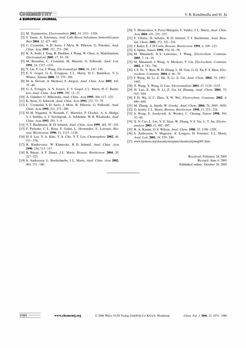
Microscopic characterization of CMC membrane : Figure 3shows the SEM and TEM images of the different mem-branes. The cross-linked chitosan membrane showed a ho-
Figure 1. Schematic diagram of the flow-injection detection system.1) Peristalytic pump, 2) auxiliary electrode, 3) working electrode, 4) refer-ence electrode, 5, 6) upper and bottom portions of flow cell, 7) flowchamber, 8) electrochemical detection instrument, 9) computer.
Figure 2. Cyclic voltammograms at 100 mV s�1 of 100 mm pH 7.4 PBScontaining 0.1m KCl and 1.0 mm ATCl at a) CMC/GCE, b) AChE/gluta-raldehyde-chitosan/GCE and c) AChE/CMC/GCE; GCE: glass carbonelectrode.
Chem. Eur. J. 2006, 12, 1074 – 1080 G 2006 Wiley-VCH Verlag GmbH & Co. KGaA, Weinheim www.chemeurj.org 1075
FULL PAPER

mogenous porous structure (Figure 3a), and MWNTs werealmost homogeneously distributed in the membrane to forma fluey structure (Figure 3b), which indicates that both themembrane and the composite on GCE were crack-free. Thehomogenous porous structure of the cross-linked chitosanmembrane favored the entrapment of MWNTs in the mem-brane. The fluey structure of the composite was beneficialto the loading or covalently linking of AChE to the free-CHO groups in CMC and the approach of substrate and in-hibitor to the immobilized enzyme, which increased the sen-sitivity of the biosensor for detection of both ATCl and OPinsecticides.
The TEM images indicated that the entrapment ofMWNTs in the membrane did not change the structure andmorphology of MWNTs. Thus this matrix displayed excel-lent conductive properties.
Applied potential and bufferpH for amperometric detection :The dependence of the steady-state current on the applied po-tential is shown in Figure 4. Thesteady-state current quickly in-creases with increasing positive-ly applied potential from +300to +550 mV and reaches amaximum current at +600 mV;this indicates that the more pos-itive applied potential facilitatesthe oxidation of the thiocholineproduced from the enzymaticreaction. The applied potentialof +600 mV was close to the
anodic peak potential of cyclic voltammogram of thiocho-line in the same system at 100 mV s�1. Subsequently, weused +600 mV as the applied potential for following am-perometric measurements.
The bioactivity of the immobilized AChE depends on thesolution pH.[3] The optimal pH is usually in the range of 7.0to 7.5.[4,5,8,20] Thus the effect of pH was examined in therange of pH 6 to 8. The results are listed in Table 1, whichshow that the optimal pH value is 7.4. Thus, pH 7.4 was se-lected for amperometric detection.
Optimization of enzyme electrode preparation : As shown inTable 1, the amperometric response of obtained biosensorincreased, with an increasing content of chitosan in the mix-ture for preparation of CMC; the response then decreasedwith a maximum value occurring at the content of 0.45 %(w/v). A lower content of chitosan made the membranemore fragile, although a higher content of chitosan de-creased the concentration of free -CHO in the membrane.The fragility of the film made the sensor response unstabledue to easy detachment of the film during the experimentsand washing. Both factors also decreased the loading ofenzyme. Furthermore, the membrane formed at high con-tent of chitosan also possibly led to a barrier to enzyme forlinkage, substrate for enzymatic hydrolysis and produced
Figure 3. SEM images of a) cross-linked chitosan and b) CMC mem-branes and TEM images of c) CMC and d) single CNT.
Figure 4. Effect of applied potential on amperometric response of biosen-sor prepared at the contents of 0.45 (w/v), 0.26 (w/v) and 2.4% (v/v) forchitosan, MWNTs and glutaraldehyde in 100 mm pH 7.4 PBS containing0.1m KCl and 0.6 mm ATCl.
Table 1. Effects of pH of detection solution, and the contents of chitosan, MWNTs, and glutaraldehyde forbiosensor preparation on amperometric response of the obtained biosensor to 600 mm ATCl at +600 mV(bold values refer to optimal conditions).
Parameter Content, response and SD [nA]
pH of detection solution 6 6.5 7 7.4 7.8 8response (n=5) 139�5 212�6.3 236�6.9 241�6 226�7.1 189�8.7chitosan (w/v) 0.31 0.45 0.67 0.9 1.12response (n=3)[a] 113�3 173�3.2 160�3.5 125�4.5 95�4.1MWNTs (w/v) 0.032 0.065 0.13 0.2 0.26response (n=3)[b] 132�5.3 165�4.5 201�4.6 184�3.6 172�6.8glutaraldehyde (v/v)[c] 0.32 0.65 0.9 1.26 1.56 1.85 2.4response (n=5)[d] 166�8.0 196�8.4 227�7.6 241�6.3 228�6.4 198�6.9 179�5.1
[a] Obtained at the MWNTs content of 0.26 (w/v) and glutaraldehyde content of 2.4 (v/v). [b] Obtained at thechitosan content of 0.45 (w/v) and glutaraldehyde content of 2.4 (v/v). [c] Volume ratio of 25% glutaraldehydeto 0.5% chitosan solution. [d] Obtained at the chitosan content of 0.45 (w/v) and MWNTs content of 0.13(w/v).
www.chemeurj.org G 2006 Wiley-VCH Verlag GmbH & Co. KGaA, Weinheim Chem. Eur. J. 2006, 12, 1074 – 10801076
V. B. Kandimalla and H. Ju
thiocholine for oxidation, thus decreased the sensor re-sponse.
Similarly with an increasing content of MWNTs in themembrane, the current increased and then decreased at thecontent of 0.13 % (w/v). This was possibly due to the de-crease of biocompatibility of the formed membrane, whichdecreased the enzymatic activity of the immobilizedenzyme, thus decreasing the response of the enzymatic prod-uct. The response also changed with the glutaraldehyde con-tent due to the fact that the amount of enzyme covalentlybound to electrode surface was mainly regulated by theamount of aldehyde groups in cross-linked chitosan mem-brane. The maximum response occurred at the glutaralde-hyde content of 1.26 % (v/v). Thus, the CMC was preparedwith the optimal chitosan, MWNTs and glutaraldehyde con-tents at 0.45 % (w/v), 0.13 % (w/v) and 1.26 % (v/v), respec-tively, in following experiments.
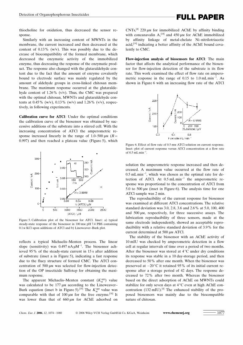
Calibration curve for ATCl : Under the optimal conditionsthe calibration curve of the biosensor was obtained by suc-cessive additions of the substrate into a stirred cell. With theincreasing concentration of ATCl the amperometric re-sponse increased linearly in the range of 1.0–500 mm (R=
0.997) and then reached a plateau value (Figure 5), which
reflects a typical Michaelis–Menten process. The linearslope (sensitivity) was 0.497 nAmM�1. The biosensor ach-ieved 95 % of the steady-state current in 15 s after additionof substrate (inset a in Figure 5), indicating a fast responsedue to the fluey structure of formed CMC. The ATCl con-centration of 500 mm was selected for flow-injection detec-tion of the OP insecticide Sulfotep for obtaining the maxi-mum response.
The apparent Michaelis–Menten constant (K appm ) value
was calculated to be 177 mm according to the Lineweaver–Burk equation (inset b in Figure 5).[35] The K app
m value wascomparable with that of 100 mm for the free enzyme.[18] Itwas lower than that of 660 mm for AChE adsorbed on
CNTs,[4] 220 mm for immobilized AChE by affinity bindingwith concanavalin A,[18] and 450 mm for AChE immobilizedby affinity linkage of metal-chelate Ni–nitrilotriaceticacid,[19] indicating a better affinity of the AChE bound cova-lently to CMC.
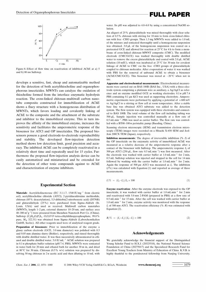
Flow-injection analysis of biosensors for ATCl : The mainfactor that affects the analytical performance of the biosen-sor for flow-injection detection of the substrate is its flowrate. This work examined the effect of flow rate on ampero-metric response in the range of 0.15 to 1.0 mL min�1. Asshown in Figure 6 with an increasing flow rate of the ATCl
solution the amperometric response increased and then de-creased. A maximum value occurred at the flow rate of0.5 mL min�1, which was chosen as the optimal rate for de-tection of ATCl. At 0.5 mL min�1 the amperometric re-sponse was proportional to the concentration of ATCl from5.0 to 500 mm (inset in Figure 6). The analysis time for oneATCl sample was 2 min.
The reproducibility of the current response for biosensorwas examined at different ATCl concentrations. The relativestandard deviation was 3.0, 2.8, 3.6 and 2.6 % at 5.0, 100, 400and 500 mm, respectively, for three successive assays. Thefabrication reproducibility of three sensors, made at thesame electrode independently, showed an acceptable repro-ducibility with a relative standard deviation of 3.9 % for thecurrent determined at 500 mm ATCl.
The stability of the biosensor with an AChE activity of10 mIU was checked by amperometric detection in a flowcell at regular intervals of time over a period of two months.After the biosensor was stored at 4 8C under dry conditionsits response was stable in a 10 day-storage period, and thendecreased to 50 % after one month. When the biosensor waspreserved at �20 8C it retained 95 % of its initial current re-sponse after a storage period of 42 days. The response de-creased to 72 % after two month. Whereas the biosensorbased on the direct adsorption of AChE on MWNTs couldstabilize for only seven days at 4 8C even at high AChE con-centration (132 mIU).[4] The enhanced stability of the pro-posed biosensors was mainly due to the biocompatiblenature of chitosan.
Figure 5. Calibration plot of the biosensor for ATCl. Inset: a) typicalsteady-state response of the biosensor in 100 mm pH 7.4 PBS containing0.1m KCl upon additions of ATCl and b) Lineweaver–Burk plot.
Figure 6. Effect of flow rate of 0.5 mm ATCl solution on current response.Inset: plot of current response versus ATCl concentration at a flow rateof 0.5 mL min�1.
Chem. Eur. J. 2006, 12, 1074 – 1080 G 2006 Wiley-VCH Verlag GmbH & Co. KGaA, Weinheim www.chemeurj.org 1077
FULL PAPERDetection of Organophosphorous Insecticides

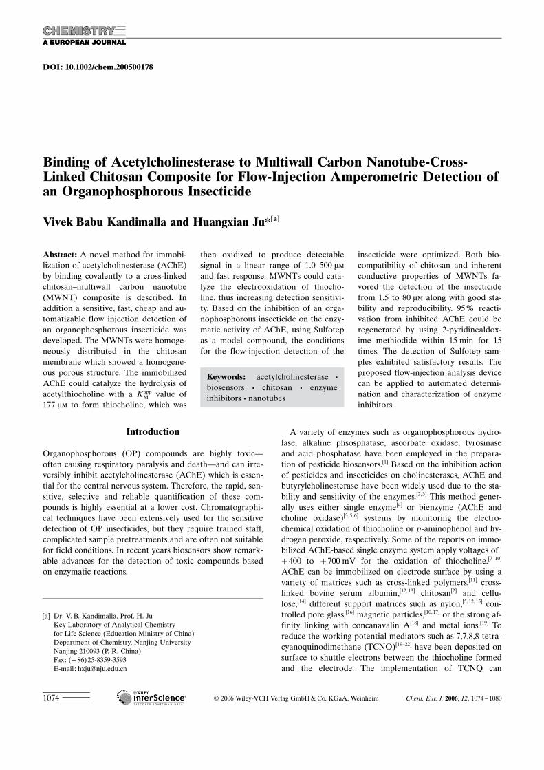
Flow-injection detection of Sulfotep : One of the most influ-encing parameters in pesticide analysis is the incubationtime for the inhibition. With increasing incubation periodthe percentage of the inhibition also increases.[5] This workused a flow-stop method[10] for the inhibition of the enzyme.The Sulfotep sample was injected at 0.5 mL min�1 and thenstopped in the cell to bind the pesticide with enzyme activesite, which led to the inhibition. The incubation time re-quired for the inhibition was checked at different time inter-vals from 5 to 20 min. With an increasing incubation timethe inhibition increased and reached a maximum value afterthe incubation time of 14 min. Thus, including the analysistime, 14 min was used for flow-stop assay of Sulfotep.
Following the incubation and washing steps the ampero-metric response of 500 mm ATCl was detected. The responsedecreased with an increasing Sulfotep concentration (insetFigure 7). At concentrations higher than 90 nm 100 % inhibi-tion occurred. The logarithmic plot of I versus Sulfotep con-centration ranging from 1.5 to 80 nm showed a good lineari-ty (Figure 7). The detection limit was calculated to be 1.0 nmat 10 % inhibition.
The presence of MWNTs greatly enhanced the oxidation
current of the enzymatically formed product. As shown inTable 2, the amperometric response of the biosensor in theflow-injection analysis of 0.5 mm ATCl concentration was2.1 times that of the modified electrode without MWNTs.Upon injection of the same concentration of Sulfotep thedecrease of amperometric response at the biosensor wasalso about twice that without MWNTs. The decreasing rateof the amperometric response was faster than that withoutMWNTs. Thus the MWNTs improved the sensitivity of thebiosensor obtained.
The reproducibility for the Sulfotep detection was evalu-ated first. The relative standard deviations for inhibition de-tection at the Sulfotep concentrations of 5.0 and 66 nm were3.3 and 0.9 %, respectively, for six independently made sen-sors; these results show a good reproducibility. To validatethe practicability of the proposed method, real samples weretested. Water from Yangzhi river and tap was filtered with a0.2 mm filter and the pH adjusted to 7.0–9.0. These watersamples were then probed with a known concentration ofSulfotep and measured with the proposed method. The re-sults are shown in Table 3, which were in good agreementwith the given concentration with an average recovery of99.5 % (n=18). This indicates that this method could beused for assay of real samples.
Reactivation of AChE : AChE, which has been irreversiblyinhibited by organophosphorous pesticides, can be com-pletely reactivated by using nucleophilic compounds such as2-PAM.[16,21, 31,32] 5.0 mm 2-PAM prepared in PBS was usedfor the reactivation. After 2-PAM flowed through the cell at0.5 mL min�1 for different periods of time, the reactivationefficiency R was estimated. With increasing reactivationtime R increased and reached a constant value after 15 min(Figure 8). After exposing the enzyme to 5, 50 and 80 nm ofSulfotep 97, 95 and 94 % of the enzymatic activity could beregained within 15 min, respectively. The time was slightlylonger than that reported by Andreescu et al.,[36] who ob-tained 90 % recovery within 5 min using 1 mm 2-PAM. Thiswas possibly due to the less toxicity of parathion methyl anddichlorovos than Sulfotep;[37] though it was still much fasterthan that of 6 h for the reactivation of AChE inhibited bycarbaryl.[15] With the reactivation procedure this biosensorcould be repeatedly used for 15 cycles with an acceptable re-producibility.
Conclusion
This work proposes a simpleand efficient method for immo-bilization of acetylcholinester-ase on an electrode surface and
Figure 7. Logarithmic plot of I [%] versus Sulfotep concentration forflow-injection detection. Inset: amperometric responses at a) 0, b) 20 andc) 50 nm Sulfotep.
Table 2. Ampermetric responses [nA] and relative standard deviations(RSD %, n=3) in flow-injection analysis of 500 mm ATCl at +600 mV atdifferent concentrations of Sulfotep.
Concentration of Sulfotep [nm] Without CNT/chitosan
Chitosan–MWNTscomposite
0 110.1 (� 2.9) 231.0 (� 1.2)1.5 98.5 (� 3.7) 205.6 (� 1.1)5 92.9 (� 4.1) 187.0 (� 1.1)10 83.3 (� 3.4) 150.0 (� 1.3)20 66.2 (� 3.3) 125.4 (� 1.0)35 54.9 (� 2.2) 95.0 (� 1.0)50 39.0 (� 2.5) 67.5 (� 1.6)66 28.7 (� 2.5) 38.8 (� 2.1)80 16.4 (� 3.5) 15.4 (� 1.6)
Table 3. Measurement of Sulfotep in tap and river water using proposed biosensor (n=3).
concentration of Sulfotep spiked [nm] 1.5 27 50
detected concentration in tap water [nm] 1.42(�0.04) 27.4(�0.1) 49.2(�0.15)recovery [%] 95 101 98detected concentration in river water [nm] 1.53(�0.05) 26.8(�0.18) 50.8(�0.1)recovery [%] 102 99 102
www.chemeurj.org G 2006 Wiley-VCH Verlag GmbH & Co. KGaA, Weinheim Chem. Eur. J. 2006, 12, 1074 – 10801078
V. B. Kandimalla and H. Ju
develops a sensitive, fast, cheap and automatizable methodfor the detection of both acetylthiocholine and organophos-phorous insecticides. MWNTs can catalyze the oxidation ofthiocholine formed from the interface enzymatic hydrolysisreaction. The cross-linked chitosan–multiwall carbon nano-tube composite constructed for immobilization of AChEshows a fluey structure with a homogeneous distribution ofMWNTs, which favors loading and covalently linking ofAChE to the composite and the attachment of the substrateand inhibitor to the immobilized enzyme. This in turn im-proves the affinity of the immobilized enzyme, increases thesensitivity and facilitates the amperometric response of thebiosensor for ATCl and OP insecticides. The proposed bio-sensors possess a good electrode-to-electrode reproducibilityand stability. The developed flow-injection detectionmethod shows low detection limit, good precision and accu-racy. The inhibited AChE can be completely reactivated in arelatively short time and repeatedly used for 15 cycles. Fur-thermore the proposed flow-injection analysis device can beeasily automatized and miniaturized and be extended forthe detection of other toxic compounds against to AChEand characterization of enzyme inhibitors.
Experimental Section
Materials : Acetylcholinesterase (EC 3.1.1.7, 1540 IU mg�1 from electriceel), acetylthiocholine chloride (ATCl), 2-pyridinealdoxime methiodide,chitosan (85 % deacetylation), 5,5-dithiobis(2-nitrobenzoic acid) (DTNB)and glutaraldehyde (25 %) were purchased from Sigma-Aldrich (St.Louis, USA) and used as received. Multiwall carbon nanotubes(MWNTs, length 1–2 mm, external diameter 10–20 nm, and surface area40–300 m2 g�1) were procured from Shenzhen Nanotech Port Co. (China).Sulfotep (C8H20O5P2S2, O,O,O’O’-tetra-ethyldithiopyrophosphate, 99.0 %pure, MW 322.31) was obtained from Sigma-Aldrich (LaborchemikalienGmbH, Seelze). All other reagents used were of analytical reagent grade.
Preparation of biosensors : Prior to immobilization of the enzyme aglassy carbon electrode (GCE, 3.0 mm diameter) was polished with 0.3and 0.05 mm alumina slurry (Behler), respectively, and rinsed thoroughlywith double distilled water. It was then successively ultrasonicated in eth-anol and double distilled water. 5.0 IU mL�1 AChE solution was preparedin 0.1m phosphate buffer solution (pH 7.4, PBS). MWNTs were sonicatedin water bath for 20 min and ethanol bath for another 30 m in, and driedat 80 8C for 30 min. Chitosan (0.5 % w/v) solution was prepared by dis-solving 50 mg chitosan in 2m acetic acid and then diluting to 10 mL with
water. Its pH was adjusted to 4.0–6.0 by using a concentrated NaOH so-lution.
An aliquot of 25% glutaraldehyde was mixed thoroughly with clear solu-tion of 0.5% chitosan with stirring for 10 min to form cross-linked chito-san with free -CHO groups. Then 1.3 mg MWNTs were added to 1.0 mLof the mixture and sonicated thoroughly until a homogeneous suspensionwas obtained. 3.0 mL of the homogeneous suspension was coated on apretreated GCE and allowed for reaction at 25 8C for 4 h to form a mem-brane of cross-linked chitosan–MWNT composite (CMC). The modifiedelectrode (CMC/GCE) was washed thoroughly with double distilledwater to remove the excess glutaraldehyde and coated with 2.0 mL AChEsolution (10 mIU), which was incubated at 25 8C for 30 min for covalentlinkage of AChE to CMC via the free -CHO groups of glutaraldehydecoupled to chitosan molecules. After evaporation of water it was washedwith PBS for the removal of unbound AChE to obtain a biosensor(AChE/CMC/GCE). This biosensor was stored at �20 8C when not inuse.
Apparatus and electrochemical measurements : Electrochemical measure-ments were carried out on BAS 100B (BAS Inc., USA) with a three-elec-trode system comprising a platinum wire as auxiliary, a Ag/AgCl as refer-ence and an enzyme modified GCE as working electrodes. 0.1m pH 7.4PBS containing 0.1 mm KCl was used as supporting electrolyte. Ampero-metric experiments were performed by applying a potential of +600 mVvs Ag/AgCl to a stirring or flow cell at room temperature. After a stablebase line was obtained ATCl substrate was added to the detectionsystem. The flow system was equipped with three electrodes and connect-ed to BAS 100B. The total volume of the flow cell including tubing was500 mL. Sample injection was controlled manually at a flow rate of0.5 mL min�1. PBS was used as carrier buffer. The flow rate was control-led with a BT00–100m peristaltic pump (Baoding, China).
Scanning electron microscopic (SEM) and transmission electron micro-scopic (TEM) images were recorded on a Hitachi X-650 SEM and Jeol-Jem 200CX TEM (Japan), respectively.
Inhibition measurements : The degree of irreversible inhibition (% I) ofthe OP insecticide on the enzymatic activity of immobilized AChE wasmeasured as a relative decrease of the amperometric response after acontact of the biosensor with Sulfotep. The amperometric response I0 of500 mm ATCl (250 mL, flow rate 0.5 mL min�1) was first measured. Afterthe electrode was washed with carrier buffer at 1.0 mL min�1 for 2 min,0.5 mL Sulfotep solution was injected and stopped in the cell for 14 minfollowed by washing with the carrier buffer at 1.0 mL min�1 for 2 min.Again the response of 500 mm ATCl was measured as It. The inhibition(I%) was calculated with Equation (1) and reported as average of threemeasurements.
I=% ¼ ðI0�I tÞ=I0 � 100 ð1Þ
Enzyme reactivation : After the enzyme electrode was exposed to the OPinsecticide, it was washed with carrier buffer at 1.0 mL min�1 for 2 minand reactivated with 5.0 mm 2-PAM (prepared in PBS) at a flow rate of0.5 mL min�1 for 15 min. After the cell was washed with carrier buffer at1.0 mL min�1 for 2 min, enzyme activity was monitored with the responseIr of 500 mm ATCl. The reactivation efficiency (% R) was estimated withEquation (2).
R=% ¼ ðIr�ItÞ=ðI0�I tÞ � 100 ð2Þ
Acknowledgements
We gratefully acknowledge the financial support of the DistinguishedYoung Scholar Fund to H.X.J. (20325518), the National Natural ScienceFoundation of China (20275017) and the Specialized Research Fund forExcellent Young Teachers from Ministry of Education of China. K.V.B. ishighly thankful to the postdoctoral fellowship from Nanjing University.
Figure 8. Effect of flow time on reactivation of inhibited AChE at a) 5and b) 80 nm Sulfotep.
Chem. Eur. J. 2006, 12, 1074 – 1080 G 2006 Wiley-VCH Verlag GmbH & Co. KGaA, Weinheim www.chemeurj.org 1079
FULL PAPERDetection of Organophosphorous Insecticides
[1] M. Trojanowicz, Electroanalysis 2002, 14, 1311 –1328.[2] S. Timur, A. Telefoncu, Artif. Cells Blood Substitutes Immobilization
Biot 2004, 32, 427 – 442.[3] C. Cremisini, A. D. Sario, J. Mela, R. Pilloton, G. Paleshci, Anal.
Chim. Acta 1995, 311, 273 –280.[4] K. A. Joshi, J. Tang, R. Haddon, J. Wang, W. Chen, A. Mulchandani,
Electroanalysis 2005, 17, 54–58.[5] M. Bernabai, C. Cremisini, M. Mascini, G. Palleschi, Anal. Lett.
1991, 24, 1317 –1331.[6] Y. Lin, F. Lu, J. Wang, Electroanalysis 2004, 16, 145 –149.[7] E. V. Gogol, G. A. Evtugyan, J. L. Marty, H. C. Budnikov, V. G.
Winter, Talanta 2000, 53, 379 –389.[8] M. A. Sirvent, A. Merkoci, S. Alegret, Anal. Chim. Acta 2001, 442,
35– 44.[9] G. A. Evtugyn, A. N. Ivanov, E. V. Gogol, J. L. Marty, H. C. Budni-
kov, Anal. Chim. Acta 1999, 385, 13– 21.[10] A. GNnther, U. Bilitewski, Anal. Chim. Acta 1995, 300, 117 –125.[11] K. Stein, G. Schwedt, Anal. Chim. Acta 1993, 272, 73 –79.[12] C. Cremisini, S. D. Sario, J. Mela, R. Pilloton, G. Palleschi, Anal.
Chim. Acta 1995, 311, 273 –280.[13] H. M. Nygamsi, N. Renault, C. Martelet, P. Clechet, A. A. Shulga,
V. I. Strikha, L. I. Netchipruk, A. Soldatkin, W. B. Wlodarski, Anal.Chim. Acta 1993, 281, 3 –8.
[14] T. T. Bachmann, R. D. Schmid, Anal. Chim. Acta 1999, 401, 95 –103.[15] F. Pariente, C. L. Rosa, F. Galan, L. Hernandez, E. Lorenzo, Bio-
sens. Bioelectron. 1996, 11, 1115 – 1128.[16] H. S. Lee, Y. A. Kim, Y. A. Cho, Y. T. Lee, Chemosphere 2002, 46,
571 – 576.[17] R. Kindervater, W. KNnnecke, R. D. Schmid, Anal. Chim. Acta
1990, 234, 113 –117.[18] B. Bucur, A. F. Danet, J. L. Marty, Biosens. Bioelectron. 2004, 20,
217 – 225.[19] S. Andreescu, L. Barthelmebs, J. L. Marty, Anal. Chim. Acta 2002,
464, 171 –180.
[20] T. Montesinos, S. Perez-Munguia, F. Valdez, J. L. Marty, Anal. Chim.Acta 2001, 431, 231 –237.
[21] F. Villatte, H. Schulze, R. D. Schmid, T. T. Bachmann, Anal. Bioa-nal. Chem. 2002, 372, 322 – 326.
[22] J. Kulys, E. J. D’Costa, Biosens. Bioelectron. 1991, 6, 109 –115.[23] S. Iijima, Nature 1991, 354, 56– 58.[24] M. Musameh, N. S. Lawrence, J. Wang, Electrochem. Commun.
2005, 7, 14–18.[25] M. Musameh, J. Wang, A. Merkoci, Y. Lin, Electrochem. Commun.
2002, 4, 743 –746.[26] J. S. Ye, Y. Wen, W. D. Zhang, L. M. Gan, G. Q. Xu, F. S. Sheu, Elec-
trochem. Commun. 2004, 6, 66 –70.[27] J. Wang, M. Li, Z. Shi, N. Li, Z. Gu, Anal. Chem. 2002, 74, 1993 –
1997.[28] Z. Wang, Y. Wang, G. Luo, Electroanalysis 2003, 15, 1129 – 1133.[29] H. Luo, Z. Shi, N. Li, Z. Gu, Q. Zhuang, Anal. Chem. 2001, 73,
915 – 920.[30] F. H. Wu, G. C. Zhao, X. W. Wei, Electrochem. Commun. 2002, 4,
690 – 694.[31] M. Zhang, A. Smith, W. Gorski, Anal. Chem. 2004, 76, 5045 –5050.[32] G. Jeanty, J. L. Marty, Biosens. Bioelectron. 1998, 13, 213 – 218.[33] S. Wong, E. Joselevich, A. Wooley, C. Cheung, Nature 1998, 394,
52– 60.[34] X. N. Cao, L. Lin, Y. Z. Xian, W. Zhang, Y. F. Xie, L. T. Jin, Electro-
analysis 2003, 15, 892 –897.[35] R. A. Kamin, G. S. Wilson, Anal. Chem. 1980, 52, 1198 –1205.[36] S. Andreescu, V. Magearu, A. Lougrre, D. Fournier, J. L. Marty,
Anal. Lett. 2001, 34, 529 –540.[37] www.inchem.org/documents/pims/chemical/pimg001.htm.
Received: February 18, 2005Revised: June 6, 2005
Published online: October 20, 2005
www.chemeurj.org G 2006 Wiley-VCH Verlag GmbH & Co. KGaA, Weinheim Chem. Eur. J. 2006, 12, 1074 – 10801080
V. B. Kandimalla and H. Ju