bifurcation stenting with bvs · bifurcation stenting with bvs maciej lesiak department of...
TRANSCRIPT

Bifurcation stenting with BVS
Maciej Lesiak Department of Cardiology
University Hospital in Poznan, Poland
Breaking the limits or just breaking the struts?

Disclosure
Speaker’s name: Maciej Lesiak
I have the following potential conflicts of interest to this
presentation:
Research contracts
Consulting: Abbott Vascular
Employment in industry
Stockholder of a healthcare company
Owner of a healthcare company
Other(s)

Background
• BVS is a promising technology (vessel reparative therapy)
• Scaffolds have not been tested in bifurcation lesions so far
• Is coronary artery bifurcation an important limitation in the use of BVS?

2.0 mm balloon 8 ATM
Strut crossing
2.5 mm balloon 8 ATM
BVS 3.0x18 mm, test in the air
connector

The importance of the crossing point location

Kissing with two NC balloons 3.0 & 2.5 @ 8 ATM
Test in the air
3.0
2.5
BVS 3.0 x 18 mm
3.88 mm

Kissing with two NC balloons (3.0 & 2.5 mm) @ 8 ATM
BVS 3.0x18 mm Silicone tube, lumen 3.0 mm
No ruptured links
3.0
NC 2.5
BVS 3.0 x 18 mm NC 3.0 BVS 3.0 x 18 mm
3.52 mm
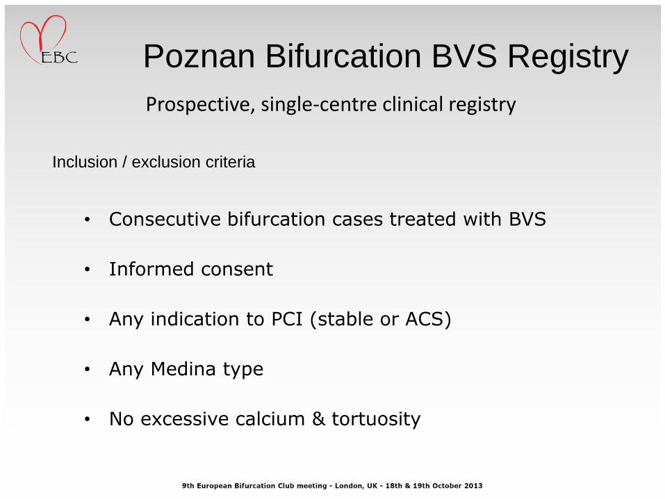
Poznan Bifurcation BVS Registry
• Consecutive bifurcation cases treated with BVS
• Informed consent
• Any indication to PCI (stable or ACS)
• Any Medina type
• No excessive calcium & tortuosity
Inclusion / exclusion criteria
Prospective, single-centre clinical registry

Baseline characteristics N = 43
Age (years) 62± 11
Male gender 33 (77%)
Diabetes mellitus 14 (32.6%)
Hypertension 33 (76.7%)
Smoking 20 (46.5%)
Previous MI 22 (52.4%)
LV Ejection Fraction (%) 51 ± 11
Previous revascularization 24 (55,8%)
Peripheral vascular disease 4 (9.3%)
Chronic Kidney Disease (eGFR<60) 6 (14.0%)
Indication for PCI
Stable angina 33 (76.7%)
Unstable angina / NSTEMI 7 (16.5%)
STEMI 3 (7.0%)
Total population

Vessel / Lesion characteristics N = 43
Bifurcation location
LM/LAD, LM/LCx 7 (16.3%)
LAD/DIA 25 (58.1%)
LCX/OM 9 (20.9%)
RCA (PL/PDA) 2 (4.7%)
True bifurcation lesion: 1,1,1 / 1,0,1 / 0,1,1 20 (46.5%)
Ostial SB only (0,0,1) 8 (18.6%)
Type B2+C 36 (83.7%)
Calcification (moderate/heavy) 10 (27%)
Thrombus (angio) 4 (9.3%)
CTO 7 (16.3%)
Vessel / lesion
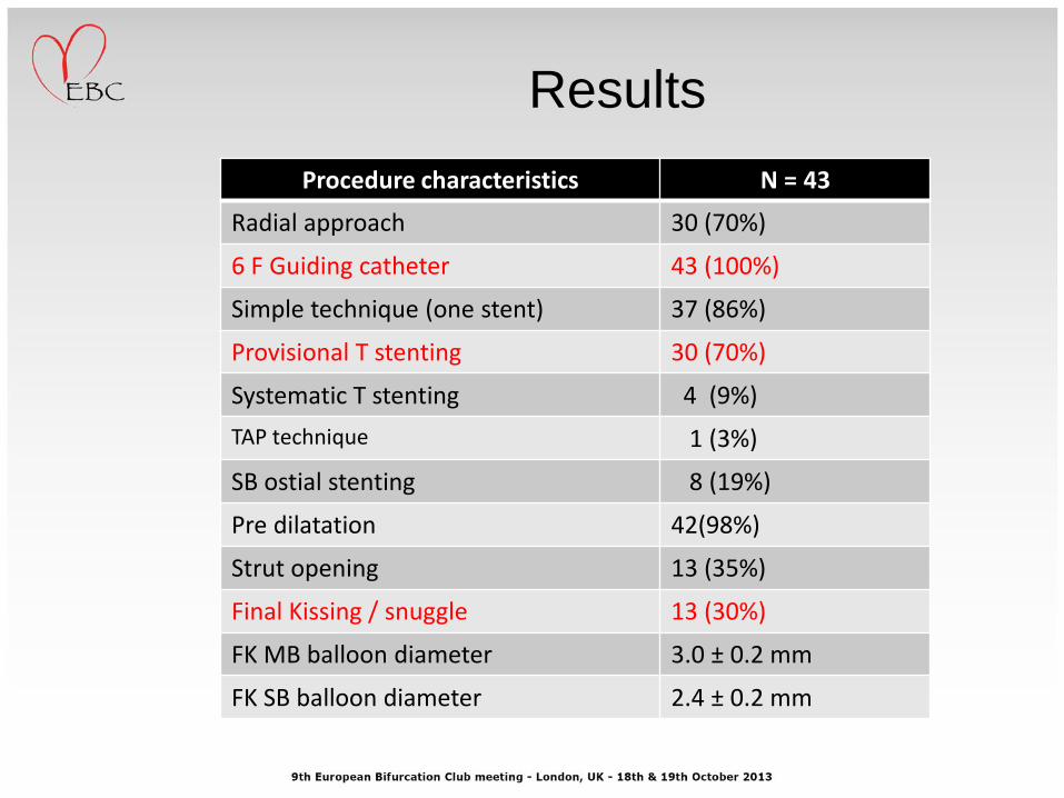
Procedure characteristics N = 43
Radial approach 30 (70%)
6 F Guiding catheter 43 (100%)
Simple technique (one stent) 37 (86%)
Provisional T stenting 30 (70%)
Systematic T stenting 4 (9%)
TAP technique 1 (3%)
SB ostial stenting 8 (19%)
Pre dilatation 42(98%)
Strut opening 13 (35%)
Final Kissing / snuggle 13 (30%)
FK MB balloon diameter 3.0 ± 0.2 mm
FK SB balloon diameter 2.4 ± 0.2 mm
Results

Vessel / Lesion characteristics N = 43
Main Vessel
Reference vessel diameter prox. 3.2 ± 0.7 mm
Reference vessel diameter dist. 2.7 ± 1.0 mm
Lesion length (mm) 11.8 ± 8.8 mm
Diameter stenosis (%) 72 ± 27 %
Side Branch
Reference vessel diameter 2.4 ± 0.5 mm
Lesion length 5.1 ± 5.4 mm
Diameter stenosis 39.6 ± 31.4 %
Angles
Proximal bifurcation angle (angle A) 140 ± 40 degrees
Distal bifurcation angle (angle B) 54± 30 degrees
Bifurcation angle < 50 degrees 26 (60.5%)
Results
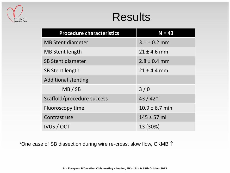
Procedure characteristics N = 43
MB Stent diameter 3.1 ± 0.2 mm
MB Stent length 21 ± 4.6 mm
SB Stent diameter 2.8 ± 0.4 mm
SB Stent length 21 ± 4.4 mm
Additional stenting
MB / SB 3 / 0
Scaffold/procedure success 43 / 42*
Fluoroscopy time 10.9 ± 6.7 min
Contrast use 145 ± 57 ml
IVUS / OCT 13 (30%)
Results
*One case of SB dissection during wire re-cross, slow flow, CKMB

In hospital Discharge-30 days
Death 0 1 (fatal stroke)
Cardiac death 0 0
Myocardial infarction 2 0
Q MI 0 0
Non-Q MI 2* 0
Stroke 0 1
Stent thrombosis (any as of ARC) 0 0
Target vessel failure 0 0
Clinical outcomes
*One case of SB dissection during wire re-cross, slow flow, CKMB
One case of septal branch closure with CKMB
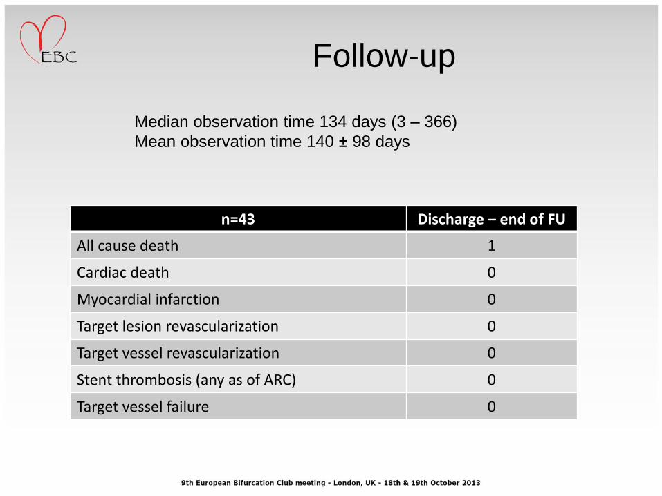
n=43 Discharge – end of FU
All cause death 1
Cardiac death 0
Myocardial infarction 0
Target lesion revascularization 0
Target vessel revascularization 0
Stent thrombosis (any as of ARC) 0
Target vessel failure 0
Follow-up
Median observation time 134 days (3 – 366)
Mean observation time 140 ± 98 days

Baseline characteristics and angiography
49YO man, stable angina class 3. Anterior MI & PCI of LAD in 2013
6 F Guide, femoral approach
Target vessel

Wired vessel
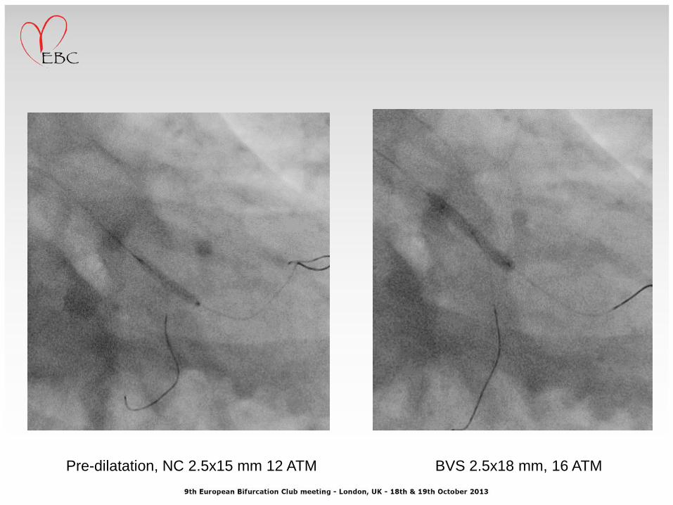
Pre-dilatation, NC 2.5x15 mm 12 ATM BVS 2.5x18 mm, 16 ATM

After BVS implantation
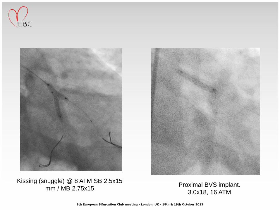
Proximal BVS implant.
3.0x18, 16 ATM
Kissing (snuggle) @ 8 ATM SB 2.5x15
mm / MB 2.75x15

Final result

OCT
SB ostium

OCT

OCT

Baseline characteristics and angiography
60 YO woman, stable angina class 2
6 F Guide, femoral approach
Target vessel

Direct BVS 3.0x18 mm, 16 ATM After implantation

POT, NC 3.25x12 mm 18 ATM

Rewiring distal strut
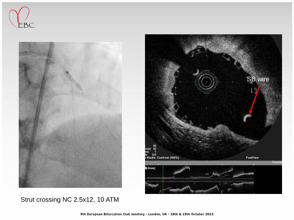
Strut crossing NC 2.5x12, 10 ATM
SB wire

BVS 2.5x18 through BVS 3.0 struts

SB BVS positioning
TAP

BVS TAP
SB BVS position Implantation:
SB BVS 2.5x18 mm, 14 ATM

BVS implantation

MB balloon 3.25x12 inflation, 4 ATM Kissing: SB BVS 2.5x18 mm, 8
ATM / MB 3.25x12, 8 ATM

IInd POT: NC 3.25x12 mm, 18 ATM

Final result
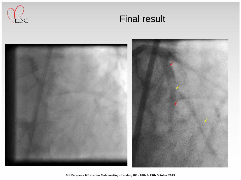
Final result

OCT
Pullback from diagonal branch Pullback from LAD

OCT
Pullback from diagonal branch Pullback from LAD
DIA LAD
Neo-carina
LAD

Breaking the limits or breaking
the struts?