single string technique for coronary bifurcation...
TRANSCRIPT

J A C C : C A R D I O V A S C U L A R I N T E R V E N T I O N S VO L . 8 , N O . 7 , 2 0 1 5
ª 2 0 1 5 B Y T H E AM E R I C A N C O L L E G E O F C A R D I O L O G Y F O U N DA T I O N I S S N 1 9 3 6 - 8 7 9 8 / $ 3 6 . 0 0
P U B L I S H E D B Y E L S E V I E R I N C . h t t p : / / d x . d o i . o r g / 1 0 . 1 0 1 6 / j . j c i n . 2 0 1 5 . 0 1 . 0 3 7
Single String Technique forCoronary Bifurcation Stenting
Detailed Technical Evaluation and Feasibility AnalysisGabor G. Toth, MD,*y Stylianos Pyxaras, MD,* Peter Mortier, MS, PHD,zx Frederic De Vroey, MD,*Giuseppe Di Gioia, MD,*k Julien Adjedj, MD,* Mariano Pellicano, MD,* Angela Ferrara, MD,*Thomas De Schryver, MS,{ Luc Van Hoorebeke, MS,{ Benedict Verhegghe, MS, PHD,zx Emanuele Barbato, MD, PHD,*kBernard De Bruyne, MD, PHD,* Matthieu De Beule, MS, PHD,zx William Wijns, MD, PHD*
ABSTRACT
Fro
Gr
Me
Un
ba
(Si
Mi
Ad
an
Ab
po
Ma
OBJECTIVES The study aimed to evaluate the adequacy and feasibility of the single string bifurcation stenting
technique.
BACKGROUND Double-stent techniques may be required for complex bifurcations. Currently applied methods all have
their morphological or structural limitations with respect to wall coverage, multiple strut layers, and apposition rate.
METHODS Single string is a novel method in which, first, the side branch (SB) stent is deployed with a single stent cell
protruding into the main branch (MB). Second, the MB stent is deployed across this protruding stent cell. The procedure
is completed by final kissing balloon dilation. The single string technique was first tested in vitro (n ¼ 20) and next
applied in patients (n ¼ 11) with complex bifurcation stenoses.
RESULTS All procedures were performed successfully, crossing a single stent cell in 100%. Procedure duration was
23.0 � 7.9 min, and the fluoroscopy time was 9.4 � 3.5 min. The results were evaluated by optical coherence tomog-
raphy, showing fully apposed struts in 83.0 � 9.2% in the bifurcation area. Residual area obstruction in the MB was
6.4 � 5.6% and 25.0 � 16.9% in the SB, as evaluated by micro computed tomography. All the human cases were
performed successfully with excellent angiographic results: the residual area stenosis was 27 � 8% and 29 � 10% in
the MB and in the SB, respectively, by 3-dimensional quantitative coronary angiography. No relevant periprocedural
enzyme increase was observed. During follow-up (6 � 4 months), no adverse clinical events (death, myocardial
infarction, target vessel revascularization) were noted.
CONCLUSIONS The single string technique for complex bifurcation dilation was shown to be adequate in vitro and
feasible in humans, with favorable results in terms of stent overlap, malapposition rate, and low residual obstruction
in both the MB and SB. (J Am Coll Cardiol Intv 2015;8:949–59) © 2015 by the American College of Cardiology
Foundation.
m the *Cardiovascular Research Centre Aalst, OLV Clinic, Aalst, Belgium; yUniversity Heart Centre Graz, Medical University of
az, Graz, Austria; zFEops Besloten Vennootschap met Beperkte Aansprakelijkheid, Ghent, Belgium; xIBiTech-bioMMeda, IMinds
dical IT, Ghent University, Ghent, Belgium; kDivision of Cardiology, Department of Advanced Biomedical Sciences, Federico II
iversity, Naples, Italy; and the {UGCT, Ghent University, Ghent, Belgium. Testing equipment, including guidewires, stents, and
lloon catheters were provided by Abbott Laboratories Inc. (Abbott Park, Illinois), Biosensors Interventional Technologies Ltd.
ngapore), Biotronik SE & Co. (Berlin, Germany), Boston Scientific Inc. (Natick, Massachusetts), and Medtronic Inc. (Minneapolis,
nnesota) without financial involvement or intellectual restriction. Drs. Verhegghe and Mortier are cofounders of FEops. Dr.
jedj is supported by a grant from Federation Française de Cardiologie. Dr. De Bruyne has received institutional grant support
d consulting fees from St. Jude Medical. Dr. De Beule is a shareholder in FEops. Dr. Wijns has received institutional grants from
bott Vascular, Biosensors Interventional Technology, Biotronik, Boston Scientific, and Medtronic. All other authors have re-
rted that they have no relationships relevant to the contents of this paper to disclose.
nuscript received September 29, 2014; revised manuscript received January 20, 2015, accepted January 29, 2015.
ABBR EV I A T I ON S
AND ACRONYMS
GW = guidewire
MB = main branch
mCT = micro computed
tomography
OCT = optical coherence
tomography
PCI = percutaneous coronary
intervention
QCA = quantitative coronary
angiography
SB = side branch
Toth et al. J A C C : C A R D I O V A S C U L A R I N T E R V E N T I O N S V O L . 8 , N O . 7 , 2 0 1 5
Single String Bifurcation Stenting Technique J U N E 2 0 1 5 : 9 4 9 – 5 9
950
T he basic concept of conventional stentsystems relies on restoration of thenative tubular geometry and expected
luminalareaofepicardialcoronaryarteries.There-fore, percutaneous coronary intervention (PCI) ofcomplex bifurcation stenosis remains a challengefor interventional cardiologists. The evolution ofbifurcation PCI resulted in several techniques, us-ing either single- and double-stent techniques (1)or a number of dedicated devices (2–5). Yet theuniversally optimal solution is still lacking,mainlydue to the highly complexmorphology with largevariation of bifurcations stenoses.
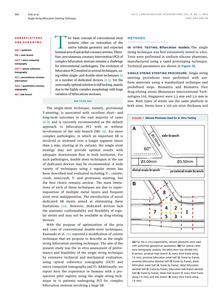
FIGURE 1 Silicone Phantoms Used for In Vitro Testing
(A) For the in vitro experiments, silicone phantoms were used
with uniformed geometrical parameters. (B) For optical coher-
ence tomography analysis, the bifurcation was divided into
8 sectors: proximal main branch (1, every third frame along
1.6 mm), proximal bifurcation ostial half (2, frame by frame),
proximal bifurcation abostial half (3, frame by frame), distal
bifurcation ostial half (4, frame by frame), distal bifurcation
abostial half (5, frame by frame), bifurcation side branch abostial
half (6, frame by frame), distal main branch (7, every third frame
along 2.4 mm), and side branch (8, every third frame along
1.6 mm).
SEE PAGE 960
The single-stent technique, namely, provisionalT-stenting, is associated with excellent short- andlong-term outcomes in the vast majority of cases(6–8) and is currently recommended as the defaultapproach to bifurcation PCI with or withoutinvolvement of the side branch (SB) (9). For morecomplex pathologies, in which an important SB isinvolved or stenosed over a longer segment (morethan 5 mm, starting at its ostium), the single stentstrategy may not provide optimal results withadequate downstream flow in both territories. Forsuch pathologies, double stent techniques or the useof dedicated devices may be recommended. A widevariety of techniques using 2 regular stents hasbeen described and evaluated including T-, culotte,crush, minicrush, T- and protrusion stenting, butthe best choice remains unclear. The main limita-tions of each of these techniques are due to super-imposition of multiple metal layers and frequentstent strut malapposition. The introduction of noveldedicated SB stents aimed at eliminating theselimitations (10). However, dedicated devices lackthe anatomic conformability and flexibility of regu-lar stents and may not be available as drug-elutingdevices.
With the purpose of optimization of the prosand cons of conventional double-stent techniques,Kawasaki et al. (11) reported a modification of culottetechnique that we propose to describe as the singlestring bifurcation stenting technique. The aim of thepresent study was the in vitro assessment of perfor-mance and feasibility of the single string techniqueby extensive technical and mechanical evaluation,using optical coherence tomography (OCT) andmicro computed tomography (mCT). Additionally, wereport here the experience in humans with a pro-spective pilot registry using the single string tech-nique in 11 patients undergoing PCI for complexbifurcation stenosis involving a large SB.
METHODS
IN VITRO TESTING. Bifurcation models. The singlestring technique was first extensively tested in vitro.Tests were performed in uniform silicone phantoms,manufactured using a rapid prototyping technique.Technical parameters are shown in Figure 1A.
SINGLE STRING STENTING PROCEDURE. Single stringstenting procedures were performed with uni-form materials using a standardized technique andpredefined steps. Biomatrix and Biomatrix Flexdrug-eluting stents (Biosensors Interventional Tech-nologies Ltd, Singapore) were 2.5 mm and 3.0 mm insize. Both types of stents use the same platform inboth sizes. Stents have a 120-mm strut thickness and
J A C C : C A R D I O V A S C U L A R I N T E R V E N T I O N S V O L . 8 , N O . 7 , 2 0 1 5 Toth et al.J U N E 2 0 1 5 : 9 4 9 – 5 9 Single String Bifurcation Stenting Technique
951
are covered by 10 mm-thick bioresorbable abluminalpolymer layer. Both platforms have 2 connectorsbetween rings and a maximal cell size of 21.2 and22.9 mm2, respectively.
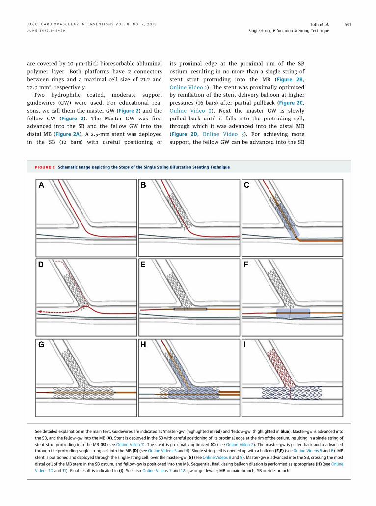
Two hydrophilic coated, moderate supportguidewires (GW) were used. For educational rea-sons, we call them the master GW (Figure 2) and thefellow GW (Figure 2). The Master GW was firstadvanced into the SB and the fellow GW into thedistal MB (Figure 2A). A 2.5-mm stent was deployedin the SB (12 bars) with careful positioning of
FIGURE 2 Schematic Image Depicting the Steps of the Single String
See detailed explanation in the main text. Guidewires are indicated as ‘ma
the SB, and the fellow-gw into the MB (A). Stent is deployed in the SB wi
stent strut protruding into the MB (B) (see Online Video 1). The stent is
through the protruding single string cell into the MB (D) (see Online Vide
stent is positioned and deployed through the single-string cell, over the m
distal cell of the MB stent in the SB ostium, and fellow-gw is positioned i
Videos 10 and 11). Final result is indicated in (I). See also Online Videos
its proximal edge at the proximal rim of the SBostium, resulting in no more than a single string ofstent strut protruding into the MB (Figure 2B,Online Video 1). The stent was proximally optimizedby reinflation of the stent delivery balloon at higherpressures (16 bars) after partial pullback (Figure 2C,Online Video 2). Next the master GW is slowlypulled back until it falls into the protruding cell,through which it was advanced into the distal MB(Figure 2D, Online Video 3). For achieving moresupport, the fellow GW can be advanced into the SB
Bifurcation Stenting Technique
ster-gw’ (highlighted in red) and ‘fellow-gw’ (highlighted in blue). Master-gw is advanced into
th careful positioning of its proximal edge at the rim of the ostium, resulting in a single string of
proximally optimized (C) (see Online Video 2). The master-gw is pulled back and readvanced
os 3 and 4). Single string cell is opened up with a balloon (E,F) (see Online Videos 5 and 6). MB
aster-gw (G) (see Online Videos 8 and 9). Master-gw is advanced into the SB, crossing the most
nto the MB. Sequential final kissing balloon dilation is performed as appropriate (H) (see Online
7 and 12. gw ¼ guidewire; MB ¼ main-branch; SB ¼ side-branch.
Toth et al. J A C C : C A R D I O V A S C U L A R I N T E R V E N T I O N S V O L . 8 , N O . 7 , 2 0 1 5
Single String Bifurcation Stenting Technique J U N E 2 0 1 5 : 9 4 9 – 5 9
952
(Online Video 4). The protruding cell was opened upto reasonable size with a 2.5-mm balloon or a1.5-mm balloon when needed (Figures 2E and 2F,Online Videos 5 and 6). Pre-dilation of the pro-truding stent cell (or the single string of the strut)allows the cell to be crossed easily with a 3.0-mmMB stent (Online Video 7). After positioning butbefore deployment of the MB stent, the fellow GWwas removed to avoid its being confined under theMB stent and the string of the SB stent. The MBstent was deployed at 12 bars (Figure 2G, OnlineVideo 8). The master GW was pulled back andpositioned in the SB after crossing the most distalcell of the MB stent facing the SB ostium (OnlineVideo 9). The fellow GW was positioned in thedistal MB. The ostium of the SB was pre-dilatedwith the previously used 2.5-mm balloon or withsmaller sized balloons when needed. Next, sequen-tial final kissing balloon dilation was performed(12 to 12 bars) with the previously used 2.5-mmballoon in the SB and the 3.0-mm stent balloon inthe MB (Figure 2H, Online Videos 10 and 11). Finaloptimization was performed in the proximal MBusing a short 3.5-mm balloon (Online Video 12).Imaging modalities. Optical coherence tomography. OCTimaging was done systematically at the end of eachin vitro procedure for the evaluation of stent strutmalapposition. Pullback runs were performed at20 mm/s with a Dragonfly Duo OCT catheter (St. JudeMedical, St. Paul, Minnesota) and analyzed by adedicated workstation (C7-XRTM OCT IntravascularImaging System, St. Jude Medical). Images wererecorded at 100 frames per second. Note that OCTpullback was only documentary and not meant toguide PCI.
For comparison purposes, analysis of the imagingdata was performed by dividing each bifurcation into8 areas, as shown in Figure 1B.
Malapposition of stent struts was calculated asdescribed previously (12), and it was graded as1) full apposition (no malapposition can be observed),2) incomplete apposition (malapposition >0 mm),3) marked malapposition (malapposition >200 mm),and 4) floating struts (malapposition >500 mm).Micro computed tomography. Final stent deformationswere visualized at a voxel resolution of 12 mm/voxel.From the scanned volumes, the stents were recon-structed by segmentation using Mimics (MaterialiseInc., Leuven, Belgium).
The following parameters were quantified for allphantoms: ostial area stenosis in the distal MB, ostialarea stenosis in the SB, wall coverage in the ostialSB, angulation between the MB and SB axes, andangulation between the MB plane and the plane of the
protruding cell (string angle). Ostial area stenosis inthe distal MB was quantified in a planar projectionperpendicular to the MB axis according to the defi-nition proposed by Ormiston et al. (13): (A1 � A2)/A1 �100%, where A1 is the total area of the distal MBostium and A2 is the largest area free of struts. Ostialarea stenosis in the SB was quantified similarly(Figure 3). Loss of wall coverage in the ostial SB wasquantified as (C2 � C1)/ C1 � 100%, where C1 is thearea of the largest fitting circle in the most distalcell of the SB stent (regular deployment), and C2is the area of the largest fitting circle in themost proximal cell at the SB (cell between the secondand third rings of the SB stent, i.e., region of stentdeformation).
PROSPECTIVE HUMAN PILOT REGISTRY. Patients withtrue bifurcation lesions, involving at least 1 of theMB segments plus the SB, with SB stenosis extend-ing for $5 mm, were selected for single string pro-cedure. The PCI procedure was performed accordingto the standardized protocol and procedural steps, asdescribed for the in vitro study. Various stent brandswere used: selection criterion was to have uniformlylarge cells of a minimum of 4.4-mm maximal cellexpansion diameter. Procedural characteristics wererecorded prospectively. Informed patient consentwas obtained for the diagnostic and PCI procedures,data collection, and reporting. Results were evalu-ated by 3-dimensional quantitative coronary angi-ography (QCA). Angiographic images were acquiredat 15 or 30 frames per second (Innova 4100,General Electric Inc., Fairfield, Ohio and AxiomArtis, Siemens Inc., Forchheim, Germany). Three-dimensional QCA was performed offline using QAn-gio XA 3D Research Edition 1.0 software package(Medis Specials BV, Leiden, the Netherlands) (14).This software allows the volumetric reconstructionof the luminal and reference diameters of theanalyzed segments from 2 different projections atleast 25� apart, preferably with the least fore-shortening and yielding the best depiction of thestenotic coronary segments.
STATISTICAL ANALYSIS. All analyses were per-formed with Prism GraphPad 5.0 (GraphPad SoftwareInc., La Jolla, California). Summary descriptive sta-tistics are reported as mean � SD, median (inter-quartile range), or n (%), as appropriate.
RESULTS
IN VITRO TESTS. Procedure . All 20 in vitro pro-cedures were successfully performed according tothe protocol; OCT and mCT analyses were completed

FIGURE 3 Results of the In Vitro Tests
Nine representative examples from the in vitro procedures: fluoroscopic results. Fluoroscopic images show the minimal overlap between the
main branch stent and the side branch stent. Additionally, good apposition and preserved strut structure are also well identifiable.
J A C C : C A R D I O V A S C U L A R I N T E R V E N T I O N S V O L . 8 , N O . 7 , 2 0 1 5 Toth et al.J U N E 2 0 1 5 : 9 4 9 – 5 9 Single String Bifurcation Stenting Technique
953
in each case. Procedure duration was 23.0 � 7.9 minwith a fluoroscopy time of 9.4 � 3.5 min. It took inaverage 1:04 min (minimum, 0:40; maximum, 2:27)to position the master GW in the distal MB throughthe string cell. In 19 cases, the initially chosenworkhorse guidewires were allowed to complete theprocedure. On average, 2.1 � 1.2 balloons were usedper procedure, including 1 for the final proximaloptimization. Of these dilation balloon catheters,1.0 � 1.2 were used for opening the string cell.Each procedure was completed with the 2 stents
intended for use. Figure 4 shows examples from invitro cases.Opt ica l coherence tomography . For the OCT runsobtained in 20 phantoms, 1402 OCT frames wereanalyzed, and 9,267 struts were identified for eval-uation. Perfect apposition was seen in 8,040struts (87%); 285 (3%) showed marked malapposition(>200 mm) and 96 (1%) were floating (>500 mm).Limiting the analysis to the bifurcation areas (area 2to 6 as shown on Figure 1B), 6,328 struts were iden-tified for evaluation. Of these, 4,368 struts (69%)
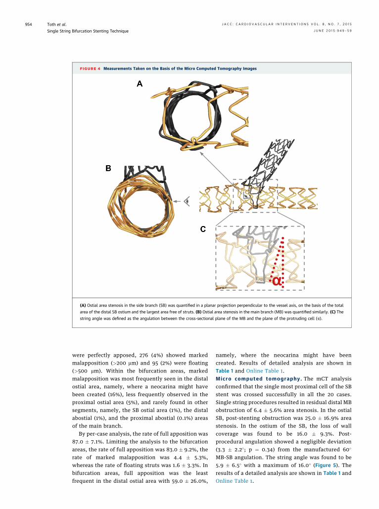
FIGURE 4 Measurements Taken on the Basis of the Micro Computed Tomography Images
(A) Ostial area stenosis in the side branch (SB) was quantified in a planar projection perpendicular to the vessel axis, on the basis of the total
area of the distal SB ostium and the largest area free of struts. (B) Ostial area stenosis in the main branch (MB) was quantified similarly. (C) The
string angle was defined as the angulation between the cross-sectional plane of the MB and the plane of the protruding cell (a).
Toth et al. J A C C : C A R D I O V A S C U L A R I N T E R V E N T I O N S V O L . 8 , N O . 7 , 2 0 1 5
Single String Bifurcation Stenting Technique J U N E 2 0 1 5 : 9 4 9 – 5 9
954
were perfectly apposed, 276 (4%) showed markedmalapposition (>200 mm) and 95 (2%) were floating(>500 mm). Within the bifurcation areas, markedmalapposition was most frequently seen in the distalostial area, namely, where a neocarina might havebeen created (16%), less frequently observed in theproximal ostial area (5%), and rarely found in othersegments, namely, the SB ostial area (1%), the distalabostial (1%), and the proximal abostial (0.1%) areasof the main branch.
By per-case analysis, the rate of full apposition was87.0 � 7.1%. Limiting the analysis to the bifurcationareas, the rate of full apposition was 83.0 � 9.2%, therate of marked malapposition was 4.4 � 5.3%,whereas the rate of floating struts was 1.6 � 3.3%. Inbifurcation areas, full apposition was the leastfrequent in the distal ostial area with 59.0 � 26.0%,
namely, where the neocarina might have beencreated. Results of detailed analysis are shown inTable 1 and Online Table 1.Micro computed tomography. The mCT analysisconfirmed that the single most proximal cell of the SBstent was crossed successfully in all the 20 cases.Single string procedures resulted in residual distal MBobstruction of 6.4 � 5.6% area stenosis. In the ostialSB, post-stenting obstruction was 25.0 � 16.9% areastenosis. In the ostium of the SB, the loss of wallcoverage was found to be 16.0 � 9.3%. Post-procedural angulation showed a negligible deviation(3.3 � 2.2�; p ¼ 0.34) from the manufactured 60�
MB-SB angulation. The string angle was found to be5.9 � 6.5� with a maximum of 16.0� (Figure 5). Theresults of a detailed analysis are shown in Table 1 andOnline Table 1.
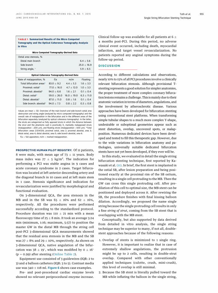
TABLE 1 Summarized Results of the Micro Computed
Tomography and the Optical Coherence Tomography Analysis
In Vitro
Micro Computed Tomography–Derived Data
Ostial area stenosis, %
Distal main branch 6.4 � 5.6
Side branch 25.0 � 16.9
String angle, � 5.9 � 6.5
Optical Coherence Tomography–Derived Data
Rate of malapposition, % f/a m/m Floating
Total bifurcation areas* 83.0 � 9.2 4.4 � 5.3 1.6 � 3.3
Proximal: ostial* 77.0 � 16.0 4.7 � 12.0 1.0 � 3.3
Proximal: abostial* 94.0 � 6.6 1.6 � 2.1 0.1 � 0.4
Distal: ostial* 59.0 � 26.0 16.0 � 19.0 6.3 � 11.0
Distal: abostial* 87.0 � 11.0 0.8 � 1.6 0.1 � 0.4
Side branch: abostial* 94.0 � 7.3 0.8 � 2.2 0.3 � 0.8
Values are mean � SD. Overview of the main branch and side branch ostial areaobstruction and string angle (analyzed by micro computed tomography) and theoverall rate of malapposition in the bifurcation and in the different areas of thebifurcation separately (analyzed by optical coherence tomography). In the latter,the struts are categorized as fully apposed struts in which the distance betweenthe strut and the phantoms wall is practically 0, markedly malapposed struts(malapposition >200 mm), and floating struts (malapposition >500 mm). *Totalbifurcation: areas 2/3/4/5/6; proximal ostial, area 2; proximal abostial, area 3;distal ostial, area 4; distal abostial, area 5; side branch abostial, area 6.
f/a ¼ full apposition; m/m ¼ marked malapposition.
J A C C : C A R D I O V A S C U L A R I N T E R V E N T I O N S V O L . 8 , N O . 7 , 2 0 1 5 Toth et al.J U N E 2 0 1 5 : 9 4 9 – 5 9 Single String Bifurcation Stenting Technique
955
PROSPECTIVE HUMAN PILOT REGISTRY. Of 11 patients,8 were male, with mean age of 73 � 11 years. Bodymass index was 27 � 5 kg/m2. The indication forperforming a PCI was stable angina in 9 cases andacute coronary syndrome in 2 cases. Target bifurca-tion was located at left anterior descending artery andthe diagonal branch in 10 cases and at left main stemin 1 case. Stenosis significance and indication forrevascularization were justified by morphological andfunctional evaluation.
On 3-dimensional QCA, the area stenosis in theMB and in the SB was 65 � 16% and 62 � 16%,respectively. All the procedures were performedsuccessfully according to the standardized protocol.Procedure duration was 110 � 21 min with a meanfluoroscopy time of 25 � 8 min. It took an average 3:24min (minimum, 1:16; maximum, 5:57) to position themaster GW in the distal MB through the string cellpost-PCI 3-dimensional QCA measurements showedthat the residual area stenosis in the MB and the SBwas 27 � 8% and 29 � 10%, respectively. As shown on3-dimensional QCA, native angulation of the bifur-cation was 38 � 19�, which was modified by 0 � 16�
(p ¼ 0.99) after stenting (Online Table 2).Equipment use consisted of 3 guidewires (IQR: 2 to
3) and 4 balloon catheters (IQR: 3 to 5). Contrast mediause was 340 � 118 ml. Figure 6 shows case examples.
Pre- and post-procedural cardiac enzyme levelsshowed no relevant periprocedural enzyme increase.
Clinical follow-up was available for all patients at 6 �4 months post-PCI. During this period, no adverseclinical event occurred, including death, myocardialinfarction, and target vessel revascularization. Nopatients reported any anginal symptoms during thefollow-up period.
DISCUSSION
According to different calculations and observations,nearly 10% to 15%of all PCI procedures involve a clinicallyrelevant bifurcation stenosis. Although provisional T-stenting represents a good solution for simpler anatomies,the proper treatment of more complex coronary bifurca-tion lesions remainsachallenge.This ismainlydue to theiranatomicvariation in termsofdiameters, angulations, andthe involvement by atherosclerotic disease. Variousapproaches have been developed for bifurcation stentingusing conventional stent platforms. When transformingsimple tubular shapes to a much more complex Y-shape,undesirable or suboptimal geometries appear such asstent distortion, overlap, uncovered spots, or malap-position. Numerous dedicated devices have been devel-oped and tested to fill this therapeutic gap. However, dueto the wide variations in bifurcation anatomy and pa-thologies, universally suitable dedicated bifurcationstents have not yet been developed, if they ever will be.
In this study, we evaluated in detail the single stringbifurcation stenting technique, first reported by Ka-wasaki et al. (11). In brief, the first stent is implanted inthe ostial SB, after lesion preparation and being posi-tioned exactly at the proximal rim of the SB ostium,resulting in a single cell protruding to theMB. Then theGW can cross this single protruding cell. After pre-dilation of this cell to optimal size, the MB stent can bepositioned and deployed across it. After rewiring theSB, the procedure finishes with final kissing balloondilation. Accordingly, we proposed the name singlestring because the single protruding cell results in onlya fine string of strut, coming from the SB stent that isoverlapping with the MB stent.
Conceptually, but also supported by data derivedfrom detailed in vitro analysis, the single stringtechnique may be superior to many, if not all, double-stent approaches because of the following reasons:
1. Overlap of stents is minimized to 1 single ring.However, it is important to realize that in case ofextremely shallow angulations, the protrusionmight be up to 2 cells, resulting in double-strutoverlap. Compared with other conventionallyapplied techniques (culotte, crush, mini-crush),this level of overlap is still minimal.
2. Because the SB stent is literally pulled toward theMB while inflating the balloon in the single string,
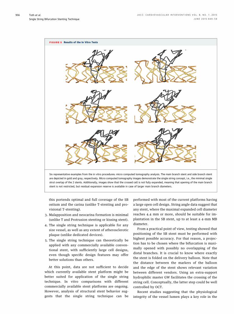
FIGURE 5 Results of the In Vitro Tests
Six representative examples from the in vitro procedures: micro computed tomography analysis. The main branch stent and side branch stent
are depicted in gold and gray, respectively. Micro computed tomography images demonstrate the single string concept, i.e., the minimal single
strut overlap of the 2 stents. Additionally, images show that the crossed cell is not fully expanded, meaning that opening of the main branch
stent is not restricted, but residual expansion reserve is available in case of larger main branch diameters.
Toth et al. J A C C : C A R D I O V A S C U L A R I N T E R V E N T I O N S V O L . 8 , N O . 7 , 2 0 1 5
Single String Bifurcation Stenting Technique J U N E 2 0 1 5 : 9 4 9 – 5 9
956
this portends optimal and full coverage of the SBostium and the carina (unlike T-stenting and pro-visional T-stenting).
3. Malapposition and neocarina formation is minimal(unlike T and Protrusion stenting or kissing stent).
4. The single string technique is applicable for anysize vessel, as well as any extent of atheroscleroticplaque (unlike dedicated devices).
5. The single string technique can theoretically beapplied with any commercially available conven-tional stent, with sufficiently large cell designs,even though specific design features may offerbetter solutions than others.
At this point, data are not sufficient to decidewhich currently available stent platform might bebetter suited for application of the single stringtechnique. In vitro comparisons with differentcommercially available stent platforms are ongoing.However, analysis of structural stent behavior sug-gests that the single string technique can be
performed with most of the current platforms havinga large open cell design. String angle data suggest thatany stent, where the maximal expanded cell diameterreaches 4.4 mm or more, should be suitable for im-plantation in the SB stent, up to at least a 4-mm MBdiameter.
From a practical point of view, testing showed thatpositioning of the SB stent must be performed withhighest possible accuracy. For that reason, a projec-tion has to be chosen where the bifurcation is maxi-mally opened with possibly no overlapping of thedistal branches. It is crucial to know where exactlythe stent is folded on the delivery balloon. Note thatthe distance between the markers of the balloonand the edge of the stent shows relevant variationbetween different vendors. Using an extra-supporthydrophilic master GW facilitates the crossing of thestring cell. Conceptually, the latter step could be wellcontrolled by OCT.
Recent studies suggesting that the physiologicalintegrity of the vessel lumen plays a key role in the

FIGURE 6 Results of the Human Study
Four representative examples from the human cases: angiographic result. (Left)
Pre-procedural (pre-PCI) anatomy. (Right) Post-procedural (post-PCI) result.
J A C C : C A R D I O V A S C U L A R I N T E R V E N T I O N S V O L . 8 , N O . 7 , 2 0 1 5 Toth et al.J U N E 2 0 1 5 : 9 4 9 – 5 9 Single String Bifurcation Stenting Technique
957
maintenance of normal flow patterns, shearstresses, and hemodynamics. Accordingly, thepresence of any disturbance (i.e., oval shape,changed angulation, malapposed struts, localizedstent deformation) might be associated with turbu-lent flow patterns and loss in driving pressure,impaired shear-stress pattern, and even abnormalplatelet activation, leading to potential risk ofadverse events (15–17). Increased risk of stentthrombosis or restenosis might be expected in caseof suboptimal post-PCI results (18). In vitro testswith the selected stent platform demonstrated bothhigh technical feasibility and excellent proceduralresults, i.e., minimal strut distortion, preservedvessel-wall coverage, optimal MB and SB openingsat the ostium, favorable malapposition rates, andintact SB angulation. These findings suggest thatpost-procedural anatomy after a single string pro-cedure can be ideally close to the native anatomicstructure. Although detailed physiological evalua-tion was beyond the scope of this project and thusremains to be investigated, it can be anticipatedthat near-normal geometry will translate into he-modynamic and pathophysiological benefits.
The numbers in the presented first-in-humanspilot registry are limited, which must be taken intoconsideration while interpreting the derived data,but clinical cases remain informative: all the caseswere performed successfully, within a reasonablelength of time and using the usual amount of contrastand radiation exposure. Furthermore, post-procedural laboratory tests did not show clinicallyrelevant periprocedural myocardial necrosis. It isimportant to emphasize that the single string tech-nique is safe to perform because, in case of diffi-culties, the procedure can be immediately convertedto the culotte technique (if the protrusion into theMB was unnecessarily long), the mini-crush tech-nique (if the protrusion into the MB was too small), orT-stenting (if no protrusion into the MB was obtainedat all).
STUDY LIMITATIONS. Our in vitro results cannot becompared directly with other bifurcation techniquesor dedicated devices, mainly due to the fact that suchstandardized and detailed evaluation with the sameimaging protocol has not been universally performed.In vitro and in vivo studies with limited sample sizesinvestigating various bifurcation techniques reportedmalapposition rates in the region of bifurcationranging between 30% and 45% (19–21), suggesting apotential advantage of the single string techniquecompared with those results. Of note, in vitro phan-toms can never represent the variety of in vivo
coronary anatomies and pathologies in terms ofangulation, disease distribution, and calibers.
Due to geometric incoherence (i.e., the angulationbetween the plane of the OCT image and the true cross-sectional plane of the SB ostium), the accuracy of OCTin evaluating malapposition is hampered by markedoverestimation, especially in the area of the carina. Inthose cases, reality might be better than measured.
PERSPECTIVES
WHAT IS KNOWN? Percutaneous coronary inter-
vention of complex bifurcation stenosis is still a chal-
lenge for interventional cardiologists. Evolution of
bifurcation interventions resulted in several approaches,
using single-stent techniques, double-stent techniques,
or dedicated devices, but the best choice remains un-
clear. The main limitations of each of these techniques
are due to superimposition of multiple metal layers and
frequent stent strut malapposition. The introduction of
novel, dedicated stents aimed at eliminating these lim-
itations; however, dedicated devices lack the anatomic
conformability and flexibility of regular stents and may
not be available as drug-eluting devices.
WHAT IS NEW? In this study, we evaluated in detail
the single string bifurcation stenting technique. In
Toth et al. J A C C : C A R D I O V A S C U L A R I N T E R V E N T I O N S V O L . 8 , N O . 7 , 2 0 1 5
Single String Bifurcation Stenting Technique J U N E 2 0 1 5 : 9 4 9 – 5 9
958
Although feasibility was thoroughly tested invitro and in humans, the number of clinical casesremains limited, and no long-term follow-up isavailable. Accordingly, medium- and long-term clin-ical safety, as well as more detailed procedural datasuch as procedure duration, radiation exposure anduse of equipment, and intravascular imaging, will beevaluated within the context of an ongoing prospec-tive registry. In the first-in-humans pilot registry, theprotocol did not include standard use of intravascularimaging methods, such as OCT. Therefore, despiteoptimal crossing of the SB stent on fluoroscopy, itcannot be confirmed that the most proximal cell wastaken in all cases. No randomized comparison withany other bifurcation technique or dedicated device isavailable yet either. Being a proof-of-concept study,comparison with any other bifurcation technique ordedicated device either in vitro or in human cases wasbeyond the scope of this work.
brief, the first stent is implanted into the ostial side
branch, resulting in a single cell protruding to the
main branch. Then the main branch stent is deployed
across this single cell, and the procedure is completed
by final kissing dilation. The concept ensures full
coverage of the whole bifurcation area in both
branches although having a minimal double layer as a
single string of a strut. In this proof-of-concept study,
the single string bifurcation technique was shown to
be feasible in vitro and in humans as well. Data indi-
cate minimal overlap, maximal wall coverage, and a
favorable malapposition rate.
WHAT IS NEXT? Although in vitro and the first-in-
humans data are encouraging, further more detailed in
human evaluation of the technique is essential. Sys-
tematic intravascular imaging will be applied within
the context of an ongoing prospective registry. Clin-
ical effectiveness and comparison with other bifurca-
tion techniques remain to be confirmed in a larger
study population.
CONCLUSIONS
Bifurcation PCI remains challenging because anytechnique needs to find the optimal balance betweenwall coverage, stent overlap, and malapposition,whereas adapting to variable anatomy. In this proof-of-concept study, single string bifurcation techniquewas shown to be feasible in vitro and in humans aswell. Data indicate minimal overlap, maximal wallcoverage, and a favorable malapposition rate. Clinicaleffectiveness remains to be confirmed in a largerstudy population.
ACKNOWLEDGMENT The authors thank Medis Inc.(Leiden, the Netherlands) for providing the QCAsoftware.
CORRESPONDENCE AND REPRINT REQUESTS: Dr.William Wijns, Cardiovascular Research Centre Aalst,OLV-Ziekenhuis, Moorselbaan 164, Aalst, B9300,Belgium. E-mail: [email protected].
RE F E RENCE S
1. Louvard Y, Thomas M, Dzavik V, et al. Classifi-cation of coronary artery bifurcation lesions andtreatments: time for a consensus! Catheter Car-diovasc Interv 2008;71:175–83.
2. Onuma Y, Müller R, Ramcharitar S, et al. Tryton I,First-In-Man (FIM) study: six month clinicaland angiographic outcome, analysis with newquantitative coronary angiography dedicated forbifurcation lesions. EuroIntervention 2008;3:546–52.
3. Latib A, Chieffo A. The Cappella Sideguard�stent. EuroIntervention 2010;6 Suppl:J143–6.
4. Grube E, Buellesfeld L, Neumann FJ, et al. Six-monthclinicalandangiographic results of adedicateddrug-eluting stent for the treatment of coronary bi-furcation narrowings. Am J Cardiol 2007;99:1691–7.
5. Gil RJ, Vassilev D, Michalek A, et al. Dedi-cated paclitaxel-eluting bifurcation stent BiOSS�(bifurcation optimisation stent system): 12-monthresults from a prospective registry of consecutive
all-comers population. EuroIntervention 2012;8:316–24.
6. Chen SL, Santoso T, Zhang JJ, et al.A randomized clinical study comparing doublekissing crush with provisional stenting for treat-ment of coronary bifurcation lesions results fromthe DKCRUSH-II (Double Kissing Crush versusProvisional Stenting Technique for Treatment ofCoronary Bifurcation Lesions) Trial. J Am CollCardiol 2011;57:914–20.
J A C C : C A R D I O V A S C U L A R I N T E R V E N T I O N S V O L . 8 , N O . 7 , 2 0 1 5 Toth et al.J U N E 2 0 1 5 : 9 4 9 – 5 9 Single String Bifurcation Stenting Technique
959
7. Chen SL, Zhang Y, Xu B, et al. Five-year clinicalfollow-up of unprotected left main bifurcationlesion stenting: one-stent versus two-stent tech-niques versus double-kissing crush technique.EuroIntervention 2012;8:803–14.
8. Maeng M, Holm NR, Erglis A, et al. Long-termresults after simple versus complex stenting ofcoronary artery bifurcation lesions: Nordic Bifur-cation Study 5-year follow-up results. J Am CollCardiol 2013;62:30–4.
9. Stankovic G, Lefèvre T, Chieffo A, et al.,European Bifurcation Club. Consensus from the7th European Bifurcation Club meeting. Euro-Intervention 2013;9:36–45.
10. Foin N, Alegria-Barrero E, Torii R, et al. Crush,culotte, T and protrusion: which 2-stent techniquefor treatment of true bifurcation lesions? Insightsfrom in vitro experiments and micro-computedtomography. Circ J 2013;77:73–80.
11. Kawasaki T, Koga H, Serikawa T. Modifiedculotte stenting technique for bifurcation lesions:the cross-stenting technique. J Invasive Cardiol2010;22:243–6.
12. Lowe HC, Narula J, Fujimoto JG, Jang IK.Intracoronary optical diagnostics: current status,limitations, and potential. J Am Coll Cardiol Intv2011;4:1257–70.
13. Ormiston JA, Webster MW, Webber B,Stewart JT, Ruygrok PN, Hatrick RI. The “crush”technique for coronary artery bifurcation stenting:insights from micro-computed tomographic im-aging of bench deployments. J Am Coll CardiolIntv 2008;1:351–7.
14. Tu S, Holm NR, Christiansen EH, Reiber JH.First presentation of 3-dimensional reconstructionand centerline-guided assessment of coronarybifurcation by fusion of X-ray angiography andoptical coherence tomography. J Am Coll CardiolIntv 2012;5:884–5.
15. Gijsen FJ, Mastik F, Schaar JA, et al. High shearstress induces a strain increase in human coronaryplaques over a 6-month period. EuroIntervention2011;7:121–7.
16. Tu S, Pyxaras SA, Li Y, Barbato E, Reiber JH,Wijns W. In vivo flow simulation at coronarybifurcation reconstructed by fusion of 3-dimen-sional X-ray angiography and optical coherencetomography. Circ Cardiovasc Interv 2013;6:e15–7.
17. Pyxaras SA, Tu S, Barbato E, Reiber JH,Wijns W. Optimization of Tryton dedicated coro-nary bifurcation system with coregistration ofoptical coherence tomography and fractional flowreserve. J Am Coll Cardiol Intv 2013;6:e39–40.
18. Samady H, Eshtehardi P, McDaniel MC, et al.Coronary artery wall shear stress is associated with
progression and transformation of atheroscleroticplaque and arterial remodeling in patients withcoronary artery disease. Circulation 2011;124:779–88.
19. Tyczynski P, Ferrante G, Moreno-Ambroj C,et al. Simple versus complex approaches to treatingcoronary bifurcation lesions: direct assessment ofstent strut apposition by optical coherence to-mography. Rev Esp Cardiol 2010;63:904–14.
20. Foin N, Torii R, Alegria E, et al. Location of sidebranch access critically affects results in bifurca-tion stenting: Insights from bench modeling andcomputational flow simulation. Int J Cardiol 2013;168:3623–8.
21. Viceconte N, Tyczynski P, Ferrante G, et al. Im-mediate results of bifurcational stenting assessedwith optical coherence tomography. Catheter Car-diovasc Interv 2013;81:519–28.
KEY WORDS coronary bifurcation,double-stent technique, first-in-human,in vitro, percutaneous intervention
APPENDIX For supplemental tablesand videos, please see the online version ofthis article.