bacterial macroscopic rope-like fibers with cytopathic and
TRANSCRIPT
Bacterial Macroscopic Rope-like Fibers with Cytopathic andAdhesive Properties*□S
Received for publication, July 7, 2010 Published, JBC Papers in Press, August 5, 2010, DOI 10.1074/jbc.M110.162248
Juan Xicohtencatl-Cortes‡, Zeus Saldana§, Wanyin Deng¶, Elsa Castaneda�, Enrique Freer**, Phil I. Tarr‡‡,B. Brett Finlay¶, Jose Luis Puente§§, and Jorge A. Giron§1
From the ‡Laboratorio de Bacteriología Intestinal, Hospital Infantil de Mexico Federico Gomez, Dr. Marquez 162, Col. Doctores,Delegacion Cuauhtemoc, Mexico D.F. 06720, Mexico, the §Department of Molecular Genetics and Microbiology, University ofFlorida, Gainesville, Florida 32610, ¶Michael Smith Laboratories, The University of British Columbia, Vancouver, British ColumbiaV6T 1Z4, Canada, the �Centro de Investigaciones en Ciencias Microbiologicas, Benemerita Universidad Autonoma de Puebla,Puebla 72000, Mexico, the **Centro de Investigacion Estructuras Microscopicas, Universidad de Costa Rica, San Jose, Costa Rica,the ‡‡Department of Pediatrics, Washington University, St. Louis, Missouri 63105, and the §§Departamento de MicrobiologíaMolecular, Instituto de Biotecnología, Universidad Nacional Autonoma de Mexico, Cuernavaca, Morelos 62210, Mexico
We present a body of ultrastructural, biochemical, andgenetic evidence that demonstrates the oligomerization of viru-lence-associated autotransporter proteins EspC or EspP pro-duced by deadly human pathogens enterohemorrhagic andenteropathogenic Escherichia coli into novelmacroscopic rope-like structures (>1 cm long). The rope-like structures showedhigh aggregation and insolubility, stability to anionic detergentsand high temperature, and binding to Congo Red and thioflavinT dyes. These are properties also exhibited by human amyloido-genic proteins. These macroscopic ropes were not observed incultures of nonpathogenic Escherichia coli or isogenic espP orespC deletion mutants of enterohemorrhagic or enteropatho-genic Escherichia coli but were produced by an Escherichia coliK-12 strain carrying a plasmid expressing espP. Purified recom-binant EspP monomers were able to self-assemble into macro-scopic ropes upon incubation, suggesting that no other proteinwas required for assembly.The ropes bound to and showedcyto-pathic effects on cultured epithelial cells, served as a substratumfor bacterial adherence and biofilm formation, and protectedbacteria from antimicrobial compounds. We hypothesize thatthese ropes play a biologically significant role in the survival andpathogenic scheme of these organisms.
Pathogenic bacteria produce a prodigious array of virulencefactors in the formof adhesins, toxins, cytoskeleton-manipulat-ing effectors, enzymes, and mediators of motility that are usu-ally secreted across the bacterial cell surface by a variety ofsimple or complex highly organized secretion mechanisms (1).In doing so, infectious bacteria ensure their ability to surviveand multiply within their host. Among the different protein
secretion mechanisms described in Gram-negative bacteria,the type 5 protein secretion system is perhaps the simplest of allbecause it does not require the repertoire of components andsophisticated structures associated with other systems (1–4).Proteins secreted via this pathway are called autotransporters(AT)2 because their export from the cytosol across the doublemembrane is a self-mediated process attributed to the presenceof defined domains with specific functions required for secre-tion. TheN terminus of ATproteins contains the signal peptidethat leads the protein toward the Sec-dependent pathway. TheC terminus contains the �-domain, which forms a �-barrelpore-like structure in the outer membrane through which thepassenger domain, corresponding to the mid-portion of theprotein, is secreted and can remain associated to the cell surfaceor be released to the milieu (2). Members of the family ofSPATE (serine protease ATs of the Enterobacteriacea) are pro-teins from Escherichia coli (E. coli) and Shigella spp., whichpossess a consensus serine protease motif (2). SPATEs are gen-erally large proteins that display an array of distinct biologicalactivities such as adhesins, hemagglutinins, cytotoxins, orenzymes with different substrate specificities (2).Enterohemorrhagic E. coli (EHEC) O157:H7 is a food-borne
pathogen implicated as a major cause of diarrheal illness indeveloped countries. The production of Shiga toxins and colo-nization of the large intestine are central aspects of the patho-genicity of EHEC strains (5). Cattle are natural hosts of EHECO157:H7, and they constitute the primary source of infectionfor humans (5). Enteropathogenic E. coli (EPEC) is a cause ofpotentially fatal infantile diarrhea in developing countries (5).EPEC and EHEC possess a pathogenicity island termed thelocus of enterocyte effacement, which contains a repertoire ofgenes encoding regulators, the adhesin intimin and its receptorTir, a type 3 secretion system (T3SS) machinery, and T3secreted effector molecules, that together with non-locus ofenterocyte effacement encoded effectors (Nles), act in concertto confer on the bacteria the ability to adhere and inflict damageto the gutmucosa (5). In addition to the outermembrane adhe-
* This work was supported, in whole or in part, by National Institutes of HealthGrants AI66012 and AI63211 (to J. A. G.). This work was also supported byDGAPA Grant IN201703-3 and CONACyT Grant 42918Q (to J. L. P.) andgrants from Canadian Institutes of Health Research and Howard HughesMedical Institute to BBF.
□S The on-line version of this article (available at http://www.jbc.org) containssupplemental text, Tables S1–S3, and Figs. S1–S3.
1 To whom correspondence should be addressed: Dept. of Molecular Genet-ics and Microbiology, Emerging Pathogens Institute, University of Florida,1600 SW Archer Rd., P.O. Box 100266, Gainesville, FL 32610. Tel.: 352-273-8892; Fax: 520-273-9420; E-mail: [email protected].
2 The abbreviations used are: AT, autotransporter; EHEC, enterohemorrhagicE. coli; EPEC, enteropathogenic E. coli; T3SS, type 3 secretion system; SEM,scanning electron microscopy; LDH, lactate dehydrogenase; CR, CongoRed; ThT, thioflavin T; HUS, hemolytic uremic syndrome.
THE JOURNAL OF BIOLOGICAL CHEMISTRY VOL. 285, NO. 42, pp. 32336 –32342, October 15, 2010© 2010 by The American Society for Biochemistry and Molecular Biology, Inc. Printed in the U.S.A.
32336 JOURNAL OF BIOLOGICAL CHEMISTRY VOLUME 285 • NUMBER 42 • OCTOBER 15, 2010
by guest on February 12, 2018http://w
ww
.jbc.org/D
ownloaded from
sin intimin (5), several other pili or non-pili adhesins have beendescribed in EPEC and EHEC O157:H7 (6–10).EspP and EspC are SPATE proteins (�100 kDa) secreted by
EHEC and EPEC, respectively, that share significant similarityat their amino acid sequence level and are two of the predomi-nant secreted proteins found in liquid culture supernatants ofthese organisms (2). The EspC protein of EPEC shows consid-erable homologywith the IgAprotease ofNeisseria gonorrhoeaeand Haemophilus influenza, although it does not display thisactivity (2, 11). Enterotoxin and cytotoxin activities on ratjejunal segments and cultured epithelial cells have been dem-onstrated for EspC (12, 13). Cytotoxicity is more efficient whenEspC is translocated during contact of EPEC with epithelialcells (14), a process that apparently also requires the participa-tion of the T3SS (15). Recently, it was also found that EspCconferred enhanced lysozyme resistance to EPEC (16). EspC andEspP display enzymatic activities, and although EspC cleavesspectrin (also called fodrin) and hemoglobin, EspP of EHECcleaves pepsin A, and human coagulation factor V (2, 11, 14,17–19). The EspP protein was also shown to influence gut col-onization in calves and contributes to adherence of EHEC tobovine primary rectal cells (20). Recently, EspP was found to bedirectly involved in biofilm formation and adherence of EHECto T84 intestinal epithelial cells (21). In this study, we providecompelling evidence that EspC and EspP proteins oligomerizeinto macroscopic rope-like structures that possess cell adhe-siveness, exert enzymatic and cytopathic effects, and serve as asubstratum for bacterial biofilm formation sheltering the bac-teria from external foes.
EXPERIMENTAL PROCEDURES
Bacterial Strains and Culture Conditions—The bacterialstrains and plasmids used in this study are listed insupplemental Table S1. The bacteria were cultured in Luria-Bertani broth or in DMEM (Invitrogen) at 37 °C with shaking,unless otherwise stated. When required, antibiotics wereadded to themedium at the following concentrations: kanamy-cin at 50 �g/ml, ampicillin at 200 �g/ml, gentamicin at 50�g/ml, and chloramphenicol at 30 �g/ml.Ultrastructural Analysis—For scanning electronmicroscopy
(SEM) and ultra thin sectioning the ropes were fixed with 2.5%glutaraldehyde and 2%paraformaldehyde in PBS and processedfor SEM and transmission electron microscopy. Ultrathin lon-gitudinal and transversal sections of ropes were used for struc-tural analysis and immunodetection of EspP with primary rab-bit anti-EspP antibodies (produced in this study againstpurified EHEC EspP) or preimmune sera and anti-rabbit IgGgold-labeled conjugates as previously described (9).Purification of Ropes, Analysis by SDS-PAGE, and Immu-
noblotting—Ropes produced by E. coli in liquid (DMEM) cul-tures were pulled out and extensively washed by vigorous andrepeated vortexing and centrifugation in distilled water toremove bacteria. The ropes were analyzed by conventionalSDS-PAGE using 10% polyacrylamide gels or immunoblottingusing rabbit anti-EspP serum as primary antibody (1:2,000) andanti-rabbit IgG horseradish peroxidase-conjugate (1:20,000). A104-kDa protein identified in the EHEC ropes was excised frompolyacrylamide gels and subjected to mass spectrometry analysis
after digestion with trypsin. To demonstrate host protein-EspPbinding, the immobilizedEspPproteinderived fromthe ropeswasincubatedwith fibronectin (5�g/ml) and reactedwith rabbit anti-fibronectin serum (Sigma) (1:20,000) and secondary antibody.Immunoblots were developed with HyGLO chemiluminescentHRP antibody detection reagent (Denville) (9).Biochemical Characterization of Ropes—Ropes produced by
E. coli K-12 DH5�(pB9-5) were subjected to different physicaland chemical treatments and then analyzed visually for stabilityand dissociation. The ropes were treated with proteolytic en-zymes proteinase K (0.2–0.8 �g/ml) and trypsin (50 �g/ml),1–4 M urea, 2–20% SDS, and 0.05–0.5 M Triton X-100 orheated at 40 or 100 °C for 2 h in PBS.Determination of Cytotoxicity on HeLa Cells—We sought to
compare the cytotoxic activity of ropes versus EspP monomer.Purification of EspP monomers and ropes is described in thesupplemental text. One hundred �g of ropes or purified EspPwere incubated with HeLa cell monolayers in high glucoseDMEM containing 1% BSA for 2, 4, 6, and 24 h at 37 °C. Thesupernatants were removed from the wells, and cell lysis wasquantified by measuring lactate dehydrogenase (LDH) releaseas outlined by themanufacturer (RocheApplied Science). HeLacells in high glucose DMEM and 1% BSA were used as lowcontrol (spontaneous LDH release). Maximum LDH release(high control) was determined by adding 100 �l/well of 2% Tri-ton X-100. For immunofluorescence microscopy, anti-EspPantibodies were added to 2% formalin-fixed samples for 1 h in10% horse serum followed by the Alexa Fluor-conjugated sec-ondary antibody and then visualized using an Axio Imager1.0Zeiss microscope as previously described (8, 9). The cytopathiceffect exerted by ropes or EspPmonomerswere compared visu-ally counting the number of rounded HeLa cells under fluores-cence microscopy.Binding of EspP versus Ropes to HeLa Cells—Different con-
centrations (two serial fold dilutions starting at 100 �g/ml) ofEspP or ropes were incubated for 6 h with monolayers of HeLacells in a black sterile polystyrene 96-well microplate (Packard).Bound ropes or EspP were detected with anti-EspP antibodiesand a secondary antibody labeled with Alexa Flour 488. Thefluorescence was read at an excitation of 485 � 9 nm.Cleavage of Pepsin A by Ropes and EspP—To determine
whether the ropes shared the enzymatic activity shown byEspP, 30 �g of porcine pepsin A (Sigma) were mixed with 5 �gof EspP or ropes in a total volume of 10 �l of 150 mM PBS (pH7.6) and incubated at 37 °C for 18 h as previously described (17).The digestion samples were analyzed by 16% SDS-PAGE andCoomassie Blue staining.Antibiotics Protection Assay—Ropes obtained from cultures
of EHEC, EPEC, or DH5�(pB9-5) were rinsed gently to removeany loosely associated bacteria and then incubated for 2 h withkanamycin, ampicillin, or gentamicin in triplicate on three dif-ferent days. The ropes were washed three times with PBS toremove residual antibiotics and then incubated with 1 ml of0.1% Triton X-100 for 30 min. Serial dilutions were plated outonto agar plates, and the colony-forming units (1 � 103) wererecorded. All of the strains used are sensitive to the antibioticsemployed, except for DH5�(pB9-5), which is resistant tokanamycin.
Fibrillar Macroscopic Structures of E. coli
OCTOBER 15, 2010 • VOLUME 285 • NUMBER 42 JOURNAL OF BIOLOGICAL CHEMISTRY 32337
by guest on February 12, 2018http://w
ww
.jbc.org/D
ownloaded from
RESULTS
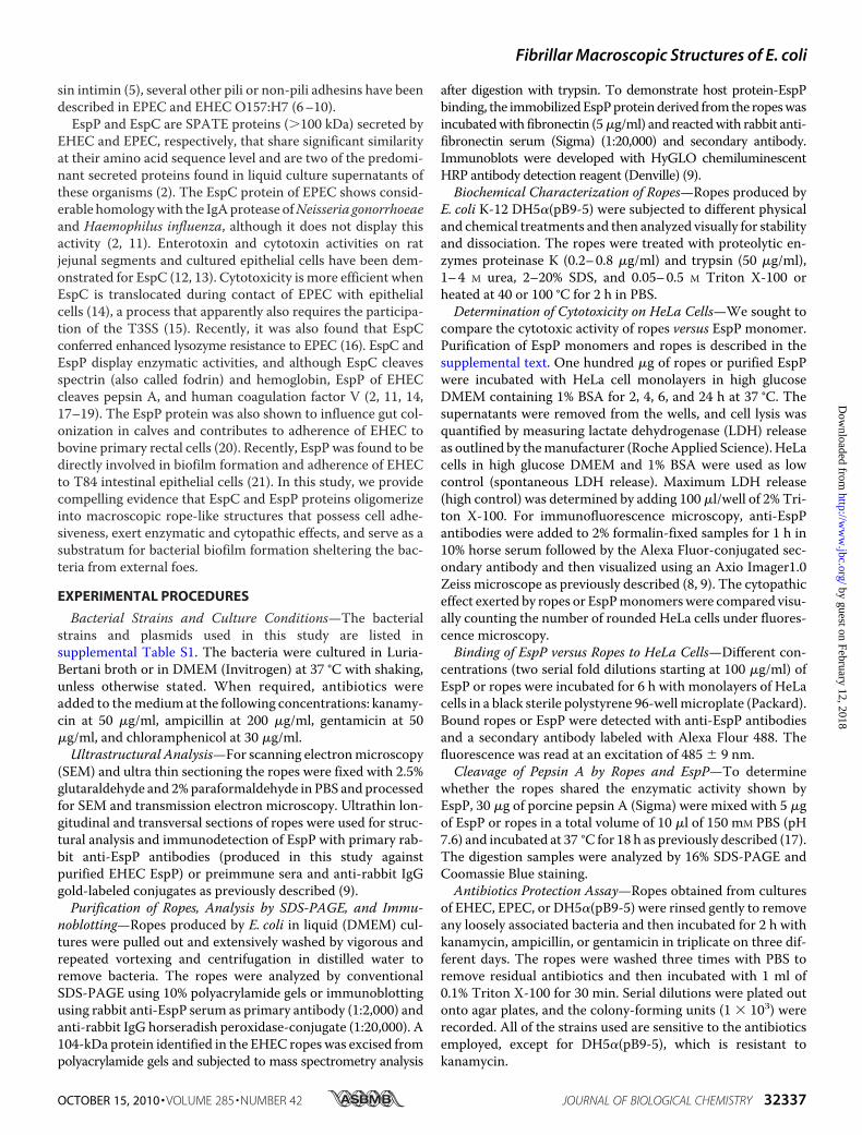
Discovery of Novel Macroscopic Structures in PathogenicE. coli—We found that macroscopic fibrillar structures areformed by EPEC strain E2348/69 (O127:H6) and EHECO157:H7 strains EDL933, 86-24, and 85-170 growing withshaking overnight at 37 °C in tissue culture DMEM(supplemental Fig. S1 andTable S1). These fibers could be visu-alized by the naked eye and pulled out for analysis. Whenextendedon a glass slide theymeasuredup to 2 cm in length andwere several microns wide (Fig. 1A). These dimensions are wellbeyond those of other bacterial filamentous superstructuressuch as flagella (20 nmwide, 5–10 �m long) (22), type 4 pili (20�m long), or T3SS needles (12 nm wide and 600 nm to several�m long) (7, 9, 23, 24). We were able to reproducibly see thesehighly flexible and sticky fibers, albeit of different dimensionson a day-to-day basis (supplemental Fig. S1).SEM analysis at low magnification of the fibers revealed a
morphology resembling ropes, which appeared to be composed
of hundreds of parallel long flexiblefilaments, which bundled up toincrease the thickness of the ropes(Fig. 2). The bundle of filamentstwisted together yielding rope-likestructures that torqued formingloops at different sites. The helixhandedness of the ropes examinedwas unambiguously determined byobservation of the orientation of thefilaments, which appeared to torquein a left-handed orientation formingcoiled and supercoiled structures(Fig. 2, A and B). The particularhandedness was not due to the rota-tion orientation (counterclockwise)of our shaking incubator becausestatic growth of large cultures ofEPECorEHECalso produced ropes.Congo Red (CR) and thioflavin T
(ThT) dye binding is a general prop-erty of highly aggregative and insol-uble human amyloid fibers and of afew bacterial amyloidogenic pro-teins such as curli fibers (25–29).These dyes are believed to specifi-cally interact in some unknown waywith the crossed-�-sheet structurecommon to amyloid structures (28,30–32). We found that, like humanamyloids, the ropes bound CR dye(Fig. 1A). We compared the absor-bance pattern of CR alone versus CRin the presence of ropes between400 and 700 nm.We found a shift inmaximal optical absorbance from475 (CR alone) to 500 (CR plusropes) nm (Fig. 1B). This pattern isin agreement with that exhibited bythe E. coli curli fibers in the pres-
ence of CR (25). For these experiments, an EPEC curli mutant(E2348/69�csgD) and its derivative complemented strain(E2348/69�csgD(pCP994)), which overproduces curli (33),were used as negative and positive controls, respectively.Observation of unstained or CR-stained bacterial ropes underlight microscopy also revealed their fibrillar nature (Fig. 1, Cand D). We also found that the fluorescent ThT dye associatedwith ropes in a protein concentration-dependent manner (Fig.1, E and F). After excitation at 450 nm, ThT exhibits a constantfluorescence emission (28, 34).WhenEspPor ropesweremixedwith ThT in suspension, both mixtures exhibited an enhancedfluorescence emission at 482 nm. However, a higher intensitywas obtained with the EspP monomers (Fig. 1F). In all, thesedata indicate that �-sheet structures are present in both theEspP monomers and the ropes.The Ropes Are Composed of a Major Secreted Protein—Re-
garding the nature of the ropes, our first thought was that theywere formed by chains of bacteria in tandem. Previous work
FIGURE 1. Binding of CR dye to E. coli macroscopic ropes. A, measurement of bacterial macroscopic fibers(lane a) and staining with CR (lane b) on a glass slide. B, CR absorbance spectrum between 400 and 700 nm ofEHEC ropes (thin solid line), CR alone (bold solid line), ropes without CR used as negative control (dashed line). Aspositive and negative controls of CR binding, we used the E2348/69�csgD(pCP994) strain, which overproducescurli and binds CR (dotted line), and the curli mutant E2348/69�csgD strain (dashed and dotted line), respec-tively. C and D, photomicrographs (taken at 100�) of CR-stained ropes or unstained ropes observed underbright field microscopy. E, binding of ThT to ropes visualized by fluorescence microscopy (micrographs takenat 100�). F, binding of 5–50 �g/ml of EspP (monomer), ropes, and BSA to 5 �M ThT and absorbance measuredafter excitation at 450 nm in a Synergy MX fluorometer (Bio-Tek Instruments, Inc.). Background values of ThTalone were subtracted from the sample values.
FIGURE 2. E. coli macroscopic fibers visualized by SEM. A–C, EPEC macrofibers observed at 50�, 100�, and8,000�, respectively.
Fibrillar Macroscopic Structures of E. coli
32338 JOURNAL OF BIOLOGICAL CHEMISTRY VOLUME 285 • NUMBER 42 • OCTOBER 15, 2010
by guest on February 12, 2018http://w
ww
.jbc.org/D
ownloaded from

reported that Bacillus subtilis could form macrofibers com-posed of biologically active strands of chained bacteria (35).Also, E. coli colonizing the urinary tract can form filamentouschains of bacteria that represent a source of infectious units(36). In the present study, analysis by high magnification SEMinside the crevices of the EPEC ropes showed a highly organizedfilamentous substratum with scores of bacteria adhering to it(Fig. 2C). Analysis of longitudinal and transversal thin sectionsof bacterial ropes by transmission electron microscopy con-firmed their stranded and sheeted architecture (Fig. 3,A and B)and that they are not composed of bacteria joined in tandem asin the case of the B. subtilis macrofibers or the filamentousuropathogenic E. coli. The presence of bacteria inside the ropessupports the provoking thought that the ropes could serve alsoas a biofilm substratum. It was necessary to discriminate thepresence of other bacterial superstructures such as flagella, pili,or T3SS needles as components of the newly discovered ropes.Isogenic deletion mutants of EPEC in fliC (flagellin gene), bfpA(type 4 pilin gene), escN (T3SS ATPase gene), espB (T3SS pore-forming protein gene), or espA (T3SS translocon sheath gene),and EHECEDL933mutants in fliC and escN retained the abilityto form ropes, indicating that rope formation was independentof pili, flagella, EspA or EspB secreted proteins, or T3S(supplemental Table S1).
SDS-PAGE analysis of individualEPEC and EHEC ropes showed thatthese fibers were largely composedin each case of a doublet proteinband of 104 kDa (Fig. 4A). TheEHEC EspP protein was analyzedby trypsin digestion and mass spec-trometry yielding peptide aminoacid sequences (KTGEGLVILGAE-KTF, VAGMQNTEADAVKQNG-NAY, and IDLHAGKNITGDGF)that matched the predicted productof the espP gene. In agreement, thedoublet band reacted with anti-
EspP antibody by immunoblotting (Fig. 4B). To investigatewhether HUS patients develop anti-EspP antibodies, we exam-ined a pool of sera obtained from patients who suffered HUS.TheHUS serum clearly reacted with EspP, whereas no reactionwas observed with a pool of normal human sera (Fig. 4C).Immuno-EM experiments using rabbit anti-EspP antibodyshowed labeling along the length of thin sections of the EPECmacrofilaments (Fig. 3C). To provide genetic evidence of therole of EspP or EspC in rope formation, we constructed isogenicdeletion mutants in EHEC espP or EPEC espC genes. Ropeswere not observed in cultures of EPEC espC or EHEC (EDL933,86-24, and 85-170) espP mutants. Laboratory strains E. coliK-12 DH5� or HB101, both naturally lacking espC or espPgenes, did not produce ropes (supplemental Table S1). Furthergenetic proof of the nature of the ropes was provided by exper-iments demonstrating rope formation in DH5� transformedwith espP harbored on plasmid pB9-5 (17) and in the EPECespC and EHEC espP mutants complemented with pB9-5(supplemental Table S1).We also found thatCitrobacter roden-tium, another attaching and effacing organism pathogenic formice (37), produced ropes, whereas the C. rodentium espCmutant was not able to assemble ropes (supplemen-tal Table S1). Rope formation was studied in the presence ofanti-EspP antibodies in filtered supernatants containing EspPmonomers obtained from overnight cultures of E. coli K-12DH5�(pB9-5), EHEC EDL933, or EPEC E2348/69 incubatedovernight with several dilutions of anti-EspP or preimmunesera. As expected, rope formation was abolished in a dose-de-pendentmanner only in the samples containing anti-EspP anti-body and not by the preimmune serum, suggesting that theantibody opsonized the soluble protein monomers blockingrope formation (supplemental Table S2).Biochemical and Physical Properties of the E. coli Ropes—CD
employing different concentrations of purified EspPmonomers(Fig. 5A) was used to evaluate the stability of the EspP protein atpH 8.0 and 20 °C using an Aviv 62DS spectropolarimeter. Adistinctive CD spectra in the far ultraviolet region between 180and 260 nmwas obtained (Fig. 5D). Low concentrations of EspP(between 0.25 and 0.5mg/ml) showed one peak at 200–205 nm,suggesting that the protein is present in a random coil struc-ture. However, at higher protein concentrations (between 1.2and 1.6 mg/ml), a mean molar ellipticity was achieved at 217and 220 nm, respectively, suggesting conformational changesfroma randomcoil structure to a�-sheet structure, which, as in
FIGURE 3. Thin sectioning of EPEC ropes. A, longitudinal sectioning. B, transversal sectioning. Note the pres-ence of one bacterium. C, immunogold labeling of sectioned rope filament with anti-EspP antibody.
FIGURE 4. Biochemical characterization of ropes. A, analysis of EPEC(E2348/69) and EHEC (EDL933) ropes by SDS-PAGE. B, immunoblotting ofEPEC and EHEC ropes with rabbit anti-EspP antibody. C, immunoreactivity ofEHEC ropes with pool sera from HUS patients and normal human (NH) sera.
Fibrillar Macroscopic Structures of E. coli
OCTOBER 15, 2010 • VOLUME 285 • NUMBER 42 JOURNAL OF BIOLOGICAL CHEMISTRY 32339
by guest on February 12, 2018http://w
ww
.jbc.org/D
ownloaded from

the case of amyloidogenic proteins, could be important for thestructural stability of the protein and for rope assembly. Thesecondary structure of the EspP passenger domain is predictedto be composed of 57% �-strands, which are likely to form�-sheet-rich intermediate fibrils that assemble into the com-plex quaternary rope structures. Experiments to determine thestability and resistance of the ropes to different physical andchemical treatments were performed. Similar to human amy-loid fibris and curli, the ropes were highly aggregative, sticky,insoluble, and stable in aqueous solutions (includingmost com-mon buffers at neutral pH) and resistant to vigorous mechani-cal shearing and disruption (e.g. vortexing or manual shaking).Bacterial ropes proved to be highly resistant to anionic deter-gents because only 75% dissociation of ropes was observed in20% SDS, and Triton X-100 had no effect on the ropes(supplemental Table S3). Urea totally dissociated the ropes
when used at 4 M, indicating thepresence of noncovalent bondsbetween the monomeric subunits(supplemental Table S3). The ropeswere sensitive to trypsin at 150�g/ml and were highly sensitive totreatment with low concentrationsof proteinase K, further provingtheir proteinaceous nature. Theaddition of 0.5 M EDTA did notdestabilize the ropes; however, thepresence of EDTA inhibited ropeformation in EspP-containingsupernatants, suggesting a role ofdivalent cations in oligomerizationof the EspP subunits into ropes.Finally, the ropes were demon-strated to be thermo-resistantbecause incubation at 100 °C for 2 hdid not disaggregate the structures(supplemental Table S3).Biological Properties Associated
with EspP Ropes—Determiningwhether or not the ropes retainedthe biological activities (cytotoxic orenzymatic) of the monomeric pro-tein constituents was an importantquestion to address. In agreementwith a previous report suggestingthat EspP is required for intestinalcolonization in calves (20), wefound that EspP interacts withfibronectin, indicating that EspPpossesses lectin properties (Fig.5C) and may recognize host pro-teins. In addition, we tested theability of rope fragments and EspPto bind to cultured epithelial cells.After 6 h of incubation, isolatedropes and EspP monomers boundclearly in a dose-dependent man-ner to cultured epithelial cells,
strongly suggesting that these structures possess cell adhe-siveness (Fig. 6D).EspP (also known as PssA) is considered by some authors to
be a cytotoxin because it causes cytotoxic effects on Vero cells(38). We found that incubation of HeLa cell monolayers withbacteria-free ropes or EspPmonomers for 24 h led to cell round-ing (Fig. 6, B and C). Cytopathic effects were more evident withEspP at 200 �g/ml, whereas the ropes showed 50% fewer cyto-phatic effects (Fig. 6E). Quantification of the cytotoxic potential ofEspP and ropes was done measuring LDH released from HeLacells (39, 40). Neither EspP nor ropes were able to significantlyincrease LDH release compared with the medium alone(supplemental Fig. S2A). Thus, the data suggest that EspP andropes exhibit a cytopathic, but not cytotoxic, effect on host cells.To determine whether the ropes retain the enzymatic activ-
ity exhibited by EspP, pepsin A was used as target substrate as
FIGURE 5. Purification of monomeric EspP and circular dichroism. A, purified EspP monomer obtained fromliquid cultures of DH5�(pB9-5) after ammonium sulfate precipitation and visualization by SDS-PAGE and Coo-massie Blue staining. B, the immobilized protein was reacted with rabbit anti-EspP antibody by immunoblot-ting. C, EspP was incubated with a fibronectin (Fn) solution (0.5 �g/ml) and then reacted with anti-fibronectinantibody. D, circular dicroism performed in a range of 180 –270 nm with different concentrations of EspPmonomer.
FIGURE 6. Binding of EspP-containing ropes to HeLa Cells. A, HeLa cells (red) alone. B and C, HeLa cellsincubated with ropes (B) or EspP (C) (green) obtained from DH5�(pB9-5) after 24 h of incubation. In both casesnote the loss of stress fibers and retraction of cell bodies indicating cytopathic activity. Photomicrographs(A–C) were taken at 60�. D, kinetics of adherence of EspP versus ropes to HeLa cells. E, cytopathic effects of EspPor ropes on HeLa cells were quantified using immunofluorescence microscopy counting the number of cellswith morphological changes (e.g. cell rounding and loss of plasmic membrane) compared with the controlwithout EspP or ropes.
Fibrillar Macroscopic Structures of E. coli
32340 JOURNAL OF BIOLOGICAL CHEMISTRY VOLUME 285 • NUMBER 42 • OCTOBER 15, 2010
by guest on February 12, 2018http://w
ww
.jbc.org/D
ownloaded from
previously described (17). The ropes did not show the samelevel of proteolytic activity on pepsin A as that seen with EspP(supplemental Fig. 2B), suggesting that the enzymatic motifs inEspP are hidden in the rope structure.In agreement with our hypothesis that EspP ropes could
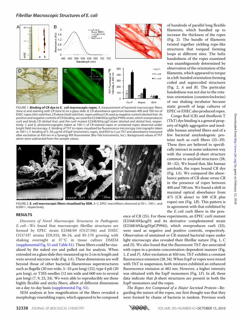
serve as a biofilm substrate, we found that exogenously addedEPEC, EHEC, and E. coli K-12 bacteria bound to bacteria-freeropes (supplemental Fig. 3). The presence of the bacteria withinthe ropes as revealed by ultrastructural studies (Fig. 2) also ledus to hypothesize that the bacteria are protected within themegastructure, presumably from host or environmental fac-tors. Thus, to test this hypothesis we performed experiments inwhich ropes from EPEC, EHEC, and DH5�(pB9-5) containingbacteria were incubated with or without several antibiotics. Itwas found that bacteria associated with the ropes were pro-tected to a significant extent from killing by the antibacterialdrugs. We were able to recover bacteria from the ropes incu-bated with antibiotics, albeit less than in the control sampleswithout antibiotics (Fig. 7). Based on these data, it is tempting tospeculate that the ropes might serve to protect bacteria fromother external foes within the host (e.g. immune system factors)or in the environment.
DISCUSSION
In this study we provide the first observation that ATproteins EspP and EspC oligomerize to form coiled-coilmegastructures that possess adhesive and cytopathic activitieson host epithelial cells. Isogenic EPEC and C. rodentium espCand EHEC espP mutants do not produce ropes, and recombi-nant EspP expressed in E. coli K-12 led to rope formation. Fur-ther, purified EspP protein assembled into ropes upon incuba-tion at 37 °C. To date, no other proteinaceous filamentousstructure (e.g. pili, flagella, and T3SS needles) in pathogenicE. coli strainsmanifests such amacroscopic nature (7, 9, 23, 24).Striking similarities, in terms of structure, biochemical and bio-physical properties, exist between the E. coli ropes and humanamyloidogenic proteins implicated in human diseases such as
Alzheimer, Parkinson, cystic fibrosis, and prion diseases. It hasbeen reported that curli fibers of uropathogenic E. coli areinvolved in the polymerization of amyloid-like fibers, whichwere suggested to participate in neurodegenerative and amy-loid-related diseases (25). The E. coli ropes showed high insol-ubility in common neutral buffer, high aggregation in aqueoussolutions, unusual stability, and binding to ThT and CR dyes.These properties are reminiscent of human amyloid fibers. TheE. coli ropes showed high resistance to mechanical forces andboiling and to solubilization and disruption at high concentra-tions of anionic detergents, urea, and mercaptoethanol. CDexperiments supported the notion that EspP monomers aggre-gate in a concentration-dependent manner and that formationof ropes involves the transition of a random coil structure into amore stable, highly organized �-sheet structure. Furthermore,we found that cationic ionsmight be important for rope forma-tion and stability because of the presence of EDTA inhibitedrope formation, although the ropes did not dissociate in thepresence of EDTA.The domains within the large AT proteins that allow self-
export across the bacterial envelope have been identified (2).However, the specific amino acid sequences in EspC or EspPthat might allow monomer-monomer interaction leading torope formation are unknown. Analysis of the primary sequenceof EspP suggests that the region between amino acids 1000 and1050 could potentially be responsible for oligomerization of themonomers, but supporting experimental data are missing.ATs are the most abundant secreted proteins found in the
supernatants of Gram-negative bacterial pathogens (2). It ispresumably unlikely that pathogenic bacteria would devoteconsiderable amounts of energy to produce and secrete ATsinto the milieu to form these megastructures without a biolog-ical purpose. Our data indicate that the ropes display severalbiological activities that could be important for the survival ofthe organism within or out of the host. For example, the ropeswere shown to bind to host epithelial cells in culture and to hostproteins such as fibronectin, to serve as a substratum for biofilmformation, to protect the bacteria within the ropes from antibi-otics, and lastly to exhibit cytopathic effects on cell monolayers.We did not find any supportive evidence of cytotoxicity by theropes or EspP using the LDH determination. Published dataindicate that EspP binds to T84 cells and that it promotes gutcolonization in calves (20, 21). Rope formation does not appearto be accidental, because several enteric pathogens, in additionto EPEC, EHEC, and C. rodentium, produce macrofilaments.Macromolecular structures consisting of extended sheets asso-ciated with enhanced T3S secretion were seen in Shigella flex-neri (41); however, their role in in vivo pathogenesis is unclear.Yersiniae enterocolitica assembles seven Yop secreted proteinsaggregated into large filaments, with unknown function (42).People naturally or experimentally infected with EPEC or
EHEC develop antibodies against EspC or EspP, respectively(38, 43). We found that a pool of sera obtained from HUSpatients reacted with the rope-forming EspP protein, whereasnormal human sera did not show any reactivity. These datasuggest that EspP-containing ropes could be produced duringinfections. However, after an effort to explore a role for theropes in vivo, we could not find differences in terms of coloni-
FIGURE 7. Ropes provide protection to the bacteria against antibiotics.The ropes obtained from EDL933, E2348/69, and DH5�(pB9-5) were incu-bated with different antibiotics for 2 h. As controls, Luria broth bacterial cul-tures were incubated with antibiotics. The bacteria within the ropes wereprotected against total killing by the indicated antibiotics. DH5�(pB9-5) (Kmr)survived the treatment with Km because of the presence of the pB9-5 plas-mid. Otherwise, all of the cultures died in the presence of the antibiotics.*, p � 0.05 with respect to the wild-type strain without antibiotic.
Fibrillar Macroscopic Structures of E. coli
OCTOBER 15, 2010 • VOLUME 285 • NUMBER 42 JOURNAL OF BIOLOGICAL CHEMISTRY 32341
by guest on February 12, 2018http://w
ww
.jbc.org/D
ownloaded from

zation betweenC. rodentium versus an isogenic espCmutant ina murine model of infection. Determining whether the ropesare produced in vivo (e.g. in the lumen of the gut) represents atechnically challenging task because of the lack of a relevantanimal model for EHEC infections and because of the lowerdensity of bacteria expected to be present on colonic tissue, incontrast to growth in liquid cultures. Although host proteindegradation and cytopathic/cytotoxic activities have beenattributed to EspC and EspP, they only constitute one of theplayers of the multi-factorial pathogenic scheme of thesepathogens. Based on the cell damage caused by EspP ropes onepithelial cells shown here, we cannot rule out the possibilitythat the ropes might contribute to the effacement and destruc-tion of the intestinal linen seen upon EPEC and EHEC infec-tions (5). That attaching and effacing pathogens, but not com-mensal E. coli, are able to assemble ropes is a remarkablephenomenon that suggests a virulence trait that must play asignificant yet undefined role in the biology and disease-caus-ing scheme of these pathogenic bacteria. The novel ropes mayrepresent a sheltering mechanism for bacteria to avoid clear-ance in biological systems (e.g. by escaping the innate andadaptive immunity mechanisms) or a substratum for adher-ence, persistence, and survival in the animal and human hostsor the environment.
Acknowledgments—We thank María Alexandra Ledesma and Ale-jandra Vázquez for technical assistance; Maribel Vargas for process-ing and analysis of samples by electron microscopy, Ben Fane foranalysis of EspP/EspC amino acid sequence; Helge Karch for kindlyproviding plasmid pB9-5; James B. Kaper for support; and Rob andMavis McKenna, Herman Gordon, S. Padmanabhan, and Neil Men-delson for valuable insights.
REFERENCES1. Coburn, B., Sekirov, I., and Finlay, B. B. (2007) Clin. Microbiol. Rev. 20,
535–5492. Henderson, I. R., Navarro-Garcia, F., Desvaux, M., Fernandez, R. C., and
Ala’Aldeen, D. (2004)Microbiol. Mol. Biol. Rev. 68, 692–7443. Christie, P. J., Atmakuri, K., Krishnamoorthy, V., Jakubowski, S., and Cas-
cales, E. (2005) Annu Rev. Microbiol. 59, 451–4854. Pukatzki, S., Ma, A. T., Revel, A. T., Sturtevant, D., and Mekalanos, J. J.
(2007) Proc. Natl. Acad. Sci. U.S.A. 104, 15508–155135. Kaper, J. B., Nataro, J. P., and Mobley, H. L. (2004) Nat. Rev. Microbiol. 2,
123–1406. Erdem, A. L., Avelino, F., Xicohtencatl-Cortes, J., and Giron, J. A. (2007) J.
Bacteriol. 189, 7426–74357. Giron, J. A., Ho, A. S., and Schoolnik, G. K. (1991) Science 254, 710–7138. Rendon,M. A., Saldana, Z., Erdem, A. L., Monteiro-Neto, V., Vazquez, A.,
Kaper, J. B., Puente, J. L., and Giron, J. A. (2007) Proc. Natl. Acad. Sci.U.S.A. 104, 10637–10642
9. Xicohtencatl-Cortes, J., Monteiro-Neto, V., Ledesma, M. A., Jordan,D.M., Francetic,O., Kaper, J. B., Puente, J. L., andGiron, J. A. (2007) J. Clin.Invest. 117, 3519–3529
10. Low, A. S., Dziva, F., Torres, A. G., Martinez, J. L., Rosser, T., Naylor, S.,Spears, K., Holden, N., Mahajan, A., Findlay, J., Sales, J., Smith, D. G., Low,J. C., Stevens, M. P., and Gally, D. L. (2006) Infect. Immun. 74, 2233–2244
11. Stein, M., Kenny, B., Stein, M. A., and Finlay, B. B. (1996) J. Bacteriol. 178,
6546–655412. Mellies, J. L., Navarro-Garcia, F., Okeke, I., Frederickson, J., Nataro, J. P.,
and Kaper, J. B. (2001) Infect. Immun. 69, 315–32413. Navarro-García, F., Canizalez-Roman, A., Sui, B. Q., Nataro, J. P., and
Azamar, Y. (2004) Infect. Immun. 72, 3609–362114. Vidal, J. E., and Navarro-García, F. (2006) Infect. Immun. 74, 2293–230315. Vidal, J. E., and Navarro-García, F. (2008) Cell Microbiol. 10, 1975–198616. Salinger, N., Kokona, B., Fairman, R., and Okeke, I. N. (2009) Appl. Envi-
ron. Microbiol. 75, 275–28017. Brunder, W., Schmidt, H., and Karch, H. (1997) Mol. Microbiol. 24,
767–77818. Drago-Serrano, M. E., Parra, S. G., and Manjarrez-Hernandez, H. A.
(2006) FEMS Microbiol. Lett. 265, 35–4019. Dutta, P. R., Cappello, R., Navarro-García, F., and Nataro, J. P. (2002)
Infect. Immun. 70, 7105–711320. Dziva, F., Mahajan, A., Cameron, P., Currie, C., McKendrick, I. J., Wallis,
T. S., Smith, D. G., and Stevens, M. P. (2007) FEMS Microbiol. Lett. 271,258–264
21. Puttamreddy, S., Cornick, N. A., and Minion, F. C. (2010) Infect. Immun.78, 2377–2384
22. Giron, J. A. (2005) in Colonization of Mucosal Surfaces (Nataro, J. P., ed)pp. 213–236, ASM Press, Herndon, VA
23. Cornelis, G. R. (2006) Nat. Rev. Microbiol. 4, 811–82524. Ogino, T., Ohno, R., Sekiya, K., Kuwae, A., Matsuzawa, T., Nonaka, T.,
Fukuda, H., Imajoh-Ohmi, S., and Abe, A. (2006) J. Bacteriol. 188,2801–2811
25. Chapman, M. R., Robinson, L. S., Pinkner, J. S., Roth, R., Heuser, J., Ham-mar, M., Normark, S., and Hultgren, S. J. (2002) Science 295, 851–855
26. Fowler, D. M., Koulov, A. V., Balch, W. E., and Kelly, J. W. (2007) TrendsBiochem. Sci. 32, 217–224
27. Frid, P., Anisimov, S. V., and Popovic, N. (2007) Brain Res. Rev. 53,135–160
28. Naiki, H., Higuchi, K., Hosokawa, M., and Takeda, T. (1989) Anal. Bio-chem. 177, 244–249
29. LeVine, H., 3rd. (1999)Methods Enzymol. 309, 274–28430. Naiki, H., Higuchi, K., Matsushima, K., Shimada, A., Chen, W. H.,
Hosokawa, M., and Takeda, T. (1990) Lab. Invest. 62, 768–77331. Naiki, H., Higuchi, K., Nakakuki, K., and Takeda, T. (1991) Lab. Invest. 65,
104–11032. Krebs, M. R., Bromley, E. H., and Donald, A. M. (2005) J. Struct. Biol. 149,
30–3733. Saldana, Z., Xicohtencatl-Cortes, J., Avelino, F., Phillips, A. D., Kaper, J. B.,
Puente, J. L., and Giron, J. A. (2009) Environ. Microbiol. 11, 992–100634. LeVine, H., 3rd. (1993) Protein. Sci. 2, 404–41035. Mendelson, N. H. (1982) J. Bacteriol. 151, 438–44936. Justice, S. S., Hunstad, D. A., Seed, P. C., and Hultgren, S. J. (2006) Proc.
Natl. Acad. Sci. U.S.A. 103, 19884–1988937. Deng, W., Puente, J. L., Gruenheid, S., Li, Y., Vallance, B. A., Vazquez, A.,
Barba, J., Ibarra, J. A., O’Donnell, P., Metalnikov, P., Ashman, K., Lee, S.,Goode, D., Pawson, T., and Finlay, B. B. (2004) Proc. Natl. Acad. Sci. U.S.A.101, 3597–3602
38. Djafari, S., Ebel, F., Deibel, C., Kramer, S., Hudel, M., and Chakraborty, T.(1997)Mol. Microbiol. 25, 771–784
39. Guyer, D. M., Radulovic, S., Jones, F. E., and Mobley, H. L. (2002) Infect.Immun. 70, 4539–4546
40. Roberts, P. H., Davis, K. C., Garstka, W. R., and Bhunia, A. K. (2001) J.Microbiol. Methods 43, 171–181
41. Parsot, C., Menard, R., Gounon, P., and Sansonetti, P. J. (1995) Mol. Mi-crobiol. 16, 291–300
42. Michiels, T., Wattiau, P., Brasseur, R., Ruysschaert, J. M., and Cornelis, G.(1990) Infect. Immun. 58, 2840–2849
43. Jarvis, K. G., and Kaper, J. B. (1996) Infect. Immun. 64, 4826–4829
Fibrillar Macroscopic Structures of E. coli
32342 JOURNAL OF BIOLOGICAL CHEMISTRY VOLUME 285 • NUMBER 42 • OCTOBER 15, 2010
by guest on February 12, 2018http://w
ww
.jbc.org/D
ownloaded from

Phil I. Tarr, B. Brett Finlay, José Luis Puente and Jorge A. GirónJuan Xicohtencatl-Cortes, Zeus Saldaña, Wanyin Deng, Elsa Castañeda, Enrique Freer,Bacterial Macroscopic Rope-like Fibers with Cytopathic and Adhesive Properties
doi: 10.1074/jbc.M110.162248 originally published online August 5, 20102010, 285:32336-32342.J. Biol. Chem.
10.1074/jbc.M110.162248Access the most updated version of this article at doi:
Alerts:
When a correction for this article is posted•
When this article is cited•
to choose from all of JBC's e-mail alertsClick here
Supplemental material:
http://www.jbc.org/content/suppl/2010/08/05/M110.162248.DC1
http://www.jbc.org/content/285/42/32336.full.html#ref-list-1
This article cites 42 references, 22 of which can be accessed free at
by guest on February 12, 2018http://w
ww
.jbc.org/D
ownloaded from