virus culture and cytopathic effect
TRANSCRIPT

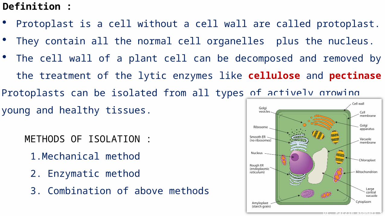
Definition :
Protoplast is a cell without a cell wall are called protoplast.
They contain all the normal cell organelles plus the nucleus.
The cell wall of a plant cell can be decomposed and removed by the treatment of the lytic
enzymes like cellulose and pectinase
Protoplasts can be isolated from all types of actively growing young and healthy tissues.
METHODS OF ISOLATION :
1.Mechanical method
2. Enzymatic method
3. Combination of above methods
Dr. Farzin Asghari Sana
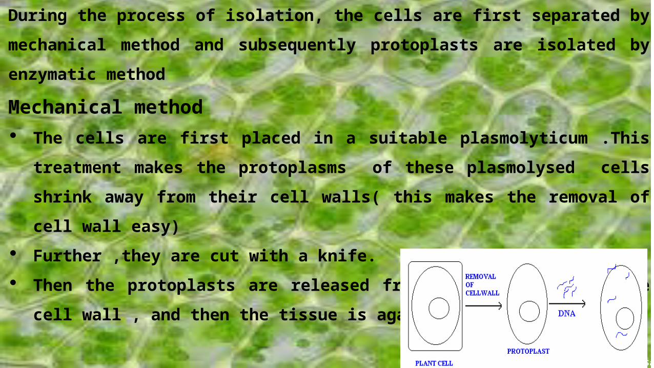
During the process of isolation, the cells are first separated by mechanical method and
subsequently protoplasts are isolated by enzymatic method
Mechanical method The cells are first placed in a suitable plasmolyticum .This treatment makes the
protoplasms of these plasmolysed cells shrink away from their cell walls( this makes
the removal of cell wall easy) Further ,they are cut with a knife. Then the protoplasts are released from the cells through the cell wall , and then the
tissue is again deplasmolysed.
Dr. Farzin Asghari Sana
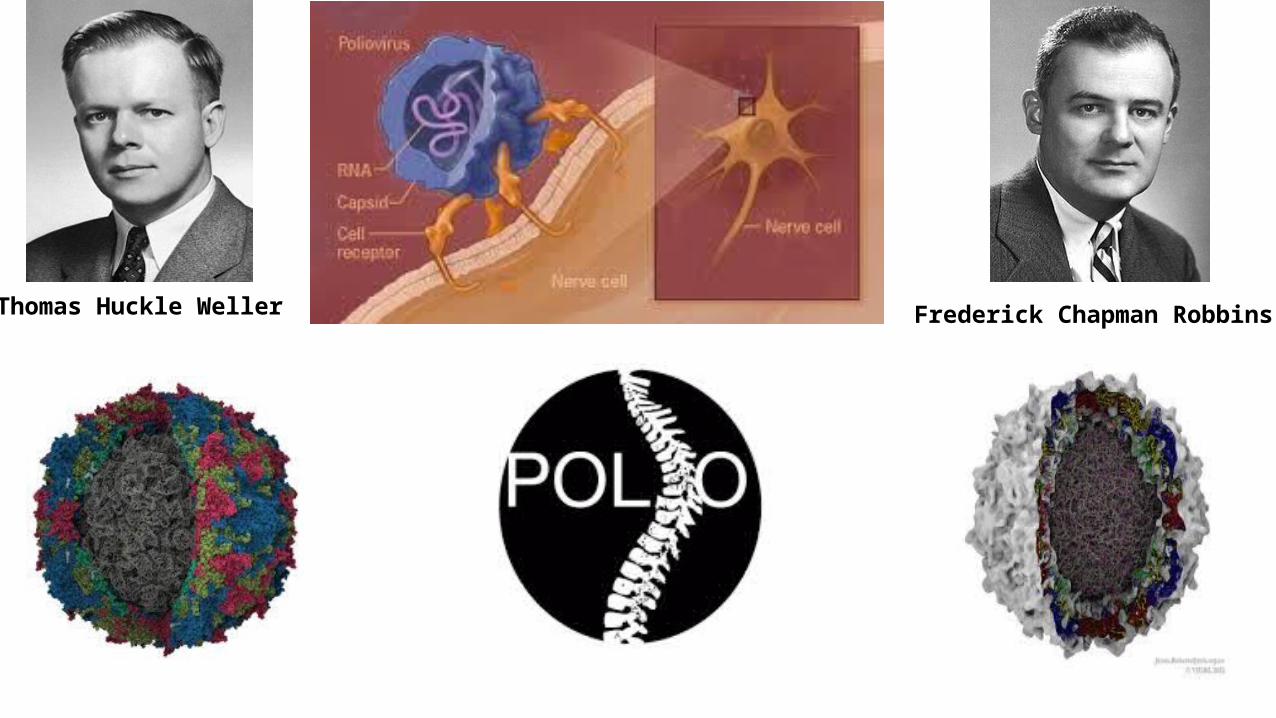
Thomas Huckle Weller Frederick Chapman Robbins
Dr. Farzin Asghari Sana
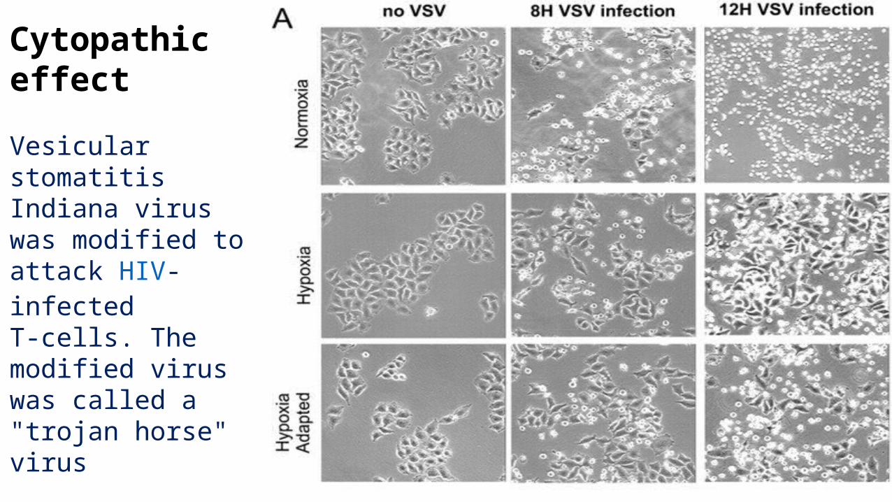
Cytopathic effect
Vesicular stomatitis Indiana virus was modified to attack HIV-infected T-cells. The modified virus was called a "trojan horse" virus
Dr. Farzin Asghari Sana

Dr. Farzin Asghari SanaDr. Farzin Asghari Sana
Dr. Farzin Asghari Sana

• The culture medium is the Combination of ingredients will support the cell growth by providing all the essential nutrients , growth factors, and hormones for cell growth, as well as regulating the pH and the osmolarity of culture.
• The three basic classes of media are: (differ in their requirement for supplementation with serum. )
Basal media
Serum-free media
Reduced-serum media
The choice of culture media is dependent on the requirements of cells being cultured.
Dr. Farzin Asghari Sana
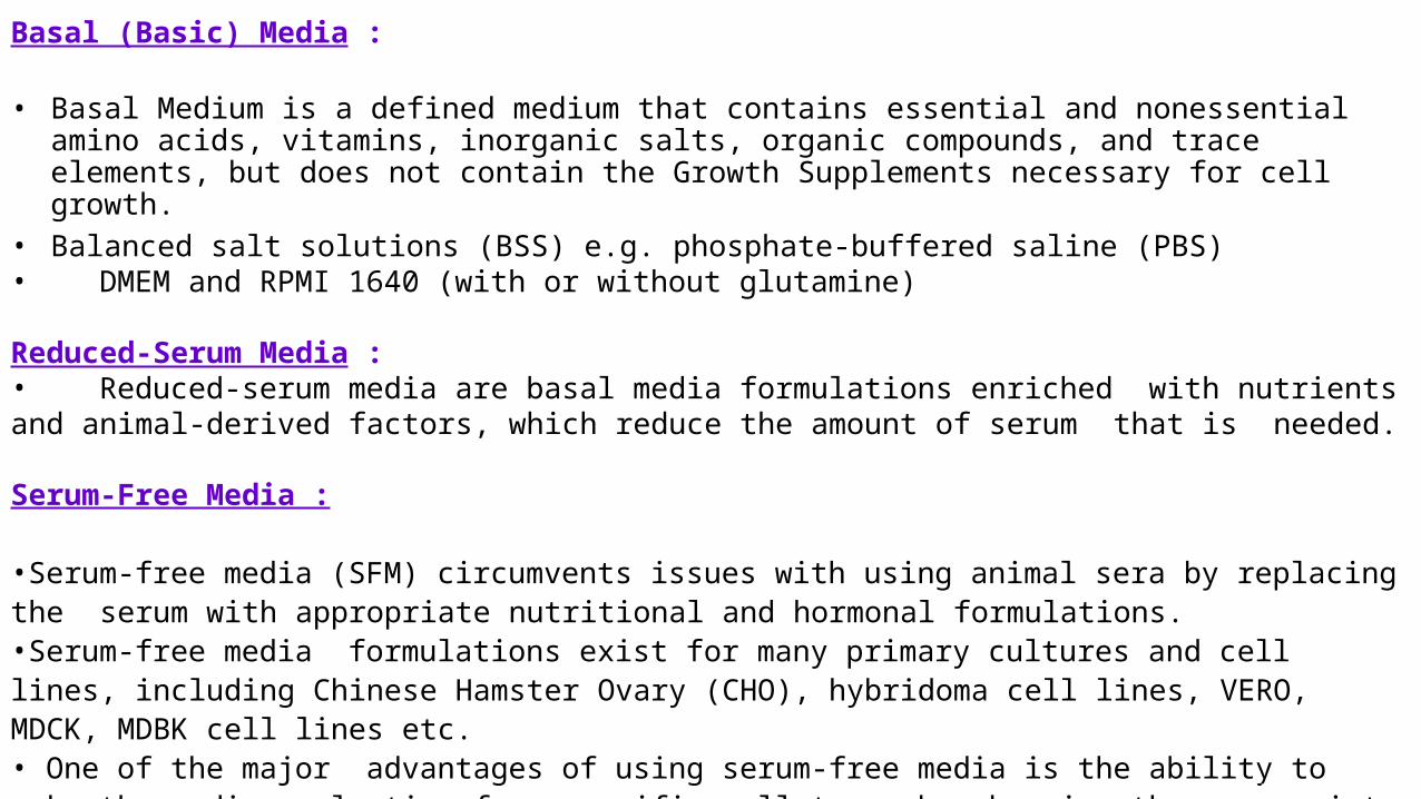
Basal (Basic) Media :
• Basal Medium is a defined medium that contains essential and nonessential amino acids, vitamins, inorganic salts, organic compounds, and trace elements, but does not contain the Growth Supplements necessary for cell growth.
• Balanced salt solutions (BSS) e.g. phosphate-buffered saline (PBS)• DMEM and RPMI 1640 (with or without glutamine)
Reduced-Serum Media :• Reduced-serum media are basal media formulations enriched with nutrients and animal-derived factors, which reduce the amount of serum that is needed.
Serum-Free Media :
•Serum-free media (SFM) circumvents issues with using animal sera by replacing the serum with appropriate nutritional and hormonal formulations. •Serum-free media formulations exist for many primary cultures and cell lines, including Chinese Hamster Ovary (CHO), hybridoma cell lines, VERO, MDCK, MDBK cell lines etc.• One of the major advantages of using serum-free media is the ability to make the medium selective for specific cell types by choosing the appropriate combination of growth factors
Dr. Farzin Asghari Sana

Types of tissue culture
Primary Continuous
Finite Indefinite
Normal cells cultured without any change in their division rate
Single cell type roughly thirty times of division, enhanced by growth factors
It is nearly the same as finite but the cells here can divide indefinitely by transformation into tumor cells, They are called cell line
Dr. Farzin Asghari Sana
Primary cultures
Cells when surgically or enzymatically removed from an organism and placed in suitable culture environment will attach and grow and are called as primary culture
Primary cells have a finite life span
Primary culture contains a very heterogeneous population of cells
Sub culturing of primary cells leads to the generation of cell lines
Cell lines have limited life span, they passage several times before they become senescent
Cells such as macrophages and neurons do not divide in -vitro so can be used as primary cultures
Continuous cell lines Most cell lines grow for a limited number of generations after which they cease
Cell lines which either occur spontaneously or induced virally are chemically transformed into continous cell lines
Dr. Farzin Asghari Sana
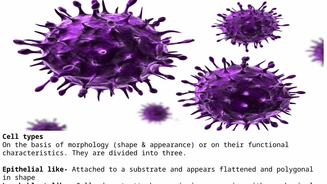
Cell types On the basis of morphology (shape & appearance) or on their functional characteristics. They are divided into three.
Epithelial like- Attached to a substrate and appears flattened and polygonal in shapeLymphoblast like- Cells do not attach, remain in suspension with a spherical shapeFibroblast like- Cells attached to a substrate, appear elongated and bipolar
Dr. Farzin Asghari Sana

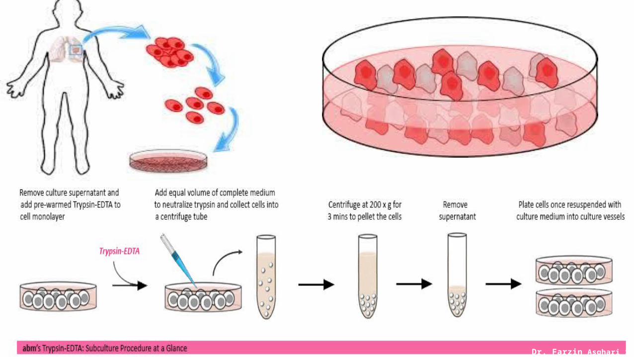
Cell Cultures/ Dispersion Cultures
Primary cell cultures
a cell line derived directly from the parent tissue. Cells in primary culture have the same karyotype and chromosome number as those in the original tissue.
Can be subculture only one or twice e.g-primary monkey, baboon kidney
• Diploid cell strains.
Which are derived from human fetal tissue and can be subcultured 20-50 times e.g-human diploid fibroblast such as MRC-5(human fetal lung fibroblast establish from normal lung tissue of 14 week old male foetus)
Some primary cells can be passed through secondary and several subsequent subcultures while retaining their original morphological characteristics and karyotype. Subcultures will have fewer cell types than primary cultures. After 20 to 50 passages in vitro, these diploid cell strains usually undergo a crisis in which their growth rate slows and they eventually die out. Diploid strains of fibroblasts derived from human fetal tissue are widely used in diagnostic virology and vaccine production.
Dr. Farzin Asghari Sana

Continuous cell lines. Derived from tumors of human or animals e.g- Vero, Hep2
Certain cultured cells, notably mouse fetal fibroblasts, kidney cells from various mammalian species, and human carcinoma cells, are able to survive the growth crisis and undergo indefinite propagation in vitro.
After several passages, the growth rate of the culture slows down; then isolated colonies of cells begin to grow more rapidly than diploid cells, their karyotype becomes abnormal (aneuploid), their morphology changes, and other poorly understood changes take place that make the cells immortal. The cells are now "dedifferentiated," having lost the specialized morphology and biochemical abilities they possessed as differentiated cells in vivo. Continuous cell lines such as KB and HeLa, both derived from human carcinomas, support the growth of a number of viruses. These lines and others derived from monkey kidneys (e.g., Vero), mouse fetuses (L929), and hamster kidneys (BHK) are widely used in diagnostic and experimental virology. Continuous cell lines have been established from many types of vertebrate and invertebrate animal tissues and are available from the American Type Culture Collection (ATCC).
Dr. Farzin Asghari Sana
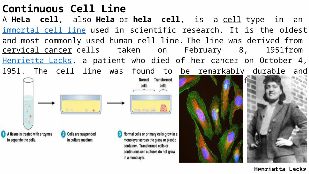
Continuous Cell LineA HeLa cell, also Hela or hela cell, is a cell type in an immortal cell line used in scientific research. It is the oldest and most commonly used human cell line. The line was derived from cervical cancer cells taken on February 8, 1951from Henrietta Lacks, a patient who died of her cancer on October 4, 1951. The cell line was found to be remarkably durable and prolific which warrants its extensive use in scientific research.
Henrietta LacksDr. Farzin Asghari Sana

Dr. Farzin Asghari Sana

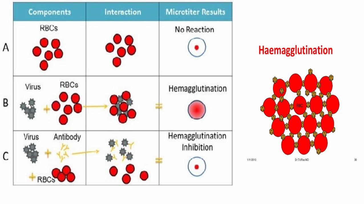
As some viruses do not cause CPE in cell lines, they can be detected by other techniques. Hemadsorption of erythrocytes onto cells infected with viruses which do not form CPE and contain hemagglutinin can be used in myxovirus and paramyxovirus detection.
Influenza viruses can be released into the culture medium and then detected by hemagglutination.
Dr. Farzin Asghari Sana

• DISADVANTAGES OF CELL CULTURES
• Long period (up to 4 weeks) required for result.
• Often very poor sensitivity, sensitivity depends on a large extent on the condition of the specimen.
• Susceptible to bacterial contamination.
• Susceptible to toxic substances which may be present in the specimen.
• Many viruses will not grow in cell culture e.g. Hepatitis B, Diarrhoeal viruses, parvovirus, papillomavirus.
Dr. Farzin Asghari Sana
Dr. Farzin Asghari Sana

Dr. Farzin Asghari Sana

Immunofluorescent staining with GTX36623_RSV-nucleoproteinDr. Farzin Asghari Sana
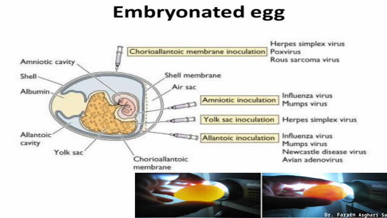
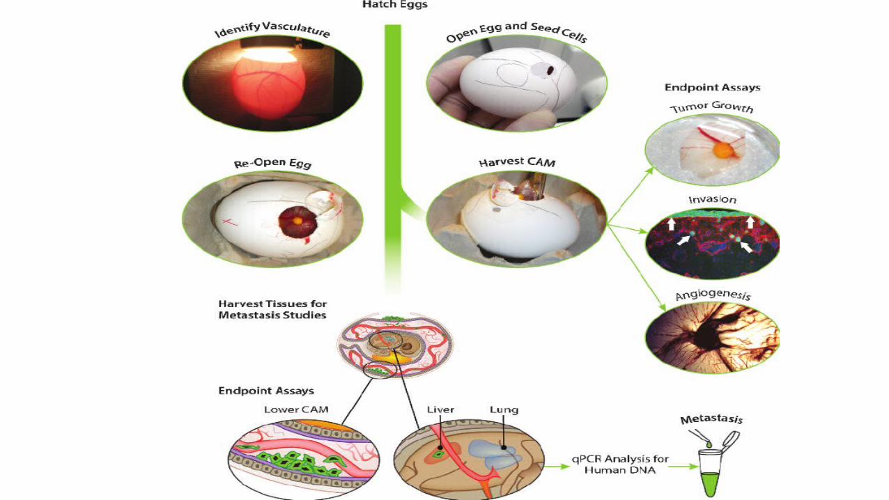
EMBRYONATED EGGS
The Embryonated hen’s egg was first used for cultivation of viruses by Good Pasteur and Burnet (1931).
Cultivation of viruses in organized tissues like chick embryo necessitates a different type of approach. For all practical purposes they all themselves behave as tissue cultures.
The process of cultivation of viruses in embryonated eggs depend on the type of egg which is used. The egg used for cultivation must be sterile and the shell should be intact and healthy.
Use embryonated chicken, duck or turkey for inoculation of viral suspension are used especially for the influenza viruses isolation. 7 - 10 days old embryonated eggs are used.
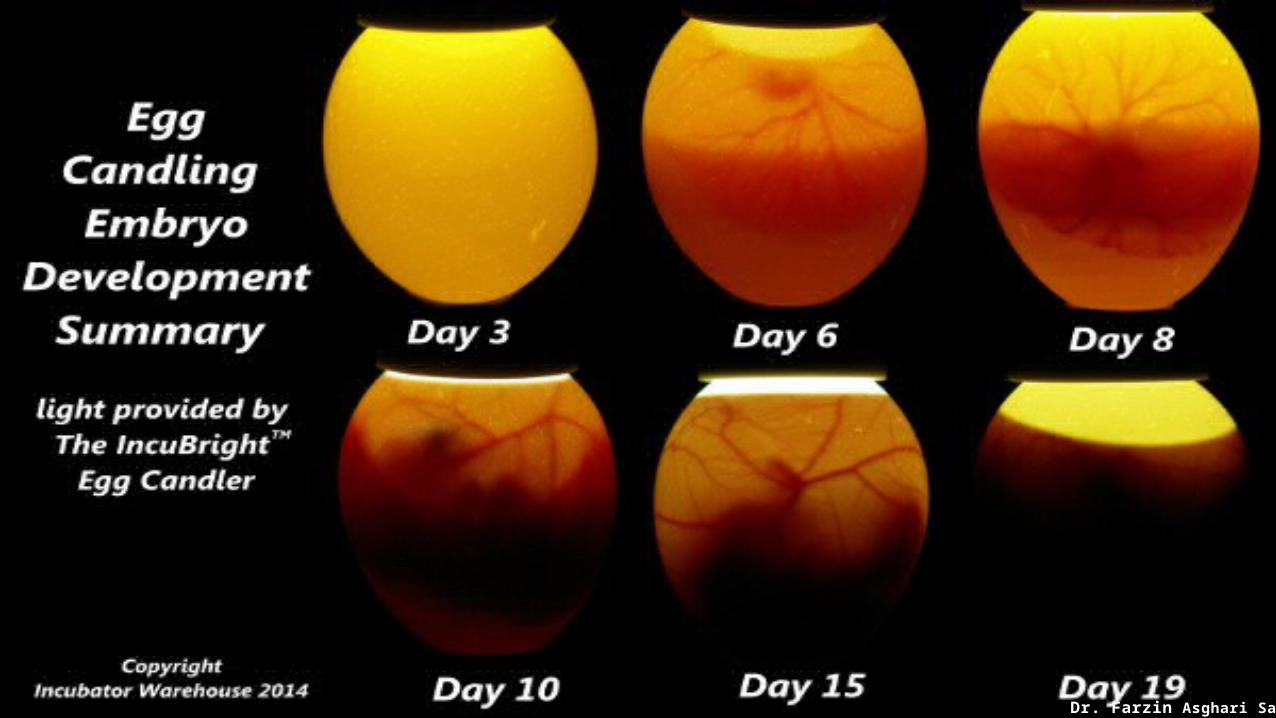
The egg must be cleaned, the shell decontaminated with a disinfectant and checked in ovoscope if it is alive. Ovoscope is the equipment used for candling.
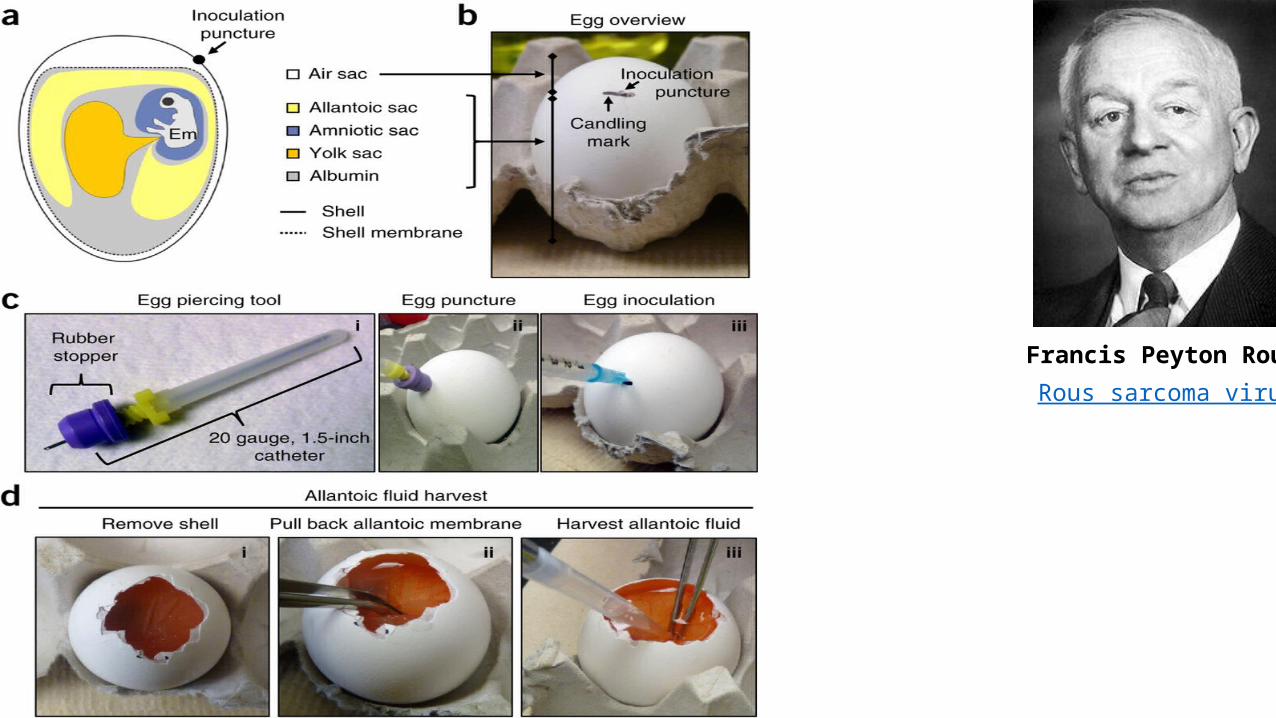
Francis Peyton Rous Rous sarcoma virus
Dr. Farzin Asghari Sana
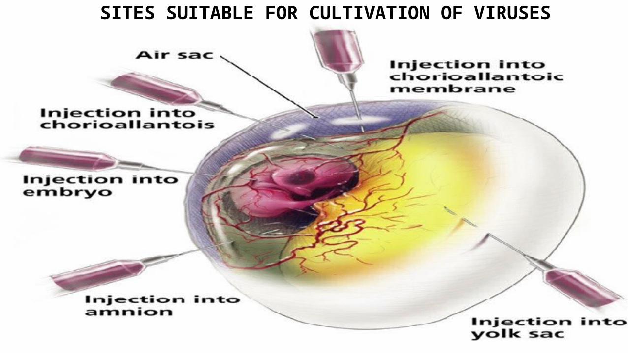
SITES SUITABLE FOR CULTIVATION OF VIRUSES
Dr. Farzin Asghari Sana
Dr. Farzin Asghari Sana
Dr. Farzin Asghari Sana
Dr. Farzin Asghari Sana
Dr. Farzin Asghari Sana
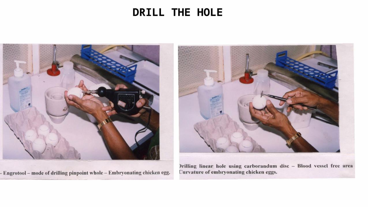
DRILL THE HOLE
Dr. Farzin Asghari Sana

INJECT THE SUSPENSION WITH SYRINGE
Dr. Farzin Asghari Sana

HOLE SEALED WITH PARAFFIN WAX
Dr. Farzin Asghari Sana

EGGS INCUBATE AT 36C FOR 2-3 DAYS
Dr. Farzin Asghari Sana

DETECTION OF VIRAL GROWTH
The signs of viral growth include:
i) Death of the embryo,
ii) Defects in embryonic development, and
iii) Localized areas of damage in the membranes, resulting in discrete, opaque spots called pocks
(a variant of pox).
iv)The embryonic fluid and tissue can be prepared for examination with an electron microscope.
v) Some can also be detected by their ability to agglutinate red blood cells or by their reaction
with an antibody of known specificity that will affix to its corresponding virus, if it is present.
Dr. Farzin Asghari Sana

POCK LESIONS ON CAM
Dr. Farzin Asghari Sana
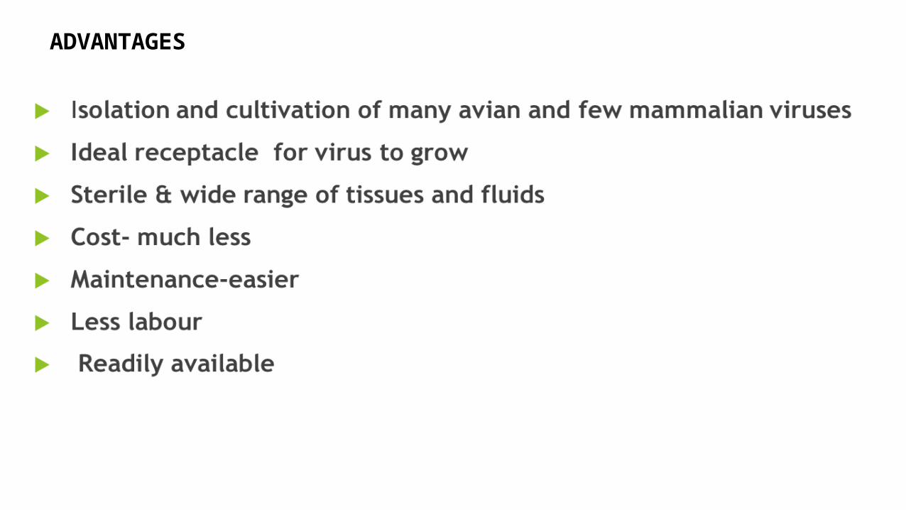
ADVANTAGES
Dr. Farzin Asghari Sana
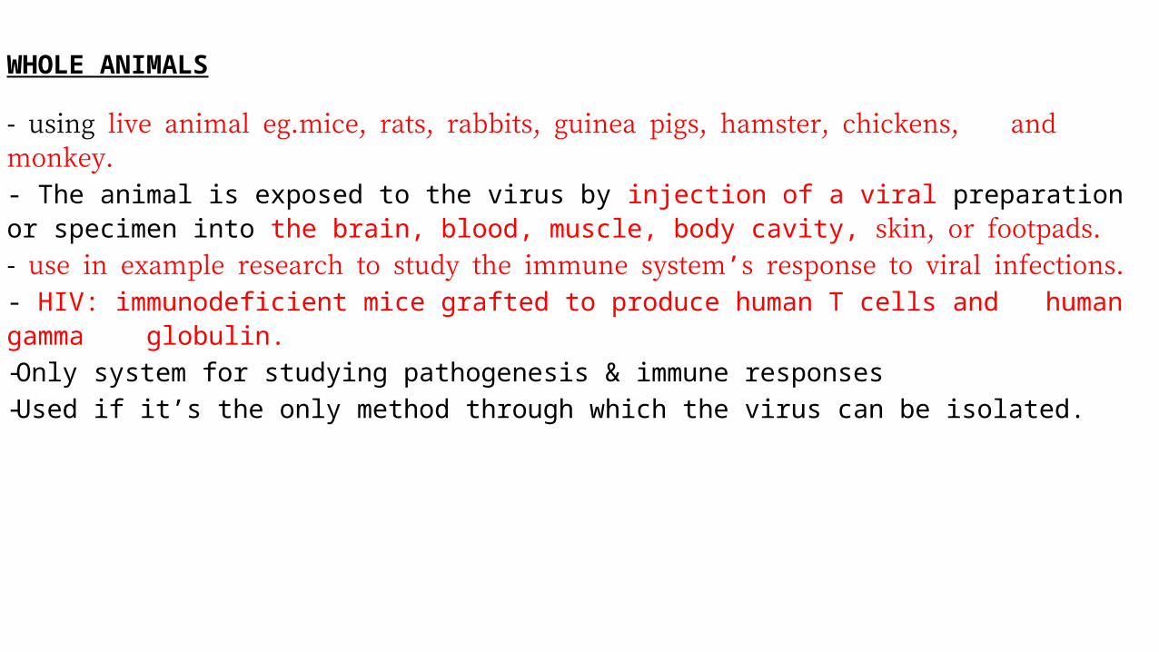
WHOLE ANIMALS
- using live animal eg.mice, rats, rabbits, guinea pigs, hamster, chickens, and monkey. - The animal is exposed to the virus by injection of a viral preparation or specimen into the brain, blood, muscle, body cavity, skin, or footpads.- use in example research to study the immune system’s response to viral infections.- HIV: immunodeficient mice grafted to produce human T cells and human gamma globulin.- Only system for studying pathogenesis & immune responses- Used if it’s the only method through which the virus can be isolated.
Dr. Farzin Asghari Sana
ANIMAL MODEL• usually a purpose-bred animal
• Mouse model• Advantages
• in-breed strains reduce genetic variability • genetics are well understood• Introduce, mutate or inactivation specific genes thought to control the immune response.
• Disadvantages• Sometimes not infected-therefore virus has to be adapted or use a closely related surrogate
virus• Does not always cause same disease state• Mice are not humans
Dr. Farzin Asghari Sana
DETECTION OF VIRAL GROWTH
• The signs of viral growth include • i)death of the animal • Ii) defects in animal development.
• The infected animal tissue can be prepared for examination with an electron microscope
Dr. Farzin Asghari Sana

VIRAL QUANTIFICATION• Virus quantification involves counting the number of viruses in a
specific volume to determine the virus concentration.• It is utilized in both (R&D) in commercial and academic laboratories
as well as production situations where the quantity of virus at various steps is an important variable
• The methods used include but not limited to: i) Hemagglutination assay ii) Plaque assay iii)TCID₅₀
Dr. Farzin Asghari Sana
• Where virus has infected the tissue culture cells, the infected cells will die causing the formation of a clear zone amongst the otherwise intact monolayer of cells
• This clear zone is called a plaque and it theoretically represents an area where one virus has infected a single tissue culture cell, has multiplied and been released, and has gone on to infect adjacent cells.
• The number of plaque forming units (pfu)/ml can be calculated based on the dilution of the original viral solution.
• The term pfu/ml is used rather than the number of viruses/ml because it is possible that occasionally more than one virus infects a single cell.
• Often the cells or plaques are stained to help in visualization of the plaques.

PLAQUE ASSAY RESULTS
Dr. Farzin Asghari Sana

CALCULATION OF PFU/Ml• Plaques are
enumerated• Plaque Counts are
averaged over wells• The average is then
divided by the dilution times the volume
(43+40+38)/3(10-4 x 0.1)
= 3,730,000 pfu/ml
43 4 1 040 3 0 038 6 2 0Plaques formed per well
Dr. Farzin Asghari Sana
TCID₅₀
• TCID50 is the measure of infectious virus titer.
• This endpoint dilution assay quantifies the amount of virus required to kill 50% of infected hosts or to produce a cytopathic effect in 50% of inoculated tissue culture cells
• This assay may be more common in clinical research applications where the lethal dose of virus must be determined or if the virus does not form plaques.
• When used in the context of tissue culture, host cells are plated and serial dilutions of the virus are added. After incubation, the percentage of cell death (i.e. infected cells) is manually observed and recorded for each virus dilution, and results are used to mathematically calculate a TCID50 result.
Dr. Farzin Asghari Sana

TCID₅₀ calculation
• The outcome of a TCID50 determination can be used• to estimate a virus titre in pfu, or vice versa, using the formula
100
75
50
1 TCID₅₀ = 0.7 pfu
10−1 10−2 10−3 10−4 10−5
10−6
TCID₅₀
Dr. Farzin Asghari Sana
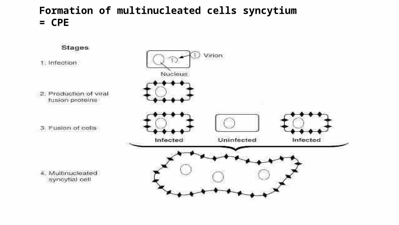
Formation of multinucleated cells syncytium = CPE
Dr. Farzin Asghari Sana
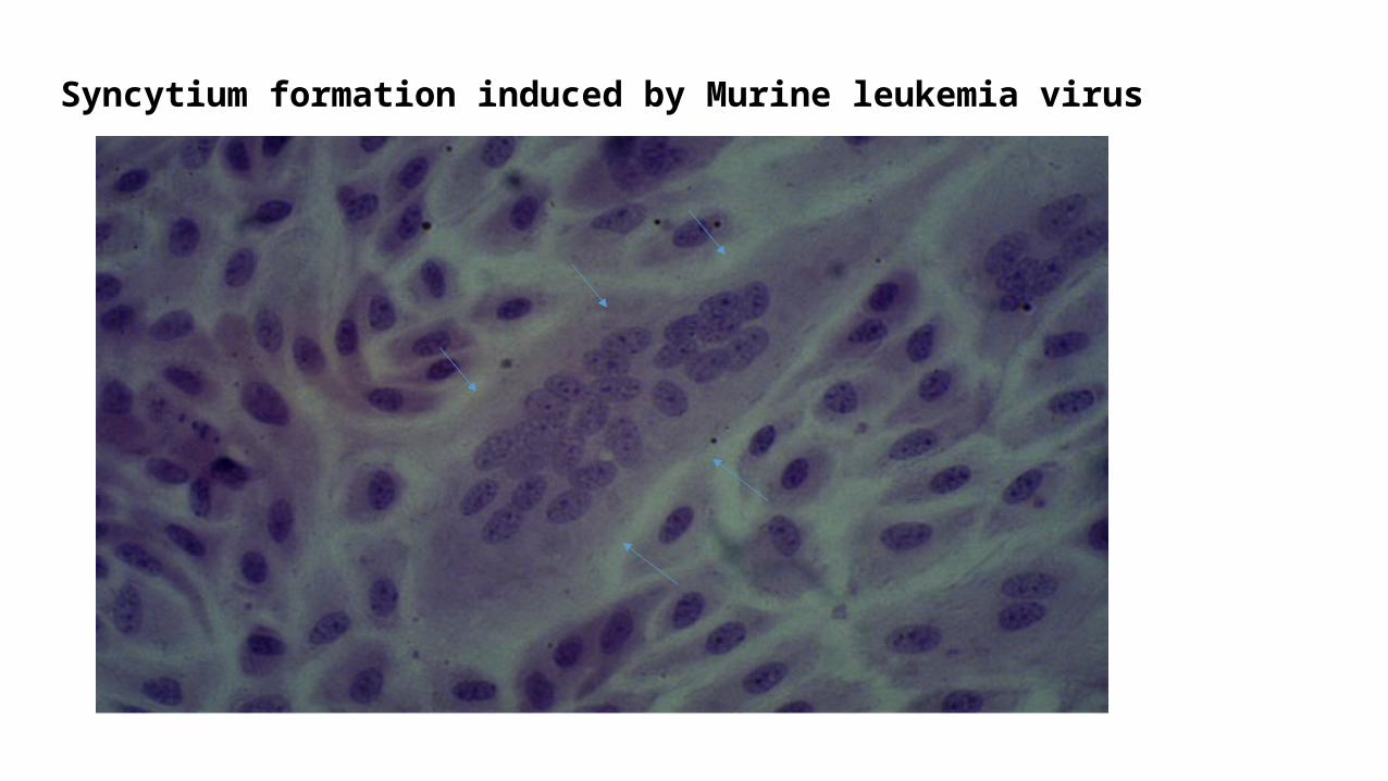
Syncytium formation induced by Murine leukemia virus
Dr. Farzin Asghari Sana

Dr. Farzin Asghari Sana
