bacterial-derived agent protects against allergic · modulator was shown to markedly reduce...
TRANSCRIPT
Transplacental immune modulation with abacterial-derived agent protects against allergicairway inflammation
Kyle T. Mincham, … , Patrick G. Holt, Deborah H. Strickland
J Clin Invest. 2018. https://doi.org/10.1172/JCI122631.
In-Press Preview
Chronic allergic inflammatory diseases are a major cause of morbidity, allergic asthmaalone affecting over 300 million people worldwide. Epidemiological studies demonstratethat environmental stimuli are associated with either promotion or prevention of disease.Major reductions in asthma prevalence are documented in European and US farmingcommunities. Protection is associated with exposure of mothers during pregnancy tomicrobial breakdown products present in farm dusts and unprocessed foods, andenhancement of innate immune competence in the children. We sought to develop ascientific rationale for progressing these findings towards clinical application for primarydisease prevention. Treatment of pregnant mice with a defined clinically-approved immune-modulator was shown to markedly reduce susceptibility of their offspring to development ofthe hallmark clinical features of allergic airway inflammatory disease. Mechanistically,offspring displayed enhanced dendritic cell-dependent airway mucosal immunesurveillance function, which resulted in more efficient generation of mucosal-homing T-regulatory cells in response to local inflammatory challenge. We provide evidence that theprincipal target for maternal treatment effects was the fetal dendritic cell progenitorcompartment, equipping the offspring for accelerated functional maturation of the airwaymucosal dendritic cell network following birth. These data provide proof-of-conceptsupporting the rationale for development of transplacental immune reprogrammingapproaches for primary disease prevention.
Research Cell biology Immunology
Find the latest version:
http://jci.me/122631/pdf

1
Transplacental immune modulation with a bacterial-derived agent protects against
allergic airway inflammation.
Authors: Kyle T. Mincham1, Naomi M. Scott1, Jean-Francois Lauzon-Joset1, Jonatan
Leffler1, Alexander N. Larcombe1,2, Philip A. Stumbles1,3,4, Sarah A. Robertson5, Christian
Pasquali6, Patrick G. Holt1† and Deborah H. Strickland1†*
Affiliations:
1Telethon Kids Institute, University of Western Australia, Nedlands, Western Australia,
Australia.
2Health, Safety and Environment, School of Public Health, Curtin University, Perth, Western
Australia, Australia.
3School of Veterinary and Life Sciences, Murdoch University, Perth, Western Australia,
Australia.
4School of Paediatrics and Child Health, University of Western Australia, Subiaco, Western
Australia, Australia.
5Robinson Research Institute and School of Medicine, University of Adelaide, Adelaide,
South Australia, Australia.
6OM Pharma, SA Geneva, Geneva, Switzerland.
†Joint senior authors.
*Corresponding author. Email: [email protected] Ph: +61 8 6319 1528
Address: Northern Entrance, Perth Children’s Hospital, 15 Hospital Avenue, Nedlands WA
6009.
2
Abstract: Chronic allergic inflammatory diseases are a major cause of morbidity, allergic
asthma alone affecting over 300 million people worldwide. Epidemiological studies
demonstrate that environmental stimuli are associated with either promotion or prevention of
disease. Major reductions in asthma prevalence are documented in European and US farming
communities. Protection is associated with exposure of mothers during pregnancy to
microbial breakdown products present in farm dusts and unprocessed foods, and
enhancement of innate immune competence in the children. We sought to develop a scientific
rationale for progressing these findings towards clinical application for primary disease
prevention. Treatment of pregnant mice with a defined clinically-approved immune-
modulator was shown to markedly reduce susceptibility of their offspring to development of
the hallmark clinical features of allergic airway inflammatory disease. Mechanistically,
offspring displayed enhanced dendritic cell-dependent airway mucosal immune surveillance
function, which resulted in more efficient generation of mucosal-homing T-regulatory cells in
response to local inflammatory challenge. We provide evidence that the principal target for
maternal treatment effects was the fetal dendritic cell progenitor compartment, equipping the
offspring for accelerated functional maturation of the airway mucosal dendritic cell network
following birth. These data provide proof-of-concept supporting the rationale for
development of transplacental immune reprogramming approaches for primary disease
prevention.
Introduction
A series of prospective birth cohort studies on the children of European traditional farming
families (1,2), now replicated in US studies contrasting Amish and Hutterite farming
populations (3), have identified striking asthma-protective properties of oral and inhalation
exposure to benign microbial stimuli present in dusts from farm barns. The target for these

3
exposures in the offspring appears to be the innate immune system, and involves modulation
of both immunoregulatory and effector cell function(s) (3-6) resulting in markedly reduced
susceptibility to the asthma-promoting effects of common respiratory allergies. The temporal
window during which these environmental stimuli exert their immunomodulatory effects
spans the period when the developing immune system is undergoing postnatal functional
maturation, but susceptibility to these effects also appears particularly high during prenatal
development as demonstrated by the strong impact of maternal microbial exposures during
pregnancy on ensuing asthma resistance in their offspring (1,7).
A broad forerunner literature supports the general principle that maternal microbial exposures
can result in transmission of transplacental signals that influence the functional phenotype of
the developing fetal immune system (8-10), but these studies have focused almost exclusively
on maternal infections, and usually on deleterious effects thereof. In contrast, in light of the
findings from the farming family studies above (1,7), we posit that benign environmental
microbial exposures during pregnancy can be read out by the maternal mucosal immune
surveillance system and transcribed into positive “immune training” signals for transplacental
transmission to their developing offspring, equipping them for more rapid adaptation after
birth to the microbial-rich postnatal environment. Moreover, we posit that this natural
mechanism can be harnessed therapeutically; notably, if these benign environmental exposure
effects could be reproduced by an agent that could be safely administered during pregnancy,
then this could open up novel possibilities for primary prevention of asthma. With this in
mind we have recently completed a proof-of-concept study in pregnant mice with a
microbial-derived therapeutic product OM-85, which has been in widespread use in Europe
in human infants and adults for >30 years for boosting resistance to airways inflammation
and attendant wheezing symptoms associated with lower respiratory infections (11-15). In

4
initial investigations to establish the safety of OM-85 use during pregnancy we demonstrated
that maternal treatment with this agent enhanced homeostatic control of innate immune and
inflammatory functions in gestational tissues at baseline and in the face of challenge with
microbial pathogens including live influenza infection and the bacterial mimic
lipopolysaccharide (LPS). Specifically, OM-85 treatment attenuated inflammatory symptoms
(which are typically exaggerated during pregnancy) and protected against fetal growth
restriction and/or pregnancy termination which can follow maternal infection (16). In the
study presented here we focus on the effects of maternal OM-85 treatment during healthy
pregnancy on the immunocompetence of offspring during the postnatal weanling period when
immune functions are typically developmentally compromised. In this regard, we focus on
the effects of maternal OM-85 treatment on the capacity of offspring to regulate airways
inflammatory responses associated with development of experimental atopic asthma during
the weanling period, which in humans represents the age range at highest risk for initiation of
what can be life-long asthma (17).
Results
Experimental model of allergic airways inflammation in sensitized weanling mice: study
rationale
For this study we utilized an experimental system developed for induction of T helper (Th) 2-
associated cell-mediated inflammation in the conducting airway mucosa in adult rodents, as a
model for the main lesional site in human asthma. Additional (albeit less extensive)
inflammation also develops in peripheral lung tissue, but the relative contribution of this to
airflow limitation in the asthmatic state is uncertain. The principal features of this model,
focusing mainly on the airway mucosa, are illustrated in Supplementary Figure 1A.
Aeroallergen delivered to the airways of pre-sensitized animals via large-droplet aerosol is
5
captured by resident mucosal dendritic cells (DC) that are functionally quiescent in the
steady-state (as marked by low-modest IAIE expression) and are specialised for antigen
sampling only, which they subsequently transport to airway draining lymph nodes (ADLN)
for presentation to allergen-specific T-memory cells (18-20). The resultant T-cell response
generates a mixture of T-effector-memory (Tmeff) and T-regulatory (Treg) cells in
proportions determined via DC programming. Representatives of these populations traffic
back to the airway mucosa, where they encounter resident mucosal DC which have recently
acquired aeroallergen, and bidirectional interactions between these three cell populations in
situ determine the intensity and duration of the ensuing T-cell dependent inflammatory
response within the airway mucosa (21,22). In particular, the capacity for local activation of
incoming Tmeffs is limited via the suppressive effects of Tregs on surface IAIE and CD86
expression by mucosal conventional DC (cDC) (18,22-24).
The principal DC population involved comprises the network of cDC within the airway
mucosa, that are responsible for the major aspects of local immune surveillance (18).
Plasmacytoid DC (pDC) have also been implicated in this process (25), particularly in
relation to pathogen surveillance (26). The airway mucosal cDC population has the property
of uniquely rapid turnover in the steady-state with 85% of the resident population turning
over every ~24 hours, being continuously depleted by migration of antigen-bearing cells to
ADLN and simultaneously replenished via incoming precursors recruited from bone marrow
(27). This orderly and highly dynamic process is rapidly accelerated during airway challenge
events, during which cDC numbers can expand markedly within the airway epithelium and
ADLN (18,28-30). This airway mucosal cDC network is developmentally compromised in
immature humans (31,32) and experimental animals (33,34), and this partially explains the
high risk of respiratory infections and aeroallergen sensitization associated with the infant

6
period (35). Our hypothesis underlying this murine study is that maternal OM-85-treatment
during pregnancy can enhance the functional maturation of this mucosal immune surveillance
system in their offspring, and as a result reduce susceptibility to initiation of inflammatory
airway disease during the high-risk early postnatal period.
To test this hypothesis, we have utilised a variant of the above mentioned allergic airways
inflammation model, modified from earlier studies assessing farm-related exposures (36),
involving sensitization of 21 day old weanling BALB/c mice to ovalbumin (OVA) employing
a prime-boost schedule followed by subsequent airways challenge with aerosolised OVA
(Supplementary Figure 1B, C). Details of the ensuing response are discussed below.
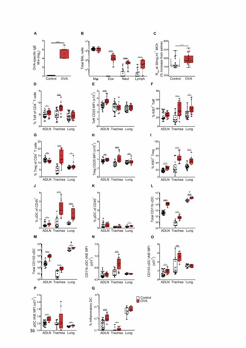
Aeroallergen-induced cellular response in the airways: baseline characteristics
Sensitized animals display high levels of OVA-specific serum IgE 24 hours following
repeated OVA-aerosol challenge (Figure. 1A). The challenged animals display gross
hypertrophy of ADLN involving in particular T-cells (see below), which is accompanied by
intense inflammatory cell infiltration into the airways encompassing eosinophils, neutrophils
and lymphocytes detectable by bronchoalveolar lavage (BAL; Figure 1B), increased levels of
Th2 cytokines in lung homogenates (not shown), and airways hyperresponsiveness (AHR)
manifesting as increased airways resistance (Raw) to methacholine (MCh; Figure 1C).
Further characterization of the phenotype of the cellular response within the airways
compartment by multi-colour flow cytometry (see Methods for gating strategies) revealed
significant increases in the total cellularity of parathymic and mediastinal ADLN and trachea,
with no observable difference in peripheral lung (Supplementary Figure 1D). This cellular
response was dominated by changes in the CD3+ T-cells (Supplementary Figure 1E) and

7
especially the CD4+ T-cell compartment (Supplementary Figure 1F). These changes in
particular involved increases in the numbers (Supplementary Figure 1G), proportions (Figure
1D) and activation status (Figure 1E, F) of CD3+CD4+CD25+FoxP3- Tmeff cells within the
ADLNs and in tracheal tissue, with much smaller parallel changes in peripheral lung
parenchyma (Figure 1D) which is consistent with deposition of the bulk of aerosol droplets in
the large and central airways. ADLN Tmeffs displayed a heightened state of activation
(Figure 1E), whereas lung and especially tracheal Tmeffs demonstrated high levels of Ki67
expression suggesting very recent (possibly local) proliferation (Figure 1F; see corresponding
cDC data below). In conjunction with the CD25+FoxP3- Tmeff response, a decrease in
CD3+CD4+CD25+FoxP3+ Tregs within the T-cell compartment of ADLNs was observed,
accompanied by a large increase within trachea and a smaller (but significant) increase in
peripheral lung tissues (Figure 1G), presumably derived by migration of these cells from
ADLN. Characterisation of Treg function-associated markers revealed increased CTLA-4+
Tregs within trachea (74.47 ± 3.78; 46.41 ± 6.05) and peripheral lung (51.16 ± 2.50; 34.9 ±
2.54) compared to naïve controls (Supplementary Figure 1H). Furthermore, an increase in
CD69+ Tregs was identified within peripheral lung samples (15.68 ± 0.53; 12.98 ± 1.54),
while CD69+ Tregs were reduced within the ADLNs (28.19 ± 0.69; 32.5 ± 1.19) compared to
naïve controls (Supplementary Figure 1I). Airways Tregs additionally displayed enhanced
CD25 expression (Figure 1H) and proliferative capacity (Figure 1I) following OVA
challenge compared to naïve controls.
Forerunner studies from our lab (18,22,37) suggest that in the early stage of recall responses
to inhaled antigen, the limiting factor determining the efficiency of generation of airway
mucosal homing Tregs is the efficiency of DC-mediated transport of antigen-specific signals
from the airway to ADLN. We therefore turned our attention to characterising the myeloid
8
cell populations localised within the airways of early life OVA sensitised and aerosol
challenged animals. For these analyses, IAIE+F4/80-CD11c+ conventional DCs (cDC) were
subdivided into CD11b+CD103- and CD11b-CD103+ populations, representing the two
dominant cDC subsets localised within the airways, having specialised roles in immunogenic
and tolerogenic responses respectively (38). We additionally quantified
IAIE+Ly6G/Clo/+F4/80-CD11c+B220+CD11b- pDC which represented a much smaller
proportion of the CD45+ population compared to cDC (Figure 1J, K). Consecutive aerosol
challenges of pre-sensitised mice induces a minor response in pDC in peripheral lung only
(Figure 1K), in contrast to a significant influx of both cDC subsets across all airways tissues
sampled, in particular the tracheal mucosa and its ADLN where numbers of these cells
displayed a logfold increase (Figure 1L, M). Characterisation of cDC based on expression
levels of surface IAIE demonstrated marked enhancement in expression intensity across the
entire cDC population within the trachea following challenge (Figure 1N, O). This
observation is consistent with allergen-driven functional maturation in situ of these cells from
strict antigen-sampling to antigen-presentation phenotype as previously observed (18,19), and
this provides a plausible mechanism for local activation of Tmeffs within the mucosa during
repeated challenge (Figure 1D, F). In parallel, ADLN displayed modest IAIE upregulation in
the CD103+ subset, while both subsets remained at baseline in the peripheral lung (Figure
1N, O). Furthermore, upregulation of pDC IAIE expression occurred both in ADLN and
peripheral lung (Figure 1P). Additionally, we observed increased numbers post challenge of
rare IAIE+F4/80intLy6G/ChiCD11b+CD11c+ inflammatory DCs (infDC) which have been
implicated in driving Th2-mediated inflammation to antigen exposure (39,40), within ADLN
and tracheal tissue (Figure 1Q).

9
Role of the bone marrow in the development of an experimental allergic airways
inflammatory response
The granulocytic and DC subsets identified above as participants in the airways response to
aerosol challenge in sensitized mice are derived from bone marrow, and we posited that (as
inferred from earlier studies on eosinophils in aeroallergen challenged human asthmatics
(41)) the dynamic changes detailed in these populations in our murine model of allergic
airways inflammation above should be mirrored by changes in respective bone marrow
precursor populations.
The scheme in Supplementary Figure 2 summarizes current understanding of the
interrelationships between relevant bone marrow progenitor compartments in the
development pathway leading to production of granulocytes, pDC and cDC. We assessed the
impact of repeated challenge with aeroallergen on the size of relevant compartments at or
beyond the myeloid precursor (MP) stage, employing multicolour flow cytometry, targeting
the markers shown. These analyses (Figure 2A-D) demonstrate firstly that early myeloid
precursor compartments up to and including the granulocyte-macrophage progenitor (GMP)
population, which are a major source of both DC and granulocyte populations (42,43), and
the macrophage-dendritic cell progenitor (MDP) compartment which is committed to pDC
and cDC production (44-46), expand significantly in response to repeated aeroallergen
challenge of sensitized animals. This finding is consistent with the data shown in Figure 1,
demonstrating the buildup of these populations in the challenged airways. Beyond this stage
the cDC compartment appeared reduced relative to baseline (Figure 2F), and this may be
expected in light of the accumulation of these cells in tracheal mucosa and at their ultimate
destination in ALDN, which displays a logfold increase in cDC numbers post-challenge
(Figure 1L, M).

10
Furthermore, cDCs remaining within the bone marrow post challenge displayed reduced
IAIE surface expression relative to baseline controls (Figure 2G), which may indicate
preferential recruitment of cDC from the more functionally mature end of the developmental
spectrum.
Maternal OM-85 treatment during pregnancy: effects on experimental allergic airways
inflammation susceptibility in sensitized offspring
We posited that treatment of pregnant mice with the microbial-derived immunomodulatory
agent OM-85 would enhance the resistance of their offspring to development of allergic
airways inflammation during the early post-weaning period. To test this hypothesis, we
utilised an OM-85 treatment protocol we have recently demonstrated to protect pregnant
mice and their fetuses against the toxic effects of bacterial and viral infections (16),
comprising oral administration of OM-85 from gestation day 9.5-17.5, followed by natural
delivery of offspring 2-3 days later. Age-matched offspring from OM-85 treated and
untreated control mothers were sensitized at weaning (21 days of age) and aerosol challenged
as per Supplementary Figure 1B and C, and their airways responses compared (Figure 3). As
previously demonstrated, early life OVA sensitisation and ensuing aerosol challenge initiates
granulocytic and lymphocytic infiltration of the airways, and these cellular responses (Figure
3A), together with accompanying development of AHR to MCh (Figure 3B), were markedly
attenuated in the offspring of mice treated during pregnancy. Of note, treatment did not affect
OVA-specific IgE levels (log2 titres 5.44 ± 0.21 and 5.54 ± 0.34 in treated versus untreated
groups respectively), implying that OM-85 treatment influences mechanism(s) downstream
of sensitization per se.
11
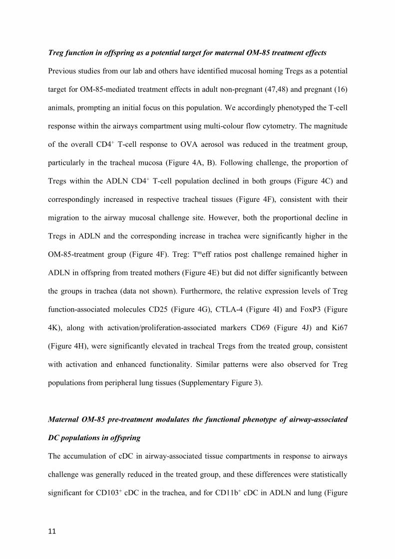
Treg function in offspring as a potential target for maternal OM-85 treatment effects
Previous studies from our lab and others have identified mucosal homing Tregs as a potential
target for OM-85-mediated treatment effects in adult non-pregnant (47,48) and pregnant (16)
animals, prompting an initial focus on this population. We accordingly phenotyped the T-cell
response within the airways compartment using multi-colour flow cytometry. The magnitude
of the overall CD4+ T-cell response to OVA aerosol was reduced in the treatment group,
particularly in the tracheal mucosa (Figure 4A, B). Following challenge, the proportion of
Tregs within the ADLN CD4+ T-cell population declined in both groups (Figure 4C) and
correspondingly increased in respective tracheal tissues (Figure 4F), consistent with their
migration to the airway mucosal challenge site. However, both the proportional decline in
Tregs in ADLN and the corresponding increase in trachea were significantly higher in the
OM-85-treatment group (Figure 4F). Treg: Tmeff ratios post challenge remained higher in
ADLN in offspring from treated mothers (Figure 4E) but did not differ significantly between
the groups in trachea (data not shown). Furthermore, the relative expression levels of Treg
function-associated molecules CD25 (Figure 4G), CTLA-4 (Figure 4I) and FoxP3 (Figure
4K), along with activation/proliferation-associated markers CD69 (Figure 4J) and Ki67
(Figure 4H), were significantly elevated in tracheal Tregs from the treated group, consistent
with activation and enhanced functionality. Similar patterns were also observed for Treg
populations from peripheral lung tissues (Supplementary Figure 3).
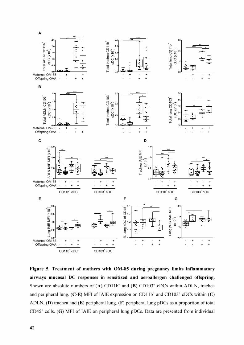
Maternal OM-85 pre-treatment modulates the functional phenotype of airway-associated
DC populations in offspring
The accumulation of cDC in airway-associated tissue compartments in response to airways
challenge was generally reduced in the treated group, and these differences were statistically
significant for CD103+ cDC in the trachea, and for CD11b+ cDC in ADLN and lung (Figure

12
5A, B). However, the most notable finding related to cDC maturational status as measured by
surface IAIE expression, which was reduced at baseline in both CD103+ and CD11b+ subsets
in ADLN (Figure 5C). Moreover, the antigen-induced surge in IAIE expression levels on
both subsets in the rapidly turning over cDC population in the tracheal mucosa, which is a
hallmark of functional activation of these cells, was likewise attenuated (Figure 5D). This
contrasted with the picture in the peripheral lung (Figure 5E), which is dominated by cDC
populations with much longer half-lives, and which displayed minimal upregulation of IAIE
in response to challenge. However as noted above, aerosol challenge does elicit a small but
significant increase in pDC in peripheral lung tissue, and this response was attenuated in the
treated group (Figure 5F, G). We also screened the groups for treatment effects on the rare
inflammatory DC subset in airway tissues, however none were detected (data not shown).
Offspring bone marrow as the primary target for maternal OM-85 treatment effects
The final series of experiments tested the hypothesis that maternal OM-85 treatment
mediated effects on cellular immune function(s) in offspring respiratory tract tissues in this
model may be associated with upstream effects on relevant precursor populations in bone
marrow. Figure 6 directly compares the aeroallergen-induced bone marrow responses of
offspring from treated versus untreated mothers. Firstly, while baseline output of pre-cDC
and cDC was comparable between groups, there were small but significant increases in the
resting GMP and MDP populations in the control offspring from treated mothers (Figure 6C,
D). However, the major treatment-associated differences were revealed by aeroallergen
challenge, notably a consistent attenuation of the expansion in all precursor compartments
spanning the MP–MDP stages which was observed in the challenged offspring from
untreated mothers (Figure 6A-D). Secondly, at the end of this developmental spectrum, the
post challenge depletion of bone marrow cDC reserves that occurs in the offspring of

13
untreated mothers was significantly attenuated (Figure 6E, F), consistent with the reduced
draw on this pool resulting from reduced recruitment to airway mucosa and ADLN in the
OM-85-treated group (Figure 5A, B).
It is additionally pertinent to note that expression levels of IAIE on cDC were reduced in the
treated group both at baseline and in particular post-challenge (Figure 6G), suggesting
maintenance of a more tightly regulated/quiescent functional state, mirroring the picture seen
for trachea and ADLN cDC in Figure 5. A similar pattern was also observed in relation to the
bone marrow pDC reservoir (data not shown).
Maternal OM-85 treatment effects at earlier ages
The data above pertains to animals sensitized at 3 weeks and challenged/sacrificed at 6
weeks. In the studies presented in Figure 7, we assessed the extent to which treatment effects
on DC populations were demonstrable at younger ages. Looking firstly at age 3 weeks, we
observed that the offspring of OM-85-treated mothers displayed higher numbers CD11b+ and
CD103+ cDC in lung tissue at baseline (Figure 7A, B) and higher levels of attendant IAIE
expression (Figure 7C), consistent with treatment-mediated acceleration of postnatal
maturation of DC networks in the respiratory tract. In a preliminary experiment we also
compared total cDC yields in granulocyte-macrophage colony-stimulating factor (GM-CSF)-
driven bone marrow cultures derived from the same animals, and the increased yields from
the treated group (Figure 7D) again point to the marrow as the likely primary site of action of
maternal OM-85 treatment.
To further extend this finding, we characterised the progenitor pool within freshly harvested
fetal bone marrow at 18.5 days gestation, 24 hours following the last maternal OM-85 oral

14
dose. The marked increase in total bone marrow cDC (Figure 7E) accompanied by parallel
expansion in the upstream MDP compartment in the treated group (Figure 7F) is consistent
with the conclusion that the bone marrow is the ultimate target for OM-85 treatment effects.
OM-85-mediated attenuation of the responsiveness of bone marrow DC precursors to
environmental inflammatory stimuli: validation of OM-85 treatment effects in an
independent inflammatory model
In the experiments illustrated in Figure 8 we cultured bone marrow from 6-week old
offspring of OM-85-treated/untreated mothers in GM-CSF-enriched medium for 7 days,
adding the archetypal pro-inflammatory agent bacterial LPS to half the cultures for the last 24
hours. Comparison of resultant activation levels of cDC by surface expression of IAIE
(Figure 8A) and the costimulator CD86 (Figure 8B) indicated marked attenuation of
upregulation of these function-associated markers, consistent with enhanced capacity for
homeostatic regulation of inflammatory responses in general in cDC from the treated group.
Discussion
In this experimental model, allergen challenge of early-life sensitized animals via aerosol
triggers accumulation of a Th2-associated inflammatory cell infiltrate in respiratory tract
tissues, and ensuing AHR, mimicking some of the hallmark features of atopic asthma.
Consistent with earlier reports (18,19), prominent within these infiltrates were activated
CD4+ Tmeffs and Treg cell populations, with the accompanying buildup of an expanded
population of cDC and their transition (especially in the mucosa) from the passive/antigen
surveillance phenotype (IAIElow) to a functionally mature (IAIEhigh) state. We also
demonstrate for the first time that these rapid changes in the population dynamics of cDC in
the challenged airways of sensitized animals are accompanied by concomitant depletion of
15
committed cDC from bone marrow, and parallel (presumably compensatory) expansion of
upstream multipotent precursor compartments.
We further demonstrate that susceptibility to this aeroallergen-induced asthma-like response
in the airways is markedly attenuated in young animals born to mothers given repeated doses
of OM-85 during pregnancy. These findings mirror those previously reported by Conrad et al.
(36) in a model of controlled maternal exposure to Acinetobacter lwoffii F78, which the
authors asserted to changes in Th1/Th2 balance (49). In contrast, in the present study,
accompanying this acquired resistant state following OM-85 treatment is increased capacity
for expansion and functional activation of Tregs in the airway mucosa in response to
aeroallergen challenge, with parallel attenuation of local cDC recruitment, activation and
trafficking to ADLN. It is pertinent to note that this OM-85 treatment is not accompanied by
overt attenuation of allergen-specific IgE production, similar to the situation described in
humans undergoing successful allergen immunotherapy in which treated patients frequently
display reduced inflammatory symptoms without major reduction in IgE production, and this
has been ascribed inter alia to boosting of Treg functions (50,51).
Moreover, underpinning these OM-85 treatment effects in respiratory tract associated tissues
are a series of parallel changes in bone marrow DC progenitor populations at various stages
of DC commitment, which are collectively consistent with the reduced draw on bone marrow
cDC reserves in the treated group as a result of more effective control of the inflammatory
milieu within the challenged mucosa. The key question is whether these changes in bone
marrow of offspring from treated mothers are primary or secondary in this process.
16
In this regard, we (48) and Navarro et al. (47) have previously demonstrated that oral OM-85
treatment of rodents can directly promote generation in gut-associated tissues of mucosal-
homing DC that can bolster systemic natural Treg populations (including those in the airway
mucosa) at baseline. Moreover, Navarro et al. (47) has demonstrated that these effects of
OM-85 are Toll-like receptor (TLR)-dependent, and similar findings have been reported in in
vitro studies in a murine model (52). In the current model, airway mucosal Treg density and
functional phenotype do not differ between OM-85- and non-treated groups at baseline, and
the stimulatory effects of treatment are only evident following aeroallergen challenge (Figure
4C-K). We posit therefore that OM-85 treatment has affected the functionality of the airway
mucosal cDC that are responsible for programming Treg:Teff balance in the aerosol induced
T-cell response, prior to their migration into the airway mucosa i.e. at bone marrow precursor
stage. Several lines of indirect evidence support the possibility of OM-85 treatment effects in
bone marrow: (i) baseline IAIE expression on 6-week old bone marrow cDC is significantly
reduced in offspring from treated mothers (Figure 6G), and corresponding GMP and MDP
precursor compartments are expanded in the same animals (Figure 6C, D); (ii) at age 3 weeks
when airway mucosal cDC networks are normally developmentally compromised with
respect to baseline density, offspring from treated mothers display higher frequency of
CD103+ and CD11b+ cDC in lung tissue digests (Figure 7A, B) and higher cDC yields from
bone marrow cultures (Figure 7D); (iii) the frequency of cDC and their MDP precursors in
fetal bone marrow are already increased in those from treated mothers by late gestation
(Figure 7E, F). Moreover, preliminary studies (Supplementary Figure 4.) have demonstrated
a direct stimulation effect of OM-85 feeding on the myeloid precursor compartment of adult
non-pregnant mice, and our forerunner studies (16) have demonstrated a range of effects on
myeloid and regulatory cell populations in maternal gestational tissues. However, the
strongest evidence for treatment related effects at the DC precursor stage comes from the
17
studies on cDC from 6-week old GM-CSF-driven 7-day bone marrow cultures (Figure 8).
The introduction of the archetypal pro-inflammatory stimulus bacterial LPS for the final 24
hours of these cultures triggers ultra-high expression on cDC from untreated controls of both
IAIE and CD86, at levels likely to result in potentially pathologic T-cell hyperstimulation.
However, this upregulation was more tightly controlled in cDC from offspring of treated
mothers, suggesting enhanced capacity to maintain homeostasis and avoid bystander damage
to host tissues during immunoinflammatory responses to environmental stimuli.
We acknowledge several limitations to this study. Firstly, we have not addressed the question
of whether OM-85 treatment influences susceptibility to primary allergic sensitization to
inhalant allergens, and this merits future investigation, given that this process has also been
shown to be controlled by ADLN-derived Tregs (53). Secondly, we have no information on
the mechanism of transmission of orally delivered OM-85-associated signals to the bone
marrow. Earlier studies from our group (48) and others (47) have demonstrated local
activation of both T-cells and myeloid cells in the gut wall and associated lymphoid tissues
following OM-85 feeding, and it is possible that trafficking of representatives of either or
both of these populations, or transmission of soluble signals generated at these sites, may be
involved. Likewise, information on the gene target(s) in fetal bone marrow DC progenitors is
not available, and will be the subject of future studies, along with possible effects on fetal
thymus. We also have not formally demonstrated TLR-dependency of OM-85, although (as
discussed above) this has been established in related models. Additionally, OM-85 dosing of
mothers in this study spanned only the second half of gestation, and future studies need to
investigate the impact of extending feeding to earlier stages. Moreover, additional dose-
response studies are required to determine the minimal dosage of OM-85 required to mediate
these effects. In this regard, recent studies (54) suggest that effective attenuation of allergic
18
airways inflammation via direct OM-85 treatment may be attainable at a log-fold lower dose
than that used in the current study.
Our goal in this study was to provide a scientific rationale for subsequent use of OM-85
during pregnancy in human mothers whose progeny are at risk of postnatal development of
persistent atopy and/or asthma. The prime initial candidates for therapy in this regard are
atopic asthmatic mothers (55,56). The severity of asthma symptomatology in this group is
exaggerated during pregnancy (57-59), likely as a consequence of the generalized Th2-
skewing of immune functions associated with the pregnant state, and asthma exacerbations
during pregnancy further increase asthma risk for their offspring (60). Moreover,
susceptibility to respiratory infections exemplified by influenza virus and its associated
symptom severity is likewise increased during pregnancy (61,62), and the latter (along with
bacterial infections) is a risk factor for fetal growth restriction (63) which in turn is associated
with increased risk for postnatal development of a range of non-communicable diseases
including asthma (64,65). In this regard, our forerunner studies on OM-85 use in pregnant
mice have demonstrated strong protection against the effects of pathogen-associated
challenge during pregnancy employing high dose LPS or live influenza, both with respect to
preservation of pregnancy per se, and maintenance of normal maternal weight gain and
associated fetal growth trajectories (16). In this system the principal treatment effect of OM-
85 involved selective attenuation within gestational tissues of the intensity of the pro-
inflammatory (particularly TNFa/IL-1/IL-6) components of the myeloid innate immune
response to pathogen, with parallel preservation of vigorous Type 1 interferon (IFN)-
associated host defense networks (16). Moreover, we (48) and others (47) have also
previously demonstrated marked attenuation of airways inflammation in OM-85-treated adult
animals in models of experimental allergic airways inflammatory disease. On this basis, it

19
can be argued that OM-85 use during pregnancy has potential direct short-term benefits to
mothers, as well as long-term benefits to their offspring in regards to reduced susceptibility to
asthma development. The latter may also include enhanced resistance to early postnatal
respiratory infections, given the OM-85-associated effects demonstrated above on increased
airway mucosal cDC numbers and IAIE expression at baseline in 3-week old weanlings, and
studies are in progress to test this possibility.
A series of additional steps are required before progression to human trials with OM-85 in
pregnancy. The first involves an independent assessment by regulators of relevant safety
issues. In this regard there is a wide body of data available on safe use during pregnancy in
experimental animals, including our own (16). Direct human safety data for pregnancy is not
presently available. However, there is a >30-year history of safe use in non-pregnant adult
humans and children down to age 6 months, and this has proven sufficient for relevant US
(66) (National Institute of Health) and Australian (67) (National Health & Medical Research
Council) authorities to endorse and publicly fund multicenter clinical trials in infants,
targeting protection against wheezing symptoms (including those associated with early
infections) and subsequent asthma development. In this regard, it is evident that airways
inflammation associated with infections and inhalant allergy act in synergy to drive asthma
pathogenesis during childhood (35), and moreover that the earlier these episodic
inflammatory events commence after birth, the greater is the risk for subsequent asthma (68-
70). This provides a compelling argument for development of protective therapeutic
strategies that can reduce susceptibility to either or both of these environmental stressors,
from birth onwards, and the possibility that this may be achievable prenatally with a readily
available therapeutic such as OM-85 merits further detailed investigation.

20
Methods
Animals
Specified pathogen-free BALB/c mice and Sprague Dawley (SD) rats were obtained from the
Animal Resource Centre (Murdoch, WA, Australia). All animals were housed under
specified pathogen-free conditions at the Telethon Kids Institute Bioresources Facility, with a
12h:12h light-dark cycle and access to an ovalbumin-free diet and water ad libitum. In-house
bred BALB/c offspring of both sexes were used in these studies.
Time-mated pregnancies
Female BALB/c mice 8 – 12 weeks of age were time-mated with male studs between 8 – 26
weeks of age. Male studs were housed separately in individual cages. One-to-two females
were housed in an individual male cage overnight, with the presence of a vaginal plug the
following morning used as an indicator of mating. The day of vaginal plug detection was
designated gestation day (GD) 0.5.
OM-85
OM-85 (OM Pharma, Geneva, Switzerland) is an endotoxin-low lyophilised extract
containing multiple TLR ligands derived from 8 major bacterial pathogens (Haemophilus
influenzae, Streptococcus pneumoniae, Streptococcus pyogenes, Streptococcus viridans,
Klebsiella pneumoniae, Klebsiella ozaenae, Staphylococcus aureus, and Neisseria
catarrhalis) frequently associated with respiratory tract infections (12,71).
Maternal OM-85 treatment protocol
Based on previously optimised dosing concentrations (16,48), time-mated pregnant female
BALB/c mice selected at random received daily feeding of lyophilised OM-85 reconstituted
21
in phosphate-buffered saline (PBS) to a concentration of 400mg.kg-1 body weight for the
second half of gestation (GD9.5-17.5). Controls were left untreated. All treatments were
performed from a single batch of OM-85 (Batch no. 1812162).
Offspring antigen sensitisation and aerosol challenge
Offspring from OM-85 treated or naïve mothers were experimentally sensitised to OVA at
the age of 21 and 35 days via intraperitoneal (i.p.) inoculation of 20µg OVA (grade V;
Sigma-Aldrich, MO, USA) emulsified in 1.3mg aluminium hydroxide (Alu-Gel-S, SERVA,
Heidelberg, Germany) in a total volume of 200µl. On days 42, 43 and 44, sensitised offspring
were exposed OVA aerosol challenge (1% weight/volume in PBS) for 30 minutes, delivered
via ultrasonic nebuliser. Mice for airways hyperresponsiveness assessment received a single
OVA aerosol challenge on day 42 for 30 minutes. All experimental mice were sacrificed 24
hours post final aerosol.
Measurement of airway hyperresponsiveness
Airway hyperresponsiveness (AHR) to inhaled methacholine (MCh) was assessed 24 hours
after a single OVA (1% weight/volume in PBS) aerosol on day 42 following pre-
sensitisation. The low-frequency forced oscillation technique (LFOT) was used to measure
respiratory system input impedance (Zrs), as determined by previously optimised protocols
(72). Briefly, BALB/c mice were anesthetised (40% ketamine 100mg.ml-1, 10% xylazine
20mg.ml-1, 50% saline; 1% body weight), tracheotomised and ventilated (Legacy flexiVent,
SCIREQ, Montreal, Canada) at 450 breaths/min with a tidal volume of 8ml.kg-1 and 2cmH20
positive end-expiratory pressure (PEEP). Lung volume history was standardised for each
individual mouse prior to measurement of experimental lung mechanics. Zrs was measured
during 16-second periods of apnea using a signal containing 19 mutually prime sinusoidal

22
frequencies ranging from 0.25 to 19.625 Hz. The constant-phase model was fit to Zrs in order
to calculate changes in airways resistance (Raw). Ventilated BALB/c mice had 5x baseline
measurements recorded, with 10-second aerosol challenge of saline followed by semi-log-
fold increasing dose concentrations of MCh ranging from 0.1 – 30mg.ml-1 to assess for AHR.
Five LFOT measurements were recorded after each MCh dose at 1 minute intervals. Dose-
response curves were generated using the maximum response recorded for Raw.
Tissue collection
Fetal. Pregnant BALB/c mice were sacrificed on GD18.5. Both horns of the uterus were
removed and fetuses were sacrificed via decapitation. Hind legs were removed, cleaned of
remaining tissue and stored in cold PBS + 0.1% BSA. Dead fetuses were excluded. 3-week
old offspring. Offspring were sacrificed as 21 days of age. Lungs were perfused via cardiac
flush with 2ml cold PBS + 0.1% BSA. Peripheral lung and the femur and tibia from both
hind legs were collected. 6-week old offspring. Offspring were sacrificed at 45 days of age.
Lungs were perfused via cardia flush with 2ml cold PBS + 0.1% BSA. Parathymic and
mediastinal (airways draining) lymph nodes (ADLN), trachea, peripheral lung and the femur
and tibia from both hind legs were collected. Blood was collected via cardiac puncture at
time of autopsy.
Passive cutaneous anaphylaxis IgE assay
In vivo passive cutaneous anaphylaxis assays were performed using male Sprague Dawley
rats >10 weeks of age. Individual BALB/c serum samples were prepared as serial 1:2
dilutions with a final sample volume of 55µl. SD rats were anaesthetised via i.p. injection of
4ml 5.71% chloral hydrate (Sigma-Aldrich, MO, USA) in PBS. Once anesthetised, rats had
their back closely shaved to remove all hair and 50µl of each sample was subcutaneously

23
injected down the back. 24 hours later, rats were anaesthetised with chloral hydrate and
intravenously (i.v.) injected with 2ml of a 1:1 antigen-dye solution containing 4mg.ml-1 OVA
in PBS and 1% Evans blue dye (Sigma-Aldrich, MO, USA). Blue subcutaneous injection
sites after 15-30 minutes indicate serum samples positive for OVA-specific IgE. The highest
positive serum dilution for each sample was recorded and animals were euthanised with
600µl Lethabarb i.v.
Single cell suspension preparation
Airways tissue and fetal bone marrow. ADLN, trachea, peripheral lung and fetal bone
marrow single cell suspensions were prepared by mincing excised tissue/bone into smaller
pieces and resuspending in 10ml GKN (11mM D-glucose, 5.5mM KCl, 137 mM NaCl,
25mM Na2HPO4) + 10% fetal calf serum (FCS; Serana, Bunbury, WA, Australia) with
collagenase IV (Worthington Biochemical Corporation, Lakewood, NJ, USA) and DNase
(Sigma-Aldrich, MO, USA) for enzymatic digestion at 37°C under gentle agitation for 30
minutes (ADLN and trachea), 60 minutes (fetal bone marrow) or 90 minutes (peripheral
lung). Following digestion, tissues were disaggregated via manual pipetting and filtered
through sterile cotton wool columns. Cell suspensions were centrifuged and pellet
resuspended in red blood cell (RBC; 17mM Tris-HCl, 0.14M NH4Cl at pH 7.2) lysis buffer
for 3 minutes. Cell were washed with cold PBS and pelleted. Supernatant was removed and
pellet resuspended in PBS + 0.1% BSA for total cell counts. 3- and 6-week old bone
marrow. Long bones were flushed with 10ml GKN + 5% FCS using a 25g needle. Cells were
disaggregated by manual pipetting and filtered through a sterile cotton wool column. Filtered
cells were washed with GKN + 5% FCS and centrifuged at 1800rpm for 8 minutes at 4°C.
Supernatant was removed and pellet resuspended in RBC lysis buffer for 5 minutes. Cells

24
were washed in cold PBS, centrifuged and pellet resuspended in PBS + 0.1% BSA for total
cell counts.
Bronchoalveolar lavage and differential cell counts
Bronchoalveolar lavage (BAL) fluid was collected via tracheal cannula flushing the lungs
three times with 800µl cold PBS + 0.1% BSA. BAL cells were resuspended in 300µl RBC
lysis buffer for 4 minutes. Cells were washed with cold PBS, spun and pellets resuspended in
100µl cold PBS + 0.1% BSA for counting. BAL samples were counted with Trypan Blue
(LabChem; Thermo Fisher Scientific, MA, USA) using a haemocytometer and counting to a
minimum of 100 leukocytes. 1x10⁵ cells for each individual sample were spun onto
SuperfrostÒ Plus microscope slides (LabServ; Thermo Fisher Scientific, MA, USA).
Cytospin cell preparations were stained using Diff-Quik (Rapid Stain Kit; Perth Scientific,
WA, Australia) and differential cell counts performed by counting ³300 cells per cytospin.
Bone marrow cultures
3-week offspring. Single-cell bone marrow suspensions (previously described) were washed
with 20ml cold PBS + 0.1% BSA and centrifuged at 1800rpm for 8 minutes at 4°C. Cells
were resuspended in RPMI-10 Complete media (RPMI 1640, 10% FCS, 2mM L-glutamine,
50µM 2-b-mercaptoethanol (Sigma-Aldrich, MO, USA), 5µg.ml-1 gentamycin (Pfizer, NY,
USA) and 10ng.ml-1 GM-CSF) at a concentration of 8x10⁵ cells.ml-1. 1ml aliquots were
seeded onto 24-well treated cell culture plates and incubated at 37°C and 5% CO2 in a water
jacketed incubator. At 48 hours, culture media was aspirated and wells were washed with 1ml
RPMI supplemented with 2mM L-glutamine, 50µM 2-b-mercaptoethanol and 5µg.ml-1
gentamycin. Wash was aspirated and 1ml of fresh RPMI-10 Complete media was added to
wells. After 6 days, cells were harvested and wells washed twice with 500µl RPMI-10. Cells
25
were centrifuged and pellets resuspended in 500µl RPMI-10 to perform total cell counts.
Following counts, cells were resuspended at a density of 1x10⁶ cells.ml-1 in RPMI-10
Complete media and 1ml aliquots were re-seeded on a 24-well treated cell culture plate. After
24 hours, cells were harvested for flow cytometric phenotypic analysis. 6-week offspring.
Culture days 1-5 were as described for 3-week offspring. On day 6, cells were harvested into
15ml conical tubes and wells washed twice with 500µl RPMI-10. Cells were centrifuged and
pellets resuspended in 500µl RPMI-10 to perform total cell counts. Following counts, cells
were resuspended at a density of 1x10⁶ cells.ml-1 in RPMI-10 Complete media. 1ml aliquots
were re-seeded on a 24-well treated cell culture plate and 1ng.ml-1 LPS was added to each
well. Cells were cultured in the presence of LPS for 24 hours. After 24 hours, cells were
harvested for flow cytometric phenotypic analysis.
Flow cytometry
Single-cell suspensions (as per above) were used for all immunostaining. Panels of
monoclonal antibodies were developed to enable phenotypic characterisation of airways T
cell, myeloid cell, bone marrow hematopoietic stem and progenitor cell and bone marrow
committed myeloid cell populations, as summarized in Supplementary Tables 1-4.
Intracellular staining for FoxP3, CTLA-4 and Ki67 was performed using a FoxP3
intracellular staining buffer set (eBiosciences, San Diego, CA, USA). Acquisition was
performed on a four-laser LSR FortessaÔ (BD Bioscience, San Jose, CA, USA). All samples
were kept as individuals and not pooled. Immune cell phenotyping was analysed using
FlowJo® software (Version 10.1, Tree Star, Sanford, CA, USA) and associated gating
strategies are outlined in Supplementary Figures 5-7. Fluorescent minus one (FMO) staining
controls were used for all panels.
26
viSNE analysis
ADLN, trachea and peripheral lung FCS files, with software compensation applied, were
uploaded to the Cytobank platform (Cytobank Mountain View, CA, USA) and analysed
using established methods (73,74). The software transformed the data to arcsinh scales.
Antibodies listed as per above were used for T cell subset identification to create viSNE maps
using a total of 10,000 (ADLN and peripheral lung) or 3,000 (trachea) cells per sample.
Statistics
Statistical analysis and graphing was performed using GraphPad Prism (GraphPad software;
version 7.0a). Statistical significance of p<0.05 was considered significant. Unpaired, two-
tailed Student’s t-test or Mann Whitney test were used based on distribution of the data as
determined by D’Agostino-Pearson omnibus normality test, unless otherwise stated. The data
was not corrected for multiple testing because the analyses were not ad hoc but at each stage
address a series of specific hypotheses, each of which is based on a priori knowledge of
underlying mechanisms.
Study approval
All animal experiments were approved and performed in accordance with the Telethon Kids
Institute Animal Ethics Committee and the National Health & Medical Research Council of
Australia guidelines for use of animals for scientific research.
Author contributions: K.T.M., P.G.H. and D.H.S. designed the study. P.G.H. and D.H.S.
supervised the study. K.T.M., N.M.S., J.-F.L.-J., J.L. and A.N.L. performed the experiments.
K.T.M., N.M.S., J.-F.L.-J., J.L., A.N.L. and D.H.S. analyzed the data. P.A.S. contributed to
the project design, methodology and discussions on data interpretation. S.A.R. contributed to

27
the project design and methodology. C.P. contributed to initial project design. K.T.M., P.G.H.
and D.H.S. wrote the manuscript. All authors reviewed the final manuscript.
Acknowledgments: The authors wish to acknowledge the animal technicians in the Telethon
Kids Institute Bioresources Facility. Funding: This study was funded by the National Health
and Medical Research Council of Australia. OM Pharma provided the OM-85 at no cost.
Conflicting interests: C.P. is an employee of OM Pharma (Vifor Pharma). C.P. had no input
into data analysis or interpretation. The remaining authors declare no conflict of interest.
References
1. von Mutius E, Radon K. Living on a Farm: Impact on Asthma Induction and Clinical
Course. Immunol Allergy Clin North Am. 2008;28(3):631-647.
2. Ege MJ, et al. Exposure to Environmental Microorganisms and Childhood Asthma. N
Engl J Med. 2011;364(8):701-709.
3. Stein MM, et al. Innate Immunity and Asthma Risk in Amish and Hutterite Farm
Children. N Engl J Med. 2016;375(5):411-421.
4. Schaub B, et al. Maternal farm exposure modulates neonatal immune mechanisms
through regulatory T cells. J Allergy Clin Immunol. 2009;123(4):774-782.e775.
5. Schuijs MJ, et al. Farm dust and endotoxin protect against allergy through A20
induction in lung epithelial cells. Science. 2015;349(6252):1106-1110.
6. Holt PG, Sly PD. Environmental Microbial Exposure and Protection against Asthma.
N Engl J Med. 2015;373(26):2576-2578.
7. Ege MJ, et al. Prenatal farm exposure is related to the expression of receptors of the
innate immunity and to atopic sensitization in school-age children. J Allergy Clin
Immunol. 2006;117(4):817-823.
28
8. Lisciandro JG, et al. Neonatal antigen-presenting cells are functionally more
quiescent in children born under traditional compared with modern environmental
conditions. J Allergy Clin Immunol. 2012;130(5):1167-1174.e1110.
9. Adegnika AA, et al. Pregnancy-associated malaria affects toll-like receptor ligand-
induced cytokine responses in cord blood. J Infect Dis. 2008;198(6):928-936.
10. Bisseye C, et al. Plasmodium falciparum infection of the placenta impacts on the T
helper type 1 (Th1)/Th2 balance of neonatal T cells through CD4(+)CD25(+)
forkhead box P3(+) regulatory T cells and interleukin-10. Clin Exp Immunol.
2009;158(3):287-293.
11. Schaad UB, Mutterlein R, Goffin H. Immunostimulation with OM-85 in children with
recurrent infections of the upper respiratory tract: a double-blind, placebo-controlled
multicenter study. Chest. 2002;122(6):2042-2049.
12. Razi CH, et al. The immunostimulant OM-85 BV prevents wheezing attacks in
preschool children. J Allergy Clin Immunol. 2010;126(4):763-769.
13. Collet JP, et al. Effects of an immunostimulating agent on acute exacerbations and
hospitalizations in patients with chronic obstructive pulmonary disease. The PARI-IS
Study Steering Committee and Research Group. Prevention of Acute Respiratory
Infection by an Immunostimulant. Am J Respir Crit Care Med. 1997;156(6):1719-
1724.
14. Orcel B, Delclaux B, Baud M, Derenne JP. Oral immunization with bacterial extracts
for protection against acute bronchitis in elderly institutionalized patients with chronic
bronchitis. Eur Respir J. 1994;7(3):446-452.
15. Solèr M, Mutterlein R, Cozma G. Double-blind study of OM-85 in patients with
chronic bronchitis or mild chronic obstructive pulmonary disease. Respiration.
2007;74(1):26-32.
29
16. Scott NM, et al. Protection against maternal infection-associated fetal growth
restriction: proof-of-concept with a microbial-derived immunomodulator. Mucosal
Immunol. 2017;10(3):789-801.
17. Holt PG, Upham JW, Sly PD. Contemporaneous maturation of immunologic and
respiratory functions during early childhood: Implications for development of asthma
prevention strategies. J Allergy Clin Immunol. 2005;116(1):16-24.
18. Huh JC, et al. Bidirectional Interactions between Antigen-bearing Respiratory Tract
Dendritic Cells (DCs) and T Cells Precede the Late Phase Reaction in Experimental
Asthma: DC Activation Occurs in the Airway Mucosa but Not in the Lung
Parenchyma. J Exp Med. 2003;198(1):19-30.
19. Stumbles PA, et al. Resting respiratory tract dendritic cells preferentially stimulate T
helper cell type 2 (Th2) responses and require obligatory cytokine signals for
induction of Th1 immunity. J Exp Med. 1998;188(11):2019-2031.
20. Vermaelen KY, Carro-Muino I, Lambrecht BN, Pauwels RA. Specific migratory
dendritic cells rapidly transport antigen from the airways to the thoracic lymph nodes.
J Exp Med. 2001;193(1):51-60.
21. Akdis M, et al. Immune responses in healthy and allergic individuals are
characterized by a fine balance between allergen-specific T regulatory 1 and T helper
2 cells. J Exp Med. 2004;199(11):1567-1575.
22. Strickland DH, et al. Reversal of airway hyperresponsiveness by induction of airway
mucosal CD4(+)CD25(+) regulatory T cells. J Exp Med. 2006;203(12):2649-2660.
23. Cederbom L, Hall H, Ivars F. CD4+CD25+ regulatory T cells down-regulate co-
stimulatory molecules on antigen-presenting cells. Eur J Immunol. 2000;30(6):1538-
1543.

30
24. Lewkowich IP, et al. CD4(+)CD25(+) T cells protect against experimentally induced
asthma and alter pulmonary dendritic cell phenotype and function. J Exp Med.
2005;202(11):1549-1561.
25. De Heer HJ, et al. Essential role of lung plasmacytoid dendritic cells in preventing
asthmatic reactions to harmless inhaled antigen. J Exp Med. 2004;200(1):89-98.
26. Gill MA, et al. Mobilization of plasmacytoid and myeloid dendritic cells to mucosal
sites in children with respiratory syncytial virus and other viral respiratory infections.
J Infect Dis. 2005;191(7):1105-1115.
27. Holt PG, Haining S, Nelson DJ, Sedgwick JD. Origin and steady-state turnover of
class II MHC-bearing dendritic cells in the epithelium of the conducting airways. J
Immunol. 1994;153(1):256-261.
28. McWilliam AS, Nelson D, Thomas JA, Holt PG. Rapid dendritic cell recruitment is a
hallmark of the acute inflammatory response at mucosal surfaces. J Exp Med.
1994;179(4):1331-1336.
29. McWilliam AS, et al. Dendritic Cells Are Recruited into the Airway Epithelium
during the Inflammatory Response to a Broad Spectrum of Stimuli. J Exp Med.
1996;184(6):2429-2432.
30. Jahnsen FL, et al. Accelerated antigen sampling and transport by airway mucosal
dendritic cells following inhalation of a bacterial stimulus. J Immunol.
2006;177(9):5861-5867.
31. Tschernig T, Debertin AS, Paulsen F, Kleemann WJ, Pabst R. Dendritic cells in the
mucosa of the human trachea are not regularly found in the first year of life. Thorax.
2001;56(6):427-431.
32. Heier I, et al. Characterisation of bronchus-associated lymphoid tissue and antigen-
presenting cells in central airway mucosa of children. Thorax. 2011;66(2):151-156.
31
33. Nelson DJ, McMenamin C, McWilliam AS, Brenan M, Holt PG. Development of the
airway intraepithelial dendritic cell network in the rat from class II major
histocompatibility (Ia)-negative precursors: differential regulation of Ia expression at
different levels of the respiratory tract. J Exp Med. 1994;179(1):203-212.
34. Nelson DJ, Holt PG. Defective regional immunity in the respiratory tract of neonates
is attributable to hyporesponsiveness of local dendritic cells to activation signals. J
Immunol. 1995;155(7):3517-3524.
35. Holt PG, Sly PD. Viral infections and atopy in asthma pathogenesis: new rationales
for asthma prevention and treatment. Nat Med. 2012;18(5):726-735.
36. Conrad ML, et al. Maternal TLR signaling is required for prenatal asthma protection
by the nonpathogenic microbe Acinetobacter lwoffii F78. J Exp Med.
2009;206(13):2869-2877.
37. Strickland DH, et al. Defective aeroallergen surveillance by airway mucosal dendritic
cells as a determinant of risk for persistent airways hyper-responsiveness in
experimental asthma. Mucosal Immunol. 2012;5(3):332-341.
38. Plantinga M, et al. Conventional and Monocyte-Derived CD11b(+) Dendritic Cells
Initiate and Maintain T Helper 2 Cell-Mediated Immunity to House Dust Mite
Allergen. Immunity. 2013;38(2):322-335.
39. Kool M, et al. Alum adjuvant boosts adaptive immunity by inducing uric acid and
activating inflammatory dendritic cells. J Exp Med. 2008;205(4):869-882.
40. Hammad H, et al. Inflammatory dendritic cells—not basophils—are necessary and
sufficient for induction of Th2 immunity to inhaled house dust mite allergen. J Exp
Med. 2010;207(10):2097-2111.
41. Denburg JA, et al. Systemic aspects of allergic disease: Bone marrow responses. J
Allergy Clin Immunol. 2000;106(5):S242-S246.

32
42. Akashi K, Traver D, Miyamoto T, Weissman IL. A clonogenic common myeloid
progenitor that gives rise to all myeloid lineages. Nature. 2000;404(6774):193-197.
43. Iwasaki H, et al. Identification of eosinophil lineage–committed progenitors in the
murine bone marrow. J Exp Med. 2005;201(12):1891-1897.
44. Fogg DK, et al. A clonogenic bone marrow progenitor specific for macrophages and
dendritic cells. Science. 2006;311(5757):83-87.
45. Auffray C, et al. CX(3)CR1(+) CD115(+) CD135(+) common macrophage/DC
precursors and the role of CX(3)CR1 in their response to inflammation. J Exp Med.
2009;206(3):595-606.
46. del Rio ML, et al. CX3CR1+ c-kit+ bone marrow cells give rise to CD103+ and
CD103- dendritic cells with distinct functional properties. J Immunol.
2008;181(9):6178-6188.
47. Navarro S, et al. The oral administration of bacterial extracts prevents asthma via the
recruitment of regulatory T cells to the airways. Mucosal Immunol. 2011;4(1):53-65.
48. Strickland DH, et al. Boosting airway T-regulatory cells by gastrointestinal
stimulation as a strategy for asthma control. Mucosal Immunol. 2011;4(1):43-52.
49. Brand S, et al. Epigenetic regulation in murine offspring as a novel mechanism for
transmaternal asthma protection induced by microbes. J Allergy Clin Immunol.
2011;128(3):618-625.e617.
50. Larché M, Akdis CA, Valenta R. Immunological mechanisms of allergen-specific
immunotherapy. Nat Rev Immunol. 2006;6(10):761-771.
51. Akdis M, Akdis CA. Mechanisms of allergen-specific immunotherapy: multiple
suppressor factors at work in immune tolerance to allergens. J Allergy Clin Immunol.
2014;133(3):621-631.

33
52. Dang AT, Pasquali C, Ludigs K, Guarda G. OM-85 is an immunomodulator of
interferon-β production and inflammasome activity. Sci Rep. 2017;7:43844.
53. Holt PG, Strickland DH, Wikstrom ME, Jahnsen FL. Regulation of immunological
homeostasis in the respiratory tract. Nat Rev Immunol. 2008;8(2):142-152.
54. Holt PG, Strickland DH. Low dose treatment of mice with bacterial extract (OM-85)
for attenuation of experimental atopic asthma in mice. Allergol Immunopathol
(Madr). 2017;45(3):310-311.
55. Tariq SM, et al. The prevalence of and risk factors for atopy in early childhood: A
whole population birth cohort study. J Allergy Clin Immunol. 1998;101(5):587-593.
56. Litonjua AA, Carey VJ, Burge HA, Weiss ST, Gold DR. Parental history and the risk
for childhood asthma. Does mother confer more risk than father? Am J Respir Crit
Care Med. 1998;158(1):176-181.
57. Schatz M, et al. The course of asthma during pregnancy, post partum, and with
successive pregnancies: a prospective analysis. J Allergy Clin Immunol.
1988;81(3):509-517.
58. Schatz M, et al. Asthma morbidity during pregnancy can be predicted by severity
classification. J Allergy Clin Immunol. 2003;112(2):283-288.
59. Murphy VE, Gibson P, Talbot PI, Clifton VL. Severe asthma exacerbations during
pregnancy. Obstet Gynecol. 2005;106(5 Pt 1):1046-1054.
60. Martel MJ, et al. Control and severity of asthma during pregnancy are associated with
asthma incidence in offspring: Two-stage case-control study. Eur Respir J.
2009;34(3):579-587.
61. Dodds L, et al. Impact of influenza exposure on rates of hospital admissions and
physician visits because of respiratory illness among pregnant women. CMAJ :
Canadian Medical Association Journal. 2007;176(4):463-468.

34
62. Jamieson DJ, et al. H1N1 2009 influenza virus infection during pregnancy in the
USA. The Lancet. 2009;374(9688):451-458.
63. McNeil SA, et al. Effect of respiratory hospitalization during pregnancy on infant
outcomes. Am J Obstet Gynecol. 2011;204(6, Supplement):S54-S57.
64. Barker DJ. Adult consequences of fetal growth restriction. Clin Obstet Gynecol.
2006;49(2):270-283.
65. Xu X-F, et al. Effect of low birth weight on childhood asthma: a meta-analysis. BMC
Pediatr. 2014;14:275-282.
66. Oral Bacterial Extract for the Prevention of Wheezing Lower Respiratory Tract
Illness - ClinicalTrials.gov. 2017; https://clinicaltrials.gov/ct2/show/NCT02148796.
67. ANZCTR - Registration. 2008;
https://www.anzctr.org.au/Trial/Registration/TrialReview.aspx?id=362459&showHist
ory=true&isReview=true.
68. Bisgaard H, et al. Childhood asthma after bacterial colonization of the airway in
neonates. N Engl J Med. 2007;357(15):1487-1495.
69. Teo SM, et al. The infant nasopharyngeal microbiome impacts severity of lower
respiratory infection and risk of asthma development. Cell Host Microbe.
2015;17(5):704-715.
70. Kusel MMH, et al. Early-life respiratory viral infections, atopic sensitization, and risk
of subsequent development of persistent asthma. J Allergy Clin Immunol.
2007;119(5):1105-1110.
71. Rozy A, Chorostowska-Wynimko J. Bacterial immunostimulants--mechanism of
action and clinical application in respiratory diseases. Pneumonol Alergol Pol.
2008;76(5):353-359.

35
72. Zosky GR, et al. Ovalbumin-sensitized mice are good models for airway
hyperresponsiveness but not acute physiological responses to allergen inhalation. Clin
Exp Allergy. 2008;38(5):829-838.
73. Kotecha N, Krutzik PO, Irish JM. Web-based analysis and publication of flow
cytometry experiments. Curr Protoc Cytom. 2010;Chapter 10:Unit10.17.
74. Diggins KE, Ferrell PB, Jr., Irish JM. Methods for discovery and characterization of
cell subsets in high dimensional mass cytometry data. Methods. 2015;82:55-63.
36
B A C
I G H
F D E
L J K
O M N
P Q
Control OVA
OVA
-spe
cific
IgE
titre
(log
2)
Control OVA
Raw
at 3
0mg.
ml-1
MC
h (%
incr
ease
from
sal
ine)
M𝛗 Eos Neut Lymph To
tal B
AL c
ells
ADLN Trachea Lung
% T
eff o
f CD
4+ T c
ells
ADLN Trachea Lung
Teff
CD
25 M
FI (x
103 )
ADLN Trachea Lung
% K
i67+ T
eff
ADLN Trachea Lung
% T
reg
of C
D4+ T
cel
ls
ADLN Trachea Lung
Treg
CD
25 M
FI (x
103 )
ADLN Trachea Lung
% K
i67+ T
reg
ADLN Trachea Lung
% c
DC
of C
D45
+
ADLN Trachea Lung
% p
DC
of C
D45
+
ADLN Trachea Lung
Tota
l CD
11b
cDC
ADLN Trachea Lung
Tota
l CD
103
cDC
ADLN Trachea Lung
CD
11b
cDC
IAIE
MFI
(x
015 )
ADLN Trachea Lung
CD
103
cDC
IAIE
MFI
(x
014 )
ADLN Trachea Lung
pDC
IAIE
MFI
(x01
5 )
ADLN Trachea Lung
% In
flam
mat
ory
DC Control
OVA

37
Figure 1. Response in early life sensitised mice to aeroallergen challenge. (A) Serum
titres of OVA-specific IgE as measured by in vivo passive cutaneous anaphylaxis assay. (B)
Absolute numbers of macrophages, eosinophils, neutrophils and lymphocytes as determined
by bronchoalveolar lavage (BAL) 24 hours post challenge. (C) Airways hyperresponsiveness
to methacholine (MCh) challenge at a dose of 30mg.ml-1 MCh. (D-F) Analysis of
CD3+CD4+CD25+FoxP3- T-effector cells (Teff) within airway draining lymph nodes
(ADLN), trachea and peripheral lung showing (D) Teff as a proportion of total CD4+ T-cells,
(E) mean fluorescence intensity (MFI) of CD25 on Teff and (F) proportion of Teff cells
Ki67+. (G-I) Analysis of CD3+CD4+CD25+FoxP3+ T-regulatory cells (Treg) within ADLN,
trachea and peripheral lung showing (G) Treg as a proportion of total CD4+ T-cells, (H) MFI
of CD25 on Treg and (I) proportion of Treg Ki67+. (J) IAIE+F4/80-CD11c+ conventional
DCs (cDCs) and (K) IAIE+Ly6G/Clo/+F4/80-CD11c+CD11b-B220+ plasmacytoid DCs (pDCs)
as a proportion of total CD45+ leukocytes in ADLN, trachea and peripheral lung samples. (L-
M) Absolute numbers of (L) CD11b+ and (M) CD103+ cDCs within ADLN, trachea and
peripheral lung samples. (N-P) MFI of IAIE expression on (N) CD11b+ cDCs, (O) CD103+
cDCs and (P) pDCs within ADLN, trachea and peripheral lung samples. (Q) Proportion of
inflammatory DCs (infDC) within ADLN, trachea and peripheral lung samples. Data are
presented from individual animals comparing naïve controls (white) versus OVA sensitised
and aerosol challenged offspring (with sample collection 24 hours post challenge; red) and
displayed as box and whisker plot showing median, Q1 and Q3 and min to max values of n≥6
independent experiments. Total peripheral lung cells displayed as cells per milligram of
tissue (#; L, M) Mj = macrophage, Eos = eosinophil, Neut = neutrophil, Lymph =
lymphocyte. Statistical significance was determined using Student’s t-test or Mann Whitney
test (A-B, E-Q) or two-way ANOVA followed by Sidak’s multiple comparison test (C) and
presented as *p<0.05, **p<0.01, ***p<0.001, ****p<0.0001.

38
Figure 2. Bone marrow cellular response following aeroallergen challenge in mice
sensitised at weaning. (A) Lin-IL7-Ra-c-Kit+Sca-1- myeloid progenitors (MP), (B) Lin-IL7-
Ra-c-Kit+Sca-1-CD16/32lo/-CD34lo/- common myeloid progenitors (CMP), (C) Lin-IL7-Ra-c-
Kit+Sca-1-CD16/32hiCD34+ granulocyte-macrophage progenitors (GMP), (D) Lin-IL7-Ra-c-
Kit+Sca-1-CD16/32hiCD34+ CX3CR1+Flt-3+ macrophage-dendritic cell progenitors (MDP),
(E) CD11c+CD11b+IAIE- pre-cDCs and (F) CD11c+CD11b+IAIE+ cDCs in bone marrow as a
proportion of total cells. (G) MFI of IAIE on bone marrow cDCs. Data are presented from
individual animals comparing naïve controls (white) versus OVA sensitised and aerosol
challenged offspring (with sample collection 24 hours post challenge; red). Data displayed as
box and whisker plot showing median, Q1 and Q3 and min to max values of n≥8 independent
experiments. Statistical significance was determined using Student’s t-test or Mann Whitney
test and presented as **p<0.01, ***p<0.001, ****p<0.0001.
B
E
C
F
A
D
G
Control OVA
% B
one
mar
row
MP
Control OVA
% B
one
mar
row
CM
P
Control OVA
% B
one
mar
row
GM
P
Control OVA
% B
one
mar
row
MD
P
Control OVA
% B
one
mar
row
pre
-cD
C
Control OVA %
Bon
e m
arro
w c
DC
Control OVA
Bone
mar
row
cD
C IA
IE
MFI
(x10
4 )

39
Figure 3. Maternal OM-85 treatment during pregnancy confers resistance to airways
inflammation in sensitized and challenged offspring. (A) Absolute numbers of
macrophages, eosinophils, neutrophils and lymphocytes within BAL 24 hours post challenge.
(B) AHR to 30mg.ml-1 MCh challenge. Data are presented from individual animals
comparing naïve controls versus OVA sensitised and aerosol challenged offspring from OM-
85 treated/untreated mothers and displayed as box and whisker plot showing median, Q1 and
Q3 and min to max values of n≥6 independent experiments. Statistical significance was
determined using Student’s t-test (A) or two-way ANOVA followed by Sidak’s multiple
comparison test (B) and presented as *p<0.05, ***p<0.001, ****p<0.0001.
B
A
Maternal OM-85 - + - + Offspring OVA - - + +
Raw
at 3
0mg.
ml-1
MC
h (%
incr
ease
from
sal
ine)
To
tal c
ells
(x10
5 )
Maternal OM-85 - + - + Offspring OVA - - + +
- + - + - - + +
Tota
l cel
ls (x
104 )
- + - + - - + +
Tota
l cel
ls (x
104 )
- + - + - - + +
Tota
l cel
ls (x
103 )

40
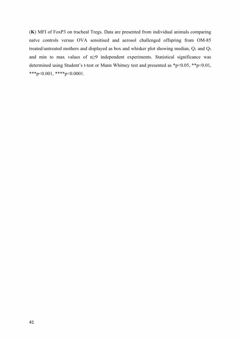
Figure 4. Maternal OM-85 treatment during pregnancy promotes Treg suppressive
phenotypes in sensitized and challenged offspring. (A-B) Absolute numbers of
CD3+CD4+CD8- T-cells in (A) ADLN and (B) trachea. (C-E) Analysis of Tregs within
ADLN showing (C) Tregs as a proportion of total CD4+ T-cells, (D) MFI of CD25 on Tregs
and (E) Treg:Teff ratio within total CD4+ T-cells. (F-J) Analysis of Tregs in the trachea
showing (F) Tregs as a proportion of total CD4+ T-cells, (G) MFI of CD25 on Tregs, (H)
proportion of Treg Ki67+, (I) proportion of Treg CTLA-4+ and (J) proportion of Treg CD69+.
E
H
K
A
C
F
I
B
D
G
J
Maternal OM-85 - + - + Offspring OVA - - + +
Tota
l AD
LN C
D4+ T
ce
lls (x
106 )
- + - + - - + +
Tota
l tra
chea
CD
4+ T
cells
(x10
6 )
Maternal OM-85 - + - + Offspring OVA - - + +
% A
DLN
Tre
g of
C
D4+ T
cel
ls
- + - + - - + +
ADLN
Tre
g C
D25
M
FI (x
103 )
- + - + - - + +
ADLN
Tre
g:Te
ff ra
tio
of C
D4+ T
cel
ls
Maternal OM-85 - + - + Offspring OVA - - + +
% T
rach
ea T
reg
of
CD
4+ T c
ells
- + - + - - + +
Trac
hea
Treg
CD
25
MFI
(x10
3 )
- + - + - - + +
% T
rach
ea K
i67+ T
reg
Maternal OM-85 - + - + Offspring OVA - - + +
% T
rach
ea C
TLA-
4+ Tr
eg
- + - + - - + +
% T
rach
ea C
D69
+ Tre
g
- + - + - - + +
Trac
hea
Treg
Fox
P3
MFI
(x10
4 )
41
(K) MFI of FoxP3 on tracheal Tregs. Data are presented from individual animals comparing
naïve controls versus OVA sensitised and aerosol challenged offspring from OM-85
treated/untreated mothers and displayed as box and whisker plot showing median, Q1 and Q3
and min to max values of n≥9 independent experiments. Statistical significance was
determined using Student’s t-test or Mann Whitney test and presented as *p<0.05, **p<0.01,
***p<0.001, ****p<0.0001.
42
Figure 5. Treatment of mothers with OM-85 during pregnancy limits inflammatory
airways mucosal DC responses in sensitized and aeroallergen challenged offspring.
Shown are absolute numbers of (A) CD11b+ and (B) CD103+ cDCs within ADLN, trachea
and peripheral lung. (C-E) MFI of IAIE expression on CD11b+ and CD103+ cDCs within (C)
ADLN, (D) trachea and (E) peripheral lung. (F) peripheral lung pDCs as a proportion of total
CD45+ cells. (G) MFI of IAIE on peripheral lung pDCs. Data are presented from individual
D C
E G F
A
B
Maternal OM-85 - + - + Offspring OVA - - + +
Tota
l AD
LN C
D11
b+ cD
C (x
104 )
- + - + - - + +
Tota
l tra
chea
CD
11b+
cDC
(x10
2 )
- + - + - - + +
Tota
l lun
g C
D11
b+ cD
C (x
105 )
Maternal OM-85 - + - + Offspring OVA - - + +
Tota
l AD
LN C
D10
3+ cD
C (x
104 )
- + - + - - + +
Tota
l tra
chea
CD
103+
cDC
(x10
2 )
- + - + - - + +
Tota
l lun
g C
D10
3+ cD
C (x
105 )
Maternal OM-85 - + - + Offspring OVA - - + +
- + - + - - + +
ADLN
IAIE
MFI
(x10
4 )
- + - + - - + +
- + - + - - + +
Trac
hea
IAIE
MFI
(x
105 )
Maternal OM-85 - + - + Offspring OVA - - + +
- + - + - - + +
Lung
IAIE
MFI
(x10
4 )
- + - + - - + +
% L
ung
pDC
of C
D45
+
- + - + - - + +
Lung
pD
C IA
IE M
FI
(x10
4 )
CD11b+ cDC CD103
+ cDC CD11b
+ cDC CD103
+ cDC
CD11b+ cDC CD103
+ cDC

43
animals comparing naïve controls versus OVA sensitised and aerosol challenged offspring
from OM-85 treated/untreated mothers and displayed as box and whisker plot showing
median, Q1 and Q3 and min to max values of n≥5 independent experiments. Total peripheral
lung cells displayed as cells per milligram of tissue (A, B). Statistical significance was
determined using Student’s t-test or Mann Whitney test and presented as *p<0.05, **p<0.01,
***p<0.001, ****p<0.0001.
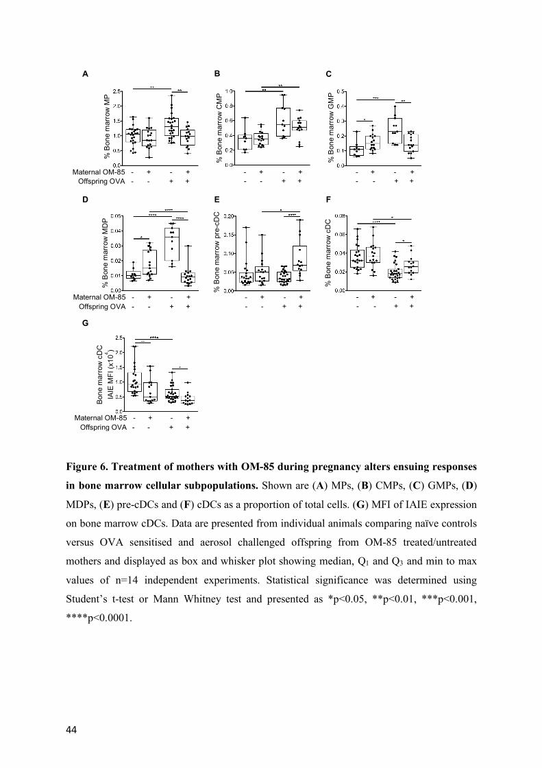
44
Figure 6. Treatment of mothers with OM-85 during pregnancy alters ensuing responses
in bone marrow cellular subpopulations. Shown are (A) MPs, (B) CMPs, (C) GMPs, (D)
MDPs, (E) pre-cDCs and (F) cDCs as a proportion of total cells. (G) MFI of IAIE expression
on bone marrow cDCs. Data are presented from individual animals comparing naïve controls
versus OVA sensitised and aerosol challenged offspring from OM-85 treated/untreated
mothers and displayed as box and whisker plot showing median, Q1 and Q3 and min to max
values of n=14 independent experiments. Statistical significance was determined using
Student’s t-test or Mann Whitney test and presented as *p<0.05, **p<0.01, ***p<0.001,
****p<0.0001.
B
E
C
F
A
D
G
Maternal OM-85 - + - + Offspring OVA - - + +
% B
one
mar
row
MP
- + - + - - + +
% B
one
mar
row
CM
P
- + - + - - + +
% B
one
mar
row
GM
P
Maternal OM-85 - + - + Offspring OVA - - + +
% B
one
mar
row
MD
P
- + - + - - + +
% B
one
mar
row
pre
-cD
C
- + - + - - + +
% B
one
mar
row
cD
C
Maternal OM-85 - + - + Offspring OVA - - + +
Bone
mar
row
cD
C
IAIE
MFI
(x10
4 )

45
Figure 7. Maternal OM-85 treatment during pregnancy supports early life development
of the myeloid compartment. (A-D) Data represent individual 3-week old naïve offspring
from mothers treated or not with OM85 during pregnancy. Shown are (A) CD103+ and (B)
CD11b+ cDCs as a proportion of total peripheral lung cells (C) MFI of IAIE expression on
peripheral lung CD11b+ cDCs (D) Frequency of cDCs generated following in vitro 7-day
GM-CSF-driven bone marrow cultures. (E-F) Data represent individual fetal bone marrow
collected at gestation day 18.5 from mothers treated or not with OM85 during pregnancy.
Shown are absolute numbers of fetal bone marrow (E) cDCs and (F) MDPs. Data are
displayed as box and whisker plot showing median, Q1 and Q3 and min to max values of n≥5
independent experiments. Statistical significance was determined using Student’s t-test or
Mann Whitney test and presented as *p<0.05, **p<0.01, ***p<0.001.
B
E
A
D
C
F
Maternal OM-85 - +
% 3
wk
lung
CD
103+ c
DC
- +
% 3
wk
lung
CD
11b+ c
DC
- +
3wk
lung
CD
11b+ c
DC
IA
IE M
FI (x
104 )
Maternal OM-85 - +
% 3
wk
bone
mar
row
cD
C
- +
Feta
l tot
al b
one
mar
row
cD
C (x
103 )
- + Fe
tal t
otal
bon
e m
arro
w
MD
P (x
104 )
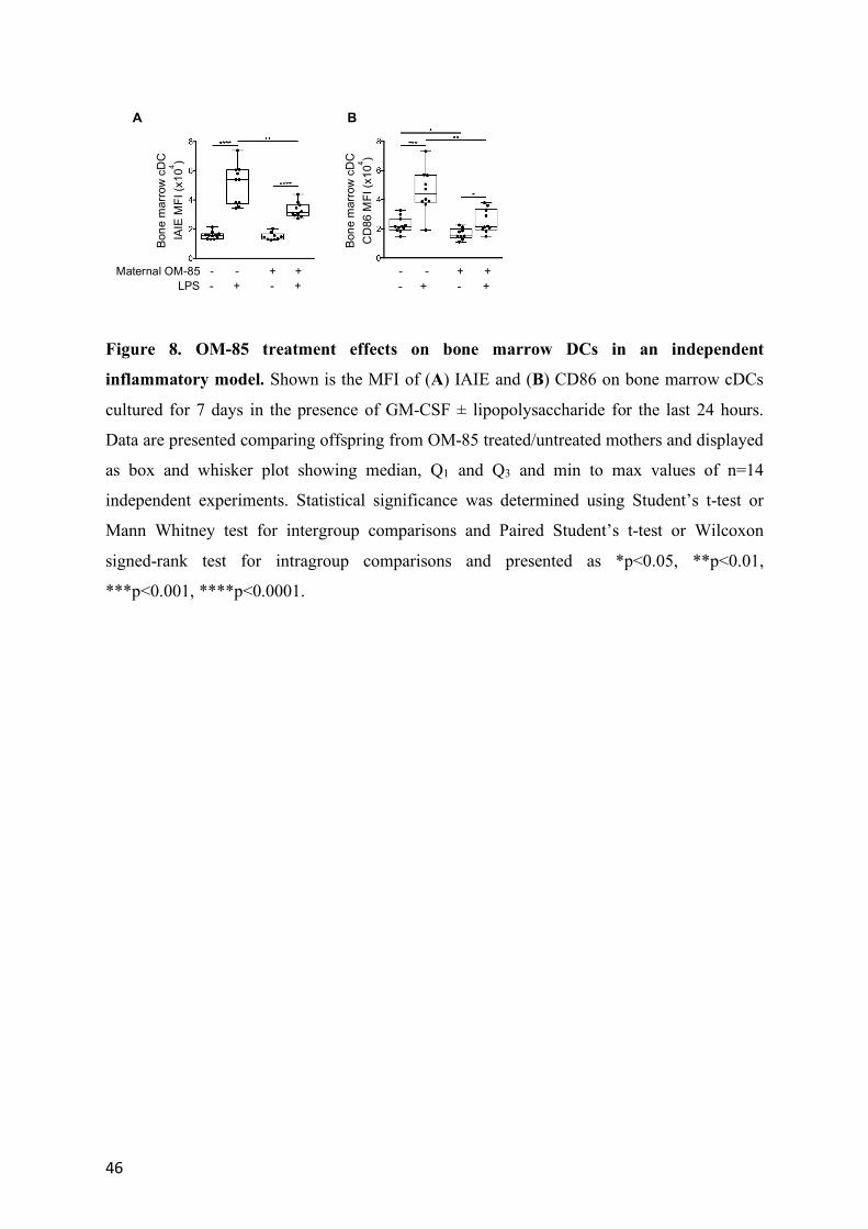
46
Figure 8. OM-85 treatment effects on bone marrow DCs in an independent
inflammatory model. Shown is the MFI of (A) IAIE and (B) CD86 on bone marrow cDCs
cultured for 7 days in the presence of GM-CSF ± lipopolysaccharide for the last 24 hours.
Data are presented comparing offspring from OM-85 treated/untreated mothers and displayed
as box and whisker plot showing median, Q1 and Q3 and min to max values of n=14
independent experiments. Statistical significance was determined using Student’s t-test or
Mann Whitney test for intergroup comparisons and Paired Student’s t-test or Wilcoxon
signed-rank test for intragroup comparisons and presented as *p<0.05, **p<0.01,
***p<0.001, ****p<0.0001.
A B
Maternal OM-85 - - + + LPS - + - +
Bone
mar
row
cD
C
IAIE
MFI
(x10
4 )
- - + + - + - +
Bone
mar
row
cD
C
CD
86 M
FI (x
104 )