back to basics for surgery neurosurgery
DESCRIPTION
Back to Basics for Surgery Neurosurgery. R. Moulton. Principles of Neurological Diagnosis. Questions. What is the lesion Where is the lesion. History Physical (Neurological) Examination Special Tests. Presentation of Neurosurgical Illness. Raised ICP Headache, vomiting papilloedema - PowerPoint PPT PresentationTRANSCRIPT

Back to Basics for SurgeryBack to Basics for SurgeryNeurosurgeryNeurosurgery
R. MoultonR. Moulton

Principles of Neurological Principles of Neurological DiagnosisDiagnosis

QuestionsQuestions
What is the lesionWhat is the lesion Where is the lesionWhere is the lesion

HistoryHistory Physical (Neurological) ExaminationPhysical (Neurological) Examination Special TestsSpecial Tests

Presentation of Neurosurgical Presentation of Neurosurgical IllnessIllness
Raised ICPRaised ICP– Headache, vomitingHeadache, vomiting– papilloedemapapilloedema
Neurological DysfunctionNeurological Dysfunction– General – level of consciousnessGeneral – level of consciousness– Focal – sensory or motor lossFocal – sensory or motor loss
SeizuresSeizures PainPain

What is the lesion – historyWhat is the lesion – history Where is the lesion – neurological Where is the lesion – neurological
examexam

History (What is the lesion?)History (What is the lesion?)
SymptomsSymptoms Mode of onsetMode of onset Speed of onsetSpeed of onset Prior relevant illnessPrior relevant illness Progression/regression of symptomsProgression/regression of symptoms

Neurological Examination Neurological Examination (Where is the Lesion?)(Where is the Lesion?)
Level of Consciousness – GCSLevel of Consciousness – GCS Mental status – orientation, memory, concentration, Mental status – orientation, memory, concentration,
abstraction, calculationabstraction, calculation Cranial NervesCranial Nerves Motor examinationMotor examination
– Upper vs. lower motor neuronUpper vs. lower motor neuron– Cerebellar functionCerebellar function– GaitGait
Sensory examinationSensory examination– light touch, pain & temp, joint position senselight touch, pain & temp, joint position sense– Cortical sensory modalities Cortical sensory modalities

Cranial NervesCranial Nerves
I I OlfactoryOlfactory IIII OpticOptic IIIIII OculomotorOculomotor IVIV TrochlearTrochlear VV TrigeminalTrigeminal VIVI AbducensAbducens VIIVII Facial Facial VIII AcousticVIII Acoustic IX IX GlossopharyngealGlossopharyngeal XX VagusVagus XIXI AccessoryAccessory XIIXII HypoglossalHypoglossal

Motor ExaminationMotor Examination
Upper Motor NeuronUpper Motor Neuron– Weakness (distal > proximal) antigravity Weakness (distal > proximal) antigravity
muscles preservedmuscles preserved– Increased reflexes and tone (spasticity)Increased reflexes and tone (spasticity)– Disuse atrophyDisuse atrophy– Loss of coordination (ataxia)Loss of coordination (ataxia)– ApraxiaApraxia– Upgoing plantar responseUpgoing plantar response

Lower Motor NeuronLower Motor Neuron– WeaknessWeakness– Decreased toneDecreased tone– Decreased reflexesDecreased reflexes– Denervation atrophyDenervation atrophy– Coordination usually intactCoordination usually intact

Sensory ExaminationSensory Examination
Special senses – cranial nervesSpecial senses – cranial nerves Basic ModalitiesBasic Modalities
– Light touch, pain & temp, vibration & Light touch, pain & temp, vibration & proprioceptionproprioception
– Dermatomes, peripheral nerve distributionDermatomes, peripheral nerve distribution
Cortical ModalitiesCortical Modalities– Graphaesthesia, stereognosis, simultaneous Graphaesthesia, stereognosis, simultaneous
appreciation of tactile stimuli, appreciation of tactile stimuli, somatotopognosis, agnosagnosia, neglectsomatotopognosis, agnosagnosia, neglect

Special TestsSpecial Tests
Biochemical, hematological, microbiologyBiochemical, hematological, microbiology– BloodBlood– CSFCSF
ImagingImaging– Plain x-raysPlain x-rays– CTCT– MRIMRI– AngiographyAngiography
ElectrophysiologyElectrophysiology– EMG, nerve conduction, EEG etc.EMG, nerve conduction, EEG etc.
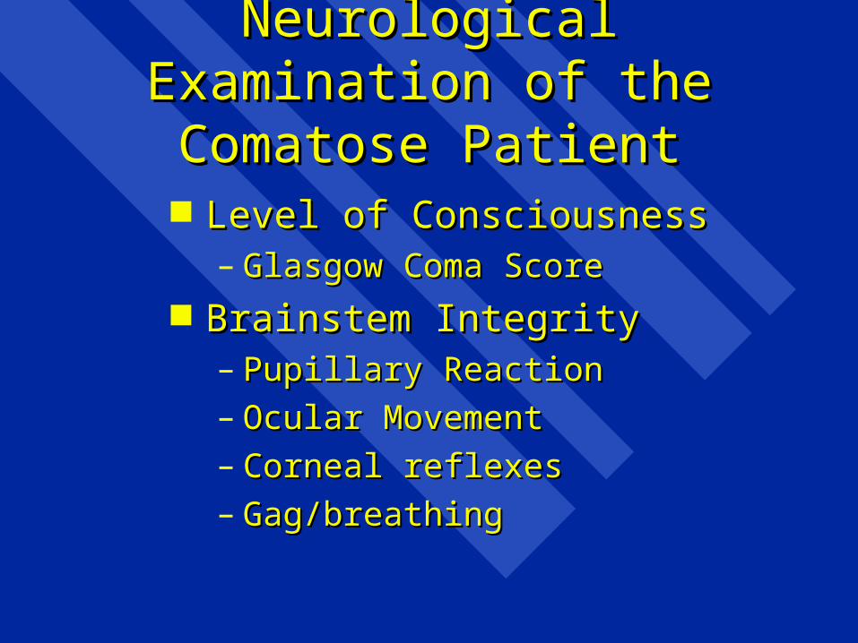
Neurological Examination of Neurological Examination of the Comatose Patientthe Comatose Patient
Level of ConsciousnessLevel of Consciousness– Glasgow Coma ScoreGlasgow Coma Score
Brainstem IntegrityBrainstem Integrity– Pupillary ReactionPupillary Reaction– Ocular MovementOcular Movement– Corneal reflexesCorneal reflexes– Gag/breathingGag/breathing

Eye OpeningEye Openingspontaneousspontaneous 44to voiceto voice 33to painto pain 22nonenone 11
Verbal ResponseVerbal Responseorientedoriented 55confused - sentencesconfused - sentences 44words onlywords only 33soundssounds 22nonenone 11
MovementMovementobeysobeys 66localiseslocalises 55flexion withdrawalflexion withdrawal44abnormal flexionabnormal flexion 33extensionextension 22nonenone 11

Rostral-Caudal DeteriorationRostral-Caudal Deterioration
MidbrainMidbrain– Bilateral pupillary abnormalitiesBilateral pupillary abnormalities– Oculomotor abnormalitiesOculomotor abnormalities
PonsPons– Loss of corneal reflexesLoss of corneal reflexes
MedullaMedulla– Loss of gag reflexesLoss of gag reflexes– Respiratory and vasomotor collapseRespiratory and vasomotor collapse

Brain Tumour ClassificationBrain Tumour Classification Intra-axial (frequently malignant)Intra-axial (frequently malignant) PrimaryPrimary
– GlialGlial– Choroid plexusChoroid plexus– Neuronal or mixed glial-neuronalNeuronal or mixed glial-neuronal– PNET/medulloblastomaPNET/medulloblastoma– CNS lymphomaCNS lymphoma– Pineal regionPineal region– hemangioblastomahemangioblastoma
MetastaticMetastatic

Brain Tumour ClassificationBrain Tumour Classification
Extra-axial (usually benign)Extra-axial (usually benign)– MeningesMeninges– Cranial nerves (Schwannoma)Cranial nerves (Schwannoma)– PituitaryPituitary– skullskull

Glial TumoursGlial Tumours
Astrocytoma (gliobastoma Astrocytoma (gliobastoma multiforme)multiforme)
OligodendrogliomaOligodendroglioma EpendymomaEpendymoma Mixed tumoursMixed tumours Gr. I - IVGr. I - IV

TreatmentTreatment
SupportiveSupportive SpecificSpecific
– Corticosteroids (dexamethasone)Corticosteroids (dexamethasone)– SurgicalSurgical
» BiopsyBiopsy
» Excision Excision
» Internal decompressionInternal decompression

Treatment contd.Treatment contd.
– RadiotherapyRadiotherapy» ConventionalConventional
» Stereotactic focusedStereotactic focused
– ChemotherapyChemotherapy» Temazolamide (malignant glial tumours)Temazolamide (malignant glial tumours)
» Lymphoma protocolsLymphoma protocols
» Specific to tissue of origin for metastasesSpecific to tissue of origin for metastases
ObservationObservation


No Contrast With Contrast


Stroke: Classification and Stroke: Classification and ManagementManagement

Stroke DefinitionStroke Definition
Sudden onset of a neurological deficit Sudden onset of a neurological deficit due to disease or injury of the blood due to disease or injury of the blood supply of the brain.supply of the brain.

Stroke ClassificationStroke Classification
IschemicIschemic– BlandBland– Hemorrhagic transformationHemorrhagic transformation
Hemorrhagic (hemorrhage is 1Hemorrhagic (hemorrhage is 100 event) event)– HypertensionHypertension– Amyloid angiopathyAmyloid angiopathy– AneurysmalAneurysmal– AVMAVM– Other Other

Ischemic Stroke (Infarction)Ischemic Stroke (Infarction)
Thrombotic (local vessel disease)Thrombotic (local vessel disease) EmbolicEmbolic
– Artery to artery (usually carotid)Artery to artery (usually carotid)– Heart to artery (atrial fibrillation)Heart to artery (atrial fibrillation)– Paradoxical (vein to artery)Paradoxical (vein to artery)– Other (air, foreign body, iatrogenic)Other (air, foreign body, iatrogenic)

Intracerebral HemorrhageIntracerebral Hemorrhage
HypertensiveHypertensive– Occurs in long narrow perforating Occurs in long narrow perforating
arteries (basal ganglia, thalamus, pons, arteries (basal ganglia, thalamus, pons, cerebellar nuclei)cerebellar nuclei)
– Charcot-Bouchard aneurysmsCharcot-Bouchard aneurysms– Related primarily to duration of Related primarily to duration of
hypertensionhypertension

Intracerebral HemorrhageIntracerebral Hemorrhage
Amyloid angiopathyAmyloid angiopathy– Age related change in cerebral vesselsAge related change in cerebral vessels– Lobar hemorrhageLobar hemorrhage– Most commonly in posterior part of Most commonly in posterior part of
cerebral hemispherescerebral hemispheres


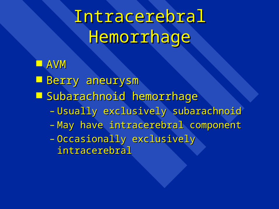
Intracerebral HemorrhageIntracerebral Hemorrhage
AVMAVM Berry aneurysmBerry aneurysm Subarachnoid hemorrhageSubarachnoid hemorrhage
– Usually exclusively subarachnoidUsually exclusively subarachnoid– May have intracerebral componentMay have intracerebral component– Occasionally exclusively intracerebralOccasionally exclusively intracerebral


ManagementManagement
DiagnosisDiagnosis– HistoryHistory– Physical ExaminationPhysical Examination– Special tests (imaging)Special tests (imaging)
TreatmentTreatment

Stroke DiagnosisStroke Diagnosis
HistoryHistory– Rapid onset fixed deficit – ischemicRapid onset fixed deficit – ischemic– Rapid onset progressive deficit – Rapid onset progressive deficit –
hemorrhagehemorrhage– Sudden severe headache, Sudden severe headache,
nausea/vomiting/photophobia +/- nausea/vomiting/photophobia +/- neurological deficit - SAHneurological deficit - SAH

Stroke Physical ExaminationStroke Physical Examination
Focal deficitsFocal deficits– Most often ischemic stroke or ICHMost often ischemic stroke or ICH– Much less common in SAHMuch less common in SAH
Alteration in level of consciousnessAlteration in level of consciousness– SAHSAH– ICHICH– Delayed swelling from large infarctsDelayed swelling from large infarcts

Stroke InvestigationStroke Investigation
CT scanCT scan– First line imaging to distinguish infarct First line imaging to distinguish infarct
from hemorrhagefrom hemorrhage– 11stst choice for confirming SAH, LP if choice for confirming SAH, LP if
negativenegative OtherOther
– Cerebral angiography, doppler for Cerebral angiography, doppler for carotidscarotids
– MRI in special circumstancesMRI in special circumstances


Acute Stroke TreatmentAcute Stroke Treatment
SupportiveSupportive– AirwayAirway– Blood pressureBlood pressure
DefinitiveDefinitive– ThrombolysisThrombolysis– Hematoma evacuation (limited Hematoma evacuation (limited
circumstances)circumstances)


Stroke TreatmentStroke Treatment
PreventionPrevention– Risk factor modificationRisk factor modification
» Hypertension, smoking, diabetes, lipids/cholesterolHypertension, smoking, diabetes, lipids/cholesterol
– Antiplatelet agents (artery-artery embolism, Antiplatelet agents (artery-artery embolism, local occlusive disease)local occlusive disease)
– Anticoagulation (heart to artery emboli)Anticoagulation (heart to artery emboli)
– Surgical preventionSurgical prevention» Carotid endarterectomy, stentingCarotid endarterectomy, stenting
» Aneurysm obliterationAneurysm obliteration
» AVM excisionAVM excision


Skull Skull FractureFracture

Primary Impact InjuryPrimary Impact Injury
Shear (diffuse) injury of axonsShear (diffuse) injury of axons
Laceration/contusion of cortical Laceration/contusion of cortical surfacesurface

Blumbergs, Head Injury, 1997:45

Cerebral ContusionsCerebral Contusions

Secondary InsultsSecondary Insults
HypoxiaHypoxia IschaemiaIschaemia Intracranial hematomasIntracranial hematomas Raised intracranial pressureRaised intracranial pressure SeizuresSeizures Infection Infection Fluid and electrolyte disturbanceFluid and electrolyte disturbance

Respiratory Changes in Head InjuryRespiratory Changes in Head Injury
Depression/abolition of gag and cough Depression/abolition of gag and cough reflexesreflexes
Hypercarbia 2Hypercarbia 2o o to respiratory centre to respiratory centre depressiondepression
Hypoxemia -- systemic causesHypoxemia -- systemic causes– inadequate airway managementinadequate airway management– chest traumachest trauma– aspirationaspiration

Recommendations for Treatment
Resuscitate aggressively with appropriate fluids Brain oedema is not a concern
Manage source of bleeding in unstable patients prior to transfer
Do not use mannitol in presence of hypotension or you will further destabilise the
patientConsider transient use of vasopressor drugs
while restoring volume and controlling haemorrhage


Trauma Craniotomy IncisionTrauma Craniotomy Incision


Pressure Volume CurvePressure Volume Curve
Pressure
Volume
Vskull = Vbrain + Vblood + VCSF + Vmass

Trans-Tentorial HerniationTrans-Tentorial Herniation

Use of MannitolUse of Mannitol
.5 - 1 gm./kg of 20% solution.5 - 1 gm./kg of 20% solution give as a bolusgive as a bolus urinary catheterurinary catheter Contraindications:Contraindications:ShockShockAnuriaAnuria

Other ICP TherapiesOther ICP Therapies
CPP therapyCPP therapy
Barbiturate ComaBarbiturate Coma
Decompressive CraniectomyDecompressive Craniectomy


Back to Basics For SurgeryBack to Basics For Surgery
SpineSpine

Pain GeneratorsPain Generators
MyofascialMyofascial DiscDisc Facet JointFacet Joint NerveNerve VisceralVisceral VascularVascular

Physical Examination: The SpinePhysical Examination: The Spine
Inspect: deformityInspect: deformity Palpate: deformity, local tendernessPalpate: deformity, local tenderness Range of motion (limitation, pain)Range of motion (limitation, pain)

MyelopathyMyelopathy
‘‘a general term denoting functional a general term denoting functional disturbance and/or pathological disturbance and/or pathological changes in the spinal cord’changes in the spinal cord’

Myelopathy Myelopathy Important QuestionsImportant Questions
Level of lesionLevel of lesion Nature of lesion Nature of lesion
– Surgical (spondylotic, neoplastic, infectious, Surgical (spondylotic, neoplastic, infectious, hematoma, traumatic)hematoma, traumatic)
– Treatment frequently curativeTreatment frequently curative– Non-surgical (degenerative, inflammatory)Non-surgical (degenerative, inflammatory)
Degree of patient disabilityDegree of patient disability Rate of progressionRate of progression History, physical examination, special History, physical examination, special
investigationsinvestigations

Myelopathy: HistoryMyelopathy: History Patient Complaints:Patient Complaints: Numbness (loss of sensation, alteration of Numbness (loss of sensation, alteration of
sensation – paraesthesia, awkwardness)sensation – paraesthesia, awkwardness) Ataxia (awkwardness, clumsiness)Ataxia (awkwardness, clumsiness)
– Usually:Usually:
– Gait (imbalance, unsteadiness, unable to move Gait (imbalance, unsteadiness, unable to move quickly)quickly)
– Fine movements of hands (doing up buttons, Fine movements of hands (doing up buttons, handwriting)handwriting)
Weakness – usually a late findingWeakness – usually a late finding

Myelopathy: HistoryMyelopathy: History
Limbs involved: lower (may be thoracic or Limbs involved: lower (may be thoracic or cervical), upper and lower (always cervical)cervical), upper and lower (always cervical)
Onset: gradual, rapid or suddenOnset: gradual, rapid or sudden Associated pain: Associated pain:
– Activity related: spondyloticActivity related: spondylotic– Nocturnal: neoplasticNocturnal: neoplastic– Associated radicular painAssociated radicular pain
Previous or concurrent neurological Previous or concurrent neurological symptoms/illnesssymptoms/illness

Myelopathy: Physical Myelopathy: Physical ExaminationExamination
Motor:Motor:– Strength: weakness is usually late finding Strength: weakness is usually late finding
in slowly evolving surgical conditions, in slowly evolving surgical conditions, occurs in corticospinal distributionoccurs in corticospinal distribution
– Reflexes (change occurs early): hyperactive Reflexes (change occurs early): hyperactive distal to lesion in gradually evolving lesionsdistal to lesion in gradually evolving lesions
» In disc disease may be hypoactive at level of In disc disease may be hypoactive at level of lesionlesion

Myelopathy: Physical Myelopathy: Physical ExaminationExamination
– Tone (early): increased distal to lesionTone (early): increased distal to lesion– Coordination (early): impaired distal to Coordination (early): impaired distal to
lesionlesion– Plantar responses: up-going (reliability?)Plantar responses: up-going (reliability?)
Sensation:Sensation:– Proprioception: frequently impaired in Proprioception: frequently impaired in
lower limbs – impossible to establish lower limbs – impossible to establish precise levelprecise level
– Pinprick: extremely useful in thoracic Pinprick: extremely useful in thoracic lesionslesions

Special InvestigationsSpecial Investigations
Plain x-rays (bone destruction, fracture, Plain x-rays (bone destruction, fracture, subluxation, spondylotic changes), n.b. no subluxation, spondylotic changes), n.b. no visualization of nervous tissuevisualization of nervous tissue
CT scan (same indications/contraindications CT scan (same indications/contraindications as x-ray)as x-ray)
MRI usually the definitive investigationMRI usually the definitive investigation CT-myelography (most useful for looking at CT-myelography (most useful for looking at
bone and disc relation to spinal cord/nerve bone and disc relation to spinal cord/nerve roots)roots)

Myelopathy: Surgical Decision-Myelopathy: Surgical Decision-MakingMaking
Nature of the lesionNature of the lesion Natural history of the lesionNatural history of the lesion
– Trauma: static/improving unless spine unstableTrauma: static/improving unless spine unstable
– Neoplastic: progressive, rate variable depending on Neoplastic: progressive, rate variable depending on histologyhistology
– Infectious: usually rapidly progressiveInfectious: usually rapidly progressive
– Spondylotic myelopathy, usually gradually progressive, rate Spondylotic myelopathy, usually gradually progressive, rate variablevariable
– Recovery usually poor with advanced deficitsRecovery usually poor with advanced deficits

Myelopathy: Surgical ApproachMyelopathy: Surgical Approach
Lesion site:Lesion site:– ExtraduralExtradural– Intra-dural, extra-medullaryIntra-dural, extra-medullary– IntramedullaryIntramedullary
Extradural:Extradural:– Anterior pathology – anterior approachAnterior pathology – anterior approach– Posterior pathology – posterior approach Posterior pathology – posterior approach
(laminectomy)(laminectomy) Intradural-extramedullary – posteriorIntradural-extramedullary – posterior Intradural-intramedullary - posteriorIntradural-intramedullary - posterior


RadiculopathyRadiculopathy
a general term denoting functional a general term denoting functional disturbance and/or pathological disturbance and/or pathological changes in a spinal nerve rootchanges in a spinal nerve root

RadiculopathyRadiculopathy
SymptomsSymptoms– Pain, paraesthesiae, sensory loss in the Pain, paraesthesiae, sensory loss in the
approximate dermatome of the involved approximate dermatome of the involved nerve rootnerve root
– Axial pain is not a symptom of nerve root Axial pain is not a symptom of nerve root involvement involvement
– Weakness in the myotome of the Weakness in the myotome of the involved nerve root – pts. frequently involved nerve root – pts. frequently can’t be specificcan’t be specific

RadiculopathyRadiculopathy
Exam findingsExam findings– Lower motor neuron findings in the Lower motor neuron findings in the
appropriate myotomeappropriate myotome– Sensory findings in the appropriate Sensory findings in the appropriate
dermatomedermatome

Radiculopathy – InvestigationRadiculopathy – Investigation
LumbarLumbar– MRI, CT scanMRI, CT scan
Cervical/thoracicCervical/thoracic– MRIMRI

Radiculopathy - Conservative TxRadiculopathy - Conservative Tx
Activity modificationActivity modification NSAIDSNSAIDS AnalgesicsAnalgesics Physiotherapy - activePhysiotherapy - active
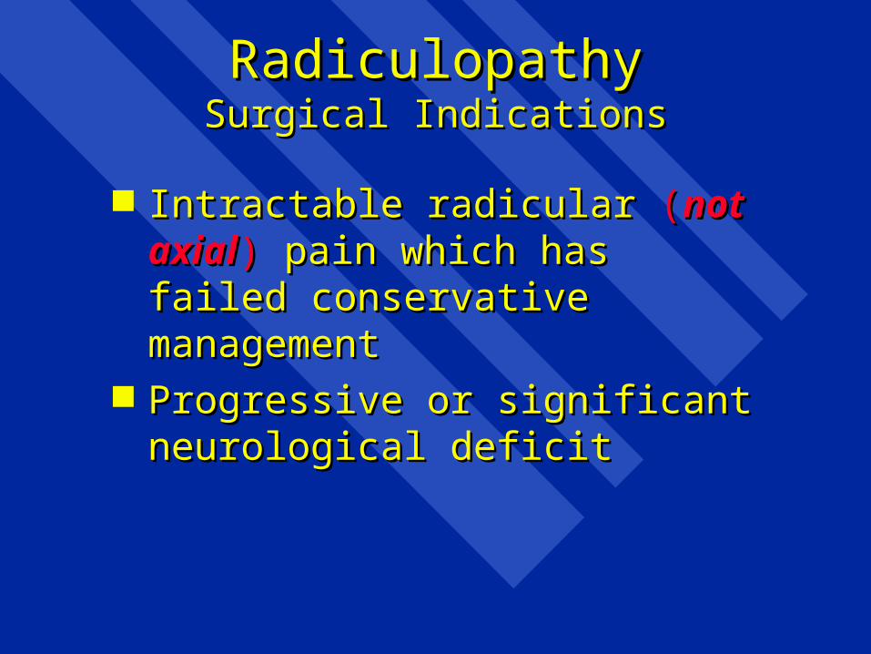
RadiculopathyRadiculopathySurgical IndicationsSurgical Indications
Intractable radicular Intractable radicular ((not axialnot axial)) pain pain which has failed conservative which has failed conservative managementmanagement
Progressive or significant neurological Progressive or significant neurological deficitdeficit


Spine Pain Spine Pain – Red Flags– Red Flags
Hx of major trauma or minor trauma in elderly, Hx of major trauma or minor trauma in elderly, osteoporotic patientsosteoporotic patients
Age < 20 or > 50Age < 20 or > 50 Hx of cancer, fever, chills, unexplained wt. lossHx of cancer, fever, chills, unexplained wt. loss Hx of recent infection, IV drug abuse, Hx of recent infection, IV drug abuse,
immunocompromiseimmunocompromise Hx of bladder or bowel incontinence, urinary Hx of bladder or bowel incontinence, urinary
retentionretention Hx of major or progressive neurological deficitHx of major or progressive neurological deficit Hx of pain worsening when supine or severe night Hx of pain worsening when supine or severe night
painpain

Spine Pain Spine Pain – Red Flags– Red Flags
Exam: major neurological deficit/signs Exam: major neurological deficit/signs of upper motor neuron dysfunctionof upper motor neuron dysfunction
Exam: peri-anal anaesthesiaExam: peri-anal anaesthesia Exam: loss of anal sphincter toneExam: loss of anal sphincter tone


Indications for SurgeryIndications for Surgery (Non-Degenerative Back Pain)(Non-Degenerative Back Pain)
Tumour Tumour – primary primary – metastaticmetastatic
Infection Infection – Discitis/osteomyelitisDiscitis/osteomyelitis– Epidural AbcessEpidural Abcess
Fracture/subluxation with instabilityFracture/subluxation with instability


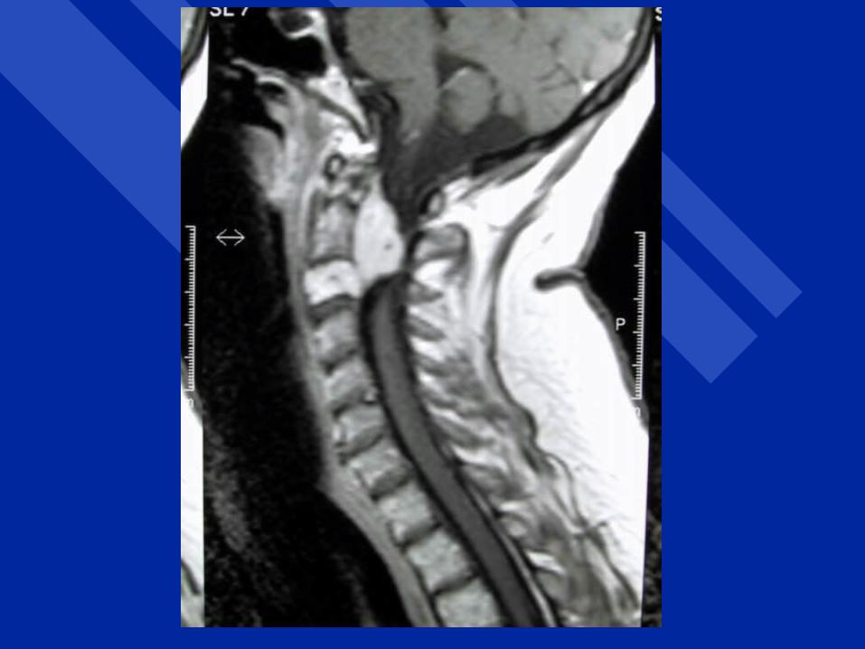
Clinical Assessment of Spinal InjuriesClinical Assessment of Spinal Injuries
HistoryHistoryMechanism of injuryMechanism of injurySpinal painSpinal painParaesthesia or motor weaknessParaesthesia or motor weakness
Physical examinationPhysical examinationLog roll, inspect and palpate entire spineLog roll, inspect and palpate entire spineTendernessTendernessMalalignment of spinous processesMalalignment of spinous processes

Traps for the UnwaryTraps for the Unwary
Patient intoxicationPatient intoxication Altered level of consciousnessAltered level of consciousness Distraction from other injuriesDistraction from other injuries Cursory examination – failure to Cursory examination – failure to
appreciate single root injuryappreciate single root injury

Cervical Spine X-raysCervical Spine X-rays
Lateral to T1Lateral to T1 APAP Open-mouth odontoidOpen-mouth odontoid CT Scan if one or more of above not CT Scan if one or more of above not
availableavailable

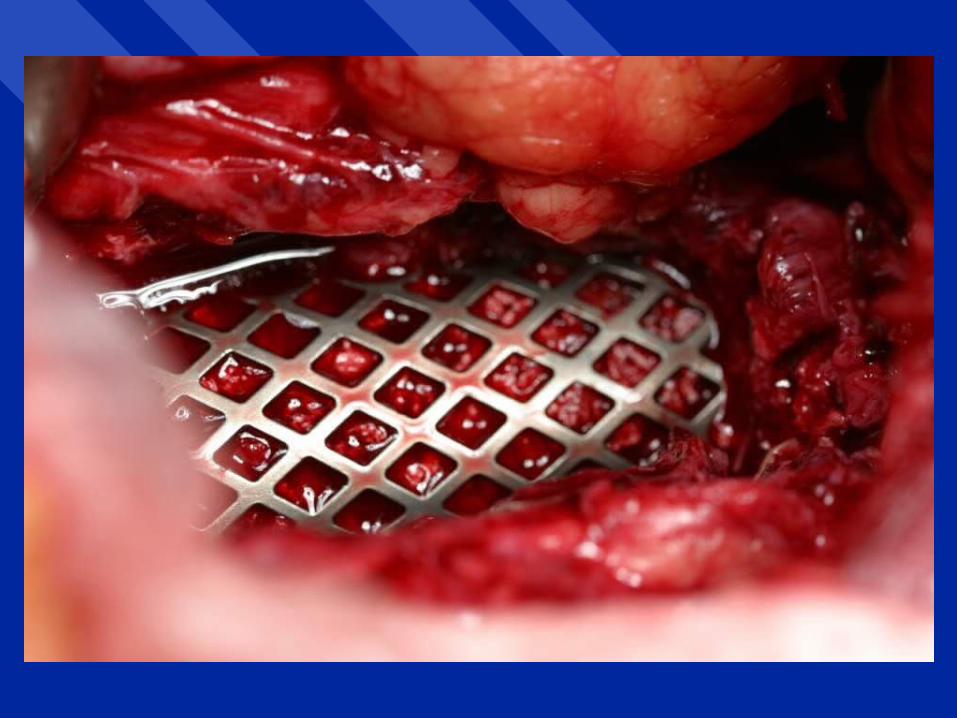
Treatment of Spine InjuriesTreatment of Spine Injuries
Immobilize patientImmobilize patient Reduce deformityReduce deformity Stabilize/fuse spineStabilize/fuse spine


Back to Basics for SurgeryBack to Basics for Surgery
Peripheral NervePeripheral Nerve

Injury Classification Injury Classification (Seddon)(Seddon)
• NeurapraxiaNeurapraxia
• AxonotmesisAxonotmesis
• NeurotmesisNeurotmesis

Peripheral Nerve InjuryPeripheral Nerve Injury
HistoryHistory– Usually immediate onset of Usually immediate onset of
symptoms/signs from time of injurysymptoms/signs from time of injury– Blunt or penetrating injuryBlunt or penetrating injury– Blunt injury frequently associated with Blunt injury frequently associated with
fracture or dislocationfracture or dislocation– May follow reduction of fracture or May follow reduction of fracture or
dislocationdislocation– Delayed onset: compartment syndrome or Delayed onset: compartment syndrome or
vascular injury to limbvascular injury to limb


Peripheral Nerve InjuryPeripheral Nerve Injury
Physical ExaminationPhysical Examination– Upper vs. lower motor neuronUpper vs. lower motor neuron– Root vs. peripheral nerveRoot vs. peripheral nerve– Which root?Which root?– Which peripheral nerve?Which peripheral nerve?

InvestigationsInvestigations
MRI/CTMRI/CT– Indirect, helpful if question of upper vs. lower Indirect, helpful if question of upper vs. lower
motor neuron, root vs. peripheral nervemotor neuron, root vs. peripheral nerve
EMGs/Nerve conductionEMGs/Nerve conduction– Former useful, latter notFormer useful, latter not
– Most sensitive in detecting early recoveryMost sensitive in detecting early recovery
– Not useful in acute managementNot useful in acute management
Extremity X-rays: Extremity X-rays: – helpful with injury site if fracture or dislocationhelpful with injury site if fracture or dislocation

InvestigationInvestigation
• EMG (all injuries)EMG (all injuries)importance of clinical vs. EMG recoveryimportance of clinical vs. EMG recovery
• Root and trunk injuriesRoot and trunk injuriesMetrizamide CT- myelogramMetrizamide CT- myelogramMRIMRI


Overall Treatment StrategyOverall Treatment Strategy
• Nerve repairNerve repairRestore movementRestore movementRestore sensationRestore sensation
• Muscle/tendon/joint reconstructive Muscle/tendon/joint reconstructive surgerysurgery
• ProstheticsProsthetics• RehabilitationRehabilitation• Educational and vocational adviceEducational and vocational advice


Timing of SurgeryTiming of Surgery
• Primary repair (penetrating injury)Primary repair (penetrating injury)immediateimmediatedelayed (2 weeks)delayed (2 weeks)
• Secondary repair (blunt injury)Secondary repair (blunt injury)3 - 4 month delay3 - 4 month delay


Reconstructive Strategies to Reconstructive Strategies to Achieve Elbow FlexionAchieve Elbow Flexion
• Steindler flexoroplastySteindler flexoroplasty
• Latissimus dorsi transferLatissimus dorsi transfer
• Pectoralis major transferPectoralis major transfer
• Triceps transferTriceps transfer

Common Wrist/Hand Common Wrist/Hand Tendon TransfersTendon Transfers
• Wrist extension -- pronator teresWrist extension -- pronator teres• Thumb extension -- palmaris longusThumb extension -- palmaris longus• MCP extension -- flexor carpi radialisMCP extension -- flexor carpi radialis• Finger flexion -- brachioradialis or Finger flexion -- brachioradialis or
extensor carpi radialis longus to flexor extensor carpi radialis longus to flexor digitorum profundusdigitorum profundus
• Thumb flexion -- BR or ECRL to FPLThumb flexion -- BR or ECRL to FPL

Results EtiologyResults EtiologyEtiology Etiology No. of PtsNo. of Pts
Lacerations 24Lacerations 24MVA 22MVA 22Winter sports 11Winter sports 11Falls 8Falls 8Gunshot wounds 4Gunshot wounds 4Others 14Others 14
Adjacent fractures in 15 patientsAdjacent fractures in 15 patients

Individual nerve outcomeIndividual nerve outcome
Nerve Nerve Inc. loss Exc. lossInc. loss Exc. loss
to f/u to f/uto f/u to f/u
Brachial plexus 33% 37.5%Brachial plexus 33% 37.5%
Axillary 42.9% 75%Axillary 42.9% 75%
Musculocutaneous 57.1% 80%Musculocutaneous 57.1% 80%
Radial 58.3% 87.5%Radial 58.3% 87.5%
Median 75% 85.7%Median 75% 85.7%
Ulnar 66% 100%Ulnar 66% 100%
Posterior tibial 50% 60%Posterior tibial 50% 60%

Outcome by EtiologyOutcome by Etiology
LacerationLaceration 87.5%87.5%
MVAMVA 32%32%
Winter sportsWinter sports 57.1%57.1%
FallsFalls 50%50%