autotaxin, an ectoenzyme that produces lysophosphatidic acid, promotes the entry of lymphocytes into...
TRANSCRIPT

Autotaxin, an ectoenzyme that produceslysophosphatidic acid, promotes the entry oflymphocytes into secondary lymphoid organs
Hidenobu Kanda1, Rebecca Newton1,3, Russell Klein1,3, Yuka Morita1,3, Michael D Gunn2,3 & Steven D Rosen1
The extracellular lysophospholipase D autotaxin (ATX) and its product, lysophosphatidic acid, have diverse functions in
development and cancer, but little is known about their functions in the immune system. Here we found that ATX had high
expression in the high endothelial venules of lymphoid organs and was secreted. Chemokine-activated lymphocytes expressed
receptors with enhanced affinity for ATX, which provides a mechanism for targeting the secreted ATX to lymphocytes undergoing
recruitment. Lysophosphatidic acid induced chemokinesis in T cells. Intravenous injection of enzymatically inactive ATX
attenuated the homing of T cells to lymphoid tissues, probably through competition with endogenous ATX and exertion of
a dominant negative effect. Our results support the idea of a new and general step in the homing cascade in which the
ectoenzyme ATX facilitates the entry of lymphocytes into lymphoid organs.
High endothelial venules (HEVs) in lymph nodes, Peyer’s patches andother secondary lymphoid tissue contribute to continuous immunesurveillance by supporting the recruitment of lymphocytes from theblood1–3. Considerable work has established that HEVs are morpho-logically and functionally specialized to capture lymphocytes circulat-ing at high speed in the bloodstream and to support their migrationinto the lymphoid organ. HEVs are characterized by high endothelialcells (HECs) with a plump morphology and a well developed Golgicomplex and rough endoplasmic reticulum1. Such features suggesthighly active biosynthetic activities, whereas endothelial cells in othertissues generally have thin morphology and quiescent phenotypes.Sites of chronic inflammation can develop HEV-like vessels that alsoserve as a gateway for lymphocyte entry2,4.
As is generally true for leukocyte-endothelium interactions, lym-phocyte-HEV interactions occur in three sequential steps: rolling oflymphocytes along the endothelium, arrest on the endothelium andtransmigration across the endothelium1,3,5. In lymph node HEVs, therolling step is governed by weak and transient interactions betweenL-selectin on the lymphocyte and the carbohydrate determinant ‘sialyl6-sulfo Lewis X’, which is presented by a family of sialomucin proteinson HEVs2. In homing to Peyer’s patches, both L-selectin and theintegrin a4b7 interact with the a4b7 ligand MAdCAM-1 on HEVs tosupport lymphocyte rolling6. The arrest step is induced by so-called‘arrest’ chemokines such as CCL21 and CCL19 (also called SLC andELC, respectively), which are immobilized and presented on the apical
aspect of HEVs3,7. These molecules trigger activation of the integrinLFA-1 on lymphocytes and increase its affinity for its ligands ICAM-1and ICAM-2, expressed on HEVs. The transendothelial migration stepis poorly understood.
The knowledge gaps about HEV function have prompted severalgene-profiling analyses of purified HECs from lymphoid organs8–10.An expressed sequence tag study of human tonsillar HEC geneexpression has shown that about 5% of the transcripts encodethe ectoenzyme autotoxin (ATX; A003157)10. ATX was originallyidentified in human melanoma cell culture medium as an autocrinemotility factor that potently induces the motility of the melanomacells11,12. ATX is overexpressed in various tumors and has been linkedto tumor cell migration, invasiveness and metastasis13. ATX is requiredfor normal development, as gene-targeted mice die in utero withprofound defects in vascular formation and other abnormalities14,15.Notable in the sequence of ATX is a phosphodiesterase domain,which is required for its motility-stimulating activity12,16. Severalyears after its cloning, ATX was found to have lysophospholipase Dactivity, which generates lysophosphatidic acid (LPA) from lyso-phosphatidylcholine17,18. As an extracellular lysophospholipid, LPAelicits a wide variety of responses in many cell types through inter-actions with a family of five G protein–coupled receptors (GPCRs)13,19.Among its most extensively studied activities, LPA regulatescytoskeletal organization and migration in many cell types13. In severalcases, it has been established that the motility-stimulating activity
Received 22 October 2007; accepted 8 February 2008; published online 9 March 2008; doi:10.1038/ni1573
1Department of Anatomy, Program in Immunology, Cardiovascular Research Institute, and 2Cardiovascular Research Institute, University of California San Francisco,San Francisco, California 94143, USA. 3Present addresses: Office of the Gene Technology Regulator, Federal Government Department of Health, Canberra 2601, Australia(R.N.), Pulmonary and Critical Care Medicine Division, University of California Irvine, Orange, California 92868, USA (R.K.), Osaka University Faculty of Medicine, Osaka565-0871, Japan (Y.M.) and Department of Medicine, Division of Cardiovascular Medicine, Duke University, Durham, North Carolina 27710, USA (M.D.G.).Correspondence should be addressed to S.D.R. ([email protected]).
NATURE IMMUNOLOGY VOLUME 9 NUMBER 4 APRIL 2008 415
A R T I C L E S©
2008
Nat
ure
Pub
lishi
ng G
roup
ht
tp://
ww
w.n
atur
e.co
m/n
atur
eim
mun
olog
y
of ATX is mediated through the production of LPA and signaltransduction through its specific GPCRs17,20.
Most interest in ATX has been directed toward its functions incancer and early development. The discovery that HECs have highexpression of ATX transcripts10 prompted our investigation of thepotential functions of this protein in the immune system. Our studiesshow a general contribution of ATX to the entry of lymphocytes fromthe blood into secondary lymphoid organs.
RESULTS
ATX expression in mouse lymph nodes
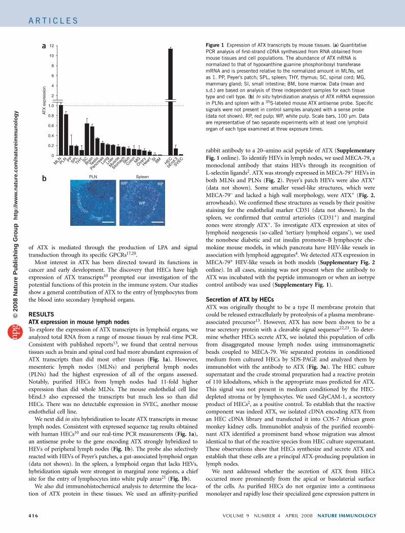
To explore the expression of ATX transcripts in lymphoid organs, weanalyzed total RNA from a range of mouse tissues by real-time PCR.Consistent with published reports13, we found that central nervoustissues such as brain and spinal cord had more abundant expression ofATX transcripts than did most other tissues (Fig. 1a). However,mesenteric lymph nodes (MLNs) and peripheral lymph nodes(PLNs) had the highest expression of all of the organs assessed.Notably, purified HECs from lymph nodes had 11-fold higherexpression than did whole MLNs. The mouse endothelial cell linebEnd.3 also expressed the transcripts but much less so than didHECs. There was no detectable expression in SVEC, another mouseendothelial cell line.
We next did in situ hybridization to locate ATX transcripts in mouselymph nodes. Consistent with expressed sequence tag results obtainedwith human HECs10 and our real-time PCR measurements (Fig. 1a),an antisense probe to the gene encoding ATX strongly hybridized toHEVs of peripheral lymph nodes (Fig. 1b). The probe also selectivelyreacted with HEVs of Peyer’s patches, a gut-associated lymphoid organ(data not shown). In the spleen, a lymphoid organ that lacks HEVs,hybridization signals were strongest in marginal zone regions, a chiefsite for the entry of lymphocytes into white pulp areas21 (Fig. 1b).
We also did immunohistochemical analysis to determine the loca-tion of ATX protein in these tissues. We used an affinity-purified
rabbit antibody to a 20–amino acid peptide of ATX (SupplementaryFig. 1 online). To identify HEVs in lymph nodes, we used MECA-79, amonoclonal antibody that stains HEVs through its recognition ofL-selectin ligands2. ATX was strongly expressed in MECA-79+ HEVs inboth MLNs and PLNs (Fig. 2). Peyer’s patch HEVs were also ATX+
(data not shown). Some smaller vessel-like structures, which wereMECA-79– and lacked a high wall morphology, were ATX+ (Fig. 2,arrowheads). We confirmed these structures as vessels by their positivestaining for the endothelial marker CD31 (data not shown). In thespleen, we confirmed that central arterioles (CD31+) and marginalzones were strongly ATX+. To investigate ATX expression at sites oflymphoid neogenesis (so-called ‘tertiary lymphoid organs’), we usedthe nonobese diabetic and rat insulin promoter–B lymphocyte che-mokine mouse models, in which pancreata have HEV-like vessels inassociation with lymphoid aggregates4. We detected ATX expression inMECA-79+ HEV-like vessels in both models (Supplementary Fig. 2online). In all cases, staining was not present when the antibody toATX was incubated with the peptide immunogen or when an isotypecontrol antibody was used (Supplementary Fig. 1).
Secretion of ATX by HECs
ATX was originally thought to be a type II membrane protein thatcould be released extracellularly by proteolysis of a plasma membrane-associated precursor13. However, ATX has now been shown to be atrue secretory protein with a cleavable signal sequence22,23. To deter-mine whether HECs secrete ATX, we isolated this population of cellsfrom disaggregated mouse lymph nodes using immunomagneticbeads coupled to MECA-79. We separated proteins in conditionedmedium from cultured HECs by SDS-PAGE and analyzed them byimmunoblot with the antibody to ATX (Fig. 3a). The HEC culturesupernatant and the crude stromal preparation had a reactive proteinof 110 kilodaltons, which is the appropriate mass predicted for ATX.This signal was not present in medium conditioned by the HEC-depleted stroma or by lymphocytes. We used GlyCAM-1, a secretoryproduct of HECs2, as a positive control. To establish that the reactivecomponent was indeed ATX, we isolated cDNA encoding ATX froman HEC cDNA library and transfected it into COS-7 African greenmonkey kidney cells. Immunoblot analysis of the purified recombi-nant ATX identified a prominent band whose migration was almostidentical to that of the reactive species from HEC culture supernatant.These observations show that HECs synthesize and secrete ATX andestablish that these cells are a principal ATX-producing population inlymph nodes.
We next addressed whether the secretion of ATX from HECsoccurred more prominently from the apical or basolaterial surfaceof the cells. As purified HECs do not organize into a continuousmonolayer and rapidly lose their specialized gene expression pattern in
MLNPLN PP
SPLTHY SC
BrainTe
stis
Pancr
easLu
ngLiv
er
Mus
cle
Stom
achSkin
ColonM
GOva
ry
Heart SI
BMHEC
bEnd
.3
SVEC
12
10
8
6
4
2
0.2
0.4
0.6
0.8
1.0
0
a
WP
RPWPb PLN Spleen
ATX
exp
ress
ion
Figure 1 Expression of ATX transcripts by mouse tissues. (a) Quantitative
PCR analysis of first-strand cDNA synthesized from RNA obtained from
mouse tissues and cell populations. The abundance of ATX mRNA is
normalized to that of hypoxanthine guanine phosphoribosyl transferase
mRNA and is presented relative to the normalized amount in MLNs, set
as 1. PP, Peyer’s patch; SPL, spleen; THY, thymus; SC, spinal cord; MG,
mammary gland; SI, small intestine; BM, bone marrow. Data (mean and
s.d.) are based on analysis of three independent samples for each tissuetype and cell type. (b) In situ hybridization analysis of ATX mRNA expression
in PLNs and spleen with a 35S-labeled mouse ATX antisense probe. Specific
signals were not present in control samples analyzed with a sense probe
(data not shown). RP, red pulp; WP, white pulp. Scale bars, 100 mm. Data
are representative of two separate experiments with at least one lymphoid
organ of each type examined at three exposure times.
416 VOLUME 9 NUMBER 4 APRIL 2008 NATURE IMMUNOLOGY
A R T I C L E S©
2008
Nat
ure
Pub
lishi
ng G
roup
ht
tp://
ww
w.n
atur
e.co
m/n
atur
eim
mun
olog
y

culture24, we used transfected MDCK canine kidney cells for thesestudies. MDCK cells form a highly polarized and tight monolayer andare widely used to study polarized secretion in epithelial cells25. Weprepared a stable MDCK transfectant that expressed ATX with asix-histidine tag at the amino terminus. These cells established a tightmonolayer, as confirmed by measurement of the passage of fluoresceinisothiocyanate–dextran across the cell layer (Fig. 3b). We collectedculture medium from above the monolayer (apical) and below themonolayer (basolateral) and assessed ATX by both immunoblotanalysis and measurement of enzymatic activity (lysophospholipaseD). By both measures, ATX was secreted mainly (over 90%) into theapical compartment (Fig. 3c,d). We also assessed the endogenousprotein clusterin (Clstr; also called ApoJ), which is secreted in theapical direction by MDCK cells25. We found that 70–80% of clusterinwas in the top compartment for both parental cells and the ATX-transfected line. On the basis of these results, we conclude that HECssecrete ATX apically and thus would add this protein to the blood. Infact, ATX is known to be present in blood plasma and serum,and these fluids have served as sources for its purification17,18. Itremains to be determined whether HEVs in various lymphoid organs,as well as ATX+ vessels in the spleen, are the main sources of ATXin the blood.
Binding of ATX to lymphocytes
As our results indicated apical secretion of ATX from HEVs, we nextsought to determine whether there is a mechanism for targeting theactivity of this ectoenzyme onto lymphocytes undergoing recruitmentinto lymphoid organs. ATX has many potential integrin-binding
motifs, including an Arg-Gly-Asp sequencefor interaction with a5b1, aVb3 and aVb5
and a Leu-Asp-Val sequence for bindinga4b1 (ref. 26). We first explored whethersoluble ATX could bind to peripheral bloodlymphocytes in suspension. With a flow cyto-metry–based assay, we were able to detectbinding to these cells in the presence ofMn2+ (a general enhancer of integrin activ-ity26) in some experiments, but the signal washighly variable (data not shown). We nextdetermined if ATX could serve as bindingsubstrate for lymphocytes in a standard adhe-sion assay for integrin-dependent inter-actions, in which we coated ATX onto plasticwells. Jurkat T cells showed considerableadhesion to immobilized ATX in the presenceof Mn2+ and much less interaction whenMn2+ was omitted (Fig. 4a). Consistent withprevious findings26, Mn2+ also enhanced thebinding of Jurkat cells to ICAM-1 andVCAM-1, the endothelial ligand for the integ-rin a4b1. Antibodies to the a4 and b1 subunitsof a4b1 substantially blocked the adhesion ofJurkat cells to ATX, whereas a function-block-ing antibody to the b2 integrin subunit and anirrelevant class-matched antibody had noeffect (Fig. 4b). To determine whether amore physiological activator of integrinswould increase the binding of Jurkat cells toATX, we exposed lymphocytes to immobi-lized CCL21, an ‘arrest’ chemokine3,7. CCL21stimulated greater adhesion of Jurkat cells to
ATX, and this interaction required a4b1 (Fig. 4c). In addition, over-night treatment of Jurkat cells with the phorbol ester PMA resulted ina4b1-dependent adhesion of Jurkat cells to ATX (data not shown).Purified human T cells (isolated from peripheral blood) also bound toimmobilized ATX. Mn2+ enhanced this interaction, as did coimmo-bilized CCL21, by about twofold. ATX was similar to VCAM-1 in itssupport of T cell adhesion per unit of input protein (Fig. 4d). Again,antibody blockade indicated involvement of a4b1 in the interactionsenhanced by CCL21 and Mn2+, although inhibition by antibody to a4
or b1 was not as strong as it was for Jurkat cells (Fig. 4e,f), possiblybecause T cells have lower a4b1 expression27. Unexpectedly, eventhough mouse lymphocytes had considerable binding to immobilizedATX (which could be inhibited by EDTA), we were unable to blockthis interaction with integrin-directed antibodies (specific for b1, b2 ora4) or with integrin-binding peptides (Leu-Asp-Val and Arg-Gly-Asp;Supplementary Fig. 3 online). Combinations of these antibodies werealso ineffective.
Effects of LPA on lymphocytes
A chief enzymatic product of ATX is LPA. Because ATX was able tobind to lymphocytes (Fig. 4) and its substrate lysophosphatidylcholineis abundant in blood plasma13, we next explored the effect of LPA onlymphocytes. LPA has been reported to stimulate cytoskeletal reorga-nization of actin in T cell lymphoma cell lines28. We used a fluorescentconjugate of phalloidin to measure the effects of LPA on actinpolymerization in purified human T cells. LPA (5 mM) induced anincrease of 40% in intracellular filamentous actin within 15 min of itsaddition (Fig. 5a). This increase was comparable to that induced in
PLN
MLN
SPL
CD31
Merged
Merged
BF
BF
ATX MECA-79
ATX
Figure 2 Localization of ATX protein in lymphoid organs. Two-color immunofluorescence of cryostat
sections of PLNs and MLNs for ATX (green) and MECA-79 (red) and on spleen sections for ATX (green)and CD31 (red). Far right, brightfield (BF) images (hematoxylin staining) of the fields at left. White
arrowheads indicate ATX+ small vessels, which are MECA-79–. Scale bars, 100 mm. Data are
representative of four independent experiments.
NATURE IMMUNOLOGY VOLUME 9 NUMBER 4 APRIL 2008 417
A R T I C L E S©
2008
Nat
ure
Pub
lishi
ng G
roup
ht
tp://
ww
w.n
atur
e.co
m/n
atur
eim
mun
olog
y

eosinophils by the same concentration of LPA29. In agreement withpublished work30, CXCL12, a highly active chemokine for lympho-cytes, also elicited actin polymerization in the T cells (Fig. 5a).
LPA promotes the invasiveness of T cell lymphomas into fibroblastmonolayers28. Additionally, in a Transwell assay, LPA has been foundto induce both the chemotaxis and the chemokinesis of Jurkat T cellsengineered to express mainly the LPA2 GPCR for LPA31. To determinewhether LPA had motility-enhancing effects on primary humanT cells, we did a conventional Transwell assay. When LPA was addedto the top well with lymphocytes, it stimulated migration to the lower
well with the greatest effect at a concentration of 1 mM (Fig. 5b). Thischemokinetic activity was inhibited by pretreatment of the lympho-cytes with pertussis toxin, consistent with the involvement of a Gprotein of the Gi family (Fig. 5c). To test for chemotactic activity, weadded LPA to the lower well alone. This had no effect on the migrationof the lymphocytes (Fig. 5d). Also, the addition of LPA at the sameconcentration to both wells produced the same effect as the additionof LPA to the upper well, consistent with a chemokinetic action ofLPA. The inability of LPA in the lower chamber to induce responsesfrom lymphocytes may have reflected its nonspecific binding to the
Rabbit IgG 110
110
(kDa)110His
Clstr
IgG
40
110
(kDa)rA
TXHEC
LN st
rom
a
HEC (–) s
trom
a
Lym
phoc
ytes
Bare filter Buffer Mock
16
14
12
10
8
Flu
ores
cenc
e (×
103 )
100
80
60
40
20
0
Lyso
PLD
act
ivity
(%
)
6
4
2
0ATX
30 minMock
Apical
Apical
Basal
Basal
Apical
Apical
Basal
Basal
ATX
90 min ATXMock
50Anti-GlyCAM
Anti-ATX
a b c d
Figure 3 Secretion of ATX by HECs and transfected MDCK cells. (a) Immunoblot analysis of ATX and GlyCAM-1 in conditioned medium from isolated HECs.
The slight difference in the molecular weights of recombinant ATX (rATX) and native ATX is probably due to glycosylation differences. LN, lymph node; HEC
(–), no HECs; kDa, kilodaltons. Bottom, membrane stripped and reprobed with normal rabbit IgG as a control. (b) Transwell assay of the diffusion offluorescein isothiocyanate–dextran across MDCK monolayers (ATX transfected or ‘mock’ transfected) for analysis of monolayer integrity. Far left, diffusion
across a bare filter. (c,d) Immunoblot analysis (c) and ATX enzymatic activity analysis (lysophospholipase D (LysoPLD)) (d) of supernatants collected from
above (Apical) or below (Basal) MDCK monolayers. (c) Analysis of ATX (histidine tag (His)) or clusterin (Clstr); below, control mouse IgG. Data are
representative of three (a) or two (b–d) independent experiments.
ATX (ng)sIC
AM
sVCAM0 100
30
20
10
0200 300 400
Jurk
at a
dhes
ion
(% o
f inp
ut)
+ Mn2+
– Mn2+
a b c
** *
* ** *
Blocking Ab:0
10
20
30
40
Substrate:
None
NoneNon
eNon
eNon
e
None
ATXATX
sVCAM
None
CCL21
ATXATX +
CCL2
1
None
α M α 4 β 1 α M β 1 β 2
α 4+β 1
NoneNon
eα M α 4 β 1
α 4+β 1 α M α 4 β 1
α 4+β 1
Jurk
at a
dhes
ion
(% o
f inp
ut)
Blocking Ab:0
4
8
12
16
20Substrate:
Jurk
at a
dhes
ion
(% o
f inp
ut)
40
30
20
10
00 400
ATXsVCAM
300200100
50
40
30
20
10
0Blocking Ab:
Substrate: ATX
sVCAM
None
Noneβ 2
α 4+β 1β 1α 4α M
α 4+β 1β 1α 4α MNon
eNon
e
T c
ell a
dhes
ion
(% o
f inp
ut)
35302520151050
Substrate:
None
CCL21
None
None
** *
* **
ATX + C
CL21
Substrate (ng)
T c
ell a
dhes
ion
(% o
f inp
ut)
d e f
T c
ell a
dhes
ion
(% o
f inp
ut)
Blocking Ab:
Figure 4 Integrin dependency of the
binding of ATX to human T cells.
(a) Static adhesion assay of the
interaction of ATX with Jurkat T cells
in ATX-coated plastic wells (input
concentration, horizontal axis) in the
presence of divalent cation–free
Hanks’ balanced-salt solution with or
without the addition of 0.5 mM Mn2+.
Right, wells coated with 200 ng
human soluble ICAM (sICAM) and
soluble VCAM (sVCAM) serve as
positive controls for adhesive substrates. (b) Adhesion analysis of Jurkat T cells pretreated with function-blocking antibody for integrin aM, a4, b1 or b2
(Blocking Ab; 5 mg/ml) in buffer containing Mn2+, tested for adhesion to ATX (200 ng). sVCAM, adhesion to wells coated with 200 ng VCAM-1. (c) Static
adhesion assay of Jurkat T cells allowed to interact with various combinations (above graph) of ATX (200 ng) and CCL21 (100 ng) coated onto the plate.Filled bars, CCL21; open bars, no CCL21. (d) Binding of human T cells to wells coated with ATX or VCAM-1 (substrate concentration, horizontal axis) in the
presence of Mn2+. (e) Effect of function-blocking antibodies to integrin subunits on the adhesion of human T cells to wells coated with ATX (200 ng) in the
presence of Mn2+. sVCAM, adhesion to wells coated with 200 ng VCAM-1. (f) Interaction of ATX with primary human T cells exposed to CCL21 and ATX
immobilized together as described in c, assessed after treatment of cells with function-blocking antibodies. *, P o 0.01, inhibitory effect of antibody
relative to control. Data (mean ± s.d. of triplicate determinations) are representative of three to four experiments (a–c), two experiments (b, left and right;
d,f) or one experiment (e).
418 VOLUME 9 NUMBER 4 APRIL 2008 NATURE IMMUNOLOGY
A R T I C L E S©
2008
Nat
ure
Pub
lishi
ng G
roup
ht
tp://
ww
w.n
atur
e.co
m/n
atur
eim
mun
olog
y

plastic filter31. When we added a known chemotactic factor (CXCL12or CCL21) to the lower well, its effects were additive with those ofLPA. Thus, with LPA in the upper well and CXCL12 or CCL21 in thelower well, migration to the lower well was greater than with LPAalone in the upper well or chemoattractant alone in the lower well(Fig. 5e,f). We obtained similar results with mouse lymphocytes(Supplementary Fig. 4 online). To determine whether LPA mighthave been increasing the expression of receptors for CXCL12 orCCL21, we measured surface expression of CXCR4 and CCR7 on Tcells after LPA treatment. There was no change in the amount of thesereceptors (Fig. 5g). We conclude that lymphocytes are able to respondsimultaneously to the chemokinetic action of LPA and the chemotac-tic action of chemokines.
Effect of inactive ATX on T cell homing
The presence of receptor sites for ATX on activated lymphocytes,together with the ability of LPA to stimulate lymphocyte migration,suggested a potential function for ATX during lymphocyte homing. Inthis paracrine model (Supplementary Fig. 5 online), ATX is secretedinto the lumens of HEVs and binds to adherent lymphocytes throughan interaction with an activated receptor (a4b1 is one example). ATXmay first bind to HEVs and subsequently interact with lymphocytes asan immobilized ligand. Next, the lymphocyte-bound ATX acts on itsabundant substrate lysophosphatidylcholine in the blood and catalyzesthe production of a high local concentration of LPA, which triggersGPCRs on the lymphocyte and promotes transendothelial migration.This model allows the prediction that an excess of catalytically inactiveATX in the blood could potentially act in a dominant negative way bycompeting with endogenous ATX for a limited number of ATX-binding sites on the lymphocyte. To test that proposal, we prepared a
mutant of ATX with a single amino acid change in the phosphodies-terase domain (T210A)16 and analyzed it together with the wild-typeprotein (Supplementary Fig. 6 online). As expected, lysophospho-lipase D activity was absent for the mutant and was readily detectedfor the wild-type protein. We found that the T210A mutant wasequivalent to wild-type ATX in its ability to support the adhesion ofMn2+-stimulated lymphocytes (Supplementary Fig. 6). We tested theeffects of these two proteins in a standard lymphocyte-homing assay.We labeled purified T lymphocytes with fluorescence, mixed withthem with inactive or active ATX or with PBS and then injected themintravenously into wild-type mice. After 15 min, we determined byflow cytometry the number of fluorescent cells that had accumulatedin lymph nodes, Peyer’s patches and spleen (Fig. 6a). Relative to PBStreatment, inactive ATX (4 mg per mouse) attenuated the homing of Tcells to lymph nodes and Peyer’s patches by 50–60% and their homingto the spleen by 30%. Active ATX at the same concentration did notaffect homing to any organ. The lower accumulation of lymphocyteswas not due to removal of lymphocytes from the blood, as treatmentwith inactive ATX had no effect on the number of labeled lymphocytesin the blood. Increasing the amount of inactive ATX to 8 mg permouse did not augment inhibition (data not shown).
To visualize the effects of ATX on the migration of lymphocytes intoand within lymphoid organs, we collected lymph nodes 15 min afterinjection of labeled lymphocytes and evaluated the numbers andpositions of fluorescent cells in sections. Consistent with the homingmeasurements, treatment of lymphocytes with mutant ATX (4 mg permouse) decreased the total number of lymphocytes in the lymphsections by 50% or more relative to treatment with the active ATX orPBS (Fig. 6b). To compare the effects of the two proteins on overalllymphocyte migration, we assessed the positions of individual cells
a b c12
10
8
6
4
2
01000 101 102 103 104
Mig
rate
d ce
lls (
% o
f inp
ut)
Mig
rate
d ce
lls (
% o
f inp
ut)
50
40
30
20
10
0
60Chemokinesis16
14121086420
Chemotaxis
1000 101 102 103 104LPA, upper:LPA, lower:
1614121086420
d
1.01.21.41.6
0.80.60.40.2
00 0.2 1 5
f
+––
–+ +
+–CXCL1
2
F-a
ctin
LPA (µM) LPA (nM)
Mig
rate
d ce
lls (
% o
f inp
ut)
PTX (–)
PTX (+)
LPA (nM)
Mig
rate
d ce
lls(%
of i
nput
)
CXCL12
e
LPA, upper:CXCL12, lower:
+ +
**
––– – ++
50
40
30
20
10
0
Mig
rate
d ce
lls(%
of i
nput
)
g
LPA, upper:CCL21, lower:
+ +––– – ++
16
12
8
4
0
* *
Mig
rate
d ce
lls(%
of i
nput
)
CXCR4100 101 102 103 104
Cou
nt
Cou
nt
0
20
40
60
80
100
100 101 102 103 104
CCR7
No LPA
0
20
40
60
80
100
Isotype
10 µM
1 µM
Figure 5 Effects of LPA on human T cells. (a) F-actin in T cells stimulated for 15 min
at 37 1C with LPA (concentration, horizontal axis) or CXCL12 (250 ng/ml), then fixed
and made permeable and then stained with Alexa Fluor 488–conjugated phalloidin.
The amount of F-actin is relative to that in untreated cells, set as 1. (b) Transwellmigration assay of T cells added to the upper chamber with LPA added either to
the same chamber (to induce chemokinesis) or to the lower chamber (to induce
chemotaxis); after 3 h of incubation at 37 1C, cells that had migrated to the bottom
chamber were quantified by flow cytometry. Right, CXCL12 (250 ng/ml in the lower
chamber) serves as a lymphocyte chemoattractant positive control. (c) Transwell
migration assay of the chemokinetic response to LPA of T cells pretreated with
pertussis toxin (PTX (+)) or not (PTX (–)), assessed as described in b. (d) Transwell
assay of the effect of LPA on the migration of T cells from the upper to lower chamber,
with 1 mM LPA added to the upper or lower chamber or both chambers, assessed after
3 h. (e) Transwell assay as described in b, with LPA (1 mM) in the upper chamber
and/or CXCL12 (50 ng/ml) in the lower chamber. (f) Human T cell migration assay
with 1 mM of LPA in the upper chamber (1 mM) and/or CCL21 (50 ng/ml) in the lower
chamber. (g) Flow cytometry of the expression of chemokine receptors by T cells
incubated for 3 h at 37 1C with LPA (concentration, key), assessed with monoclonal
anti-CXCR4 or anti-CCR7. *, P o 0.01, chemokine (CXCL12 or CCL21) plus LPA
versus chemokine alone. Data (mean and s.d. of triplicate determinations, a–f) are
representative of two independent experiments (a,b,g) or one experiment each (c–f).
NATURE IMMUNOLOGY VOLUME 9 NUMBER 4 APRIL 2008 419
A R T I C L E S©
2008
Nat
ure
Pub
lishi
ng G
roup
ht
tp://
ww
w.n
atur
e.co
m/n
atur
eim
mun
olog
y

relative to the nearest HEV (visualized by MECA-79 staining) anddetermined the distance migrated. Mutant ATX inhibited overallmigration relative to treatment with PBS or wild-type ATX(Fig. 6c). Thus, the introduction of a low concentration of inactiveATX into the circulation caused a general decrease in the homing oflymphocytes to secondary lymphoid organs and impeded the dispersalof lymphocytes in at least one type of organ.
DISCUSSION
Despite the fact that naive lymphocytes are intrinsically nonmotile instandard in vitro conditions32, these cells constitutively enter lymphoidorgans. Culture of lymphocytes together with HECs promotesthe efficient passage of lymphocytes across the endothelial layer,which suggests HECs express a ‘lymphocyte migration stimulus’33,34.Although a soluble factor that corresponds to this activity has beendescribed34, the biochemical identity and mechanism of action of this‘stimulus’ have not been defined. We report here that ATX is a likelycandidate for the postulated lymphocyte motility stimulus.
Following up on earlier gene profiling analysis of HECs10, we foundhere that that mouse lymphoid organs had abundant expression ofATX transcripts and confirmed that HECs were a particularlyrich source of this. We confirmed the expression of ATX protein inlymph node HEVs by immunocytochemistry and biochemical analysisof isolated HECs. We also found ATX expression in HEV-like vessels intwo different models of lymphoid neogenesis. Many proteinsexpressed by HEVs (including selectin ligand scaffolding proteins,fucosyltransferases, sulfotransferases and chemokines), which arecritically involved in the lymphocyte homing cascade, are elementsof an HEV differentiation program4. Lymphotoxin signaling isrequired for this program during organogenesis and for the inductionof HEV-like vessels at sites of lymphoid neogenesis4,35. Notably, thegene encoding ATX is one of a limited subset of genes suppressed inlymph nodes by blockade of lymphotoxin signaling36, which suggeststhat it may be an element of the HEV differentiation program.
Our finding that ATX was secreted by HECs in an apical orientationindicated a possible parallel with ‘arrest’ chemokines. These aresecreted by HEVs, become associated with apical receptors and exerttheir effects on adherent lymphocytes7. Noting a potential a4b1-binding motif (Leu-Asp-Val) in the sequence of ATX, we sought to
determine whether ATX could bind to lymphocytes through a4b1.Indeed, we found that Jurkat cells and primary human T cellsactivated in three different ways showed a4b1-dependent adhesionto immobilized ATX. This finding suggests that ATX could be targetedto HEV-adherent lymphocytes whose integrins have been activatedduring the arrest step of the recruitment cascade. Many a4b1 ligandsare known, including fibronectin, VCAM-1 and osteopontin37. ATXalso has an Arg-Gly-Asp sequence, which suggests that ATX may beable to ‘partner’ with other integrins26. Further experiments areneeded to define the nature of the divalent cation–dependent ATXreceptors on mouse lymphocytes. It is conceivable that the versatilityof ATX may include the ability to bind simultaneously to bothHEV and lymphocyte, which could help ‘bridge’ the lymphocyteto the endothelium.
To arrive at a rationale for how ATX might influence lymphocyteactivity, we examined the effects of LPA on primary T cells. Wefocused on LPA because many of the biological activities of ATX canbe attributed to its production of this phospholipid13. Moreover, ATXaccounts for the basal concentration of LPA in the blood14,15.Published reports have investigated the responses of lymphocytes(usually lymphoma populations) to LPA19,28,31,38–40. Most pertinentto our work here was the rapid action of LPA in inducing chemokin-esis of Jurkat cells31 and in promoting shape change and invasivenessof mouse lymphoma lines28. Our experiments with primary lympho-cytes were consistent with those cell line studies28,31. Thus, we haveshown that LPA induced actin polymerization in suspended cells andwas chemokinetic in a Transwell assay. The dose-response curves weresimilar to those reported for LPA in other bioassays17,20,31. Notably,the optimal concentration for LPA in our experiments (about 1 mM)exceeds the basal concentration in blood plasma for both mice andhumans13–15. We found that the chemokinetic effect of LPA wasadditive with the chemoattractant effects of CXCL12 and CCL21,which suggests the possibility of ‘cooperative’ interactions betweenLPA and chemokine signaling in migrating lymphocytes.
We obtained our most convincing functional results in short-termhoming experiments. We found that intravenous injection of anenzymatically inactive form of ATX blocked the homing of blood-borne lymphocytes to lymph nodes, spleen and Peyer’s patches. Ourmodel posits that the inactive ATX would interfere with the function
a b c1.4
1.2
1.0
0.8
Hom
ing
inde
x
0.6
0.4
0.2
PBSW
TT21
0APBS
WTT21
0APBS
WTT21
0APBS
WTT21
0APBS
WTT21
0A
PBS0
20
40
60
Flu
ores
cent
cel
ls/fi
eld
80
100
120
WTT21
0A
PBS WT T210A
T210AWT
NS*
PBS
250
200
150
100
50Dis
tanc
e tr
avel
edfr
om H
EV
(µm
)
00
MLN PLN SPL PP Blood
* *
**
*
Figure 6 Migration of T cells to and within lymphoid organs in the presence of exogenous ATX. (a) Flow cytometry of CFSE-labeled T cells pretreated with
PBS, active ATX (WT) or enzymatically inactive ATX (T210A) and injected intravenously (4mg active or inactive ATX per mouse) into recipient mice, then
quantified 15 min later in tissues from recipient mice. For lymphoid organs, n ¼ 6 mice (PBS) and n ¼ 8 mice (active and inactive ATX); for blood, n ¼ 4
mice (each condition). Values are relative to those of PBS, set as 1. *, P o0.01, versus PBS control. Data are representative of five experiments (mean
and s.d. of results pooled from two experiments). (b) Entry of lymphocytes into PLNs, assessed in cryostat sections of lymph nodes processed 15 min after
injection of labeled lymphocytes into mice treated as described in a; total lymphocytes were counted in at least ten nonsequential sections for eachtreatment (n ¼ 3 mice per treatment condition). Data (mean and s.d.) are representative of two independent experiments. (c) Lymphocyte migration within
PLNs, assessed by measurement of the distance between extravasated lymphocytes (outside HEVs) and the nearest HEV in lymph nodes processed 15 min
after injection of fluorescent lymphocytes into mice treated as described in a; HEVs are identified by staining with MECA-79. Left, microscopy of cryosatat
sections; right, distances measured in ten nonsequential sections (n ¼ 3 mice for each treatment condition). Original magnification, �20. *, P o 0.05;
NS, nonsignificant. Data are representative of two independent experiments.
420 VOLUME 9 NUMBER 4 APRIL 2008 NATURE IMMUNOLOGY
A R T I C L E S©
2008
Nat
ure
Pub
lishi
ng G
roup
ht
tp://
ww
w.n
atur
e.co
m/n
atur
eim
mun
olog
y

of endogenous ATX by displacing it from a limited number of bindingsites on lymphocytes (and possibly HECs), thus exerting a dominantnegative effect. The endogenous ATX is proposed to emanate fromHEVs. For spleen, the proposed sources are marginal zones, whichsurround white pulp regions and are known sites of lymphocyte entry.The net result of this competition from the inactive ATX would be lesslocally produced LPA and consequently less lymphocyte entry.Whether LPA affects cell motility, cell adhesion (for example, effectson LFA-1 after arrest), cell shape or transendothelial migration duringthe complex process of entry remains to be investigated. It should benoted that locally produced LPA at the lymphocyte-HEV nexus couldalso have effects on HEV function during lymphocyte recruitment, asLPA is able to elicit responses in endothelial cells41.
The activity of ATX in regulating lymphocyte migration may extendbeyond the entry phase, as we detected ATX in stromal regions oflymphoid organs. Lymphocytes show considerable motility in lym-phoid organs, much of it apparently random in direction3,21,42,43. Thechemokine receptor CCR7 and its chemokine ligand CCL21 accountfor a portion of T cell motility, probably through a chemokineticeffect44–47. LPA is a plausible candidate as an additional motility-stimulating factor in lymphoid organs. We found that treatment ofmice with inactive ATX caused a decrease in the distance betweenlabeled lymphocytes and the nearest HEVs. Whether this effect can beattributed to delayed entry into the lymph node or less motility in thenode is not known. As for a possible contribution by ATX to the egressof lymphocytes from lymph nodes, we did not detect ATX in vesselspositive for the lymphatic endothelial cell marker LYVE-1 (data notshown). Finally, it should be noted that ATX may be stably tethered tolymphoid stromal elements and thus could provide an adhesivesubstrate for migrating lymphocytes.
LPA signals cells through five known GPCRs (LPA1–LPA5), the firstthree of which (LPA1, LPA2 and LPA3) are members of the ‘EDG’family to which the sphingosine 1-phosphate receptors alsobelong19,48,49. Lymphocytes express mainly LPA1 (refs. 40,48), LPA2
(refs. 38,48) and LPA5 (ref. 48), with subset ‘preferences’. Unlikechemokine GPCRs, which signal mainly through G proteinsof the Gi family21, LPA receptors can use a variety of G proteinfamilies, including the Gi family19. We found that the LPA-inducedchemokinesis of lymphocytes was blocked by pertussis toxin, whichindicated involvement of Gi proteins in this response. However,pertussis toxin has no effect on lymphoma invasiveness, whereas Gproteins of the Gq and G12-G13 families have been linked to thisresponse28. The LPA signaling pathways involved in lymphocytemigration into and within lymphoid organs remain to be identified.
The recruitment of lymphocytes into lymphoid organs is a multistepprocess. The molecular elucidation of this process over the past 20 yearsrepresents a triumph of immunology. Here we have proposed a newstep in this process involving the regulation of lymphocyte migrationby the ectoenzyme ATX. Our model of ATX action lends itself totesting with genetically engineered mice and pharmacological agents.As inhibitors of the exit of lymphocytes from lymphoid organs are ofclinical value for achieving immunosuppression19, inhibitors of theproposed ATX pathway may also be of therapeutic value insome settings by preventing entry of lymphocytes into secondarylymphoid organs or into induced tertiary lymphoid organs at sites ofchronic inflammation50.
METHODSHuman subjects. Peripheral venous blood was obtained from normal donors
after they provided informed consent with approval from Committee for
Human Research at the University of California, San Francisco. For some
experiments, human T cells were prepared from peripheral blood purchased
from Blood Centers of the Pacific (San Francisco).
Mice. All mouse experiments were in accordance with protocols approved by
the Committee for Animal Research of the University of California, San
Francisco. For real-time PCR, C57BL/6 female mice (8 weeks of age) were
used for RNA extraction. For homing assays, CD-1 female mice (6–8 weeks
of age) were used as both recipient and donor mice. Mice were from Charles
River Laboratories.
Reagents, antibodies and proteins. Fatty acid–free BSA, L-a-lysophosphatidic
acid, L-a-lysophosphatidylcholine, choline oxidase and peroxidase were from
Sigma. The polyclonal antibody used for immunocytochemistry was prepared
by immunization of rabbits with the peptide CKKPDQHFKPYMKQHLPKRL
(ProSci) and purification of the resultant serum on a peptide affinity column as
described51. The specificity of the antibody was confirmed by enzyme-linked
immunosorbent assay and peptide inhibition (Supplementary Fig. 1). A
published antibody to ATX directed against the same peptide sequence51 was
used for immunoblot analysis of ATX. Other antibodies are described below.
Recombinant human and mouse VCAM-immunoglobulin fusion proteins were
from R&D Systems. Recombinant human CXCL12 and human CCL21 were
from Leinco Technologies.
Cells. HECs purified from mouse lymph nodes were prepared as described9,10.
Mouse T cells were prepared from CD-1 mouse MLNs and PLNs. Samples were
depleted of non–T cells with mouse and human Pan T Cell Isolation kits
(Miltenyi Biotec). At least 98% of the resulting cells expressed CD3. Jurkat
T cells were maintained in RPMI-1640 medium (Mediatech) supplemented
with 10% (vol/vol) FBS (Invitrogen), penicillin (100 units/ml), streptomycin
(100 mg/ml) and 2-mercaptoethanol (25 mM).
Cloning of human cDNA encoding ATX and expression of ATX protein.
Full-length cDNA encoding ATX was cloned from a human tonsil HEC library52
by a pool-selection technique in conjunction with PCR, based on published
procedures52. The cDNA was sequenced in both directions and the protein
encoded exactly matched the teratocarcinoma form of human ATX, which
differs from melanoma ATX in lacking a 52–amino acid insertion. Native ATX
produced by melanoma cells has an amino terminus beginning at residue 49
(ref. 12). A cDNA construct encoding a 51-residue amino-terminal-truncated
version of ATX was cloned into the pSecTag expression vector (Invitrogen) with
a six-histidine tag at the amino terminus. PCR-based mutagenesis was used for
the preparation of cDNA encoding enzymatically inactive ATX in which the
critical amino acid residue threonine at position 210 was replaced with alanine
(T210A)16. Proteins were purified from supernatants of transfected COS-7 cells
by chromatography on nickel–nitrilotriacetic acid sepharose (Qiagen). The
purified proteins produced a single band by both gel staining (GelCode, Pierce)
and immunoblot analysis after SDS-PAGE. Protein concentrations were deter-
mined by the Bradford assay with BSA as the standard.
ATX secretion assay. For analysis of ATX secreted from purified HECs,
conditioned medium was prepared by culture of HECs for 96 h in Opti-
MEM I (Invitrogen) with concentration by Vivaspin (Vivascience). Proteins
from conditioned medium were separated by SDS-PAGE (2 mg/lane; 4–20%
gradient gel; Bio-Rad) in reducing conditions and were transferred to ProBlott
membranes (Applied Biosystems). After blockade with 3% (wt/vol) BSA in
PBS, filters were probed with antibody to ATX or with antibody to GlyCAM-1
(ref. 53), followed by biotin-conjugated goat antibody to rabbit immuno-
globulin (goat anti–rabbit IgG; 111-065-008; Jackson ImmunoResearch) and
streptavidin–horseradish peroxidase (Jackson ImmunoResearch) with chemi-
luminescent detection (GE Healthcare). MDCK cells stably transfected with
ATX were prepared by transfection with the ATX pSecTag vector mentioned
above using FuGENE 6 (Roche) followed by selection in Zeocin (0.5 mg/ml;
Invitrogen). MDCK cells were cultured for 4 d in Transwell chambers (pore
size, 0.4 mm; Corning) to allow confluent monolayers to form. The medium
was replaced with Opti-MEM I and cells were cultured for an additional 4 d.
Supernatants from the upper and lower chambers were analyzed for lyso-
phospholipase D activity and were separated by SDS-PAGE and analyzed by
immunoblot. Antibodies to a five-histidine tag (MCA1396; AbD Serotec) and
NATURE IMMUNOLOGY VOLUME 9 NUMBER 4 APRIL 2008 421
A R T I C L E S©
2008
Nat
ure
Pub
lishi
ng G
roup
ht
tp://
ww
w.n
atur
e.co
m/n
atur
eim
mun
olog
y

clusterin (SC-6419; Santa Cruz) were used as primary antibodies, followed by
horseradish peroxidase–conjugated secondary antibodies with detection by
enhanced chemiluminescence. The membrane used for analysis with the
monoclonal antibody to the five-histidine tag was stripped and reprobed with
mouse IgG (02-6502; Zymed) as a control.
Real-time PCR. Total RNA was extracted from tissues of three 8-week-old
female C57BL/6 mice by Trizol (Invitrogen) and was subjected to reverse-
transcription with Superscript II reverse transcriptase (Invitrogen) and random
hexamers (Invitrogen). Real-time PCR was done in the Biomolecular Resource
Center at University of California, San Francisco with a 7900HT TaqMan
(Applied Biosystems). For measurement of ATX mRNA expression, the
following primer set and probe were used: forward primer, 5¢-TGTGGAAGGC
AGCTCTATTCCTGT-3¢; reverse primer, 5¢-ATTGTCAGGTCGGTGAG GAAG
GAT-3¢; probe, 5¢-(5-carboxyfluorescein)-ACTTCACTCAGCCTGCAGACA
AGTGT-(black-hole quencher)-3¢. PCR conditions were as follows: 10 min at
95 1C, then 40 cycles of 15 s at 95 1C and 2 min at 60 1C. Results were analyzed
with SDS 2.1 software. ATX expression was normalized to that of hypoxanthine
guanine phosphoribosyl transferase.
In situ hybridization. Paraffin sections (5 mm in thickness) of peripheral lymph
nodes from C57BL/6 mice were deparaffinized, were fixed in 4% (vol/vol)
paraformaldehyde and were treated with proteinase K. After being washed in
saline citrate buffer, sections were incubated in hybridization solution overnight
with sense or antisense 35S-labeled ATX riboprobe52. After hybridization,
sections were washed, were dipped in photographic emulsion NTB2 (Kodak),
were developed and were counterstained with hematoxylin and eosin.
Immunofluorescence. Mouse tissues were embedded and frozen in optimum
cutting temperature compound. Cryostat sections 10 mm in thickness were
fixed in acetone and blocked with 3% (wt/vol) BSA in PBS containing
10% (vol/vol) goat serum, then were stained with anti-ATX as a primary
antibody, followed by biotin-conjugated goat anti–rabbit IgG and carbo-
cyanine-streptavidin (016-220-084; Jackson ImmunoResearch Laboratories).
For simultaneous MECA-79 staining, indocarbocyanine-labeled goat anti–rat
IgM (112-165-075; Jackson ImmunoResearch Laboratories) was used as a
secondary detection antibody. Digital images were captured with an Optiphot
microscope (Nikon) equipped with a digital camera system (AxioCam; Zeiss).
Lysophospholipase D assay. Lysophospholipase D activity was measured
as described17. ATX protein was incubated for up to 4 h at 37 1C with
1 mM lysophosphatidylcholine and the choline liberated was assessed color-
imetrically by linked reactions with choline oxidase (2 U/ml) and peroxidase
(5 U/ml).
Static ATX adhesion assay. Wells of a 96-well Immulon 2HB (Thermo) plate
were coated overnight at 4 1C with ATX in Mg2+- and Ca2+-free PBS. After the
plates were rinsed with PBS, free binding sites were blocked for 2 h at 22 1C
with 0.2% (wt/vol) polyvinylpyrrolidone (molecular weight, 360 kilodaltons;
Sigma). When Mn2+ was used for the assay, Jurkat T cells or human peripheral
blood T cells labeled with 5-chloromethylfluorescein diacetate (Invitrogen)
were pretreated for 30 min at 22 1C with antibody at a concentration of 5 mg/ml
in Hanks’ balanced-salt solution containing 25 mM HEPES, pH 7.4, 0.5 mM
Mn and 0.5% BSA (fatty acid–free). For CCL21 stimulation, ATX (200 ng) and
CCL21 (100 ng) were immobilized together on wells as described above. Cells
were then added to wells and were allowed to settle for 1 h at 37 1C. After
nonadherent cells were removed by inversion of the plate, adherent cells were
made soluble with 0.2% (vol/vol) Nonidet P-40 in PBS and the fluorescence
of the wells was measured with a Cytofluor II (Perseptive Biosystems).
The following function-blocking antibodies were used: ICRF44 (aM sub-
unit; 301311; BioLegend), HP2/1 (a4 subunit; MAB1383; Chemicon),
P5D2 (b1 subunit; SC-13590; Santa Cruz) and TS1/18 (b2 subunit;
302111; BioLegend).
Transwell assay. Human T cells (1 � 106) were added to the upper chambers of
Transwells (pore size, 5 mm; Costar) and were allowed to migrate for 3 h at
37 1C. For analysis of the chemokinetic response of T cells, cells were incubated
with various concentrations of LPA in the upper chamber. In some experi-
ments, CXCL12 or CCL21 was simultaneously added to the lower chambers.
For pertussis toxin inhibition experiments, T cells were preincubated for 2–3 h
at 37 1C with pertussis toxin (250 ng/ml; Fluka). Migrated cells were quantified
by flow cytometry.
F-actin-reorganization assay. Purified T cells were incubated for 15 min at
37 1C with or without LPA. Cells were fixed and made permeable in PBS
containing 2% (vol/vol) paraformaldehyde and 0.2% (vol/vol) Triton X-100
and were stained with Alexa Fluor 488–conjugated phalloidin (Invitrogen).
The incorporation of fluorescent phalloidin into T cells was analyzed by
flow cytometry.
Chemokine receptor expression. The expression of chemokine receptors by
T cells was assessed by flow cytometry. T cells were treated for 3 h at 37 1C with
LPA, then were incubated with anti-CXCR4 (12G5; 555972; BD Pharmingen),
anti-CCR7 (3D12; 552175; BD Pharmingen) or isotype-matched control IgG
(02-6502; Zymed). Cells were incubated with phycoerythrin-conjugated sec-
ondary antibody (Jackson ImmunoResearch Laboratories) and were analyzed
by flow cytometry.
In vivo T cell–homing assay. Purified T cells from PLNs and MLNs of 6- to
8-week-old CD-1 mice were labeled for 30 min at 37 1C with the cytosolic dye
CFSE (carboxyfluorescein diacetate succinimidyl ester; Invitrogen). Cells were
incubated for 2 h at 37 1C with ATX protein (wild-type or inactive; 40 mg/ml) or
with PBS. Human ATX protein was used; it is 94% identical to mouse ATX.
Cells (5 � 107) in 100 ml of PBS plus ATX protein (total, 4 mg) were injected
through the tail vein into age-matched CD-1 recipient mice. Then,
15 min later, mice were killed and suspensions of MLNs, PLNs, Peyer’s patches
and spleen were prepared by mechanical dissociation. Blood samples were also
collected. CFSE+ cells in each sample were quantified by flow cytometry (as
CFSE+ cells per 1 � 106 total lymphocytes for MLNs, PLNs, spleen and blood or
CFSE+ cells per 5 � 105 total cells for Peyer’s patches). Data were acquired with
CellQuest (BD Bioscience) and analyzed with FlowJo (Tree Star). For analysis of
the number and distribution of injected lymphocytes in lymph nodes, frozen
blocks were prepared after mice were killed and sections 10 mm in thickness were
prepared and fixed with 4% (vol/vol) parafornaldehyde. Fluorescent cells in
many fields were counted in ten nonsequential sections. For identification of
HEVs, sections were stained with biotin-conjugated MECA-79 followed by
streptavidin–horseradish peroxidase and NovaRED (Vector). For analysis
of the distance between fluorescent cells and HEVs, fluorescent cells outside
HEVs were identified and the distance to the center of the nearest HEV was
measured for many lymphocytes in each section for at least ten different
nonsequential sections. Measurements were made by investigator ‘blinded’ to
sample identity.
Statistical analysis. The Student’s t-test was used for statistical analysis.
Accession code. UCSD-Nature Signaling Gateway (http://www.signaling-
gateway.org): A003157.
Note: Supplementary information is available on the Nature Immunology website.
ACKNOWLEDGMENTSWe thank B. Fuss (Virginia Commonwealth University Medical Center) forpolyclonal anti–rat ATX; J.G. Cyster (University of California, San Francisco)for monoclonal anti–mouse a4 and anti–mouse aL and for rat insulinpromoter–B lymphocyte chemokine mice; J. Bluestone (University of California,San Francisco) for nonobese diabetic mice; M. Singer and D. Tsay for assistancewith homing assays; and E.J. Goetzl and J.G. Cyster for advice and criticalreading of this manuscript. Supported by the National Institute of Health(RO1-GM57411 and RO1-GM23547 to S.D.R.) and the Uehara MemorialFoundation, Japan (H.K.).
AUTHOR CONTRIBUTIONSH.K. and S.D.R. conceptualized and designed the research and prepared themanuscript; S.D.R. supervised the research and provided intellectual guidance;H.K. did experiments and analyzed data; R.N. and R.K. participated in theearly phases of this project; Y.M. quantified ATX homing; and M.D.G.supervised the in situ hybridization.
422 VOLUME 9 NUMBER 4 APRIL 2008 NATURE IMMUNOLOGY
A R T I C L E S©
2008
Nat
ure
Pub
lishi
ng G
roup
ht
tp://
ww
w.n
atur
e.co
m/n
atur
eim
mun
olog
y

Published online at http://www.nature.com/natureimmunology
Reprints and permissions information is available online at http://npg.nature.com/
reprintsandpermissions
1. Miyasaka, M. & Tanaka, T. Lymphocyte trafficking across high endothelial venules:dogmas and enigmas. Nat. Rev. Immunol. 4, 360–370 (2004).
2. Rosen, S.D. Ligands for L-selectin: homing, inflammation, and beyond. Annu. Rev.Immunol. 22, 129–156 (2004).
3. von Andrian, U.H. & Mempel, T.R. Homing and cellular traffic in lymph nodes. Nat. Rev.Immunol. 3, 867–878 (2003).
4. Drayton, D.L., Liao, S., Mounzer, R.H. & Ruddle, N.H. Lymphoid organ development:from ontogeny to neogenesis. Nat. Immunol. 7, 344–353 (2006).
5. Ley, K., Laudanna, C., Cybulsky, M. & Nourshargh, S. Getting to the site of inflamma-tion: the leukocyte adhesion cascade updated. Nat. Rev. Immunol. 7, 678–689(2007).
6. Butcher, E.C. & Picker, L.J. Lymphocyte homing and homeostasis. Science 272,60–66 (1996).
7. Ley, K. Arrest chemokines. Microcirculation 10, 289–295 (2003).8. Girard, J.-P. & Springer, T.A. Cloning from purified high endothelial venule cells of
hevin| a close relative of the antiadhesive extracellular matrix protein SPARC. Immunity2, 113–123 (1995).
9. Izawa, D. et al. Expression profile of active genes in mouse lymph node high endothelialcells. Int. Immunol. 11, 1989–1998 (1999).
10. Palmeri, D., Zuo, F.R., Rosen, S.D. & Hemmerich, S. Differential gene expressionprofile of human tonsil high endothelial cells: implications for lymphocyte trafficking.J Leuk. Biol. 75, 910–927 (2004).
11. Stracke, M.L. et al. Identification, purification, and partial sequence analysis ofautotaxin, a novel motility-stimulating protein. J. Biol. Chem. 267, 2524–2529(1992).
12. Murata, J. et al. cDNA cloning of the human tumor motility-stimulating protein,autotaxin, reveals a homology with phosphodiesterases. J. Biol. Chem. 269,30479–30484 (1994).
13. Mills, G.B. & Moolenaar, W.H. The emerging role of lysophosphatidic acid in cancer.Nat. Rev. Cancer 3, 582–591 (2003).
14. Tanaka, M. et al. Autotaxin stabilizes blood vessels and is required for embryonicvasculature by producing lysophosphatidic acid. J. Biol. Chem. 281, 25822–25830(2006).
15. van Meeteren, L.A. et al. Autotaxin, a secreted lysophospholipase D, is essential forblood vessel formation during development. Mol. Cell. Biol. 26, 5015–5022 (2006).
16. Lee, H.Y. et al. Stimulation of tumor cell motility linked to phosphodiesterase catalyticsite of autotaxin. J. Biol. Chem. 271, 24408–24412 (1996).
17. Umezu-Goto, M. et al. Autotaxin has lysophospholipase D activity leading to tumor cellgrowth and motility by lysophosphatidic acid production. J. Cell Biol. 158, 227–233(2002).
18. Tokumura, A. et al. Identification of human plasma lysophospholipase D, a lysophos-phatidic acid-producing enzyme, as autotaxin, a multifunctional phosphodiesterase.J. Biol. Chem. 277, 39436–39442 (2002).
19. Goetzl, E.J. & Rosen, H. Regulation of immunity by lysosphingolipids and theirG protein-coupled receptors. J. Clin. Invest. 114, 1531–1537 (2004).
20. Hama, K. et al. Lysophosphatidic acid and autotaxin stimulate cell motility ofneoplastic and non-neoplastic cells through LPA1. J. Biol. Chem. 279,17634–17639 (2004).
21. Cyster, J.G. Chemokines, sphingosine-1-phosphate, and cell migration in secondarylymphoid organs. Annu. Rev. Immunol. 23, 127–159 (2005).
22. Jansen, S. et al. Proteolytic maturation and activation of autotaxin (NPP2), asecreted metastasis-enhancing lysophospholipase D. J. Cell Sci. 118, 3081–3089(2005).
23. Koike, S., Keino-Masu, K., Ohto, T. & Masu, M. The N-terminal hydrophobic sequenceof autotaxin (ENPP2) functions as a signal peptide. Genes Cells 11, 133–142(2006).
24. Baekkevold, E.S. et al. Culture characterization of differentiated high endothelialvenule cells from human tonsils. Lab. Invest. 79, 327–336 (1999).
25. Cohen, D., Rodriguez-Boulan, E. & Musch, A. Par-1 promotes a hepatic mode of apicalprotein trafficking in MDCK cells. Proc. Natl. Acad. Sci. USA 101, 13792–13797(2004).
26. Ruoslahti, E. RGD and other recognition sequences for integrins. Annu. Rev. Cell Dev.Biol. 12, 697–715 (1996).
27. Kitani, A. et al. Soluble VCAM-1 induces chemotaxis of Jurkat and synovial fluidT cells bearing high affinity very late antigen-4. J. Immunol. 161, 4931–4938(1998).
28. Stam, J.C., Michiels, F., van der Kammen, R.A., Moolenaar, W.H. & Collard, J.G.Invasion of T-lymphoma cells: cooperation between Rho family GTPases and lysopho-spholipid receptor signaling. EMBO J. 17, 4066–4074 (1998).
29. Idzko, M. et al. Lysophosphatidic acid induces chemotaxis, oxygen radical production,CD11b up-regulation, Ca2. mobilization, and actin reorganization in human eosinophilsvia pertussis toxin-sensitive G proteins. J. Immunol. 172, 4480–4485 (2004).
30. Bleul, C.C., Fuhlbrigge, R.C., Casasnovas, J.M., Aiuti, A. & Springer, T.A. A highlyefficacious lymphocyte chemoattractant, stromal cell-derived factor 1 (SDF-1). J. Exp.Med. 184, 1101–1109 (1996).
31. Zheng, Y., Kong, Y. & Goetzl, E.J. Lysophosphatidic acid receptor-selective effects onJurkat T cell migration through a Matrigel model basement membrane. J. Immunol.166, 2317–2322 (2001).
32. Wilkinson, P.C. The locomotor capacity of human lymphocytes and its enhancement bycell growth. Immunology 57, 281–289 (1986).
33. Ager, A. Regulation of lymphocyte migration into lymph nodes by high endothelialvenules. Biochem. Soc. Trans. 25, 421–428 (1997).
34. Harris, H. The stimulation of lymphocyte motility by cultured high endothelial cells andits inhibition by pertussis toxin. Int. Immunol. 3, 535–542 (1991).
35. Browning, J.L. et al. Lymphotoxin-b receptor signaling is required for the homeostaticcontrol of HEV differentiation and function. Immunity 23, 539–550 (2005).
36. Huber, C. et al. Lymphotoxin-b receptor-dependent genes in lymph node and folliculardendritic cell transcriptomes. J. Immunol. 174, 5526–5536 (2005).
37. Bayless, K.J. & Davis, G.E. Identification of dual a4b1 integrin binding sites within a 38amino acid domain in the N-terminal thrombin fragment of human osteopontin. J. Biol.Chem. 276, 13483–13489 (2001).
38. Goetzl, E.J., Kong, Y. & Voice, J.K. Cutting edge: differential constitutive expression offunctional receptors for lysophosphatidic acid by human blood lymphocytes.J. Immunol. 164, 4996–4999 (2000).
39. Rieken, S. et al. Lysophospholipids control integrin-dependent adhesion in splenicB cells through Gi and G12/G13 family G-proteins, but not through Gq/G11. J. Biol.Chem. 281, 36985–36992 (2006).
40. Zheng, Y., Voice, J.K., Kong, Y. & Goetzl, E.J. Altered expression and functional profileof lysophosphatidic acid receptors in mitogen-activated human blood T lymphocytes.FASEB J. 14, 2387–2389 (2000).
41. Wu, H.-L. et al. Lysophosphatidic acid stimulates thrombomodulin lectin-like domainshedding in human endothelial cells. Biochem. Biophys. Res. Commun. 367,162–168 (2008).
42. Miller, M.J., Wei, S.H., Parker, I. & Cahalan, M.D. Two-photon imaging of lymphocytemotility and antigen response in intact lymph node. Science 296, 1869–1873 (2002).
43. Bajenoff, M. et al. Highways, byways and breadcrumbs: directing lymphocyte traffic inthe lymph node. Trends Immunol. 28, 346–352 (2007).
44. Worbs, T., Mempel, T.R., Bolter, J., von Andrian, U.H. & Forster, R. CCR7 ligandsstimulate the intranodal motility of T lymphocytes in vivo. J. Exp. Med. 204, 489–495(2007).
45. Okada, T. & Cyster, J.G. CC chemokine receptor 7 contributes to Gi-dependent T cellmotility in the lymph node. J. Immunol. 178, 2973–2978 (2007).
46. Huang, J.H. et al. Requirements for T lymphocyte migration in explanted lymph nodes.J. Immunol. 178, 7747–7755 (2007).
47. Woolf, E. et al. Lymph node chemokines promote sustained T lymphocyte motilitywithout triggering stable integrin adhesiveness in the absence of shear forces.Nat. Immunol. 8, 1076–1085 (2007).
48. Kotarsky, K. et al. Lysophosphatidic acid binds to and activates GPR92, a G protein-coupled receptor highly expressed in gastrointestinal lymphocytes. J. Pharmacol. Exp.Ther. 318, 619–628 (2006).
49. Lee, C.W., Rivera, R., Gardell, S., Dubin, A.E. & Chun, J. GPR92 as a new G12/13- andGq-coupled lysophosphatidic acid receptor that increases cAMP, LPA5. J. Biol. Chem.281, 23589–23597 (2006).
50. Luster, A.D., Alon, R. & von Andrian, U.H. Immune cell migration in inflammation:present and future therapeutic targets. Nat. Immunol. 6, 1182–1190 (2005).
51. Fox, M.A., Collello, R.J., Macklin, W.B. & Fuss, B. Phosphodiesterase-Ialpha/autotaxin:a counteradhesive protein expressed by oligodendrocytes during onset of myelination.Mol. Cell. Neurosci. 23, 507–519 (2003).
52. Bistrup, A. et al. Sulfotransferases of two specificities function in the reconstitution ofhigh-endothelial-cell ligands for L-selectin. J. Cell Biol. 145, 899–910 (1999).
53. Lasky, L.A. et al. An endothelial ligand for L-selectin is a novel mucin-like molecule.Cell 69, 927–938 (1992).
NATURE IMMUNOLOGY VOLUME 9 NUMBER 4 APRIL 2008 423
A R T I C L E S©
2008
Nat
ure
Pub
lishi
ng G
roup
ht
tp://
ww
w.n
atur
e.co
m/n
atur
eim
mun
olog
y