the role of lysophosphatidic acid in radiation injury
TRANSCRIPT
Tiirki,1i Neiirosiirgen} 7: 20 - 23, 1997
EXPERIMENTAL RE SEARCH
Bavbek: P/iosp/iolipids iii Radiatioii Iiijiln}
The Role of Lysophosphatidic Acid in Radiation Injury:Pilot Study
Radyasyon Hasannda Lizofosfatidik Asitin Yeri: Ön çalisma
MURAD BAVBEK, TOSHIFUMI KAMIRYO, MARCEL DURIEUX
Baskent University Medical Facu1ty, Department of Neurosurgery (MB), Ankara, Turkey,New York University Medical Facu1ty, Department of Radiology (TK), New York, NY, USA,
University of Virginia Medical Facu1ty, Department of Anesthesiology (MD), Charlottesville, VA, USA
Abstract: Lysophosphatidic acid is the simplestphospholipid known. it is produced by platelets,endothelial cells, leukocytes, some cancer cells, astrocytes,and injured fibrablasts. Lysophosphatidic acid is thoughtto play a role in platelet aggregation and fibrosis. Since inradiation injury, platelet aggregation on endothelial cellsurfaces and fibrin formation are the main events,lysophosphatidic acid might play a major role in radiationinjury.Key Worrls: Lysophosphatidic acid, phospholipids,radiation
INTRODUCTION
Although lysohophatidic acid (LPA) (Figure la)is known as a membrane phospholipid its role iscompletely different from other phopholipids. LPAis not only a key intermediate in phospholipidmetabolism but also a signalling molecule, probablyacting through different pathways (6, 7, LO, 11). Themost prominent effed is its aggregating activity onplatelets (6, 7). Jalink et aL.reported that LPA wasalso released from iniured fibroblasts (lO). Bindingof fibroneetin to cells is enhanced in a dose
dependent manner by LPA (3, 16).
Following radiation, the earliest effect iserythema due to dilatation of capillaries. Radiationpromotes endothelial cells to prod uce chemotadicfactors leading to adherence of polymorphonuclearleukocytes (PMN) to endothelial cell monolayers (5).
20
Özet: Lizofosfatidik asit bilinen en basit fosfolipid oluptrombositlerden, endotel hücrelerinden, lökositlerden, bazikanser hücrelerinden, astrasitlerden ve zedelenmisfibroblastlardan salgilanir. Lizofosfatidik asitin trambositagregasyonu ve fibroziste görevaldigi düsünülmektedir.Radyasyon yaralanmasinda, endotel hücre yüzeylerindetrombosit agregasyonu ile fibrin olusumu ana olaylaroldugundan lizofasfatidik asitin radyasyonyaralanmasinda önemli bir rol oynama si olasidir.Anahtar Sözcükler: Fosfolipidler, lizofosfatidik asit,radyasyon
Capillary obliteration by fibrin and platelets isobserved at 2-6 months after radiation. Next, after 6months, endothelial proliferation, thickening of thebasement membrane, and the replacement of thecapillary lumen by collagen is observed. Plateletaggregation on the endothelial cell surface, areas offocal necrosis, lymphocyte and macrophageinfiltration, and focal calcification may be seen (13).
Many theories of the pathogenesis of radiationiniury and necrosis have been discussed in theliterature. A possible role of microvascular iniury anddisruption of blood brain barrier (l7), an allergicreaction with resulting alterations in proteins andincrease in vascular permeability which causes aprogressive damage to endothelium resulting ingeneralized failure of the microcirculation (4),expression of specific cytokines andimmunoregulatory molecules cause cytotoxicity (13),

Turkish Neul'OslIrgenj 7: 20 - 23, 1997 Bavbek: P/lOsp/iolipids il1 Radiatiol1 I1ijiiry
MATERIALS AND METHODS
increase in vascular permeability leading to edemaand fibrin deposition, later replaced by co Ilagen fibersresulting in fibrosis (lS) are the main explanationssuggested for radiation injury.
it is c1ear tha t the underlying mechanism is stillunknown. But in all steps of radiation injury,adhesion and fibrin formation are the main events
which suggests c10se relationship between thecytokins, surface adhesion molecules, and membranephospholipids. The aim of this pilot study is to showthe possible role of LPA in radiation injury.
Study Design and Animals
The experimental protocol was approved by theUniversity of Virginia Animal Research Committee.4 male New Zealand White Rabbits, (3.6 to 3.9 kg)were randomly allocated to one of 2 groups.
Rabbits were anesthetized by intramuscularinjection wi th a mixture of ketamine (Ketaset, 50 mg /kg) and xylazine (Rompun, 10 mg/kg), and intubatedwith an endotracheal tube. A 23-gauge butterflyneedle was inserted percutaneously into the cisternamagna. Af ter withdrawel of 1.0 ml of cerebrospinalfluid (CSF) for determination of pretreatment basa!LPA !evel, rabbits were observed for respiratorydistress for 15 minutes. In the first group (n:2), after
positioning of a rabbit skull frame, animals weretransferred to the magnetic resonance imaging study.Stereotactic radiosurgery planning was made byusing Leksell Gamma Plan 3.00. The irradiation wasdone by gamma knife (Elekta AB, Stockholm,Sweden). The collimator was 4 mm size and the cen ter
maximum dose was 140 Gy. This dose derived fromhuman radiosurgery experience to aim at radiationnecrosis within a few weeks. The target was the right
c
ci-
PIP'=ir~Ca2'
'P'_6
~200nA10 see
LPAr-
b
LP A Bio-assay
A blinded investigator determinedconcentrations of LPA in the CSF samples using theXenopus laevis oocyte bioassay.
LP A aetivates an endogenous G protein-coupledreceptor in these cells, which leads to activation ofphospholipase C and generation of inositoltrisphosphate (IP3) (Figure 1 b). The IP3 aets on itsreceptor on intracellular Ca2+ stores, and induces anincrease in intracellular Ca2+ concentration. This changein Ca2+ concentration can be measured convenientlyusing the endogenously present Ca2+-activated CL'channel as areporter. The amplitude of the resulting
Ca2+-activated ci- current (icl(c) is integrated, and the
fronto-parietal cortex, 5 mm from the midline. Mostof the irradiation energy was delivered to 4 mmdiameter volume of the brain and peripheral dose ofsuch volume was 70 Gy. Next day CSF samples wereobtained and repeated cistemal punetures were doneonce a week following gamma therapy.
The second group (n:2) did not get any radiationtherapy but CSF samples were taken at the same timeas from the treated rabbits for control LPAassessment.
a
H2 H H2C-C-Ci i i
O O Oi H i
® ?=OiFigure 1, a) Molecular structure of Iysophosphatidic acid, b) LPA activates an endogenous C-protein coupled receptor in
oocytes which leads to activation of phospholipase C and generation of inositol triphosphate, c) a typicalcurrent induced by LPA application.
21

Turkish Neurosiirgery 7: 20 - 23, 1997 Bavbek: Phospholipids iii Radiatioii liijim)
RESULTS
General Observations
Figure 2. Lysophosphatidate levels increased in 48 hoursafter the gamma-radiation.
The earliest effect after high radiation therapyis seen in a few hours as erythema due to dilatationof capillaries. The sizes of the gaps between theendothelial cells increases as the radiation dose isincreased (12). Secretion of chemotactic factors leadsto adherence of PMN to endothelial cell layers (5).
Intermediate changes are observed at 2-6months. Thrombi, consisting of fibrin and platelets,often obliterate capillaries. Leakage of plasma proteininto the walls of the arterioles may lead tohyalinization (9).LPA enhances binding of fibrinogento glycoprotein I1b-I1la which causes plateletaggregation thus forming plugs in the vessels (16).
Late changes are observed at 6 months and later.Endothelial proliferation obliterates capillaries, thecapillary lumen is replaced by collagen, the basementmembrane is thickened, and telangiectatic vessels canbe seen. Arteries became tortuous, with regions ofdilatation and constriction. Increased amounts of
acellular material, including collagen, are depositedin the intima and media, cell depletion leads toinsudation of vessel walls by fibrin, which is thenreplaced by collagen resulting in vessel wallsthickening and luminal narrowing (13, 14). BroutyBoye et aL.observed the production of large amountsof oncofetal fibronectin in human breast fibroblasts
isolated from post-radiation fibrotic tissues (2). LPAis one of the most potent mitogens for fibroblastsknown. Binding of fibronectin to cells is enhancedby LPA in a dose-dependent manner (3, 16). LPA isproduced by platelets, endothelial cells, leukocytes,and injured fibroblasts (6, 7, 10). LPA might play amajor role in post-radiation fibrosis. In our study weobserved an early LPA elevation 24-48 hours afterradiation which subsided progressively.
There is an apparent microvascular injury dueto the increase in vascular permeability which leadsto impairment in the microcirculation, vaso-occlusionand complete tissue destmction (17). Fibrinoidnecrosis and thrombosis are the endstage lesions (4).Increases in vascular permeability lead to edema anddepo sition of fibrin in the interstitial spaces and bloodvessel walls, which is later replaced by collagenfibers, resulting in the fibrosis (15).
This mechanism is controlled by a complicatedsignaling and mediator system. The effect ofcytokines on lymphocytes, fibroblasts, and tumor
12-24 hours after performing the radiation andgradually decreased in the following days (Figure 2).
DISCUSSION
10 15 20 25 30 35 40
Days after treatment
5
i1
o
4
~3():::J
o
a>if)co~ 2~a>
>-uo°1
Weekly repeated cisternal puncture and CSFwithdrawal continued for 4 months. During that timerabbits were observed carefully for any kind of injurydue to puncture and side effects of radiation. Thephysiological parameters (heart rate, blood pres sur e,blood gas analysis) of both treated and controlgroups were completely in normal ran ge but in thefirst week after the gamma treatment, rabbits werefatigued, drowsy and decreased appetite. Thisimproved af ter the first week and the animalsremained neurologically and physiologically normal.
LPA-Bioassay
Oocyte responses to CSF samples from controlanimals (n: 2) were less then 1.5 microCoulombs overthe 96 h of the experiment (Figure 2). This indicates
that multiple cisternal punctures for CSF samplingdid not influence CSF LPA concentrations. In
contrast, responses to CSF from the radiation-treatedgroup were increased more than 2 microCoulombs,
resulting charge movement in microCoulombs (iLC) isproportional to the LPA concentration in the sample.This measure is specific for LPA, as no other G proteincoupled receptors we know to be express ed in theoocyte. Figure 1 c shows a typical current induced byLPA application.
22
Turkish Neurosurgenj 7: 20 - 23, 1997
celIs is by binding to celI surface receptors to initiatesignaling pathways to induce fibroblast proliferation,recruitment of inflammatory celIs, and activation ofendothelial celIs (8).
Radiation-induced fibrosis is poorlyunderstood. Most characteristic features in fibrotic
lesions are infiltrating inflammatory celIs, atypicalfibroblasts, abundant deposition of extracellularmatrix components which were caused bycontinuous signals originating mainly from cytokinesand growth factors (1).
Radiation-induced platelet aggregation andfibrosis are the main events and are probablycontrolled by numerous mediators and signalingsystems. Adhesion molecules and phospholipidsmight play a major role in the effects of radiation bothin the earlyand Iate phases.
In this pilot study we obtained significant earlyelevation of LPA compared with the control groupwhich proves its prominent role in radiation injury.In the follow-up period of 3 months we did notobserve another elevation of LP A levels which mightindicate Iate radiation injury. The future aspects ofthis study is to use LPA bioassay for early radiationinjury determination in cerebrospinal fluid frompatients after radiation therapy in suspected cases.
Correspondence: Murad BavbekBaskent ÜniversitesiBeyin ve Sinir Cerrahisi Klinigi12. Sokak 7/3, Bahçelievler06490 Ankara, Turkey
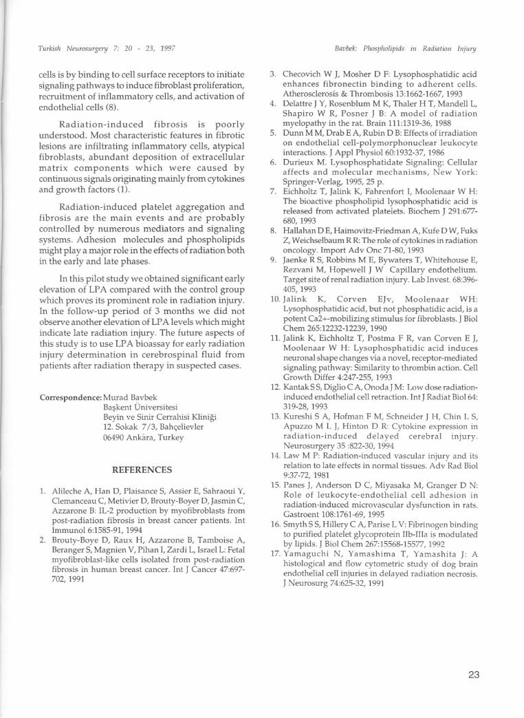
REFERENCES
1. Alileche A, Han D, Plaisance S, Assier E, Sahraoui Y,Clemanceau C, Metivier D, Brouty-Boyer D, Jasmin C,Azzarone B: IL-2 production by myofibroblasts frompost-radiation fibrosis in breast cancer patients. IntImmunol 6:1585-91, 1994
2. Brouty-Boye D, Raux H, Azzarone B, Tamboise A,Beranger S, Magnien V, Pihan I, Zardi L, Israel L:Fetalmyofibroblast-like ceiis isolated from post-radiationfibrosis in human breast cancer. Int J Cancer 47:697702, 1991
Bavbek: Phospholipids in Radiation Injury
3. Checovich W J, Mosher D F: Lysophosphatidic acidenhances fibroneetin binding to adherent ceiis.Atherosclerosis & Thrombosis 13:1662-1667, 1993
4. Delattre J Y, Rosenblum M K, Thaler H T, Mandeii L,Shapiro W R, Posner J B: A model of radiationmyelopathy in the rat. Brain 111:1319-36, 1988
5. Dunn M M, Drab E A, Rubin D B:Effects of irradiationon endothelial ceii-polymorphonuclear leukocyteinteractions. J Appl Physiol 60:1932-37, 1986
6. Durieux M. Lysophosphatidate Signaling: Ceiiularaffects and molecular mechanisms, New York:Springer-Verlag, 1995,25 p.
7. Eichholtz T, Jalink K, Fahrenfort I, Moolenaar W H:The bioactive phospholipid Iysophosphatidic acid isreleased from activated platelets. Biochem J 291:677680, 1993
8. Haiiahan D E, Haimovitz-Friedman A, Kufe D W, FuksZ, Weichselbaum R R: The role of cytokines in radiationoncology. Import Adv One 71-80,1993
9. Jaenke R S, Robbins M E, Bywaters T, Whitehouse E,Rezvani M, Hopeweii J W Capillaryendothelium.Target site of renal radiation injury. Lab Invest. 68:396405, 1993
10. Jalink K, Corven EJv, Moolenaar WH:Lysophosphatidic acid, but not phosphatidic acid, is apotent Ca2+-mobilizing stimulus for fibroblasts. J BiolChem 265:12232-12239,1990
lL. Jalink K, Eichholtz T, Postma F R, van Corven E J,Moolenaar W H: Lysophosphatidic acid inducesneuronal shape changes via a novel, receptor-mediatedsignaling pathway: Similarity to thrombin action. CeiiGrowth Differ 4:247-255, 1993
12. Kantak S S,Diglio C A, Onoda J M: Low dose radiationinduced endothelial ceii retraction. Int J Radiat Biol 64:319-28, 1993
13. Kureshi S A, Hofman F M, Schneider J H, Chin L S,Apuzzo M L J, Hinton D R: Cytokine expression inradiation-induced delayed cerebral injury.Neurosurgery 35 :822-30, 1994
14. Law M P: Radiation-induced vascular injury and itsrelation to Iate effects in normal tissues. Adv Rad Biol9:37-72, 1981
15. Panes J, Anderson D C, Miyasaka M, Granger D N:Role of leukocyte-endothelial ceii adhesion inradiation-induced microvascular dysfunction in rats.Gastroent 108:1761-69, 1995
16. Smyth S S, Hillery C A, Parise L V: Fibrinogen bindingto purified platelet glycoprotein Hb-Illa is modulatedby lipids. J Biol Chem 267:15568-15577,1992
17. Yamaguchi N, Yamashima T, Yamashita J: Ahistological and flow cytometric study of dog brainendothelial ceii injuries in delayed radiation necrosis.J Neurosurg 74:625-32, 1991
23