automatic segmentation of the epicardium in late gadolinium
TRANSCRIPT

Automatic Segmentation of the Epicardium in Late Gadolinium EnhancedCardiac MR Images
Kjersti Engan1, Valery Naranjo2, Trygve Eftesøl1, Stein Ørn3, Leik Woie3
1 University of Stavanger, Stavanger, Norway2 Labhuman, I3BH, Universitat Politecnica de Valencia, Valencia, Spain
3 Stavanger University Hospital, Stavanger, Norway
Abstract
A novel method for fully automatic segmentation of theepicardium of the left ventricle in Late Gadolinium En-hanced Cardiac MR images (LGE-CMR), short axis viewis presented. No other images or measurements are used.The method uses prior information about typical heart sizeand shape making an a priori probability map around theautomatically detected heart center (HC). An a posterioriprobability map is made iteratively by combining the cur-rent probability model with the preprocessed image andlow-pass filtering over consecutive slices. The a posterioriprobability map is used as the input for segmentation. Us-ing the detected HC as origin, a radial evaluation is per-formed for all possible angles [0− 2π], giving a candidateborder point on the epicardium. The final segmentationcombines the information from all slices. The presentedmethod is tested on all slices of 54 patients, and gives amean Dice index of 0.86 and mean Jaccard index of 0.76.
1. Introduction
Figure 1. Consecutive MRI slices of an example patientwith myocardial scar.
Late Gadolinium Enhanced Cardiac Magnetic Reso-nance images (LGE-CMR) are used to assess the scarredareas of the myocardium after a myocardial infarction.
Healthy myocardium appears very dark in CMR images,however the edges of the heart in LGE-CMR images wherethe patient has a scar in the myocardium are sometimesvery weak or non-existing since the scarred areas will takeintensity levels close to the blood pool or the surround-ing areas. This makes automatic segmentation of the my-ocardium in LGE-CMR images difficult. In many hospi-tals today the segmentation of the myocardial muscle isperformed manually or semi-automatically by expert car-diologists. This can be time-consuming work, and the re-sults will have a degree of inter- and intra-observer vari-ability. There have been attempts to solve the problem ofautomatic segmentation reported in the literature, but to theauthors knowledge a fully automatic method for segment-ing LGE-CMR images does not exist today. Some methodsrequire manual input in form of landmarks or cropping ofregion of interest etc. seems to be necessary [1, 2]. Oth-ers make use of additional data as the corresponding cineCMR [3, 4].
This work proposes a new method for automatic seg-mentation of the epicardium. A priori knowledge of typicalheart size and shape is used as the input, and an a posteri-ori probability map is made iteratively. The final segmen-tation is based on the a posteriori probability maps, andinformation from all slices are taken into account. The De-partment of Cardiology at Stavanger University Hospitalprovided LGE-CMR images of 54 patients, all with previ-ous myocardial infarction. CMR was performed using 1.5T Philips Intera R 8.3, pixel size of 0.82× 0.82mm2, cov-ering the whole ventricle with short-axis slices of 10 mmthickness, without inter-slice gaps. An example of all the8 slices from one patient is seen in Figure 1, and the epi-cardium is the outer border of the dark ring (partly brightbecause of scar) approximately in the middle of each slice.
2. Proposed method
Let f i(x), i = 1 . . . Nslice represent the left ven-tricle short axis LGE-CMR images of a patient. x =[xrow xcolumn]T is the pixel position, and i represents
ISSN 2325-8861 Computing in Cardiology 2013; 40: 631-634.631

Algorithm 1: Iterative probability map algorithm
Data: CMR images f i(x) ∈ RM×N , Prior prob.map pi0(x) ∈ RN×M i = 1, . . . , Nslice,
Result: Posteriori prob. map,pipost(x) ∈ RN×M , i = 1, . . . , Nslice
initialization: kfinal;for i← 1 to Nslice dof iprep(x)← βBmc(f
i) %morph noise removal;f iinv(x) =scale(ones(N,M)− f iprep(x));
end forfor k ← 0 to kfinal − 1 doftemp(x) = pk(x) ◦ finv(x);ftemp(x)← LPfiltZ(ftemp(x)) %filter slices;pk+1(x)← scale(ftemp(x));
end forp2(x) = p0(x) ◦ (finv(x))◦(kfinal) ;ppost(x) = scale(pkfinal(x) + p2(x));return(ppost) ;
the slice number. We define the morphological operations[5] of dilation and erosion as [δBf ](x) and [εBf ](x) re-spectively, where B(x) is a structuring element, and open-ing and closing as γB(f) = δB(εB(f)) and φB(f) =εB(δB(f)) respectively. Morphological center, (β(f)), isa morphological filter used in our preprocessing algorithmsdefined as:
βBmc(f)(x) = min(max(f(x), f1(x)), f2(x)) (1)
where f1(x) = γBmc(φBmc(γBmc(f))) and f2(x) =φBmc(γBmc(φBmc(f))). In geodesic transformations twoimages are required, the reference f(x) and the markerg(x). The morphological reconstruction, γrec(g, f), or re-construction by dilation is the successive geodesic dilationof the marker regarding the reference up to idempotence[5]. Using the morphological reconstruction, we can definethe close-hole operator which fills all holes in a gray-scaleimage f(x) that do not touch the image border, fborder(x),defined as: fborder(x) = f(x), for x ∈ {xborder} and 0everywhere else.
φch(f) = [γrec(f c(x), fborder(x))], (2)
where f c(x) denotes the inverse of the image f(x).
2.1. A priori probability map
Some general knowledge of the nature of the my-ocardium needs to be exploited. We are only consider-ing left ventricle short axis slices, and in these images themyocardial muscle has the approximated shape of a ring.Typical sizes of the endocardium and epicardium is also
known. We propose to make a crude a priori model of theheart using a Gaussian prior in the radial direction from theheart center for every possible angle θ ∈ [0− 2π]:
pθ(r) =1√
2πσ2e
(r−µi)2
2σ2 (3)
where µi is the a priori radius at slice number i accordingto knowledge of a typical heart size, and σ is the variance,and the same for all slices. In the experiments σ = 15 andµ = [16 24 29 32 35 35 . . . 35 34]T pixels. Convertingfrom polar to cartesian for all radii (within interest) and allangles, and thereafter quantize to form a digital image andscaled to make all pixel values∈ [0, 1], a 2D probability forbeing myocardium map is made pi0(x). The approximatedcenter of the heart is an important input to make the a prioriprobability map. We defineHCij = [HCrow HCcolumn]T
as the heart center for patient j in slice i. In our previouswork [6] two alternative methods to find the heart center isproposed, one based on morphological preprocessing fol-lowed by the Circular Hough Transform (CHT), and an-other method based on the morphological method of greylevel distance, both performing very good.
2.2. A posteriori probability map
Using the a priori probability map as input, we pro-pose an iterative approach to refine the map and makean a posteriori probability map for the myocardium. Ateach iteration the probability map is combined with the in-verse of the original (preprocessed) slices. The inverse isused since the myocardium has very low values in the im-ages, and we want to express the probability of being my-ocardium. A filtering over the neighboring slices is per-formed an the output is scaled to a new probability map.The final probability map is a weighted sum between oneversion filtering over the neighboring slices in every itera-tion, and another with no filtering. Without filtering overthe slices, the a posteriori map can be found directly asp2(x) = p0(x) ◦ (finv(x))◦(kfinal), where A ◦ B denotesthe Hademard product, and A◦(k) denotes the Hadamardproduct of A with itself k times. A pseudo code of the al-gorithm is depicted in Algorithm 1, where scale(A) set allvalues in A ∈ [0, 1]. An example of the a priori map and aposteriori map of a patient can be seen in figure 2.
2.3. Segmentation of epicardium
The final segmentation step is challenging. The patients(all) have myocardial scars which appears as bright areaswhereas the healthy parts of the myocardial muscle ap-pears dark. Therefore scarred areas might be very dark inthe a posteriori probability images and the intensity levelsare very uneven at different angles. Thus a global thresh-olding technique will not work. We need to evaluate the
632

different probability levels radially at each possible angleinstead. To simplify and spatially smooth the probabilitymap, it is quantized using an Otsu’s thresholding basedmethod (slice by slice) for nonuniform quantization with5 bins, giving f iq(x). Using the position HCij as origin,the values from f iq(x) are interpreted in polar coordinatesgiving f iqpol(θ, r). The partial derivative of f iqpol(θ, r) withrespect to r is found for all θ, Dr(θ, r), and the first neg-ative value of the derivative (smallest radius) is markedas a candidate epicardium border point, Riepi(θ), for eachθ. For some angles the algorithm will fail to find sucha point, and an alternative is found interpolated from theother slices and from the other angles. Finally the con-vex hull of the candidate borders is found giving the epi-cardium mask, Maskepicard(x).
Figure 2. A priori and a posteriori probability map of allslices of a patient.
2.4. Postprocessing
Postprocessing is done excluding parts of theMaskiepicard(x) not present in any of the other slices,Maskjepicar(x), for all j 6= i, and includingparts in Maskiepicard(x) present in all other slicesMaskjepicar(x), for all j 6= i. An additional postprocess-ing is low-pass filtering of the Fourier Descriptor (FD) ofthe boundary, giving a new mask MaskFDi
epicard(x).
3. Experiments and results
The method is tested on all 54 patients, 503 images,and evaluated compared to consensus manual segmenta-
Algorithm 2: Final segmentation of epicardium
Data: CMR images, f i(x) ∈ RN×M , a post prob.map, pipost(x) ∈ RN×M , i = 1, . . . Nslice ,heart center coordinates HCi;
Result: Maskiepicard(x)
initialization: Riepi(θ) = 0, θ ∈ [0, 2π];fch(x) = φch(ppost(x)) ;for i = 1 to Nslice dof iq(x) = Quantinonuni(f
ich(x), L) % Nonuniform
quantization in L bins ;for θ = 0 to 2π dof iqpol(θ, r) = car2polar(f iq(x), θ,HCi) ;
Dr(θ, r) =δfiqpolar
(θ,r)
δr ;Repi(θ) = argminr(Dr < 0) ;
end forRiepi(θ)← smooth(Riepi(θ)) ;
end forfor i = 1 to Nslice do
for θ = 0 to 2π doif Riepi(θ) = 0 thenRiepi(θ)← mean(Rjepi(θ) 6= 0)
end ifif Riepi(θ) = 0 thenRiepi(θ)← min|∆θ|(R
iepi(θ −∆θ) 6= 0)
end ifxiθ = poly2cart(Riepi(θ)) ;f ip(x
iθ) = 1;
end forMaskiepicard(x) = ConvexHull(f ip(x));
end forreturn(Maskepicard(x)) ;
tion of two experienced cardiologists (LW,SØ). The inputis the full images, as seen in Figure 1. Firstly the approx-imated heart centers, HCj are found using the CircularHough Transform (CHT) method presented in [6]. Theimages are automatically cropped to a region of interestbased on the HCj . Subsequently the a posteriori proba-bility maps are found from the iterative probability mapalgorithm, 1, followed by the final segmentation algorithm2. The number of iterations kfinal = 8 was chosen empir-ically. The low-pass filter, performed pixel by pixel overthe slices, LPfiltZ(ftemp(x)), is a simple anti causal FIRfilter: H(z) = 0.15z−1 + 0.7 + 0.15z1. The results areevaluated by the Dice index and the Jaccard index, as wellas visually. The first and last slice of each patient in thedata material are of poor quality. They are used in the al-gorithm, filtering over slices, but they are excluded fromthe evaluation and in the figures, leaving 395 images for
633
evaluation.The mean and variance of the Dice and Jaccard indices
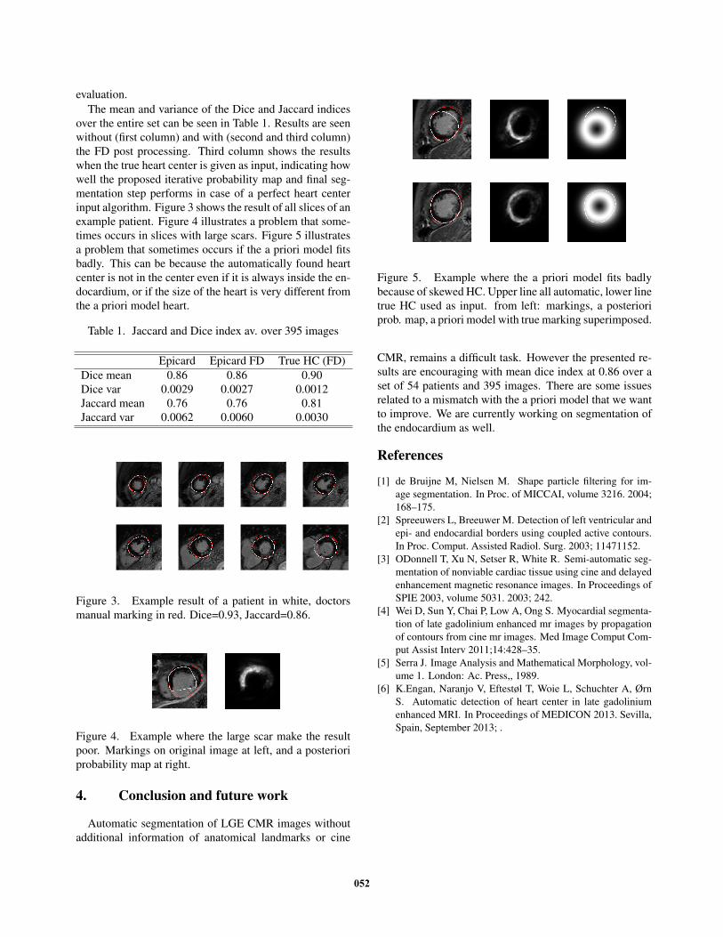
over the entire set can be seen in Table 1. Results are seenwithout (first column) and with (second and third column)the FD post processing. Third column shows the resultswhen the true heart center is given as input, indicating howwell the proposed iterative probability map and final seg-mentation step performs in case of a perfect heart centerinput algorithm. Figure 3 shows the result of all slices of anexample patient. Figure 4 illustrates a problem that some-times occurs in slices with large scars. Figure 5 illustratesa problem that sometimes occurs if the a priori model fitsbadly. This can be because the automatically found heartcenter is not in the center even if it is always inside the en-docardium, or if the size of the heart is very different fromthe a priori model heart.
Table 1. Jaccard and Dice index av. over 395 images
Epicard Epicard FD True HC (FD)Dice mean 0.86 0.86 0.90Dice var 0.0029 0.0027 0.0012Jaccard mean 0.76 0.76 0.81Jaccard var 0.0062 0.0060 0.0030
Figure 3. Example result of a patient in white, doctorsmanual marking in red. Dice=0.93, Jaccard=0.86.
Figure 4. Example where the large scar make the resultpoor. Markings on original image at left, and a posterioriprobability map at right.
4. Conclusion and future work
Automatic segmentation of LGE CMR images withoutadditional information of anatomical landmarks or cine
Figure 5. Example where the a priori model fits badlybecause of skewed HC. Upper line all automatic, lower linetrue HC used as input. from left: markings, a posterioriprob. map, a priori model with true marking superimposed.
CMR, remains a difficult task. However the presented re-sults are encouraging with mean dice index at 0.86 over aset of 54 patients and 395 images. There are some issuesrelated to a mismatch with the a priori model that we wantto improve. We are currently working on segmentation ofthe endocardium as well.
References
[1] de Bruijne M, Nielsen M. Shape particle filtering for im-age segmentation. In Proc. of MICCAI, volume 3216. 2004;168–175.
[2] Spreeuwers L, Breeuwer M. Detection of left ventricular andepi- and endocardial borders using coupled active contours.In Proc. Comput. Assisted Radiol. Surg. 2003; 11471152.
[3] ODonnell T, Xu N, Setser R, White R. Semi-automatic seg-mentation of nonviable cardiac tissue using cine and delayedenhancement magnetic resonance images. In Proceedings ofSPIE 2003, volume 5031. 2003; 242.
[4] Wei D, Sun Y, Chai P, Low A, Ong S. Myocardial segmenta-tion of late gadolinium enhanced mr images by propagationof contours from cine mr images. Med Image Comput Com-put Assist Interv 2011;14:428–35.
[5] Serra J. Image Analysis and Mathematical Morphology, vol-ume 1. London: Ac. Press,, 1989.
[6] K.Engan, Naranjo V, Eftestøl T, Woie L, Schuchter A, ØrnS. Automatic detection of heart center in late gadoliniumenhanced MRI. In Proceedings of MEDICON 2013. Sevilla,Spain, September 2013; .
634