atypical pyoderma gangrenosum in a patient with ... · atypical pyoderma gangrenosum in a patient...
TRANSCRIPT
Volumen 64, Broj 11 VOJNOSANITETSKI PREGLED Strana 787
Correspondence to: Dubravka Živanović, Clinical Center of Serbia, Institute of Dermatology and Venereology, 11 000 Belgrad, Serbia.
C A S E R E P O R T UDC: 616.5-002.3:616.71/74-006.327
Atypical pyoderma gangrenosum in a patient with osteomyelofibrosisAtipična pyoderma gangrenosum kod bolesnika sa osteomijelofibrozom
Dubravka Živanović*, Srđan Tanasilović*, Dušan Škiljević*, Maja Tomović*,Andrija Bogdanović†, Sonja Vesić*, Ljiljana Medenica*
Clinical Center of Serbia, *Institute of Dermatology and Venereology,†Institute of Haematology, Belgrade
Abstract
Background. Atypical forms of pyoderma gangrenosum gener-ally appear on the upper extremities; most frequently theyare associated with myeloproliferative disorders, includingosteomyelofibrosis. A response to systemic steroids is morepronounced than in classical form. Sometimes it may be thefirst sign of an underlying malignancy. Case report. We re-ported a patient with atypical pyoderma gangrenosum developedduring the course of a myeloid malignancy – osteomyelofibrosis.The lesions occurred after a minor trauma. Painful blisteringplaques, with an elevated, bluish-gray border were locatedon the dorsal aspect of hands. No skin malignancy wasfound. The lesions resolved rapidly to systemic steroids.Conclusion. Considering the unusual clinical presentationwhich makes the diagnosis difficult, as well as the fact thatatypical forms of pyoderma gangrenosum can be the first sign ofmalignancies, especially myeloproliferative ones, recognizingthis entity enables timely guiding future investigations to-ward their prompt detection.
Key words:pyoderma gangrenosum; myeloproliferative disorders;diagnosis; drug therapy; adrenal cortex hormones;treatment outcome.
Apstrakt
Uvod. Atipične forme pyoderma gangrenosum obično se javljajuna gornjim ekstremitetima. Najčešće su udružene sa mijelop-roliferativnim oboljenjima uključujući i osteomijelofibrozu.Odgovor na opštu kortikosteroidnu terapiju mnogo je bržinego kod klasične forme. Ponekad mogu biti prvi znak pos-tojećeg maligniteta. Prikaz bolesnika. Prikazali smo bolesni-ka sa atipičnom formom pyoderma gangrenosum koja se razvilatokom osnovne bolesti – osteomijelofibroze. Lezije na šaka-ma javile su se nakon manje traume. Na dorzumu šaka nalazilisu se bolni plakovi sa bulama i uzdignutim, plavosivim, livid-nim ivicama. Evaluacijom promena na koži nisu otkriveniznaci kutanog maligniteta. Primenom opšte kortikosteroidneterapije postignuta je brza klinička regresija kutanih lezija.Zaključak. S obzirom na neuobičajenu kliničku sliku kojaotežava dijagnostiku, kao i činjenicu da atipične formepyoderma gangrenosum mogu biti prvi znak prisustva maligniteta,naročito mijeloproliferativnih maligniteta, poznavanje ovogentiteta omogućava pravovremeno usmeravanje daljih ispiti-vanja u pravcu njihovog promptnog otkrivanja.
Ključne reči:pioderma gangrenozum; mijeloproliferativniporemećaji; dijagnoza; lečenje lekovima;kortikosteroidni hormoni; lečenje, ishod.
Introduction
The diagnosis of pyoderma gangrenosum (PG) ismostly based on the appearance of typical lesions – raisedplaques with progressive central necrosis and a bluish-rednecrotising border 1, 2. Histopathological features are variableand not specific; the presence of vasculitis is thought bysome authors to exclude the diagnosis 1.
In 50% of the cases, PG is associated with bowel dis-eases, although atypical forms are mostly reported with hae-matologic diseases 3, 4.
Diagnostic difficulties may arise in the absence of sys-temic disease or with uncommon associations such as mye-
liod malignancies 1, 2. Patients with myeloproliferative disor-ders often have an atypical clinical presentation of PG, withblisters, and relatively superficial involvement, obscuring thecorrect clinical diagnosis 1, 2, 5, 6.
Myelofibrosis with myeloid metaplasia (chronic myelo-fibrosis, osteomyelofibrosis) is a primary, Philadelphianegative, myeloproliferative disorder arising from the levelof the haematopoetic stem cell. It is characterized withhepato/splenomegaly, anemia and the tear drop erythrocyteswith leukoerythroblastic changes in typical cases. The hall-mark is bone marrow fibrosis and myeloid metaplasia in thespleen and liver 1, 7. It is a rare disease of adults and the eld-erly. Generally, the disorder is progressive, after several
Strana 788 VOJNOSANITETSKI PREGLED Volumen 64, Broj 11
Živanović D, et al. Vojnosanit Pregl 2007; 64(11): 787–789.
years, insufficiency of hematopoesis and spleen enlargementbecomes the hallmark of the disease 1, 7.
Skin changes in osteomyelofibrosis are infrequent.Dermatitis resembling Sweet's syndrome or PG has alreadybeen described 1–3.
Case report
A 66-year-old Caucasian man was admitted to our In-stitute in November 2006, with lesions on the dorsa al sidesof hands, which occurred about a month earlier, after a minortrauma (working on the farm). Lesions enlarged symmetri-cally and were accompanied by painful sensations.
Family history was unremarkable. In personal historyhe had osteomyelofibrosis with myeloid metaplasia since2003. The patient was treated with hydroxyurea (1–1.5 gdaily). Because of severe anemia and almost transfusion de-pendence, the treatment with danazole started (400 mg daily)in the beginning of September 2006. During the episodes ofsevere anaemia, he also suffered from pneumonia.
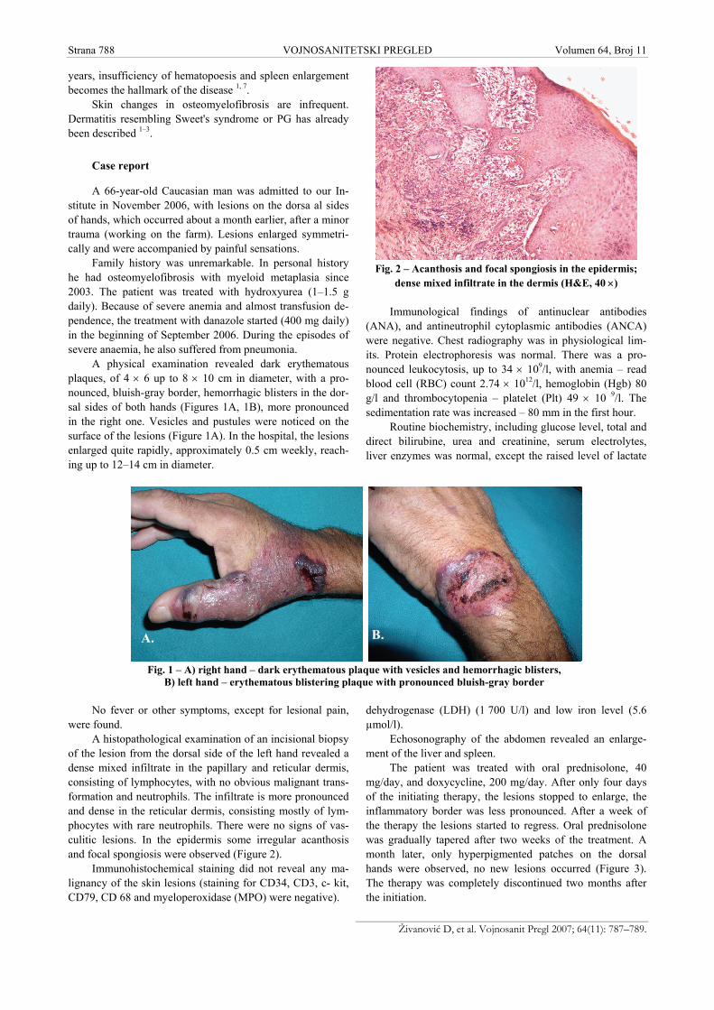
A physical examination revealed dark erythematousplaques, of 4 × 6 up to 8 × 10 cm in diameter, with a pro-nounced, bluish-gray border, hemorrhagic blisters in the dor-sal sides of both hands (Figures 1A, 1B), more pronouncedin the right one. Vesicles and pustules were noticed on thesurface of the lesions (Figure 1A). In the hospital, the lesionsenlarged quite rapidly, approximately 0.5 cm weekly, reach-ing up to 12–14 cm in diameter.
No fever or other symptoms, except for lesional pain,were found.
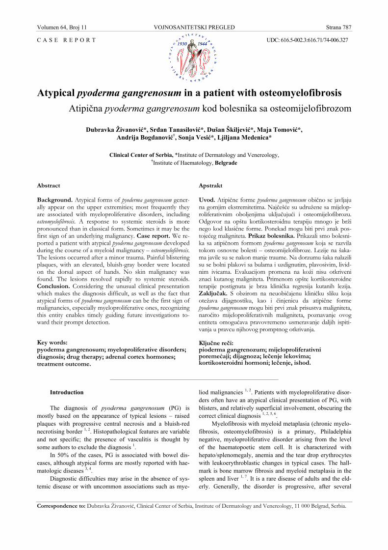
A histopathological examination of an incisional biopsyof the lesion from the dorsal side of the left hand revealed adense mixed infiltrate in the papillary and reticular dermis,consisting of lymphocytes, with no obvious malignant trans-formation and neutrophils. The infiltrate is more pronouncedand dense in the reticular dermis, consisting mostly of lym-phocytes with rare neutrophils. There were no signs of vas-culitic lesions. In the epidermis some irregular acanthosisand focal spongiosis were observed (Figure 2).
Immunohistochemical staining did not reveal any ma-lignancy of the skin lesions (staining for CD34, CD3, c- kit,CD79, CD 68 and myeloperoxidase (MPO) were negative).
Fig. 2 – Acanthosis and focal spongiosis in the epidermis;dense mixed infiltrate in the dermis (H&E, 40 ×)
Immunological findings of antinuclear antibodies(ANA), and antineutrophil cytoplasmic antibodies (ANCA)were negative. Chest radiography was in physiological lim-its. Protein electrophoresis was normal. There was a pro-nounced leukocytosis, up to 34 × 109/l, with anemia – readblood cell (RBC) count 2.74 × 10¹²/l, hemoglobin (Hgb) 80g/l and thrombocytopenia – platelet (Plt) 49 × 10 9/l. Thesedimentation rate was increased – 80 mm in the first hour.
Routine biochemistry, including glucose level, total anddirect bilirubine, urea and creatinine, serum electrolytes,liver enzymes was normal, except the raised level of lactate
dehydrogenase (LDH) (1 700 U/l) and low iron level (5.6µmol/l).
Echosonography of the abdomen revealed an enlarge-ment of the liver and spleen.
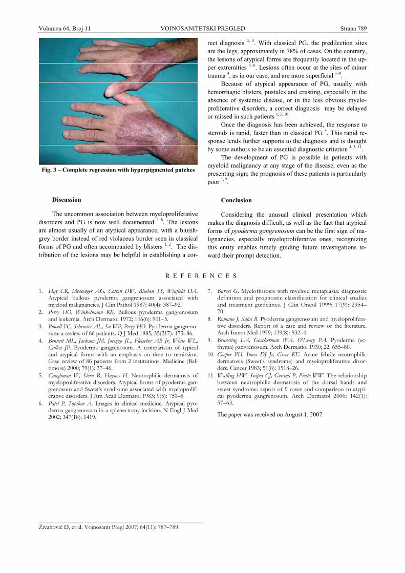
The patient was treated with oral prednisolone, 40mg/day, and doxycycline, 200 mg/day. After only four daysof the initiating therapy, the lesions stopped to enlarge, theinflammatory border was less pronounced. After a week ofthe therapy the lesions started to regress. Oral prednisolonewas gradually tapered after two weeks of the treatment. Amonth later, only hyperpigmented patches on the dorsalhands were observed, no new lesions occurred (Figure 3).The therapy was completely discontinued two months afterthe initiation.
A. B.
Fig. 1 – A) right hand – dark erythematous plaque with vesicles and hemorrhagic blisters,B) left hand – erythematous blistering plaque with pronounced bluish-gray border
Volumen 64, Broj 11 VOJNOSANITETSKI PREGLED Strana 789
Živanović D, et al. Vojnosanit Pregl 2007; 64(11): 787–789.
Fig. 3 – Complete regression with hyperpigmented patches
Discussion
The uncommon association between myeloproliferativedisorders and PG is now well documented 1–8. The lesionsare almost usually of an atypical appearance, with a bluish-grey border instead of red violaceus border seen in classicalforms of PG and often accompanied by blisters 1, 2. The dis-tribution of the lesions may be helpful in establishing a cor-
rect diagnosis 3, 5. With classical PG, the predilection sitesare the legs, approximately in 78% of cases. On the contrary,the lesions of atypical forms are frequently located in the up-per extremities 4, 6. Lesions often occur at the sites of minortrauma 4, as in our case, and are more superficial 1–9.
Because of atypical appearance of PG, usually withhemorrhagic blisters, pustules and crusting, especially in theabsence of systemic disease, or in the less obvious myelo-proliferative disorders, a correct diagnosis may be delayedor missed in such patients 1, 5, 10.
Once the diagnosis has been achieved, the response tosteroids is rapid, faster than in classical PG 4. This rapid re-sponse lends further supports to the diagnosis and is thoughtby some authors to be an essential diagnostic criterion 4, 5, 11.
The development of PG is possible in patients withmyeloid malignancy at any stage of the disease, even as thepresenting sign; the prognosis of these patients is particularlypoor 1, 7.
Conclusion
Considering the unusual clinical presentation whichmakes the diagnosis difficult, as well as the fact that atypicalforms of pyoderma gangrenosum can be the first sign of ma-lignancies, especially myeloproliferative ones, recognizingthis entity enables timely guiding future investigations to-ward their prompt detection.
R E F E R E N C E S
1. Hay CR, Messenger AG, Cotton DW, Bleehen SS, Winfield DA.Atypical bullous pyoderma gangrenosum associated withmyeloid malignancies. J Clin Pathol 1987; 40(4): 387–92.
2. Perry HO, Winkelmann RK. Bullous pyoderma gangrenosumand leukemia. Arch Dermatol 1972; 106(6): 901–5.
3. Powell FC, Schroeter AL, Su WP, Perry HO. Pyoderma gangreno-sum: a review of 86 patients. Q J Med 1985; 55(217): 173–86.
4. Bennett ML, Jackson JM, Jorizzo JL, Fleischer AB Jr, White WL,Callen JP. Pyoderma gangrenosum. A comparison of typicaland atypical forms with an emphasis on time to remission.Case review of 86 patients from 2 institutions. Medicine (Bal-timore) 2000; 79(1): 37–46.
5. Caughman W, Stern R, Haynes H. Neutrophilic dermatosis ofmyeloproliferative disorders. Atypical forms of pyoderma gan-grenosum and Sweet's syndrome associated with myeloprolif-erative disorders. J Am Acad Dermatol 1983; 9(5): 751–8.
6. Patel P, Topilow A. Images in clinical medicine. Atypical pyo-derma gangrenosum in a splenectomy incision. N Engl J Med2002; 347(18): 1419.
7. Barosi G. Myelofibrosis with myeloid metaplasia: diagnosticdefinition and prognostic classification for clinical studiesand treatment guidelines. J Clin Oncol 1999; 17(9): 2954–70.
8. Romano J, Safai B. Pyoderma gangrenosum and myeloprolifera-tive disorders. Report of a case and review of the literature.Arch Intern Med 1979; 139(8): 932–4.
9. Brunsting LA, Goeckerman WA, O'Leary PA. Pyoderma (ec-thyma) gangrenosum. Arch Dermatiol 1930; 22: 655–80.
10. Cooper PH, Innes DJ Jr, Greer KE. Acute febrile neutrophilicdermatosis (Sweet's syndrome) and myeloproliferative disor-ders. Cancer 1983; 51(8): 1518–26.
11. Walling HW, Snipes CJ, Gerami P, Piette WW. The relationshipbetween neutrophilic dermatosis of the dorsal hands andsweet syndrome: report of 9 cases and comparison to atypi-cal pyoderma gangrenosum. Arch Dermatol 2006; 142(1):57–63.
The paper was received on August 1, 2007.