pyoderma gangrenosum diagnosis, differential diagnosis and … u027... · learning objectives •...
TRANSCRIPT

Pyoderma Gangrenosum Diagnosis,
Differential Diagnosis and Management
Jeffrey P. Callen, MD
Professor of Medicine (Dermatology)
University of Louisville

Jeffrey P. Callen, MD
Disclosure (previous 12 months)
• Consultant – Amgen, Lilly, Argenx
• Speakers Bureau - Biogen/IDEC (Discussion of Drug Eruptions in
patients treated with an approved drug for MS)
• Equity Holdings (Personal/Spouse): Celgene; Pfizer; 3M; Johnson and
Johnson; Merck; Abbott Laboratories; AbbVie; Procter and Gamble;
CVS; Walgreens; Allergen; Amgen
• None of the above relationships are relevant to my presentation
• I will discuss “off-label” uses of some of the currently available agents
and will identify which are labeled v. off-labeled uses.
June 2017

Learning Objectives
• Following this lecture, the attendee will be able to:
– Effectively diagnose pyoderma gangrenosum
– Differentiate pyoderma gangrenosum from other causes of
cutaneous ulceration
– Develop an algorithm for effective management of pyoderma
gangrenosum

Pyoderma Gangrenosum
• PG is a rare, painful ulcerating condition associated with a
variety of co-morbid conditions in roughly 50%
• Variants – classical, atypical, peristomal, mucosal
• PG is a diagnosis of exclusion
• Associations – IBD, arthritis, hematologic diseases, other
• Neutrophilic infiltrates may occur in other organs

PG Subtypes
Subtype Clinical features Location
Ulcerative (classic) Ulcer, undermined violaceous
border, purulent base,
cribriform scarring
Trunk, extremities
Bullous (atypical) Bulloussuperficial ulcers
Hematologic malignancy
Arms, face
Pustular Pustules and erosions
Inflammatory bowel disease
Same as ulcerative
Vegetative Superficial, localized
vegetative plaques, ulcers
Head, neck

Clinical features suggestive of PG
• Site: legs or peristomal location
• Presence of systemic disease (e.g. IBD, arthritis, hematologic)
• Presence of pathergy
• Pustular lesions at onset of lesion
• Formation of purulent discharge
• Undermined borders
• Crater-like holes/Cribriform scarring
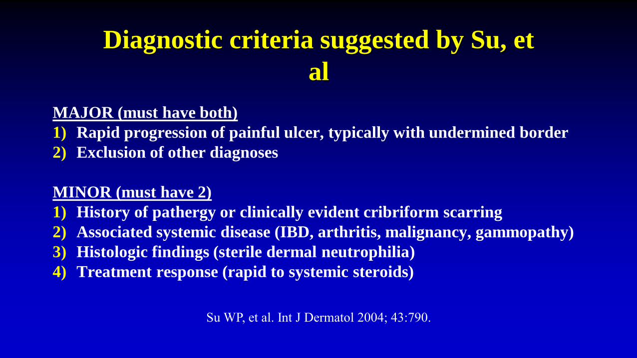
Diagnostic criteria suggested by Su, et
al
MAJOR (must have both)
1) Rapid progression of painful ulcer, typically with undermined border
2) Exclusion of other diagnoses
MINOR (must have 2)
1) History of pathergy or clinically evident cribriform scarring
2) Associated systemic disease (IBD, arthritis, malignancy, gammopathy)
3) Histologic findings (sterile dermal neutrophilia)
4) Treatment response (rapid to systemic steroids)
Su WP, et al. Int J Dermatol 2004; 43:790.

• Review of 240 patients with a presumed diagnosis of PG
• 49 had a different diagnosis
– Vasculopathy – livedoid vasculitis, APS, venous ulceration, etc.
– Vasculitis – WG, PAN, LCV, Cryo-assoc.
– Malignancy – lymphoma/leukemia
– Infection – deep fungal, Tb, HSV, etc.
– Miscellaneous – NLD, Crohn’s, hydroxyurea-induced, spider bite
» NEJM 2002; 347: 1412-8


Acta Derm Venereol 2015

Genetic abnormalities in patients with PG might direct
therapy
• Protein Tyrosine Phosphatase Nonreceptor Type 6 (PTPN6/SHP1) (Am J Pathol 2011; 178: 1434)
• E250K mutation in CD2BP1 gene [PAPA patient) (Clin Exp
Rheumatol 2012:452)
• A230T & E250Q in the threonine phosphatase-interacting protein 1 (Anakinra might be useful) (Inflamm Bowel Dis 2011;17: e41)
• Janus kinase 2 (JAK2V617F) mutation (Myelofibrosis patient) (Clin Exp Dermatol. 2013 Jan;38(1):44-6.) (ruxolitinib might be used)
• PSTPIP1 Mutation (PAPA patient) (Canakinumab was used) (JAMA Dermatol 2013; 149(2):209-215)


Future therapies ?
• Canukinumab - NCT01302795 – completed report in the British J. Dermatol
• Xilonix - NCT01965613 – completed no reports of results
• Gevokizumab - NCT01882504 – studies halted
• Secukinumab – NCT02733094 – Recruiting (Germany)
• Ixekizumab - NCT03137160 recruting
• Etrasimod - NCT03072953 – recruiting (Australia)http://clinicaltrials.gov/ct2/results?term=pyoderma+gangrenosum&Search=Search

Conclusions
• PG is a diagnosis of exclusion
• Associated diseases include IBD, RA and other arthritides,
hematologic malignancy
• Neutrophilic infiltrates may affect other organs
• Multiple treatments are effective