assessment of the immune system cells at the end of this seminar you will be able to answer the...
TRANSCRIPT

Assessment of the immune system cells
At the end of this seminar you will be able to answer
the following questions:
How to determine cell count?
How to separate and isolate different cell populations?
Which in vitro and in vivo tests are used for the
assessment of the immune system

Cell count determination
Based on morphology and staining chambers and blood smears
Based on size and density automated cell counters
Based on specific cell markers T lymphocytes: CD3, CD4, CD8....
B lymphocytes: Ig, CD19, CD20...
NK cells: CD16 and 56, CD161

Flow cytometry

Flow cytometry (FACS®)
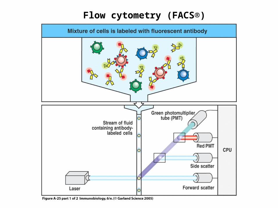
Flow cytometry (FACS®)

Dot plot
Histogram

MonocyteIg -CD3 -
CD14+
Distribution of different cell population in PBMCs

• Immunophenotyping (research, lymphoproliferative diseases, immunodeficiencies )
• Functional surface molecule detection• Activation marker detection• Intracellular protein detection• Activation status (intracellular Ca2+
concentration, protein phosphorylation, ROS , NOS)
• DNA content – cell cycle• Apoptosis detection and quantification
Applications of flow cytometry

Isolation of cells of the immune system Based on different density (specific
gravity) using density gradient (Ficoll®)
Based in different cell markers using paramagnetic beads-labeled antibodies MACS®
Based on different cell markers using fluorescence-labeled antibodies FACS® (flow cytometer)

centrifugation
PBMCs isolation using density gradient

PBMCs isolation using density gradient

Cell separation/isolation using paramagnetic beads

• Chemotaxis
Functional analysis of phagocytes

Boyden chamber assay

• Chemotaxis• Ingestion• Respiratory burst • Intracellular killing
Functional analysis of phagocytes

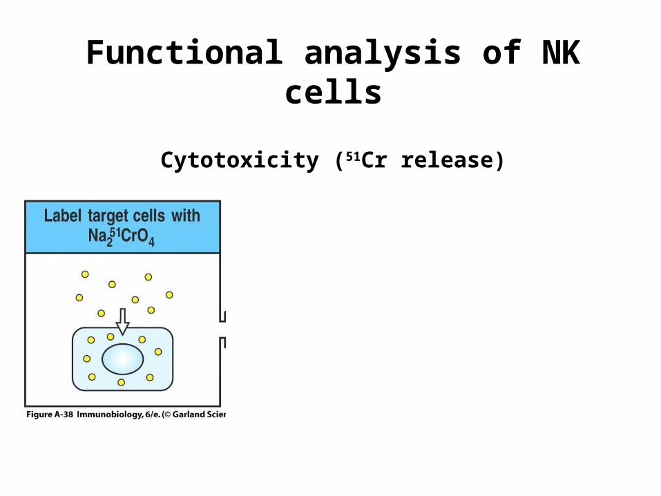
Functional analysis of NK cells
Cytotoxicity
K 562(Cytotoxicity test)
Cytokine production
IFN-γ(ELISA)
Functional analysis of NK cells
Cytotoxicity (51Cr release)

Functional analysis of NK cells
Cytotoxicity (51Cr release)

Functional analysis of NK cells
Cytotoxicity (51Cr release)

Phenotyping and determination of cell count
T cells
(proliferation, cytotoxicity, cytokine production)
B cells
(proliferation, antibody production)
Testing of cells of adaptive immunity (T and B lymphocytes)

Functional analysis of T cells
Proliferation
Stimulation:polyclonal activator (mitogen: Con A, PHA.....)
antigen
Based on incorporation of 3H thymidine in DNA


Patient medium ConA
A.B. 430±3143905±389
D.G. 330±84 1200±227

Cytotoxicity (T cells)
Cytoxicity test
Cytokine production (T cells)
ELISA
Antibody production (B cells)
ELISA, Nephelometry, RID
Functional analysis of T and B cells

Immediate hypersensitivity skin tests Prick test Intradermal test
Delayed hypersensitivity (DTH) skin tests
Assesment of humoral and cellular immune response afterimmunization
Provocation tests
Assessment of immune response in vivo


15 min.
Prick test (HSR type I)
1. Desinficate the skinand mark the spots
2. Put a drop of alergenpositive and negative control
3. Prick the skin by lancet
3. Remove the excess of alergen
edema is measured, not the redness
2mm › neg. control – positive result
≥ 5mm – clinical relevance
5-10 mm mild sensitivity10-15 mm moderate sensitivity
› 15 mm pronounced sensitivity
Flare and wheel

Assessment of immune response in vivoImmediate hypersensitivity skin tests
Prick test Intradermal test
Delayed hypersensitivity (DTH) skin tests Patch test
Assesment of humoral and cellular immune response after immunization
Provocation tests

Patch test (DTH)
Negative no changesweak positive rednessmoderate positive redness, vesiclespronounced positive redness, vesicles, edema
redness, vesicles and edema
after 48h (again after 72h)

Immediate hypersensitivity skin tests Prick test Intradermal test
DTH skin tests Patch test Tuberculin test
Provocation tests Bronchial provocation test DBPCFC (Double-Blind Placebo-Controlled Food Challenge)
Assessment of immune response in vivo

10.__9.__8.__7.__6.__5.__4.__3.__2.__1.__
CD16 and CD56j.T-cell proloferation can be assessed by10.
stimulation with T-cell mitogensi.Tuberculin test is9.
delayed type hypersensitivity testh.DTH tests determine8.
membrane Ig, CD19, CD20g.Immediate type hypersensitivity test determine
7.
CD3 and CD4 or CD8f.Respiratory burst is decreased in6.
difference in density or surface markers
e.Cytotoxic activity of NK cells can be measured by
5.
cytotoxicity testd.Surface markers of NK cells are4.
chronic granulomatous diseasec.Surface markers of B-cells are3.
presence of antigen-specific T-cellsb.Surface markers of T-cells are2.
presence of allergen-specific IgEa.Cell isolation is based on1.
e f g j d c a b h i