ascorbic acid prevents ischemia-reperfusion injury in the rat small intestine
TRANSCRIPT

Introduction
It is well known that structural and functional injuriesare caused by ischemia-reperfusion (I/R) in various or-gans [5, 22, 30]. The exact details of the underlyingmechanism are still unclear, but some oxygen-derivedfree radicals generated during reperfusion are believedto play an important role [2, 3, 35]. McCord [16] firstpostulated that oxygen-derived free radicals may causetissue injuries during reperfusion. According toMcCord’s theory, ATP undergoes degradation duringischemia and leads to the generation of superoxide an-ions which may eventually cause injury to tissues duringreperfusion. Superoxide anions first change to hydrogenperoxide in the presence of superoxide dismutase andthen to hydroxyl radicals with ferrous ions (Habour-Weiss reaction [10]). In recent years, however, superox-ide anions have been shown not to be able to extract hy-
drogen atoms from unsaturated fatty acids [20], high-lighting the importance of both hydroxyl radicals andiron-oxygen complexes. These are thought to be morereactive free radicals and to possibly react with cellmembranes, producing lipid peroxides and peroxidativeintermediate metabolites. Lipid peroxides, as well asalkoxy/peroxy radicals, may directly injure the cellmembrane [14, 25, 32, 38]. In fact, many investigatorshave already reported that peroxidative products areobserved in damaged tissues during the postischemicperiod [8, 25, 38]. Because such radicals have highly re-active properties, they immediately react with cell mem-branes consisting of polyunsaturated fatty acids(PUFA), and they may initiate lipid peroxidation of themembranes. Peroxidized lipids generate many poten-tially cytotoxic products [14], among which stoichiomet-rically the major products are lipid hydroperoxides(LPO). LPO are highly toxic in vivo [2]; they are also ca-
Michio NakamuraMichitaka OzakiShohei FuchinoueSatoshi TeraokaKazuo Ota
Ascorbic acid prevents ischemia-reperfusion injury in the rat small intestine
Received: 21 June 1996Received after revision: 8 October 1996Accepted: 12 November 1996
M. Nakamura ()) ⋅ M. OzakiS. Fuchinoue ⋅ S. Teraoka ⋅ K. OtaDepartment of Surgery III,Tokyo Women’s Medical College,8–1, Kawada-cho, Shinjuku-ku,Tokyo 162, Japan
Abstract Ischemia-reperfusion in-jury by free radicals and lipid perox-ides is observed in various organs.Ascorbic acid (AsA) or glutathione(GSH) in various doses (AsA:2, 0.5,0.1 mmol/kg, GSH:2 mmol/kg) wasintraperitoneally administered tomale Wistar rats. The entire small in-testines were resected just before is-chemia, after ischemia, and after20 min of reperfusion (n = 7–10 ateach time point). At each time point,the specimens were subjected to as-says of lipid peroxides, GSH, andglutaminase activity of the tissues;they were also examined histologi-cally. In the AsA group, the produc-tion of lipid peroxides after reperfu-sion was significantly suppressed in a
dose-dependent manner, and the ra-tio of oxidized GSH to total GSHwas also significantly low. Tissueglutaminase activity decreased to alesser extent, and the degree of in-jury was apparently less marked inthe AsA group. This study indicatesthat AsA acts as an antioxidantagainst peroxidative tissue injury,possibly by scavenging radicals, pre-serving reduced GSH, and reducingthe peroxidative reaction.
Key words Small intestine, ascorbicacid, reperfusion ⋅ Ascorbic acid,small intestine, reperfusion ⋅Reperfusion, ascorbic acid, smallintestine ⋅ Ischemia, ascorbic acid,reperfusion
Transpl Int (1997) 10: 89–95 Springer-Verlag 1997 ORIGINAL ARTICLE
pable of inactivating enzymes in vitro [7] and furtherpromoting free radical-mediated destruction of PUFA[32] and proteins [14].
Ascorbic acid (AsA), a water-soluble vitamin, hasboth reducing and chelating properties. In the very earlystage of reperfusion, AsA radicals (monodehydro-AsA)have been observed in an e. p. r.-spin trapping study, fol-lowed by hydroxyl radicals [34]. Their appearance maybe explained by a washing out of the radicals after oxida-tion by free radicals produced during ischemia, and byAsA scavenging of reactive oxygen intermediates pro-duced immediately after reperfusion [34]. With its strongreducing property and its property of scavenging freeradicals, AsA is well known as a strong antioxidant agent[37]. One of its major functions is to protect tissues fromharmful oxidative products and to keep certain enzymesin their reduced states [27]. It has also been demon-strated that, in addition to reacting with and scavengingsuperoxide and hydroxyl radicals [27], AsA can alsoscavenge singlet oxygen [28]. AsA is known to promoteperoxidation at low concentrations [1, 13] but to inhibitit at higher concentrations [15, 27, 28, 37].
This peroxidative reaction at an early stage after re-perfusion, i. e., primary I/R injury, may cause a varietyof secondary events, such as the release of several typesof interleukins and adhesion molecules of the cell mem-brane, and leukocyte infiltration to the tissues, second-ary I/R injury [5]. I/R injury to the small intestine hasbeen researched biochemically and histologically withregard to the production of free radicals and the adher-ence and accumulation of neutrophils [6, 16, 21, 31]. Al-most the same mechanism may be responsible for I/Rinjury in the case of the small intestine.
Though the small intestinal tissue in rats is commonlyknown to contain AsA, the effects of AsA on postische-mic lipid peroxidative injury are not yet thoroughly un-derstood. At present, much remains unknown aboutthe in vivo effects of AsA on I/R injury. In the presentstudy, we examined the effects of AsA on I/R injury tothe rat small intestine both biochemically and histologi-cally. We then considered the possible mechanism un-derlying these effects on postischemic tissue injurycaused by the lipid peroxidative reaction.
Materials and methods
Materials
The reduced form of glutathione (GSH), glyoxalase l, methylgly-oxal, NADPH, glutathione reductase, and thiobarbituric acid(TBA) were obtained from Sigma (St. Louis, Mo., USA). The he-moglobin-methylene blue (HMB) test kit for the LPO assay wasobtained from Kyowa Medex (Tokyo, Japan) and the ascorbicacid (AsA) from Wako Pure Chemical Industries (Osaka, Japan).All other chemicals were of analytical grade and were used withoutfurther purification.
Methods
AsA or GSH, dissolved at various concentrations in physiologicalsaline, was intraperitoneally administered (2 ml in total) to maleWistar rats weighing 250–300 g after 1 day of fasting. The same vol-ume of physiological saline was intraperitoneally administered torats in a control group. Animals were divided into the followinggroups: (1) Controls (n = 30); (2) AsA: 2 mmol/kg (n = 30),0.5 mmol/kg (n = 21), 0.1 mmol/kg (n = 21); and (3) GSH:2 mmol/kg (n = 30). Parentheses indicate the number of rats exam-ined in total.
Laparotomy was performed 1 h after administration of thedrugs under anesthesia with Nembutal (pentobarbital sodium,60 mg/100 body weight g), and the superior mesenteric artery wasclamped to induce ischemia over the entire small intestine. The ar-tery was unclamped after maintaining ischemia for 1 h. Each of thefive groups of rats was divided into three subgroups based on thetime points just before ischemia (B. I.), after ischemia (A.I.), andafter 20 min of reperfusion (A.R.; K = 7–10 at each time point).At each time point, the entire small intestine was resected. The re-sected tissues were subjected to the following assays and histologi-cal examination.
Assay of thiobarbituric acid reactive substance (TBA-RS)and LPO of the small intestine tissue
Small intestinal tissues were homogenized with 10 volumes of1.15% KCl solution and used to determine TBA-RS and LPO.TBA-RS was assayed following the method of Ohkawa et al. [23].
Assay of tissue LPO with the HMB test kit
The HMB test has a higher specificity for peroxides than the TBAmethod because it uses the peroxidase activity of hemoglobin.When hemoglobin reduces peroxides to their corresponding alco-hols, N-methylcarbamoyl derivatives of methylene blue (leucoform) are oxidized and colored blue [11].
Assay of the reduced form of glutathione (GSH) and the oxidizedform of glutathione (GSSG) of the small intestinal tissue
Small intestinal tissues were homogenized with 5 volumes of 6%perchlorate and subjected to the assay following the method ofBergmeyer et al . [12].
Assay of AsA and dehydro-AsA of the small intestinal tissue
Small intestinal tissues were also homogenized with 5 volumes of5% TCA and centrifuged at 3000 rpm for 10 min. The supernatantwas subjected to the assay of AsA and dehydro-AsA following themethod of Okamura et al. [24].
Glutaminase activity of the intestinal tissue
Small intestinal tissues were homogenized in 125 mM potassiumphosphate, 330 mM sucrose, and 2 mM dithiothreitol and sub-jected to the assay following the method of Pinkus and Wind-mueller [29].
90
Histological grading
Small intestinal tissues were fixed in 10% buffered formalin andprocessed for standard light microscopy. The severity of the patho-logical lesions in small intestinal tissues resected 20 min after re-perfusion ranged from 0 to 3, based on the following four parame-ters: (1) cell infiltration, (2) separation of the villus epitheliumfrom the lamina propria, (3) villus bases and crypts, and (4) edema(Table 1) [19].
This study was performed according to the principles of labora-tory animal care and was approved by the animal studies commit-tee of Tokyo Women’s Medical College.
Results
The following AsA group means AsA 2 mmol/kgtreated group except for Fig. 2.
Lipid peroxidation of small intestinal tissue duringischemia and reperfusion. (Fig. 1, Table 2)
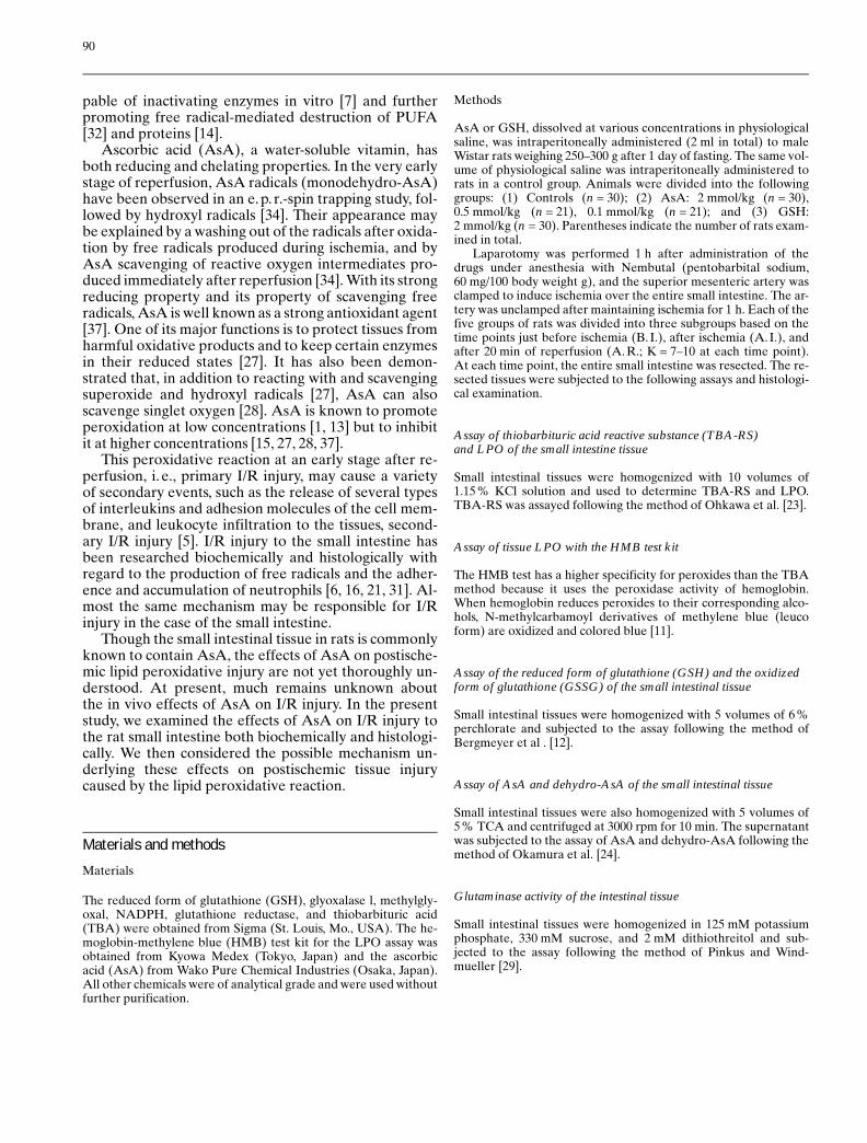
In the control group, the production of TBA-RS was sig-nificantly increased after 20 min of reperfusion of thesmall intestinal tissue (B. I. 0.020 ± 0.002, A.I.0.081 ± 0.001, A.R. 0.155 ± 0.049 absorbance at 535 nm;P < 0.01 vs B. I.; P < 0.05 vs A.I.). The production wasalso significantly increased in the AsA group (B.I.
0.029 ± 0.005, A.I. 0.084 ± 0.004, A.R. 0.078 ± 0.005;P < 0.05, vs B. I.). The production of TBA-RS in smallintestinal tissues obtained 20 min after reperfusion wassignificantly suppressed in the AsA groups comparedwith the control group (P < 0.01; Fig. 1), while in theGSH group the production of TBA-RS was significantlyincreased (B. I. 0.039 ± 0.005, A.I. 0.078 ± 0.008, A.R.0.114 ± 0.008; P < 0.05 vs B. I. and A. I.). LPO produc-tion was also significantly increased in the small intesti-nal tissue after reperfusion in the control group. How-ever, the administration of AsA significantly suppressedthe production of LPO (Table 2).
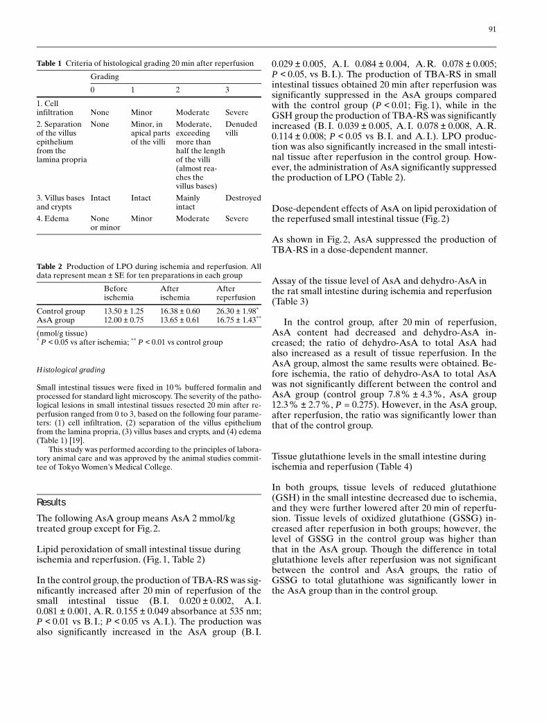
Dose-dependent effects of AsA on lipid peroxidation ofthe reperfused small intestinal tissue (Fig. 2)
As shown in Fig.2, AsA suppressed the production ofTBA-RS in a dose-dependent manner.
Assay of the tissue level of AsA and dehydro-AsA inthe rat small intestine during ischemia and reperfusion(Table 3)
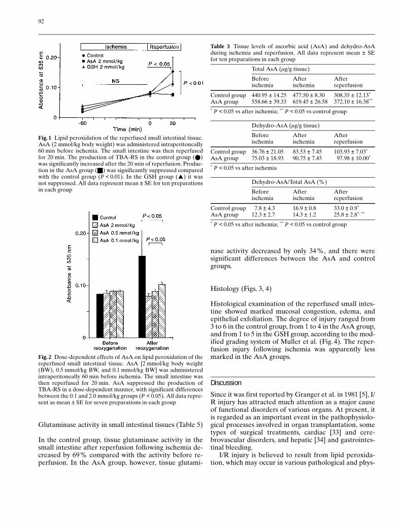
In the control group, after 20 min of reperfusion,AsA content had decreased and dehydro-AsA in-creased; the ratio of dehydro-AsA to total AsA hadalso increased as a result of tissue reperfusion. In theAsA group, almost the same results were obtained. Be-fore ischemia, the ratio of dehydro-AsA to total AsAwas not significantly different between the control andAsA group (control group 7.8% ± 4.3 %, AsA group12.3 % ± 2.7 %, P = 0.275). However, in the AsA group,after reperfusion, the ratio was significantly lower thanthat of the control group.
Tissue glutathione levels in the small intestine duringischemia and reperfusion (Table 4)
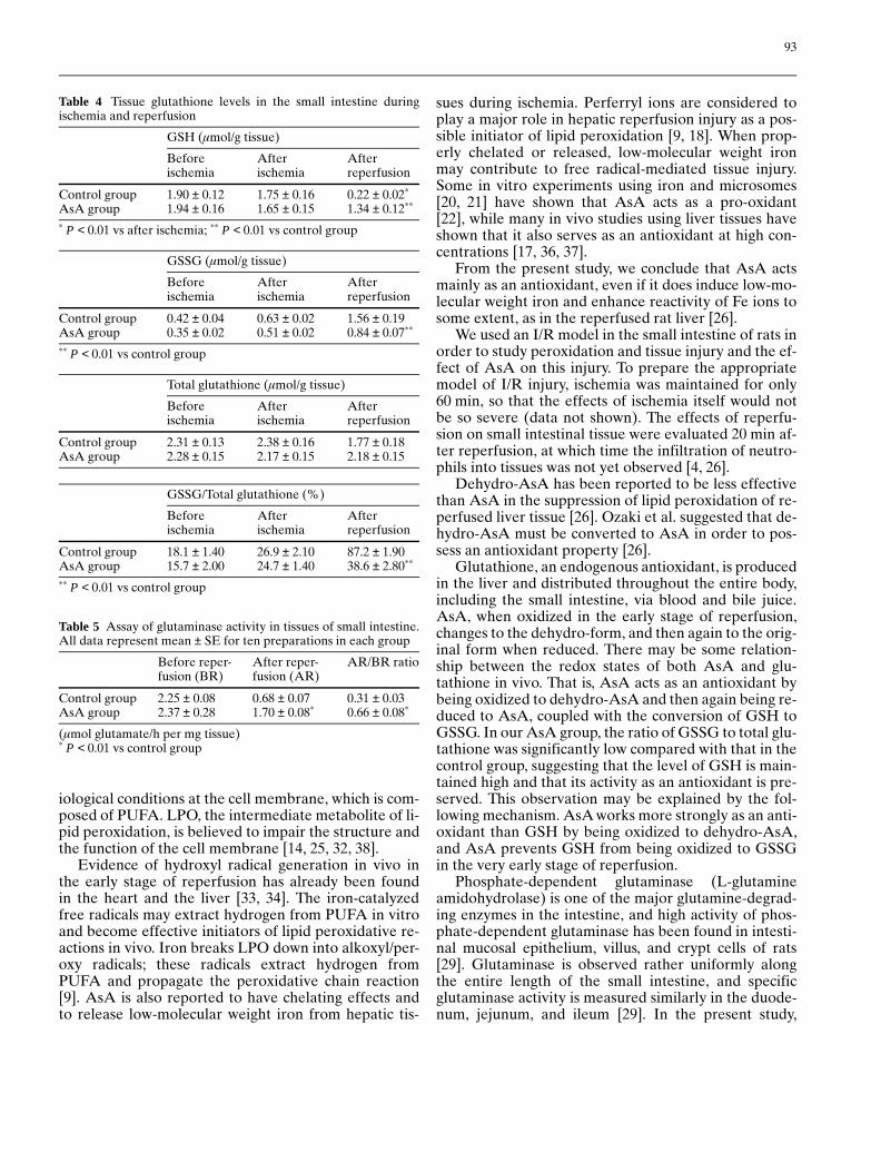
In both groups, tissue levels of reduced glutathione(GSH) in the small intestine decreased due to ischemia,and they were further lowered after 20 min of reperfu-sion. Tissue levels of oxidized glutathione (GSSG) in-creased after reperfusion in both groups; however, thelevel of GSSG in the control group was higher thanthat in the AsA group. Though the difference in totalglutathione levels after reperfusion was not significantbetween the control and AsA groups, the ratio ofGSSG to total glutathione was significantly lower inthe AsA group than in the control group.
91
Table 1 Criteria of histological grading 20 min after reperfusion
Grading
0 1 2 3
1. Cellinfiltration None Minor Moderate Severe
2. Separationof the villusepitheliumfrom thelamina propria
None Minor, inapical partsof the villi
Moderate,exceedingmore thanhalf the lengthof the villi(almost rea-ches thevillus bases)
Denudedvilli
3. Villus basesand crypts
Intact Intact Mainlyintact
Destroyed
4. Edema Noneor minor
Minor Moderate Severe
Table 2 Production of LPO during ischemia and reperfusion. Alldata represent mean ± SE for ten preparations in each group
Beforeischemia
Afterischemia
Afterreperfusion
Control group 13.50 ± 1.25 16.38 ± 0.60 26.30 ± 1.98*
AsA group 12.00 ± 0.75 13.65 ± 0.61 16.75 ± 1.43**
(nmol/g tissue)* P < 0.05 vs after ischemia; ** P < 0.01 vs control group
Glutaminase activity in small intestinal tissues (Table 5)
In the control group, tissue glutaminase activity in thesmall intestine after reperfusion following ischemia de-creased by 69 % compared with the activity before re-perfusion. In the AsA group, however, tissue glutami-
nase activity decreased by only 34 %, and there weresignificant differences between the AsA and controlgroups.
Histology (Figs. 3, 4)
Histological examination of the reperfused small intes-tine showed marked mucosal congestion, edema, andepithelial exfoliation. The degree of injury ranged from3 to 6 in the control group, from 1 to 4 in the AsA group,and from 1 to 5 in the GSH group, according to the mod-ified grading system of Muller et al. (Fig.4). The reper-fusion injury following ischemia was apparently lessmarked in the AsA groups.
Discussion
Since it was first reported by Granger et al. in 1981 [5], I/R injury has attracted much attention as a major causeof functional disorders of various organs. At present, itis regarded as an important event in the pathophysiolo-gical processes involved in organ transplantation, sometypes of surgical treatments, cardiac [33] and cere-brovascular disorders, and hepatic [34] and gastrointes-tinal bleeding.
I/R injury is believed to result from lipid peroxida-tion, which may occur in various pathological and phys-
92
Fig. 1 Lipid peroxidation of the reperfused small intestinal tissue.AsA (2 mmol/kg body weight) was administered intraperitoneally60 min before ischemia. The small intestine was then reperfusedfor 20 min. The production of TBA-RS in the control group (U)was significantly increased after the 20 min of reperfusion. Produc-tion in the AsA group (R) was significantly suppressed comparedwith the control group (P < 0.01). In the GSH group (X) it wasnot suppressed. All data represent mean ± SE for ten preparationsin each group
Fig. 2 Dose-dependent effects of AsA on lipid peroxidation of thereperfused small intestinal tissue. AsA [2 mmol/kg body weight(BW), 0.5 mmol/kg BW, and 0.1 mmol/kg BW] was administeredintraperitoneally 60 min before ischemia. The small intestine wasthen reperfused for 20 min. AsA suppressed the production ofTBA-RS in a dose-dependent manner, with significant differencesbetween the 0.1 and 2.0 mmol/kg groups (P < 0.05). All data repre-sent as mean ± SE for seven preparations in each group
Table 3 Tissue levels of ascorbic acid (AsA) and dehydro-AsAduring ischemia and reperfusion. All data represent mean ± SEfor ten preparations in each group
Total AsA (mg/g tissue)
Beforeischemia
Afterischemia
Afterreperfusion
Control group 440.95 ± 14.25 477.50 ± 8.30 308.35 ± 12.13*
AsA group 558.66 ± 39.33 619.45 ± 26.58 372.10 ± 16.38**
* P < 0.05 vs after ischemia; ** P < 0.05 vs control group
Dehydro-AsA (mg/g tissue)
Beforeischemia
Afterischemia
Afterreperfusion
Control group 36.76 ± 21.05 83.53 ± 7.45 103.93 ± 7.03*
AsA group 75.03 ± 18.93 90.75 ± 7.45 97.98 ± 10.00*
* P < 0.05 vs after ischemia
Dehydro-AsA/Total AsA (%)
Beforeischemia
Afterischemia
Afterreperfusion
Control group 7.8 ± 4.3 16.9 ± 0.8 33.0 ± 0.9*
AsA group 12.3 ± 2.7 14.3 ± 1.2 25.8 ± 2.8*, **
* P < 0.05 vs after ischemia; ** P < 0.05 vs control group
iological conditions at the cell membrane, which is com-posed of PUFA. LPO, the intermediate metabolite of li-pid peroxidation, is believed to impair the structure andthe function of the cell membrane [14, 25, 32, 38].
Evidence of hydroxyl radical generation in vivo inthe early stage of reperfusion has already been foundin the heart and the liver [33, 34]. The iron-catalyzedfree radicals may extract hydrogen from PUFA in vitroand become effective initiators of lipid peroxidative re-actions in vivo. Iron breaks LPO down into alkoxyl/per-oxy radicals; these radicals extract hydrogen fromPUFA and propagate the peroxidative chain reaction[9]. AsA is also reported to have chelating effects andto release low-molecular weight iron from hepatic tis-
sues during ischemia. Perferryl ions are considered toplay a major role in hepatic reperfusion injury as a pos-sible initiator of lipid peroxidation [9, 18]. When prop-erly chelated or released, low-molecular weight ironmay contribute to free radical-mediated tissue injury.Some in vitro experiments using iron and microsomes[20, 21] have shown that AsA acts as a pro-oxidant[22], while many in vivo studies using liver tissues haveshown that it also serves as an antioxidant at high con-centrations [17, 36, 37].
From the present study, we conclude that AsA actsmainly as an antioxidant, even if it does induce low-mo-lecular weight iron and enhance reactivity of Fe ions tosome extent, as in the reperfused rat liver [26].
We used an I/R model in the small intestine of rats inorder to study peroxidation and tissue injury and the ef-fect of AsA on this injury. To prepare the appropriatemodel of I/R injury, ischemia was maintained for only60 min, so that the effects of ischemia itself would notbe so severe (data not shown). The effects of reperfu-sion on small intestinal tissue were evaluated 20 min af-ter reperfusion, at which time the infiltration of neutro-phils into tissues was not yet observed [4, 26].
Dehydro-AsA has been reported to be less effectivethan AsA in the suppression of lipid peroxidation of re-perfused liver tissue [26]. Ozaki et al. suggested that de-hydro-AsA must be converted to AsA in order to pos-sess an antioxidant property [26].
Glutathione, an endogenous antioxidant, is producedin the liver and distributed throughout the entire body,including the small intestine, via blood and bile juice.AsA, when oxidized in the early stage of reperfusion,changes to the dehydro-form, and then again to the orig-inal form when reduced. There may be some relation-ship between the redox states of both AsA and glu-tathione in vivo. That is, AsA acts as an antioxidant bybeing oxidized to dehydro-AsA and then again being re-duced to AsA, coupled with the conversion of GSH toGSSG. In our AsA group, the ratio of GSSG to total glu-tathione was significantly low compared with that in thecontrol group, suggesting that the level of GSH is main-tained high and that its activity as an antioxidant is pre-served. This observation may be explained by the fol-lowing mechanism. AsA works more strongly as an anti-oxidant than GSH by being oxidized to dehydro-AsA,and AsA prevents GSH from being oxidized to GSSGin the very early stage of reperfusion.
Phosphate-dependent glutaminase (L-glutamineamidohydrolase) is one of the major glutamine-degrad-ing enzymes in the intestine, and high activity of phos-phate-dependent glutaminase has been found in intesti-nal mucosal epithelium, villus, and crypt cells of rats[29]. Glutaminase is observed rather uniformly alongthe entire length of the small intestine, and specificglutaminase activity is measured similarly in the duode-num, jejunum, and ileum [29]. In the present study,
93
Table 4 Tissue glutathione levels in the small intestine duringischemia and reperfusion
GSH (mmol/g tissue)
Beforeischemia
Afterischemia
Afterreperfusion
Control group 1.90 ± 0.12 1.75 ± 0.16 0.22 ± 0.02*
AsA group 1.94 ± 0.16 1.65 ± 0.15 1.34 ± 0.12**
* P < 0.01 vs after ischemia; ** P < 0.01 vs control group
GSSG (mmol/g tissue)
Beforeischemia
Afterischemia
Afterreperfusion
Control group 0.42 ± 0.04 0.63 ± 0.02 1.56 ± 0.19AsA group 0.35 ± 0.02 0.51 ± 0.02 0.84 ± 0.07**
** P < 0.01 vs control group
Total glutathione (mmol/g tissue)
Beforeischemia
Afterischemia
Afterreperfusion
Control group 2.31 ± 0.13 2.38 ± 0.16 1.77 ± 0.18AsA group 2.28 ± 0.15 2.17 ± 0.15 2.18 ± 0.15
GSSG/Total glutathione (%)
Beforeischemia
Afterischemia
Afterreperfusion
Control group 18.1 ± 1.40 26.9 ± 2.10 87.2 ± 1.90AsA group 15.7 ± 2.00 24.7 ± 1.40 38.6 ± 2.80**
** P < 0.01 vs control group
Table 5 Assay of glutaminase activity in tissues of small intestine.All data represent mean ± SE for ten preparations in each group
Before reper-fusion (BR)
After reper-fusion (AR)
AR/BR ratio
Control group 2.25 ± 0.08 0.68 ± 0.07 0.31 ± 0.03AsA group 2.37 ± 0.28 1.70 ± 0.08* 0.66 ± 0.08*
(mmol glutamate/h per mg tissue)* P < 0.01 vs control group

glutaminase activity in the reperfused rat small intesti-nal tissues was well preserved by the administration ofAsA.
Histopathologically, in the AsA group, the structureof the intestinal mucosa was generally preserved. Thereperfused small intestine of rats in the control groupshowed severe cellular infiltration, separation of the ep-ithelia from the lamina propria down to the villus base,mostly disarrayed villus bases, and moderate edema. Se-vere reperfusion injury following ischemia mainly oc-currs in the mucosal layer of the small intestine in rats[19]. The mucosa contains over 90 % of the total glutam-inase activity [29]. This is why glutaminase activity re-mained so high in the AsA group. These results indicatethat by administering AsA, the reperfused small intesti-nal tissues of rats were effectively protected from reper-fusion injury, both biochemically and histologically.
In this in vivo study, AsA worked exclusively as anantioxidant and protected the postischemic intestinaltissue from peroxidative tissue injury. This protectivemechanism may be explained, in part, as follows: AsAprotects postischemic small intestinal tissue by scaveng-ing radicals and/or reducing the peroxidative reactions.It also preserves the high glutathione level of postische-mic small intestinal tissue. As the result, the cellular in-tegrity of postischemic small intestinal tissue is well pre-served.
In conclusion, our findings show that AsA acts as anantioxidant in the small intestine and reduces injurydue to reperfusion. These effects should be very usefulin preserving mucosal function or accelerating its recov-ery after certain types of surgical treatments, such as en-terectomy, and especially small bowel transplantation.
94
Fig. 3A, B Histological appearance of the small intestine 20 minafter reperfusion. After 60 min of ischemia followed by 20 min ofreperfusion, full-thickness small intestinal tissues were fixed in10% buffered formalin and processed for standard H&E stainand light microscopic examination ( × 20): A control group: thereis severe cell infiltration, separation of the epithelium from thelamina propria down to the villus base, mostly disarrayed villusbases, and moderate edema; B AsA group: there is mild cell infil-tration, minor separation of the epithelium from the lamina pro-pria, almost intact villus bases, and minor edema
Fig. 4 Histological grading of the reperfused small intestinal tissue.The severity of pathological lesions in small intestinal tissues as-sessed 20 min after reperfusion was graded 0–3, based on the fol-lowing four parameters: (1) cell infiltration, (2) separation of thevillus epithelium from the lamina propria, (3) villus bases andcrypts, and (4) edema. The degree of injury was graded from 3 to6 in the control group (U), from 1 to 4 in the AsA group (R), andfrom 1 to 5 in the GSH group (X)
95
Reference
1. Bodannes RS, Chan PC (1979) Ascor-bic acid as a scavenger of singlet oxy-gen. FEBS Lett 105: 195–196
2. Caraceni P, Rosenblum ER, ThielDHV, Borle AB (1994) Reoxygenationinjury in isolated rat hepatocytes: rela-tion to oxygen free radicals and lipidperoxidation. Am J Physiol 266: G799–G806
3. Flecha BG, Cutrin JC, Boveris A (1993)Time course and mechanism of oxida-tive stress and tissue damage in rat liversubjected to in vivo ischemia-reperfu-sion. J Clin Invest 91: 456–464
4. Grace PA (1994) Ischemia-reperfusioninjury. Br J Surg 81: 637–647
5. Granger DN, Rutili G, McCord JM(1981) Superoxide radicals in feline in-testinal ischemia. Gastroenterology 81:22–29
6. Granger DN, Hollwarth ME, Parks DA(1986) Ischemic-reperfusion injury: roleof oxygen-derived free radicals. ActaPhysiol Scand 126: 47–63
7. Green RC, Little C, O’Brien PJ (1971)The inactivation of isocitrate dehydro-genase by a lipid peroxide. Arch Bio-chem Biophys 142: 598–605
8. Grune T, Siems WG, Schonheit K, Bla-sig IE (1993) Release of 4-hydroxynon-enal, an aldehydic mediator of inflam-mation, during postischemic reperfu-sion of the myocardium. Int J TissueReact 15: 145–150
9. Halliwell B, Gutteridge JMC (1984)Oxygen toxicity, oxygen radicals, tran-sition metals and disease. Biochem J219: 1–14
10. Harbour JR, Chow V, Bolton JR (1974)An electron spin resonance study of thespin adducts of OH and HO2 radicalswith nitrons in the ultraviolet photolysisof aqueous hydrogen peroxide solu-tions. Can J Chem 52: 3549–3553
11. Kanazawa K, Inoue N, Ashida H, Mi-zuno M, Natake M (1989) What dothiobarbituric acid and hemoglobin-methylene blue tests evaluate in the en-dogenous lipid peroxidation of rat liver? J Clin Biochem Nutr 7: 69–79
12. Klotsh H, Bergmeyer HU (1974) Meth-ods in enzymatic analysis, vol.4. Aca-demic Press, New York
13. Laudicina DC, Marnett LJ (1990) En-hancement of hydroperoxide-depen-dent lipid peroxidation in rat liver mi-crosomes by ascorbic acid. Arch Bio-chem Biophys 278: 73–80
14. Logani MK, Davies RE (1980) Lipidoxidation: biologic effects and antioxi-dants, a review. Lipids 15: 485–495
15. Mak IT, Weglicki WB (1985) Charac-terization of iron-mediated peroxida-tive injury in isolated hepatic lysoso-mes. J Clin Invest 75: 58–63
16. McCord JM (1985) Oxygen-derivedfree radicals in postischemic tissue in-jury. N Engl J Med 312: 159–163
17. Miller DM, Aust SD (1989) Studies ofascorbate-dependent, iron-catalyzed li-pid peroxidation. Arch Biochem Bio-phys 271: 113–119
18. Miller DM, Spear NH, Aust SD (1992)Effects of deferrioxamine on iron-cata-lyzed lipid peroxidation. Arch BiochemBiophys 295: 240–246
19. Muller AR, Nalesnik M, Platz KP, Lan-grehr JM, Hoffman RA, Schraut WH(1994) Evaluation of preservation con-ditions and various solutions for smallbowel preservation. Transplantation 57:649–655
20. Niki H (1987) Kasseisanso. IshiyakuPress, Tokyo
21. Nilsson UA, Schoenberg MH, AnemanA, Poch B, Magadum S, Beger HG,Lundgren O (1994) Free radicals andpathogenesis during ischemia and re-perfusion of the cat small intestine.Gastroenterology 106: 629–636
22. Nordstrom G, Seeman T, HasselgrenPO (1985) Beneficial effect of allopu-rinol in liver ischemia. Surgery 97: 679–683
23. Ohkawa H, Ohishi N, Yagi K (1979)Assay for lipid peroxides in animal tis-sues by thiobarbituric acid reaction.Anal Biochem 95: 351–358
24. Okamura M (1980) An improvedmethod for determination of L-ascorbicacid and L-dehydroascorbic acid inblood plasma. Clin Chim Acta 103: 259–268
25. Ozaki M, Fuchinoue S, Teraoka S, OtaK (1994) Mobilization of low-molecu-lar-weight iron and peroxidative dam-age during ischemia and reoxygenationof the rat liver. Transplant Proc 26: 918–921
26. Ozaki M, Fuchinoue S, Teraoka S, OtaK (1995) The in vivo cytoprotection ofascorbic acid against ischemia/reoxy-genation injury of rat liver. Arch Bio-chem Biophys 318: 439–445
27. Padh H (1990) Cellular functions ofascorbic acid. Biochem Cell Biol 68:1166–1173
28. Palamanda JR, Kehrer JP (1993) In-volvement of vitamin E and proteinthiols in the inhibition of microsomal li-pid peroxidation by glutathione. Lipids28: 427–431
29. Pinkus LM, Windmueller HG (1977)Phosphate-dependent glutaminase ofsmall intestine: localization and role inintestinal glutamine metabolism. ArchBiochem Biophys 182: 506–517
30. Rao PS, Mueller HS (1983) Lipid per-oxidation and acute myocardial is-chemia. Adv Exp Med Biol 161: 347–363
31. Schoenberg MH, Poch B, Younes M,Schwarz A, Baczako K, Lundberg C,Haglund U, Beger HG (1991) Involve-ment of neutrophils in postischaemicdamage to the small intestine. Gut 32:905–912
32. Shimasaki H, Privett OS (1975) Studieson the role of vitamin E in the oxidationof blood components by fatty hydro-peroxides. Arch Biochem Biophys 169:506–512
33. Sun JZ, Kaur H, Halliwell B, Li XY,Bolli R (1993) Use of aromatic hydrox-ylation of phenylalanine to measureproduction of hydroxyl radicals aftermyocardial ischemia in vivo. Circ Res73: 534–549
34. Togashi H, Shinzawa H, Yong H, Taka-hashi T, Noda H, Oikawa K, Kamada H(1994) Ascorbic acid radical, superox-ide, and hydroxyl radical are detected inreperfusion injury of rat liver usingelectron spin resonance spectroscopy.Arch Biochem Biophys 308: 1–7
35. Weinberg JM, Davis JA, Abarzua M,Rajan T (1987) Cytoprotective effectsof glycine and glutathione against hy-poxic injury to renal tubules. J Clin In-vest 80: 1446–1454
36. Younes M, Kayser E, Strubelt O (1992)Effect of antioxidants on hypoxia/reox-ygenation induced injury in isolatedperfused rat liver. Pharmacol Toxicol71: 278–283
37. Zeng LH, WU J, Carey D, Wu TW(1991) Trolox and ascorbate: are theysynergistic protecting liver cells in vitroand in vivo? Biochem Cell Biol 69: 198–201
38. Zimmerman BJ, Granger DN (1994)Mechanism of reperfusion injury. Am JMed Sci 307: 284–292