apoptosis and caspases after ischemia-reperfusion...
TRANSCRIPT
Apoptosis and Caspases after Ischemia—ReperfusionInjury in Rat Retina
Tim T. Lam,1'2 Andrew S. Abler,1 and Mark 0. M. Tso2
PURPOSE. Extensive cell loss in the retinal ganglion cell layer (RGCL) and the inner nuclear layer(INL) was noted in a rat model of retinal ischemia-reperfusion injury by transient elevatedintraocular pressure (IOP). The possible involvement of apoptosis and caspases was examined inthis model of neuronal loss.
METHODS. Transient elevated IOP was induced in albino Lewis rats through the insertion of a needleinto the anterior chamber connected to a saline column. Elevated IOP at 110 mm Hg wasmaintained for 60 minutes. Groups of animals were euthanatized at various times after reperfusion,and their retinas were evaluated by morphology, agarose gel electrophoresis of DNA, in situterminal deoxynucleotidyl transferase-mediated biotin-deoxyuridine triphosphate nick-end label-ing (TUNEL), immunohistochemistry of caspases II (ICH1) and III (CPP32), and morphometry.YVAD.CMK, a tetrapeptide inhibitor of caspases, was used to examine the involvement of caspases.
RESULTS. A marked ladder pattern in retinal DNA gel analysis, typical of internucleosomal DNAfragmentation and characteristic of apoptosis, was present 12 and 18 hours after reperfusion.Labeling of nuclei in the RGCL and the inner nuclear layer (INL) by TUNEL was noted between 8and 18 hours after reperfusion. Histologic and ultrastructural features typical of apoptosis were alsoobserved in the inner retina after ischemia. YVAD.CMK administered during the ischemic periodinhibited apoptotic fragmentation of retinal DNA and ameliorated the tissue damage. Whenadministered intravitreally 0, 2, or 4 hours after reperfusion, YVAD.CMK was also effective inpreserving the inner retina but had no significant effect when administered 6 or 8 hours afterreperfusion. The inner retina showed transient elevated immunoreactivity of caspases II and HI 4and 8 hours after reperfusion.
CONCLUSIONS. Retinal ischemia-reperfusion after transient elevated IOP induced apoptosis of cells inthe retinal ganglion cell layer and the INL. Caspases may have a pivotal role in the early events ofthe apoptotic pathway(s). Rescue by using anti-apoptotic agents after ischemia-reperfusion isfeasible. (Invest Ophthalmol Vis Sci. 1999;40:967-975)
Apoptosis, a highly regulated and energy-dependentform of cell death, has characteristic histologic andmorphologic features showing scattered involvement
of individual cells in an asynchronous pattern, a minimum oran absence of inflammatory reaction around the dying cells,intact cytoplasmic membrane, perinuclear chromatin con-densation with subsequent nuclear disintegration, and apo-ptotic cell bodies.1'2 Biochemically, it is characterized byinternucleosomal double-stranded DNA cleavage, producingDNA fragments that are multiples of 180 bp to 200 bp inlength and therefore show a typical ladder pattern in DNAgel electrophoresis.1'2 In contrast, the random breakdown
From the 'Department of Ophthalmology and Visual Science,University of Illinois at Chicago; 2Department of Ophthalmology andVisual Sciences, The Chinese University of Hong Hong.
Supported in part by Grants EYO1903 and EYO6761 from theNational Institutes of Health, Bethesda, Maryland; Campus ResearchGrant, University of Illinois at Chicago; and RGC Grant CUHK295/96M,Hong Kong.
Submitted for publication October 30, 1997; revised December 4,1998; accepted January 11, 1999.
Proprietary interest category: N.Reprint requests: Tim T. Lam, Department of Ophthalmology and
Visual Sciences, Hong Kong Eye Hospital, 147K Argyle Street 3/F,Kowloon, Hong Kong.
of DNA in necrosis shows a smear in gel electrophoresis.1'2
The double-stranded DNA nicks in apoptosis can be demon-strated histochemically in situ with the terminal deoxynu-cleotidyl transferase (TdT)-mediated biotin-deoxyuridinetriphosphate (dUTP) nick-end labeling (TUNEL) techniquein paraffin sections.3 Using these criteria and methods, wenoted apoptosis in the retinas of the retinal dystrophic RoyalCollege of Surgeons (RCS) rat,4 and in rat retinas after lightinjury 5 or JV-methyl-D-aspartate-induced excitotoxicity.6
Others have also noted its involvement in retinal degenera-tion in the RCS rat,7 rd7'9 and rds mice,9 and transgenicmice carrying mutations in the rhodopsin gene.9 Clinically,apoptosis has been shown to occur in retinas with glauco-ma,10 traumatic detachment," macular degeneration,12 ret-inoblastoma,13 or retinitis pigmentosa.14 Therefore, apopto-sis seems to be the final common pathway of retinalneuronal degeneration.
Retinal degeneration after ischemia-reperfusion injury bytransient elevation of intraocular pressure GOP) in rats showsextensive loss of neuronal elements of the retinal ganglion celllayer (RGCL) and the inner nuclear layer (INL).15 These fea-tures closely resemble those after retinal ischemic insult16 andacute glaucoma.17 Certain ultrastructural features typical ofapoptosis have been noted among the dying cells in a similaranimal model.18 Therefore, it seems possible that apoptosis
Investigative Ophthalmology & Visual Science, April 1999, Vol. 40, No. 5Copyright © Association for Research in Vision and Ophthalmology 967
Downloaded From: http://iovs.arvojournals.org/pdfaccess.ashx?url=/data/journals/iovs/933211/ on 05/17/2018
968 Lam et al. IOVS, April 1999, Vol. 40, No. 5
may have an important role in ischemia-reperfusion insult tothe retina.
Yuan and Horvitz19 reported the requirement of ced-3 andced-4 genes for apoptotic cell death in the nematode Caeno-rhabditis elegans. Subsequent sequence analysis of the geneproducts by Miura et al.20 found a highly conserved nonserine-rich region of the CED-3 protein homologous to the sequenceof the human interleukin-lj3-converting enzyme (ICE), a cys-teine protease, and suggested that ICE may have a central rolein apoptotic cell death in mammalian cells. The exact humangene(s) corresponding to ced-3 in C. elegans remains unde-fined, but various studies have shown that ICE-like enzymes orcaspases may play a pivotal role in apoptosis in mammaliancells.2122 Whether caspases play similar significant roles inretinal degeneration is not clear.
In the present study, we showed that the loss of innerretinal elements after retinal ischemia-reperfusion insult withtransient elevated IOP involved apoptosis. In addition, we usedimmunohistochemistry and a caspase inhibitor, the tetrapep-tide YVAD.CMK,23 to probe the possible involvement ofcaspases in apoptosis of retinal neurons after ischemia-reper-fusion insult. We also explored the timing of the window foreffective treatment with YVAD.CMK after the insult.
MATERIALS AND METHODS
Induction of Transient Elevated IntraocularPressure
Transient elevated IOP was induced with an establishedmethod15 in adult male albino Lewis rats (Harlan, Indianapolis,IN), 45 to 50 days old. Briefly, the animals were anesthetizedwith intraperitoneal injections of 400 mg/kg chloral hydrate(Sigma, St. Louis, MO). After topical application of 0.5% pro-paracaine hydrochloride, the anterior chamber was cannulatedwith a 26-gauge needle connected to a normal saline containerby Silastic tubing (Dow-Corning, Midland, MI). Intraocularpressure was raised to 110 mm Hg by elevating the salinecontainer. Body temperature of the animals was maintained at37.5°C with a probe and heating pad during the experimentalperiod. The infusion needle was removed from the anteriorchamber 60 minutes later. Reperfusion of the retinal vascula-ture was confirmed by fundus examination. The animals werethen euthanatized at various times, and their eyes were enu-cleated for morphologic, morphometric, and immunohisto-chemical studies; DNA agarose gel electrophoresis analysis; orTUNEL assay. All procedures involving animals were in accor-dance with the ARVO Statement on the Use of Animals inOphthalmic and Vision Research.
Agarose Gel Electrophoresis of Retinal DNA
The eyes enucleated for DNA agarose gel electrophoresis werebisected at the ora serrata, and the lenses were removed. Theretinas were dissected from the underlying tissues, transferredto liquid nitrogen, and kept frozen in — 70°C until analysis witha previously described procedure.4 Briefly, the frozen retinaswere thawed and vortexed in a buffer containing 50 mM EDTA,0.5% sodium dodecyl sulfate (SDS), and 20 mM Tris-HCl (pH8.0). RNase A was added to a final concentration of 50 jag/ml.After incubation for 30 minutes at 42°C, proteinase K wasadded to a final concentration of 400 /ug/ml. The samples werethen incubated at 55°C until clear lysates were produced. The
lysates were extracted with phenol/chloroform/isoamyl alco-hol (25:24:1) and chloroform/isoamyl alcohol (24:1), respec-tively. DNA was precipitated with 3 M sodium acetate andice-cold ethanol and analyzed for internucleosomal cleavage byelectrophoresis using a 2% agarose gel (10 jag DNA per lane).DNA in the gel was visualized by UV light after ethidiumbromide staining and photographed (MP-4 system; Polaroid,Cambridge, MA).
Morphologic Study
Eyes enucleated for morphologic and immunohistochemicalstudies were fixed in 10% buffered formaldehyde. The anteriorsegment was removed, and the posterior segment was pre-pared for paraffin sectioning. Morphologic evaluations wereperformed on 5-ju.m sections stained with hematoxylin andeosin.
Eyes were used for electron microscopy 18 hours afterreperfusion. The enucleated eyes were fixed in 1% glutaralde-hyde and 4% paraformaldehyde in phosphate buffer for 1 hour.The anterior segment was removed, and the whole eyecup wasdivided into four strips, one each from the nasal, inferior,temporal, and superior quadrants. Samples were postfbced by1% osmium tetroxide, dehydrated by graded alcohol and pro-pylene oxide, embedded in epoxy resin, sectioned, and stainedwith Mallory's azure II-methylene blue. Ultrathin sections con-taining representative areas were obtained for electron micros-copy.
In Situ TUNEL
In situ TUNEL was performed according to the proceduredescribed by Gavrieli et al.3 except that the proteinase Kdigestion step was omitted. The 5-/u,m paraffin sections ob-tained from the preceding section were used. The tissue sec-tions were deparaffinized, rehydrated, and incubated in meth-anol containing 3% H2O2 to block the endogenous peroxidase.They were washed in distilled water, and TdT buffer (30 mMTrizma base [pH 7.2], 140 mM sodium cacodylate, 1 mM cobaltchloride). The sections were incubated with TdT buffer con-taining TdT and dUTP for 60 minutes, and then with TB buffer(300 mM sodium chloride, 30 mM sodium citrate) to terminatethe reaction. They were washed with double-distilled water,incubated with peroxidase-conjugated streptavidin, and devel-oped with 3,3-diaminobenzidine tetrahydrochloride (DAB;Sigma, St. Louis, MO).
Caspase Inhibitor Study
The effects of YVAD.CMK, a tetrapeptidyl caspase inhibitor,on apoptotic and tissue responses were evaluated. Vehicle(0.1 M phosphate-buffered saline [PBS]; pH 7.2), or 100 JLLMYVAD.CMK23 24 (Bachem, Philadelphia, PA) in PBS was intro-duced into the anterior chamber during the induction of ele-vated IOP through the needle and Silastic tubing, as previouslydescribed.25 Animals were killed 12 hours after reperfusion forDNA analysis by electrophoresis, when the characteristic lad-der pattern was apparent, or at 7 days after reperfusion formorphometric evaluation, when the tissue responses wereestablished.
A second series of experiments was conducted to deter-mine the timing of the window for the effectiveness of thecaspase inhibitor YVAD.CMK. Groups of animals received 2 /xlof 500 ixM YVAD.CMK intravitreally at 0, 2, 4, 6, or 8 hours
Downloaded From: http://iovs.arvojournals.org/pdfaccess.ashx?url=/data/journals/iovs/933211/ on 05/17/2018
IOVS, April 1999, Vol. 40, No. 5
after the initial reperfusion of retinas previously exposed to 60minutes of ischemia and were euthanatized 7 days after reper-fusion, when the tissue responses had stabilized. The effective-ness of the treatment was evaluated morphometrically withinner retinal thickness (IRT) measurements.15
MorphometryTissue response after retinal ischemia-reperfusion injury wasevaluated using a previously published method: measurementof the IRT.'5 Tissue sections were prepared as described in themorphologic study with electron microscopy. The IRT wasassessed by measuring the thickness between the internallimiting membrane and the interface of the outer plexiformlayer and outer nuclear layer (ONL) on the projected image ofa stained 1-jam section onto a digitizing pad coupled with acustomized image-processing work station. Two readings wereobtained for each tissue sample. The average readings of thefour quadrants of the same eye represented the mean of thewhole eye. Analysis of variance and Student's f-test were usedfor statistical comparisons.
Immunohistochemistry of CaspasesParaffin sections obtained from the morphologic study weredeparaffinized, rehydrated, and incubated in 10 mM sodiumcitrate buffer pH 6.0 at 90°C for 40 minutes. Endogenousperoxidases were blocked with incubation in 3% H2O2 for 4minutes. The sections were permeabilized in 0.3% Triton X-100for 1 hour. Anti-caspase II (Anti-ICHl; 1:100) or anti-caspaseIII (Anti-CPP32; 1:100; Transduction Laboratory, Lexington,KY) was added to the sections separately and incubated at 4°Covernight. Immunoreactivity was detected using the standardABC complex (StreptABC Complex; Dako, Carpinteria, CA)and DAB.
RESULTS
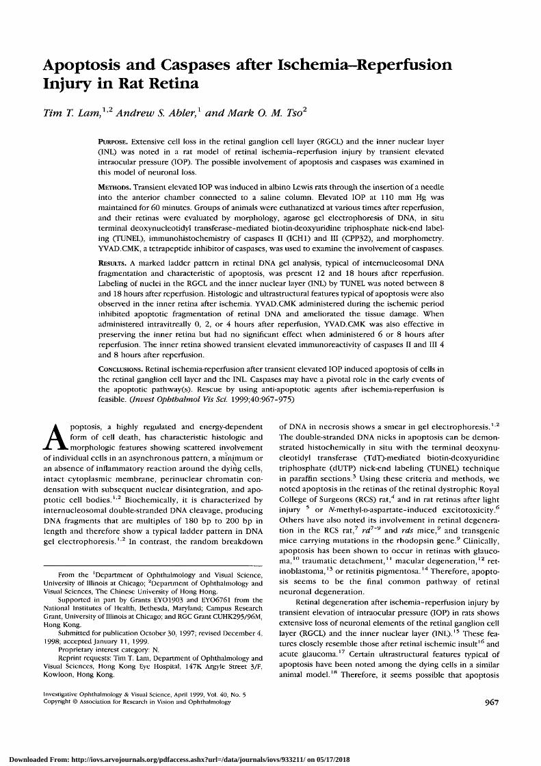
Agarose gel electrophoresis of DNA samples from retinas at 0to 48 hours of reperfusion after 60 minutes of ischemia byelevated IOP showed a recognizable ladder pattern at 12 and18 hours after reperfusion (Fig. 1; lanes 5 and 6, respectively).
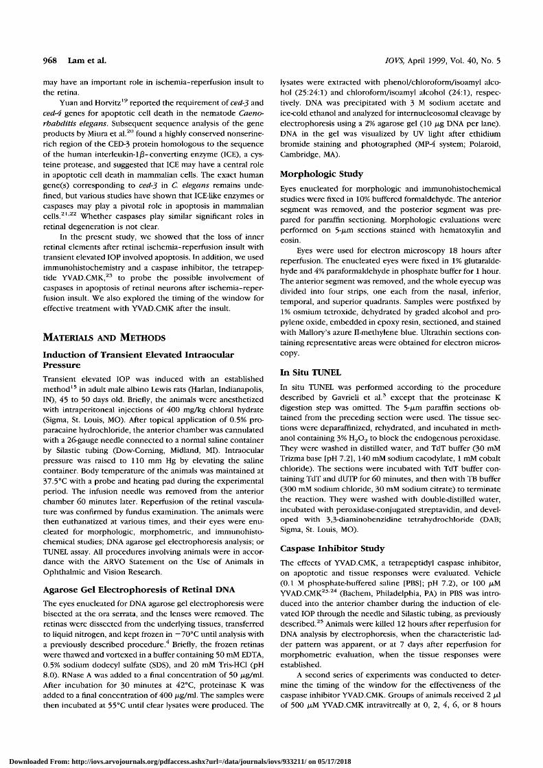
Histopathologically, at 4 hours, the retina showed occa-sionally condensed nuclei in the RGCL, markedly edematousinner plexiform layer (IPL) and condensed nuclei with variousdegrees of cytoplasmic vacuolation in the INL, especially in theinner part of the INL (Fig. 2B). The outer retina was unremark-able. At 8 hours (Fig. 2C), condensed nuclei scattering in theRGCL and the INL and various degrees of cytoplasmic vacuo-lation in the INL persisted. At 12 (Fig. 2D) and 18 (Fig. 2E)hours, scattered condensed nuclei remained in the RGCL andthe INL, whereas vacuolation in the INL subsided. After 48hours (Fig. 2F), only occasionally condensed nuclei were stillseen in the RGCL. The IPL was markedly atrophic. Thinning ofthe INL was noted. The ONL remained unremarkable through-out the observation period.
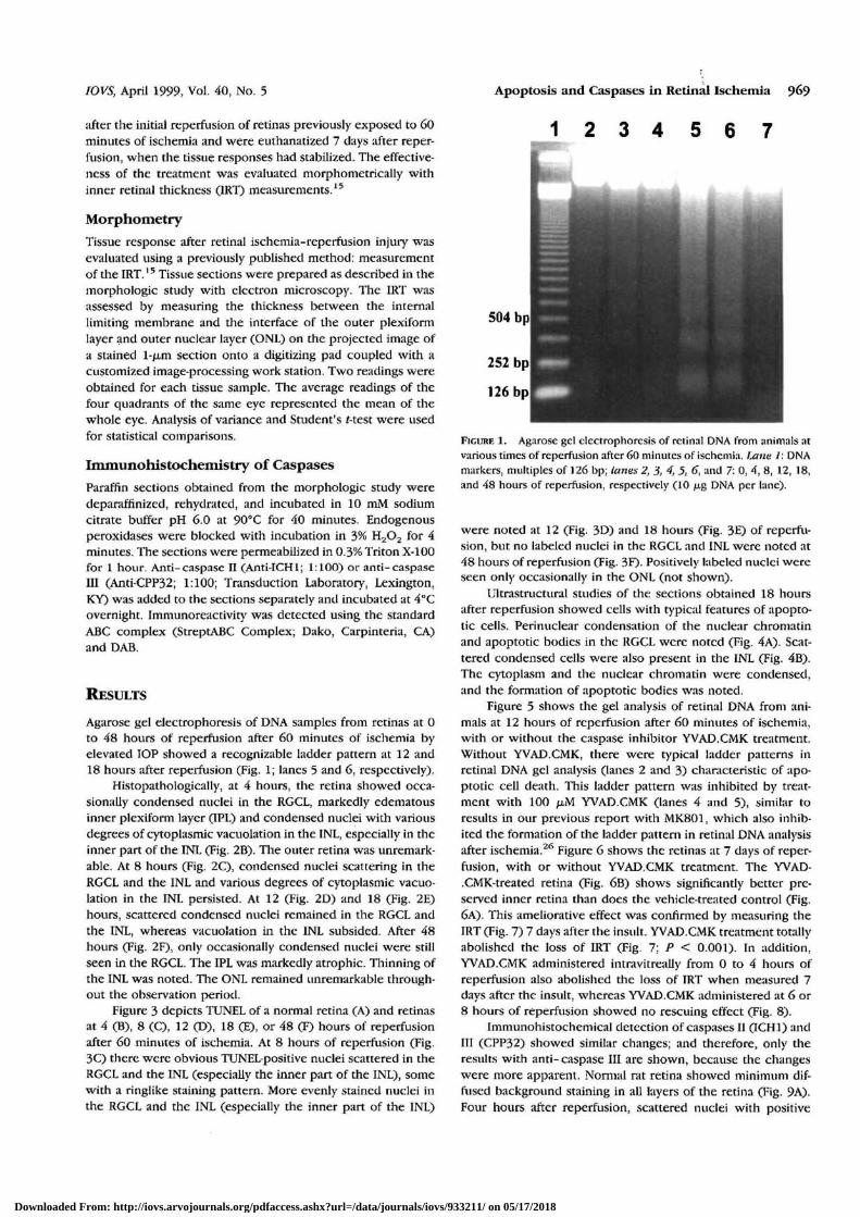
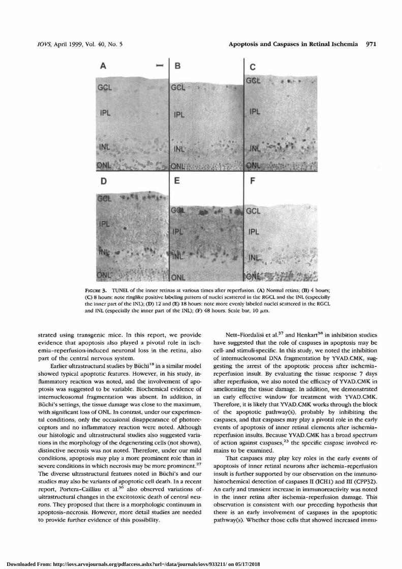
Figure 3 depicts TUNEL of a normal retina (A) and retinasat 4 (B), 8 (C), 12 (D), 18 (E), or 48 (F) hours of reperfusionafter 60 minutes of ischemia. At 8 hours of reperfusion (Fig.3C) there were obvious TUNEL positive nuclei scattered in theRGCL and the INL (especially the inner part of the INL), somewith a ringlike staining pattern. More evenly stained nuclei inthe RGCL and the INL (especially the inner part of the INL)
Apoptosis and Caspases in Retinal Ischemia 969
1 2 3 4 5 6 7
FIGURE 1. Agarose gel electrophoresis of retinal DNA from animals atvarious times of reperfusion after 60 minutes of ischemia. Lane 1: DNAmarkers, multiples of 126 bp; lanes 2, 3, 4, 5, 6, and 7: 0, 4, 8, 12, 18,and 48 hours of reperfusion, respectively (10 jug DNA per lane).
were noted at 12 (Fig. 3D) and 18 hours (Fig. 3E) of reperfu-sion, but no labeled nuclei in the RGCL and INL were noted at48 hours of reperfusion (Fig. 3F). Positively labeled nuclei wereseen only occasionally in the ONL (not shown).
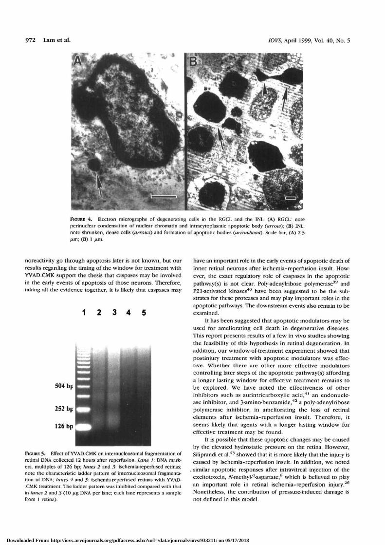
Ultrastructural studies of the sections obtained 18 hoursafter reperfusion showed cells with typical features of apopto-tic cells. Perinuclear condensation of the nuclear chromatinand apoptotic bodies in the RGCL were noted (Fig. 4A). Scat-tered condensed cells were also present in the INL (Fig. 4B).The cytoplasm and the nuclear chromatin were condensed,and the formation of apoptotic bodies was noted.
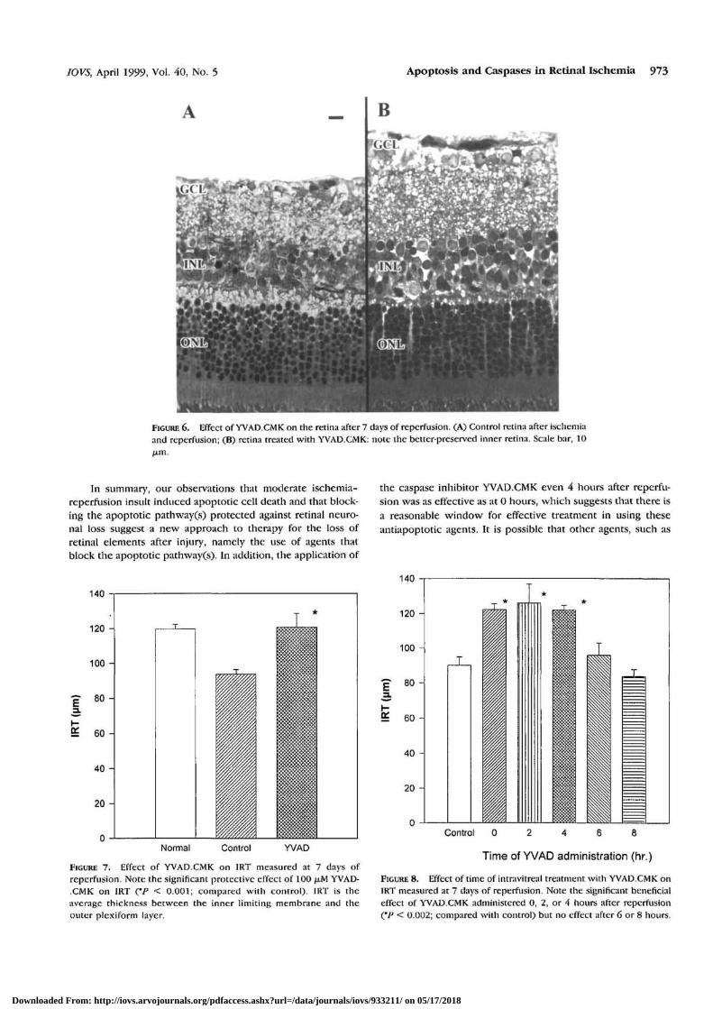
Figure 5 shows the gel analysis of retinal DNA from ani-mals at 12 hours of reperfusion after 60 minutes of ischemia,with or without the caspase inhibitor YVAD.CMK treatment.Without YVAD.CMK, there were typical ladder patterns inretinal DNA gel analysis (lanes 2 and 3) characteristic of apo-ptotic cell death. This ladder pattern was inhibited by treat-ment with 100 yM YVAD.CMK (lanes 4 and 5), similar toresults in our previous report with MK801, which also inhib-ited the formation of the ladder pattern in retinal DNA analysisafter ischemia.26 Figure 6 shows the retinas at 7 days of reper-fusion, with or without YVAD.CMK treatment. The YVAD-.CMK-treated retina (Fig- 6B) shows significantly better pre-served inner retina than does the vehicle-treated control (Fig.6A), This ameliorative effect was confirmed by measuring theIRT (Fig. 7) 7 days after the insult. YVAD.CMK treatment totallyabolished the loss of IRT (Fig. 7; P < 0.001). In addition,YVAD.CMK administered intravitreally from 0 to 4 hours ofreperfusion also abolished the loss of IRT when measured 7days after the insult, whereas YVAD.CMK administered at 6 or8 hours of reperfusion showed no rescuing effect (Fig. 8).
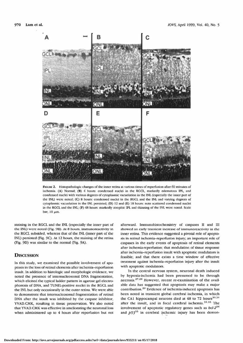
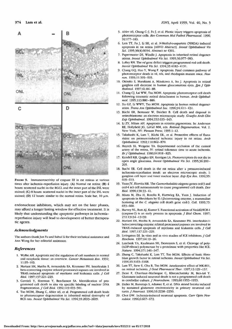
Immunohistochemical detection of caspases II (ICH1) andIII (CPP32) showed similar changes; and therefore, only theresults with anti- caspase III are shown, because the changeswere more apparent. Normal nit retina showed minimum dif-fused background staining in all layers of the retina (Fig. 9A).Four hours after reperfusion, scattered nuclei with positive
Downloaded From: http://iovs.arvojournals.org/pdfaccess.ashx?url=/data/journals/iovs/933211/ on 05/17/2018
970 Lam et al. IOVS, April 1999, Vol. 40, No. 5
FIGURE 2. Histopathologic changes of the inner retina at various times of reperfusion after 60 minutes ofischemia. (A) Normal; (B) 4 hours: condensed nuclei in the RGCL, markedly edematous IPL, andcondensed nuclei with various degrees of cytoplasmic vacuolation in the INL (especially the inner part ofdie INL) were noted; (C) 8 hours: condensed nuclei in the RGCL and the INL and varying degrees ofcytoplasmic vacuolation in the INL persisted; (D) 12 and (E) 18 hours: note scattered condensed nucleiin the RGCL and the INL; (F) 48 hours: markedly atrophic IPL and thinning of the INL were noted. Scalebar, 10 jam.
staining in the RGCL and the INL (especially the inner part ofthe INL) were noted CFig- 9B> At 8 hours, immunoreactivity inthe RGCL subsided, whereas that of the INL (inner part of theINL) persisted (Fig. 9C). At 12 hours, the staining of the retina(Fig. 9D) was similar to the normal (Fig. 9A).
DISCUSSION
In this study, we examined the possible involvement of apo-ptosis in the loss of retinal elements after ischemia-reperftisioninsult. In addition to histologic and morphologic evidence, wenoted the presence of internucleosomal DNA fragmentation,which elicited the typical ladder pattern in agarose gel electro-phoresis of DNA, and TUNEL-positive nuclei in the RGCL andthe INL but only occasionally in the outer retina. We were ableto demonstrate that internucleosomal fragmentation of retinalDNA after the insult was inhibited by the caspase inhibitor,YVAD.CMK, resulting in tissue preservation. We also notedthat YVAD.CMK was effective in ameliorating the neuronal losswhen administered up to 4 hours after reperfusion but not
afterward. Immunohistochemistry of caspases II and IIIshowed an early transient increase of immunoreactivity in theinner retina. This evidence suggested a pivotal role of apopto-sis in retinal ischemia-reperfusion injury; an important role ofcaspases in the early events of apoptosis of retinal elementsafter ischemia-reperfusion; that modulation of tissue responseafter ischemia-reperfusion insult with apoptotic modulators isfeasible; and that there exists a time window of effectivetreatment against ischemia-reperfusion injury after the insultwith apoptotic modulators.
In the central nervous system, neuronal death inducedby hypoxia-ischemia had been presumed to be throughnecrosis.27'28 However, recent re-examination of the avail-able data has suggested that apoptosis may make a majorcontribution.29 Evidence of ischemia-induced apoptosis hasbeen noted in transient global cerebral ischemia, in whichthe CA1 hippocampal neurons died at 48 to 72 hours30'31
after the insult, and in focal cerebral ischemia.32 33 Theinvolvement of apoptotic regulatory genes such as bcl-234
and p5335 in cerebral ischemic injury has been demon-
Downloaded From: http://iovs.arvojournals.org/pdfaccess.ashx?url=/data/journals/iovs/933211/ on 05/17/2018
IOVS, April 1999, Vol. 40, No. 5 Apoptosis and Caspases in Retinal Ischemia 971
INL
ONL
B
GCL
IPL
QNL
G(M
IPL
ONL
# •
IPL
INL
GCL
IPL
INL
FIGURE 3. TUNEL of the inner retinas at various times after reperfusion. (A) Normal retina; (B) 4 hours;(C) 8 hours: note ringlike positive labeling pattern of nuclei scattered in the RGCL and the INL (especiallythe inner part of the INL); (D) 12 and (E) 18 hours: note more evenly labeled nuclei scattered in the RGCLand INL (especially the inner part of the INL); (F) 48 hours. Scale bar, 10 yam.
strated using transgenic mice. In this report, we provideevidence that apoptosis also played a pivotal role in isch-emia-reperfusion-induced neuronal loss in the retina, alsopart of the central nervous system.
Earlier ultrastructural studies by Buchi18 in a similar modelshowed typical apoptotic features. However, in his study, in-flammatory reaction was noted, and the involvement of apo-ptosis was suggested to be variable. Biochemical evidence ofinternucleosomal fragmentation was absent. In addition, inBiichi's settings, the tissue damage was close to the maximum,with significant loss of ONL. In contrast, under our experimen-tal conditions, only the occasional disappearance of photore-ceptors and no inflammatory reaction were noted. Althoughour histologic and ultrastructural studies also suggested varia-tions in the morphology of the degenerating cells (not shown),distinctive necrosis was not noted. Therefore, under our mildconditions, apoptosis may play a more prominent role than insevere conditions in which necrosis may be more prominent.27
The diverse ultrastructural features noted in Biichi's and ourstudies may also be variants of apoptotic cell death. In a recentreport, Portera-Cailliau et al.36 also observed variations ofultrastructural changes in the excitotoxic death of central neu-rons. They proposed that there is a morphologic continuum inapoptosis-necrosis. However, more detail studies are neededto provide further evidence of this possibility.
Nett-Fiordalisi et al.37 and Henkart38 in inhibition studieshave suggested that the role of caspases in apoptosis may becell- and stimuli-specific. In this study, we noted the inhibitionof internucleosomal DNA fragmentation by YVAD.CMK, sug-gesting the arrest of the apoptotic process after ischemia-reperfusion insult. By evaluating the tissue response 7 daysafter reperfusion, we also noted the efficacy of YVAD.CMK inameliorating the tissue damage. In addition, we demonstratedan early effective window for treatment with YVAD.CMK.Therefore, it is likely that YVAD.CMK works through the blockof the apoptotic pathway(s), probably by inhibiting thecaspases, and that caspases may play a pivotal role in the earlyevents of apoptosis of inner retinal elements after ischemia-reperfusion insults. Because YVAD.CMK has a broad spectrumof action against caspases,23 the specific caspase involved re-mains to be examined.
That caspases may play key roles in the early events ofapoptosis of inner retinal neurons after ischemia-reperfusioninsult is further supported by our observation on the immuno-histochemical detection of caspases II (ICH1) and III (CPP32).An early and transient increase in immunoreactivity was notedin the inner retina after ischemia-reperfusion damage. Thisobservation is consistent with our preceding hypothesis thatthere is an early involvement of caspases in the apoptoticpathway(s). Whether those cells that showed increased immu-
Downloaded From: http://iovs.arvojournals.org/pdfaccess.ashx?url=/data/journals/iovs/933211/ on 05/17/2018
972 Lam et IOVS, April 1999, Vol. 40, No. 5
FIGURE 4. Electron micrographs of degenerating cells in the RGCL and the INL, (A) RGCL: noteperinuclear condensation of nuclear chromatin and intracytoplasmic apoptotic body (arrow); (B) INL:note shrunken, dense cells (arrows) and formation of apoptotic bodies (arrowhead). Scale bar, (A) 2.5ju,m; (B) 1 jam.
noreactivity go through apoptosis later is not known, but ourresults regarding the timing of the window for treatment withYVAD.CMK support the thesis that caspases may be involvedin the early events of apoptosis of those neurons. Therefore,taking all the evidence together, it is likely that caspases may
1 2 3 4 5
FIGURE 5- Effect of YVAD.CMK on intemucleosomal fragmentation ofretinal DNA collected 12 hours after reperfusion. Lane /: DNA mark-ers, multiples of 126 bp; lanes 2 and 3- ischemia-reperfused retinas;note the characteristic ladder pattern of intemucleosomal fragmenta-tion of DNA; lanes 4 and 5: ischemia-reperfused retinas with YVAD-.CMK treatment. The ladder pattern was inhibited compared with thatin lanes 2 and 3 (10 jug DNA per lane; each lane represents a samplefrom 1 retina).
have an important role in the early events of apoptotic death ofinner retinal neurons after ischemia-reperfusion insult. How-ever, the exact regulatory role of caspases in the apoptoticpathway(s) is not clear. Poly-adenylribose polymerase39 andP21-activated kinases40 have been suggested to be the sub-strates for these proteases and may play important roles in theapoptotic pathways. The downstream events also remain to beexamined.
It has been suggested that apoptotic modulators may beused for ameliorating cell death in degenerative diseases.This report presents results of a few in vivo studies showingthe feasibility of this hypothesis in retinal degeneration. Inaddition, our window-of-treatment experiment showed thatpostinjury treatment with apoptotic modulators was effec-tive. Whether there are other more effective modulatorscontrolling later steps of the apoptotic pathway(s) affordinga longer lasting window for effective treatment remains tobe explored. We have noted the effectiveness of otherinhibitors such as aurintricarboxylic acid,4' an endonucle-ase inhibitor, and 3-amino-benzamide,42 a poly-adenylribosepolymerase inhibitor, in ameliorating the loss of retinalelements after ischemia-reperfusion insult. Therefore, itseems likely that agents with a longer lasting window foreffective treatment may be found.
It is possible that these apoptotic changes may be causedby the elevated hydrostatic pressure on the retina. However,Siliprandi et al.43 showed that it is more likely that the injury iscaused by ischemia-reperfusion insult. In addition, we noted
. similar apoptotic responses after intravitreal injection of theexcitotoxcin, Ar-methyl-tl-aspartate,6 which is believed to playan important role in retinal ischemia-reperfusion injury.26
Nonetheless, the contribution of pressure-induced damage isnot defined in this model.
Downloaded From: http://iovs.arvojournals.org/pdfaccess.ashx?url=/data/journals/iovs/933211/ on 05/17/2018
JOVS, April 1999, Vol. 40, No. 5 Apoptosis and Caspases in Retinal Ischemia 973
FIGURE 6. Effect of YVAD.CMK on the retina after 7 days of reperfusion. (A) Control retina after ischemiaand reperfusion; (B) retina treated with YVAD.CMK: note the better-preserved inner retina. Scale bar, 10
In summary, our observations that moderate ischemia-reperfusion insult induced apoptotic cell death and that block-ing the apoptotic pathway(s) protected against retinal neuro-nal loss suggest a new approach to therapy for the loss ofretinal elements after injury, namely the use of agents thatblock the apoptotic pathway(s). In addition, the application of
Normal Control WAD
FIGURE 7. Effect of YVAD.CMK on IRT measured at 7 days ofreperfusion. Note the significant protective effect of 100 p,M YVAD-.CMK on IRT (*P < 0.001; compared with control). IRT is theaverage thickness between the inner limiting membrane and theouter plexiform layer.
the caspase inhibitor YVAD.CMK even 4 hours after reperfu-sion was as effective as at 0 hours, which suggests that there isa reasonable window for effective treatment in using theseantiapoptotic agents. It is possible that other agents, such as
140
120 -
100
80 -
9= 6 0 -
40 -
20 -
Control 0 2 4 6 8
Time of YVAD administration (hr.)
FIGURE 8. Effect of time of intravitreal treatment with YVAD.CMK onIRT measured at 7 days of reperfusion. Note the significant beneficialeffect of YVAD.CMK administered 0, 2, or 4 hours after reperfusion(*P < 0.002; compared with control) but no effect after 6 or 8 hours.
Downloaded From: http://iovs.arvojournals.org/pdfaccess.ashx?url=/data/journals/iovs/933211/ on 05/17/2018
974 Lam et al.
A B
ONL ;"';' ; *
C
INL "!*
ONL
D
ONL
GCL
INL
ONL
FIGURE 9- Immunoreactivity of caspase III in rat retinas at varioustimes after ischemia-reperfusion injury-. (A) Normal rat retina; (B) 4hours: scattered nuclei in the RGCL and the inner part of the INL werestained; (C) 8 hours: scattered nuclei in the inner part of the INL werestained; (D) 12 hours: similar to the normal retina. Scale bar, 10 fxm.
endonuclease inhibitors, which may act on the later phases,may afford a longer lasting window for effective treatment. It islikely that understanding the apoptotic pathways in ischemia-reperfiision injury will lead to development of better therapeu-tic agents.
Acknowledgments
The authors thank Jun Fu and Suhui Li for their technical assistance andAnn Wong for her editorial assistance.
References1. Wyllie AH. Apoptosis and the regulation of cell numbers in normal
and neoplastic tissue: an overview. Cancer Metastasis Rev. 1992;11:95-103.
2. Mariani SM, Matiba B, Armandola EA, Krammer PH. Interleukin Ibeta-converting enzyme related proteases/caspases are involved inTRAIL-induced apoptosis of myeloma and leukemia cells. J CellBiol. 1997;137:221-229.
3. Gavrieli Y, Sherman Y, Ben-Sasson SA. Identification of pro-grammed cell death in situ via specific labeling of nuclear DNAfragmentation./ Cell Biol. 1992;119:493-501.
4. Tso MOM, Zhang C, Abler AS, et al. Programmed cell death leadsto photoreceptor degeneration in inherited retinal dystrophy ofRCS rats. Invest Ophthalmol Vis Sci. 1994;35:2693-2699-
/OVS, April 1999, Vol. 40, No. 5
5. Abler AS, Chang C-J, Fu J, et al. Photic injury triggers apoptosis ofphotoreceptor cells. Res Commun Mol Pathol Pharmacol. 1996;92:177-183.
6. Lam TT, Fu J, Li SH, et al. iV-Methyl-D-aspartate (NMDA) inducedapoptosis in rat retina [ARVO Abstract]. Invest Ophthalmol VisSet. 1995;36(4):S934. Abstract nr 4301.
7. Papermaster DS, Windle J. Apoptosis in inherited retinal degener-ations. Invest Ophthalmol Vis Sci. 1995:36:977-983.
8. Lolley RN. The rd gene defect triggers programmed rod cell death.Invest Ophthalmol Vis Sci. 1994;35:4l82-4l91.
9. Chang GQ, Hao Y, Wong F. Apoptosis. Final common pathway ofphotoreceptor death in rd, rds, and rhodopsin mutant mice. Neu-ron. 1993; 11:595-605.
10. Okisaka S, Murakami A, Mizukawa A, Ito J. Apoptosis in retinalgarglion cell decrease in human glaucomatous eyes. Jpn J Oph-thalmol. 1997;4l:84-88.
11. Chang CJ, Lai WW, Tso MOM. Apoptotic photoreceptor cell deathfollowing traumatic retinal detachment in human. Arch Ophthal-mol. 1995;113:880-886.
12. Xu GZ, Li WWY, Tso MOM, Apoptosis in human retinal degener-ation. Trans Am Ophthalmol Soc. 1996;94:4l 1-431.
13. Biichi ER, Bernauer W, Daicker B. Cell death and disposal inretinoblastoma: an electron microscopic study. Graefes Arch ClinExp Ophthalmol. 1994;232:635-645.
14. Li ZY, Milam AH. Apoptosis in retinitis pigmentosa. In: AndersonRE, Hollyneld JG, LaVail MM, eds. Retinal Degeneration. Vol. 2.New York, NY: Plenum Press; 1995:1-12.
15. Takahashi K, Lam T, Biichi ER., et al. Protective effects of fluna-rizine on pressure-induced ischemic injury in rat retina. ArchOphthalmol. 1992; 110:862-870.
16. Hayreh SS, Weigeist TA. Experimental occlusion of the centralarter}' of the retina, IV: retinal tolerance time to acute ischemia.Br j Ophthalmol. 1980;64:818-825.
17. Kendell KR, Quigley EN, Kerrigan LA. Photoreceptors do not die inopen angle glaucoma. Invest Ophthalmol Vis Sci. 1995;36:200-205.
18. Buchi ER. Cell death in the rat retina after a pressure-inducedischaemia-reperfusion insult: an electron microscopic study, 1:ganglion cell layer and inner nuclear layer. Exp Eye Res. 1992;55:605-613.
19. Yuan JY, Horvitz HR. The Caenorhabditis elegans genes ced-3 andced-4 act cell autonomously to cause programmed cell death. DevBiol. 1990:138:33-41.
20. Miura M, Zhu H, Rotello R, Hartwieg EA, Yuan J. Induction ofapoptosis in fibroblasts by IL-ljS-converting enzyme, a mammalianhomolog of the C. elegans cell death gene ced-3. Cell. 1993;75:653-660.
21. Harvey NL, Butt AJ, Kumar S. Functional activation of Nedd2/ICH-1(caspase-2) is an early process in apoptosis. / Biol Chem. 1997;272:13134-13139.
22. Mariani SM, Matiba B, Armandola EA, Krammer PH. Interleukin-1-beta-converting enzyme related proteases/caspases are involved inTRAIL-induced apoptosis of myeloma and leukemia cells. / Celtmot. 1997; 137:221-229-
23. Livingston DJ. In vitro and in vivo studies of ICE inhibitors. / CellBiochem. 1997;64:19-26.
24. Lazebnik YA, Kaufmann SH, Desnoyers S, et al. Cleavage of poly-(ADP-ribose) polymerase by a proteinase with properties like ICE.Nature. 1994;371:346-347.
25. Zhang C, Takahashi K, Lam TT, Tso MOM. Effects of basic fibro-blast growth factor in retinal ischemia. Invest Ophthalmol Vis Sci.1994;35:3l63-3l68.
26. Lam TT, Siew E, Chu R, Tso MOM. Ameliorative effect of MK-801,on retinal ischemia. / Ocul Pharmacol Ther. 1997;13:129-137.
27. Dessi F, Charriaut-Marlangue C, Khrestchatisky M, Ben-Ari Y.Glutamate-induced neuronal death is not a programmed cell deathin cerebellar culture. J Neurocbem. 1993;60:1953-1955.
28. Didier M, Bursztajn S, Adamec E, et al. DNA strand breaks inducedby sustained glutamate excitotoxicity in primary neuronal cul-tures. / Neurosci. 1996; 16:2238 -2250.
29. Choi DW. Ischemia-induced neuronal apoptosis. Curr Opin Neu-robiol. 1996;6:667-672.
Downloaded From: http://iovs.arvojournals.org/pdfaccess.ashx?url=/data/journals/iovs/933211/ on 05/17/2018
IOVS, April 1999, Vol. 40, No. 5 Apoptosis and Caspases in Retinal Ischemia 975
30. Heron A, Pollard H, Dessi F, et al. Regional variability in DNAfragmentation after global ischemia evidenced by combined histo-logical and gel electrophoresis observations in the rat brain. JNeu-rochem. 1993;6l:1973-1976.
31. Nitatori T, Sato N, Waguri S, et al. Delayed neuronal death inthe CA1 pyramidal cell layer of the gerbil hippocampus follow-ing transient ischemia is apoptosis. / Neurosci. 1995;15:1OO1-1011.
32. Linnik MD, Zobrist RH, Hatfield MD. Evidence supporting a rolefor programmed cell death in focal cerebral ischemia in rats.Stroke. 1993;24:2002-2009.
33. Li Y, Sharov VG, Jiang N, et al. Ultrastructural and light micro-scopic evidence of apoptosis after middle cerebral artery occlu-sion in the rat. AmJPatbol. 1995;l46:1045-1051.
34. Martinou J-C, Dubois-Duaphin M, Staple JK, et al. Overexpressionof BCL-2 in transgenic mice protects neurons from naturally oc-curring cell death and experimental ischemia. Neuron. 1994; 13:1017-1030.
35. Crumrine RC, Thomas AL, Morgan PF. Attenuation of p53 expres-sion protects against focal ischemic damage in transgenic mice./ Cereb Blood Flow Metab. 1994;l4:887-891.
36. Portera-Cailliau C, Price DL, Martin LJ. Non-NMDA andNMDA receptor-mediated excitotoxic neuronal deaths in adultbrain are morphologically distinct: further evidence for an
apoptosis-necrosis continuum./ Comp Neurol. 1997;378:88-104.
37. Nett-Fiordalisi M, Tomaselli K, Russell JH, Chaplin DD. Macro-phage apoptosis in the absence of active interleukin-1 beta-con-verting enzyme. / Leukoc Biol. 1995;58:717-24.
38. Henkart PA. ICE family proteases: mediators of all apoptotic celldeath? Immunity. 1996;4:195-201.
39- Mariani SM, Matiba B, Armandola EA, Krammer PH. Interleukin-l-beta-converting enzyme related proteases/caspases are involved inTRAIL-induced apoptosis of myeloma and leukemia cells. / CellBiol. 1997; 137:221-229.
40. Rudel T, Bokoch GM. Membrane and morphological changes inapoptotic cells regulated by caspase-mediated activation of PAK2.Science. 1991\2l6:\yj\-\5l4.
41. Lam TT, Fu J, Hrynewycz M, Tso MOM. The effect of aurintricar-boxylic acid, an endonuclease inhibitor, on ischemia/reperfusiondamage in rat retina./ OCM/ Pharmacol Ther. 1995;ll:253-260.
42. Lam TT. The effect of 3-aminobenzamide, a poly(ADP-ribose) poly-merase inhibitor, on ischemia/reperfusion damage in rat retina.Res Commun Mol Patbol Pharmacol. 1997;95:24l-252.
43. Siliprandi R, Canella R, Carmignoto G. Nerve growth factor pro-motes functional recovery of retinal ganglion cells after ischemia.Invest Ophthalmol Vis Sci. 1993;34:3232-3245.
Downloaded From: http://iovs.arvojournals.org/pdfaccess.ashx?url=/data/journals/iovs/933211/ on 05/17/2018