aptamer-functionalized gold nanoparticles as photoresponsive nanoplatform for co-drug delivery
TRANSCRIPT

Aptamer-Functionalized Gold Nanoparticles As PhotoresponsiveNanoplatform for Co-Drug DeliveryYi-Syun Shiao, Huai-Hsuan Chiu, Pin-Hsuan Wu, and Yu-Fen Huang*
Department of Biomedical Engineering and Environmental Sciences, National Tsing Hua University, Hsinchu, Taiwan ROC
*S Supporting Information
ABSTRACT: Various platforms have been developed asinnovative nanocarriers to deliver therapeutic agents to thediseased sites. Multifunctional surface modification allows anenhanced recognition and uptake of drug carriers by targetedcells. However, the development of drug resistance in sometumor cells plays a major role in the failure of chemotherapy.Drugs given in combination, called multidrug delivery approach,was designed to improve the therapeutic efficacy and has becomean increasingly used strategy that is of great importance in clinicalcancer treatments. In this study, aptamer-functionalized gold nanoparticles (Au NPs) have been used as a nanoplatform tocodeliver two different anticancer drugs for improving the drug effectiveness. The surface of Au NPs (13 nm in diameter) wasassembled with AS1411 aptamers, which tethered with 21-base pairs of (CGATCGA)3 sequence approached to the Au NPs.Both the photosensitizer 5,10,15,20-tetrakis(1-methylpyridinium-4-yl) porphyrin (TMPyP4) and the chemotherapeutic drugdoxorubicin (Dox) were then physically attached to the AS1411-conjugated Au NPs (T/D:ds-NPs) and delivered to the targettumor cells such as HeLa and Dox-resistant MCF-7R cell lines. When exposed to a 632 nm light, reactive oxygen species inducedby TMPyP4 molecules were generated inside the living cells, followed by cell damage. In addition, triggered release of thecomplementary drugs also occurred simultaneously during the photodynamic reaction. In the presence of Dox molecules, thetoxicity toward the target cells was superior to individual drug treatment. Overall, a co-drug delivery platform was successfullyestablished to improve the therapeutic efficacy in tumor cells. The improvement of the photodynamic-stimulated triggeredrelease was enhanced, thus highly promising precise drug release in targeted drug delivery.
KEYWORDS: aptamer, gold nanoparticle, photodynamic therapy, chemotherapy, multidrug resistant
■ INTRODUCTION
Photodynamic therapy (PDT) has emerged as a popular,noninvasive treatment for numerous cancers such as inoperableesophageal tumors and malignancies of the head and neck.1−3 Italso gained worldwide popularity in adjunctive treatments forother cancer types, including breast, prostate, and ovarian inpreclinical and clinical trials.4−6 The basic principle of PDTinvolves the administration of photosensitizing agents followedby local illumination of the target tissue with a light source. Asufficient amount of molecular oxygen (normally present in thetumor) is also required to achieve effective treatment. Thecollision of an oxygen molecule with the photoexcited sensitizerresults in the formation of 1O2 and other reactive oxygenspecies (ROS) that cause oxidative damage to cellularcomponents. It may also destroy the tumor vasculature, therebydepriving the tumor of oxygen and nutrients and ultimately,leading to tumor cell death.Most photosensitizers used in PDT are water-insoluble.7,8
They exhibit high level of dark toxicity, rapid degradability, andconsequent inactivity under irradiation, therefore restrictingtheir clinical applications. To address these issues, numerousdrug formulations have been extensively studied as potentialPDT agents. The use of nanoparticles as delivery vehicles islikely one of the most promising strategy in cancer
research.9−13 A wide range of biocompatible, nontoxicnanoparticles such as liposomes and polymer-, silica-,magnetic-, gold-, and carbon-based NPs have been developedto improve the efficacy of PDT.14−29 Nanoparticles localize tothe tumor via the enhanced permeability and retention (EPR)effect or active targeting over free drugs. The achievement ofhigh drug accumulation into a tumor allows effective reductionof side effects associated with PDT treatment. Furthermore,nanoparticles can protect photosensitizers from leakage,degradation, or modification in the biological environmentprior to delivery of the drug to the infected tissues.With the recent interests in multimodality, nanoparticles that
can incorporate different functionalities are attractive candi-dates for advanced medical applications. For example, PDTbased on upconversion nanoparticles (UCNPs)30−33 has beendeveloped as a new approach to deliver near-infrared light intodeeper tissues for PDT treatment. UCNPs, which offer uniqueupconversion luminescence, are beneficial for remarkably
Special Issue: Materials for Theranostics
Received: April 29, 2014Accepted: June 11, 2014
Forum Article
www.acsami.org
© XXXX American Chemical Society A dx.doi.org/10.1021/am5026243 | ACS Appl. Mater. Interfaces XXXX, XXX, XXX−XXX
sensitive in vivo biomedical imaging and cell tracking. Severalother image-guided involvements of PDT deliveries, e.g.,magnetic resonance (MR) imaging have also been presentedby different groups in recent years.22,34−36 The combination ofboth therapeutic and diagnostic capabilities in one dose makesthese nanoparticles ideal platforms for theranostics. In addition,the simultaneous delivery of anticancer drugs and photo-sensitizers to the tumor cells is expected to overcome thehurdles of traditional single-drug treatments.37−40 Nano-particle-mediated combination of chemotherapy and PDT hasbeen investigated to overcome drug resistance in mouse tumormodel by invoking multiple anticancer mechanisms.41 Asignificant enhancement of ROS production and cytotoxicityusing nanoparticles contributes to the greatly improved efficacyof combination treatment.Despite the above-mentioned advantages, photodynamic
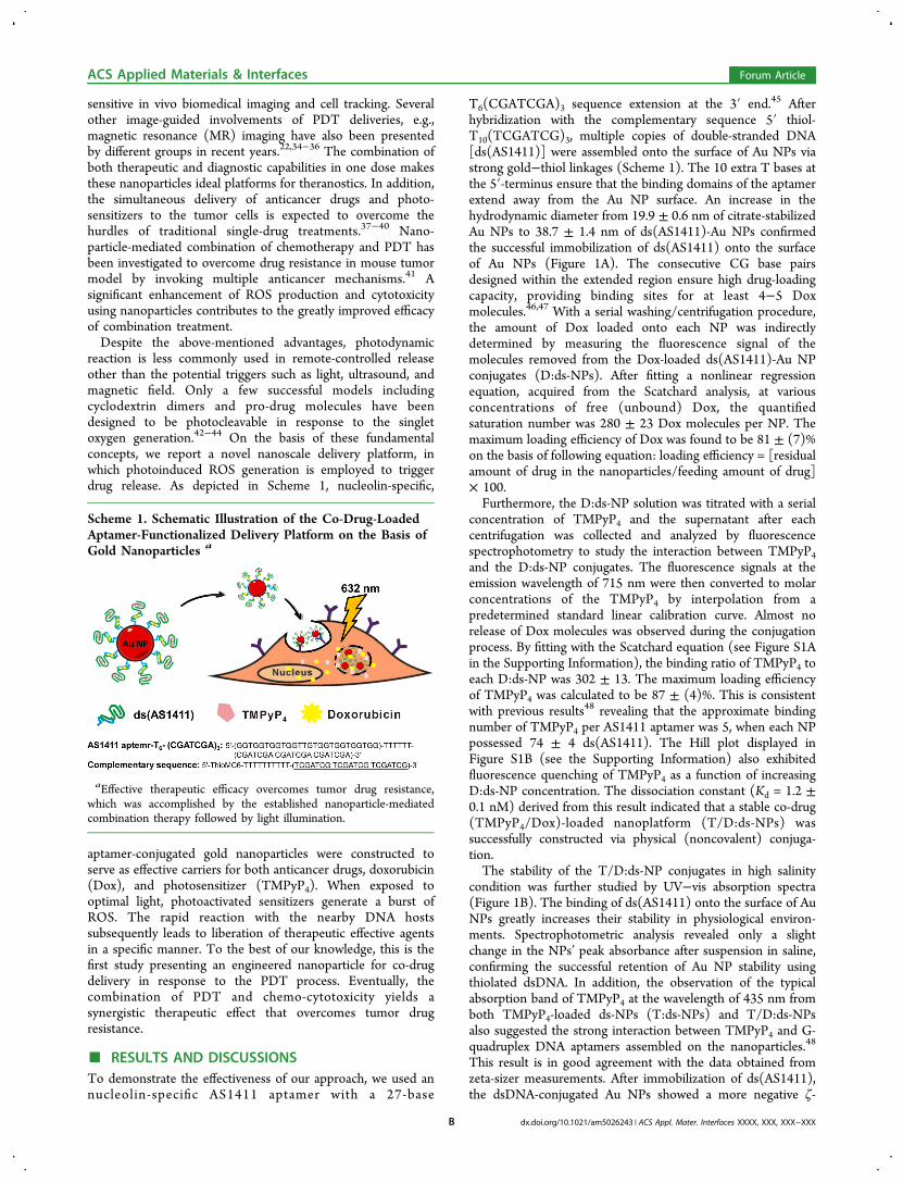
reaction is less commonly used in remote-controlled releaseother than the potential triggers such as light, ultrasound, andmagnetic field. Only a few successful models includingcyclodextrin dimers and pro-drug molecules have beendesigned to be photocleavable in response to the singletoxygen generation.42−44 On the basis of these fundamentalconcepts, we report a novel nanoscale delivery platform, inwhich photoinduced ROS generation is employed to triggerdrug release. As depicted in Scheme 1, nucleolin-specific,
aptamer-conjugated gold nanoparticles were constructed toserve as effective carriers for both anticancer drugs, doxorubicin(Dox), and photosensitizer (TMPyP4). When exposed tooptimal light, photoactivated sensitizers generate a burst ofROS. The rapid reaction with the nearby DNA hostssubsequently leads to liberation of therapeutic effective agentsin a specific manner. To the best of our knowledge, this is thefirst study presenting an engineered nanoparticle for co-drugdelivery in response to the PDT process. Eventually, thecombination of PDT and chemo-cytotoxicity yields asynergistic therapeutic effect that overcomes tumor drugresistance.
■ RESULTS AND DISCUSSIONSTo demonstrate the effectiveness of our approach, we used annucleolin-specific AS1411 aptamer with a 27-base
T6(CGATCGA)3 sequence extension at the 3′ end.45 Afterhybridization with the complementary sequence 5′ thiol-T10(TCGATCG)3, multiple copies of double-stranded DNA[ds(AS1411)] were assembled onto the surface of Au NPs viastrong gold−thiol linkages (Scheme 1). The 10 extra T bases atthe 5′-terminus ensure that the binding domains of the aptamerextend away from the Au NP surface. An increase in thehydrodynamic diameter from 19.9 ± 0.6 nm of citrate-stabilizedAu NPs to 38.7 ± 1.4 nm of ds(AS1411)-Au NPs confirmedthe successful immobilization of ds(AS1411) onto the surfaceof Au NPs (Figure 1A). The consecutive CG base pairsdesigned within the extended region ensure high drug-loadingcapacity, providing binding sites for at least 4−5 Doxmolecules.46,47 With a serial washing/centrifugation procedure,the amount of Dox loaded onto each NP was indirectlydetermined by measuring the fluorescence signal of themolecules removed from the Dox-loaded ds(AS1411)-Au NPconjugates (D:ds-NPs). After fitting a nonlinear regressionequation, acquired from the Scatchard analysis, at variousconcentrations of free (unbound) Dox, the quantifiedsaturation number was 280 ± 23 Dox molecules per NP. Themaximum loading efficiency of Dox was found to be 81 ± (7)%on the basis of following equation: loading efficiency = [residualamount of drug in the nanoparticles/feeding amount of drug]× 100.Furthermore, the D:ds-NP solution was titrated with a serial
concentration of TMPyP4 and the supernatant after eachcentrifugation was collected and analyzed by fluorescencespectrophotometry to study the interaction between TMPyP4and the D:ds-NP conjugates. The fluorescence signals at theemission wavelength of 715 nm were then converted to molarconcentrations of the TMPyP4 by interpolation from apredetermined standard linear calibration curve. Almost norelease of Dox molecules was observed during the conjugationprocess. By fitting with the Scatchard equation (see Figure S1Ain the Supporting Information), the binding ratio of TMPyP4 toeach D:ds-NP was 302 ± 13. The maximum loading efficiencyof TMPyP4 was calculated to be 87 ± (4)%. This is consistentwith previous results48 revealing that the approximate bindingnumber of TMPyP4 per AS1411 aptamer was 5, when each NPpossessed 74 ± 4 ds(AS1411). The Hill plot displayed inFigure S1B (see the Supporting Information) also exhibitedfluorescence quenching of TMPyP4 as a function of increasingD:ds-NP concentration. The dissociation constant (Kd = 1.2 ±0.1 nM) derived from this result indicated that a stable co-drug(TMPyP4/Dox)-loaded nanoplatform (T/D:ds-NPs) wassuccessfully constructed via physical (noncovalent) conjuga-tion.The stability of the T/D:ds-NP conjugates in high salinity
condition was further studied by UV−vis absorption spectra(Figure 1B). The binding of ds(AS1411) onto the surface of AuNPs greatly increases their stability in physiological environ-ments. Spectrophotometric analysis revealed only a slightchange in the NPs’ peak absorbance after suspension in saline,confirming the successful retention of Au NP stability usingthiolated dsDNA. In addition, the observation of the typicalabsorption band of TMPyP4 at the wavelength of 435 nm fromboth TMPyP4-loaded ds-NPs (T:ds-NPs) and T/D:ds-NPsalso suggested the strong interaction between TMPyP4 and G-quadruplex DNA aptamers assembled on the nanoparticles.48
This result is in good agreement with the data obtained fromzeta-sizer measurements. After immobilization of ds(AS1411),the dsDNA-conjugated Au NPs showed a more negative ζ-
Scheme 1. Schematic Illustration of the Co-Drug-LoadedAptamer-Functionalized Delivery Platform on the Basis ofGold Nanoparticles a
aEffective therapeutic efficacy overcomes tumor drug resistance,which was accomplished by the established nanoparticle-mediatedcombination therapy followed by light illumination.
ACS Applied Materials & Interfaces Forum Article
dx.doi.org/10.1021/am5026243 | ACS Appl. Mater. Interfaces XXXX, XXX, XXX−XXXB

potential (−44.9 ± 2.8) than the citrate-stabilized Au NPs(−36.0 ± 2.7). Moreover, the subsequent loading of drugmolecules (both Dox and TMPyP4) lead to a slight decline inthe negative intensity of ζ-potential (−39.4 ± 1.6 mV), whereasthe size of the conjugates increased slightly to 40.1 ± 1.4 nm.These results suggested that dsDNA and drug molecules weresequentially introduced onto the surface of Au NPs.After confirming the physiochemical properties of the drug-
loaded ds-NPs, the interaction between the aptamer-function-alized nanocarrier toward cancer cells was investigated usingdark-field microscopy coupled to a CCD digital camera. HeLacells, which overexpress nucleolin on the cell surface,49 wereincubated with T/D:ds-NPs for 4 h, followed by repeatedwashing steps to remove excess conjugates from the cells. Asshown in Figure S2A (see the Supporting Information), theyellow color representing the scattering light from Au NPs wasobserved in/on the cells, while the untreated control cellsappeared dim white because of the intrinsic cellular scattering.To further confirm the intracellular uptake of aptamer-basednanocomplex by HeLa cells, we used trypsin to remove surface-bound particles. The number of ds(AS1411)-NPs taken up by
Hela cells was then evaluated via atomic absorptionspectrometry (AAS; AAnalyst 600; PerkinElmer, Waltham,MA, USA). Our results showed that the maximum number ofds(AS1411)-NPs inside HeLa cells was 1.5 ± 0.3 × 10⁵ percells, whereas <6.1 ± 0.9 × 10⁴ Au NPs were determined fornonspecific intracellular uptake using dsDNA-Au NPs lackingAS1411 [denoted as ds(ctrl)-NPs]. To evaluate the function ofds(AS1411) in the cellular uptake of the aptamer-conjugatedcomplex, a simple competition assay was conducted. HeLa cellswere first incubated with ds(AS1411)-NPs at 4 °C for 30 minand then labeled with a Cy5-modified AS1411 (Cy5-apt).Fluorescence intensity was ultimately determined by flowcytometry. Compared with the control ds(ctrl)-NPs, thefluorescence signal from ds(AS1411)-NP-treated cells was0.33-fold less intense (see Figure S2B in the SupportingInformation). Considering the specific recognition of AS1411to its target protein, this results suggests that the interactionbetween ds(AS1411)-NPs toward HeLa cells was through thenucleolin-binding and internalization pathway.To investigate the photo-, chemo-, and combined therapeutic
efficacy of these drug-loaded ds(AS1411)-NP conjugates, HeLa
Figure 1. (A) Hydrodynamic size distribution and (B) UV−vis spectra of Au NPs (in 4 mM citrate buffer), ds-NPs, T:ds-NPs, D:ds-NPs, and T/D:ds-NPs (1 × ). Buffer: 10 mM phosphate buffer, 100 mM NaCl, pH 7.4. The concentration of the as-prepared Au NP is denoted as 1×, whichcorresponds to the concentration of approximately 13 nM. Inset: TEM images of the as-prepared T/D:ds-NPs (scale: 25 nm).
Figure 2. (A) Dependence of cytotoxicity of treated HeLa cells as a function of exposure time (632 nm red light). Cells were incubated with T:ds-NPs and T/D:dsNPs (4×) in washing buffer (1% BSA) at 37 °C for 4 h. After treatments, cells were subsequently grown in fresh culture medium(10% FBS) for 24 h. Cytotoxicity was measured with the MTT assay. (B) Cell viability of HeLa cells under different treatments in washing buffer(1% BSA) at 37 °C for 4 h, followed by red light exposure (632 nm, 20 min).
ACS Applied Materials & Interfaces Forum Article
dx.doi.org/10.1021/am5026243 | ACS Appl. Mater. Interfaces XXXX, XXX, XXX−XXXC

cells were incubated with T:ds-NPs and T/D:ds-NPs,respectively. After removal of the excess reagents, cells wereirradiated with a red light (632 nm, 15 mW/cm2) generatedfrom a halogen lamp after passing through a bandpass filterwith a center wavelength of 632 nm for serial time periods.Fresh medium (10% FBS) was added for further cell growth(24 h). The relative viability of cells with different treatmentswas then determined by performing the 3-(4,5-dimethylthiazol-2-yl)-5-(3-carboxymethoxyphenyl)-2-(4-sulfophenyl)-2H-tetra-zolium) (MTT) assay. As depicted in Figure 2A, no or littlecytotoxicity was observed for T:ds-NPs-treated cells before
light exposure. Viability began to decline to approximately 82.4± 7.0% after 10 min irradiation, and decreased to around 51.0± 11.0% after 40 min light illumination because of theexcitation of the photosensitizer, leading to the photodynamickilling of cancer cells. The cell killing activity of co-drugdelivery using T/D:ds-NPs was further assessed in HeLa cells.A dramatic increase in cell death was observed depending onthe light exposure time. Approximately 74.0 ± 4.2% of cellswere dead when T/D:ds-NPs attached HeLa cells wereirradiated with red light for 10 min. The superior cytotoxicityof T/D:ds-NPs compared with that of T:ds-NPs for all
Figure 3. (A) Flow cytometry analysis of the production of reactive oxygen species (ROS) in (a) nontreated HeLa cells, and cells treated with 4×(b) ds-NPs, (c) D:ds-NPs, (d) T:ds-NPs, (e) T/D:ds-NPs, and (f) H2O2 (300 μM, 2h). After incubation in culture medium (10% FBS) at 37 °Cand 5% CO2 for 4 h, DCFH-DA (50 μM, 40 min) was added to each cell suspension, followed by red light exposure (632 nm, 50 min). (B)Increased intracellular ROS and Dox signals (ΔF) induced by H2O2. Nontreated and treated cells under different conditions were exposed to 300μM H2O2 for 2 h. DCFH-DA was used to quantify the intracellular ROS production.
Figure 4. Fluorescence images of HeLa cells incubated with 2× (A) T/D:ds-NPs and (B) D:ds-NPs. After incubation in washing buffer (1% BSA) at37 °C for 1 h, cells were rinsed with DPBS, followed by red light irradiation (632 nm, 30 min). For microscopic analysis, cells were fixed with 4%paraformaldehyde solution for 5 min and stained with 4,6-diamidino-2-phenylindole (DAPI,1.0 μM) for 5 min. The fluorescence images weremonitored for (a) nucleus (DAPI), (b) Dox, and (c) overlay of these two channels. TMPyP4 does not exhibit detectable fluorescence signals in thesechannels. The scale length of each image is 20 μm.
ACS Applied Materials & Interfaces Forum Article
dx.doi.org/10.1021/am5026243 | ACS Appl. Mater. Interfaces XXXX, XXX, XXX−XXXD

irradiation periods suggested that a synergistic effect wasinvolved in the cellular death.To further confirm the combined therapeutic efficacy, cells
were incubated with serial concentrations of D:ds-NPs, T:ds-NPs, and T/D:ds-NPs (Figure 2B). Compared with the cellswithout drug and light treatment, the increase of T:ds-NPsconcentration lead to a higher cell death following a red lightexposure. The cell viability of cells incubated with 4× T:ds-NPswas 64.4 ± 5.2% after 20 min irradiation. The control cells,which were incubated with ds-Au NPs, showed no cellulardamage under the same illumination condition (data notshown). In addition, the dose-dependent survival curve forD:ds-NPs-treated cells was also examined. Dox-loaded ds-NPsexhibited less potent antiproliferative properties than T/D:ds-NPs at equal Dox concentration. Only 17.1 ± 2.9% of HeLacells were killed by D:ds-NPs (4×) after light exposure for 20min. However, an obvious loss of cell viability (21.4 ± 4.3%)had been observed for T/D:ds-NP conjugates under the sameexperimental condition. Collectively, the therapeutic efficacy ofT/D:ds-NPs significantly improved to 2.5- and 4.6-foldcompared to PDT and chemotherapy alone, indicating asynergistic enhancement of cancer therapy via the combinationof photo- and chemocytotoxic reactions. It should also be notedthat red light exposure showed no harm to cells withoutphotosensitizers. The cell viability of (D:ds-NPs)-treated HeLacells was light-independent with irradiation less than 1 h (datanot shown).To account for the different cytotoxicity of these drug
nanocarriers, the intracellular ROS formation was studied using2′,7′-dichlorodihydrofluorescein diacetate (DCFH-DA) as aprobe during the combined and single treatment.50 The cells,which were incubated with different drug conjugates, wereirradiated to allow the production of significant amounts ofROS by the photosensitizer. Our experimental results (Figure3A) clearly show that the elevated ROS was generated in bothT:ds-NPs (1.5-fold, d)- and T/D:ds-NPs (1.6-fold, e)-treatedcells following 50 min irradiation. However, no production ofROS was expected for cells incubated with nanoconjugateslacking photoactivated drugs (a−c) after light exposure. Inaddition, hydrogen peroxide (H2O2; 300 μM, f) was used as apositive control to stimulate intracellular ROS accumulation.An obvious fluorescence change was found in the treated cells,which was 1.5-fold higher than that without H2O2 (a). Incontrast, subsequent exposure of the same cells to a red lightleads to a decrease in the fluorescence level. The signal decaydue to light illumination (column f of Figure 3A) suggested apossible photobleaching reaction of DCFH-DA under con-tinuous irradiation. The same phenomenon was also observedin nontreated cells (a) after light exposure. Collectively, the celldeath following exposure to the red light could be attributed tothe accumulation of intracellular ROS induced by TMPyP4-containing nanocarriers.Further, to investigate the mechanism of the therapeutic
synergism elicited by the combination of TMPyP4 and Doxagainst HeLa cells, we monitored the intracellular signal of Doxmolecules during the PDT process. As shown in Figure 4A, thefluorescence of Dox was primarily detected around the nucleus,possibly through an endolysosomal pathway after theincubation of cells with T/D:ds-NPs for 1 h. When exposedto the red light, the fluorescence emitted from Dox moleculeswas visible across the nucleus and predominantly colocalizedwith the 4, 6-diamidino-2-phenylindole (DAPI) staining. Thechange in the intracellular distribution of Dox in the presence
of scattered light arising from Au NPs remained unchanged,implying that the intracellular release of drug payloads from T/D:ds-NPs could be triggered by red light exposure. In contrast,the fluorescence signal from cells treated with D:ds-NPs wasmostly accumulated in the nucleus following cellular uptake andno change was observed after light exposure (Figure 4B). Theaccumulation of Dox molecules in the nucleus is likelyattributable to the noncontrollable release of drugs from thecomplexes by diffusion or through degradation of the AS1411aptamer inside the cells. The obvious signal difference betweenHeLa cells incubated with T/D:ds-NPs and D:ds-NPs beforelight exposure suggested that the incorporation of TMPyP4 intothe G-quadruplex structures of these aptamer-tethered nano-conjugates effectively trapped guest molecules. According tothe flow cytometric analysis, less intense fluorescence signals(29.9 au) were detected from cells incubated with T/D:ds-NPsthan those detected in D:ds-NPs-treated cells (43.9 au).Because the fluorescence intensity of Dox was largely quenchedby Au NPs, the recovery of Dox signals could be directlycorrelated to the release of Dox inside the cells. Additionally,results obtained by AAS exhibited similar uptake efficiency ofboth nanoconjugates toward HeLa cells (data not shown).Collectively, these results confirmed a unique “gate keeper”property of TMPyP4 molecules in our developed nanoplatform.Furthermore, upon light illumination, the capping effect wasremoved, thus allowing a subsequent release of the entrappedcargos, as evidenced by the significant change in fluorescencedistribution inside the cells. Although the ROS generated bythe photoactive compound could lead to endolysosomalmembrane damage, the rapid transfer of small drug moleculesfrom the endolysosome to the nucleus during illumination isconsistent with the data previously reported.51−53
From the intracellular analysis, it was quite evident that Doxmolecules could be liberated from the TMPyP4-loadednanoconjugates under light irradiation. However, only a slightincrease in the fluorescence signal inside the treated HeLa cellsafter illumination was examined by flow cytometry (data notshown). One possible explanation for the limited intensitychange is due to the photochemical destruction of drugmolecules during irradiation.54,55 In addition, the occurrence ofoxidative reactions through the ROS also results in the declineof the fluorescence signal.56 Among the ROS, H2O2 is a lesseffective oxidant than singlet oxygen (1O2) and other freeradicals such as superoxide anion (O2
•−), and hydroxyl radical(·OH).57,58 Therefore, we tested the ability of H2O2 to induceDox release inside the cells. HeLa cells were incubated withdifferent nanoconjuagtes at the same Au NP concentration (4 ×) for 4 h. Cells were then carefully washed to remove unbounddrugs and reincubated with H2O2 (300 μM) for 2 h. Flowcytometry data (Figure 3B) revealed an apparent increase ofDox fluorescence in both D:ds-NPs- and T/D:ds-NPs-treatedcells (c and e), whereas the others remained almost unchanged.The elevation of intracellular ROS levels was also assessedusing DCFH-DA, with the resulting fluorescence showing nosignificant difference between the study groups. Here, theresults supported the indication that the release of Dox fromthe T/D:ds-NP nanocomposites could be triggered by anexternal red light, likely due to the production of significantamounts of ROS.It is widely accepted that ROS can attack DNA primarily
through the reaction of guanine residues in DNA fragments.Oxidative DNA damage, which results in base mispairings,strand breaks, and DNA cleavage, is considered as the major
ACS Applied Materials & Interfaces Forum Article
dx.doi.org/10.1021/am5026243 | ACS Appl. Mater. Interfaces XXXX, XXX, XXX−XXXE

cause of cancer.59−61 Moreover, cationic porphyrins, whichpossess strong binding affinity for nucleic acids, have attractedconsiderable attention as effective photodynamic sensitizers.They can achieve effective DNA photocleavage by lightirradiation.62,63 To confirm that the photoresponsive Doxrelease is attributable to the PDT process, the photoinducedDNA cleavage activity of T:ds-NPs exposed to red light wasstudied by agarose gel electrophoresis in 0.5× TBE buffer. Theelectrophorogram given in Figure S3A (see the SupportingInformation) shows that the T:ds-NP conjugate (Lane b)moves faster than the T:ds-NP conjugate without lighttreatment (Lane a). Because the surface charges of theTMPyP4-loaded ds-NPs remain unchanged following lightillumination (data not shown), the slight increase in electro-phoretic mobility indicated a small reduction in hydrodynamicdiameter. This result is in contrast to that obtained for the ds-NPs without TMPyP4 because no apparent shift in theelectrophorogram was observed under a similar irradiationcondition (Lane c and d). In addition, dynamic light-scattering(DLS) measurements (see Figure S3B in the SupportingInformation) showed that the size of T:ds-NPs decreased from54.6 ± 1.9 nm to 43.9 ± 2.0 nm with light illumination,whereas the size of ds-NPs decreased from 44.6 ± 2.0 nm to41.5 ± 3.1, respectively. This result correlates with theconsiderable increase of DNA signal from the supernatant ofT:ds-NPs compared with that of ds-NPs (see Figure S3C in theSupporting Information). After exposure to visible light, thesupernatants of the sample solution were isolated bycentrifugation. The photoinduced DNA cleavage was quantifiedusing OliGreen fluorescent stain. When exposed to 20 minirradiation, approximately 2.8-fold enhanced fluorescence signalwas obtained from T:ds-NPs, whereas ds-NPs showed limitedincrease (1.5-fold). Collectively, the ROS-mediated DNAcleavage induced by photoactivation of TMPyP4 providespossibilities for payload release from the double-strandstructures of DNA molecules.Recent studies have suggested that photosensitizers could
overcome the problems associated with P-glycoprotein (P-gp)-mediated drug efflux, resulting in high photodynamic activityagainst multidrug resistant (MDR) cells.41 On the basis of theseproperties, we tested our constructed drug delivery platform inMDR-MCF7 cells (MCF-7R), which shows high expression
level of nucleolin.49 In addition, the up-regulation of P-gp onthe surface of drug-resistant cell lines was also examined by flowcytometry analysis. A high florescence intensity of APC-conjugated anti-P-gp antibody64 was observed in MCF-7R cellscompared to that in MCF-7 cells (Figure 5A), indicatingapproximately 12-fold up-regulation of P-gp in MCF-7R cellscompared to MCF-7 cells. MCF-7R cells were then incubatedwith different drug-loaded conjugates at serial concentrationsfollowed by red light illumination for 20 min. The highproliferation rate (second column of Figure 5B) suggested aless pronounced cytotoxic effect of free Dox toward drug-resistant MCF-7R cells compared with MCF-7 cells (firstcolumn). However, cellular viability declined when MCF-7Rcells were treated with TMPyP4 and then irradiated. This resultconfirmed that the drug-resistant cells were more susceptible toPDT-induced cellular damage as reported. Moreover, incomparison with PDT treatment alone, codelivery ofTMPyP4/Dox (T/D) displayed a greater potency to MCF-7Rcells. This result is consistent with previous findings that thephotosensitizer-mediated membrane disruption can greatlyimprove the anticancer efficacy of chemo drugs by overcomingthe MDR of cancer cells.51,53 The killing efficiency was furtherinvestigated by simultaneous delivery of T/D by applying ds-NPs to the MDR tumor cells. Although the cytotoxic effect ofT/D:ds-NPs was similar or slightly less toxic than that of freeT/D (Figure 5B), severe side effects on normal tissues could beminimized by tumor-targeted delivery through the formulationof aptamer-conjugated nanoplatform. Moreover, the cytotox-icity to MCF-7R cells in response to the codelivery of TMPyP4and Dox was also confirmed by using the G-quadruplexstructure (dsAS1411) as a drug carrier. The physicallyconjugated drug:apt complex exhibited high stability and wasan ideal vector for nucleolin targeting and drug delivery.48 Incontrast to the dramatic cytotoxic effect of our T/D:ds-NPs,the killing efficacy of T/D:dsDNA complex toward MCF-7Rcells was significantly less pronounced. This result suggests thatnanoscale carriers, which can achieve sufficient drug accumu-lation via endocytic uptake on the cellular level and promoteresponsive intracellular drug release, can deliver chemo-therapeutic drugs to overcome MDR.
Figure 5. (A) Flow cytometry assay for the binding of the UIC2 antibody in (a) MCF-7 cells and (b) resistant MCF-7R cells. (B) MTT assay ofMCF-7 cells (Dox only) and resistant MCF-7R cells incubated with Dox, TMPyP4, TMPyP4/Dox (T/D), T/D:dsDNA, and T/D:ds-NPs in washingbuffer (1% BSA) at 37 °C and 5% CO2 for 4 h. After drug treatments, cells were exposed to red light irradiation (632 nm) for 20 min andsubsequently grown in fresh medium (10% FBS) for 24 h. The drug concentration of Dox and TMPyP4 denoted as 1× corresponds to 3.6 and 3.9μM, respectively.
ACS Applied Materials & Interfaces Forum Article
dx.doi.org/10.1021/am5026243 | ACS Appl. Mater. Interfaces XXXX, XXX, XXX−XXXF

■ EXPERIMENTAL DETAILSChemicals. Gold(III) chloride hydrate, tris, doxorubicin hydro-
chloride, 6-mercapto-1-hexanol, (97%), trisodium citrate 2-hydrate,sodium dodecyl sulfate (SDS), sodium chloride, and 5,10,15,20-tetrakis(1-methylpyridinium 4-yl)porphyrin (TMPyP4) were obtainedfrom Sigma−Aldrich (St. Louis, MO, USA). Fetal bovine serum andpenicillin/streptomycin were obtained from GIBCO (Grand Island,NY, USA). Dulbecco’s phosphate-buffered saline was purchased fromBiosource (Camarillo, CA, USA). The 5′-thiol-modified aptamer,ds(AS1411) (strand 1:5′-thiol-TTT TTT TTT TTC GAT CGT CGATCG TCG ATC G)-3′, strand 2:5′-GGT GGT GGT GGT TGTGGT GGT GGT GGT TTT TTC GAT CGA CGA TCG ACG ATCGA-3′), was purchased from Integrated DNA Technology Incorpo-rated (Coralville, IA, USA). Deionized water (18.2 MΩ cm) was usedto prepare all of the aqueous solutions. For the cellular experiments, allof the reagents, buffers, and culture medium were sterilized by steamautoclave (121 °C, 40 min) or filtration (0.22 μm pore size, Millipore),and maintained under a sterile condition.Synthesis of Au NPs. Thirteen nanometer Au NPs were
synthesized according to the method developed by Frens.65 Briefly,0.1 mL of 1.0 M chloroauric acid was added to 100 mL of deionizedwater, and the solution boiled. Next, 1.0 mL of 0.4 M trisodium citratewas added to the solution to obtain 12.6 (±0.8)-nm Au NPs. The sizesand absorption spectra of Au NPs were verified using a Hitachi H-7100 transmission electron microscope (Tokyo, Japan) and a Cary100 UV−Vis spectrophotometer (Varian, Palo Alto, CA, USA). Theconcentration of Au NPs in each aliquot was also determined by UV−Vis spectrophotometric measurements via Beer’s law (A = εbc).66 Theconcentration of the as-prepared Au NP is denoted as 1×, whichcorresponds to the concentration of approximately 13 nM.Synthesis of Drug-Loaded ds(AS1411)-Au NPs. To stabilize
Au NPs during the conjugation, we added 10% SDS (5 μL) into 0.5mL of Au NPs (1 × ) for 24 h. Twenty micromolar ds(AS1411) wasfirst heated at 95 °C for 5 min and gradually anneal to roomtemperature in 28 mM Tris-acetate buffer (pH 7.2), containing 0.1 MNaCl, 0.1 M KCl, and 4.0 mM MgCl2. The ds(AS1411) solution wasthen added into an aqueous Au NP solution (0.1% SDS and 0.06 MNaCl in 10 mM phosphate buffer, pH 7.0) to obtain the resultingconjugates, ds-NPs. After reaction for 1 h, 0.5 M NaCl (in 10 mMphosphate, pH 7.0) was added to bring the salt concentration to 0.1M. This solution was incubated for an additional 12 h. Next, themixture was equilibrated with 4 μM 6-mercaptohexanol for 30 min andcentrifuged at 13 000 rpm for 20 min to remove excess dsDNA. DNAconjugated-NPs were then resuspended in phosphate-buffered saline(PBS, 0.1 M NaCl). For drug loading, Dox (4 μM) and/or TMPyP4 (4μM) was subsequently added into the solution of ds-NPs (1 × ).Following 2 h incubation, the mixture was subjected to two centrifuge/wash cycles (13 000 rpm, 20 min) to remove excess drug andresuspended in 1% BSA containing washing buffer [4.5 g/L glucoseand 5 mM MgCl2 in Dulbecco’s PBS with calcium chloride andmagnesium chloride (Sigma−Aldrich)]. The amount of unbound drugmolecules in the supernatant was calculated from the emissionintensity of Dox at 596 nm (excitation at 480 nm) and TMPyP4 at 715nm (excitation at 432 nm), respectively.Characterization of DNA Loading. The loading of DNA onto
the Au NP surface was determined by fluorescence measurement(Tecan Safire Plate Reader, Tecan Group AG, Basel, Switzerland) offluorescein-labeled DNA (strand 2). The fluorescence maxima(measured at 520 nm) of the supernatant, containing free DNAremoved from the particle, were converted to molar concentrations offluorescein-modified DNA by interpolation from a standard linearcalibration curve. Standard curves were prepared with knownconcentrations of fluorescein-labeled DNA using identical buffer pHand salt concentrations. Finally, the mean number of DNA per particlewas obtained by dividing the measured DNA molar concentration bythe original Au NP concentration.Cell Lines and Buffers. HeLa cells (ATCC CCL-2 human cervical
carcinoma) and MCF-7 (HTB-22 breast adenocarcinoma) wasobtained from American Type Culture Collection (ATCC, Manassas,
VA, USA). The resistant cell line MCF-7R to adriamycin was kindlyprovided by Professor Chi-Shiun Chiang, National Tsing HuaUniversity, Taiwan. All the cells were cultured in suspension inDMEM medium supplemented with 10% FBS and 1% penicillin-streptomycin (Invitrogen, Carlsbad, CA, USA) at 37° in a balanced airhumidified incubator with an atmosphere of 5% CO2. Cell density wasdetermined using a hemocytometer, and this was done beforeexperimentation. All experiments were performed on cells passaged12 h prior.
Dark-Field and Fluorescence Microscopic Analyses. Toobserve cellular uptake, we seeded HeLa cells at a density of 1.0 ×10⁵ cells per well on 10 × 10 mm sterile cover glasses inserted into 48-well plates for 12 h. The culture medium was replaced with washingbuffer (1% BSA) containing Dox (2 μM), D:ds-NPs (2×) and T/D:ds-NPs (2×), respectively. After 1 h incubation, the cells were rinsedwith DPBS twice, fixed with 4% paraformaldehyde solution for 5 min.Cover glasses were then placed on the slide glasses. The scatteringlight of Au NP conjugates inside HeLa cells was recorded using aninverted microscope (Olympus IX71) with a highly numerical aperturedark-field condenser (U-DCW, Olympus). The Internalization of Doxand TMPyP4 into live cells was monitored by fluorescence using aninverted fluorescence microscope. Nuclei were visualized after stainingwith 4′,6-diamidino-2-phenylindole (DAPI, 1.0 μM) for 5 min.
Intracellular Reactive Oxygen Species Generation by RedLight Irradiation. Intracellular ROS generation was determined bythe increase in fluorescence due to DCFH-DA oxidation. HeLa cellswere seeded at a density of 2 × 10⁴ cells per well into 96-well plates for12 h. The culture medium was replaced with complete culture mediumcontaining ds-NPs (4×), D:ds-NPs (4×), T:ds-NPs (4×) and T/D:ds-NPs (4×), respectively. Following 4 h incubation, the cells werewashed twice in washing buffer (1% BSA). 50 μM DCFH-DA(Molecular Probes) prepared in serum-free DMEM medium wasadded to the cells for 40 min at 37 °C. The cells were then divided intotwo groups under different conditions: (1) no irradiation, and (2)irradiation for 50 min using a 632 nm light (15 mW/cm2). Tocompare intracellular ROS levels, H2O2 (300 μM, 2 h)-treated cellswere used as a positive control. Finally, cells were trypsinized andcollected in the tube by centrifugation (980 g, 5 min) and resuspendedin 200 μL DPBS for flow cytometry analysis with excitation andemission settings of 488 and 530 nm, respectively. For each analysis, atleast 10 000 events were counted.
Intracellular Drug Release Based on ROS Generation. Toobserve intracellular Dox release, HeLa cells were grown in a 96-wellculture plate of 1.0 × 10⁵ cells per well for 12 h. Cells were treatedwith different conjugates (4× ds-NPs, D:ds-NPs, T:ds-NPs, and T/D:ds-NPs) prepared in complete culture medium for 4 h, respectively.Excess conjugates were then replaced by washing buffer supplementedwith 1% BSA. To increase the intracellular ROS level, treated cellswere incubated with 300 μM H2O2 for 2 h. The triggered drug releaseprocess was further observed by flow cytometric analysis to monitorDox fluorescence.
Cytotoxicity Assay. HeLa, MCF-7 and MCF-7R cells were seededinto 96-well plate (8 × 10³ cells per well) for 12 h. The cells werewashed once and incubated with various concentration of differentdrug conjugates (Dox, TMPyP4, T/D, T:ds-NPs, D:ds-NPs, T/D:dsDNA, and T/D:ds-NPs) at 37 °C, respectively. Following 4 htreatment, the well contents not specifically bound or taken-up by thecells were removed by rinsing the cells twice in washing buffer (1%BSA). One group of the treated cells was kept in the dark, while theother was exposure to a 632 nm red light (15 mW/cm2) for 20 min.After irradiation, the cells were kept in complete culture medium foran additional growth at 37 °C in a 5% CO2 atmosphere for 24 h. Forcytotoxicity measurement, 10 μL Cell Titer reagent (Promega,Madison, WI, USA) was added to each well and incubated for 2 h.Using a plate reader, the absorption was recorded at 570 and 600 nm,respectively. The percentage of cell viability was determined bycomparing cells treated with drug conjugates with the untreatedcontrol.
ACS Applied Materials & Interfaces Forum Article
dx.doi.org/10.1021/am5026243 | ACS Appl. Mater. Interfaces XXXX, XXX, XXX−XXXG

■ CONCLUSIONSIn conclusion, Au NPs assembled with a nucleolin-boundaptamer-incorporated DNA motif was constructed as aneffective nanocarrier for co-drug delivery. When exposed tovisible light, the photodynamic action induced by thephotoactivated sensitizers produced abundant ROS. TheDNA moiety, which serves as a molecular switch of thesedrug nanocarriers, scavenges a number of ROS, thus driving thespecific release of complementary drug molecules intercalatedinto the dsDNA onto the Au NP surfaces. The combination ofPDT and chemotherapy leads to an improvement in thetherapeutic inhibition of tumor cell growth over individualtreatment. Moreover, this nanoscale delivery system isimmensely advantageous in combatting tumor drug resistancewith effective intracellular transport as well as optimalantitumor efficacy.
■ ASSOCIATED CONTENT*S Supporting InformationCo-drug loading analysis, microscopic and flow cytometricassay, photoirradiation effects on aptamer-modified Au NPs.This material is available free of charge via the Internet athttp://pubs.acs.org.
■ AUTHOR INFORMATIONCorresponding Author*E-mail: [email protected] ContributionsThe manuscript was written through contributions of allauthors. All authors have given approval to the final version ofthe manuscript.NotesThe authors declare no competing financial interest.
■ ACKNOWLEDGMENTSThis work was supported by National Tsing Hua University(101N7046E1) and the Ministry of Science and Technology(NSC 102-2113-M-007-005-MY3, NSC 102-2627-M-007-004)of Taiwan, ROC.
■ REFERENCES(1) Panjehpour, M.; Overholt, B. F.; Haydek, J. M.; Lee, S. G. Resultsof Photodynamic Therapy for Ablation of Dysplasia and Early Cancerin Barrett’s Esophagus and Effect of Oral Steroids on StrictureFormation. Am. J. Gastroenterol. 2000, 95, 2177−2184.(2) Varma, S.; Wilson, H.; Kurwa, H. A.; Gambles, B.; Charman, C.;Pearse, A. D.; Taylor, D.; Anstey, A. V. Bowen’s Disease, SolarKeratoses and Superficial Basal Cell Carcinomas Treated byPhotodynamic Therapy Using a Large-Field Incoherent Light Source.Br. J. Dermatol. 2001, 144, 567−574.(3) Rigual, N. R.; Thankappan, K.; Cooper, M.; Sullivan, M. A.;Dougherty, T.; Popat, S. R.; Loree, T. R.; Biel, M. A.; Henderson, B.Photodynamic Therapy for Head and Neck Dysplasia and Cancer.Arch. Otolaryngol. Head Neck Surg. 2009, 135, 784−788.(4) Dougherty, T. J.; Gomer, C. J.; Henderson, B. W.; Jori, G.;Kessel, D.; Korbelik, M.; Moan, J.; Peng, Q. Photodynamic Therapy. J.Natl. Cancer Inst. 1998, 90, 889−905.(5) Brown, S. B.; Brown, E. A.; Walker, I. The Present and FutureRole of Photodynamic Therapy in Cancer Treatment. Lancet Oncol.2004, 5, 497−508.(6) Triesscheijn, M.; Baas, P.; Schellens, J. H.; Stewart, F. A.Photodynamic Therapy in Oncology. Oncologist 2006, 11, 1034−1044.(7) O’Connor, A. E.; Gallagher, W. M.; Byrne, A. T. Porphyrin andNonporphyrin Photosensitizers in Oncology: Preclinical and Clinical
Advances in Photodynamic Therapy. Photochem. Photobiol. 2009, 85,1053−1074.(8) Ethirajan, M.; Chen, Y.; Joshi, P.; Pandey, R. K. The Role ofPorphyrin Chemistry in Tumor Imaging and Photodynamic Therapy.Chem. Soc. Rev. 2011, 40, 340−362.(9) Bechet, D.; Couleaud, P.; Frochot, C.; Viriot, M. L.; Guillemin,F.; Barberi-Heyob, M. Nanoparticles as Vehicles for Delivery ofPhotodynamic Therapy Agents. Trends Biotechnol. 2008, 26, 612−621.(10) Chatterjee, D. K.; Fong, L. S.; Zhang, Y. Nanoparticles inPhotodynamic Therapy: an Emerging Paradigm. Adv. Drug DeliveryRev. 2008, 60, 1627−1637.(11) Wilson, B. C.; Patterson, M. S. The Physics, Biophysics andTechnology of Photodynamic Therapy. Phys. Med. Biol. 2008, 53,R61−R109.(12) Master, A.; Livingston, M.; Sen Gupta, A. PhotodynamicNanomedicine in the Treatment of Solid Tumors: Perspectives andChallenges. J. Controlled Release 2013, 168, 88−102.(13) Manthe, R. L.; Foy, S. P.; Krishnamurthy, N.; Sharma, B.;Labhasetwar, V. Tumor Ablation and Nanotechnology. Mol. Pharm.2010, 7, 1880−1898.(14) Roy, I.; Ohulchanskyy, T. Y.; Pudavar, H. E.; Bergey, E. J.;Oseroff, A. R.; Morgan, J.; Dougherty, T. J.; Prasad, P. N. Ceramic-Based Nanoparticles Entrapping Water-Insoluble PhotosensitizingAnticancer Drugs: a Novel Drug−Carrier System for PhotodynamicTherapy. J. Am. Chem. Soc. 2003, 125, 7860−7865.(15) Tang, W.; Xu, H.; Kopelman, R.; Philbert, M. A. PhotodynamicCharacterization and in Vitro Application of Methylene Blue-Containing Nanoparticle Platforms. Photochem. Photobiol. 2005, 81,242−249.(16) Wieder, M. E.; Hone, D. C.; Cook, M. J.; Handsley, M. M.;Gavrilovic, J.; Russell, D. A. Intracellular Photodynamic Therapy withPhotosensitizer-Nanoparticle Conjugates: Cancer Therapy Using a’Trojan Horse’. Photochem. Photobiol. Sci. 2006, 5, 727−734.(17) Cheng, Y.; Samia, A. C.; Meyers, J. D.; Panagopoulos, I.; Fei, B.W.; Burda, C. Highly Efficient Drug Delivery with Gold NanoparticleVectors for in Vivo Photodynamic Therapy of Cancer. J. Am. Chem.Soc. 2008, 130, 10643−10647.(18) Zhang, M.; Murakami, T.; Ajima, K.; Tsuchida, K.; Sandanayaka,A. S. D.; Ito, O.; Iijima, S.; Yudasaka, M. Fabrication of ZnPc/ProteinNanohorns for Double Photodynamic and Hyperthermic CancerPhototherapy. Proc. Natl. Acad. Sci. U.S.A. 2008, 105, 14773−14778.(19) Khdair, A.; Gerard, B.; Handa, H.; Mao, G. Z.; Shekhar, M. P.V.; Panyam, J. Surfactant-Polymer Nanoparticles Enhance theEffectiveness of Anticancer Photodynamic Therapy. Mol. Pharm.2008, 5, 795−807.(20) Rossi, L. M.; Silva, P. R.; Vono, L. L. R.; Fernandes, A. U.; Tada,D. B.; Baptista, M. S. Protoporphyrin IX Nanoparticle Carrier:Preparation, Optical Properties, and Singlet Oxygen Generation.Langmuir 2008, 24, 12534−12538.(21) Camerin, M.; Magaraggia, M.; Soncin, M.; Jori, G.; Moreno, M.;Chambrier, I.; Cook, M. J.; Russell, D. A. The in Vivo Efficacy ofPhthalocyanine-Nanoparticle Conjugates for the PhotodynamicTherapy of Amelanotic Melanoma. Eur. J. Cancer 2010, 46, 1910−1918.(22) McCarthy, J. R.; Korngold, E.; Weissleder, R.; Jaffer, F. A. ALight-Activated Theranostic Nanoagent for Targeted MacrophageAblation in Inflammatory Atherosclerosis. Small 2010, 6, 2041−2049.(23) Koo, H.; Lee, H.; Lee, S.; Min, K. H.; Kim, M. S.; Lee, D. S.;Choi, Y.; Kwon, I. C.; Kim, K.; Jeong, S. Y. In Vivo Tumor Diagnosisand Photodynamic Therapy via Tumoral pH-Responsive PolymericMicelles. Chem. Commun. 2010, 46, 5668−5670.(24) Couleaud, P.; Morosini, V.; Frochot, C.; Richeter, S.; Raehm, L.;Durand, J.-O. Silica-Based Nanoparticles for Photodynamic TherapyApplications. Nanoscale 2010, 2, 1083−1095.(25) Lee, S. J.; Koo, H.; Lee, D. E.; Min, S.; Lee, S.; Chen, X.; Choi,Y.; Leary, J. F.; Park, K.; Jeong, S. Y.; Kwon, I. C.; Kim, K.; Choi, K.Tumor-Homing Photosensitizer-Conjugated Glycol Chitosan Nano-particles for Synchronous Photodynamic Imaging and Therapy Basedon Cellular On/Off System. Biomaterials 2011, 32, 4021−4029.
ACS Applied Materials & Interfaces Forum Article
dx.doi.org/10.1021/am5026243 | ACS Appl. Mater. Interfaces XXXX, XXX, XXX−XXXH

(26) Jin, C. S.; Zheng, G. Liposomal Nanostructures for Photo-sensitizer Delivery. Lasers Surg. Med. 2011, 43, 734−748.(27) Xiao, L.; Gu, L.; Howell, S. B.; Sailor, M. J. Porous SiliconNanoparticle Photosensitizers for Singlet Oxygen and Their Photo-toxicity against Cancer Cells. ACS Nano 2011, 5, 3651−3659.(28) Yoon, H. Y.; Koo, H.; Choi, K. Y.; Lee, S. J.; Kim, K.; Kwon, I.C.; Leary, J. F.; Park, K.; Yuk, S. H.; Park, J. H.; Choi, K. Tumor-Targeting Hyaluronic Acid Nanoparticles for Photodynamic Imagingand Therapy. Biomaterials 2012, 33, 3980−3989.(29) Khaing Oo, M. K.; Yang, Y.; Hu, Y.; Gomez, M.; Du, H.; Wang,H. Gold Nanoparticle-Enhanced and Size-Dependent Generation ofReactive Oxygen Species from Protoporphyrin IX. ACS Nano 2012, 6,1939−1947.(30) Zhang, P.; Steelant, W.; Kumar, M.; Scholfield, M. VersatilePhotosensitizers for Photodynamic Therapy at Infrared Excitation. J.Am. Chem. Soc. 2007, 129, 4526−4527.(31) Shan, J. N.; Budijono, S. J.; Hu, G. H.; Yao, N.; Kang, Y. B.; Ju,Y. G.; Prud’homme, R. K. Pegylated Composite NanoparticlesContaining Upconverting Phosphors and Meso-Tetraphenyl porphine(TPP) for Photodynamic Therapy. Adv. Funct. Mater. 2011, 21, 2488−2495.(32) Idris, N. M.; Gnanasammandhan, M. K.; Zhang, J.; Ho, P. C.;Mahendran, R.; Zhang, Y. In vivo Photodynamic Therapy UsingUpconversion Nanoparticles as Remote-Controlled Nanotransducers.Nat. Med. 2012, 18, 1580−1585.(33) Wang, C.; Cheng, L.; Liu, Z. Upconversion Nanoparticles forPhotodynamic Therapy and Other Cancer Therapeutics. Theranostics2013, 3, 317−330.(34) Delehanty, J. B.; Boeneman, K.; Bradburne, C. E.; Robertson,K.; Medintz, I. L. Quantum Dots: a Powerful Tool for Understandingthe Intricacies of Nanoparticle-Mediated Drug Delivery. Expert Opin.Drug Delivery 2009, 6, 1091−1112.(35) Rai, P.; Mallidi, S.; Zheng, X.; Rahmanzadeh, R.; Mir, Y.;Elrington, S.; Khurshid, A.; Hasan, T. Development and Applicationsof Photo-Triggered Theranostic Agents. Adv. Drug Delivery Rev. 2010,62, 1094−124.(36) Huang, P.; Li, Z. M.; Lin, J.; Yang, D. P.; Gao, G.; Xu, C.; Bao,L.; Zhang, C. L.; Wang, K.; Song, H.; Hu, H. Y.; Cui, D. X.Photosensitizer-Conjugated Magnetic Nanoparticles for in VivoSimultaneous Magnetofluorescent Imaging and Targeting Therapy.Biomaterials 2011, 32, 3447−3458.(37) Peterson, C. M.; Lu, J. M.; Sun, Y.; Peterson, C. A.; Shiah, J. G.;Straight, R. C.; Kopecek, J. Combination Chemotherapy andPhotodynamic Therapy with N-(2-hydroxypropyl) MethacrylamideCopolymer-Bound Anticancer Drugs Inhibit Human OvarianCarcinoma Heterotransplanted in Nude Mice. Cancer Res. 1996, 56,3980−3985.(38) Hongrapipat, J.; Kopeckova, P.; Liu, J.; Prakongpan, S.;Kopecek, J. Combination Chemotherapy and Photodynamic Therapywith Fab’Fragment Targeted HPMA Copolymer Conjugates inHuman Ovarian Carcinoma Cells. Mol. Pharm. 2008, 5, 696−709.(39) Peng, C. L.; Lai, P. S.; Lin, F. H.; Yueh-Hsiu Wu, S.; Shieh, M. J.Dual Chemotherapy and Photodynamic Therapy in an HT-29 HumanColon Cancer Xenograft Model Using SN-38-Loaded Chlorin-CoreStar Block Copolymer Micelles. Biomaterials 2009, 30, 3614−3625.(40) Lau, J. T.; Lo, P. C.; Fong, W. P.; Ng, D. K. A Zinc(II)Phthalocyanine Conjugated with an Oxaliplatin Derivative for DualChemo- and Photodynamic Therapy. J. Med. Chem. 2012, 55, 5446−5454.(41) Khdair, A.; Chen, D.; Patil, Y.; Ma, L.; Dou, Q. P.; Shekhar, M.P.; Panyam, J. Nanoparticle-Mediated Combination Chemotherapyand Photodynamic Therapy Overcomes Tumor Drug Resistance. J.Controlled Release 2010, 141, 137−144.(42) Ruebner, A.; Yang, Z.; Leung, D.; Breslow, R. A CyclodextrinDimer with a Photocleavable Linker as a Possible Carrier for thePhotosensitizer in Photodynamic Tumor Therapy. Proc. Natl. Acad.Sci. U.S.A. 1999, 96, 14692−14693.
(43) Baugh, S. D.; Yang, Z.; Leung, D. K.; Wilson, D. M.; Breslow, R.Cyclodextrin Dimers as Cleavable Carriers of PhotodynamicSensitizers. J. Am. Chem. Soc. 2001, 123, 12488−12494.(44) Jiang, M. Y.; Dolphin, D. Site-Specific Prodrug Release UsingVisible Light. J. Am. Chem. Soc. 2008, 130, 4236−4237.(45) Bates, P. J.; Kahlon, J. B.; Thomas, S. D.; Trent, J. O.; Miller, D.M. Antiproliferative Activity of G-rich Oligonucleotides Correlateswith Protein Binding. J. Biol. Chem. 1999, 274, 26369−26377.(46) Kim, D.; Jeong, Y. Y.; Jon, S. A Drug-Loaded Aptamer-GoldNanoparticle Bioconjugate for Combined CT Imaging and Therapy ofProstate Cancer. ACS Nano 2010, 4, 3689−3696.(47) Luo, Y.-L.; Shiao, Y.-S.; Huang, Y.-F. Release of PhotoactivatableDrugs from Plasmonic Nanoparticles for Targeted Cancer Therapy.ACS Nano 2011, 5, 7796−7804.(48) Shieh, Y.-A.; Yang, S.-J.; Wei, M.-F.; Shieh, M.-J. Aptamer-BasedTumor-Targeted Drug Delivery for Photodynamic Therapy. ACSNano 2010, 4, 1433−1442.(49) Bates, P. J.; Laber, D. A.; Miller, D. M.; Thomas, S. D.; Trent, J.O. Discovery and Development of the G-rich Oligonucleotide AS1411as a Novel Treatment for Cancer. Exp. Mol. Pathol. 2009, 86, 151−164.(50) Senturker, S.; Tschirret-Guth, R.; Morrow, J.; Levine, R.;Shacter, E. Induction of Apoptosis by Chemotherapeutic Drugswithout Generation of Reactive Oxygen Species. Arch. Biochem.Biophys. 2002, 397, 262−272.(51) Hogset, A.; Prasmickaite, L.; Selbo, P. K.; Hellum, M.;Engesaeter, B. O.; Bonsted, A.; Berg, K. Photochemical Internalisationin Drug and Gene Delivery. Adv. Drug Delivery Rev. 2004, 56, 95−115.(52) Nishiyama, N.; Iriyama, A.; Jang, W. D.; Miyata, K.; Itaka, K.;Inoue, Y.; Takahashi, H.; Yanagi, Y.; Tamaki, Y.; Koyama, H.; Kataoka,K. Light-Induced Gene Transfer from Packaged DNA Enveloped in aDendrimeric Photosensitizer. Nat. Mater. 2005, 4, 934−941.(53) Lee, C. S.; Park, W.; Park, S. J.; Na, K. EndolysosomalEnvironment-Responsive Photodynamic Nanocarrier to EnhanceCytosolic Drug Delivery via Photosensitizer-Mediated MembraneDisruption. Biomaterials 2013, 34, 9227−9236.(54) Wood, M. J.; Irwin, W. J.; Scott, D. K. Photodegradation ofDoxorubicin, Daunorubicin and Epirubicin Measured by High-Performance Liquid Chromatography. J. Clin. Pharm. Ther. 1990, 15,291−300.(55) Lee, C. M.; Tannock, I. F. Inhibition of EndosomalSequestration of Basic Anticancer Drugs: Influence on Cytotoxicityand Tissue Penetration. Br. J. Cancer 2006, 94, 863−869.(56) Zheng, Q.; Jockusch, S.; Zhou, Z.; Blanchard, S. C. TheContribution of Reactive Oxygen Species to the Photobleaching ofOrganic Fluorophores. Photochem. Photobiol. 2014, 90, 448−454.(57) Blokhina, O.; Virolainen, E.; Fagerstedt, K. V. ntioxidants,Oxidative Damage and Oxygen Deprivation Stress: a Review. Ann. Bot.2003, 91, 179−194.(58) Toledano, M. B.; Fourquet, S.; D’AutreauxB. In SignalTransduction: Pathways, Mechanisms and Diseases; Sitaramayya, A.,Ed.; Springer: Berlin, 2010; Chapter 17, pp 313−336.(59) Wiseman, H.; Halliwell, B. Damage to DNA by Reactive Oxygenand Nitrogen Species: Role in Inflammatory Disease and Progressionto Cancer. Biochem. J. 1996, 313, 17−29.(60) Kamiya, H. Mutagenic Potentials of Damaged Nucleic AcidsProduced by Reactive Oxygen/Nitrogen Species: Approaches UsingSynthetic Oligonucleotides and Nucleotides: Survey and Summary.Nucleic Acids Res. 2003, 31, 517−531.(61) Waris, G.; Ahsan, H. Reactive Oxygen Species: Role in theDevelopment of Cancer and Various Chronic Conditions. J. Carcinog.2006, 5, 14.(62) Tada-Oikawa, S.; Oikawa, S.; Hirayama, J.; Hirakawa, K.;Kawanishi, S. NA Damage and Apoptosis Induced by Photo-sensitization of 5,10,15,20-Tetrakis (N-methyl-4-pyridyl)-21H,23H-porphyrin via Singlet Oxygen Generation. Photochem. Photobiol. 2009,85, 1391−1399.(63) Hussain, A.; Gadadhar, S.; Goswami, T. K.; Karande, A. A.;Chakravarty, A. R. Photoactivated DNA Cleavage and Anticancer
ACS Applied Materials & Interfaces Forum Article
dx.doi.org/10.1021/am5026243 | ACS Appl. Mater. Interfaces XXXX, XXX, XXX−XXXI

Activity of Pyrenyl-Terpyridine Lanthanide Complexes. Eur. J. Med.Chem. 2012, 50, 319−331.(64) Silva, R.; Carmo, H.; Dinis-Oliveira, R.; Cordeiro-da-Silva, A.;Lima, S. C.; Carvalho, F.; Bastos Mde, L.; Remiao, F. In Vitro Study ofP-Glycoprotein Induction as an Antidotal Pathway to PreventCytotoxicity in Caco-2 Cells. Arch. Toxicol. 2011, 85, 315−326.(65) Frens, G. Controlled Nucleation for the Regulation of theParticle Size in Monodisperse Gold Suspensions. Nature 1973, 241,20−22.(66) Hurst, S. J.; Lytton-Jean, A. K. R.; Mirkin, C. A. MaximizingDNA Loading on a Range of Gold Nanoparticle Sizes. Anal. Chem.2006, 78, 8313−8318.
ACS Applied Materials & Interfaces Forum Article
dx.doi.org/10.1021/am5026243 | ACS Appl. Mater. Interfaces XXXX, XXX, XXX−XXXJ