photoresponsive liposomes for anticancer photothermal...
TRANSCRIPT

저 시-비 리- 경 지 2.0 한민
는 아래 조건 르는 경 에 한하여 게
l 저 물 복제, 포, 전송, 전시, 공연 송할 수 습니다.
다 과 같 조건 라야 합니다:
l 하는, 저 물 나 포 경 , 저 물에 적 된 허락조건 명확하게 나타내어야 합니다.
l 저 터 허가를 면 러한 조건들 적 되지 않습니다.
저 에 른 리는 내 에 하여 향 지 않습니다.
것 허락규약(Legal Code) 해하 쉽게 약한 것 니다.
Disclaimer
저 시. 하는 원저 를 시하여야 합니다.
비 리. 하는 저 물 리 목적 할 수 없습니다.
경 지. 하는 저 물 개 , 형 또는 가공할 수 없습니다.

약학석사학위논문
Photoresponsive Liposomes for Anticancer Photothermal Therapy
광감응 리포좀을 이용한
광열 항암 치료
2016년 2월
서울대학교 약학대학원
약학과 물리약학
황 지 현

Photoresponsive Liposomes for Anticancer Photothermal Therapy
광감응 리포좀을 이용한
광열 항암 치료
지도교수 오 유 경
이 논문을 약학석사학위논문으로 제출함
2015년 12월
서울대학교 대학원
약학과 물리약학전공
황 지 현
황지현의 석사학위논문을 인준함
2015년 12월
위 원 장 이 봉 진 (인)
부 위 원 장 변 영 로 (인)
위 원 오 유 경 (인)

Abstract
Photoresponsive Liposomes for Anticancer Photothermal Therapy
Jehyun Whang, Physical Pharmacy, Seoul National University
This thesis discusses the delivery of photo-responsive liposomes,
lipid-based nanoparticles, to cancer cells for an anticancer photothermal
effect with simple near-infrared light radiation. Dopamine (DA) is a
catecholamine neurotransmitter hormone (153 Da) that may be oxidized
and self-polymerized into polydopamine (PDA) under basic conditions,
in this case pH of 8.5. From the previous studies, PDA has strong
adhesive properties towards solids of any chemical composition through
covalent or non-covalent interactions, therefore, it was used for
bio-inspired surface modifications on liposomes, which also brought its
photo-responsive nature to NIR light at 808nm at the same time; this
type of adhesive coating method was selectively derived from the
adhesive mechanism of the mussels and is widely used for its

biocompatibility. The experimental results showed various promising
effects. First the temperature of modified photo-responsive liposomes
increased about 69 oC (at maximum) above that of the control group
under the NIR light, which in turn brought photo-thermal effect of the
cancer cells. The results of in vitro efficacy tests of the modified
liposomes showed low cell viability compared to other two groups -
untreated and normal liposomes. Lastly, the liposomal delivery of
Edelfosine - one of the most common anti-cancer drugs - reduced
viability of the cancer cells remarkably after PTT. Overall, this
development of the photo-responsive liposomes, encapsulating anticancer
drug, suggests combinatorial chemo- and photothermal- therapy as a
new approach to the development of future anticancer medicines.
Key words: Liposomes, Near infrared light, Bio-inspired surface
modification, Biocompatibility, Photothermal therapy, Chemotherapy
Student number : 2014-21056

Contents
Ⅰ. Introduction
Ⅱ. Materials and methods
2. 1. Cell culture....................................................................3
2. 2. Preparation of PDA......................................................3
2. 3. Preparation of liposomes..............................................4
2. 4. Characterization of photoresponsive liposomes..........5
2. 5. Laser irradiation and photothermal imaging...............5
2. 6. In vitro uptake study....................................................6
2. 7. Quantitative cell viability assay following
NIR laser irradiation.....................................................7
2. 8. Statistics..........................................................................8

Ⅲ. Results
3. 1. Synthesis and characterization of PDA......................9
3. 2. Photothermal capacities of PDA................................13
3. 3. Cellular uptake of photoresponsive liposomes.........15
3. 4. Photoresponsive liposomes effect on in vitro
NIR laser-induced photothermal activity...................18
3. 5. Anticancer drug encapsulated photoresponsive
liposomes effect on in vitro NIR laser-induced
photothermal activity...................................................21

Ⅳ. Discussion
Ⅴ. Conclusion
Ⅵ. References
Ⅶ. Abstract (in Korean)

Lists of Figures
Figure 1. Schematic illustration.
Figure 2. Synthesis schemes of the PDA.
Figure 3. Characterization of photoresponsive liposomes.
Figure 4. Photothermal capacity of the PDA.
Figure 5. In vitro cellular uptake of the liposomes and
photoresponsive liposomes.
Figure 6. Photothermal effects of photoresponsive
liposomes.
Figure 7. Combined photothermal-chemo cancer cell-killing
effects of the anticancer drug edelfosine
encapsulated photoresponsive liposomes.

- 1 -
Ⅰ. Introduction
Photothermal therapy (PTT), one of the recently developed cancer
treatments, has been considered as an alternative method for nonsurgical
cancer therapy [1]. PTT has been used to treat tumor cells both selectively
and remotely through irradiation at a specific wavelength of light [2]. On the
other hand, chemotherapy results systemic side effects due to its unspecific
drug delivery to all tissues including normal and abnormal ones [3-5].
Moreover, cancer chemo- treatment may results in drug resistance [6]; to
overcome its side effects, PTT can be promising assistant cancer treatment for
optimized efficacy.
Generally, gold, iron oxide, carbon and other inorganic nanomaterials are
used as photothermal nanoparticles responsive to near-infrared (NIR) light
between wavelength of 700 and 1100nm [7-8]. Photoresponsive materials
abosorb NIR light and they go under conversion process from light to heat
[3,9]. With biocompatibility and capability of high performance as a PTT
agent, PDA are explored as photo-absorbers [10] for anticancer therapy.
Although PDA itself provides a high capacity of anticancer photothermal
effect, it may not maximize the therapeutic efficacy; therefore, the
combination of chemo and photothermal therapy is a useful strategy for
optimizing efficacy and minimizing the dosage-related side effects in the
treatment of solid tumors [11].
In this study, we explored the use of photoresponsive and heat-producing,
material, PDA, a mimic of mussel adhesive proteins, as a substance for
surface modification on nanoparticles [12]. In addition, the anticancer drug of
- 2 -
edelfosine as a chemotherapeutic agent was encapsulated in nanoparticles.
Therefore, our new photothermal multifunctional nanoparticles for cancer
thermo-chemo therapy will be discussed in the thesis.
- 3 -
Ⅱ. Materials & Methods
2. 1. Cell culture
The BT-474 (human breast carcinoma) cells were maintained in
Rosewell Park Memorial Institute Medium (Gibco BRL life Technologies,
Carlsbad, CA, USA) supplemented with 10% fetal bovine serum (FBS;
Gibco), 100 units/ml of penicillin, 100 mg/ml of streptomycin (complete
RPMI media). The A375 (human melanoma) cells were maintained in
Dulbecco's Modified Eagle's Medium (Gibco) supplemented with 10% fetal
bovine serum (FBS; Gibco), 100 units/ml of penicillin, 100 mg/ml of
streptomycin (complete DMEM media). Both cells were placed in 5% CO2
and 95% oxygen at 37°C in a humidified incubator.
2. 2. Preparation of PDA.
Dopamine hydrochloride (10mg/ml ; SigmaAldrich) was dissolved in
10mM Tris-HCl (pH8.5). PDA was prepared by oxidizing dopamine solution
for an hour at room temperature. It can self-polymerize at basic pH 8.5
buffer solution, and the coating was performed with this simple mixing
method with a ratio of 1:1 (PDA:liposome). PDA can spontaneously deposit
on liposomes' surface form a conformal layer.

- 4 -
2. 3. Preparation of liposomes
Liposomes were prepared by the previous described method
modifying the composition and ratio of lipids. To prepare anionic
liposomes, egg-L-a-phosphatidyl-DL-glycerol (PG; Avanti Lipids,
Birmingham, AL, USA) and cholesterol (Chol; Avanti Lipids) were
mixed at a molar ratio of 10:5 (PG:Chol) with total 15 mmole of
lipids. To prepare edelfosine (Tocris Bioscience, Bristol, UK)
encapsulated liposomes, 19mM of edelfosine in chloroform was
combined with the mixed lipids at a molar ratio of 10:5:5
(PG:Chol:Edelfosine). To prepare fluorescent liposomes,
N-(7-nitrobenz-2-oxa-1,3-diazol-4-yl)-1,2-dihexadecanoyl-sn-gly
cero-3-phosphoethanolamine (triethylammonium salt) (NBD-PE,
Invitrogen, Carlsbad, CA, USA) was used by simply adding 6.2 mmole
of the described lipid mixtures. After all the lipids were mixed, then
all organic solvents must be removed in order to make thin lipid films.
It hydrated with Tris-buffer at pH 8.5 and extruded (Northern Lipids,
British Columbia, Canada) three times through 0.2 mm polycarbonate
membrane filters (Millipore, Bdford, MA, USA).

- 5 -
2. 4. Characterization of the photoresponsive liposomes
To characterize the photoresponsive liposomes, ELS-Z instrument
(Photal, Osaka, Japan) was used for size and zeta potential measurement. The
sizes of liposomes and photoresponsive liposomes were determined using
dynamic light scattering (DLS). The samples were diluted with Tris buffered
and placed in ELS-Z. Zeta potential values of liposomes and photoresponsive
liposomes were determined by laser Doppler microelectrophoresis at an angle
of 22oC using ELS-Z.
2. 5. Laser irradiation and photothermal imaging
Liposomes and PDA/liposomes were irradiated using an 808 nm
continuous wave NIR diode laser beam (BWT Beijing LTD, Beijing, China)
with an output power of 1.5W. The temperature and photothermal images of
the liposomes and PDA coated liposomes suspensions during laser irradiation
were recorded using an infrared thermal imaging system every 30 seconds
(FLIR T420, FLIR Systems Inc., Danderyd, Sweden).
- 6 -
2. 6. In vitro cellular uptake study
The cellular uptakes of liposomes or PDA/liposomes were determined
using confocal microscopy. The cellular uptake was visualized by labeling the
liposomes with NBD-PE lipids. BT-474 human breast carcinoma cells were
seeded onto 24-well plates at a density of 1.5x10⁷ cell per well. When the
cells reached 70% confluence, NBD-PE-labeled liposomes or PDA/liposomes
were added to each well. After 2h, the cells were washed by cold
phosphate-buffered saline (PBS), fixed with 4% paraformaldehyde for 15 min,
and stained with 4,6-diamidino-2-phenylindole (DAPI, Sigma-Aldrich). The
fluorescence of the cells was observed using a confocal laser scanning
microscope (LSM 5 Exciter; Carl Zeiss, Inc., Jena, Germany). Flow cytometry
measurements were conducted by harvesting the cells and washing twice with
cold PBS containing 2% fetal bovine serum. The cells were analyzed using a
BD FACSCalibur flow cytometer using the Cell Quest Pro software (BD
Bioscience, San Jose, CA, USA).

- 7 -
2. 7. Quantitative cell viability assay following NIR laser irradiation
A375 human melanoma cells were seeded onto 24-well plates at a
density of 1x10⁵ cells per well. The following day, cells were untreated or
treated with liposomes, PDA/liposomes, edelfosine encapusulating liposomes,
or PDA/edelfosine encapsulating liposomes at a edelfosine concentration of
5mM. After 2h incubation at 37oC, the cells were washed twice with cold
PBS and re-suspended in complete DMEM media. Divide each group of the
cells into two for distinguish between at condition with or without NIR
irradiation. The cell suspensions were irradiated with an 808nm
continuous-wave NIR diode laser at an output power of 1.5 W for five
minutes. Immediately after irradiation, the cells were diluted 20-fold using
complete DMEM media and seeded to 48-well plates for 24h. The next day,
the cell viability was quantified using a
3-(4,5-dimethythiazol-2-yl)-2,5-diphenyltetrazoli
-um bromide (MTT) assay (Sigma-aldrich, St. Louis, MO, USA). MTT
solution (500 mM) was added and plate was incubated for an additional 1 h.
The resulting crystal were dissoleved in 100 ml of 0.06 N HCl in
isopropanol, and the absorbance was measured at 570 nm using a microplate
reader (Sunrise Basic; TECAM, Männedorf, Switzerland). The values are
expressed as a percentage of the cell viability measured in the control
groups.

- 8 -
2. 8. Statistics
ANOVA techniques were used to statistically evaluate the experimental data.
The Student-Newman-Keuls test was used as a post-hoc test. All statistical
analyses were performed using the SigmaStat software (version 3.5, Systat
Software, Richmond, CA, USA), and a p-value of < 0.05 was considered
significant.

- 9 -
Ⅲ. Results
3. 1. Synthesis and characterization of PDA
The deposition of PDA on liposomes was prepared for photothermal
effects (Fig. 1) by self-polymerization of dopamine via oxidation in 10mM
Tris buffer at pH 8.5 for an hour (Fig. 2A). The color of dopamine solution
(10mg/ml) turned into clear first, whereas the color of PDA became
dark-black after self-polymerization (Fig. 2B). The dynamic light scattering
(DLS) data showed that diameters of the liposomes increased after the PDA
coating (Fig. 3B). Also, the surface charges of the liposomes were changed
after the PDA coating (Fig. 3C). Moreover, the UV-vis spectra showed that
the PDA-coated liposomes had a higher NIR absorption rate compared to that
of the non-modified liposomes (Fig. 3A).

- 10 -
Figure 1. Schematic illustration.
Schematic illustration of the preparation PDA coated liposomes for PTT.

- 11 -
Figure 2. Synthesis schemes of the PDA.
Reaction mechanism for dopamine polymerization into PDA (A) was only
take an hour. The photographs of dopamine solution (concentration of 10
mg/ml) oxidized into PDA in Tris buffered at pH 8.5 (B).

- 12 -
Figure 3. Characterization of photoresponsive liposomes.
UV-vis absorbance spectra (A) was measured by UV spectrometer. The sizes
(B) and zeta potential values (C) of liposomes and PDA-liposomes were
measured by dynamic light-scattering.

- 13 -
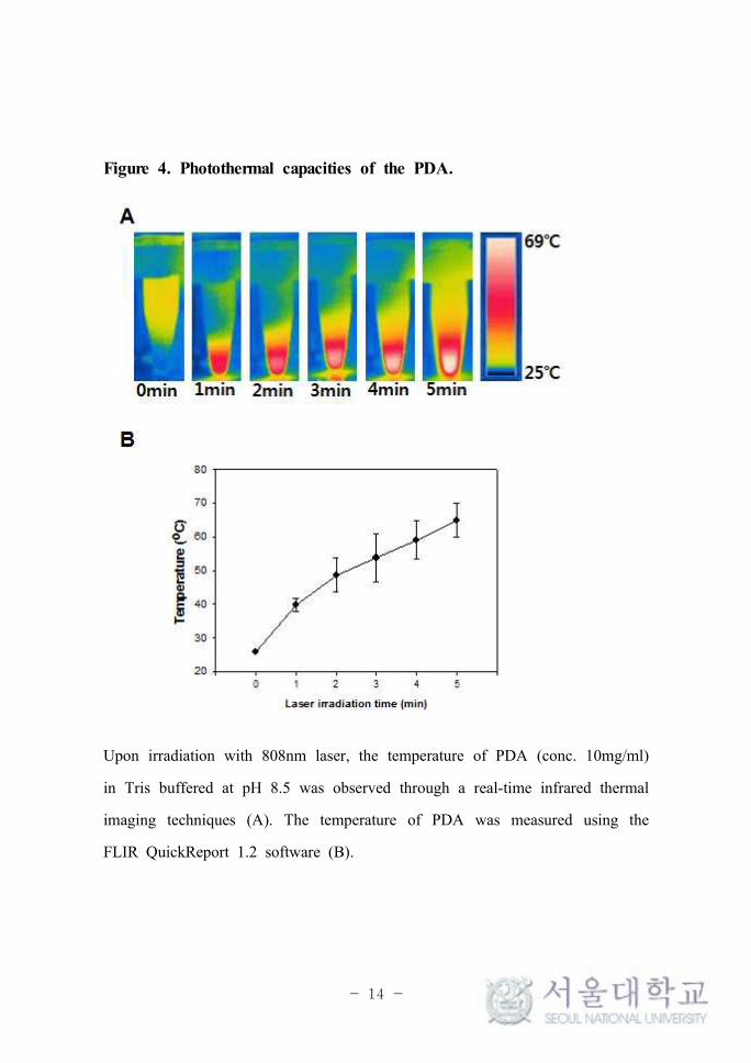
3. 2. Photothermal capacities of PDA
The photothermal capacity was determined through real-time infrared
thermal imaging of irradiated the PDA solution (10mg/ml) in 10mM
Tris-buffer at pH 8.5 (Fig. 4A). The temperature of the PDA was observed
in relation to the irradiation time of NIR laser (808nm). After five minutes
of irradiation, the temperature of the PDA reached 64.9 ± 4.5 oC (Fig. 4B).
- 14 -
Figure 4. Photothermal capacities of the PDA.
Upon irradiation with 808nm laser, the temperature of PDA (conc. 10mg/ml)
in Tris buffered at pH 8.5 was observed through a real-time infrared thermal
imaging techniques (A). The temperature of PDA was measured using the
FLIR QuickReport 1.2 software (B).

- 15 -
3. 3. Cellular uptake of photoresponsive liposomes
BT-474 cells took up PDA-liposomes to a greater degree than they did
for conventional liposomes (Fig. 5). As shown in (Fig. 5A), two groups of
harvested cell pellets - one treated with conventional liposomes and the other
with PDA-liposomes - displayed clearly different colors, white and black
respectively. These qualitative observations indicated effective cellular uptake
of photoresonsive liposomes for photothermal activity, which was
quantitatively measured by using NBD-PE, a fluorescent dye. As observed,
fluorescence confocal microscopy images revealed higher fluorescence intensity
in the cells with just dye-labeled liposomes (Fig. 5C) than the cells treated
with NBD-PE-labeled liposomes (Fig. 5B). Also, flow cytometry
measurements of the cells revealed a 4.7-fold higher fluorescence intensity of
the NBD-PE signal from the NBD-PE labeled liposomes (Fig. 5D, 5E).

- 16 -
Figure 5. In vitro cellular uptake of the liposomes and photoresponsive
liposomes.
BT-474 cells were left untreated or treated with liposomes or PDA-
liposomes. After 2h, the appearances of the pellets formed from untreated,

- 17 -
liposomes, or PDA/liposomes-treated cells were photographed (A).
Fluorescence confocal microscopy images of the BT-474 cells treated with
NBD-PE labeled liposomes (B) or PDA coated NBD-PE labeled liposomes
(C). Representative flow cytometry data (D), and the quantitation of the
fluorescence cellular intensity data (E) are presented. The scale bar indicates
20mm. *Significantly higher (p < 0.05) compared to the other groups
(assessed by the ANOVA and the Student-Newman-Keuls test).
- 18 -
3. 4. Photoresponsive liposomes effect on in vitro NIR laser-induced
photothermal activity
The photothermal effects produced from the irradiation of liposomes and
PDA-liposomes in the treated cells were examined through real-time infrared
thermal imaging (Fig. 6A). The temperature of the cell suspension was
observed to increase during the five minute NIR laser (808nm) irradiation
(Fig. 6A). The untreated cells showed an increase of 2.1 oC with the same
irradiation time (Fig. 6B). The temperature of the cells treated with liposomes
increased with the irradiation time to the same extent as did the temperature
of the untreated cells. The cells treated with liposomes showed a temperature
increase of 2.5 oC after five minutes of irradiation. By contrast, the cells
treated with PDA-liposomes increased in temperature by 37.3 oC after five
minutes of irradiation as well(Fig. 6B).

- 19 -
Figure 6. Photothermal effects of the photoresponsive liposomes.
Temperature increases induced by the presence of liposomes or
PDA-liposomes treated cells upon irradiation were observed using real-time
- 20 -
infrared thermal imaging techniques (A). The temperature of cell suspension
was measured using the FLIR QuickReport 1.2 software (B). *Significantly
higher (p < 0.05) compared to the other groups (assessed by the ANOVA
and the Student-Newman_Keuls test).

- 21 -
3. 5. Anticancer drug encapsulated photoresponsive liposomes effect on
in vitro NIR laser-induced photothermal activity
NIR laser-induced anticancer photothermal and/or chemo effects of the
PDA and Edelfosine were measured using the viability test of the A375
human melanoma carcinoma cells. The survival of the tumor cells after NIR
laser irradiation was found to be dependent upon the existence of PDA or
Edelfosine drug in the cells (Fig. 7). The irradiation of cells with a NIR
laser over five minutes did not affect the viability of the untreated cells. As
a result, the photothermal effect of the NIR irradiation was shown to be
effective only on the liposome groups that were modified with PDA. The
liposomes that consisted of both Edelfosine and PDA showed significantly
lower cell viability due to chemotherapy and PTT effects.

- 22 -
Figure 7. Combined photothermal-chemo cancer cell-killing effects of
the anticancer drug Edelfosine-encapsulated photoresponsive liposomes.
A375 cells were left untreated or treated with liposomes, PDA-liposomes,
Edelfosine encapsulated liposomes and PDA-Edelfosine encapsulated
liposomes. The viabilities of the cells were measured using the MTT assay
with and without laser irradiation for five minutes and then incubation for 24
hr. *Significantly higher (p < 0.05) compared to the other groups (assessed
by the ANOVA and the Student-Newman_Keuls test).

- 23 -
Ⅳ. Discussion
In this study, we demonstrated that combinatorial therapy of photothermal
and chemo using bio-inspired surface modification materials and anticancer
drug has higher cancer cell killing potency than single therapy [11].
The PDA, which was the key part of our system was easily synthesized
and modified on all kinds of organic and inorganic surfaces in aqueous state
[10,12-15]. The photoresponsive liposomes were prepared by simple and
versatile coating method with a uniform PDA film by dispersing in basic
condition of dopamine solution at room temperature for an hour [15].
Importantly, PDA - mussel-inspired adhesion proteins - acts as an excellent
redox mediator [16] and as a nontoxic [17,18] surface modification material,
which makes its self-polymerization process harmless [18]. Also, dopamine
contains catechol and amine groups; self-polymerization of dopamine proceeds
via oxidation of catechol into dopaminequinone followed by oxidative
oligomerization and further self-assembly [19]. After all, PDA can be easily
distinguished from dopamine solution by color change (Fig. 2B), white and
black, respectively.
The photothermal capacity of PDA was initially confirmed by UV
absorbance spectra (Fig. 3A). The PDA-liposomes displayed a significantly
higher NIR absorption compared to the non-PDA liposomes. This means PDA
absorb NIR light and further it generates enough heat for anticancer effect.
The high photothermal effects were shown by PDA (Fig. 4). The highest
temperature of PDA solution reached to 69 oC after five minutes irradiation

- 24 -
at 808nm.
As previously mentioned above, PDA is a mussel mimicking adhesive
proteins. Adhesiveness is relative to the cellular uptake efficiency [20,21]
which means the existence of PDA can be influenced [22]. The cell
internalization mechanisms of PDA have not been clarified [23], however, its
cellular uptake was confirmed by confocal microscopy images (Fig. 5B, 5C).
More uptake means higher photothermal anticancer effects due to the light
energy PDA absorb itself and convert them into heat [1]. The temperature
between 50 oC to 52 oC for four to six minutes may damage the cells by
denaturing proteins [24]. At least, the temperature above 45 oC may kill
tumor cells directly [1]. Therefore, the higher temperature and longer
irradiation time may bring dramatic anticancer effects. However, if the
temperature is too high for long time may bring serious burn in skin; so,
finding the optimized irradiation time is unavoidable.
Not only PDA solution itself, also the cells treated with PDA-liposomes
were displayed high photothermal activity (Fig.6). The results that the
modified liposomes treated group was only responsive group to NIR light
with regard to cellular uptake. The amount of the PDA adhesive to or
internalized into the cell can be influenced its photothermal activities due to
absorption of light energy [25].
As a result, treatment with the anticancer drug Edelfosine, encapsulated
in liposomes, reduced viability of A375 human melanoma carcinoma cells
compared to the cells treated with liposomes with no drug [26-29]. Also, the
cellular damage have occurred with the PDA's photothermal activities.
However, the anticancer effects of neither chemotherapy nor photothermal got

- 25 -
to the insufficient anticancer acitivity. Only the group with liposomes that
carried of both Edelfosine and PDA showed lowest cell viability among other
groups due to combinatorial therapies. Thus, PDA will be used and applied
as a biocompatible material to the numerous way of developing
multifunctional nano platforms in future medical field.

- 26 -
Ⅴ. Conclusion
In this study, we formulated liposomes, which were modified with PDA -
mussel-inspired photoresponsive materials - for adherence and absorbance of
NIR light to convert heat. These altered liposomes were then used as carriers
for Edelfosine and NBD-PE, a fluorescent dye. The small size (nano-scale) of
the modified liposomes enabled the enhanced permeability and retention effect
(EPR) for targeting during in vitro studies. The results of this study
suggested the usefulness of PDA-modification in the field of photothermal
cancer therapy. Moreover, the technique utilizes combinatorial photothermal
and chemotherapy, which can bring effective biomedical applications of
multifunctional anticancer effect nanoparticles of high performance.

- 27 -
Ⅵ. References
1. Zhang Z, Wang J, Chen C. Near-infrared light-mediated
nanoplatforms for cancer thermo-chemotherapy and optical
imaging. Adv Mater. 2013;25:3869-80.
2. Lee SE, Liu GL, Kim F, Lee LP. Remote optical switch for
localized and selective control of gene interference. Nano Lett
2009;9:562-70.Zhang Z, Wang J, Chen C. Near-infrared
light-mediated nanoplatforms for cancer thermo-chemotherapy and
optical imaging. Adv Mater. 2013;25:3869-80.
3. Maltzahn GV, Park JH, Agrawal A, Bandaru NK, Das SK,
Sailor MJ, Bhatia SN. Computationally guided photothermal
tumor therapy using long-circulating gold nanorod antennas.
Cancer Res. 2009;69:3892-900.
4. Iancu C, Mocan L. Advances in cancer therapy through the use
of carbon nanotube-mediated targeted hyperthermia. Int J
Nanomed. 2011;6:1675-84.
5. Choi WI, Kim JY, Kang C, Byeon CC, Kim YH, Tae G.
Tumor regression in vivo by photothermal therapy based on
gold-nanorod-loaded, functional nanocarriers. ACS Nano. 2011;5:
1995-2003.

- 28 -
6. Min Y, Mao C, Chen S, Ma G, Wang J, Liu Y. Combating
the drug resistance of cisplatin using a platinum prodrug based
delivery system. Angew Chem Int Ed. 2012;51:6742-47.
7. Helmchen F, Denk W. Deep tissue two-photon microscopy. Nat
Methods. 2005;2:932-40.
8. Welsher K, Liu Z, Sherlock SP, Robinson JT, Chen Z,
Daranciang D. A route to brightly fluorescent carbon nanotubes
for near-infrared imaging in mice. Nat Nanotechnol
2009;4:773-80.
9. Wang Y, Black KC, Luehmann H, Li W, Zhang Y, Cai X.
Comparison study of gold nanohexapods, nanorods, nanorods,
and nanocages for photothermal cancer treatment. ACS Nano.
2013;7:2068-77.
10. Lin L, Cong Z, Cao J, Ke K, Peng Q, Gao J, Yang H, Liu
G, Chen X. Multifunctional Fe3O4@Polydopamine Core-Shell
Nanocomposites for Intracellular mRNA Detection and
Imaging-Guided Photothermal Therapy. ACS Nano.
11. Zhang W, Guo Z, Huang D, Liu Z, Guo X, Zhong H.
Synergstic effect of chemo-photothermal therapy using PEGylated
graphene oxide. Biomaterials. 2012;336(6085):1124-1128.
12. Lee HS, Dellatore SM, Miller WM, Messersmith PB.
Mussel-Inspired surface chemistry for multifunctional coatings.
Science. 2007;318:426-30.

- 29 -
13. Sa R, Yan Y, Wei Z, Zhang L, Wang W, Tian M. Surface
Modification of Aramid Fibers by Bio-inspired Poly(dopamine)
and Epoxy Functionalized Silane Grafting. Appl Mater Interfaces.
2014;6:21730-38.
14. Lynge ME, Ogaki R, Laursen AO, Lovmand J, Sutherland DS,
Stadler B. Polydopamine/Liposome coatings and their interation
with myoblast cells. ACS Apppl Mater Interfaces.
2011;3:2142-47.
15. Liu Q, Wang N, Caro J, Huang A. Bio-inspired polydopamine:
a versatile and powerful platform for covalent synthesis of
molecular sieve membranes. J Am Chem Soc.
2013;135:17679-82.
16. Kim JH, Lee M, Park CB. Polydopamine as a Biomimetic
Electron Gate for Artificial Photosynthesis. Angew Chem Int Ed.
2014;53:6364-6368.
17. Hong S, Kim KY, Hwang JW, Park SY, Lee KD, Lee DY,
Lee HS. Attenuation of the in vivo toxicity of biomaterials by
polydopamine surface modification. Nanomedicine.
2011;6:793-801.
18. Sa R, Yan Y, Wei Z, Zhang L, Wang W, Tian M. Surface
Modification of Aramid Fibers by Bio-inspired Poly(dopamine)
and Epoxy Functionalized Silane Grafting. Appl Mater Interfaces.
2014;6:21730-38.

- 30 -
19. Zhou J, Wang P, Wang C, Goh YT, Fang Z, Merssersmith PB,
Duan H. Versatile core - Shell Nanoparitle@Metal - organic
framework nanohybrids: exploiting mussle-inspired polydopamine
for tailored structural integration. ACS Nano. 2015;9:6951-60.
20. Lesniak A, Salvati A, Santos-Martinez MJ, Radomski MW,
Dawson KA, Aberg C. Nanoparticle adhesion to the cell
membrane and its effect on nanoparticle uptake efficiency. J Am
Cem Soc. 2013;135:1438-44.
21. Yuan H, Li J, Bao G, Zhang S. Variable nanoparticle-cell
adhesion strength regulates cellular uptake. Phys Rev Lett.
2010;105:138101.
22. Lynge ME, Fernandez-Medina M, Postma A, Stadler B.
Liposomal drug deposits in poly(dopamine) coatings: effect of
their composition, cell type, uptake pathway considerations, and
shear stress. Macromol Biosci. 2014;14:1677-87.
23. Lynge ME, Fernandez-Medina M, Postma A, Stadler B.
Liposomal drug deposits in poly(dopamine) coatings: effect of
their composition, cell type, uptake pathway considerations, and
shear stress. Macromol Biosci. 2014;14:1677-87.
24. Zhou J, Chen JK, Zhang Y. Theoretical analysis of thermal
damage in biological tissues caused by laser irradiation. Mol
Cell Biomech. 2007;4:27-39.
25. Liu Y, Ai K, Liu J, Deng M, He Y, Lu L. Dopamine-Melanin

- 31 -
Colloidal Nanospheres: An efficient Near-Infrared photothermal
therapeutic agent for in vivo cancer therapy. Adv Mater.
2013;52:2012-16.
26. Lee S, Kim J, Shim G, Kim S, Han SE, Kim K, Kwon IC,
Choi Y, Kim YB, Kim CW, Oh YK. Tetraiodothyroacetic
acid-tagged liposomes for enhanced delivery of anticancer drug
to tumor tissue via integrin receptor. J Control Release.
2012;164:213-20.
27. Ahmand I, Filep JJ, Franklin JC, Janoff AS, Masters GR,
Pattassery J, Peters A, Schupsky JJ, Zha Y, Mayhew E.
Enhanced therapeutic effects of liposome-associated
1-O-octadecyl-2-O-methyl-sn-glycero-3-phosphocoline. Cancer Res.
1997;57:1915-21.
28. Castro BM, Fedorov A, Hornillos V, Delgado J, Acuna AU,
Mollinedo F, Prieto M. Edelfosine and miltefosine effects on
lipid raft properties: membrane biophysics in cell death by
antitumor lipids. J Phys Chem B 2013;117:7929-40.
29. Busto JV, Canto-Janez E, Goni FM, Mollinedo F, Alonso A.
Combination of the anti-tumour cell ether lipid edelfosine with
sterols abolishes haemolytic side effects of the drug. J Chem
Biol. 2008;1:89-94.
- 32 -
Abstract (in Korean)
광감응 리포좀을 이용한
광열 항암 치료
황지현, 약학과 물리약학전공, 약학대학, 서울대학교
이 논문은 지질 기반 나노 전달체인 리포좀(liposome)을 광 감응성
물질로 코팅하여 암세포로의 전달을 통해 광열항암치료효과에 대해
연구를 수행하였다. 본 연구에서는 최근 큰 각광을 받고 있는 생체
모방 표면 개질 소재중 하나인 폴리도파민을 리포좀에 코팅하는 동
시에 808nm 근적외선에 감응하는 물질로써 암세포 광열치료에 사
용하였다. 도파민은 카테콜과 아민 작용기를 가지는 분자량
153(Da)의 단 분자 물질인데, 염기성 수용액상 조건 (pH 8.5)에서
는 카테콜의 산화에 의해 자발적으로 반응이 진행되어 폴리도파민
(polydopamine, pDA)을 형성하여 표면의 화학적 성질에 관계없이
금속, 고분자 등 다양한 소재 표면의 강하게 흡착되는 뛰어난 표면
부착능력이 있다. 홍합의 접착 메커니즘의 화학적 작용기만을 선택
적으로 모방하여 도입한 생체 모방 표면 개질 기법 (bio-inspired

- 33 -
surface modification)은 접착성뿐만 아니라 생체재료의 가장 중요
한 요소인 생체친화적인 (biocompatible) 코팅 기법으로써 이미 다
양한 선행연구들에서 표면개질효능과 세포독성이 없다는 점이 세포
배양실험을 통해 밝혀져 왔다. 본 연구는 선행연구와의 차별성과
학술적 발전에 기여하기 위하여 리포좀 표면의 폴리도파민을 흡착
시켜 코팅 했을 뿐만 아니라 암세포로 전달한 후, 외부에서 근적외
선을 조사하여 광열효과를 유도하였다. 대조군과 비교했을 때 광
감응성 리포좀의 온도는 최고 69 oC까지 올라 세포실험 결과 암세
포들이 사멸하는 것을 확인하였다. 또한 현재 널리 쓰이고 있는 항
암제인 Edelfosine을 리포좀에 봉입하여 폴리도파민으로 코팅하고
암세포에 전달한 뒤 근적외선의 광열효과를 통해 세포생존율의 감
소를 확이 할 수 있었다. 본 연구의 의의는 리포좀을 이용하여 항
암제를 전달하고 동시에 광 감응성 물질을 코팅함으로써 근적외선
조사에 의한 광열효과까지 갖춘 리포좀을 개발하였다. 이러한 광
감응성 리포좀은 봉입한 항암제에 의한 화학요법과 함께 광열치료
의 상승작용을 기대할 수 있어 미래의 새로운 항암 의약재료의 가
능성을 확인하였다. Key words: Liposomes, Near infrared light, Bio-inspired surface
modification, Biocompatibility, Photothermal therapy, Chemotherapy
학 번 : 2014-21056