application of silver degeneration stains for ... of silver degeneration stains for neurotoxicity...
TRANSCRIPT

TOXICOLOGIC PATHOLOGY, vol 28 , no I, pp 70-83, 2000Copyright © 2000 by the Society of Toxi cologic Path ologi sts
Application of Silver Degeneration Stains for Neurotoxicity Testing
ROBERT C. SWITZER III
NeuroScience Associates, Knoxville, Tennessee 37922
ABSTRACT
Silver sta ining procedures have been used in num erou s way s to render a varie ty of phy sical and biol ogic al features vis ible . Inbiol ogi cal tissue, hist olo gic protocols use silver to visu alize diverse structures or features, such as reticulin, melanin, fun gi, chromosomebands, nucl eolar organizing regions , and different features in the nervous system. A comparison of the specific steps in these protocolsindicates that the silver is " directed" to stain any given feature by the type of fixation, the pretreatment ("morda nting"), the composition of the silver-co ntaining solution(s ), and the form of development (reduction). Sin ce the mechanisms of staining have not beenunderstood historically (nor are they now) , each method was developed by trial and error. Keystone methods such as thos e of Bodi anand Bielschow sky exploit the nervous system's affinity for silver (argyrophilia). The beginning of a new era in brain research ca mewith the recognition that distin ct silver-impregnated morphologic changes occurring in damaged axons could be used for trac ing axo npathways in experime ntal animals with specifically placed lesion s. Improvements in sta ining methods used to selec tively imp regnatethe di sintegratin g axons but to leave normal axon s unstained were achieved by Nauta and Gyg ax (early workers with these procedures)and spawned a host of meth od var iations known as the " Nauta" methods. Of the se , the Fink-Heime r and de Olmos cupric-silvermethod s were able to unambiguously dem onstrate di sintegratin g synaptic terminals, thereby allowi ng complete tracin g of axo n pathway s. The late 1970s and 1980s witnessed innovative applications of the se techniques . Th e silve r methods once used to trace axonpathways became indicators of the extreme endpoint of neurotoxicity : disintegrative degeneration of neur ons induced by neurotoxicchemicals that were administered sys temically. The hallmark of neurotoxic subs tances is the selec tivity with which each destroysspec ific populat ions or subpopulations of neurons. Th e high contras t and sens itivi ty of the silver degeneration stains greatly facilitatethe sc ree ning pro cess to de tect these affected populations, especially when there is no basis for know ing where in the brain to lookfor dam age . More recentl y, in addition to expanded use in screening for neu rotoxi c effects, the silver degeneration stains are be ingused to chart the neur on population s undergo ing pro grammed cell death in the dev elop ing brain . Other newly developed silver methodshave been refined to show nondisinte grative degeneration , such as the plaqu es and tangl es of Alzhe imer 's di sease.
Keywords. Amino cupric silver; apoptosis: brain ; elec tro n microscopy; fragmentation of protein s; MultiBrain Technolo gy; neuraltracin g; neurohi stology : neurotoxins; risk assessment
INTRODUCTION
The use of silver in staining protocols throughout thehistory of histology has been extensive. Parallel disciplines within the area of histology developed specializedsilver staining methods that reflect those requirementsunique to each, Increasingly in the last several decades,there has been a divergence in the information that isimportant to and shared by those laboratories with broadclinical demands vs those specialty laboratories that require new methods with which to explore the structureof the central nervous system. Perhaps, then, it is notsurprising that the so-called " reduced silver methods"(which require external reducing agent vs tissue that"auto" -reduces silver) are not part of the standard histopathologic armamentarium.
The purpose of this review is to give a perspective on
Address correspond ence to : Dr Robe rt C. Sw itzer III, NeuroScie nceAssociates, 10915 Lake Rid ge Dr ive, Knoxv ille, Tenn essee 3792 2.
the evolution of these silver stains and on their significance for contemporary use in assessing neurotoxicity.Some concepts of candidate mechanisms of these silverstains will be considered as well as suggestions for thedesign of studies for the detection of neurotoxicity.
HISTORICAL B ACKGROUND
Axon Tracing
Excellent accounts of the historic trace of the neurohistologic silver methods have been provided by Ebbesson (19), Heimer (36), Leonard (49, 50), de Olmos et al(16 ), and Beltramino et al (4). Drawing upon these accounts, the following historic vignette is provided.
Waller (82) obs erved in 1850 that transection of thehypoglossal and glossopharyngeal nerves produced a" kind of coagulation or curdling" of the nerve fiber s.This "downstream" or anterograde degeneration came tobe known as " Wallerian degeneration. " The first person,however, to actually use the morphology of anterograde
70 0192 -62 33/00$3.00 + $0 .00

Vol. 28, No.1, 2000 APPLICAnON OF SILVER DEGENERAnON STAINS 71
degeneration as a means of tracing axon pathways wasLudwig Turck, who preceded Waller in his report on spinal cord tracts by 1 year (49) . According to Leonard (50),Cajal later found that the se coagulated particles had ahigh affinity for impregnation by silver, but he did notfurther utilize this property.
In 1873 , Camillo Golgi exposed brain tissue that hadbeen hardened in potassium dichromate to silver nitrateand achieved results that revolutionized the study of thenervous system . A small percent of the neurons becameimpregnated with silver in their entirety such that the finest detail of the dendritic and axonal processes was revealed. This was the first of the so-called "reduced silver" methods. Cajal exploited this discovery, andthrough his work, he validated the neuron doctrine ortheory of the structure of the nervous system. Ironically,Golgi was an advocate of the opposing line of thoughtthe reticular theory (68). The neuron doctrine stated thatneurons with dendrites and axons as their parts are thebasic units of structure of the nervous system. On theother hand, the reticular theory stated that the fine ,branchlike structures (axons and dendrites) were continuou s with one another and that neuron cell bodies provided a trophic/nourishing role (68).
The period from 1870 to 1910 wa s a golden era in thatwe gained an understanding of the structural elements ofthe brain. During thi s period, the Golgi and other stainswere used to identify components of the nervous systemand to ascertain the pathways ofaxons. Bielschowsky (5)introduced a silver method that, in contrast to the Golgimethods, essentially stained all types of cells and neuralprocesses (Figure 1). The Bielschowsky stain becamewidely used, as it still is today, to identify abnormal features in the context of normally stained components.
Eventually these methods became limited because ofthe difficulty in finding the degenerating axonal elementsamong the normal axons that were also stained-todaywe call it poor "signal to noise" or just lack of contrast.It is the implicit goal of any histologic stain to providecontrast to different components of the tissue and, at thelimits of this concept, to stain only tho se entities that areof interest while leaving other components unstained.Contemporary antibody staining exemplifies this process.
The solution to this lack of contrast came with thediscovery of the way in which to utilize the fact thatdegenerating axons have a somew hat higher affinity forsilver than do normal axons. This property was fully exploited in the 1940s by Glees ' modification (3 1) of theBielschowsky method (5) and then again by Nauta andGygax in the early 1950s (59). The common feature inthe se successes wa s that the tissue was pretreated withvarious solutions prior to exposure to silver solutions .The result was a " suppress ion" of the silver impregnation in the normal axons and a rather intense impregnation of the degenerated axons; thereby, high contrast wa sachieved. These manipulations essentially "directed" thestructures to be stained by the silver. Fink and Heimer(23) further refined the pretreatment strategy to obtain atechnique that for the first time revealed degeneratingsynaptic boutons without the ambiguities that plaguedother silver methods. Almost concurrently with the ef-
FI GURE l.-Bielschowsky-stained sec tio n from human cerebellum.Ce ll bodies . de ndr ites, and axo ns are visi ble; \OJ.!- paraffin section.
forts of Fink and Heimer, de Olmos developed the cupricsilver method (15 ), which, in its current form, providesthe most sensitivity and the highest contrast (high signalto-noise) images.
Later protocols developed by Gallyas took the varietyof pretreatments to new levels of directed staining (2629) . It is interesting to observe that the principles underlying the steps used to achieve "directed staining" areanalogous to those associated with the use of mordants(definition: to treat with a chemical that confers the ability to combine with dyes) in other disciplines of histology.
Contemporary Use f or Neurotoxicity Detection
Beginning in the late 1970s, other methods for tracingaxon pathways bec ame available (tritiated amino acids,horseradish peroxidase, and fluorescent markers), and thefrequency of use of silver degeneration methods rapidlydeclined. At about the same time, however, a new use ofthe silver degeneration stains began to evolve: detectionof damage to the nervous system caused by chemicalagents. The level of damage registered by the se silverstains is that characterized by destruction of neural elements. Loss of neurons is the ultimate endpoint of thevarious measures of neurotoxicity, because neurons donot regenerate (75) . Table 1 lists the first studies to systemically administer a known or suspected neurotoxinand to use a silver degeneration stain to detect the areasof damage.

72 SWITZER T OXICOLOGIC P ATH OLOGY
TABLE I.-Chronology of use of silver degen erat ion stains to detect neurotoxic d amage fo llowing system ic ex posure to differen t agents .
Substance/agent Target/s usce ptible areas Authors Year Reference
Tetanus tox in somatosen sory fiber s Ill is and Mitchell 1970 426-Hydroxydopamine (6-0 HDA) substantia nigra Hedreen and Chalmers 1972 353-Acetyl pyridine inferior olive, n. ambiguou s Desclin and Esc ubi 1974 18High-pressure ox yge n auditory nucl ei Sw itze r 1980 69Capsa icin somatose nso ry fibers Jancso and Kiraly 1980 43Soman (ne rve gas organophos pha te) hippocampus, pyrifo rm cortex Petras 1981 66Methamphetam ine stria tum, cortex Ricaurt e et al 1982 67Alcohol entor hinal cortex, hippocam pus Sw itzer et al 1982 72Kainate hippocampus, pyrifo rm co rtex Jarrard 1989 45MPTP (I -me thy l-4-phenyl-1 ,2,3 ,6-tet-
rahydropyridine) substantia nigra , thal amu s, n. sag ulum Switzer and Cam pbe ll 1987 70Trime thy ltin hippocampu s, cortex Balaban et al 1988 1Tri-ortho- cr esyl- ph osphat e (TOCP;
Type 1 OPIDN) spinal cord , brain stem, cerebellum Tanaka and Bursian 1989 80Tri-phenyl phosphit e (Type II OPIDN) spinal cord, brain stem, cerebe llum, thalamu s, Tanaka et al 1990 81
cortexMDMA (methylen e-dio xymethamphet- cor tex Jen sen et al 1990 46
amine)Glutamate coc hlea Janssen et al 1991 44Cocaine fasciculus retroflexus Ellison 1992 20MK- 801 (dizoci lpine maleate) cingulate, olfactory cortex , hippocampu s Corso et al 1992 10Phencyclidine (PCP) cingulate, entorh inal cortex, hippocampu s Ellison and Switzer 1993 2 1Domoic aci d hippocampus Peng et al 1994 62High ene rgy (Me V) elec tro ns and 13 astrocy tes Switzer et al 1994 77
pro tons3-Nitropro pio nic acid striatum, cortex , hippocampus Mill er and Zaborszky 1997 56
Silver Staining and How a Neuron Dies. " Do the silver degeneration stains show neurons that died by apoptosis?" is a frequently posed question. As is becomingincreasingly evident, there are numerous routes, pathways, or mechanisms that can lead to the death of thecell (52, 55 ). After a neuron is "dead," it disintegrates,and, as will be discussed later, it is the debris from thisdisintegration that is registered by the silver degenerationstains, This multi-option process is depicted in Figure 2and is compatible with insights recently expressed byLevin et al (52). One or more of the hypothetical sequences shown could be apoptotic or oncotic in nature.The term necrosis is reserved for a "final common pathway" of di ssolution (disintegration) leading to the products that are the sugges ted moieties " stained" by the silver. At this point in the sequence, the silver degenerationstain indicates that cell death occurred, but it cannot provide the means to determine if the events that initiatedthe sequence leading to cell death were oncotic or apoptotic.
Evidence is being accumulated in our laboratory thatindicates that the appearance of neuronal debris is a function of how the cell died. For example, the debris ofneurons in cingulate cortex that were " killed" by anacute exposure to soman is not in evidence 4 weeks afterexposure. However, a 5-day subchronic exposure to soman yields cell body-sized packets of silver-impregnateddebris in the same location (73). Evidently, differencesin the agonal process can yield different presentations ofneuronal cell debris. This is consistent with and complements the findings that the morphology of dying neuronsis a function of the form of insult (5 1, cited in 52 ).
Currently, the disruption of the chronology of apoptosis (or programmed cell death) in the developing brain isan issue of concern. The silver degeneration methodshave recently been shown (57, 79 , 83) to be a useful toolin assessing the timetable of " cell death-days" for neu-
ronal populations as a complement to the well-documented patterns and chronology of "cell birthdays. " Thistabulation may identify other critical periods duringwhich development may be perturbed.
Interpretation ofSilver Degeneration Staining. Duringthe time when silver degeneration methods were usedextensively for axon tract tracing (1950-1970), a numberof articles gave in-depth discu ssions, with extensive illustrations of silver-impregnated features that were associated with disintegrative degeneration (4 , 16, 19, 36 ,50). Brain sections stained with the Fink-Heimer and deOlmos cupric silver methods had excellent signal-tonoise characteristics, such that it could be generally saidthat " if it wa s impregnated by silver, it is degenerated. "The dichotomy of the features stained offered enormousvalue for detecting degeneration-hence, the value forindicating neurotoxicity.
With the development of more contemporary silverprotocols, the sensitivity of the staining has increased.Features not visible with earlier methods can now be seensoon after the insult occurs (14, 30). While this may beadvantageous from some perspectives, it can potentiallyles sen the dichotomous " all or nothing" characteristicand requires that the investigator use appropriate sur vivaltimes to avoid ambiguity. For example, the new methodscan silver-impregnate neurons that have been traumatizedonly a matter of minutes to hours prior to sacrifice (14 ,30). This is certainly of value in terms of detecting theneuronal populations perturbed by a particular form ofinsult, but with very short survival times, a conclusioncannot be reached as to whether the neurons will subsequently die. A similar situation exists for neurons thatexpress heat-sho ck protein, HSP-70, or c-fos, in that bothreveal perturbed neurons, but which, if any , would diecannot be determined.
The valuable dichotomy of silver-stained features ispreserved if the investigator judiciously selects the times

Vol. 28, No.1, 2000 APPLICATION OF SILVER DEGENERATION STAINS 73
Schematic of cellular events that follow various perturbations methods seem less influential with the amino cupric silvermethod.
Appearance of Silver StainingWith the aid of relatively few silver-stained slides, a
pathologist can become acquainted with the characteristics of the silver degeneration stain in just a few hours.Some of the photographs shown in Figure 3 illustrate thekind of material such a study set would comprise.
With the amino cupric silver stain, degenerative features undergoing various degrees of disintegration are selectively impregnated and are seen as black or blackbrown profiles against a clear background. With de Olmos's original version, as well as with most of the socalled "Nauta variants," the background is dark to lightamber (Figure 3A). The time of appearance of disintegrated neural components is dependent on the durationof survival following the neurotoxic insult. Degeneratingdendrites and axon terminals (Figure 3A, B, F) appearearly (after a few hours), but their debris is no longerevident after about 3-7 days. Degenerating cell bodies(Figure 3A , B, D) can be seen shortly after the appearance of degenerating terminals, but cell body debris isalso cleared by 7-10 days after the insult. Degeneratingaxons (Figure 3C, D, E) and associated debris are the lastto be observed and may persist for remarkably long periods of time (33) . These elements can be seen beginningat about 2-3 days after the insult, and this debris can stillbe stained 9 months later in rats. In primates, especiallyhumans, the axonal debris can be stained after more than1-2 years (33). Therefore, if debris associated with thedeath of neuron cell bodies is no longer present, the axonal debris of these cells may still be stainable and willprovide conspicuous evidence of the neurotoxic event.
An axon can degenerate and the degeneration can bedetected even though the parent cell body survives . Oftensuch neuron cell bodies display changes known as chromatolysis, wherein the nucleus is displaced to the periphery, the cell is swollen, and the Nissl bodies are reducedand located at the cell margin (22) . Chromatolysis ismore extensive and extreme in the immature nervous system. The incident causing the distal axon to degeneratecan be transection or trauma somewhere along the lengthof the axon (causing Wallerian degeneration) or at theterminus of the axon, wh ich causes a dying back phenomenon (retrograde degeneration) (34).
Conceptually, in long-term neurodegenerative diseases,there could be routes to cell death that generate disintegration products at such a slow rate that at any giventime, not enough debris is present to yield much bindingof silver (thus resulting in a weak signal). For example,in our staining of brains from Parkinson's disease victims,degenerating axons from substantia nigra to the striatumare not seen.
M ECHA NISM OF S TAINING
Historic PerspectiveThe question of mechanism of staining is usually one
of the first questions asked by those individuals first presented with the potential of using these silver stains toasses s neurotoxicity. Similar questions were asked during
Disintegrative
, • "Degeneration"
leading to Cell Death
. 1
methods
( 'Holy Grail' components)
Domain of changes
~ " ..... Changes;Silver Degeneration ,,/ ......
-- Forms of Perturbation --
Domain 4 Q Q00~ CD 0 1 Different
of changes \ \ I (!) Sequences.& CD 0 2detectable , ', : ,' of Effects or
with Antibodies ~0 0 >2> Responses\ \ / / following Trauma
& iri situ methods ~~~t
, Final c~::::i:)athWay
~
detectable with .~- _ _ J' - _ _ __ _ -. -
F IGURE 2.-Schem atic of potent ially neurotoxic biochemical changesthat fo llow different form s of perturbat ion : stages of applicability ofantibod y markers and silver degeneration sta ins. Th e spec trum of neurotoxic effect s encompasses a broad range of changes in a cell that canculminate in its death. Depending on the form of insult , a differentsequence of effects may ensue, all of which may be reversible . In theext reme , any given pertu rbation or traum a (the geometric forms at thetop ) can result in the death of the cell , eve n though it may occur bydiffe rent sequences , as ind icated by the strings of sma ll, numbered geo metric forms. All of the se, however, co nve rge on the sti ppled poly gonto traverse a final common path of changes (sma ll stippled polygo ns)that res ults in ce ll death . Ant ibody prob es hav e been devel oped aga instdi ffer ent proteins (depicted by triangle-2 or diamond-I ) that are constituents of the sequence of biochemical changes that foll ow a part icularform of trauma. Unless an antibody is dire cted against a componentalong the " final common path " to cell death, the pre sence of immunore act ivity of prot eins in the earli er sequences does not neces saril ymean ce ll death. Since it is uncertain whether or not even a polyspecificant ibody could be ra ised against the amorphous disintegrative debris ofa degenerated cell , the silver degeneration stains currently remai n thebest probe to mark this end point of neu ro toxici ty.
of sacrifice after insult. By allowing a set of animals subject to the same trauma to survive 2-4 days, the morphology of any silver-stained neurons will indicate thedeath of that neuronal element.
With de Olmos's new amino cupric silver method (14),the silver impregnation of degenerated structures is moreintense, and the background is clear. The amino cupricsilver method also has the advantage of being able tostain brain tissue that has not been optimally fixed, suchas the case in human autopsies. Other unidentifiable technical aspects that perturbed earlier silver degeneration

74
'..
. .'" ~ :-.. . -...~ .'
~.~ ~ : .." .
.'
SWITZER T OXICOLOGIC P ATHOLOGY

Vol. 28 , No . I, 2000 APPLICAnON OF SILVER DEGENERAnON STAINS 75
the 1960s and 1970s as new silver methods arose. In fact,it seems that as any given silver method became moredirected, questions regarding validity and explanations ofmechanisms of staining were expected. It is not withinthe scope of this review, nor it is within the expertise ofthe author, to provide a definitive explanation for whysilver can reveal any given feature , since at best, themechanism of staining is poorly understood, in sp ite ofconsiderable efforts (63- 65).
In Table 2, several silver methods are presented, alongwith their generic steps. Progressing from left to right, itcan be seen that as the methods acquire more or differentsteps (especially pretreatments), they become more ordifferently " directed" in terms of the features that arestained. The features stained are dependent on the composition of the solutions at each of the steps: pretreatment, silver incubation, a second silver (if present), thereducer (developer), bleaching, and stabilization.
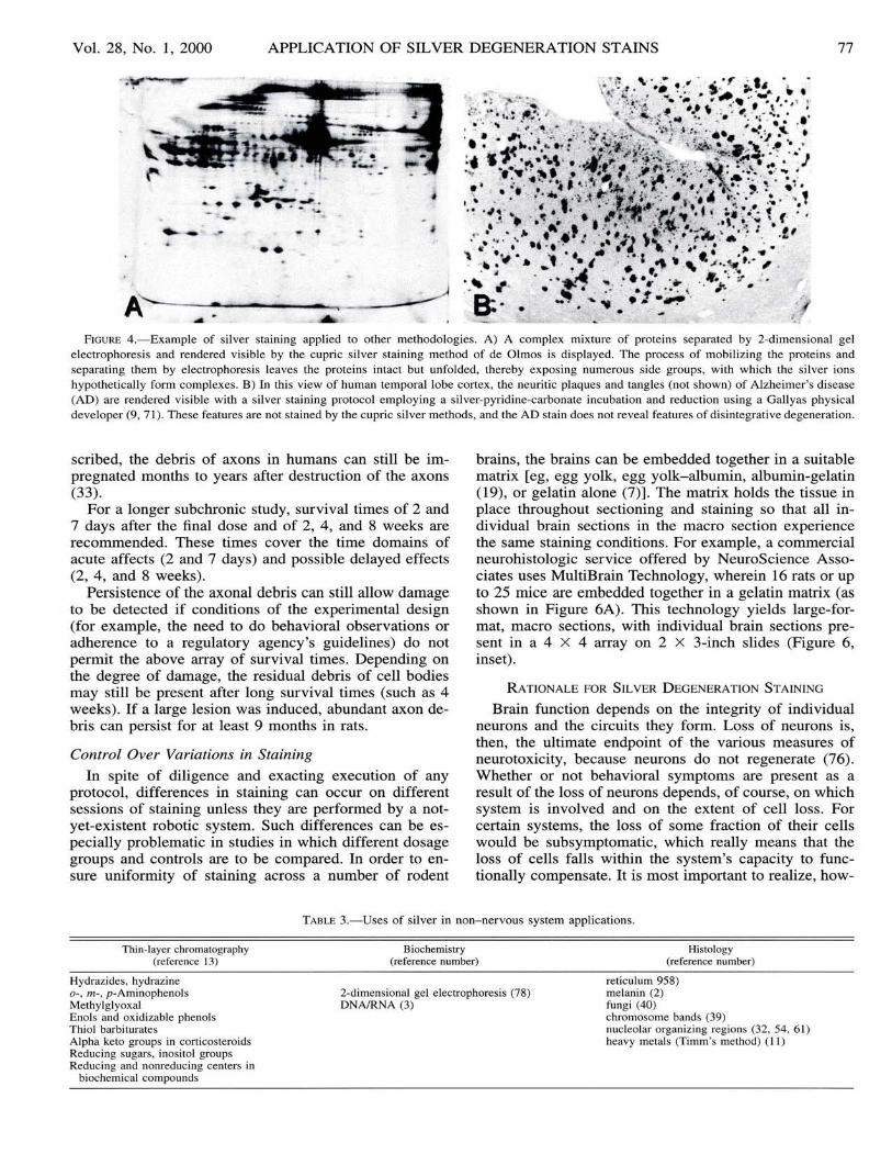
In order to illustrate the complexity of factors involvedin any given silver stain, consider that the same methodused to reveal disintegrative degeneration (the cupric silver method) was successfully applied to reveal complexmixtures of proteins that were separated by 2-dimensional gel electrophoresis (Figure 4A) (78 ). The proteins inthe se gel s are not " disintegrating" ; that is , they are notfragmented, but they are unfolded, and side groups areexposed. Furthermore, in brain sections from victims ofAlzheimer's disease, the silver degeneration stains do notstain the plaques and tangles that are revealed with othersilver stains (Figure 4B ).
These few examples illustrating the diversity of silveralso provide some insight regarding the reputation of thesilver methods as being "capricious. " It is ironic that thevirtue of versatility of silver can also be a curse. We haveseen that numerous factors can direct silver to stain features of interest. However, if unintended factors (contaminants) are present, the silver staining can be misdirectedand may yield undesirable results or outright failure. Asis true for procedures in biochemistry and biotechnologyin general, so it is for silver methods that purity of reagents and exacting execution of the protocol are necessary for consistent and reliable results.
It can be inferred from the comparison of methods inTable 2 that although some common principles underliethe mechanisms of staining, there is likely to be a different set of mechanisms for each stain. Sil ver is onl y Ifactor (albeit, a crucial one) in the ensemble of directivefactors for each method. In order to gain a perspectiveon just some of the possibilities for mechanism of staining , other applications of silver as a " stain" are shownin Table 3.
Working Hypothesis
Although an explicit mechanism cannot be providedfor the silver degeneration stain, a working hypothesis orconceptual vehicle can be offered (Figure 5); this hypothesis was inspired in part by experiments using puromycin as a "lesioning" agent (74). Single and shortchains of amino acids are known to form complexes withsilver ions as well as with other metals, such as copper(8, 25) . Different amino acids have different affinities forthe binding of silver (6) that can even provide a colordimension. It is commonly thought that those amino acidswith single sulfide groups have the largest affinity, butthrough the formation of complexes with amino acid carboxyl and amine terminal groups, as well as side groups,the opportunities for silver ions to form complexes arenumerous.
In the environment of an intact protein that is more orless globular in shape, many of the se groups are not accessible to silver ion s. In a cell undergoing degeneration,proteolytic mechanisms begin to dismantle proteins, andin the process, they expose more sites for silver to formcomplexes. This scenario is depicted at the bottom ofFigure 5.
Each site at which the complexes form can be thoughtof as a nucleation point around which other silver ion saggregate, as wa s suggested by Liesegang (53) when heput forth this hypothesis in 1911. This is comparable tothe events in the photographic process. From the physicochemistry of photography we learn that photons striking a silver-laden photographic emulsion create a nucleation point around which other silver ions gather during" development" (reduction), and these nucleation points
F IGURE 3.-Examples of silver degeneration sta ining. A) Th e ce ll body, dendrites, and portio ns of their axo ns are impregnated in this pair ofpyramidal ce lls in rat neocortex. Dend ritic frag ments are see n as either con tinuo us lines or linear arrays of dots from other ce ll bodies out of theplane of section. Th e lesion was induced by purom ycin 2 days prior to sacrifice (74) ; Stain : de Olm os cupric silve r method; 40-fL freeze-cut sections.B) Pyramidal ce lls of the hippocampal CA I reg ion in a ra t ex pose d to the organophos phate so man. Clusters of pyramidal ce ll bod ies wit h largesprays of dendri tes are visible in a degenerated sta te. Unaffected pyramidal ce lls are see n as gray ish profi les in line with the si lver-impreg nate dce ll bodie s; Stai n: amino cupric silver method of de Olmos, with neut ral red co unters ta in; 40 -fL freeze-cut sec tio ns. C) Cunea te tract in the dorsalspinal co rd from a rat. Silve r-imp reg nated degenerated axo ns are shown in transverse section. Lesion was of unkn own or igin; Stai n: amino cupricsilver meth od of de Olm os; 40-fL freeze-cut sec tions. D) Rat cerebe llum in corona l plane displaying degenerated ce ll bod ies and axo ns followingacrylamide expos ure . Purkinje ce ll bod ies and their dendrites are visible in the lower left quadrant of the image. Linear arrays of degenerated axonsare num erou s in the right half of the image. If the plan e of sec tio n had been in the sagi ttal plan e, the fan-sha ped geo me try of the Purkinj e ce lldend rites would have been visi ble; Stain : amino cupric silver meth od of de Olm os, with neut ral red co unters ta in. E) Rat trapezoid bod y in coro nalplane displ ayin g degenerated axo ns foll owing acry lamide expos ure. Both large and sma ll ca liber axons are degenerated. In norm al rats and otherspec ies, a few of the large axons ca n routinely be found as degenerated . Thi s can serve as a partial positi ve co ntro l for the silve r sta ining; Stain:amino cupric silver method of de Olm os. F) Degenerated synaptic terminals (boutons) in layer I of rat neocortex after MK-80l intoxication . Th echarac teristic punctate appearance of silver-impregnated degenerated sy naptic terminal s is displayed in this image of the layer I in the retros plenialagranul ar/vi sual cortex. Sacrifi ce of the rat was 2 days after exposure to MK -801 . Aft er 3-4 day s, the debri s associated with these terminals wouldno longer be present and therefore would not be impregnated by silver.

76 SWITZER T O XI CO LO GI C P ATHO LO G Y
T ABLE 2.-Comparison of silver staining me thods by comparison of steps and stai ning : res ults for degeneration and neuritic plaques and tanglesof Alzheime r's disease.
Stain ing met hods (reference nu mber)
CupricCa mp bell- Silver-
Bielschowsky Bod ian Sw itzer deOlmosType of step (5) (4 1) (9,7 1) (17)
Pret reatment NH40H dilut e
Primary incu ba tio n AgNO, prot argol AgNO, AgNO,(si lver-protein) pyridine CuN O ,metalli c K,CO, pyri dineco ppe r ethano l
Interm ed iate citric acidacetic acid
Secondary incubation AgNO, AgNO,NH40H NaOH
NH40H
Redu ction/d evelopmen t formaldeh yde hydroquinone for malde hyde forma ldehydeAgNO, + Na sulfite Ag NO, eth anolNH40H tun gstosilic ic acid ci tric acidcitric acid Na,CO,nitric acid NH4CO,
Ton ing goldchloride
Postreduction Na K fer roc yanide(co lor different iation ) oxalic ac id thiosulfa te Na borate
Fea tures sta ined
Neurona l degeneration no no no yesNe ur itic plaques and
neurofibrill ary tang les yes yes yes no
Bac kg ro und fibers yes yes no no
subsequently become macroscopically visible aggregates(47 ). This is a process of considerable amplification, andit gives silver enormous benefits . Similar hypotheses havebeen made regarding the mechanism of the Timms silversulfide staining for zinc by Danscher et al (12) and byGallyas (26) to explain the mechanism underlying thefamily of physical developers that he formulated.
Electron Mi croscopy Evidence of Silver DepositionSites
Is the silver really impregnating degenerating elements? As the ability of any of the silver stains becamemore selective for the features impregnated, issues of validity arose. This was especially true for the silver degeneration methods that claimed to stain degeneratingsynaptic terminals, such as the Fink-Heimer method. Inresponse to thi s challenge, Heimer and Peters (38) demonstrated by electron microscopy that silver was preferentially deposited in synapses that were degenerating, asdetermined by their ultrastructural characteristics. Curiously, one of these characteristics is higher osmiophilia.This raises the following question: Are the underlyingcauses for osmiophilia the same as those that cause thede generating structures to be argyrophilic? Other reports(33 , 36 , 83) corroborate the findings of Heimer and Peters .
S UG G E STIO NS FOR OPTIMIZI NG E X PERIMENTAL D ESIGN
Among the strategies for assessing neurotoxicity, thenext most important experimental variable in terms of the
parameters of exposure is survival time: the interval oftime between treatment exposure and the sacrifice of theanimal (75). Some induced neurotoxic changes can occurin a matter of minutes, as is the case with the c-fo s andheat-shock proteins, and then disappear almost as qui ckly(or in a matte r of hours). Other changes have time frameson the order of days and weeks. These are more experimentally desirable in that there is a broader " window"of time during which the y can be ob served, which reduces the risk of fal se-negative results. The approachesdi scus sed here have broad windows of opportunity forobservation. This helps ensure the likelihood that degenerating neurons will be stained by silver and that the positive findings will be detected by the pathologist.
Cupric SilverThe silver degeneration methods are generally applied
to sections from animals that have been sacrificed 2-3days after acute treatment. By this time, disintegration ofneural elements has already begun. In some instances, thesilver impregnation can be observed in les s than 24hours. In order to en sure that the silver-impregnated fea tures are actually in an irreversible disintegrative stateand not merely a perturbed state, it is important to selecta survival time (after exposure) that will allow enoughtime for the disintegrative process to take place.
Optimal sur vival times for observing degeneratedstates for different neural elements are as follows: Synaptic terminals, 1-3 days; cell bodies and dendrites, 1-4days; and axons, 3-7 days. However, as previou sly de-
Vol. 28 , No .1 , 2000 APPLICATION OF SILVER DEGENERAnON STAINS 77
FIGURE 4.- Exam ple of si lver stai ning app lied to other methodo log ies. A) A complex mixture of prot eins sepa rated by 2-di me nsiona l ge lelectropho res is and re ndered visi ble by the cupric silver staining meth od of de O lmos is displayed. The process of mobilizin g the prote ins andseparating them by elec tro phoresis leaves the prote ins intact but un folded, thereb y exposing nu merou s side groups , wit h wh ich the silver ionshypothet icall y form complexes. B) In this vie w of human temporal lobe co rtex , the neu ritic plaques and tangles (no t shown) of Alzhe imer's di sease(AD) are re nde red visibl e wit h a si lve r staining protocol em ploying a si lver-py ridi ne-carbonate incuba tion and red uc tion using a Gallyas physicaldevel oper (9, 7 1). Thes e features are not stai ned by the cupric si lver meth od s, and the A D stain does not reveal fea tures of disint egrative degen erati on .
scribed, the debris ofaxons in humans can still be impregnated months to years after destruction of the axons(33).
For a longer subchronic study, survival times of 2 and7 days after the final dose and of 2, 4, and 8 weeks arerecommended. These times cover the time domains ofacute affects (2 and 7 days) and possible delayed effects(2 , 4 , and 8 weeks).
Persistence of the axonal debris can still allow damageto be detected if conditions of the experimental de sign(for example, the need to do behavioral observations oradherence to a regulatory agency's guidelines) do notpermit the above array of survival times. Depending onthe degree of damage, the residual debris of cell bodiesmay still be present after long sur vival times (such as 4weeks). If a large lesion was induced, abundant axon debri s can persist for at least 9 months in rats.
Control Over Variations in Staining
In spite of diligence and exacting execution of anyprotocol, differences in staining can occur on differentsessions of staining unless they are performed by a notyet-exis tent robotic system . Such differences can be especially problematic in studies in which different dosagegroups and controls are to be compared. In order to ensure uniformity of staining across a number of rodent
brains, the brains can be embedded together in a suitablematrix [eg , egg yolk, egg yolk-albumin, albumin-gelatin(19) , or gelatin alone (7)] . The matrix holds the tissue inplace throughout sectioning and staining so that all individual brain sections in the macro section experiencethe same staining conditions. For example, a commercialneurohistologic service offered by NeuroScience As sociates uses MultiBrain Technology, wherein 16 rats or upto 25 mice are embedded together in a gelatin matrix (asshown in Figure 6A). This technology yields large-format, macro sections , with individual brain sections present in a 4 X 4 array on 2 X 3-inch slides (Figure 6,inset).
R ATIONALE FOR SILVER D EGENERATION S TAINING
Brain function dep ends on the integrity of individualneurons and the circuits they form. Lo ss of neurons is ,then, the ultimate endpoint of the variou s measures ofneurotoxicity, because neurons do not regenerate (76 ).Whether or not behavioral symptoms are present as aresult of the loss of neurons depends, of course, on whichsys tem is involved and on the extent of cell loss. Forcertain systems, the loss of some fra ction of their cellswould be sub symptomatic, wh ich really means that theloss of cells fall s within the system's capacity to functionally compensate. It is most important to realize, how-
Thin-layer chromatography(reference 13)
TABLE 3.-Uses of silver in non-nervous system app lic at ions.
Biochemistry(reference number)
Histology(reference number)
Hydrazides, hydrazine0- , m- , p -Amino phenolsMethylglyoxalEnols and oxidizable phenolsThiol barbituratesAlpha keto groups in corticosteroidsReducing sugars , inosi tol groupsReducing and nonreducing centers in
biochemical compo unds
2-dimens ional gel electrophoresis (78)DNA/RNA (3)
reticulum 958)melanin (2)fungi (40)chromosome bands (39)nucleolar organi zing regio ns (32, 54, 61)heavy metals (Timrn's method) (I I)

78 SWITZER TOXICOLOGIC PATHOLOGY
Suggested Sequence for Basis ofSilver Degeneration Stains
-- -AA Ag AA- - -
Silver forms complexes withindividual Amino Acids
- --AA AA AA Ag AA Ag AA AA Ag AA AA Ag AA- -
Silver complexes with individual Amino Acids or sequences ofAmino Acids (peptides) to form short to long chain lengths
.,Ag•I \
-AS Ag-\ / /'
<,Ag- . -Ag1 I" -,
Ag AIg-..-- \
Silver ions form nucleation points around whichother Silver ions aggregate
Disintegration of protein s provides morenucleation points for Silver ions
F IGURE 5.- Hy pothetica l basis for silver bindin g to di sint egrationproducts of prot ein s in degeneratin g neurons. Thi s figure dep icts I)s ilver fo rming co mplexes with individual amino aci ds; 2) complexes ofsilver and sho rt cha ins of amino acids (8, 25); 3) silver as a nucleati onpoint for furth er silver ion agg reg ation ( 12, 26, 53); and 4) co mpariso nof silve r binding oppo rtunities in intact and disint egrated prot ein s. Pro gressive fragm entation of protein s (prot eol ysis) in disintegrat ing neuro ns lead s to increased si tes for silver to form co mplexes (74) .
ever, that because of this cell loss, the capacity for compensation for future insults is diminished. Neurologicsymptoms that occur later in life may be the deferredmanifestation of an earlier exposure to a neurotoxic agentor condition.
It is important, then, to identify any cell loss or destruction of neural components, regardless of the absenceof behavioral signs. (Do we have an armamentarium ofbehavioral tests that is sensitive to all cerebral functions ?I think not.) Identification of the system with damage willat least give direction to the behavioral scientist in termsof predicting the functions that might be perturbed by theloss or damage of a particular set of cells. The behavioralscientist or clinical neurologist can then select an appropriate behavioral test.
Exposure to a chemical or to external influences (suchas ionizing or non-ionizing radiation or contaminated atmospheres) can certainly be expected to have the potential to induce changes in the brain that do not involve thedeath of neurons. Some of these changes may be reversible whereas others may not . The changes can be structural (eg , pruning the branches of dendritic trees) or biochemical, affecting cell metabolism and neurotransmitterpathways, etc. Detecting changes that do no t cause thedeath of neurons poses the greatest challenge to the neu rotoxico logist.
There are many specific probes that have been developed to detect the changes that take place in ne uronsfollowing a given level of perturbation. Some of theseoccur in succession to one another, thereby forming asequence. The length or duration of this sequence maybe dependent on the nature and degree of perturbation orinsult to the system. For example, subtle perturbationsmay induce the expression of intermediate early genes orheat-shock proteins. If the insult cau ses more permanentdamage, transcription for molecular en tities associatedwith repair will occur. In spite of these cellular responses,
~:,~~~~~~~
~~~,) '~ ~~~
~~~~ tft~~)
~j~ ~.~
F IGURE 6.-An array of 16 rat bra ins is shown embedded in a matr ix, composed prim arily of ge latin usin g MultiBrain Technology (Neu roScienceAssociates, Knoxvill e, TN). Th e inset displays a macro or multibrain sec tion that was freeze-cut from the block, mounted , and stai ned with thi onin efor Niss l substance. Uniform staining acro ss the ind ividu al brains is a prime adv antage of these large-format sect ions.

Vol. 28 , No.1, 2000 APPLICAnON OF SILVER DEGENERAnON STAINS 79
the form and/or degree of insult may overwhelm the cellbeyond recovery, thereby resulting in the cell 's death anddisintegration. As described earlier, Figure 2 depicts hypothetical levels, or a "flow chart, " of such sequences.
The particular form of an insult that ultimately causesthe cell to die can be expected to determine the premortem sequence of changes that takes place. Death by mitochondrial poisoning, blockage of protein synthesis [eg ,puromycin (74) ], blockage of transcription from DNA(eg, actinomycin-D), or ion channel blockers (to suggestbut a few ) can result in different sequences of chemicalchanges that can be markers of a particular route to death.Clearly, the " Holy Grail" of markers of irreversible neuronal injury is that which is present only when the cellwill subsequently die.
Changes in cells other than neurons constitute indirectmarkers of neuronal injury and death. For example, proliferation of glia (gliosis) at the site of damage and increased synthesis of the astrocyte-specific protein glialfibrillary acid protein (GFAP) have been commonly usedas evidence of neurotoxic effects (60) . Often, the occurrence of either of these is coincident with the destructionof neural elements. However, there are also examples ofincreased GFAP expression that is not attended by degeneration of neural elements (48).
Classical histopathologic methods stain various components of an injured or dying neuron differently thanthey do tho se of a normal neuron. Using the hematoxylinand eosin (H&E) staining method (the gold standard ofhistopathology), the term " red and dead" is an apt expression applied to the appearance of the cytoplasm in aneuron that has just died. If such cells are not coalescedand part of a massive lesion but instead comprise a dispersed population among normal-appearing cells, detection is difficult, since it requires microscopic examinationat relatively high magnifications (24). These classicalmethods are also incapable of revealing little, if anything,about the demise of synaptic terminals, dendrites, andaxons (especially unmyelinated axons) . Traditionally, theH&E stain is the only stain applied to sections in a toxicity assessment. As shown below, exclusive use of classical histopathologic techniques is fraught with the potential for false-negative results.
Shown in Figure 7A through D are examples of classical H&E and cell body stains compared with silver degeneration staining. Figure 7B shows a section stainedwith thionine (the staining of this dye is comparable tothat of cresyl violet) to reveal the nucleic acids of Nisslsubstance and DNA in cell bodies. Figure 7A displays anadjacent section that wa s stained with the amino cupricsilver method, which reveals disintegrative degenerationof neural elements resulting from treatment with the organophosphate nerve gas soman.
The thionine-stained section appears normal at this relatively low magnification. No loss of cell bodies is evident. In the amino cupric silver-stained section, however,the black profiles and attendant dendritic debris indicatethat a subpopulation of neuron cell bodies has degenerated. Examination of the section stained for cell bodiesalone would most likely have yielded a false-negativeresult unless this sample was examined at high magnifi-
cations in order to detect pyknotic or karyoclasic nuclei .The high contrast offered by the silver degenerationstains greatly accelerates the pathologic review process,and this process can be completed with a greater likelihood that nothing has been missed.
Another display of degeneration can be induced in theolfactory cortex by transection of the olfactory bulb (37).Because of a transneuronal effect, the layer I cells of theolfactory cortex undergo a rather fulminant degeneration.In spite of the high density of cells visible with the cupricsilver stain (Figure 7C), the same appreciation of theseevents is not obvious in an adjacent section stained withH&E. At higher magnifications, however, neurons showing pyknotic or karyoclasic nuclear profiles were moreclearly evident in the same location as the silver-impregnated cells. With increasing time after the transection ofthe olfactory bulb, these nuclear fragments in each cellbecame more numerous and smaller, and by the fifth day,they could no longer be seen. This pattern wa s paralleledby a diminishing intensity of the eosinophilia, which wasvirtually extinguished by the fourth or fifth day. Debrisof these cells continued to be impregnated by silverthrough the fifth day, thereby providing a broader window of time for detection of the degeneration.
Both of the se examples illu strate that in Nis sl- orH&E-stained sections, unless the observer knows whereto look, degenerative events may be missed. Another excellent example of the advantage of the high contrast provided by the silver degeneration stain is found in thedegeneration induced by I-methyl-4-phenyl ,1,2,3,6-tetrahydropyridine (MPTP), which induces a Parkinson's disease-like lesion by destroying substantia nigra neurons(Figure 7E, F). Since MPTP was believed to only affectdopaminergic neurons, it was quite unexpected to findthe neurons of the nucleus sagulum, as well as some thalamic cell groups (70), in a state of disintegrative degeneration, as is shown in Figure 7F.
Paraffin vs Freeze-Cut Sections,These above comparisons have been made with freeze
cut 40-f.L-thick sections, and the point has been made thatstandard H&E paraffin sections would have shown features that the H&E-stained freeze-cut section did not.Paraffin sections of nervous system tissue are typically5-10 f.L thick, and freeze-cut sections are typically 30-40f.L thick. The same tinctorial changes in H&E staining canbe seen in both types of sections, but the contrast of the sechanges and the cytological detail visible in the thinnerparaffin sections is superior to that associated with freezecut sections in terms of making cell-by-cell examinations.On the other hand, there is a spatial dilution with thethinner paraffin sections, so that a lesser volume of tissueis being examined. In freeze-cut sections, more cells withpotential changes are available within a given field ofview, and subtle changes in the cytoarchitectonics canmore readily be appreciated. The ability to do silver degeneration stains on freeze-cut sections provides a highcontrast image of degeneration that is visible at low magnifications-this provides an enormous advantage interms of analysis.

80 SW ITZER T OXICOLOGIC P ATHOLOGY
" :/ "
~ ,• I : ~~ .I . \ "1 .. ,
-, \·.f
E
~ .
..

Vol. 28, No.1, 2000 APPLICAnON OF SILVER DEGENERAnON STAINS 81
CONCLUSION
Nearly all known neurotoxins have been discoveredthrough human incident that was attributable, perhaps, tothe lack of sensitive and efficient means for conductingneurotoxicity screening. Now, however, the use of thesilver degeneration stains early in the toxicologic screening process provides researchers with the potential to detect neurotoxic effects that might otherwise only be discovered later (by effects on humans).
The silver degeneration stains have unmatched properties for detecting early as well as chronically occurringneural degeneration, and they should be considered anessential part of the complete neurotoxicologic assessment battery. A great deal of credit is owed to all thescientists who, by developing and using silver stains during the last century, have brought this staining technologyto its current state. We are now poised to witness yetanother major contribution to be made through the application of the silver degeneration methods to screeningfor potential neurotoxicants in routine safety assessmentsconducted for chemicals and new drug entities.
ACKNOWLEDGMENTS
The author thanks Dana Wheat and Julie Switzer fortheir assistance and editorship in the preparation of thismanuscript. Drs Brad Bolon, Karen Regan, and SteveSparenborg contributed valuable, early discussions oncontent, and Dr Andrew Fix critically reviewed the finaldraft.
REFERENCES
I. Balaban CD, O'Callaghan JP, Billingsley ML (1988). Trimethyltininduced neuronal damage in the rat brain: Comparative studies using silver degeneration stains, immunocytochemistry and immunoassay for neuronotypic and gliotypic proteins. Neuroscience 26 :337-361.
2. Bancroft JD (1975). Masson Fontana method for melanins and argentaffin cells. In: Histochemical Techniques, 2nd ed . Butterworths, Boston, p 191.
3. Bassam BJ, Caetano-Anolles G, Gresshoff PM (1991). Fast andsensitive silver staining of DNA in polyacrylamide gels. AnalBiochem 198: 217 .
4. Beltramino CA , de Olmos JS, Gallyas F, Heimer L, Zaborszky L(1993). Silver staining as a tool for neurotoxic assessment. In : Asses sing Neurotoxicity of Drugs of Abuse, Erinoff L (ed). NationalInstitute of Drug Abuse Monograph 136, Rockville, Maryland, pp101-126.
5. Bielschowsky M (1904). Silberimpregnation der neurofibrillen. JPsychol Neurol 3: 169-188.
6. Black MM, Ansley HR (1966). Histone specificity revealed by ammoniacal silver staining. J Histo chem Cytochem 14(2) : 177-18 I.
7. Blackstad TW, Heimer L, Magnaini E (1981). Experimental neuroanatomy: General approaches and laboratory procedures. In :Neuroanatomical Tract-Tracing Methods, Heimer L, Robards MJ(eds). Plenum, New York , pp 1-53.
8. Breslow E (1973). Met al-protein complexes. In: Inorganic Biochemistry, Eichhorn GL (ed) . Elsevier, Amsterdam, The Netherlands, pp 227-249.
9. Campbell SK, Switzer RC III, Martin TL (1987) . Alzheimer'splaques and tangles: Control of silver staining through physicaldevelopment. Neuroscience 13: 678 (abstract).
10. Corso T, Neafsey EL, Collins M (1992). Ethanol-induced degeneration of dentate gyrus, entorhinal cortex and other olfactory related areas in rats: Effects of co-administration of MK-801 , DNQX,or nimodipine. Soc Neurosci Abstr 18: 540.
1I. Danscher G (1981). Histochemical demonstration of heavy metals:A revised version of the sulphide silver method suitable for bothlight and electron microscopy. Histochemistry 71 : 1-16.
12. Danscher G, Stoltenberg M, Juhl S (1994). How to detect gold,silver and mercury in human brain and other tissues by autometallographic silver amplification. Neuropathol Appl Neurobiol 20:454-467.
13. Dawson RMC, Elliott DC, Elliot WH, Jones KM (1969). Methodsfor the detection of biochemical compounds on paper and thin layerchromatograms, with some notes on separation. In: Data for Biochemical Research, Oxford University Press, New York , pp 509591.
14. de Olmos JD, Beltramino CA, de Olmos-de Lorenzo S (1994). Useof an amino-cupric-silver technique for the detection of early andsemiacute neuronal degeneration caused by neurotoxicants, hypoxia, and physical trauma. Neurotoxicol Teratol 16: 545 -561.
15. de Olmos JS (1969). A cupric-silver method for impregnation ofterminal axon degeneration and its further use in staining granularargyrophilic neurons. Brain Behav Evol 2: 213-237.
16. de Olmos JS , Ebbesson SOE, Heimer L (1981). Silver methods forthe impregnation of degenerating axoplasm. In : NeuroanatomicalTract-Tracing Methods, Heimer L, Robards MJ (eds). Plenum, NewYork , pp 117-170.
17. de Olmos JS , Ingram WR (1972). An improved cupric-silver method for impregnation of axonal and terminal degeneration. BrainRes 33: 523-529.
18. Desclin JC, Escubi J (1974). Effects of 3-acetylpyridine on thecentral nervous system of the rat , as demonstrated by silver methods. Brain Res 77: 349-364.
19. Ebbesson SOE (1970). The selective silver-impregnation of degenerating axons and their synaptic endings in nonmammalian species .In: Contemporary Research Methods in Neuroanatomy, NautaWJH, Ebbesson SOE (eds). Springer-Verlag, Berlin, pp 132-162.
20. Ellison G (1992). Continuous amphetamine and cocaine have similar neurotoxic effects in lateral habenular nucleus and fasciculusretroflexus. Brain Res 598: 353-356.
21. Ellison G, Switzer RC III (1993). Dissimilar patterns of degenera-
FIGURE 7.-Comparisons of silver degeneration staining with standard Nissl or H&E staining. A-D) 40-fL freeze-cut sections. E-F) 30-fL freezecut sections. A and B) The medial frontal cortex of a soman-intoxicated rat displays abundant amino cupric silver-stained degenerated cell bodiesdistributed in layers IV and VI (shown in A) , but this level of damage is not evident in the adjacent section, which was stained with thionine-Nissl.Since the affected neurons are somewhat dispersed and are not concentrated, features of these cells that might be revealed by the Nissl stain arenot readily apparent. C and D) The olfactory cortex of this rat displays fulminant degeneration on the second day following transection of theolfactory bulb, as displayed in the cupric silver-stained section in C. In an adjacent section, which was stained with H&E, pyknotic nuclei can beseen at higher magnifications, but not in this view. Under conditions in which slides are scanned at low magnification, the ease with whichdegeneration can be detected with the silver degeneration methods poses distinct advantages. E and F) Detection of degeneration in unexpectedareas. E) This image displays the MPTP-induced degeneration of dopaminergic neurons in the pars compacta of the substantia nigra and in theventral tegmentum of the C57/Balb c mouse, as revealed by the cupric silver stain of de Olmos. F) The cupric silver stain also reveals degeneratingneurons in the nucleus sagulum (an auditory system-related nucleus) , which is not dopaminergic in its innervation. This illustrates the advantageof the high signal-to-noise properties of the silver generation stains and thereby their value in detecting neurotoxic effects.

82 SWITZER TOXICOLOGIC PATHOLOGY
tion in brain following four different addictive stimulants. Neuroreport 5: 17-20.
22 . Escourolle R, Poirier J (1978) . Basic pathology of the central nervous system. In: Manual of Basic Neuropathology, W. B. SaundersCompany, Philadelphia, Pennsylvania, pp 1-17.
23 . Fink Rp, Heimer L (1967). Two methods for selective silver impregnation of degenerating axons and their synaptic endings in thecentral nervous system. Brain Res 4: 369-374.
24 . Fix AS , Ross JR , Stitzel SR , Switzer RC (1996). Integrated evaluation of central nervous system lesions: Stains for neurons, astrocytes, and microglia reveal the spatial and temporal features ofMK-801 -induced neuronal necrosis in the rat cerebral cortex . Toxicol Pathol 24 : 291-304.
25 . Freeman HC (1973) . Metal complexes of amino acid and peptides.In: Inorganic Biochemistry, Eichhorn GL (ed). Elsevier, Amsterdam, The Netherlands, pp 12 I-166.
26 . Gallyas F (1982). Physico-chemical mechanism of the argyrophilIII reaction . Histochemistry 74 : 409-421.
27. Gallyas F, Wolff JR , Bottcher H, Zaborszky L (1980). A reliableand sensitive method to locate terminal degeneration and Iysosomesin the CNS . Stain Technol 55: 299-306.
28. Gallyas F, Wolff JR, Bottcher H, Zaborszky L (1980). Selectivedemonstration of axonal degeneration. Stain Technol 55 : 291-297.
29 . Gallyas F, Zaborszky L, Wolff JR (1980). Experiments on the mechanism of impregnation methods demonstrating axonal and terminaldegeneration . Stain Technol 55: 281-290.
30 . Gallyas G, Hsu M, Buzsaki G (1993). Four modified silver methodsfor thick sections of formaldehyde-fixed mammalian central nervous tissue: 'Dark ' neurons, perikarya of all neurons, microglialcells and capillaries. J Neurosci Methods 50: 159-164.
31. Glees P (1946). Terminal degeneration within the central nervoussystem as studied by a new silver method. J Neuropathol ExpNeurol 5: 54-59.32. Goodpasture C, Bloom SE (1975). Visualization of nucleolar organizer regions in mammalian chromosomesusing silver stainng. Chromosoma 53: 37-50.
33 . Grafe MR , Leonard CM (1980). Successful silver impregnation ofdegenerating axons after long survivals in the human brain. J Neuropathol Exp Neurol 39: 555-574.
34. Grant G (1970). Neuronal changes central to the site of axon transection: A method for the identification of retrograde changes inperkarya, dendrites and axons by silver impregnation. In : Contemporary Research Methods in Neuroanatomy, Nauta WJH, EbbessonSOE (eds). Springer-Verlag, Berlin, pp 173-185.
35. Hedreen JC , Chalmers JP (1972). Neuronal degeneration in ratbrain induced by 6-hydroxydopamine: A histological and biochemical study. Brain Res 47 : 1-36.
36 . Heimer L (1970). Selective silver-impregnation of degeneratingaxoplasm. In : Contemporary Research Methods in Neuroanatomy,Nauta WJH, Ebbesson SOE (eds). Springer-Verlag, Berlin, pp 106131.
37 . Heimer L, Kalil R (1978). Rapid transneuronal degeneration anddeath of cortical neurons following removal of the olfactory bulbin adult rats. J Comp Neurol 178: 559-609.
38 . Heimer L, Peters A (1968). An electron microscope study of asilver stain for degenerating boutons. Brain Res 8: 337-346.
39. Howell WM, Denton TE, Diamond JR (1975) . Differential stainingof the satellite regions of human acrocentric chromosomes . Experientia 3 I : 260-262.
40. Humason GL (1972). Gomori methenamine-silver nitrate method(Grocott adaptation, 1955; Mowry modification, 1959). In : AnimalTissue Techniques, Kennedy D, Park RB (eds). W. H. Freeman andCompany, San Francisco, pp 397-399.
41. Humason GL (1972). Silver impregnating II: Bodian method. In :Animal Tissue Techniques, D Kennedy, Park RB (eds). W. H. Freeman and Company, San Francisco, pp 227-229.
42. IIlis LS, Mitchell J (1970). The effect of tetanus toxin on boutonsterminaux. Brain Res 18: 283-295.
43. Jancso G, Kiraly E (1980) . Distribution of chemosensitive primarysensory afferents in the central nervous system of the rat. J CompNeurol 190: 781-792.
44 . Janssen R, Schweitzer L, Jensen KF (199 I) . Glutamate neurotoxicity in the developing rat cochlea: Physiological and morphological approaches. Brain Res 552: 255-264.
45. Jarrard LE (1989). On the use of ibotenic acid to lesion selectivelydifferent components of the hippocampal formation. J NeurosciMethods 29: 251-259.
46. Jensen KF, Miller DB, Olin JK, O'Callaghan JP (1990). Evidencefor the neurotoxicity of methylenedioxymethamphetamine(MDMA) using a cupric-silver stain for neuronal degeneration. SocNeuroscience 16: 256 .
47. John DHO, Field GTJ (1963) . In: Photographic Chemistry, Reinhold, New York, p 93 , Figure 14.
48 . Khurgel M, Switzer RC III, Teskey GC, Spiller AE, Racine RJ, IvyGO (1995). Activation of astrocytes during epileptogenesis in theabsence of neuronal degeneration. Neurobiol Dis 2: 23-35 .
49 . Leonard C (1979). Degeneration methods in neurobiology. TrendsNeurosci I: 156-159.
50. Leonard C (198 I). Silver degeneration methods. In: Current Trendsin Morphological Techniques, Johnson JE Jr (ed) . CRC Press, BocaRaton, Florida, pp 93-140.
51 . Levi-Montalcini R, Aloe L (198 I). Mechanism(s) of action of nervegrowth factor in intact and lethally injured sympathetic nerve cellsin neonatal rodents . In: Cell Death in Biology and Pathology, Bowen ID, Lockshin RA (eds). Chapman and Hall, London, pp 295328.
52. Levin S, Bucci TJ, Cohen SM , Fix AS, Hardisty JF, LeGrand EK ,Maronpot RR, Trump BF (1999). The nomenclature of cell death:Recommendations of an ad hoc committee of the Society of Toxicologic Pathologists. Toxicol Pathol: 27 : 484-490.
53. Liesegang RE (1911). Die kolloid-chemie der histologischen silberfarbung. Kolloid Beitr 3: 1-46.
54. Likovsky Z, Smetana K (198 I). Further studies on the cytochemistry of the standardized silver staining of interphase nucleoli insmear preparations of Yoshida ascitic sarcoma cells in rats. Histochemistry, 72: 301-313.
55 . Lockshin RA (1999). Commentary: The utility of apoptosis terminology. Toxicol Pathol 24: 492-493.
56 . Miller PJ, Zaborszky L (1997). 3-Nitropropionic acid neurotoxicity:Visualization by silver staining and implications for use as an animal model of Huntington's disease. Exp Neurol 146: 212-229.
57 . Myer J , Ellison G (1999). Mapping the neuronal degeneration innew-born rat pups using silver stains. Soc Neurosci Abstr 25: 1776 .
58. Naoumenko J, Feigin I (1974). A simple silver solution for stainingreticulin. Stain Technol 49: 153-155.
59. Nauta WJH, Gygax PA (195 I). Silver impregnation of degeneratingaxon terminals in the central nervous system (I) Technic (2) Chemical notes. Stain Technol 26: 5-1 I.
60 . O'Callaghan Jp, Jensen KF (1992). Enhanced expression of glialfibrillary acidic protein and the cupric silver degeneration reactioncan be used as sensitive and early indicators of neurotoxicity. Neurotoxicology 13: 113-122.
61 . Ofner D, Aubele M, Biesterfeld S, Derenzini M, Giminez-Mas JA ,Hufnagl P, Ploton D, Trere D, Ruschoff J (1995). Guidelines ofAgNOR quantitation-First update. Virchows Arch 427 : 341.
62. Peng YG, Taylor TB, Finch RE, Switzer RC , Ramsdell JS (1994).Neuroexcitatory and neurotoxic actions of the amnesic shellfishpoison, domoic acid. Neuroreport 5: 981-985.
63. Peters A (1955). Experiments on the mechanism of the silver staining . Part I. Impregnation. J Microsc Sci 96 : 84-102.
64. Peters A (1955). Experiments on the mechanism of the silver staining. Part II. Development. J Microsc Sci 96: 103-115.
65 . Peters A (1955). Experiments on the mechanism of the silver staining. Part III. Quantitative studies. J Microsc Sci 96: 301-316.
66. Petras JM (1981). Soman neurotoxicity. Fundam Appl Toxicol I :242.
67. Ricaurte GA, Guillery RW, Seiden LS, Schuster CR, Moore RY(1982). Dopamine nerve terminal degeneration produced by highdoses of methamphetamine in the rat brain. Brain Res 235: 93103.
68 . Shepherd GM (1991). Foundations of the Neuron Doctrine. Oxford

Vol. 28, No.1, 2000 APPLICATION OF SILVER DEGENERATION STAINS 83
Universi ty Pres s, New York . Fidia Resea rch Foundation , (ed). History of Neuroscience . No .6.
69. Switzer RC (1980). Tox ic ity of oxygen in iron-rich areas of ratbrain . Soc Neurosci Abstr 6: 738 .
70 . Switzer RC , Ca mpbell SK (1987). MPTP induces degeneration innucleus sagulum, thalamus, dorsal raphe and of non-cortical projecting dopaminergic brainstem neurons. Soc Neurosci Abstr 13:247 .
7 1. Switzer RC , Campbell SK , Murdock TM (1993). A histologicmethod for staining Alzheimer pathology . US Patent 5192688.
72 . Switzer RC , Majchrowicz E, Weight FW (1982). Eth anol inducedargyrophilia in neu rons of entorhinal cortex of rat. Anat Rec Abstr
73. Sw itzer RC , Murphy MR , Ca mpbell SK , Kerenyi SZ , Miller SA,Hartgraves SL (1988). Soman in mu ltiple low doses: Dam age toselected popu lations of neurons in rat brain. Soc Neurosci Abstr14: 774 .
74 . Switzer RC III (1976). Neural argyrophilia induced by puromycin :A directed Golgi-like method. Neurosci Lett 2: 301-305 .
75 . Switzer RC III (1991 ). Strategies for assessing neurotoxicity. Neurosci Biobehav Rev 15: 89-93.
76 . Switzer RC III (1993). Silver staining methods : Their role in de tecting neurotoxicity . In: Mark ers of Neuronal Injury and Degeneration, Vol 679 , Johann essen IN (ed). Ann als of the New YorkAcadem y of Sciences, New York, pp 341-348.
77 . Switzer RC III, Bogo V, Mickley GA (1994). Histologic effects ofhigh energy electron and pro ton irradiation of rat brain dete ctedwith a silver-degeneration stain. Adv Spa ce Res 14: 443-451.
78. Switzer RC III, Merril CR , Shjifrin S (1979). A highly sensit ivesilver stain for detecting proteins and peptides in polyacrylamidegels. Ana l Biochem 98: 231-237.
79. Switzer RC III, Wheat DL, Turner JC, Baker CA (1999). Patternsof programmed cell death in rat brain nuclei during postnatal day s1-10 as revealed with a silver degeneration stain. Soc NeurosciAbstr 25 : 1776 .
80. Tanaka D Jr, Bursian SJ (1989). Degeneration patterns in the chicken central ner vous system induced by ingestion of the organophosphorus de layed neurotoxin tri-orth o-tolyl phosphate: A silverimpregnation study. Brain Res 484 : 240 -256.
8 1. Tanaka D Jr, Bursian SJ, Lehning EJ , Auler ich RJ (1990). Exposureto triphenyl phosphite results in widespread degeneration in themammalian central nervous system. Bra in Res 531 : 294 -298.
82 . Waller AV (1850). Exp eriments on the section of the glossopharyn geal and hypoglossal nerves of the frog , and observations ofthe alterations produced thereby in the structure of their primitivefibers . Phil os Tran s R Soc Land (Bioi ) 140: 423-469.
83. Yamamoto T, Iwasaki Y, Konno H, Ilzuka H (1986). Identificat ionof cells undergoing phy sio logi cal neuronal death in the neonatalrat brain by the Fink-Heimer method. Bra in Res 374: 419-424.