aportes de la genÉtica molecular a la clÍnica de ... · aportes de la genÉtica molecular ......
TRANSCRIPT
APORTES DE LA GENÉTICA MOLECULAR A LA CLÍNICA
de losdefectoscongénitos
IÊDA MARIA ORIOLI ECLAMC
UNIVERSIDADE FEDERAL DO RIO DE JANEIRO 2012

http://www.geneclinics.org
Mutación
Pérdida o ganancia
Cariótipo FISH Fluorescence In
Situ Hybridization
CGH Comparative
genomic hybridization
DNA Array MLPA Multiplex
Ligation-dependent Probe Amplification
RFLP restriction fragment
length polymorphism
Sequenciación DNA array Exoma Genoma
Expresión Q-PCR Real-Time quantitative PCR
Array de metilación Transcriptoma Proteoma
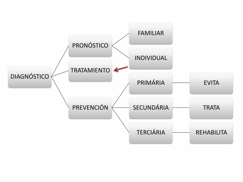
DIAGNÓSTICO
PRONÓSTICO
FAMILIAR
INDIVIDUAL
TRATAMIENTO
PREVENCIÓN
PRIMÁRIA EVITA
SECUNDÁRIA TRATA
TERCIÁRIA REHABILITA

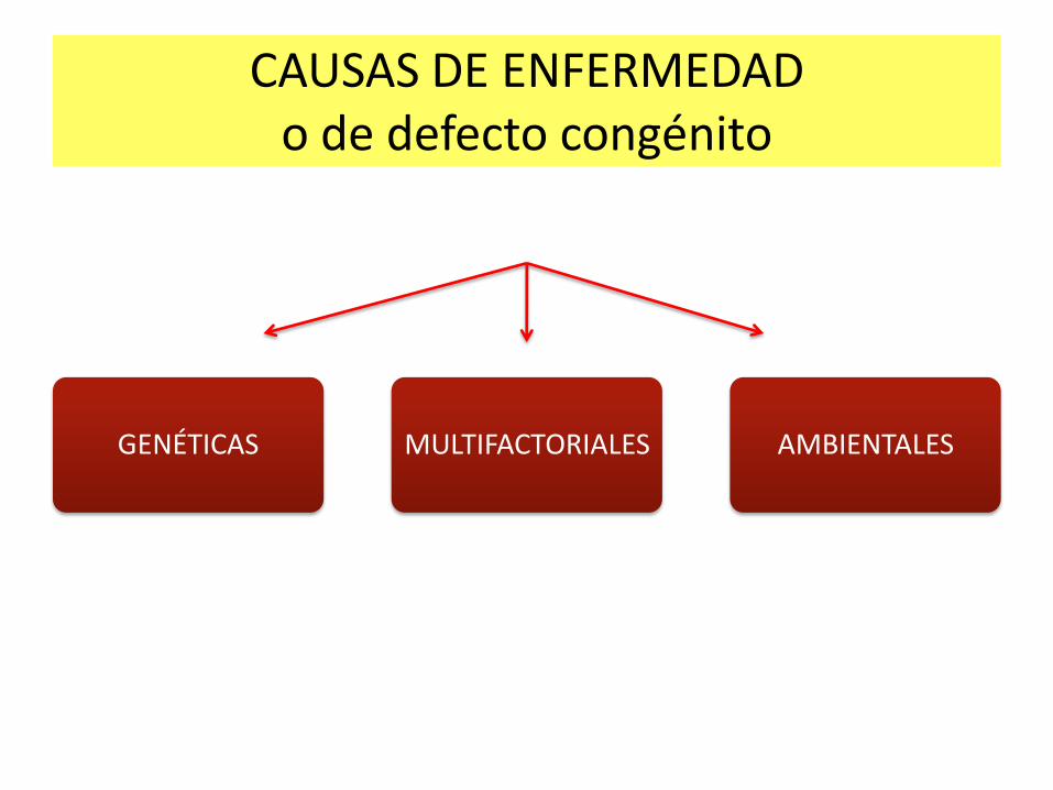
GENÉTICAS MULTIFACTORIALES AMBIENTALES
CAUSAS DE ENFERMEDAD o de defecto congénito
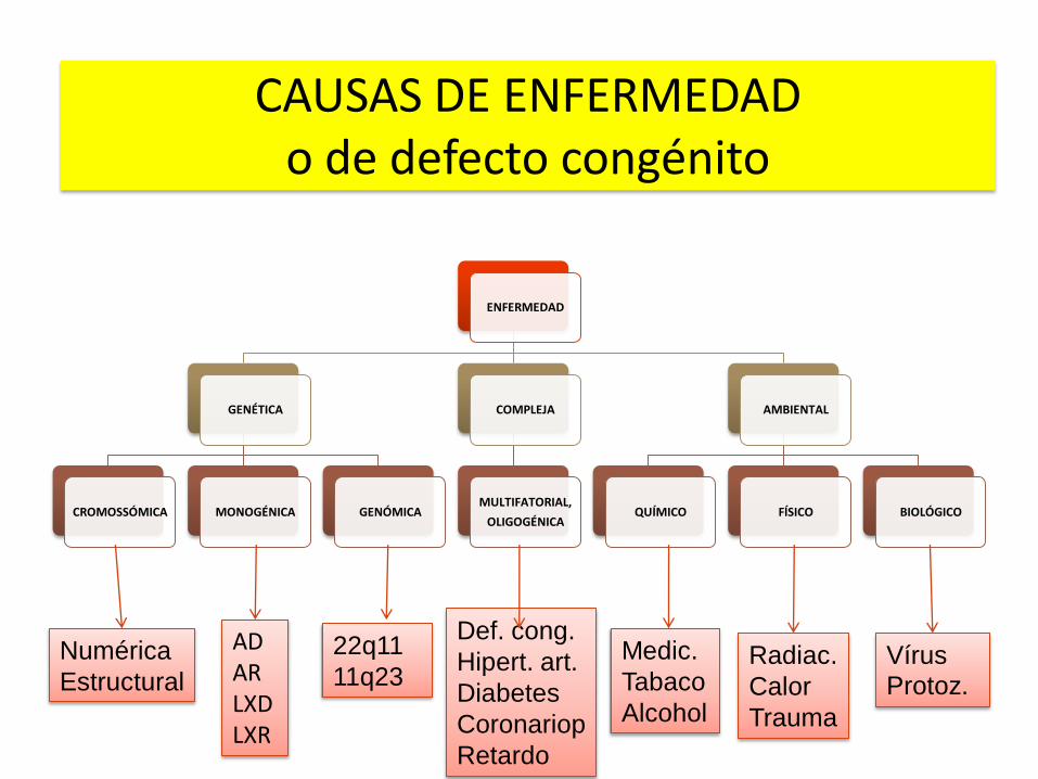
ENFERMEDAD
GENÉTICA
CROMOSSÓMICA MONOGÉNICA GENÓMICA
COMPLEJA
MULTIFATORIAL,
OLIGOGÉNICA
AMBIENTAL
QUÍMICO FÍSICO BIOLÓGICO
Numérica
Estructural
AD AR LXD LXR
22q11
11q23
Def. cong.
Hipert. art.
Diabetes
Coronariop
Retardo
Medic.
Tabaco
Alcohol
Radiac.
Calor
Trauma
Vírus
Protoz.
CAUSAS DE ENFERMEDAD o de defecto congénito

ANOMALIAS CROMOSÓMICAS
Trisomia 13
ANOMALIAS GENÓMICAS Síndrome VCF (22q21-)
ANOMALIAS MONOGÉNICAS Síndrome EEC (P63)
ANOMALIAS AMBIENTALES Embriopatia por isotretinoina
HETEROGENEIDAD ETIOLÓGICA: FISURAS ORALES
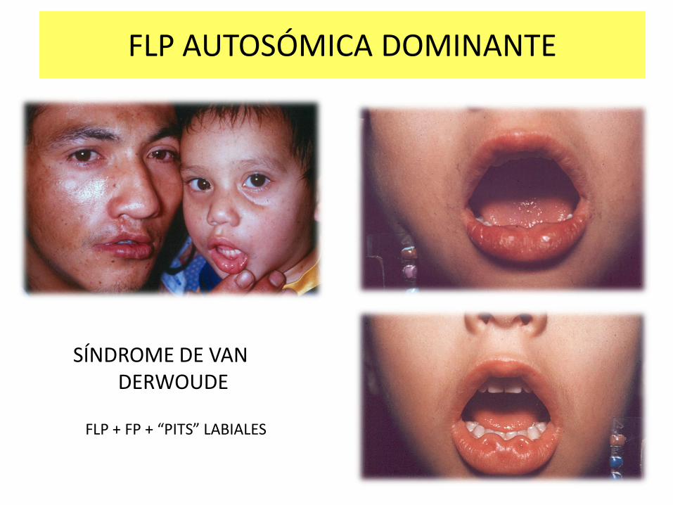
SÍNDROME DE VAN DERWOUDE
FLP AUTOSÓMICA DOMINANTE
FLP + FP + “PITS” LABIALES

PENETRANCIA Y EXPRESIVIDAD VARIABLE: SVW
NO PENETRANCIA FISURAS MISTAS
Cleft lip
Lip pits Obligate Carrier
Cleft palate
132
150
C A
G
C C
T
A
G
NSCLP
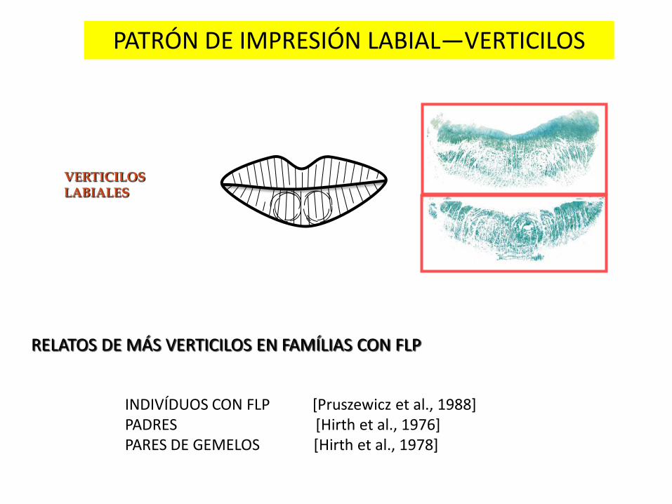
PATRÓN DE IMPRESIÓN LABIAL—VERTICILOS
VERTICILOS LABIALES
RELATOS DE MÁS VERTICILOS EN FAMÍLIAS CON FLP
INDIVÍDUOS CON FLP [Pruszewicz et al., 1988] PADRES [Hirth et al., 1976] PARES DE GEMELOS [Hirth et al., 1978]
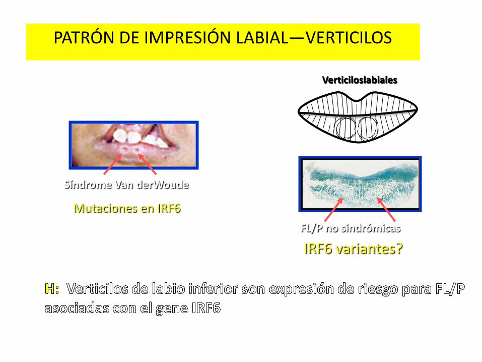
PATRÓN DE IMPRESIÓN LABIAL—VERTICILOS
Síndrome Van derWoude
Mutaciones en IRF6
FL/P no sindrómicas
IRF6 variantes?
Verticiloslabiales
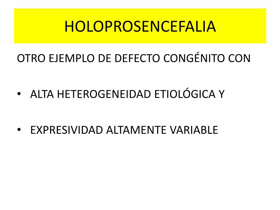
HOLOPROSENCEFALIA
OTRO EJEMPLO DE DEFECTO CONGÉNITO CON
• ALTA HETEROGENEIDAD ETIOLÓGICA Y
• EXPRESIVIDAD ALTAMENTE VARIABLE
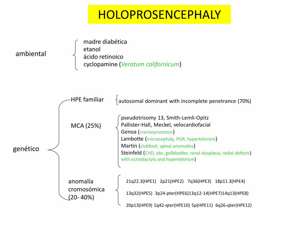
HOLOPROSENCEPHALY
ambiental
madre diabética
genético
MCA (25%) pseudotrisomy 13, Smith-Lemli-Opitz Pallister-Hall, Meckel, velocardiofacial Genoa (craniosynostosis) Lambotte (microcephaly, PGR, hypertelorism) Martin (clubfoot, spinal anomalies) Steinfeld (CHD, abs. gallbladder, renal dysplasia, radial defects) with ectrodactyly and hypertelorism)
anomalía cromosómica (20- 40%)
HPE familiar
21q22.3(HPE1) 2p21(HPE2) 7q36(HPE3) 18p11.3(HPE4) 13q32(HPE5) 3p24-pter(HPE6)13q12-14(HPE7)14q13(HPE8) 20p13(HPE9) 1q42-qter(HPE10) 5p(HPE11) 6q26-qter(HPE12)
autosomal dominant with incomplete penetrance (70%)
etanol ácido retinoico cyclopamine (Veratum californicum)

Genes HPE: • SHH (7q36) •ZIC2 (13q32) •SIX3 (2p21) •TGIF (18p11.3) •PTCH (9q22.3) •TDGF1 (3p21.3) •GLI2 (2q14) •DHCR7(11q13) •FAST1/FOXH1 (8q24.3) •DISP1 (1q42) •FGF8 (10q24)
AUTOSÓMICA DOMINANTE CON PENETRANCIA INCOMPLETA
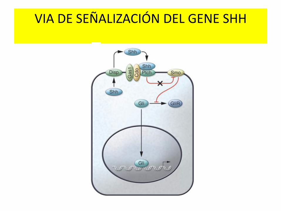
VIA DE SEÑALIZACIÓN DEL GENE SHH

ESPECTRO DE ANOMALÍAS FACIALES EN LA HOLOPROSENCEFALIA
1 2 3 4
5 6 7 8
9 10 11
1. Ciclopía 2. Ciclopía 3. Ciclopía con sinoftalmos 4. Ciclopía con sinoftalmos 5. Ciclopía con sinoftalmos 6. Ciclopía con sinoftalmos 7. Etmocefalia 8. Cebocefalia 9. Cebocefalia 10. Agenesia de la pre maxila 11. Agenesia de la pre maxila
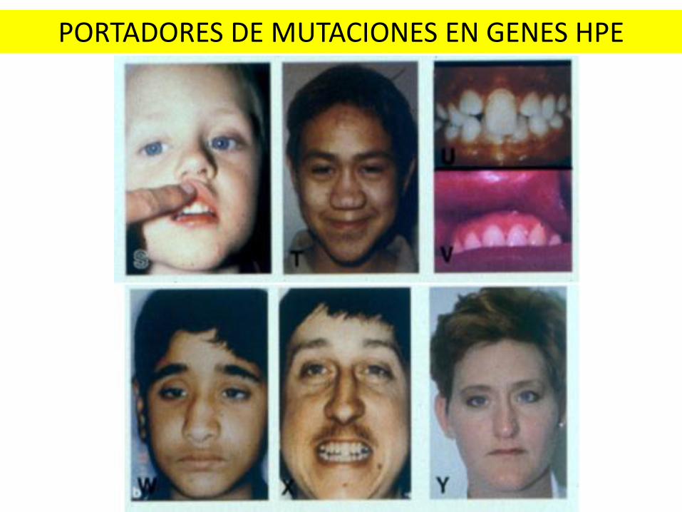
PORTADORES DE MUTACIONES EN GENES HPE
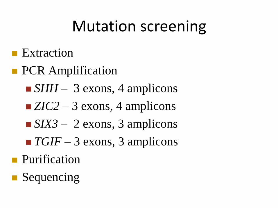
Mutation screening
Extraction
PCR Amplification
SHH – 3 exons, 4 amplicons
ZIC2 – 3 exons, 4 amplicons
SIX3 – 2 exons, 3 amplicons
TGIF – 3 exons, 3 amplicons
Purification
Sequencing

MLPA (Multiplex Ligation-Dependent Probe Amplification):
•Multiplex PCR for copy number quantification •Probes of target regions are amplified in the case of hybridization •Analysis is performed by capillary electrophoresis •Cases are compared with controls of the same type of extraction

MLPA (Multiplex Ligation-Dependent Probe Amplification):
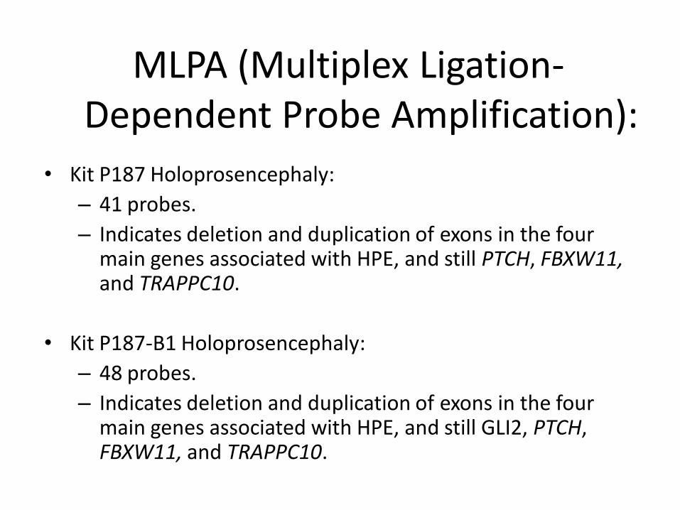
• Kit P187 Holoprosencephaly:
– 41 probes.
– Indicates deletion and duplication of exons in the four main genes associated with HPE, and still PTCH, FBXW11, and TRAPPC10.
• Kit P187-B1 Holoprosencephaly:
– 48 probes.
– Indicates deletion and duplication of exons in the four main genes associated with HPE, and still GLI2, PTCH, FBXW11, and TRAPPC10.

Screening for trisomies by QF-PCR:
• QF-PCR
(quantitativefluorescentpolymerasechainreaction) - in the early exponential phase of amplification, the quantity of product is proportional to the amount of target sequences present in the initial sample.
• The kit uses STR markers to determine the incidence of trisomies of chromosomes 13, 18, 21 and the sex chromosomes aneuploidies.
• STR markers are amplified by PCR with primers labeled with fluorescence. – 4 markers for each chromosome
– Specific kits for each chromosome are used to confirm the results
• The alleles are determined by capillary electrophoresis.
• The amount of fluorescent PCR product is a numerical value which corresponds to the peak area viewed in the electropherogram.
Screening for trisomies by QF-PCR:
Screening for trisomies by QF-PCR:
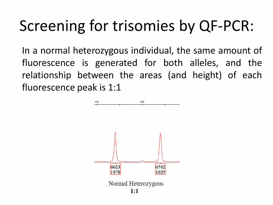
In a normal heterozygous individual, the same amount of fluorescence is generated for both alleles, and the relationship between the areas (and height) of each fluorescence peak is 1:1

Screening for trisomies by QF-PCR:
In trisomy: three peaks with same fluorescence intensity and a ratio between the peak areas (or heights) of 1:1:1 (in the case of trissomictriallelic), or two fluorescence peaks with an unbalanced area/height ratio of 2:1 (trissomicdiallelic).

Estudio de Holoprosencefalía
Methodologia
Numero de casos
estudiados
Numero de casos
estudiados del ECLAMC
Mutation screening of SHH, ZIC2, SIX3,
and TGIF gene 111 56
Screening of microdeletions by MLPA 50 17
Screening for trisomiesby QF-PCR 52 36
Cariotipo
No SHH, SIX3, ZIC2, TGIF
No MLPA
No ?
Si
Si
Si
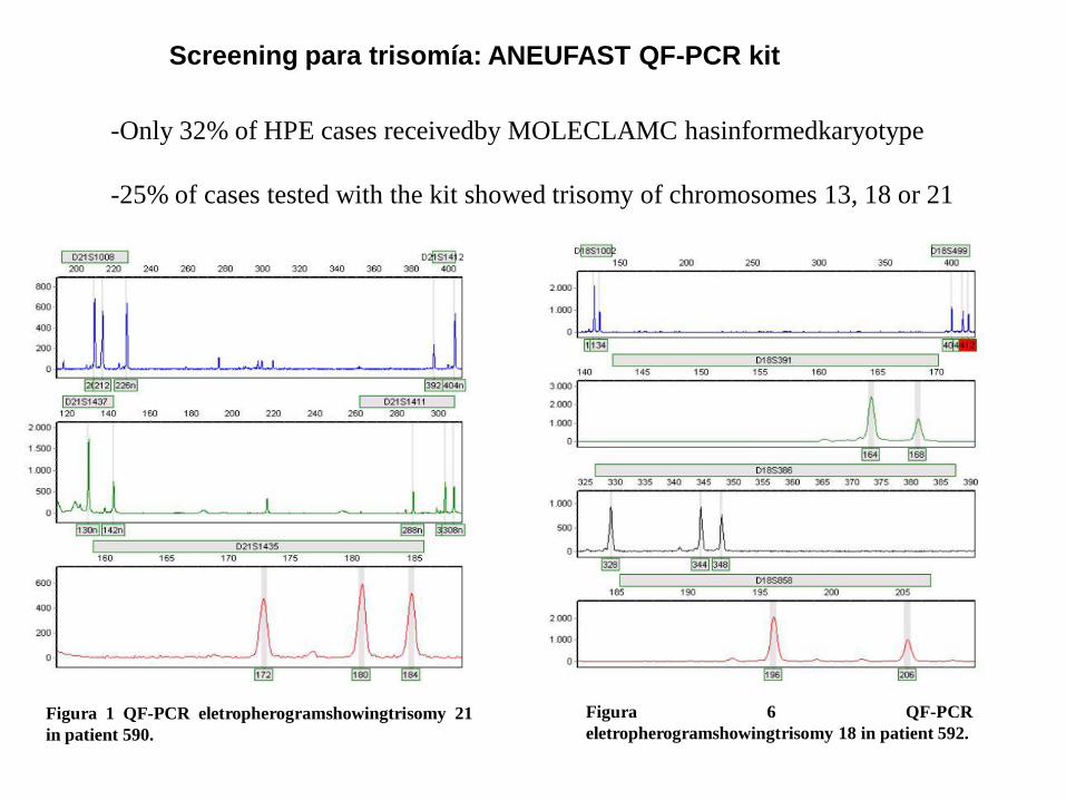
Figura 1 QF-PCR eletropherogramshowingtrisomy 21
in patient 590.
Figura 6 QF-PCR
eletropherogramshowingtrisomy 18 in patient 592.
-Only 32% of HPE cases receivedby MOLECLAMC hasinformedkaryotype
-25% of cases tested with the kit showed trisomy of chromosomes 13, 18 or 21
Screening para trisomía: ANEUFAST QF-PCR kit
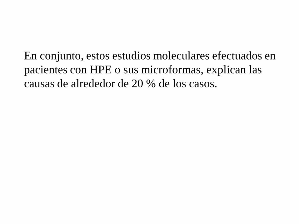
En conjunto, estos estudios moleculares efectuados en
pacientes con HPE o sus microformas, explican las
causas de alrededor de 20 % de los casos.

SÍNDROMES CON CRANIOSINOSTOSIS
SÍNDROME • Apert
• Crouzon
• Muenke
• Pfeiffer
• Jackson-Weiss
• Crouzonconacanthosisnigricans
• Saethre-Chotzen
• Beare Stevenson
• Displasia craniofrontonasal
GENES • FGFR2
• FGFR2
• FGFR3, pro250arg
• FGFR1, FGFR2
• FGFR2
• FGFR2
• TWIST
• FGFR2
• EFNB1
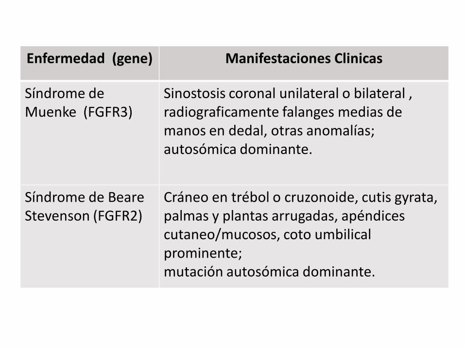
Enfermedad (gene) Manifestaciones Clinicas
Síndrome de Muenke (FGFR3)
Sinostosis coronal unilateral o bilateral , radiograficamente falanges medias de manos en dedal, otras anomalías; autosómica dominante.
Síndrome de Beare Stevenson (FGFR2)
Cráneo en trébol o cruzonoide, cutis gyrata, palmas y plantas arrugadas, apéndices cutaneo/mucosos, coto umbilical prominente; mutación autosómica dominante.
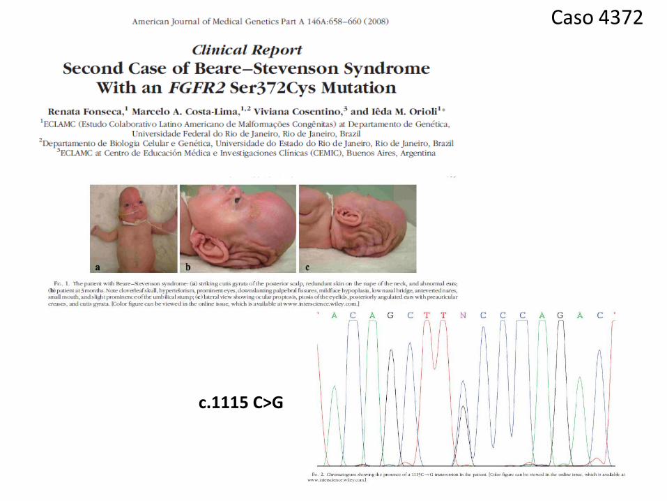
Caso 4372
c.1115 C>G
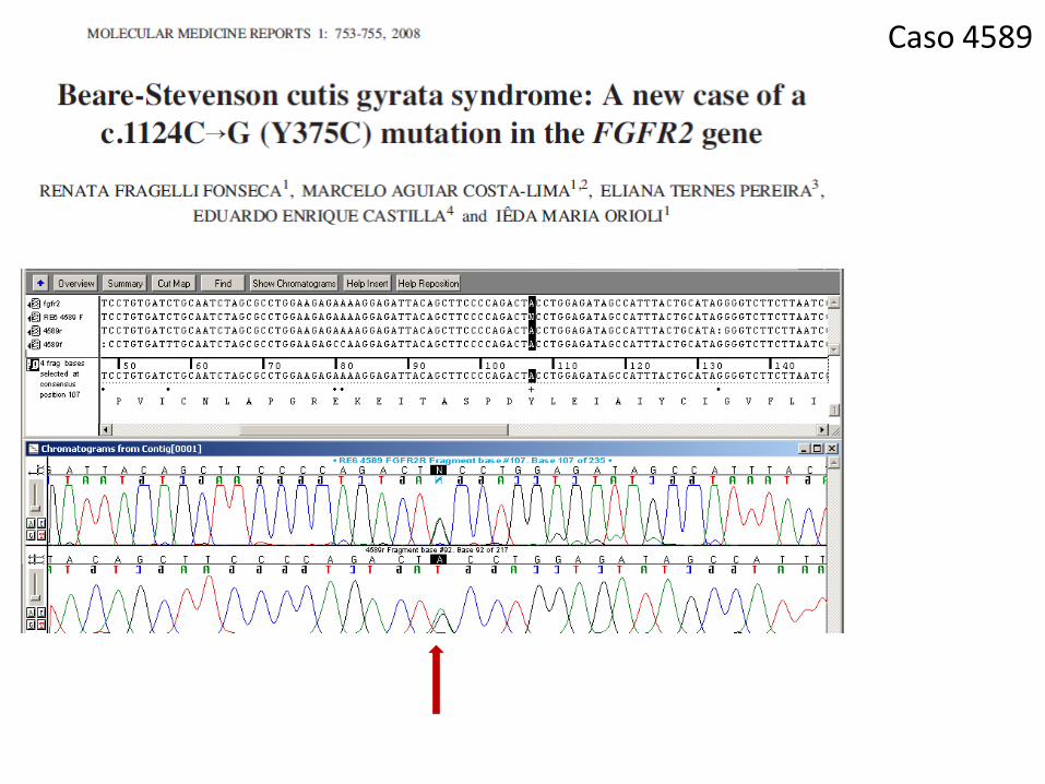
Caso 4589
Agradecimientos