anterior and posterior pituitary cells and its functions
TRANSCRIPT

12/27/20151
ANTERIOR AND
POSTERIOR PITUITARY
CELLS & ITS
FUNCTIONS
DR. VIJAY JAISWAL, Asso. Professor ,
Fellow Pediatric . Endocrinology

The pituitary gland is the central regulator of growth,
reproduction,and homeostasis. MASTER GLAND ‘
PEEYUSH GRANTHEE’
It functions through hormone signaling pathways that
co-ordinate signals from the brain and the
hypothalamus to target organs, such as the adrenals,
thyroid, and gonads.
The pituitary lies within the sella turcica at the base of
the brain, and the mature gland consists of the
adenohypophysis (anterior and intermediate lobes)
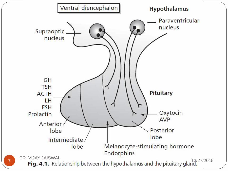
and the neurohypophysis (posterior lobe).12/27/2015DR. VIJAY JAISWAL2

The posterior lobe consists of axons of neurons,
the cell bodies of which reside in the
hypothalamus.
The posterior pituitary stores and releases
oxytocin, which is required during parturition and
lactation, and arginine vasopressin (AVP), which
regulates water balance
12/27/2015DR. VIJAY JAISWAL3

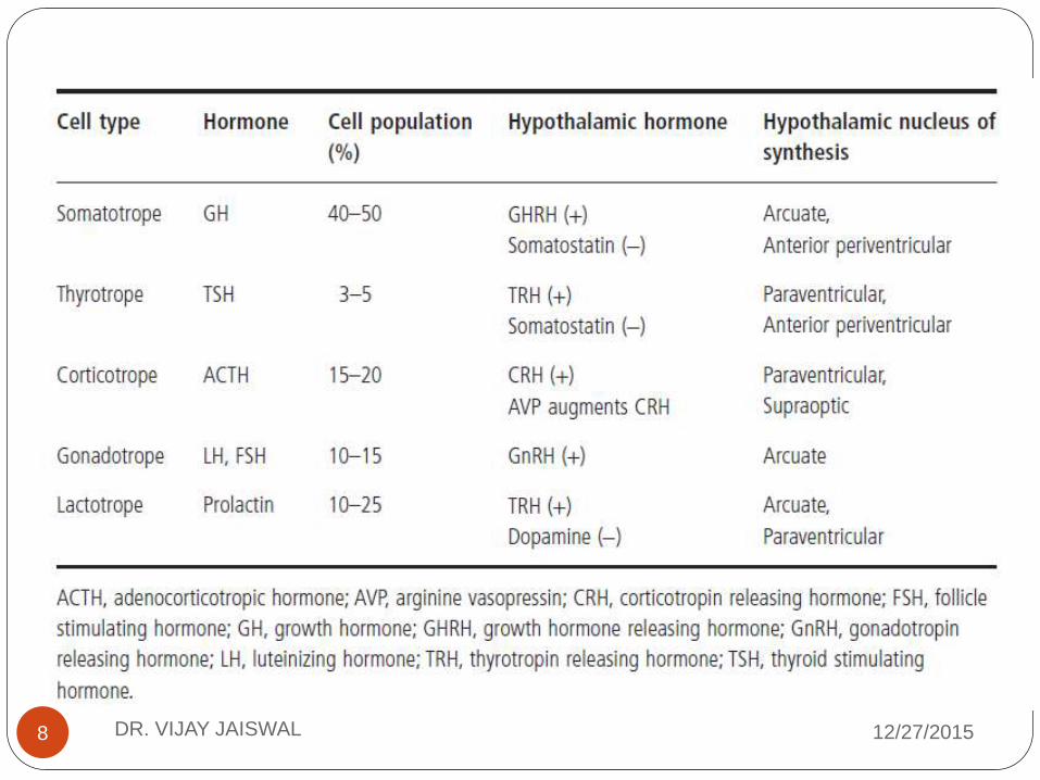
The function of the pituitary gland is intricately linked
with that of the hypothalamus. Stimulatory and
inhibitory releasing hormones are secreted from the
hypothalamus, which regulate hypothalamo-pituitary–
target gland axes.
These include
• corticotrophin-releasing hormone (CRH)
•thyrotrophin-releasing hormone (TRH),
•GH-releasing hormone(GHRH)
•gonadotropin-releasing hormone (GnRH)
inhibitory hormones dopamine and somatostatin.
The hormones secreted by the posterior lobe are
synthesized in the magnocellular neurons of the
paraventricular and supraoptic nuclei that lie within
the hypothalamus12/27/2015DR. VIJAY JAISWAL4

12/27/2015DR. VIJAY JAISWAL5

12/27/2015DR. VIJAY JAISWAL6
12/27/2015DR. VIJAY JAISWAL
7
12/27/2015DR. VIJAY JAISWAL8

Hormonal deficits can be one part of a syndrome, with
patients manifesting abnormalities in extra pituitary
structures,
usually in structures sharing a common embryological
origin, such as the eye and forebrain.
12/27/2015DR. VIJAY JAISWAL9
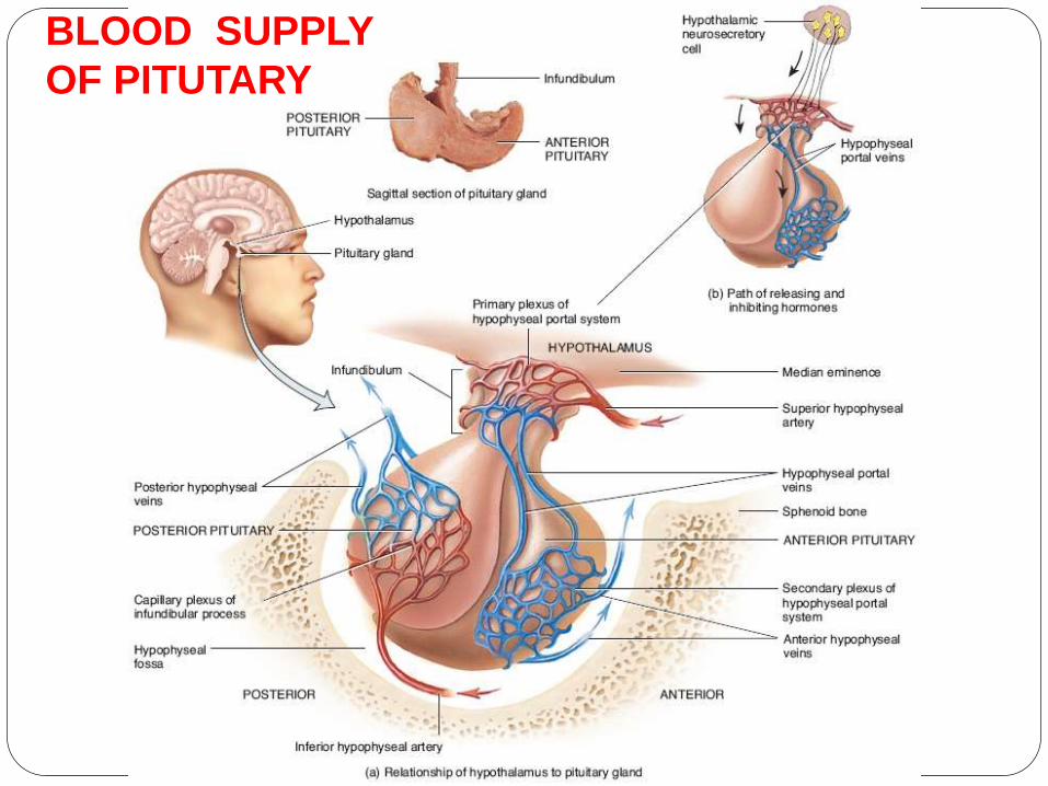
BLOOD SUPPLY
OF PITUTARY

BLOOD SUPPLY
The hypothalamus is supplied by circle of Willis and
most venous return into vein of Galen.
Blood from supr. Hypophyseal artery, which arise
from internal carotid arteries , flow through capillary
plexus in the median eminence to enter in a
sinusoidal network in the pituitary stalk. Blood
passes from these sinusoids into a second capillary
network plexus in anterior pituitary. This venous
portal system linking these two capillary network is
called the Hypothalamo – pituitary portal system .
The infundibulum or pituitary stalk carries both
the portal blood supply and the neural tracts to the
pituitary gland, so damage to the pituitary stalk
results in anterior and posterior pituitary dysfunct12/27/2015DR. VIJAY JAISWAL11
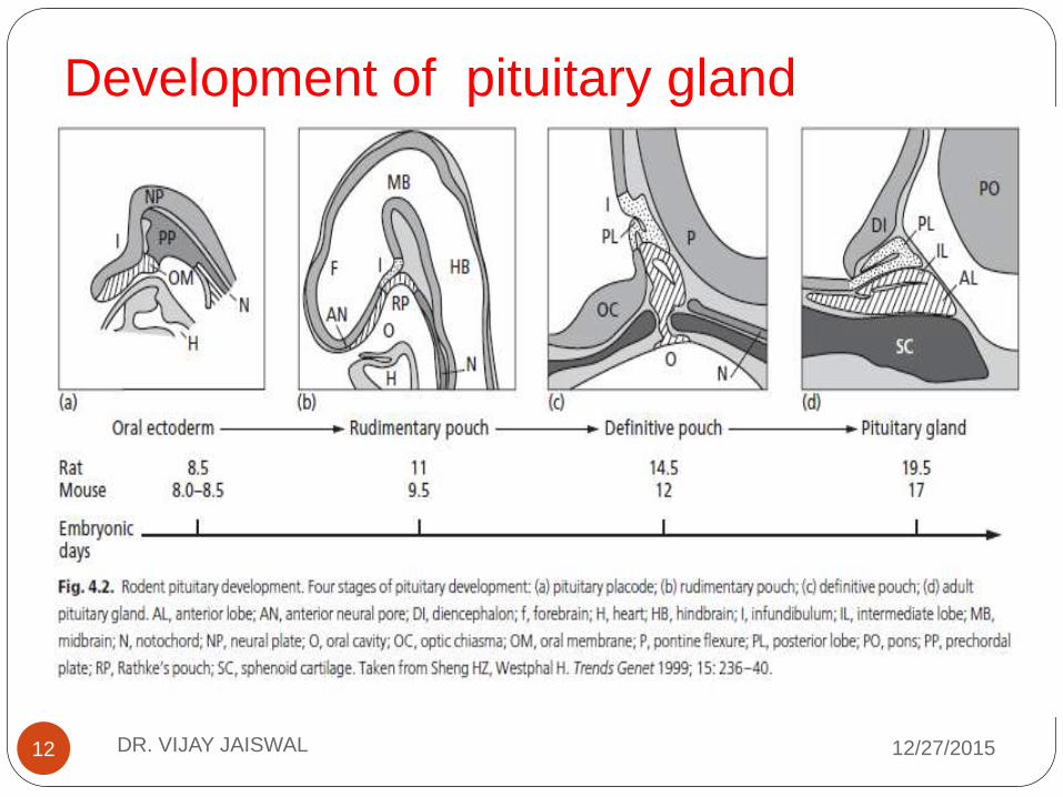
Development of pituitary gland
12/27/2015DR. VIJAY JAISWAL12

1 Formation of the pituitary placode derived from
the ectoderm
at the roof of the primitive oral cavity, which makes
contact with the floor of the ventral diencephalon.
2 Invagination of the oral ectoderm and formation
of a rudimentary Rathke’s pouch with evagination of
the ventral diencephalon to form the posterior
pituitary.
3 Formation of the definitive Rathke’s pouch.
4 Spatial and temporal differentiation of the
various cell types within the mature anterior pituitary
gland.12/27/2015DR. VIJAY JAISWAL13

Early developmental genes and transcription
factors
A number of signaling molecules and transcription
factors
are implicated in early pituitary organogenesis and
lineage
differentiation.
They are expressed sequentially at critical periods of
pituitary development, and the expression of many is
subsequently attenuated
12/27/2015DR. VIJAY JAISWAL14

Morphogenetic signals (BMP, FGF, SHH, Wnt)
Close interaction between the oral and neural
ectoderm is required for initial pituitary development.
Extrinsic molecules within the ventral diencephalon
and the surrounding structures,such as
• bone morphogenetic proteins 2 and 4 (BMP 2,4)
• fibroblast growth factor 8 (FGF8),
•Sonic Hedgehog (SHH)
•Wingless (Wnt4),
•thyroid transcription factor (Ttf1; also called Nkx2.1),
play critical roles in early organogenesis.
Rathke’s pouch develops in a two-step process
requiring at least two sequential inductive signals from
the diencephalon.12/27/2015DR. VIJAY JAISWAL15

12/27/2015DR. VIJAY JAISWAL16

12/27/2015DR. VIJAY JAISWAL17

Hesx1
Homeobox gene Expressed in embryonic stem cells
Hesx1 is one of the earliest markers of the pituitary
primordium,suggesting that it has a critical role in
early determination and differentiation of the gland. It
is also called Rpx (Rathke’s pouch homeobox) and
is a member of the paired like class of homeobox
genes.
12/27/2015DR. VIJAY JAISWAL18

Isolated hormone abnormalities
GH1 Isolated growth hormone deficiency R, D
GHRHR Isolated growth hormone deficiency R
TSH beta Isolated TSH deficiency and secondary hypothyroidism
R
TRHR Isolated TSH deficiency and secondary hypothyroidism R
T-PIT ACTH deficiency R
PC 1 ACTH deficiency, hypoglycemia, impaired glucose tolerance,
HH, obesity R
POMC ACTH deficiency, obesity, red hair R
DAX1 Adrenal hypoplasia congenital and HH XL
GnRHR Isolated gonadotropin deficiency and HH R
KAL-1 Kallman syndrome XL
FSH beta Primary amenorrhea; defective spermatogenesis R
LH beta Delayed puberty R
AVP Diabetes insipidus R
12/27/2015DR. VIJAY JAISWAL19
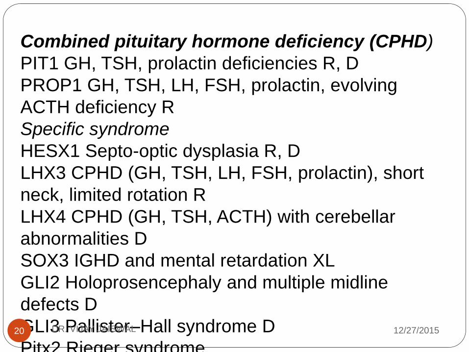
Combined pituitary hormone deficiency (CPHD)
PIT1 GH, TSH, prolactin deficiencies R, D
PROP1 GH, TSH, LH, FSH, prolactin, evolving
ACTH deficiency R
Specific syndrome
HESX1 Septo-optic dysplasia R, D
LHX3 CPHD (GH, TSH, LH, FSH, prolactin), short
neck, limited rotation R
LHX4 CPHD (GH, TSH, ACTH) with cerebellar
abnormalities D
SOX3 IGHD and mental retardation XL
GLI2 Holoprosencephaly and multiple midline
defects D
GLI3 Pallister–Hall syndrome D
Pitx2 Rieger syndrome12/27/2015DR. VIJAY JAISWAL20

Prop1 is a pituitary-specific paired-like homeodomain
activator. Both transcription factors are believed to bind
to the same DNA response elements.
Hesx1 and Prop1 function as opposing transcription
factors
and that the careful temporal regulation of their
expression
is critical for normal pituitary development
Premature expression of Prop1 can block pituitary
organogenesis. Prolonged expression of Hesx1 with the
obligate co-repressor TLE1 can block Prop1-dependent
activation, which normally results in the appearance of
somatotrophs, lactotrophs, thyrotrophs, and
gonadotrophs.
switch between binding of a paired homeodomain
repressor Hesx1 for a paired homeodomain activator
12/27/2015DR. VIJAY JAISWAL21

Pitx1 and Pitx2
Pitx1 and Pitx2 are paired-like homeobox genes
expressed in the fetal pituitary and in most cells of
the adult gland. They play an important role in the
development of Rathke’s pouch and anterior
pituitary gland.
Pitx2 is first expressed in the embryo in oral
epithelium and oral ectoderm. Pitx2 is expressed
in the developing Rathke’s pouch in addition to
the mesenchyme near the optic eminence, the
basal plate of the central nervous system (CNS),
the forelimbs, and domains of the abdominal
cavity. It appears to be required for pituitary
development shortly after formation of the
committed pouch.
12/27/2015DR. VIJAY JAISWAL22
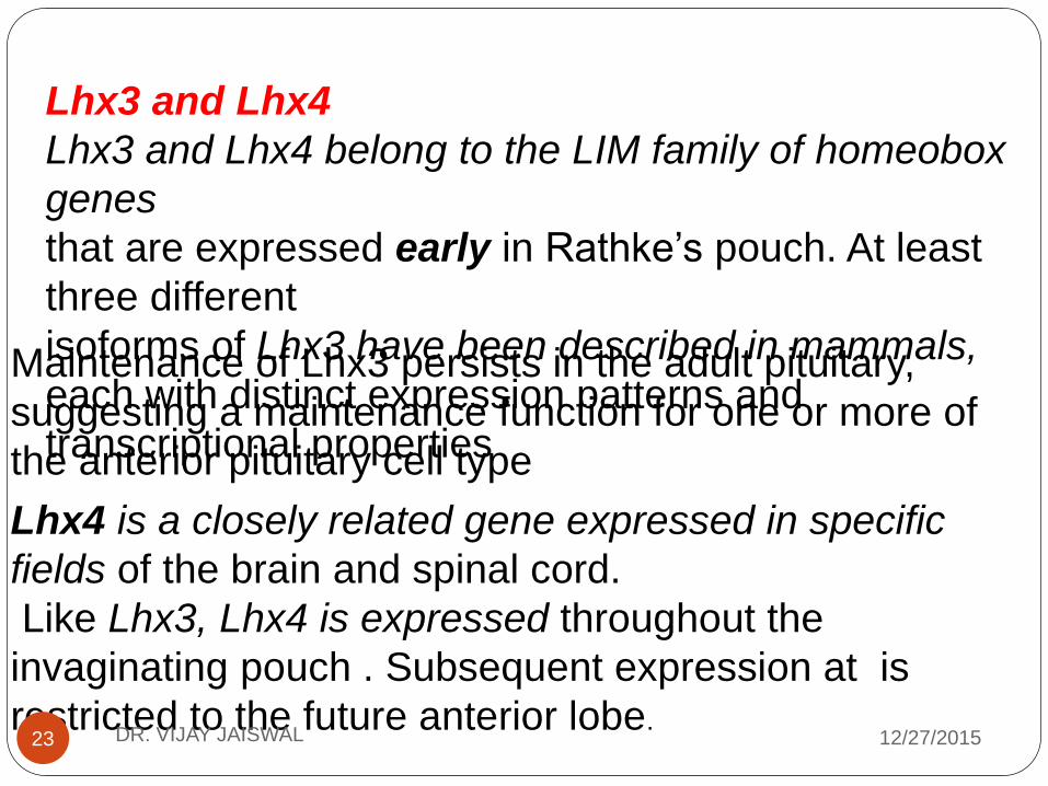
Lhx3 and Lhx4
Lhx3 and Lhx4 belong to the LIM family of homeobox
genes
that are expressed early in Rathke’s pouch. At least
three different
isoforms of Lhx3 have been described in mammals,
each with distinct expression patterns and
transcriptional properties
Maintenance of Lhx3 persists in the adult pituitary,
suggesting a maintenance function for one or more of
the anterior pituitary cell type
Lhx4 is a closely related gene expressed in specific
fields of the brain and spinal cord.
Like Lhx3, Lhx4 is expressed throughout the
invaginating pouch . Subsequent expression at is
restricted to the future anterior lobe.12/27/2015DR. VIJAY JAISWAL23
12/27/2015DR. VIJAY JAISWAL24
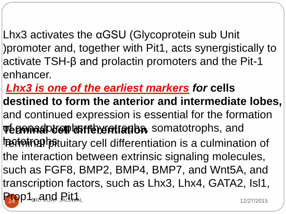
Lhx3 activates the αGSU (Glycoprotein sub Unit
)promoter and, together with Pit1, acts synergistically to
activate TSH-β and prolactin promoters and the Pit-1
enhancer.
Lhx3 is one of the earliest markers for cells
destined to form the anterior and intermediate lobes,
and continued expression is essential for the formation
of gonadotrophs, thyrotrophs, somatotrophs, and
lactotrophsTerminal cell differentiation
Terminal pituitary cell differentiation is a culmination of
the interaction between extrinsic signaling molecules,
such as FGF8, BMP2, BMP4, BMP7, and Wnt5A, and
transcription factors, such as Lhx3, Lhx4, GATA2, Isl1,
Prop1, and Pit1

12/27/2015DR. VIJAY JAISWAL25
Prop1
Prop1 (Prophet of Pit1) is a pituitary-specific
paired-like
homeodomain transcription factor, first expressed
in the
dorsal portion of Rathke pouch,
It is believed to be required for the expression of
Pit1, as there is a failure of determination of Pit-1
lineages, lack of Pit-1 gene activation, and
absence of progression to mature cells which
harbors a homozygous missense mutation (S83P)
in the Prop1 gene.
The size of the pituitary gland is considerably
reduced less than 1% of the normal complement
of somatotrophs, markedly reduced numbers of
lactotrophs and thyrotrophs, and reduced
expression of gonadotropins .

Pit1
Pit1 (now called POU1F1) is a pituitary-specific
transcription
factor belonging to the POU homeodomain family. It has
also
been called GH factor 1 (GHF1), because it was first
identified
as a regulator of GH1 transcription.
Apart from GH1, Pit1 binding sites have been identified
in the promoters of the prolactin and TSH-β genes. Pit1
is expressed relatively late during pituitary development
and expression persists throughout life.
Pit1 is sufficient to activate the minimal elements in
the GH1 promoter necessary for cell-specific
expression, it requires other factors, such as Zn-15, a
zinc finger transcription factor, for synergistic
activation of the GH1 gene.Pit1 is essential for the
development of somatotrophs, lactotrophs, and
thyrotrophs in the anterior pituitary. 12/27/2015DR. VIJAY JAISWAL26

Anterior pituitary hormones and their
deficiencies
Growth hormone
Somatotrop account for 40–50% of anterior pituitary
gland. The human GH gene (GH-N or GH1) forms part
of a cluster of five homologous genes [GH1, hCS-
L(CSHP1), hCS-A (CSH1), hGH-V (GH2), hCS-B
(CSH2)] located on the long arm of chromosome 17
(17q22–24),The full-length transcript from the GH-N gene encodes a
191-amino-acid 22 kDa protein that contains two
disulfide bridges and accounts for 85–90% of
circulating GH.
Within both the proximal promoter and the LCR are
located
binding sites for the pituitary-specific transcription factor
Pit1
12/27/2015DR. VIJAY JAISWAL27

•The half-life of hGH is less than 20 min. It binds to
the
GH receptor (GHR), which is present in a number of
tissues.
•The hormone sequentially dimerizes its receptor,
activating
a receptor-associated tyrosine kinase JAK2 that in
turn is
autophosphorylated and also phosphorylates the
GHR. This
leads to signal transduction using the mitogen-
activated
protein kinase (MAPK), signal transducer and
activator of
transcription (STAT) and phosphatidylinositol (PI) 3-
kinase
These include early response genes encoding
transcription factors (c-jun, c-fos, and c-myc implicated
in cell growth, proliferation, and differentiation) and
insulin-like growth factor I (IGF-I), which mediates the
growth-promoting effects of GH
12/27/2015DR. VIJAY JAISWAL28

Actions
GH is secreted in a pulsatile fashion. Peak
concentrations are achieved during sleep, but
secretion is also increased during emotional stress,
exercise, hypoglycemia, protein meals, and prolonged
fasting. Pharmacological agents used to increase hGH
secretion include insulin, glucagon, clonidine, L-dopa,
and propranolol.
Apart from its actions on linear growth, GH is anabolic,
lipolytic, and diabetogenic. It increases calcium
absorption and is believed to improve bone density.
Administration of hGH results in a reduction in body fat
and
an increase in muscle mass .12/27/2015DR. VIJAY JAISWAL29

GH acts indirectly on bone growth by stimulating the
synthesis of IGF-1, the main GH-dependent growth
factor. IGF-1 Is a single-chain polypeptide containing
70 amino acids. It shares considerable homology with
insulinIt is synthesized in the liver and circulates bound to
several binding glycoproteins.
The principal binding protein is IGFBP-3, the
secretion of which is also regulated by GH.
Measurement of IGF-1 correlates well with spontaneous
GH secretion and is used in the diagnosis of GH
deficiency, but its concentration is altered in a number of
other disease states, such as hypothyroidism,
malnutrition, poorly controlled diabetes, and chronic
disease12/27/2015DR. VIJAY JAISWAL30
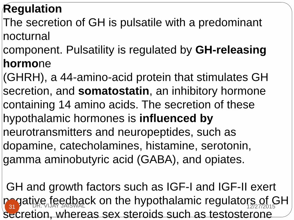
Regulation
The secretion of GH is pulsatile with a predominant
nocturnal
component. Pulsatility is regulated by GH-releasing
hormone
(GHRH), a 44-amino-acid protein that stimulates GH
secretion, and somatostatin, an inhibitory hormone
containing 14 amino acids. The secretion of these
hypothalamic hormones is influenced by
neurotransmitters and neuropeptides, such as
dopamine, catecholamines, histamine, serotonin,
gamma aminobutyric acid (GABA), and opiates.
GH and growth factors such as IGF-I and IGF-II exert
negative feedback on the hypothalamic regulators of GH
secretion, whereas sex steroids such as testosterone
and estrogen increase hGH secretion.
12/27/2015DR. VIJAY JAISWAL31

Thyrotrophin or thyroid-stimulating hormone
Thyrotrophin is a glycoprotein consisting of two non
covalently bound chains of amino acids (α and β) that is
synthesized and stored within the thyrotrophs of the
pituitary gland.
The α-chain consists of 92 amino acids and shares
homology with other pituitary glycoproteins FSH and
LH.The β-chain contains 110 amino acids and is TSH
specific
Actions
The primary function of TSH is to stimulate the thyroid
gland
to secrete triiodothyronine (T3) and thyroxine (T4 ). Its
actions include stimulation of the iodide pump on the cell
membrane transporting iodide into the cell, stimulation of
the synthesis of the thyroidal storage protein
thyroglobulin, and stimulation and synthesis of T4 and
12/27/2015DR. VIJAY JAISWAL32

TSH binds to its cell membrane receptor, which
consists of seven transmembrane domains, four
intracellular domains, and a long extracellular
sequence with six potential glycosylation sites. It is
a G-protein-coupled receptor that stimulates adenyl
cyclase activity, activation of protein kinase A, and
subsequent phosphorylation.
12/27/2015DR. VIJAY JAISWAL33

Regulation
TSH secretion is pulsatile with peak
concentrations at night. Its secretion is stimulated
by hypothalamic TRH acting via its Gprotein coupled
receptor and inhibited by somatostatin and
dopamine.
The thyroid hormones negatively feedback both at
the
pituitary level on TSH secretion and at the
hypothalamic level on TRH. Other factors impinging
on TSH secretion include estrogen which increases
the number of TRH receptors on the thyrotropes
decrease in the ambient temperature acting as a
potent stimulator of TSH.12/27/2015DR. VIJAY JAISWAL34

Adrenocorticotropic hormone
ACTH is a 39 amino acid polypeptide with a short
biological
half-life of approximately 8 min. It is synthesized and
stored
within the corticotropes of the anterior pituitary that
account for about 10% of the adenohypophysis.
Actions and regulation
Actions
The primary function of ACTH is to stimulate the zona
fasciculata
and zona reticularis of the adrenal glands to produce
glucocorticoids (mainly cortisol) and adrenal
androgens.
Like other peptide hormones, ACTH binds to its specifi c
membrane receptor on the adrenocortical cells to
increase the formation of cyclic AMP and activation of
various protein kinases
12/27/2015DR. VIJAY JAISWAL35

12/27/2015DR. VIJAY JAISWAL36
Regulation
The secretion of ACTH follows a circadian rhythm with
peak
concentrations in the early hours of the morning and
low concentrations in the late evening.
As a result, cortisol secretion is circadian with peak
concentrations at around 0800 hours and a nadir
at midnight.
This rhythm can be disrupted by shifts in day–night
patterns.

Gonadotropins
The reproductive system is unique because of changes
in the secretion
of reproductive hormones taking place throughout life.
The
gonadotropins, FSH and LH, are glycoproteins
composed of two
subunits: α and β. The α-subunit is identical to the α-
subunit of
TSH (gene encoding the α-subunit is located on
chromosome
6q12-q21) and the specific biological activity of both
hormones resides in the β-subunit.
LH secretion is pulsatile in both sexes but sexual
dimorphism in
physiological secretory patterns becomes evident with
maturity of
the hypothalamo-pituitary-gonadal axis. An increased
nocturnal
LH release is the first sign of the onset of puberty.
.
12/27/2015DR. VIJAY JAISWAL37

12/27/2015DR. VIJAY JAISWAL38
Actions and regulation
Actions
Both hormones bind to membrane receptors in their
ovarian and
testicular cells, activate the G-protein coupled complex
and stimulate adenyl cyclase.
FSH regulates gametogenesis in males and females
while LH is thought to be primarily responsible for
gonadal steroid secretion

Regulation
Pulsatile release of hypothalamic GnRH regulates the
secretion of the pituitary hormones LH and FSH which
stimulate the testis
and ovary at puberty to increase the gonadal steroid
secretion and develop secondary sexual characteristics.
There is a surge in gonadotropin and gonadal steroid
secretion in the neonatal period with concentrations
similar to those reached during puberty following the first
few months of life, the gonadotropin axis
remains quiescent until puberty, when concentrations
rise again.
GnRH-synthesizing neuronal migration, from their first
appearance
in the embryonic medial olfactory placode to their final
position in the medio basal hypothalamus, is complete
by around
12/27/2015DR. VIJAY JAISWAL39

Estradiol and progesterone act via both the pituitary
and hypothalamus to have a negative effect on
gonadotropin
secretion.
However, if plasma estradiol concentrations are very
high for a period greater than approximately 36 h in
the absence of plasma progesterone, a positive
feedback influence is exerted with an LH surge as
seen in the mid-menstrual cycle in females.
FSH secretion is also regulated by inhibin, a protein
molecule secreted by the follicular granulosa cells in
the female and Sertoli cells in the male.
12/27/2015DR. VIJAY JAISWAL40

Prolactin
Action, regulation and deficiency
Prolactin is a 199 amino acid protein with its gene
located on
chromosome 6p22.2-21.3.
The principal functions of prolactin are growth and
development of the breasts and initiation and
maintenance of lactation in postpartum women.
It also has some role in regulation of gonadal function
by stimulating the generation of LH receptors in the
gonads in both sexes.
The mechanism of action of prolactin is similar to
other protein molecules by stimulating the tyrosine
kinase pathway and subsequent intracellular protein
phosphorylation.
12/27/2015DR. VIJAY JAISWAL41

12/27/2015DR. VIJAY JAISWAL42
The release of prolactin is under the control of the
hypothalamus with afferent impulses from sensory
receptors, primarily around the nipples.
The dominant hypothalamic influence is
inhibitory and the principal inhibitory hormone is
dopamine.
Other molecules exerting an inhibitory role are
norepinephrine, histamine and serotonin acting at
either a hypothalamic or pituitary level.
TRH, in addition to stimulating the release of TSH,
is also the principal prolactin stimulatory hormone.
Thyroxine and estrogen can modulate the number
of TRH receptors in the lactotropes, thereby
influencing prolactin release
Posterior pituitary hormones
The neurohypophysis consists of the supraoptic and
paraventricular hypothalamic nuclei containing the
cell bodies of the magnocellular neurosecretory
neurons that secrete vasopressin and oxytocin, the
supraoptico-hypophyseal tract that includes the
axons of these neurons, and the posterior pituitary
where the axons terminate on capillaries of the
inferior hypophyseal artery.
Arginine vasopressin
Vasopressin is a basic nano peptide with a disulfide
bridge between
the cysteine residues at positions 1 and 6. Most
mammals have
the amino acid arginine at position 8. 12/27/2015DR. VIJAY JAISWAL43

12/27/2015DR. VIJAY JAISWAL44
The preprohormone is synthesized in the magnocellular
neuron cell body, following which the signal peptide is
cleaved.
Stimulation of vasopressinergic neurons results in the
opening of voltage gated Calcium channels in the nerve
terminals, which through
transient calcium influx results in fusion of the neuro
secretory
granules with the nerve terminal membrane and release
of their
contents into the circulation.
The half-life of vasopressin is short, approximately 5–15
min.

Actions and regulation
Vasopressin acts via three G-protein coupled
receptors:
• pressor effects via V1 receptors,
• renal effects via V2 receptors (V2-R)
• action on corticotropes to secrete ACTH in synergy
with CRH via V3 receptors. Activation of the V2-Rleads to a biphasic increase in the expression of the
water channel
protein aquaporin 2. This then allows reabsorption of
water from
the duct lumen along an osmotic gradient, with excretion
of
concentrated urine.
The main regulatory factors in determining vasopressin
secretion
are osmotic status, blood pressure and circulating
12/27/2015DR. VIJAY JAISWAL45

Oxytocin
The oxytocin gene lies on chromosome 20p13 and
consists of
three exons, which, like vasopressin, encodes a
polypeptide precursor with an amino-terminal signal
peptide, the oxytocin
peptide, neurophysin and a carboxy-terminal peptide.
The half-life of oxytocin is short. Oxytocin binds to a G-
protein coupled cell surface receptor on target cells to
mediate a variety of physiological effects largely
concerned with reproductive function, namely the
regulation of lactation, parturition and reproductive
behavior.
In humans, women lacking posterior pituitary function
can breast-feed normally, illustrating that oxytocin not
12/27/2015DR. VIJAY JAISWAL46

12/27/2015DR. VIJAY JAISWAL47
•Pituitary gland is the central regulator of growth ,
Reproduction and homeostasis.
•Anterior Pituitary Consist of 5 cell types
and 6 hormones.
Somatotrop constitute major bulk of Anterior
Pituitary (40-50%)
•Anterior Pituitary develops from oral ectoderm while
Posterior develop from neural ectoderm.
•Any damage to Pituitary stalk can result Anterior and
Posterior Pitutary dysfunction

12/27/2015DR. VIJAY JAISWAL48
•Clinical Features of Hypopituitarism are
Variable both in Severity
and Number of hormone Deficiencies
•Complete Evaluation of H-P Axis is indicated
in any patient suspected of having one hormonal
Deficiency
•HesX1 is one of the earliest markers of early
determination & differentiation of the pituitary gland.
•Cascade of signaling molecules and transcription
factors have crucial role in organ commitment, cell
proliferation and terminal differentiation and the final
product is culmination of this coordinated process

THANK
YOU