ankle fractures ram pulavarti 4 th april 2005 foot and ankle term spr teaching day sunderland royal...
Post on 20-Dec-2015
221 views
TRANSCRIPT

Ankle Fractures
Ram Pulavarti
4th April 2005
Foot and Ankle Term
Spr Teaching Day
Sunderland Royal Hospital
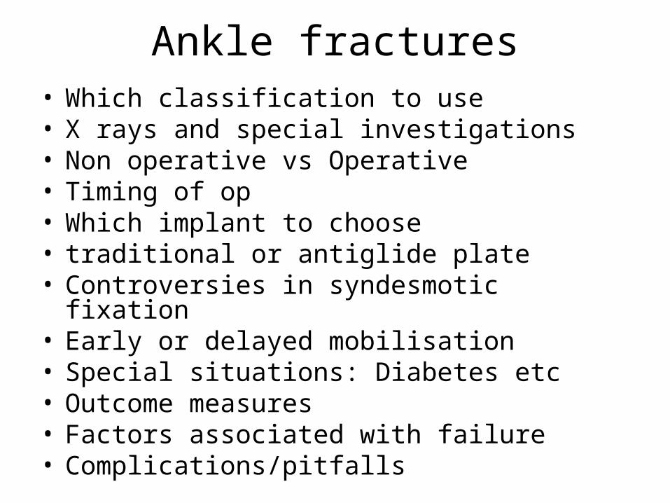
Ankle fractures• Which classification to use• X rays and special investigations• Non operative vs Operative• Timing of op• Which implant to choose• traditional or antiglide plate• Controversies in syndesmotic fixation• Early or delayed mobilisation• Special situations: Diabetes etc • Outcome measures• Factors associated with failure• Complications/pitfalls
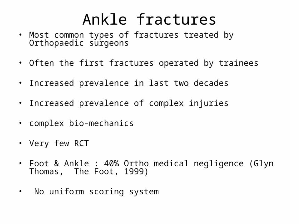
Ankle fractures• Most common types of fractures treated by Orthopaedic surgeons
• Often the first fractures operated by trainees
• Increased prevalence in last two decades
• Increased prevalence of complex injuries
• complex bio-mechanics
• Very few RCT
• Foot & Ankle : 40% Ortho medical negligence (Glyn Thomas, The Foot, 1999)
• No uniform scoring system

Bio mechanics

Limitations of radiology
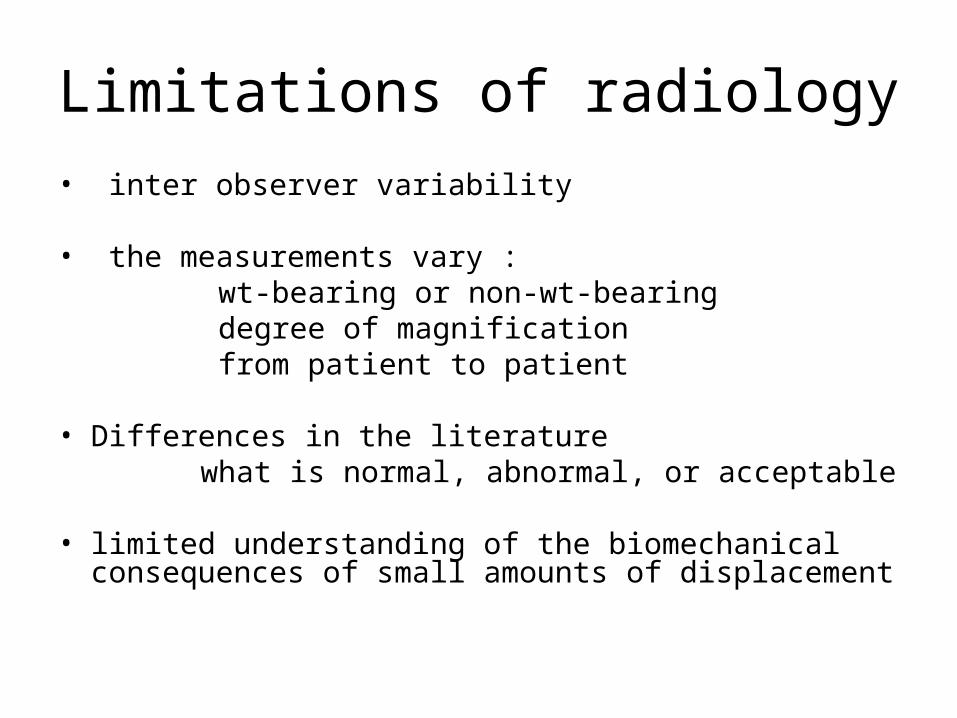
• inter observer variability
• the measurements vary : wt-bearing or non-wt-bearing degree of magnification from patient to patient
• Differences in the literature what is normal, abnormal, or acceptable
• limited understanding of the biomechanical consequences of small amounts of displacement

Special investigations
• Stress radiographs
• Tomography
• CT scan
• MRI
• Isotope bone scan
• Arthrography
• Arthroscopy

Classification
• Lauge-Hansen (1949) classification
• AO classification (Danis 1949, Weber 1972)
Both Lauge-Hansen and Weber systems are not predictive of prognosis
Moderate reproducibility (40-60%)
No classification gives information about stability
Lauge - Hansen classification
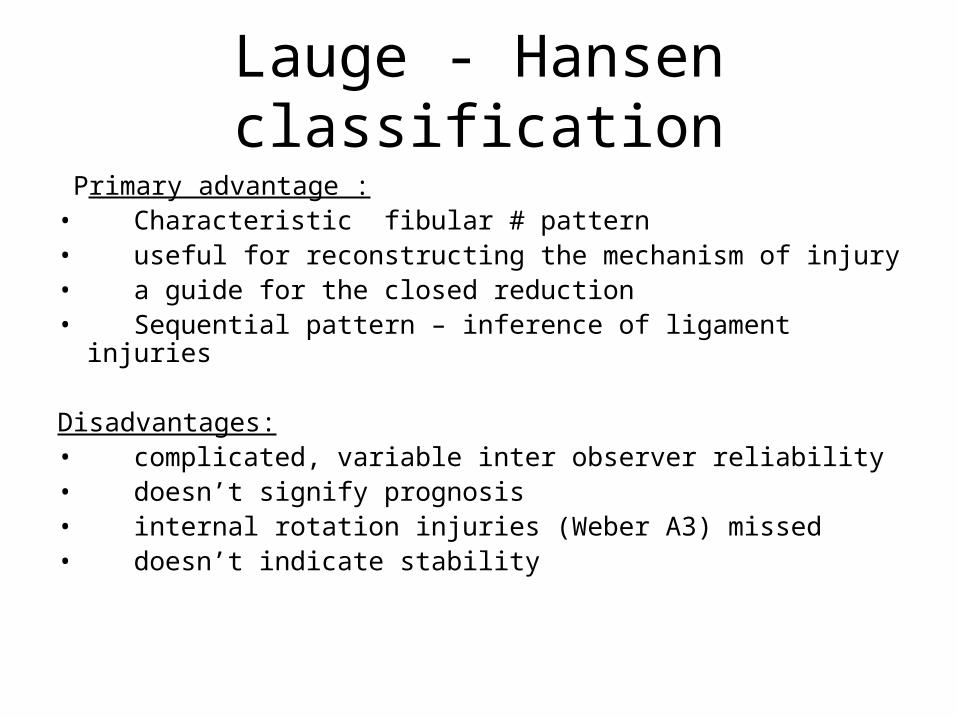
Primary advantage : • Characteristic fibular # pattern • useful for reconstructing the mechanism of injury• a guide for the closed reduction • Sequential pattern – inference of ligament injuries
Disadvantages: • complicated, variable inter observer reliability• doesn’t signify prognosis• internal rotation injuries (Weber A3) missed• doesn’t indicate stability
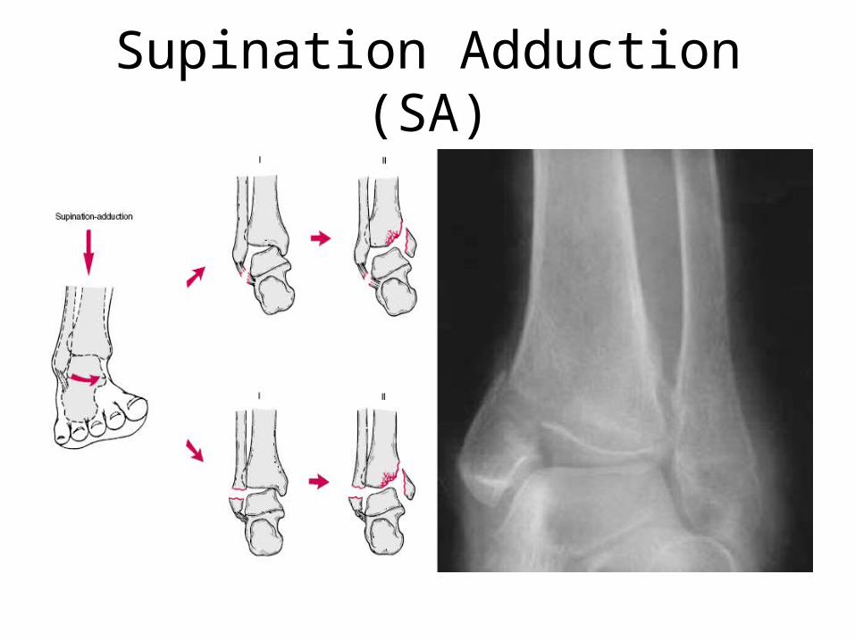
Supination Adduction (SA)
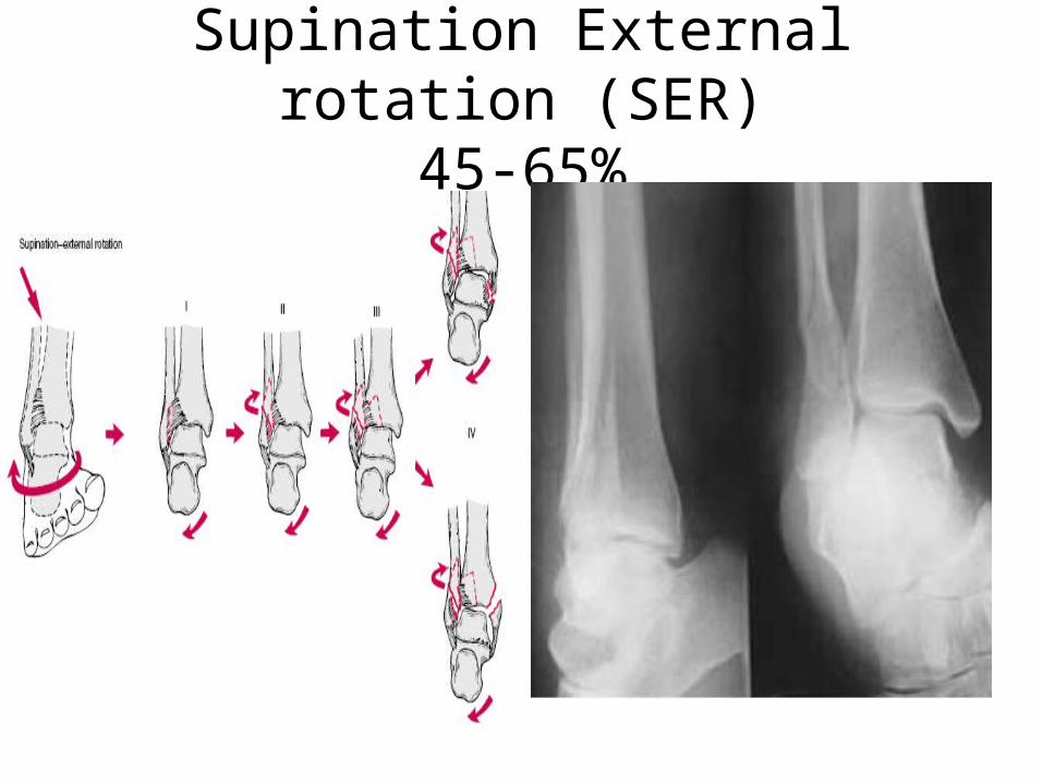
Supination External rotation (SER)45-65%
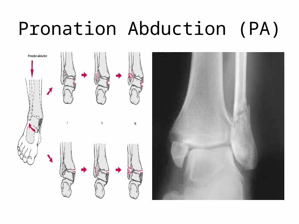
Pronation Abduction (PA)

Pronation External Rotation (PER)

Weber classification Advantages:
• easy to use • provides information about the lateral fracture
Disadvantages:
• ignores the medial injury The AO classification system
• a modification of the Weber system • subdivided on the basis of the presence of medial or posterior
injury

Correlation between the two

Management
Majority of
clinical decisions
regarding ankle fractures
are not made on the basis of
a formal classification

Surgeon’s judgment
State of soft tissues
Displacement & stabilityThe patient in general
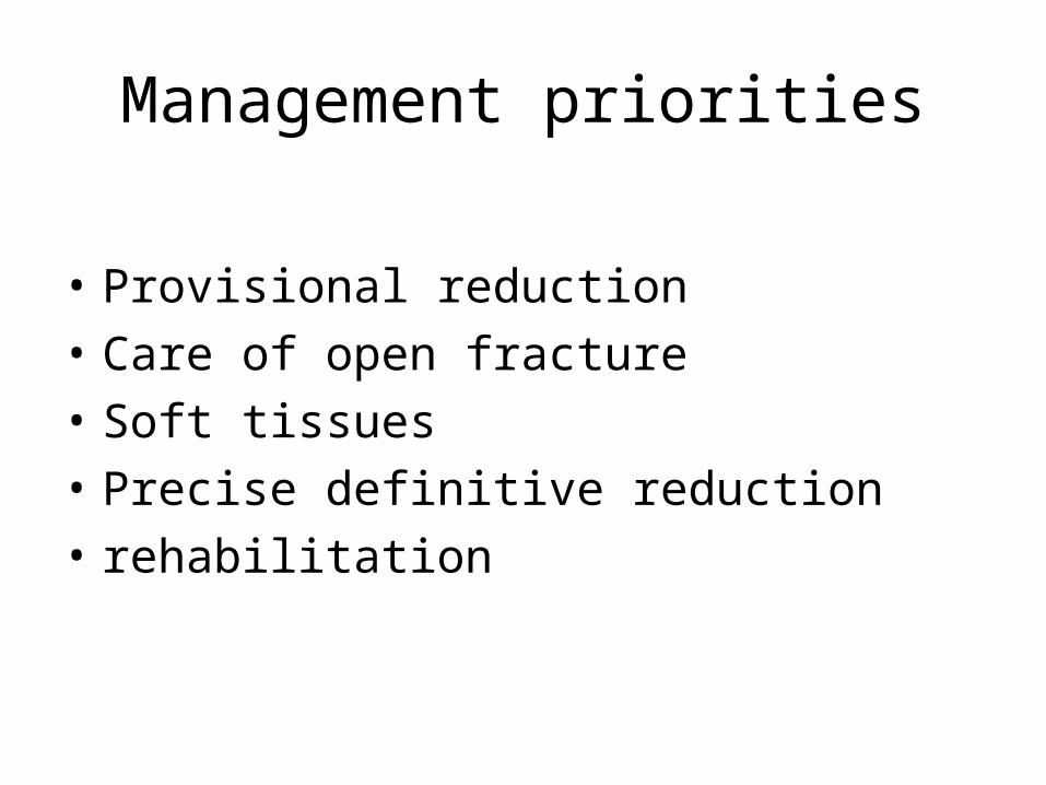
Management priorities
• Provisional reduction
• Care of open fracture
• Soft tissues
• Precise definitive reduction
• rehabilitation

Goals of treatment of Ankle fractures
• Healed fracture
• Ankle that moves and functions normally without pain
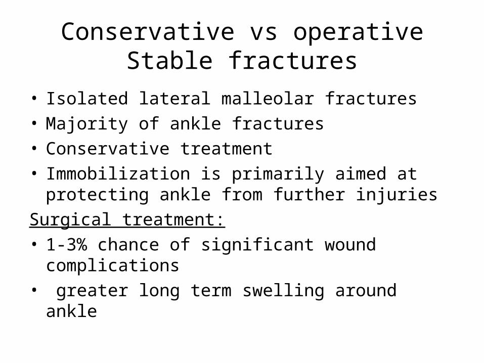
Conservative vs operativeStable fractures
• Isolated lateral malleolar fractures• Majority of ankle fractures• Conservative treatment• Immobilization is primarily aimed at protecting
ankle from further injuries
Surgical treatment: • 1-3% chance of significant wound complications• greater long term swelling around ankle

Conservative vs operativeUnstable fractures
• In a patient with lateral fractures and medial tenderness, any lateral talar shift signifies instability
• In the absence of talar shift, individual clinical judgment dictates treatment
• Closed reduction yields satisfactory results in up to 65% cases
• Routine repair of deltoid does not improve outcome and may lead to worse long term outcome
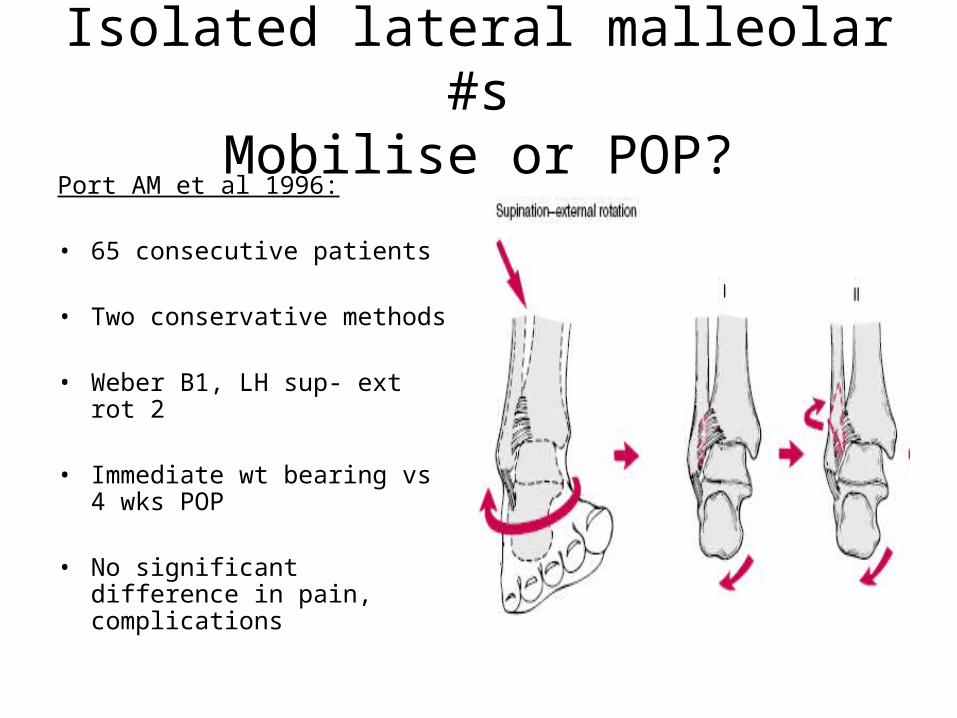
Isolated lateral malleolar #sMobilise or POP?
Port AM et al 1996:
• 65 consecutive patients
• Two conservative methods
• Weber B1, LH sup- ext rot 2
• Immediate wt bearing vs 4 wks POP
• No significant difference in pain, complications

Displaced fracturesConservative vs Operative treatment
• Because even small changes significantly influence the joint contact area, restoration of the anatomy of the fibula and consequently the ankle joint is important
BUT
• How anatomical should the reduction be??
Neither clinical or bio mechanical studies give absolute values
• Values range from mandatory anatomical reduction to acceptable 2-3mm
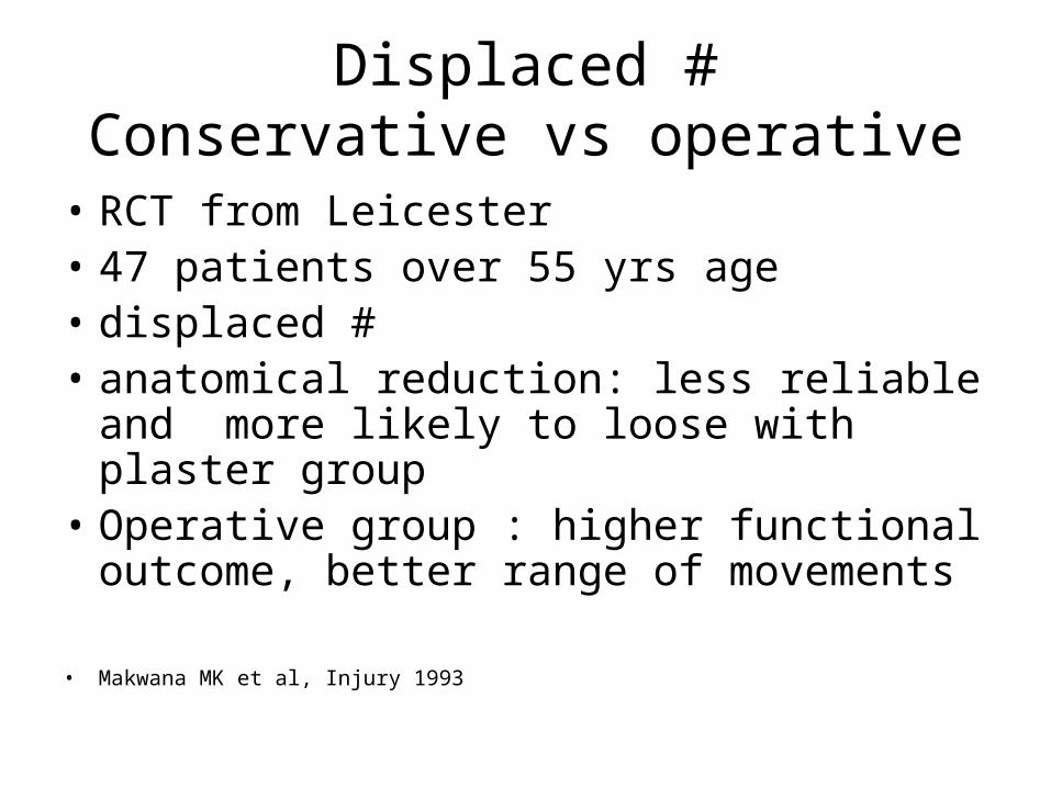
Displaced #Conservative vs operative
• RCT from Leicester• 47 patients over 55 yrs age• displaced #• anatomical reduction: less reliable and
more likely to loose with plaster group• Operative group : higher functional
outcome, better range of movements
• Makwana MK et al, Injury 1993
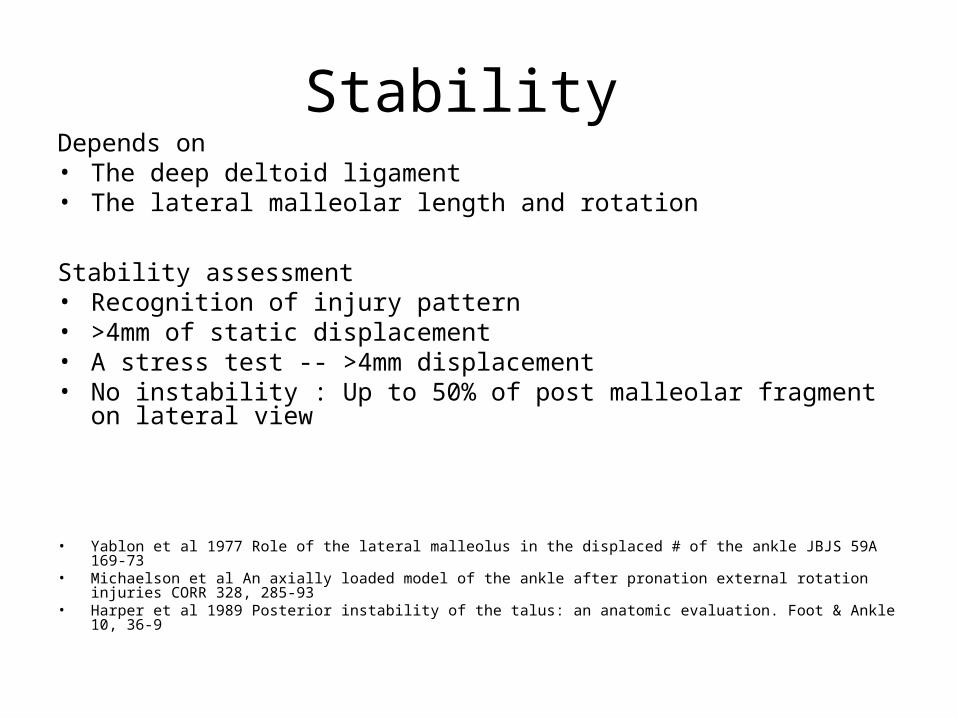
Stability Depends on • The deep deltoid ligament• The lateral malleolar length and rotation
Stability assessment • Recognition of injury pattern• >4mm of static displacement• A stress test -- >4mm displacement• No instability : Up to 50% of post malleolar fragment on lateral view
• Yablon et al 1977 Role of the lateral malleolus in the displaced # of the ankle JBJS 59A 169-73• Michaelson et al An axially loaded model of the ankle after pronation external rotation injuries CORR 328, 285-93• Harper et al 1989 Posterior instability of the talus: an anatomic evaluation. Foot & Ankle 10, 36-9

Instability
Most reliable criteria:
• a lateral talar shift in AP or mortise view medial clear space measurably larger than superior clear space
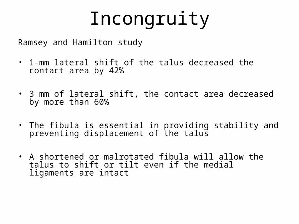
IncongruityRamsey and Hamilton study
• 1-mm lateral shift of the talus decreased the contact area by 42%
• 3 mm of lateral shift, the contact area decreased by more than 60%
• The fibula is essential in providing stability and preventing displacement of the talus
• A shortened or malrotated fibula will allow the talus to shift or tilt even if the medial ligaments are intact
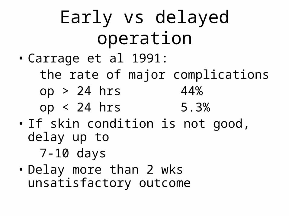
Early vs delayed operation
• Carrage et al 1991: the rate of major complications op > 24 hrs 44% op < 24 hrs 5.3% • If skin condition is not good, delay up to 7-10 days• Delay more than 2 wks unsatisfactory
outcome
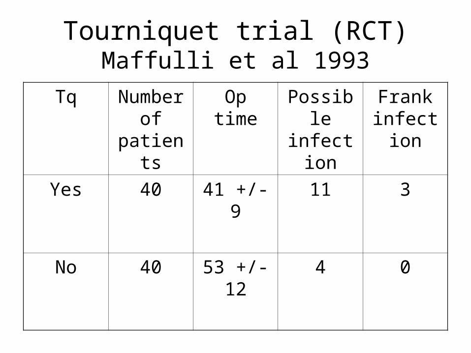
Tourniquet trial (RCT)Maffulli et al 1993
Tq Number of
patients
Op time Possible infection
Frank infection
Yes 40 41 +/- 9 11 3
No 40 53 +/- 12 4 0
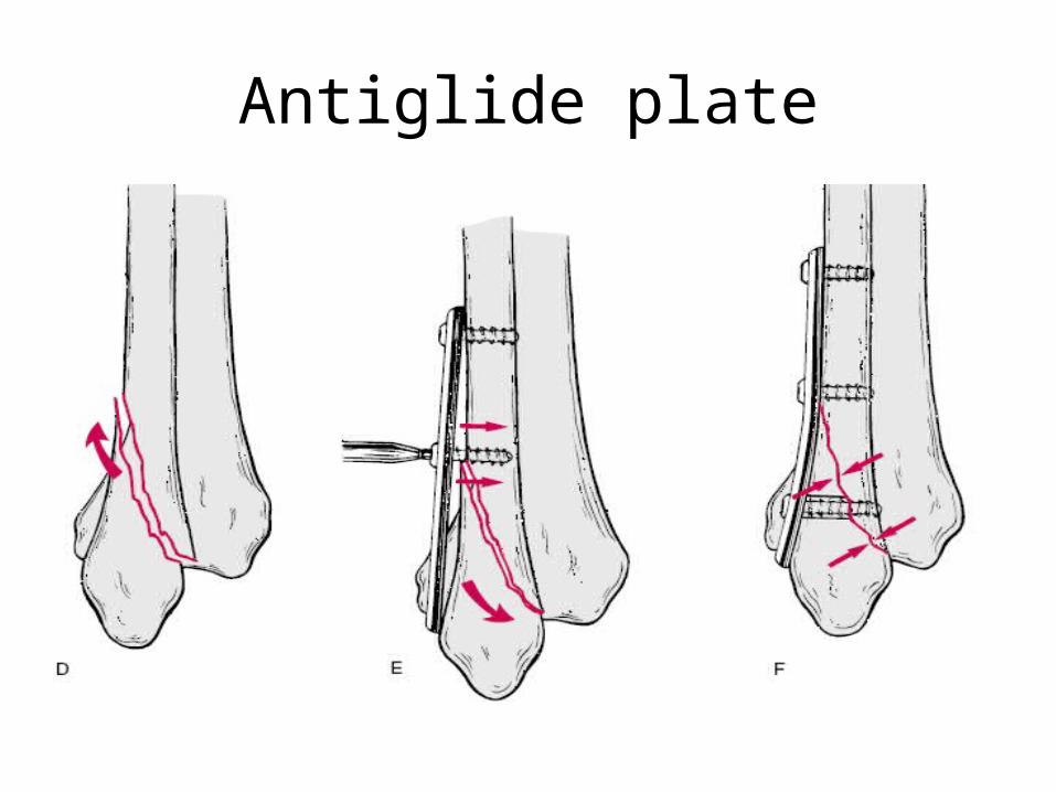
Antiglide plate
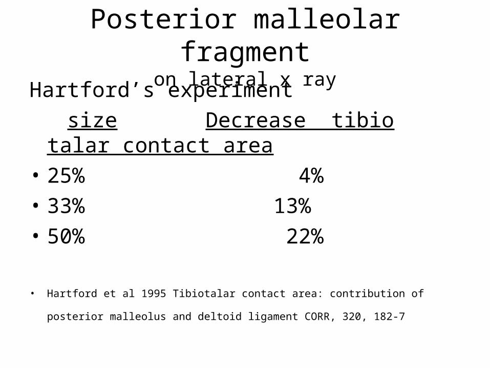
Posterior malleolar fragmenton lateral x ray
Hartford’s experiment
size Decrease tibio talar contact area
• 25% 4%
• 33% 13%
• 50% 22%
• Hartford et al 1995 Tibiotalar contact area: contribution of posterior malleolus and
deltoid ligament CORR, 320, 182-7
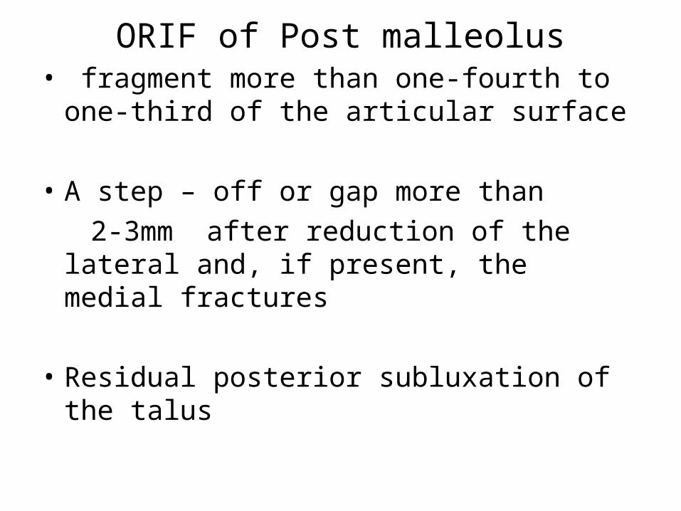
ORIF of Post malleolus• fragment more than one-fourth to one-
third of the articular surface
• A step – off or gap more than
2-3mm after reduction of the lateral and, if present, the medial fractures
• Residual posterior subluxation of the talus

Syndesmotic injury
most commonly occurs in
• pronation-external rotation (PER),
• pronation-abduction (PA),
• rarely supination-external rotation (SER)
(type-C and some type-B injuries)

Syndesmotic injuries
Burns et al., biomechanical study• only the syndesmotic ligaments were cut
- no change in the loading of the ankle • both the syndesmotic and the medial ligaments
were cut:
- increase in talar shift
- increase in joint-contact pressure
- 39 per cent decrease in joint-contact area
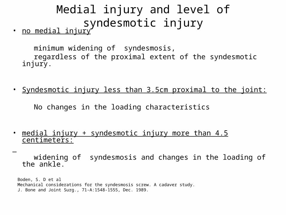
Medial injury and level of syndesmotic injury• no medial injury minimum widening of syndesmosis, regardless of the proximal extent of the syndesmotic injury.
• Syndesmotic injury less than 3.5cm proximal to the joint:
No changes in the loading characteristics
• medial injury + syndesmotic injury more than 4.5 centimeters: widening of syndesmosis and changes in the loading of the ankle.
Boden, S. D et al Mechanical considerations for the syndesmosis screw. A cadaver study. J. Bone and Joint Surg., 71-A:1548-1555, Dec. 1989.

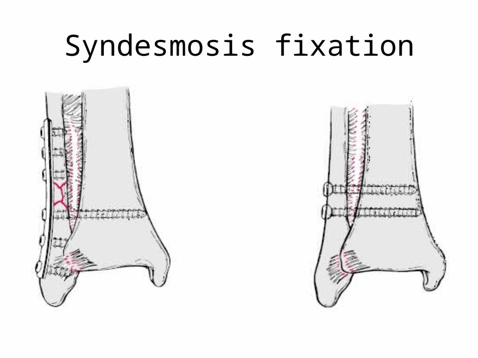
Principles of syndesmosis fixation
• Anatomical reduction is necessary
• If the fibula is fractured, its length, rotation, and alignment are restored first,
• If the medial malleolus is fractured, it should be reduced and fixed as well
• The reduction of the tibio fibular joint must be maintained during trans-syndesmotic fixation.
• 1.5-2cm proximal to plafond, 300 angle
• 3.5mm, 4.0mm, 4.5mm cortical or cancellous screws, three or four cortices
Syndesmosis – the controversy• Dorsiflexion of the ankle when fixing the syndesmosis (Tornetta et al)
• Three versus four cortices
• Hook test: Which direction, coronal or sagittal? Candal-Couto JJ et al: Injury 2004 Aug;35(8):814-8 • what type of syndesmotic fixation is needed eg: k wires, sutures, screws (metal or absorbable), endo buttons
• how the patient should be managed postoperatively
• Removal of syndesmotic screw
Syndesmosis fixation

Syndesmotic injury
Clinical tests• Squeeze test, Point test, ER test
Radiographic parameters
• widening of the medial clear space >4mm• stress test (medial clear space >4mm)• MRI

Syndesmotic screw removal?
• The syndesmotic screw prevents the normal physiologic motion of the fibula, particularly external rotation
• Several studies have shown that weight bearing with the syndesmotic screw in place had no adverse effects with no increased incidence of pain or syndesmotic ossification, provided a functional range of motion had been regained.
• Others have recommended that the syndesmotic screw be removed before weight bearing to restore the physiologic movement of the fibula and prevent fatigue failure of the screw.
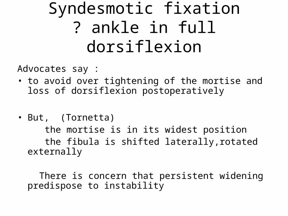
Syndesmotic fixation? ankle in full dorsiflexion
Advocates say : • to avoid over tightening of the mortise and loss of
dorsiflexion postoperatively
• But, (Tornetta) the mortise is in its widest position the fibula is shifted laterally,rotated externally There is concern that persistent widening predispose to
instability

Early or delayed mobilisation
The clinical evidence to support early mobilisation or splinting of any ankle fracture treated operatively is inconclusiveinconclusive
• Godsiff et al 1993: a comparative study of early motion and immediate plaster splintage after internal fixation of unstable fractures of the ankle. Injury, 24, 529-30
• Tropp and Norlin 1995 Ankle performance after ankle fracture: a randomized study of early mobilization
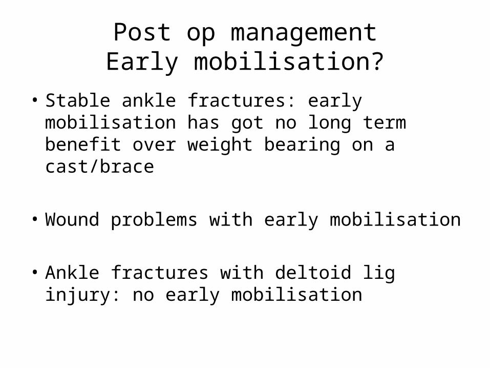
Post op managementEarly mobilisation?
• Stable ankle fractures: early mobilisation has got no long term benefit over weight bearing on a cast/brace
• Wound problems with early mobilisation
• Ankle fractures with deltoid lig injury: no early mobilisation

Special circumstancesDiabetes mellitus: • often older pts with vascular problems or neuropathy • Complication rate 40% with relative risk 2.76• Inadequate reduction and immobilization leads to
neuropathic destruction of joint• Both closed and open methods have role to playOsteoporotic ankle fractures:• Antiglide plate• Hooked fibular plate and tibia-pro-fibula screws• Locking platesOpen fracturesSudek’s osteodystrophy

Outcome
Poor outcome if:• Large medial space• Large post malleolar fracture with step• Delayed surgery >2 wks• Missed or improperly treated syndesmotic injury• Elderly females have less satisfactory results
Commonly used outcome measures:• AOFAS ankle score (Kitioka et al 1994)• Olerud and Molander score (CORR, 1986)• SF36
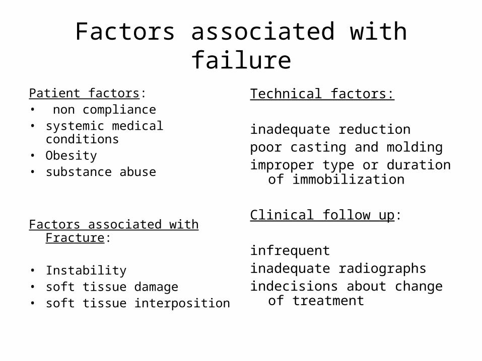
Factors associated with failure
Patient factors:• non compliance• systemic medical conditions• Obesity• substance abuse
Factors associated with Fracture:
• Instability• soft tissue damage• soft tissue interposition
Technical factors:
inadequate reductionpoor casting and moldingimproper type or duration of
immobilization
Clinical follow up:
infrequentinadequate radiographsindecisions about change of
treatment

Complications
• Mal union
• Non union
• Wound problems:
Oedema, prolonged Tq time, poor handling/dissection, wound closure without tension;
haematoma;
constrictive dressings.

Complications
Degenerative changes:• 10% of fractures that were adequately reduced • 85% of those inadequately reduced• radiographic changes were usually present within 18 months of injury.• Considering the frequency of ligament and bony injuries involving the ankle, the incidence of degenerative arthritis of the ankle is surprisingly low.
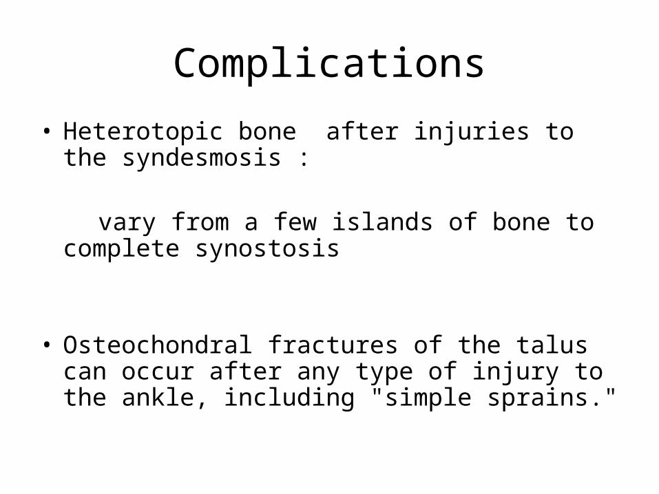
Complications
• Heterotopic bone after injuries to the syndesmosis :
vary from a few islands of bone to complete synostosis
• Osteochondral fractures of the talus can occur after any type of injury to the ankle, including "simple sprains."

PitfallsExperiments vs real life
variables involved in producing an injury• dynamic forces of muscles
• different magnitudes and rates of loading
• different degrees of weight bearing
• differences in the quality of bone and soft tissues

Thank you
Any views?