animal health constraints in dairy goats kept under ... · animal health constraints in dairy goats...
TRANSCRIPT

Vol. 6(11), pp. 268-279, November 2014 DOI: 10.5897/JVMAH2014.0312 Article Number: FE2CFC948307 ISSN 2141-2529 Copyright © 2014 Author(s) retain the copyright of this article http://www.academicjournals.org/JVMAH
Journal of Veterinary Medicine and Animal Health
Full Length Research Paper
Animal health constraints in dairy goats kept under smallholder farming systems in Kongwa and Mvomero
Districts, Tanzania
Dismas Said Ngasa Shija1, Lughano Jeremy Moses Kusiluka2, Sebastian Wilson Chenyambuga1*, Deogratias Shayo1 and Faustin Paul Lekule1
1Department of Animal Science and Production, Sokoine University of Agriculture, P.O. Box 3004, Morogoro, Tanzania.
2Nelson Mandela African Institute of Science and Technology, P.O. Box 447, Arusha, Tanzania.
Accepted 29 August, 2014; Received 7 July, 2014
This study was conducted to determine animal health constraints for dairy goats kept by small-scale farmers in Kongwa and Mvomero districts, Tanzania. A total of 129 dairy goats belonging to 108 farmers were screened for gastrointestinal nematode (GIN) infection, coccidiosis, haemoparasites, brucellosis and contagious caprine pleuropneumonia (CCPP) over a period of 11 months. Other clinical diseases and mortalities were recorded. The goats used were Norwegian crosses and Toggenburg crosses. The mean prevalence of GIN infection and coccidiosis in all goats were 54.8 and 57.4%, respectively. Prevalence of GIN infection was higher (P ≤ 0.05) during the rainy months than in the dry months, but the prevalence of coccidiosis did not differ (P > 0.05) between the dry and rainy seasons. The EPG in goats did not differ (P > 0.05) between Kongwa (169.79 ± 0.03 EPG) and Mvomero (171.51 ± 0.04 EPG) districts, but the OPG differed significantly (P ≤ 0.05) with values of 793.15 ± 0.04 (Kongwa) and 364.02 ± 0.05 (Mvomero). The prevalence of CCPP in the goats was 26.4%. Other clinical diseases included respiratory diseases, infectious keratoconjunctivitis and orf (scabby lesions around mouth and nostrils). Both tests for haemoparasites and brucellosis indicated negative results for all goats tested. Mortality rate during the study period was 15.5% and the major causes of deaths were respiratory diseases, bloat and food poisoning. In conclusion, gastrointestinal nematodes are prevalent in both districts, but the burdens are relatively low to justify mass treatment. The Norwegian goats are more susceptible to GIN infection and coccidiosis compared to Toggenburg goats. Key words: Coccidiosis, diseases, gastrointestinal nematodes, mortality, Norwegian goats, Toggenburg goats.
INTRODUCTION In Tanzania, promotion of small-scale dairy goat pro-duction for poverty alleviation and combating malnutrition
malnutrition started in early 1980s. Since then the num-ber of dairy goats in the country has increased mainly
*Corresponding author. E-mail: [email protected]. Tel: +255 784 754574. Author(s) agree that this article remain permanently open access under the terms of the Creative Commons Attribution License 4.0 International License
through the goat-in-trust schemes promoted by non-governmental organisations (NGOs), church organisations and research institutions/Universities. At the moment, the number of dairy goats in the country is estimated at 419,533 (United Republic of Tanzania (URT), 2012) and are predominantly kept by smallholder farmers in rural areas, especially women. The common dairy goat breeds include Toggenburg, Saanen, Norwegian, Anglo-Nubian and Alpine (Ministry of Livestock Development (MLD), 2006). The introduction of dairy goats in rural areas has provided an alternative source of milk to poor households, which cannot afford keeping dairy cattle.
Dairy goat production under smallholder production system is constrained by many factors, including poor husbandry practices, inadequate nutrition and disease challenges. The most common diseases affecting small ruminants in different parts of Tanzania are gastrointestinal parasitism, respiratory infections (especially pneumonic pasteurellosis and contagious caprine pleuropneumonia (CCPP)) and contagious ecthyma (Mtenga and Kusiluka, 1997; Kusiluka, 2002; Magona and Musisi, 2002). Some vector-borne diseases such as Rift valley fever (RVF) and bluetongue are associated with epidemic episodes (Sindato et al., 2011; Swai and Schoonman, 2009).
Gastrointestinal nematode (GIN) infection in small ru-minants is of considerable significance in a wide range of agro-climatic zones and affects production through losses resulting from mortalities, reduced weight gain, milk yield and reproduction efficiency (Fabiyi, 1987; Bekele et al., 1992; Singla, 1995). The effects of GIN on production depend mostly upon the age of the animals, breed, parasite species involved and the worm burden in the affected animal (Wadhwa et al., 2011). Gastrointestinal nematode infection is the most serious health challenge that limits goat production, especially in rural areas of developing countries (Kusiluka and Kambarage, 1996; Githiori et al., 2006). This is mainly because most developing countries located in the warm tropical zone, which has favourable climate for the survival and deve-lopment of GINs, have poor management practices and inadequate animal health control programmes (Akhtar et al., 2000). The impact of GINs is manifested by morbidity, mortality, cost of treatment and control measures against the syndrome (Mahusoon et al., 2004; Nwosu et al., 2007). However, most of the economic losses caused by GINs are mainly due to production losses rather than mortality (Waller, 2004) because the more prevalent subclinical infections cause sub-optimal productivity and insidious losses with negative impact on long-term animal productivity. Most cases of GINs infection rarely get veterinary attention due to their chronic and insidious nature (Sanyal, 1998; Dimander et al., 2000) and clinical signs may be evident only during terminal stages (Valentine
Shija et al. 269 et al., 2007).
Another important disease is coccidiosis, caused by different species of Eimeria. This is the most common form of coccidial infections in small ruminants, often occurring concurrently with GINs (Kusiluka, 1995; Kambarage et al., 1996; Kusiluka et al., 1998). Clinical coccidiosis is less common under traditional smallholder production system, except where poor hygiene leads to gross contamination of the environment that favours the build up of heavy oocyst burdens. This precipitates a clinical disease in young and animals with concurrent infections (Assoku, 1981; Barger et al., 1994). Most species of Eimeria affecting small ruminants have limited pathogenicity, however, in the presence of pathogenic GINs, their additive parasitic effect may lead to a clinical disease. The environmental factors that favour the establishment, survival and development of GINs and coccidiosis are very similar and quite often animals have mixed infections (Kusiluka, 1995), making it difficult to quantify the pathogenic effects of the individual parasite. Because of this, it is often advisable to study the epidemiology of both groups of parasites concurrently.
Contagious Caprine pleuropneumonia is another important disease affecting goats in Tanzania. The disease has been suspected to be in the country since early 1980s and was confirmed by isolation of Mycoplasma capri pneumoniae by Msami et al. (1998) and then Kusiluka et al. (2007). Hence, the disease has been in the country for over three decades and probably a longer time. Symptoms such as high fever, anorexia, laboured breathing, productive coughing, purulent nasal mucous emanating from the nose and reluctance to walk are considered as being indicative of CCPP and the predisposing factors include animal contact when sharing water and pasture (Swai and Neselle, 2010). Brucellosis is a disease with important effects on both public health and animal health. In goats, it is mainly caused by Brucella melitensis. The organisms persist in the genital system of the males and the disease is transmitted to the females at the time of service. The disease is of economic importance because it causes loss of milk production and abortion. The disease is characterized by abortion after third month of gestation and birth of weak kids.
In order to develop sustainable strategies for control of small ruminant diseases, there is a need to determine the most important diseases affecting the animals in different areas. This can be achieved by undertaking longitudinal studies to determine the spatial and temporal patterns of diseases of socio-economic importance with the aim of identifying appropriate intervention strategies. The present study was intended to establish the diseases of socio-economic importance in Toggenburg and Norwegian crossbred dairy goats kept under smallholder farming systems in Kongwa and Mvomero districts of
270 J. Vet. Med. Anim. Health
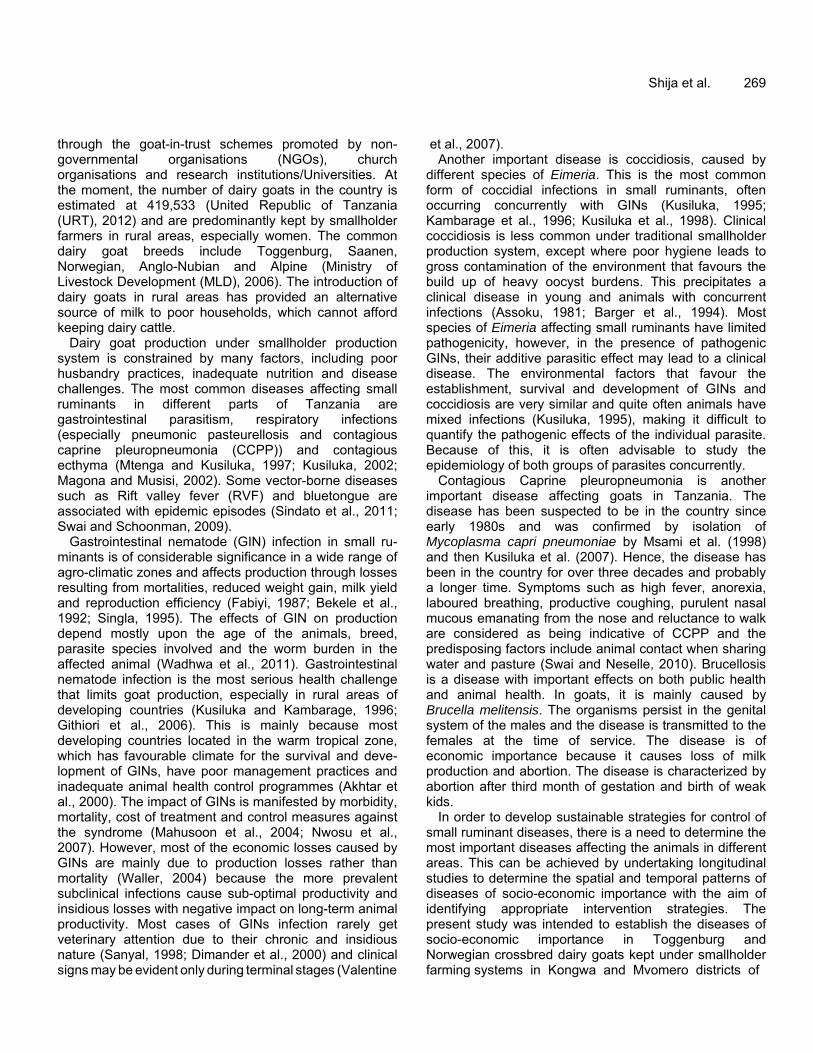
Figure 1. Map of Tanzania showing the study areas in Kongwa and Mvomero districts.
Tanzania. Toggenburg and Norwegian goats are the predominant dairy breeds in Tanzania. The animals were introduced in the rural areas of the two districts in order to improve human food and nutritional security and income of the resource-poor families. More specifically, the study was undertaken to: (i) determine the causes of mortality of dairy goats in semi-arid and sub-humid environments; (ii) determine seasonal patterns of nematode and eimeria infections and, (iii) compare the tolerance to common diseases between Toggenburg and Norwegian crossbred dairy goats introduced in the study areas. The epidemiological data gathered through the study could form the basis of designing disease control strategies in the study areas. MATERIALS AND METHODS Description of the study areas The present study was conducted in Masinyeti and Ihanda villages of Kongwa district located in northeast of Dodoma region and Kunke and Wami-Luhindo villages of Mvomero district located in the north-eastern part of Morogoro region (Figure 1). Kongwa district is located in the semi-arid area and has an altitude ranging from 900 to 1000 m above sea level (asl), mean annual temperature of 26.5°C and rainfall of 400 to 800 mm per annum. Mvomero is
located in the sub-humid tropical zone at an altitude of 600 to 2000 m asl, has temperatures that range from 18 to 30°C and receives annual rainfall of 600 to 2000 mm. Crosses of Toggenburg with the Small East African (SEA) goats were obtained from Babati district, Manyara region in northern Tanzania, which has a drier climate while the crosses of Norwegian goats with the SEA goats were obtained from Mgeta division, which is located on the slopes of the Uluguru Mountains in Morogoro region and has a cool mountain climate. The two types of crossbred goats were introduced in the study areas in order to improve food and nutritional security status of the communities. Experimental animals A total of 72 Norwegian (65 females; 7 males) and 57 Toggenburg (52 female; 5 males) crossbred goats were distributed between March and April, 2012 to 108 small-scale farmers willing to participate in the project in the four project villages (Table 1). In each village, half of the farmers received Norwegian crosses and the remaining farmers received Toggenburg crosses. For both breeds, the proportion of dairy goat blood was 75% while that of SEA goats was 25%. Before distribution to the project farmers, all animals were ear-tagged for identification and screened to know their health status with regard to GINs and coccidia burdens. Before the beginning of data collection, all goats were treated with an anthelmintic drug (Ivomec®) to control endoparasites and sprayed with acaricides to control ectoparasites. In both breeds, the animals were classified as young if they were below one year and adult if they were above one year of age.
Shija et al. 271
Table 1. Number of dairy crossbred goats distributed to farmers in the study areas.
District Village Breed Does Bucks Total
Kongwa
Ihanda Norwegian 19 3 22 Toggenburg 16 1 17
Masinyeti Norwegian 15 1 16 Toggenburg 15 2 17
Mvomero
Kunke Norwegian 16 2 18 Toggenburg 16 1 17
Wami Norwegian 15 1 16 Toggenburg 5 1 6
Total 117 12 129 Screening for gastrointestinal nematode eggs and coccidia oocysts Screening of goats for GINs, coccidiosis and haemoparasites and general health monitoring of the animals was done from June, 2012 to April, 2013. During this period, field visits were made every month to the study areas and faecal samples were collected from rectum of each animal. Each faecal sample was placed in a separate polythene bag and then all samples were packed and stored in a cool box and transported within 24 h to the laboratory at Sokoine University of Agriculture (SUA) where they were stored at 4°C until when analysis was done. The presence of gastrointestinal nematode eggs and coccidia occysts in faeces were determined using the McMaster counting technique using saturated salt solution with specific gravity of 1.200 as the floating medium (Hansen and Perry, 1994). The number of eggs and oocysts counted in the McMaster slide was multiplied by 100 and expressed as nematode eggs per gram of faeces (EPG) and oocysts per gram of faeces (OPG), respectively. The nematode egg load of each animal was graded as low (≤ 500 EPG), medium (500 to 1,000 EPG) and high (> 1,000 EPG). Similarly, the oocyst burdens were graded as low (≤ 50,000 OPG), medium (50,000 to 100,000 OPG) and high (> 100,000 OPG) according to Soulsby (1982). Animals with medium to high rate of infections were treated. For each village, faecal samples from all positive samples were pooled and cultured. Nematode larvae were harvested using sedimentation technique and species of the nematodes were identified using standard keys (Soulsby, 1982; Uhlinger, 1991; Foreyt, 2001). For identification of Eimeria species, faecal samples were allowed to sporulate and species identification was performed according to Duszynski and Wilber (1997). Other clinical diseases, conditions and symptoms observed in dairy goats kept by small-scale farmers Blood sample for each animal was collected from jugular vein using 10 ml vacutainer tubes containing Ethylenediaminetetraacetic acid (EDTA). The blood samples were stored in a cool box containing ice during the field work and were transported to the laboratory for analysis within 24 h. A thin blood smear was prepared for each sample, air dried, fixed in methanol and stained with Giemsa. The smears were examined for the presence of parasites (Quinn et al.,
1994). The parasites of interest were Trypanosoma, Babesia and Anaplasma. Also packed cell volume (PCV) and haemoglobin concentration (HB) were determined as complementary tests for parasitism. Collection of blood samples was done every month concurrently with the collection of faecal samples. Blood samples for serological screening were collected in vacutainer tubes containing heparin as anticoagulant. Serum samples were screened for brucellosis using the Rose Bengal test with the Brucella abortus as an antigen (Alton et al., 1988; Robinson, 2003). Presence of antibodies against Mycoplasma capricolum subspecies capri pneumoniae (M. capri pneumoniae), the causative agent for CCPP in serum samples was screened using the M. capri pneumoniae Antibody Test Kit (c-ELISA kit) developed based on the method by Thiaucourt et al. (1994). Farmers reported any clinical sign of diseases in the study animals to the village extension officers who then visited the respective farmers, verified and recorded the symptoms such as diarrhoea, rough hair coat, coughing, nasal and ocular discharges and skin lesions. Deaths and their causes (if established) were also recorded as they occur by the village extension workers. Statistical analyses Data on EPG, OPG, PCV and HB were analysed using the General Linear Model procedures of SAS (2009) to determine the effect of location (village), season, breed, sex, age of the goats and their interactions on EPG, OPG, PCV and HB. Before conducting statistical analysis, the EPG and OPG values were logarithmically transformed using log10 (EPG + 100) and log10 (OPG + 100) to normalize the distribution. No transformation was done for HB and PCV. Data on prevalence of various diseases and mortality rate were analysed using the chi-square test to test the significance of the differences between the breeds and among the villages. RESULTS Gastrointestinal nematode and coccidia infections Goats purchased from Babati and Mgeta were screened for nematode and Eimeria infections before being
272 J. Vet. Med. Anim. Health
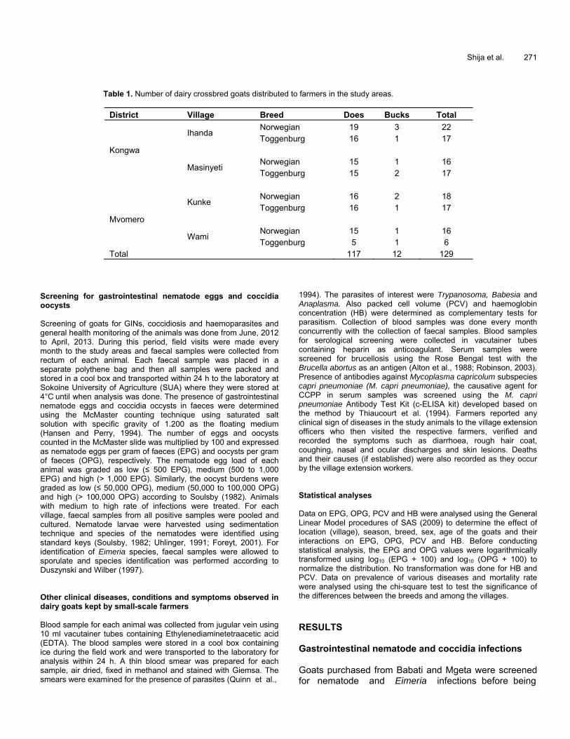
Table 2. Least squares means ± SE for EPG, OPG, HB and PCV of Toggenburg and Norwegian crosses before being transported to the study villages.
Factor Parameter Variable
EPG OPG HB (g/dl) PCV (%)
Breed Toggenburg 465.41b ± 0.07 5561.78 a ± 0.1057 9.22a ± 0.38 27.87a ± 1.15 Norwegian 792.35a ± 0.06 722.62b ± 0.089 7.91b ± 0.36 23.67b ± 1.08 P-value 0.0008 0.0480 0.0008 0.0004
Age group Adult 501.50 ± 0.09 1172.47b ± 0.13 8.54 ± 0.36 25.80 ± 1.07 Young 756.26 ± 0.11 5111.93a ± 0.15 8.59 ± 0.42 25.74 ± 1.27 P-value 0.6822 0.0361 0.9120 0.9640
Sex Female 281.61 ± 0.06 2985.62 ± 0.08 8.76 ± 0.23 26.48 ± 0.70 Male 976.16 ± 0.16 3298.79 ± 0.23 8.38 ± 0.63 25.07 ± 1.91 P-value 0.0820 0.2690 0.5928 0.5133
a,bThe means with different letters in the same column within a factor differ significantly (P ≤ 0.05). PCV = packed cell volume; HB = haemoglobin concentration; EPG = nematode eggs per gram of faeces; OPG = Eimeria oocysts per gram of faeces; n = Number of animals observed; P = Probability value.
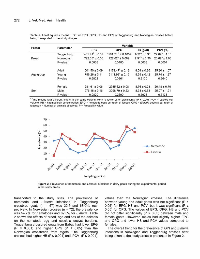
Figure 2. Prevalence of nematode and Eimeria infections in dairy goats during the experimental period in the study areas.
transported to the study sites. The prevalence of nematode and Eimeria infections in Toggenburg crossbred goats (n = 57) was 32.6 and 63.0%, res-pectively. In Norwegian crosses (n = 72), the prevalence was 54.7% for nematodes and 62.5% for Eimeria. Table 2 shows the effects of breed, age and sex of the animals on the nematode egg and coccidia oocyst burdens. Toggenburg crossbred goats from Babati had lower EPG (P ≤ 0.001) and higher OPG (P ≤ 0.05) than the Norwegian crossbreds from Mgeta. The Toggenburg crosses had higher HB (P ≤ 0.001) and PCV (P ≤ 0.001)
values than the Norwegian crosses. The difference between young and adult goats was not significant (P > 0.05) for EPG, HB and PCV, but it was significant (P ≤ 0.05) for OPG. The values of EPG, OPG, HB and PCV did not differ significantly (P > 0.05) between male and female goats. However, males had slightly higher EPG and OPG and lower HB and PCV values compared to females.
The overall trend for the prevalence of GIN and Eimeria infections in Norwegian and Toggenburg crosses after being taken to the study areas is presented in Figure 2.
Shija et al. 273
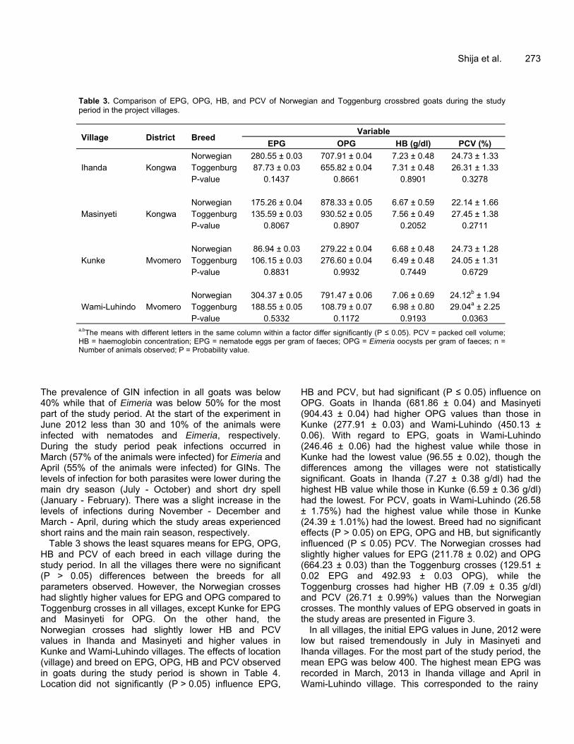
Table 3. Comparison of EPG, OPG, HB, and PCV of Norwegian and Toggenburg crossbred goats during the study period in the project villages.
Village District Breed Variable
EPG OPG HB (g/dl) PCV (%)
Ihanda Kongwa Norwegian 280.55 ± 0.03 707.91 ± 0.04 7.23 ± 0.48 24.73 ± 1.33 Toggenburg 87.73 ± 0.03 655.82 ± 0.04 7.31 ± 0.48 26.31 ± 1.33 P-value 0.1437 0.8661 0.8901 0.3278
Masinyeti Kongwa Norwegian 175.26 ± 0.04 878.33 ± 0.05 6.67 ± 0.59 22.14 ± 1.66 Toggenburg 135.59 ± 0.03 930.52 ± 0.05 7.56 ± 0.49 27.45 ± 1.38 P-value 0.8067 0.8907 0.2052 0.2711
Kunke Mvomero Norwegian 86.94 ± 0.03 279.22 ± 0.04 6.68 ± 0.48 24.73 ± 1.28 Toggenburg 106.15 ± 0.03 276.60 ± 0.04 6.49 ± 0.48 24.05 ± 1.31 P-value 0.8831 0.9932 0.7449 0.6729
Wami-Luhindo Mvomero Norwegian 304.37 ± 0.05 791.47 ± 0.06 7.06 ± 0.69 24.12b ± 1.94 Toggenburg 188.55 ± 0.05 108.79 ± 0.07 6.98 ± 0.80 29.04a ± 2.25 P-value 0.5332 0.1172 0.9193 0.0363
a,bThe means with different letters in the same column within a factor differ significantly (P ≤ 0.05). PCV = packed cell volume; HB = haemoglobin concentration; EPG = nematode eggs per gram of faeces; OPG = Eimeria oocysts per gram of faeces; n = Number of animals observed; P = Probability value.
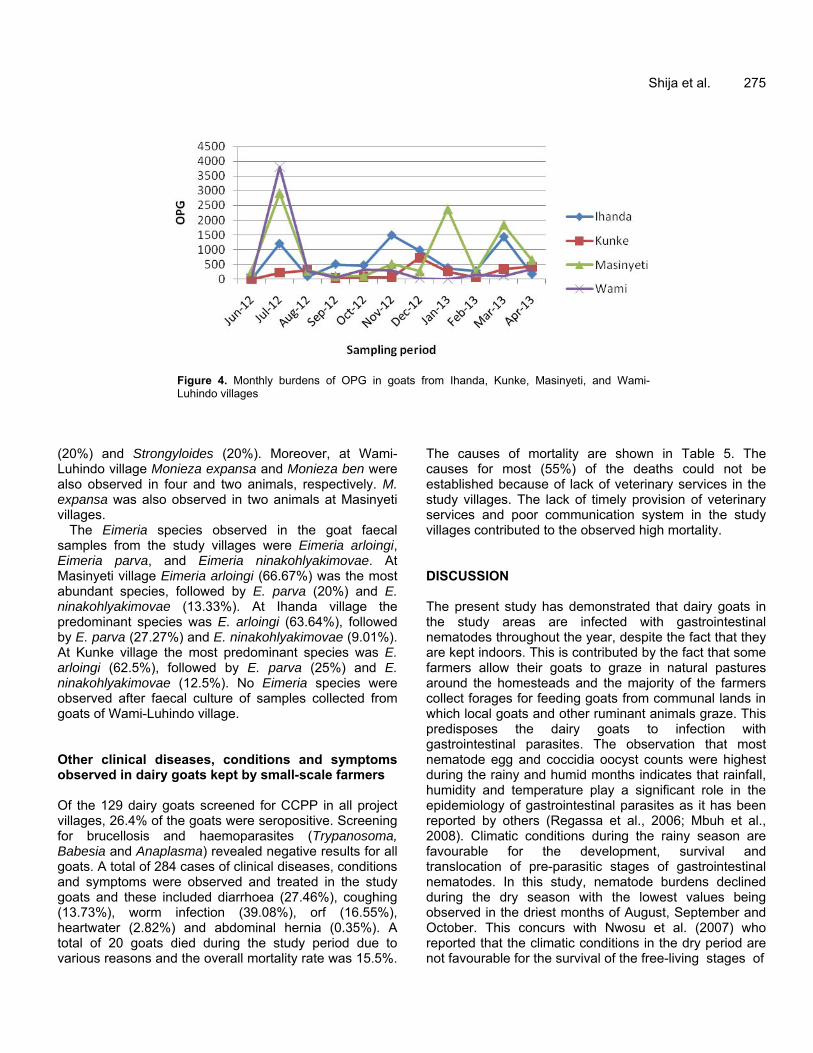
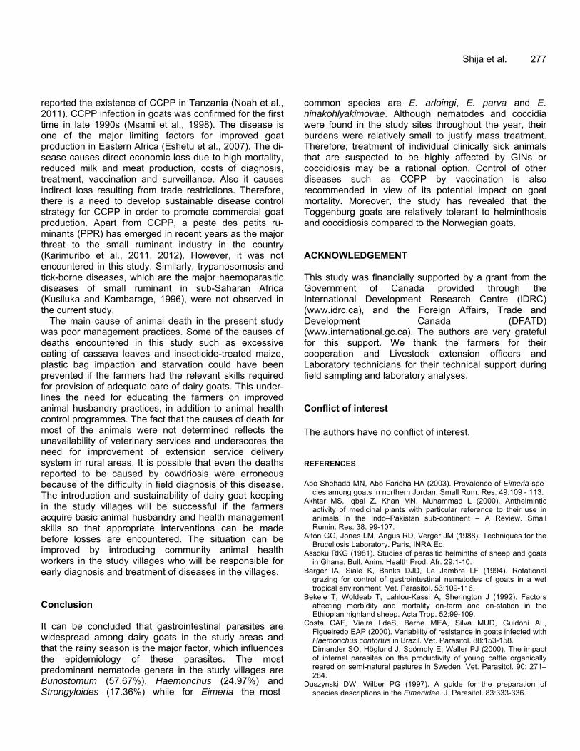
The prevalence of GIN infection in all goats was below 40% while that of Eimeria was below 50% for the most part of the study period. At the start of the experiment in June 2012 less than 30 and 10% of the animals were infected with nematodes and Eimeria, respectively. During the study period peak infections occurred in March (57% of the animals were infected) for Eimeria and April (55% of the animals were infected) for GINs. The levels of infection for both parasites were lower during the main dry season (July - October) and short dry spell (January - February). There was a slight increase in the levels of infections during November - December and March - April, during which the study areas experienced short rains and the main rain season, respectively.
Table 3 shows the least squares means for EPG, OPG, HB and PCV of each breed in each village during the study period. In all the villages there were no significant (P > 0.05) differences between the breeds for all parameters observed. However, the Norwegian crosses had slightly higher values for EPG and OPG compared to Toggenburg crosses in all villages, except Kunke for EPG and Masinyeti for OPG. On the other hand, the Norwegian crosses had slightly lower HB and PCV values in Ihanda and Masinyeti and higher values in Kunke and Wami-Luhindo villages. The effects of location (village) and breed on EPG, OPG, HB and PCV observed in goats during the study period is shown in Table 4. Location did not significantly (P ˃ 0.05) influence EPG,
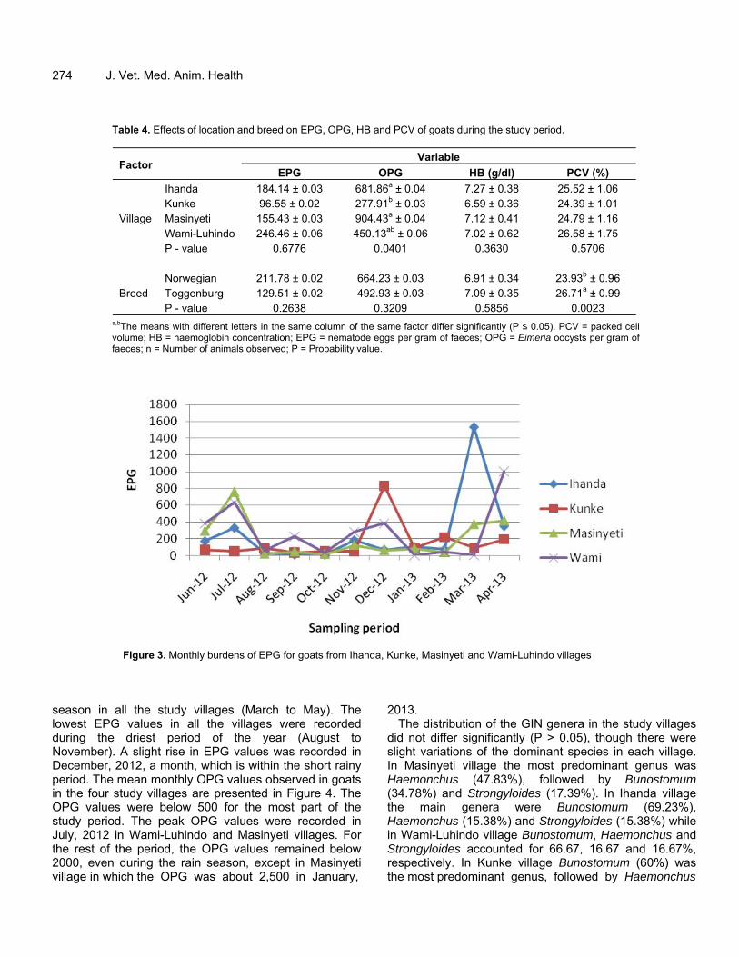
HB and PCV, but had significant (P ≤ 0.05) influence on OPG. Goats in Ihanda (681.86 ± 0.04) and Masinyeti (904.43 ± 0.04) had higher OPG values than those in Kunke (277.91 ± 0.03) and Wami-Luhindo (450.13 ± 0.06). With regard to EPG, goats in Wami-Luhindo (246.46 ± 0.06) had the highest value while those in Kunke had the lowest value (96.55 ± 0.02), though the differences among the villages were not statistically significant. Goats in Ihanda (7.27 ± 0.38 g/dl) had the highest HB value while those in Kunke (6.59 ± 0.36 g/dl) had the lowest. For PCV, goats in Wami-Luhindo (26.58 ± 1.75%) had the highest value while those in Kunke (24.39 ± 1.01%) had the lowest. Breed had no significant effects (P ˃ 0.05) on EPG, OPG and HB, but significantly influenced (P ≤ 0.05) PCV. The Norwegian crosses had slightly higher values for EPG (211.78 ± 0.02) and OPG (664.23 ± 0.03) than the Toggenburg crosses (129.51 ± 0.02 EPG and 492.93 ± 0.03 OPG), while the Toggenburg crosses had higher HB (7.09 ± 0.35 g/dl) and PCV (26.71 ± 0.99%) values than the Norwegian crosses. The monthly values of EPG observed in goats in the study areas are presented in Figure 3.
In all villages, the initial EPG values in June, 2012 were low but raised tremendously in July in Masinyeti and Ihanda villages. For the most part of the study period, the mean EPG was below 400. The highest mean EPG was recorded in March, 2013 in Ihanda village and April in Wami-Luhindo village. This corresponded to the rainy
274 J. Vet. Med. Anim. Health
Table 4. Effects of location and breed on EPG, OPG, HB and PCV of goats during the study period.
Factor Variable
EPG OPG HB (g/dl) PCV (%)
Village
Ihanda 184.14 ± 0.03 681.86a ± 0.04 7.27 ± 0.38 25.52 ± 1.06 Kunke 96.55 ± 0.02 277.91b ± 0.03 6.59 ± 0.36 24.39 ± 1.01 Masinyeti 155.43 ± 0.03 904.43a ± 0.04 7.12 ± 0.41 24.79 ± 1.16 Wami-Luhindo 246.46 ± 0.06 450.13ab ± 0.06 7.02 ± 0.62 26.58 ± 1.75 P - value 0.6776 0.0401 0.3630 0.5706
Breed Norwegian 211.78 ± 0.02 664.23 ± 0.03 6.91 ± 0.34 23.93b ± 0.96 Toggenburg 129.51 ± 0.02 492.93 ± 0.03 7.09 ± 0.35 26.71a ± 0.99 P - value 0.2638 0.3209 0.5856 0.0023
a,bThe means with different letters in the same column of the same factor differ significantly (P ≤ 0.05). PCV = packed cell volume; HB = haemoglobin concentration; EPG = nematode eggs per gram of faeces; OPG = Eimeria oocysts per gram of faeces; n = Number of animals observed; P = Probability value.
Figure 3. Monthly burdens of EPG for goats from Ihanda, Kunke, Masinyeti and Wami-Luhindo villages
season in all the study villages (March to May). The lowest EPG values in all the villages were recorded during the driest period of the year (August to November). A slight rise in EPG values was recorded in December, 2012, a month, which is within the short rainy period. The mean monthly OPG values observed in goats in the four study villages are presented in Figure 4. The OPG values were below 500 for the most part of the study period. The peak OPG values were recorded in July, 2012 in Wami-Luhindo and Masinyeti villages. For the rest of the period, the OPG values remained below 2000, even during the rain season, except in Masinyeti village in which the OPG was about 2,500 in January,
2013. The distribution of the GIN genera in the study villages
did not differ significantly (P > 0.05), though there were slight variations of the dominant species in each village. In Masinyeti village the most predominant genus was Haemonchus (47.83%), followed by Bunostomum (34.78%) and Strongyloides (17.39%). In Ihanda village the main genera were Bunostomum (69.23%), Haemonchus (15.38%) and Strongyloides (15.38%) while in Wami-Luhindo village Bunostomum, Haemonchus and Strongyloides accounted for 66.67, 16.67 and 16.67%, respectively. In Kunke village Bunostomum (60%) was the most predominant genus, followed by Haemonchus
Shija et al. 275
Figure 4. Monthly burdens of OPG in goats from Ihanda, Kunke, Masinyeti, and Wami-Luhindo villages
(20%) and Strongyloides (20%). Moreover, at Wami-Luhindo village Monieza expansa and Monieza ben were also observed in four and two animals, respectively. M. expansa was also observed in two animals at Masinyeti villages.
The Eimeria species observed in the goat faecal samples from the study villages were Eimeria arloingi, Eimeria parva, and Eimeria ninakohlyakimovae. At Masinyeti village Eimeria arloingi (66.67%) was the most abundant species, followed by E. parva (20%) and E. ninakohlyakimovae (13.33%). At Ihanda village the predominant species was E. arloingi (63.64%), followed by E. parva (27.27%) and E. ninakohlyakimovae (9.01%). At Kunke village the most predominant species was E. arloingi (62.5%), followed by E. parva (25%) and E. ninakohlyakimovae (12.5%). No Eimeria species were observed after faecal culture of samples collected from goats of Wami-Luhindo village. Other clinical diseases, conditions and symptoms observed in dairy goats kept by small-scale farmers Of the 129 dairy goats screened for CCPP in all project villages, 26.4% of the goats were seropositive. Screening for brucellosis and haemoparasites (Trypanosoma, Babesia and Anaplasma) revealed negative results for all goats. A total of 284 cases of clinical diseases, conditions and symptoms were observed and treated in the study goats and these included diarrhoea (27.46%), coughing (13.73%), worm infection (39.08%), orf (16.55%), heartwater (2.82%) and abdominal hernia (0.35%). A total of 20 goats died during the study period due to various reasons and the overall mortality rate was 15.5%.
The causes of mortality are shown in Table 5. The causes for most (55%) of the deaths could not be established because of lack of veterinary services in the study villages. The lack of timely provision of veterinary services and poor communication system in the study villages contributed to the observed high mortality. DISCUSSION The present study has demonstrated that dairy goats in the study areas are infected with gastrointestinal nematodes throughout the year, despite the fact that they are kept indoors. This is contributed by the fact that some farmers allow their goats to graze in natural pastures around the homesteads and the majority of the farmers collect forages for feeding goats from communal lands in which local goats and other ruminant animals graze. This predisposes the dairy goats to infection with gastrointestinal parasites. The observation that most nematode egg and coccidia oocyst counts were highest during the rainy and humid months indicates that rainfall, humidity and temperature play a significant role in the epidemiology of gastrointestinal parasites as it has been reported by others (Regassa et al., 2006; Mbuh et al., 2008). Climatic conditions during the rainy season are favourable for the development, survival and translocation of pre-parasitic stages of gastrointestinal nematodes. In this study, nematode burdens declined during the dry season with the lowest values being observed in the driest months of August, September and October. This concurs with Nwosu et al. (2007) who reported that the climatic conditions in the dry period are not favourable for the survival of the free-living stages of

276 J. Vet. Med. Anim. Health
Table 5. Causes of death of dairy goats in the study villages.
Cause of death Breed
Total Norwegian crosses
Toggenburg crosses
Pneumonia - 1 1 Plastic bag impaction - 1 1 Starvation - 1 1 Excessive consumption of cassava leaves - 1 1 Consumption of insecticide treated maize grain - 1 1 Abscess - 1 1 Cowdriosis 2 - 2 Bloat of unknown origin 1 - 1 Cause of death not well established 10 1 11 Total 13 7 20
the parasites. The nematode genera observed in the present study have been reported in previous studies in Tanzania (Kusiluka, 1995; Kusiluka et al., 1996, 1999) and in neighbouring countries. Ng’ang’a et al. (2004) and Odoi et al. (2007) reported that in Kenya, the widely reported nematode genera of small ruminants include Haemonchus, Trichostrongylus, Cooperia and Oesophagostomum.
The prevalence of coccidiosis in dairy goats from the four villages observed in this study is similar to what has been reported in other sub-Saharan African countries (Kanyari et al., 2009; Kanyari et al., 2010). In this study, the dairy goats were infected with Eimeria throughout the year in all four villages, suggesting that the environmental conditions were conducive for the survival and development of the Eimeria species throughout the year. Most of the dairy goats in the study villages are kept indoors under the cut-and-carry system. This system increases the risk of goats for being infected with coccidiosis. The finding that both prevalence and loads of coccidia in this study were higher in young goats compared to adult goats agrees with the findings reported by several authors (Abo-Shehada and Abo-Ferieha, 2003; Regassa et al., 2006; Mbuh et al., 2008). This is due to the fact that coccidiosis is mainly a disease of young goats that have not yet developed immunity against coccidia (Matjila and Penzhorn, 2003). It is possible that the mixed infection of nematodes and Eimeria increased the susceptibility of young goats to coccidiosis.
The proportion of animals infected with nematodes and Eimeria and the respective EPG and OPG values were very low at the beginning of the study in June, 2012, simply because the animals were given treatment against gastrointestinal parasites before being distributed to the farmers. The high burdens of both EPG and OPG
recorded in July, 2012 indicates that the animals got new infections after being introduced in the research villages, implying that the project villages are infested with nematodes and Eimeria. The higher OPG values observed during the month of July compared to EPG reflect the resistance of coccidia oocysts to desiccation. Kanyari (1993) observed that sporulated oocysts are more resistant to desiccation compared to helminth eggs or larvae and consequently oocyst counts in goats during the dry season are higher than the nematode egg counts. The overall low prevalence and burdens of nematodes and Eimeria in goats during the entire study period in all villages and both breeds may be due to the low stocking rate of goats which did not favour a build up of high nematode egg and oocyst burdens because on average, each household had one or two goats. Moreover, presence of few animals per household coupled with zero grazing minimized the risk of goats for being infected with the nematodes.
The persistently higher levels of EPG and OPG among the Norwegian crossbred goats compared to Toggenburg crosses may suggest that the Norwegian goats are more susceptible to gastrointestinal nematodes than the Toggenburg goats. Breed differences with respect to nematode infection in dairy goats have been reported by other studies (Richard et al., 1990; Costa et al., 2000). The Toggenburg goats have been in the country for lon-ger time (since early 1960s) compared to the Norwegian goats, which were introduced in the late 1980s. Hence, the Toggenburg goats may have adapted better to the local conditions and developed traits for tolerance to endemic diseases compared to the Norwegian goats.
Screening for M. capripneumoniae revealed that a significant proportion of goats in the project villages were infected with CCPP. The observation in the present study supports the findings of previous studies which have
reported the existence of CCPP in Tanzania (Noah et al., 2011). CCPP infection in goats was confirmed for the first time in late 1990s (Msami et al., 1998). The disease is one of the major limiting factors for improved goat production in Eastern Africa (Eshetu et al., 2007). The di-sease causes direct economic loss due to high mortality, reduced milk and meat production, costs of diagnosis, treatment, vaccination and surveillance. Also it causes indirect loss resulting from trade restrictions. Therefore, there is a need to develop sustainable disease control strategy for CCPP in order to promote commercial goat production. Apart from CCPP, a peste des petits ru-minants (PPR) has emerged in recent years as the major threat to the small ruminant industry in the country (Karimuribo et al., 2011, 2012). However, it was not encountered in this study. Similarly, trypanosomosis and tick-borne diseases, which are the major haemoparasitic diseases of small ruminant in sub-Saharan Africa (Kusiluka and Kambarage, 1996), were not observed in the current study.
The main cause of animal death in the present study was poor management practices. Some of the causes of deaths encountered in this study such as excessive eating of cassava leaves and insecticide-treated maize, plastic bag impaction and starvation could have been prevented if the farmers had the relevant skills required for provision of adequate care of dairy goats. This under-lines the need for educating the farmers on improved animal husbandry practices, in addition to animal health control programmes. The fact that the causes of death for most of the animals were not determined reflects the unavailability of veterinary services and underscores the need for improvement of extension service delivery system in rural areas. It is possible that even the deaths reported to be caused by cowdriosis were erroneous because of the difficulty in field diagnosis of this disease. The introduction and sustainability of dairy goat keeping in the study villages will be successful if the farmers acquire basic animal husbandry and health management skills so that appropriate interventions can be made before losses are encountered. The situation can be improved by introducing community animal health workers in the study villages who will be responsible for early diagnosis and treatment of diseases in the villages. Conclusion It can be concluded that gastrointestinal parasites are widespread among dairy goats in the study areas and that the rainy season is the major factor, which influences the epidemiology of these parasites. The most predominant nematode genera in the study villages are Bunostomum (57.67%), Haemonchus (24.97%) and Strongyloides (17.36%) while for Eimeria the most
Shija et al. 277 common species are E. arloingi, E. parva and E. ninakohlyakimovae. Although nematodes and coccidia were found in the study sites throughout the year, their burdens were relatively small to justify mass treatment. Therefore, treatment of individual clinically sick animals that are suspected to be highly affected by GINs or coccidiosis may be a rational option. Control of other diseases such as CCPP by vaccination is also recommended in view of its potential impact on goat mortality. Moreover, the study has revealed that the Toggenburg goats are relatively tolerant to helminthosis and coccidiosis compared to the Norwegian goats. ACKNOWLEDGEMENT This study was financially supported by a grant from the Government of Canada provided through the International Development Research Centre (IDRC) (www.idrc.ca), and the Foreign Affairs, Trade and Development Canada (DFATD) (www.international.gc.ca). The authors are very grateful for this support. We thank the farmers for their cooperation and Livestock extension officers and Laboratory technicians for their technical support during field sampling and laboratory analyses. Conflict of interest The authors have no conflict of interest. REFERENCES Abo-Shehada MN, Abo-Farieha HA (2003). Prevalence of Eimeria spe-
cies among goats in northern Jordan. Small Rum. Res. 49:109 - 113. Akhtar MS, Iqbal Z, Khan MN, Muhammad L (2000). Anthelmintic
activity of medicinal plants with particular reference to their use in animals in the Indo–Pakistan sub-continent – A Review. Small Rumin. Res. 38: 99-107.
Alton GG, Jones LM, Angus RD, Verger JM (1988). Techniques for the Brucellosis Laboratory. Paris, INRA Ed.
Assoku RKG (1981). Studies of parasitic helminths of sheep and goats in Ghana. Bull. Anim. Health Prod. Afr. 29:1-10.
Barger IA, Siale K, Banks DJD, Le Jambre LF (1994). Rotational grazing for control of gastrointestinal nematodes of goats in a wet tropical environment. Vet. Parasitol. 53:109-116.
Bekele T, Woldeab T, Lahlou-Kassi A, Sherington J (1992). Factors affecting morbidity and mortality on-farm and on-station in the Ethiopian highland sheep. Acta Trop. 52:99-109.
Costa CAF, Vieira LdaS, Berne MEA, Silva MUD, Guidoni AL, Figueiredo EAP (2000). Variability of resistance in goats infected with Haemonchus contortus in Brazil. Vet. Parasitol. 88:153-158. Dimander SO, Höglund J, Spörndly E, Waller PJ (2000). The impact of internal parasites on the productivity of young cattle organically reared on semi-natural pastures in Sweden. Vet. Parasitol. 90: 271– 284.
Duszynski DW, Wilber PG (1997). A guide for the preparation of species descriptions in the Eimeriidae. J. Parasitol. 83:333-336.
278 J. Vet. Med. Anim. Health Eshetu L, Yigezu Y, Asfaw W (2007). A study on Contagious Caprine
Pleuropneumonia (CCPP) in goats at an export oriented abattoir, Debre Zeit, Ethiopia. Trop. Anim. Health Prod. 39:427-432.
Fabiyi JP (1987). Production losses and control of helminths in ruminants of tropical regions. Int. J. Parasitol. 17:435- 540.
Foreyt WJ (2001). Veterinary Parasitology Reference Manual. Blackwell Publishers, Iowa, USA.
Githiori JB, Athanasiadou S, Thamsborg SM (2006). Use of plants in novel approaches for control of gastrointestinal helminths in livestock with emphasis on small ruminants. Vet. Parasitol. 139:308-320.
Hansen J, Perry B (1994). The Epidemiology, Diagnosis and Control of Helminth Parasites of Ruminants, 2nd Edition. Nairobi, Kenya; ILRAD.
Kambarage DM, Kimera SI, Kusiluka LJM, Mtambo MMA (1996). Pre-valence of Eimeria and Cryptosporidium oocysts in cattle, sheep and goats in Morogoro region, Tanzania. J. Appl. Anim. Res. 9:73- 78.
Kanyari PWN (1993). The relationship between coccidial and helminth infections in sheep and goats in Kenya. Vet. Parasitol. 51:137-141.
Kanyari PWN, Kagira JM, Mhoma RJ (2009). Prevalence and intensity of endoparasites in small ruminants kept by farmers in Kisumu Municipality, Kenya. Livestock Research for Rural Development 21(11), Article #202. Available at: http://www.lrrd.org/lrrd21/11/kany21202.htm
Kanyari PWN, Kagira JM, Mhoma JRL (2010). Prevalence of endoparasites in cattle within urban and peri-urban areas of Lake Victoria Basin, Kenya with special reference to zoonotic potential. Sci. Parasitol. 11(4):171-178.
Karimuribo ED, Sayalel K, Beda E, Short N, Wambura P, Mboera LG (2012). Towards One Health disease surveillance: The Southern African Centre for Infectious Disease Surveillance approach. Onderstepoort J. Vet. Res. 79(2):454.
Karimuribo E, Wambura P, Mounier-Jack S, Sonoiya S, Short N, Aanensen D (2011). Contrasting features and opportunities for “One Health” infectious disease surveillance system in Tanzania. EcoHealth 7:49.
Kusiluka LJM (1995). Management Systems and Health Problems of Goats in Morogoro District, Tanzania. MPhil Thesis. The University of Edinburgh, UK.
Kusiluka LJM (2002). A review of Contagious caprine pleuropneumonia in Tanzania and potential for spread to Southern Africa. Zimbabwe Vet. J. 33: 101 - 107.
Kusiluka LJM, Kambarage DM (1996). A Handbook of Diseases of Small Ruminants in Sub-Saharan Africa. ODA, London & VETAID, Edinburgh, UK.
Kusiluka LJM, Kambarage DM, Harrison LJS, Matthewman RW, Daborn CJ (1996). Gastrointestinal helminths of goats and sheep in Tanzania. Tanzania Vet. J. 16: 53-58.
Kusiluka LJM, Kambarage DM, Harrison LJS, Matthewman RW, Daborn CJ (1998). Prevalence and seasonal patterns of coccidial infections in two ecoclimatic areas in Morogoro, Tanzan. Small Rum. Res. 30: 85 - 91.
Kusiluka LJM, Kambarage DM, Harrison LJS, Matthewman RW, Daborn CJ (1999). Gastrointestinal nematodosis in goats kept under the tethering, stall-feeding and pastoral management systems in two ecoclimatic areas in Morogoro District, Tanzania. Tanzania Vet. J. 19: 6 - 15.
Kusiluka LJM, Kimaryo SJ, Nsengwa G, Kazwala RR, Kambarage DM (2007). Serological and microbiological studies of contagious caprine Pleuropneumonia in selected districts of Tanzania. Bull. Anim. Health. Prod. Africa 55:88-95.
Magona JW, Musisi G (2002). Influence of age, grazing system, season and agroclimatic zone on the prevalence and intensity of gastrointestinal strongylosis in Ugandan goats. Small Rumin. Res. 44:187-192.
Mahusoon MM, Perera ANF, Perera ERK, Perera KA (2004). Effect of molybdenum supplementation on circulating mineral levels, nematode infection and body weight in goats as related to season. Trop. Agric. Res. 16:128-136.
Matjila PT, Penzhorn BL (2003). Occurrence and diversity of bovine
coccidia at three localities in South Africa. Vet. Parasitol. 104:93- 102.
Mbuh JV, Ndamukong KJN, Ntonifor N, Nforlem GF (2008). Parasites of sheep and goats and their prevalence in Bokova, a rural area of Buea Sub Division, Cameroon. Vet. Parasitol. 156:350-352.
MLD (2006). National Livestock Policy, United Republic of Tanzania. Ministry of Livestock Development, Dar es Salaam, Tanzania. P 43.
Msami HM, Kapanga AM, Bolske G, Kimaro RT, Mundogo J, Mbise A (1998). Occurence of contagious caprine pleuropneumonia in Tanzania. Tanzan. Vet. J. 18: 285-297.
Mtenga LA, Kusiluka LJM (1997). Small ruminant production and health in Tanzania. In: Proceedings of the Workshop to mark 20 years of the Bachelor of Veterinary Medicine Degree Programme (1976-1996) held in Morogoro, 13-14 May. pp 148-169.
Ng'ang'a CJ, Maingi N, Kanyari PWN, Munyua WK (2004). Development, survival and availability of gastrointestinal nematodes of sheep on pastures in a semi-arid area of Kajiado District of Kenya. Vet. Res. Commun. 28:491-501.
Noah EY, Kusiluka LJM, Wambura P, Kimera SI (2011). Field isolation of Mycoplasma capripneumoniae in central zone of Tanzania. Int. J. Anim. Vet. Adv. 3(6):434 -442.
Nwosu CO, Madu PP, Richards WS (2007). Prevalence and seasonal changes in the population of gastrointestinal nematodes of small ruminants in the semi-arid zone of north-eastern Nigeria. Vet. Parasitol. 144:118-124.
Odoi A, Gathuma JM, Gachuiri CK , Omore A (2007). Risk factors of gastrointestinal nematode parasite infections in small ruminants kept in smallholder mixed farms in Kenya. BMC Vet. Res. 3:6
Quinn PJ, Carter ME, Markey B, Carter GR (1994). Bacterial pathogens: microscopy, culture and identification. In: Clinical Veterinary Microbiology. Wolfe Publishing, Mosby-Year Book Europe Limited. pp 21-30.
Regassa F, Sori T, Dhuguma R, Kiros Y (2006). Epidemiology of Gastrointestinal Parasites of Ruminants in Western Oromia, Ethiopia. Int. J. Appl. Res. Vet. Med. 4:51-57.
Richard S, Cabaret J, Cabourg C (1990). Genetic and environmental factors associated with nematode infection of dairy goats in North-western France. Vet. Parasitol. 36:237-243.
Robinson A (2003). Guidelines for coordinated human and animal brucellosis surveillance. FAO Animal Production and Health Paper No.156.
Sanyal PK (1998). Integrated gastrointestinal parasite management in dairy animals in Gujarat by self medication. J. Vet. Parasitol. 12:17- 20.
SAS (2009). SAS/STAT 9.2 user’s guide, second edition. SAS Institute Inc., North Carolina, USA.
Sindato C, Karimuribo E, Mboera LEG (2011). The epidemiology and socio-economic impact of rift valley fever epidemics in Tanzania: a review. Tanzan. J. Health Res. 13(Suppl1):1-16.
Singla LD (1995). A note on sub-clinical gastro-intestinal parasitism in sheep and goats in Ludhiana and Faridkot districts of Punjab. Indian Vet. Med. J. 19: 61-62
Soulsby EJL (1982). Helminths, Arthropods and protozoa of domesticated animals. Lea and Febiger, Philadelphia, USA.
Wadhawa A, Tanwar RK, Singla LD, Eda S, Kumar N, Kumar Y (2011). Prevalence of gastrointestinal helminthes in cattle and buffaloes in Bikaner, Rajasthan, India. Vet. World 4(9):417-419.
Swai ES, Neselle MO (2010). Using participatory epidemiology tools to investigate Contagious Caprine Pleuropneumonia (CCPP) in Maasai Flocks, Northern Tanzania. Int. J. Anim. Vet. Adv. 2(4):141-147.
Swai ES, Schoonman L (2009). Prevalence of Rift Valley Fever immunoglobulin G antibody in various occupational groups before the
2007 outbreak in Tanzania. Vector-borne Zoonotic Dis. 9: 579-582. Thiaucourt F, Bolske G, Libeau G, Le Goff C, Lefevre PC (1994). The
use of monoclonal antibodies in the diagnosis of contagious caprine pleuropneumonia (CCPP). Vet. Microbiol. 41:191-203.
Uhlinger CA (1991). Equine small strongyles: epidemiology, pathology and control. Compend. Contin Educ. Pract. Vet. 13:332-338.

URT (2012). United Republic of Tanzania - National sample census of
agriculture 2007/2008. Smallholder agriculture volume III – livestock sector national report. Ministry of Agriculture, Food Security and Cooperatives, Ministry of Livestock and Fisheries Development, the National Bureau of Statistics, Dar es Salaam, Tanzania and the Office of the Chief Government Statistician, Zanzibar. P 176.
Valentine BA, Cebra CK, Taylor GH (2007). Fatal gastrointestinal parasitism in goats: 31 cases (2001–2006). J. Am. Vet. Med. Assoc. 231:1098-1103.
Shija et al. 279 Wadhwa A, Tanwar RK, Singla LD, Eda S, Kumar N, Kumar Y
(2011). Prevalence of gastrointestinal helminthes in Cattle and buffaloes in Bikaner, Rajasthan, India. Vet. World 4:417-419.
Waller PJ (2004). Management and control of nematode parasites of small ruminants in the face of total anthelmintic failure. Trop. Biomed. 21: 7-13.