angiography arteriography aortograms and venography spring 2011 final
TRANSCRIPT

Angiography Angiography Arteriography Arteriography AortogramsAortograms
and Venography and Venography
SPRING 2011 SPRING 2011
FINALFINAL

AngiographyAngiography
Is the general term that Is the general term that describes the radiologic describes the radiologic examination of vascular examination of vascular
structures within the body structures within the body after the introduction of an after the introduction of an
iodinated contrast medium or iodinated contrast medium or gasgas

33
Types of Angiographic Types of Angiographic ProceduresProcedures

44

55
Angiography TeamAngiography Team
RadiologistRadiologist
CIT (Radiologic Technologist)CIT (Radiologic Technologist)– Sometimes more than oneSometimes more than one
Other specialists (if needed)Other specialists (if needed)
Nurse Nurse
Anesthesiologist (if needed)Anesthesiologist (if needed)
66
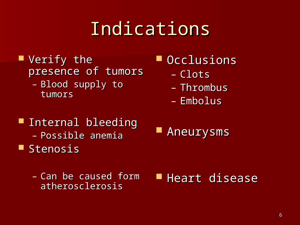
IndicationsIndications
Verify the presence of Verify the presence of tumorstumors– Blood supply to tumorsBlood supply to tumors
Internal bleedingInternal bleeding– Possible anemiaPossible anemia
StenosisStenosis
– Can be caused form Can be caused form atherosclerosisatherosclerosis
OcclusionsOcclusions– ClotsClots– ThrombusThrombus– EmbolusEmbolus
AneurysmsAneurysms
Heart diseaseHeart disease

77
ContraindicationsContraindications
Previous severe reaction to contrast Previous severe reaction to contrast
Impaired renal functionImpaired renal function
Impaired blood clotting factorsImpaired blood clotting factors
Inability to undergo surgical Inability to undergo surgical procedureprocedure

88
Contrast MediaContrast Media
Iodinated contrast media is usedIodinated contrast media is used– Can produce nausea & an Can produce nausea & an
uncomfortable burning sensationuncomfortable burning sensation
– Allergic reactionsAllergic reactions Severe: anaphylactic shockSevere: anaphylactic shock
– Shock, rapid shallow breathing, high pulse rate & Shock, rapid shallow breathing, high pulse rate & ALOCALOC
Mild: Hives or slight difficulty breathingMild: Hives or slight difficulty breathing

99
What is this?What is this?

1010
1111
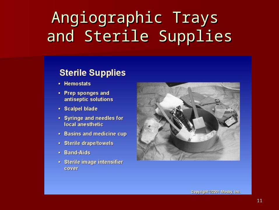
Angiographic Trays Angiographic Trays and Sterile Suppliesand Sterile Supplies

1212
Other Supplies for Other Supplies for AngiographyAngiography

1313
NeedlesNeedles
Vascular access Vascular access needlesneedles
Size based on Size based on external diameter of external diameter of needleneedle
Allows for Allows for appropriate appropriate Guidewires Guidewires matchingmatching– So internal diameter So internal diameter
must also be knownmust also be known

1414
GuidewiresGuidewires Used as a platform Used as a platform
over which a catheter over which a catheter is to be advancedis to be advanced
Once positioned Once positioned guidewire is fixed and guidewire is fixed and catheter is advanced catheter is advanced until it meets the tip until it meets the tip of the guidwireof the guidwire
Mostly constructed on Mostly constructed on stainless steel & stainless steel & coated with Tefloncoated with Teflon

1515
Introducer SheathsIntroducer Sheaths
Short catheters Short catheters used when multiple used when multiple catheters will be catheters will be usedused
Placed in lieu of a Placed in lieu of a cathetercatheter

1616
CathetersCatheters

1717

1818
DSA DSA
A subtraction mask is A subtraction mask is taken before contrast taken before contrast injectedinjected
Each of digitized Each of digitized image is from the image is from the maskmask
Images acquired Images acquired formform– 1 image every 2-3 sec1 image every 2-3 sec– Up to 30 images per Up to 30 images per
secsec

1919
Three Dimensional (3-D) Three Dimensional (3-D) Intraarterial AngiographyIntraarterial Angiography

2020
What Method is this?What Method is this?

2121
Catherization: Selinger Catherization: Selinger TechniqueTechnique

2222
Selinger Technique Catheters Selinger Technique Catheters and Guidewiresand Guidewires

2323
Pre-ProcedurePre-Procedure PT’s are usually limited to a liquid diet and PT’s are usually limited to a liquid diet and
routine medicationsroutine medications
Adequate hydrationAdequate hydration
An IV line placedAn IV line placed– Sedative may be givenSedative may be given
History taken and vitals takenHistory taken and vitals taken
Informed consentInformed consent

2424
Preparing the Patient RoomPreparing the Patient Room
Must be extensively cleanedMust be extensively cleaned
Equipment checkedEquipment checked
Room thoroughly stockedRoom thoroughly stocked
Extra supplies as neededExtra supplies as needed

2525
Radiation ProtectionRadiation Protection
PT is protected by no less than 2.5 PT is protected by no less than 2.5 mm of Aluminummm of Aluminum
Beam restrictionBeam restriction Avoidance of repeat exposureAvoidance of repeat exposure Cardinal rulesCardinal rules
– Time Time – DistanceDistance– ShieldingShielding

2626
Post ProcedurePost Procedure
PTs usually can resume normal PTs usually can resume normal activity after 24 hoursactivity after 24 hours
Most often can go home after 24 hoursMost often can go home after 24 hours– Because internal bleeding can be life Because internal bleeding can be life
threateningthreatening Vitals are monitoredVitals are monitored Puncture site is monitored for bleedingPuncture site is monitored for bleeding

2727
Stent PlacementStent Placement
http://images.google.com/imgres?http://images.google.com/imgres?imgurl=http://www.nhlbi.nih.gov/health/dci/imgurl=http://www.nhlbi.nih.gov/health/dci/images/stent_restenosis.gif&imgrefurl=http://images/stent_restenosis.gif&imgrefurl=http://www.nhlbi.nih.gov/health/dci/Diseases/stents/www.nhlbi.nih.gov/health/dci/Diseases/stents/stents_all.html&usg=__xDlbsaX9JhuYbpVojLcz19stents_all.html&usg=__xDlbsaX9JhuYbpVojLcz19apr-apr-I=&h=513&w=450&sz=59&hl=en&start=20&tbI=&h=513&w=450&sz=59&hl=en&start=20&tbnid=vWwqaG-RNW7M-nid=vWwqaG-RNW7M-M:&tbnh=131&tbnw=115&prev=/images%3FqM:&tbnh=131&tbnw=115&prev=/images%3Fq%3Dabdominal%2Bstents%26gbv%3D2%26hl%3Dabdominal%2Bstents%26gbv%3D2%26hl%3Den%3Den

2828
AortogramAortogram

2929

3030
AORTOGRAMAORTOGRAM

3131
Abdominal AortoraphyAbdominal Aortoraphy

3232
Abdominal AngiographyAbdominal Angiography

3333
AAA Pre and Post Stent AAA Pre and Post Stent PlacementPlacement

3434

3535
Abdominal StentAbdominal Stent

3636
AAAAAA

3737
Pulmonary CirculationPulmonary Circulation

3838
Pulmonary ArteriogramPulmonary Arteriogram

Celiac AteriogramCeliac Ateriogram
3939

Hepatic ArteriogramHepatic Arteriogram
4040

Splenic ArteriorgramSplenic Arteriorgram
4141

4242
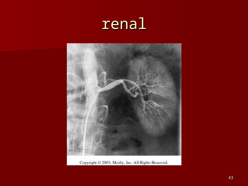
Renal ArteriogramRenal Arteriogram
4343
renalrenal

4444

4545

4646

4747
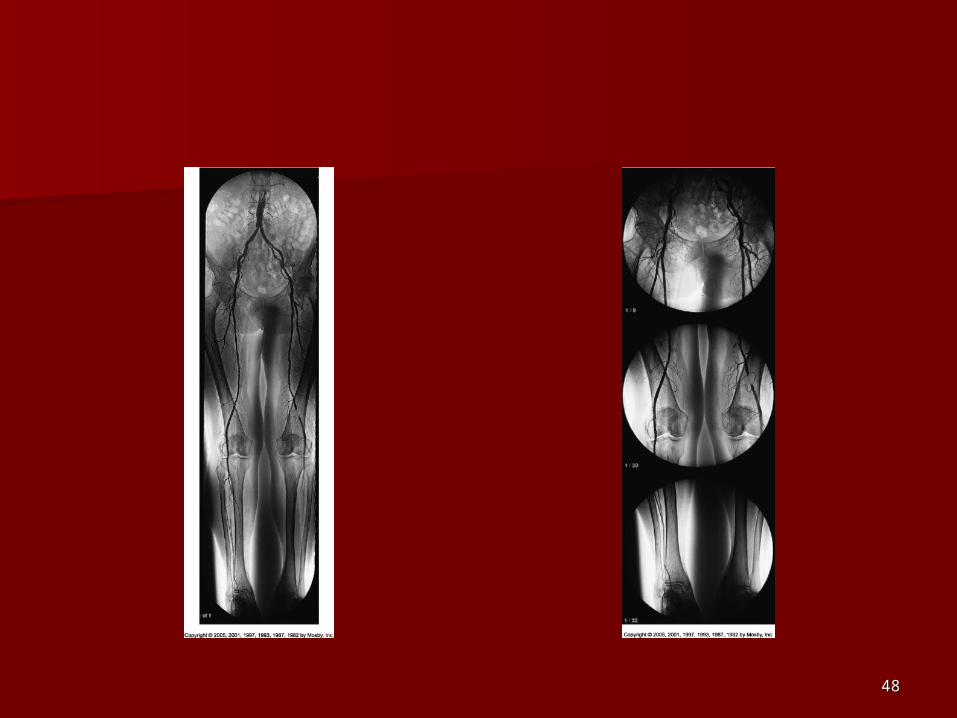
Lower Limb ArteriesLower Limb Arteries
4848

4949
Leg AtherosclerosisLeg Atherosclerosis

5050
Atherosclerosis Left LegAtherosclerosis Left Leg

5151
Upper Limb ArteriesUpper Limb Arteries

5252
Upper Extremity AnatomyUpper Extremity Anatomy

5353
Brachial and Axillary Brachial and Axillary ArteriogramArteriogram

5454
Axillary ArteriogramAxillary Arteriogram

5555
Hand ArteriogramHand Arteriogram

5656
Hand Arteriogram with Hand Arteriogram with OcclusionOcclusion

5757

5858
Balloon AngioplastyBalloon Angioplasty

5959

6060
Balloon Angioplasty Balloon Angioplasty ProcedureProcedure

6161
Femoral Artery AngioplastyFemoral Artery Angioplasty

6262
Placing a Stent after Placing a Stent after Angioplasty with BalloonAngioplasty with Balloon

6363
Intravascular StentsIntravascular Stents

Let’s ReviewLet’s Review

6565
B
C

6666
What is the name of this What is the name of this Procedure?Procedure?
What is it done for?What is it done for?

6767
What is the name of this What is the name of this pathology?pathology?

6868
What part of the body is being What part of the body is being imaged?imaged?
What is the pathology is this What is the pathology is this image?image?

6969

7070
What is this method callled?What is this method callled?

7171
A
B
C

Venography Venography

Venous Venous CirculationCirculation

What is Venography?What is Venography?
Vein study using x-ray and contrast Vein study using x-ray and contrast mediamedia– Fluoroscopy and still imagesFluoroscopy and still images
One of the most accurate tests for One of the most accurate tests for deep vein thrombosis (DVT)deep vein thrombosis (DVT)
Most commonly done in legs for DVTMost commonly done in legs for DVT

Thrombosis and EmbolismThrombosis and Embolism
Intravascular clotIntravascular clot Commonly in veins Commonly in veins
more than arteriesmore than arteries
3 factors3 factors– Where blood is slowWhere blood is slow– Change in the wall Change in the wall
of vesselsof vessels– Change in the blood Change in the blood
itselfitself
Thrombus that Thrombus that becomes detached becomes detached from the vessel wallfrom the vessel wall
Can easily flow to Can easily flow to heart causing PEheart causing PE
Severity depends Severity depends on location of on location of embolismembolism
Pulmonary EmbolismPulmonary Embolism Occurs when a clot forms or becomes lodged in Occurs when a clot forms or becomes lodged in
the pulmonary arterythe pulmonary artery
Most commonly thrombus originates in the lower Most commonly thrombus originates in the lower limbs and migrateslimbs and migrates
Can lead to resp distress, heart failure or Can lead to resp distress, heart failure or cardiogenic shockcardiogenic shock
Symptoms are acute:Symptoms are acute:– Sudden coughingSudden coughing– SOBSOB– Chest painChest pain

Pulmonary Emboli (PE)Pulmonary Emboli (PE)

IndicationsIndications Diagnose deep vein Diagnose deep vein
thrombosis thrombosis – Prevent pulmonary Prevent pulmonary
embolismembolism
Distinguish blood clots from Distinguish blood clots from obstructions in the veins obstructions in the veins
Evaluate congenital vein Evaluate congenital vein problems problems
Assess the functioning of Assess the functioning of deep leg vein valves deep leg vein valves
Identify a vein for arterial Identify a vein for arterial bypass graftingbypass grafting

Risk Factors and Risk Factors and ComplicationsComplications
Previous thrombosis Previous thrombosis
Dilution of the contrast dye in the Dilution of the contrast dye in the lower limb lower limb
Difficulty accessing the veins due to: Difficulty accessing the veins due to: – ObesityObesity– Severe swelling (edema) Severe swelling (edema) – Inflammation in the cells ( cellulitis ) Inflammation in the cells ( cellulitis )

ContraindicationsContraindications
Bleeding disordersBleeding disorders
Allergy to iodineAllergy to iodine
CHFCHF
Severe pulmonary hypertensionSevere pulmonary hypertension

Prior to ProcedurePrior to Procedure
Fast or drink only clear fluids for four Fast or drink only clear fluids for four hours before the testhours before the test
Thorough PT history obtainedThorough PT history obtained
Informed consentInformed consent
If you are nervous about the test, your If you are nervous about the test, your doctor may give you a sedative.doctor may give you a sedative.

During ProcedureDuring Procedure
PT will lie on a tilting x-ray tablePT will lie on a tilting x-ray table
Area of interest will be Area of interest will be shaved and cleaned shaved and cleaned
Local anestheticLocal anesthetic
Catheter will be inserted.Catheter will be inserted.– A small incision may be A small incision may be made in that area as wellmade in that area as well

Explanation of Procedure: Explanation of Procedure: LegsLegs The catheter is inserted The catheter is inserted
into PT veininto PT vein– (usually a vein in the foot) (usually a vein in the foot)
Contrast is slowly injected. Contrast is slowly injected.
A tight band may be tied A tight band may be tied around your ankle and around your ankle and upper thighupper thigh– or your lower body may be or your lower body may be
tiltedtilted– Fluoro and/or x-ray images Fluoro and/or x-ray images
takentaken
The procedure takes about The procedure takes about 30 - 45 minutes 30 - 45 minutes
Post ProcedurePost Procedure
Rest and avoid strenuous activityRest and avoid strenuous activity
Increase fluid intakeIncrease fluid intake
Stop bleeding with pressure Stop bleeding with pressure – Call DR if it won’t stop bleedingCall DR if it won’t stop bleeding
Observe for signs of infectionObserve for signs of infection
PT will be sore for a few daysPT will be sore for a few days
Resume normal activity 24 hours after procedureResume normal activity 24 hours after procedure

Possible Post Procedure Possible Post Procedure ComplicationsComplications
Infection at the Infection at the injection site injection site
Tissue damage Tissue damage
Phlebitis Phlebitis (inflammation of a (inflammation of a vein) vein)
Allergic reactions to Allergic reactions to the contrast dye the contrast dye
Congestive heart Congestive heart failure failure
Acute renal Acute renal insufficiency insufficiency
Venous thrombosis in a Venous thrombosis in a healthy leg healthy leg
Dislodging a clot, Dislodging a clot, perhaps resulting in perhaps resulting in pulmonary embolus or pulmonary embolus or other complicationsother complications

Lower Limb VeinsLower Limb Veins

Lower Limb VenogramsLower Limb Venograms
To rule out thrombosis of the deep To rule out thrombosis of the deep veins of the leg veins of the leg – Deep vein thrombosis (DVT)Deep vein thrombosis (DVT)
Contrast media injected in superficial Contrast media injected in superficial veins of the foot with a needleveins of the foot with a needle

Lower Limb Lower Limb VenogramsVenograms

DVTDVT

Inferior VenacavagramInferior Venacavagram
Primarily to rule out thrombus or Primarily to rule out thrombus or occlusionocclusion
Catheter inserted into femoral vein and Catheter inserted into femoral vein and positioned inside the common iliac vein positioned inside the common iliac vein or inferior aspect of inferior vena cavaor inferior aspect of inferior vena cava
Contrast injected at 20 ml/sec for total of Contrast injected at 20 ml/sec for total of 40ml40ml

Upper Limb VeinsUpper Limb Veins

Upper Limb VenogramsUpper Limb Venograms Most often for thrombosis or Most often for thrombosis or
occlusionocclusion Contrast injected in a superficial vein Contrast injected in a superficial vein
in the elbow or wristin the elbow or wrist– Using a catheter or needleUsing a catheter or needle– 40-80ml at a rate of 1-4ml/sec40-80ml at a rate of 1-4ml/sec

Superior VenacavagramSuperior Venacavagram Primarily done to rule out thrombus or Primarily done to rule out thrombus or
occlusionocclusion
Needle or catheter is introduced into Needle or catheter is introduced into antecubital fossaantecubital fossa– Catheter is positioned in the axillary or Catheter is positioned in the axillary or
subclavian vein and contrast is injectedsubclavian vein and contrast is injected– 30-50ml at 10-15ml/sec30-50ml at 10-15ml/sec
X-rays should include:X-rays should include:– Brachicephalic veinBrachicephalic vein– Subclavian veinSubclavian vein– Superior vena cavaSuperior vena cava– RT AtriumRT Atrium

Superior VenacavagramSuperior Venacavagram

Stenosis on a Superior Stenosis on a Superior VenacavogramVenacavogram

Inferior VenacavagramInferior Venacavagram

Inferior VenacavagramInferior Venacavagram

Inferior Vena Cava FiltersInferior Vena Cava Filters

Inferior Vena Cava Filter Inferior Vena Cava Filter PlacementPlacement
Designed to trap Designed to trap thrombus before thrombus before causing an causing an embolizationembolization
When When anticoagulants are anticoagulants are contraindicated contraindicated this can be usedthis can be used

Inferior Vena Cava Filter Inferior Vena Cava Filter PlacementPlacement


Hepatic VenogramHepatic Venogram
Performed to rule out stenosis or Performed to rule out stenosis or thrombus of the hepatic veinsthrombus of the hepatic veins
Obtain pressure measurements of Obtain pressure measurements of the veins inside the liverthe veins inside the liver
Usually catheter enters jugular vein Usually catheter enters jugular vein or upper limb veinsor upper limb veins

Hepatic VenogramHepatic Venogram

Portal VenogramPortal Venogram

Portal SystemPortal System
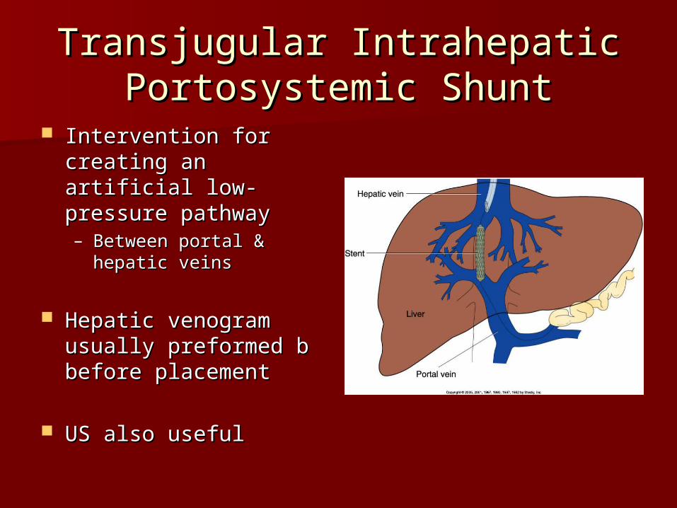
Transjugular Intrahepatic Transjugular Intrahepatic Portosystemic ShuntPortosystemic Shunt
Intervention for Intervention for creating an artificial creating an artificial low-pressure pathwaylow-pressure pathway– Between portal & Between portal &
hepatic veinshepatic veins
Hepatic venogram Hepatic venogram usually preformed b usually preformed b before placementbefore placement
US also usefulUS also useful

Transjugular Intrahepatic Transjugular Intrahepatic Portosystemic ShuntPortosystemic Shunt

Renal VenogramRenal Venogram
Rule out thrombosis of renal veinRule out thrombosis of renal vein
Renal vein catheterized to take bloodRenal vein catheterized to take blood– Measure the production of reninMeasure the production of renin– Catheter insertion site: femoral veinCatheter insertion site: femoral vein
Contrast injected 8ml/sec for 16ml Contrast injected 8ml/sec for 16ml totaltotal– 2 images per second for 4 seconds2 images per second for 4 seconds

Renal VenogramRenal Venogram