an atypical case of oral squamous cell carcinoma of
TRANSCRIPT
Case ReportAn Atypical Case of Oral Squamous Cell Carcinoma ofMandibular Alveolus
Seba Abraham , Bhagyalekshmi Mallika , Arunima Reshma,and ReejaMol Mohammed Kassim
PMS College of Dental Science and Research, India
Correspondence should be addressed to Bhagyalekshmi Mallika; [email protected]
Received 1 June 2019; Revised 21 September 2019; Accepted 30 September 2019; Published 13 November 2019
Academic Editor: Ronald S. Brown
Copyright © 2019 Seba Abraham et al. This is an open access article distributed under the Creative Commons Attribution License,which permits unrestricted use, distribution, and reproduction in any medium, provided the original work is properly cited.
Oral squamous cell carcinoma is the most common type of oral malignant neoplasm. As per literature, squamous cell carcinomas ofthe alveolar ridge account for 9% of all the oral carcinomas. The oral squamous cell carcinoma shares clinical similarity with variousforms of inflammatory gingival lesions and is often misdiagnosed in our routine dental practice. The dentist should have preciseknowledge regarding the clinical manifestation of this deadly disease as early diagnosis and prompt treatment can reducemorbidity and mortality of the disease.
1. Introduction
Oral cancer accounts for the eleventh most common cancerworldwide [1]. Oral squamous cell carcinoma (OSCC) isthe common oral carcinoma with varied clinical presenta-tions. It accounts for more than 90% of all malignant lesionsin the oral cavity [2]. The affected population group was<40 years of age in high-incidence countries such as India,Pakistan, and Sri Lanka [3]. Alveolar ridge SSC accountsfor 9% of all cases with oral SCC [4]. Alveolar ridge SSCaccounts for the second position, with the first being carci-noma of the tongue as per site specificity. As per local recur-rence rate by site, mandibular alveolus carcinoma has thehighest local recurrence rate (26/42), second being carci-noma of the tongue (20/47). Squamous cell carcinomas canoccur in all areas of the body, but they are most common inthe skin and oral cavity.
Oral squamous cell carcinoma of the mandibular regionis found to have the lowest survival rate when compared withall other oral carcinomas [5]. Various aids used for assessingmandibular invasion include clinical examination, radio-graphic examination (OPG, CT dentascan, MRI, bone scans,ultrasound, and PET CT), and histopathological examina-tion. Histopathologic analysis of squamous cell carcinoma
reveals two basic patterns of tumor invasion: invasive pattern(infiltrative pattern) and erosive pattern (compressivepatterns). In the invasive pattern, islands of tumor infiltratecancellous spaces and have little osteoclastic activity and nointervening connective tissue. However, in the erosivepattern, the tumor advances as a broad front with active oste-oclasts separating the tumor from the bone and the connec-tive tissue layer separating the tumor/bone [6].
Marginal resection is considered as the best treatmentoption if the carcinoma reaches close to the bone withoutany invasion or with mild invasion [5]. Marginal resectioninvolves the removal of 1 cm of margins from all sides [7].Nomura et al. in 2005 reported that in case of oral squamouscell carcinoma, the tumor extended to the periosteum in 33%,to the cortical bone in 23%, and to the bone marrow in 9% ofthe patients who underwent mandibular resection. Theremaining 35% of the patients had no evidence of mandibu-lar invasion. The area of bone resorption on preoperativeclinical and radiographic examinations often disagreedwith the extent of mandibular invasion on histopathologicexamination [8].
The clinical presentation of oral squamous cellcarcinoma can range from a white plaque to an ulceratedlesion [9]. Malignant lesions in the gingiva may resemble
HindawiCase Reports in DentistryVolume 2019, Article ID 2521685, 6 pageshttps://doi.org/10.1155/2019/2521685

frequently seen inflammatory lesions of the gingiva. Oral car-cinomas can often be misdiagnosed as other inflammatorylesions in the oral cavity leading to delay in delivering prompttreatment. Thus, the early diagnosis and treatment of oralcarcinoma by health care providers is mandatory foroptimum treatment outcome [10]. Treatment of squamouscell carcinoma is mainly a surgical excision and a radical neckdissection in the case of lymph node metastasis. Radiother-apy is considered as adjunct postoperative treatment alongwith chemotherapy and is a definite treatment of choice incase of advanced stages of cancer.
Oral carcinoma has emerged as a major health problemin India mainly because of the high prevalence of smokelesstobacco use. Oral SCC metastases to regional lymph nodesin the neck and distant metastases are seen in the laterstages as diseases progress [11]. Here, we are reporting anontobacco-related case of SCC of the lower alveolus withatypical presentation of desquamative gingival lesions andoften more chance to misdiagnose the case.
2. Case Report
A 48-year-old female patient presented to the Department ofPeriodontics with the chief complaint of acute pain inrelation to the lower right back teeth region for the past 2months. The patient gave a history of antibiotic and analgesicmedication for one week after consulting with a nearbyhospital. Pain was the throbbing type and continuous innature, and it aggravates on opening the mouth, duringmastication, and while brushing teeth.
General clinical examination revealed normal mouthopening. Multiple tender lymph nodes were noted on theright side of the face and neck region. On palpation, sub-mandibular and cervical lymph nodes were tender andpalpable (2 cm in diameter) and were firm and fixed tothe skin.
The intraoral examination revealed desquamativechanges in relation to the 43, 44, 45, and 46 regions(Figure 1). Grade 2 mobility in relation to the 44, 46, and47 regions and grade 3 mobility in relation to 45. Noassociated paraesthesia was reported. An ulceroproliferativelesion extending from the 44 to 47 regions with mixed kera-totic and erythematous area with granular appearance was
noted (Figure 2). On palpation, the lesion was tender withmild induration. Desquamative lesion was noted in relationto 13 extending to 15 regions (Figure 3). Periodontal probingresulted in profuse bleeding in relation to 45 and 46 regions.Deep pockets (>6mm) in relation to the 16, 17, 26, 27, 44, 45,46, and 47 regions.
Hard tissue examination revealed an end-on-endocclusion. A comprehensive periodontal examination wasdone which revealed heavy deposits of calculus on the lingualsurface of mandibular posteriors. Generalized cervical abra-sion and attrition were present. The oral hygiene mainte-nance was compromised due to severe pain while brushinginterfering with proper plaque removal.
2.1. Radiographic Evaluation. OPG revealed generalized hor-izontal bone loss with severe bone loss in relation to the 45and 46 regions with widening of PDL space and furcationradiolucency in relation to 46. Impacted 18 and 28 werenoted (Figure 4). Diffused poorly defined radiolucency wasnoted in relation to 45 and 46 extending up to the superiorborder of the mandibular canal (Figure 5).
Based on clinical and radiographic examination, aprovisional diagnosis of chronic generalized periodontitisand carcinoma alveolus was made. Differential diagnosis iserosive lichen planus.
Staging of oral squamous cell carcinoma is done based onthe American Joint Committee on Cancer. cTNM is the stagegiven after the clinical examination of the patient, while
Figure 1: Desquamative changes in relation to the 43, 44, 45, and46 regions.
Figure 2: Ulceroproliferative lesion extending from the 44 to 47regions.
Figure 3: Desquamative lesion noted in relation to 13 extending tothe 15 regions.
2 Case Reports in Dentistry

pTNM is the stage after the histopathological examination ofthe surgical specimen. Based on the American Joint Commit-tee on Cancer January 2018, the cTNM cancer staging of thiscase is AJCC Stage IVA and stage grouping T4aN1M0 asthere is involvement to the jaw bone and presence of a palpa-ble lymph node of size 2 cm.
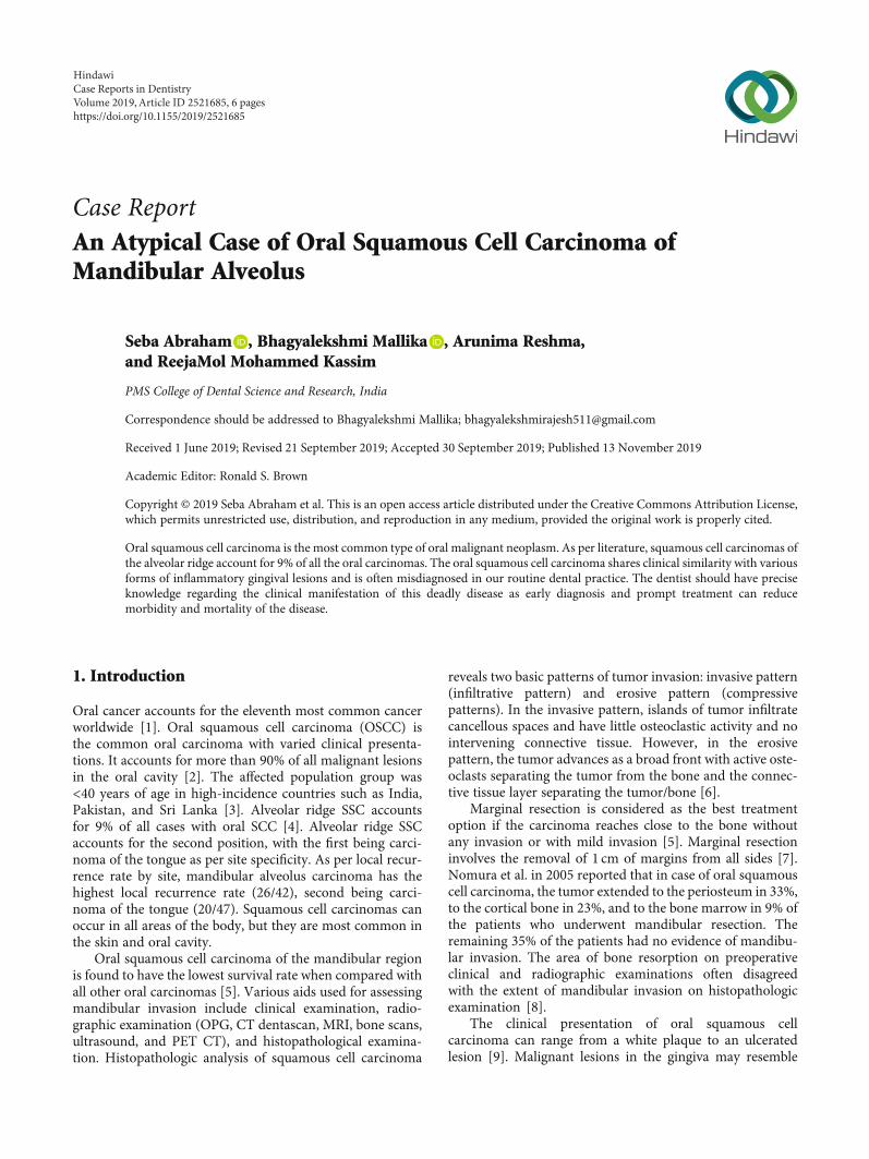
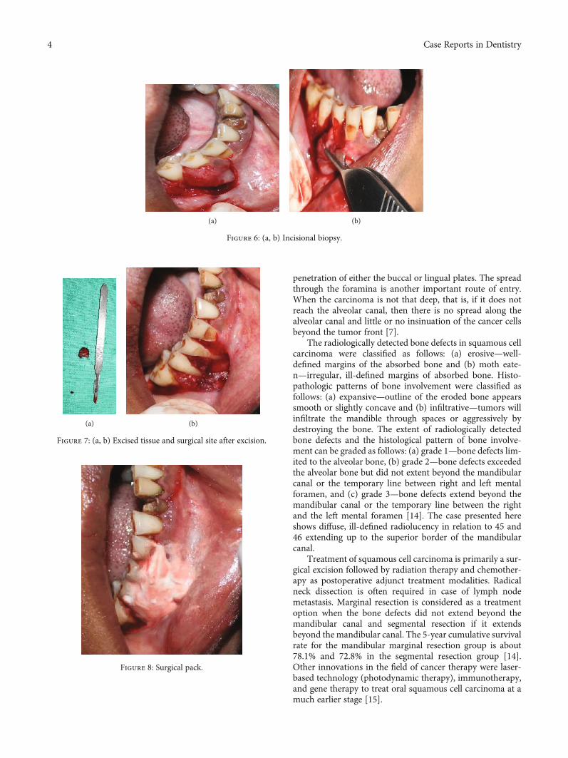
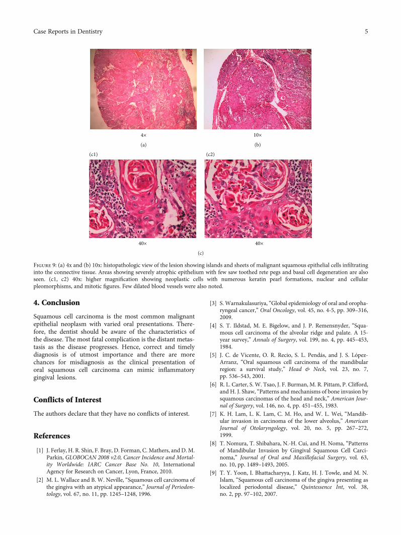
2.2. Treatment. Initially, nonsurgical periodontal therapyincluding scaling and root planing was performed. Inci-sional biopsy of the oral lesion extending from the mar-ginal gingiva of 44 to 46 to the mucogingival junction(measuring about 1:4 ∗ 1:3 ∗ 0:6 cm in size) was performedunder LA (Figure 6). The excised tissue was sent for histo-pathological examination (Figure 7). 3-0 vicryl sutures placedand a surgical pack given (Figure 8). The patient was keptunder antibiotic and anti-inflammatory therapy for one week.
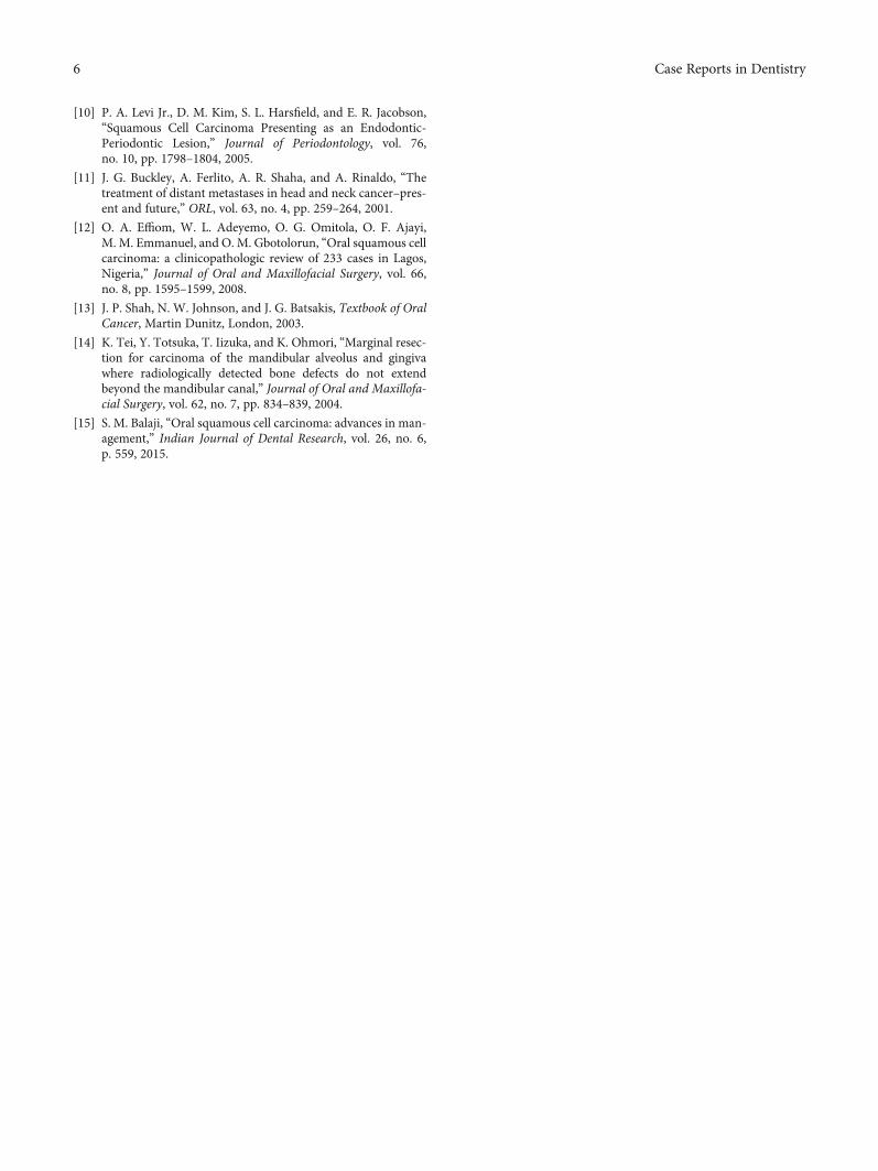
2.3. Histopathological Examination. Histopathological exam-ination of the given soft tissue section revealed islands andsheets of malignant squamous epithelial cells infiltrating intothe connective tissue (Figures 9(a) and 9(b)). The neoplasticcells exhibit numerous keratin pearl formation, nuclear andcellular pleomorphism, and mitotic figures (Figure 9(c) (c1,c2) 40x). Areas of severely atrophic epithelium with few
saw toothed rete pegs and basal cell degeneration are alsoseen. Based on clinical, radiographic, and histopathologicalexaminations, the case is diagnosed as squamous cell carci-noma (SCC). The patient was referred to the Regional CancerCentre (RCC), Thiruvananthapuram, for further treatments.We tried to contact the patient for further follow up but wasin vain.
3. Discussion
Squamous cell carcinoma is considered as the most com-mon malignant neoplasm of the oral cavity. The tongue,oropharynx, and floor of the mouth are the most commonsites, and SCC of the gingiva and lips is rarely seen. SCCof the mandibular gingiva is more common than themaxillary gingiva [12].
Most of the cases of oral carcinoma are associated withtobacco chewing habit and usually appear as a premalignantlesion like leukoplakia before progressing to the malignantstage, but rare cases have also been reported withnontobacco-associated squamous cell carcinoma. The casereported here is one without the history of tobacco chewinghabit.
Carcinoma of the gingival region often mimics desqua-mative lesions of the gingiva and other inflammatory gingivallesions. The gingiva is one of the most common sites forchronic inflammation as it is often associated with irritantslike calculus and abundant microbial flora. Attached gingivais more involved than free gingiva [4]. Misdiagnosis is oftenencountered in the absence of detailed clinical examinationand radiographic investigation.
Mandibular alveolus is the second most common site fororal carcinoma. Oral squamous cell carcinoma is morefrequently seen among men compared to women as menare often exposed to high risk habits such as smoking andtobacco chewing (Liviu Feller et al. 2012). Age is another crit-ical factor for oral SCC; as age advances, pronounced geneticand epigenetic changes take place. The case presented here isa female patient of age 48 years with a lesion mimicking des-quamative lesion in relation to the lower right back teethregion. Another desquamative lesion was noted in the maxil-lary region in relation to 13, 14, and 15 without the clinicalpresentation of SCC.
Regional lymph node metastasis is another feature ofsquamous cell carcinoma. Cervical lymph nodes of the sub-mandibular triangle and upper jugular regions have strongerpredilection of regional lymph node metastasis in the case ofSCC of the lower alveolus [13]. Tender and palpable subman-dibular and cervical lymph nodes (which are firm and fixedto the skin) of about 2 cm in diameter were detected in thepresent case. Prognosis is better in early oral SCC, especiallythose that are well-differentiated and without metastasis, butthe worst part is that most cases of OSCC are not diagnosedat the earlier stage of the disease. The prognosis can varybased on a number of factors that are related to the tumoror treatment or to the patient.
Squamous cell carcinoma was found to enter the medul-lary cavity through the upper border of the mandible, eitherthrough the occlusal ridge alone or in combination with
Figure 5: Diffused poorly defined radiolucency was noted inrelation to 45 and 46 extending up to the superior border of themandibular canal.
Figure 4: Generalized horizontal bone loss with severe bone loss inrelation to the 45 and 46 regions with widening of the PDL spaceand furcation involvement in relation to 46. Impacted 18 and 28.
3Case Reports in Dentistry
penetration of either the buccal or lingual plates. The spreadthrough the foramina is another important route of entry.When the carcinoma is not that deep, that is, if it does notreach the alveolar canal, then there is no spread along thealveolar canal and little or no insinuation of the cancer cellsbeyond the tumor front [7].
The radiologically detected bone defects in squamous cellcarcinoma were classified as follows: (a) erosive—well-defined margins of the absorbed bone and (b) moth eate-n—irregular, ill-defined margins of absorbed bone. Histo-pathologic patterns of bone involvement were classified asfollows: (a) expansive—outline of the eroded bone appearssmooth or slightly concave and (b) infiltrative—tumors willinfiltrate the mandible through spaces or aggressively bydestroying the bone. The extent of radiologically detectedbone defects and the histological pattern of bone involve-ment can be graded as follows: (a) grade 1—bone defects lim-ited to the alveolar bone, (b) grade 2—bone defects exceededthe alveolar bone but did not extent beyond the mandibularcanal or the temporary line between right and left mentalforamen, and (c) grade 3—bone defects extend beyond themandibular canal or the temporary line between the rightand the left mental foramen [14]. The case presented hereshows diffuse, ill-defined radiolucency in relation to 45 and46 extending up to the superior border of the mandibularcanal.
Treatment of squamous cell carcinoma is primarily a sur-gical excision followed by radiation therapy and chemother-apy as postoperative adjunct treatment modalities. Radicalneck dissection is often required in case of lymph nodemetastasis. Marginal resection is considered as a treatmentoption when the bone defects did not extend beyond themandibular canal and segmental resection if it extendsbeyond the mandibular canal. The 5-year cumulative survivalrate for the mandibular marginal resection group is about78.1% and 72.8% in the segmental resection group [14].Other innovations in the field of cancer therapy were laser-based technology (photodynamic therapy), immunotherapy,and gene therapy to treat oral squamous cell carcinoma at amuch earlier stage [15].
(a) (b)
Figure 6: (a, b) Incisional biopsy.
(a) (b)
Figure 7: (a, b) Excised tissue and surgical site after excision.
Figure 8: Surgical pack.
4 Case Reports in Dentistry
4. Conclusion
Squamous cell carcinoma is the most common malignantepithelial neoplasm with varied oral presentations. There-fore, the dentist should be aware of the characteristics ofthe disease. The most fatal complication is the distant metas-tasis as the disease progresses. Hence, correct and timelydiagnosis is of utmost importance and there are morechances for misdiagnosis as the clinical presentation oforal squamous cell carcinoma can mimic inflammatorygingival lesions.
Conflicts of Interest
The authors declare that they have no conflicts of interest.
References
[1] J. Ferlay, H. R. Shin, F. Bray, D. Forman, C. Mathers, and D.M.Parkin, GLOBOCAN 2008 v2.0, Cancer Incidence and Mortal-ity Worldwide: IARC Cancer Base No. 10, InternationalAgency for Research on Cancer, Lyon, France, 2010.
[2] M. L. Wallace and B. W. Neville, “Squamous cell carcinoma ofthe gingiva with an atypical appearance,” Journal of Periodon-tology, vol. 67, no. 11, pp. 1245–1248, 1996.
[3] S. Warnakulasuriya, “Global epidemiology of oral and oropha-ryngeal cancer,” Oral Oncology, vol. 45, no. 4-5, pp. 309–316,2009.
[4] S. T. Ildstad, M. E. Bigelow, and J. P. Remensnyder, “Squa-mous cell carcinoma of the alveolar ridge and palate. A 15-year survey,” Annals of Surgery, vol. 199, no. 4, pp. 445–453,1984.
[5] J. C. de Vicente, O. R. Recio, S. L. Pendás, and J. S. López-Arranz, “Oral squamous cell carcinoma of the mandibularregion: a survival study,” Head & Neck, vol. 23, no. 7,pp. 536–543, 2001.
[6] R. L. Carter, S. W. Tsao, J. F. Burman, M. R. Pittam, P. Clifford,and H. J. Shaw, “Patterns and mechanisms of bone invasion bysquamous carcinomas of the head and neck,” American Jour-nal of Surgery, vol. 146, no. 4, pp. 451–455, 1983.
[7] K. H. Lam, L. K. Lam, C. M. Ho, and W. L. Wei, “Mandib-ular invasion in carcinoma of the lower alveolus,” AmericanJournal of Otolaryngology, vol. 20, no. 5, pp. 267–272,1999.
[8] T. Nomura, T. Shibahara, N.-H. Cui, and H. Noma, “Patternsof Mandibular Invasion by Gingival Squamous Cell Carci-noma,” Journal of Oral and Maxillofacial Surgery, vol. 63,no. 10, pp. 1489–1493, 2005.
[9] T. Y. Yoon, I. Bhattacharyya, J. Katz, H. J. Towle, and M. N.Islam, “Squamous cell carcinoma of the gingiva presenting aslocalized periodontal disease,” Quintessence Int, vol. 38,no. 2, pp. 97–102, 2007.
4×
(a)
10×
(b)
40×
(c1)
40×
(c2)
(c)
Figure 9: (a) 4x and (b) 10x: histopathologic view of the lesion showing islands and sheets of malignant squamous epithelial cells infiltratinginto the connective tissue. Areas showing severely atrophic epithelium with few saw toothed rete pegs and basal cell degeneration are alsoseen. (c1, c2) 40x: higher magnification showing neoplastic cells with numerous keratin pearl formations, nuclear and cellularpleomorphisms, and mitotic figures. Few dilated blood vessels were also noted.
5Case Reports in Dentistry
[10] P. A. Levi Jr., D. M. Kim, S. L. Harsfield, and E. R. Jacobson,“Squamous Cell Carcinoma Presenting as an Endodontic‐Periodontic Lesion,” Journal of Periodontology, vol. 76,no. 10, pp. 1798–1804, 2005.
[11] J. G. Buckley, A. Ferlito, A. R. Shaha, and A. Rinaldo, “Thetreatment of distant metastases in head and neck cancer–pres-ent and future,” ORL, vol. 63, no. 4, pp. 259–264, 2001.
[12] O. A. Effiom, W. L. Adeyemo, O. G. Omitola, O. F. Ajayi,M. M. Emmanuel, and O. M. Gbotolorun, “Oral squamous cellcarcinoma: a clinicopathologic review of 233 cases in Lagos,Nigeria,” Journal of Oral and Maxillofacial Surgery, vol. 66,no. 8, pp. 1595–1599, 2008.
[13] J. P. Shah, N. W. Johnson, and J. G. Batsakis, Textbook of OralCancer, Martin Dunitz, London, 2003.
[14] K. Tei, Y. Totsuka, T. Iizuka, and K. Ohmori, “Marginal resec-tion for carcinoma of the mandibular alveolus and gingivawhere radiologically detected bone defects do not extendbeyond the mandibular canal,” Journal of Oral and Maxillofa-cial Surgery, vol. 62, no. 7, pp. 834–839, 2004.
[15] S. M. Balaji, “Oral squamous cell carcinoma: advances in man-agement,” Indian Journal of Dental Research, vol. 26, no. 6,p. 559, 2015.
6 Case Reports in Dentistry

DentistryInternational Journal of
Hindawiwww.hindawi.com Volume 2018
Environmental and Public Health
Journal of
Hindawiwww.hindawi.com Volume 2018
Hindawi Publishing Corporation http://www.hindawi.com Volume 2013Hindawiwww.hindawi.com
The Scientific World Journal
Volume 2018Hindawiwww.hindawi.com Volume 2018
Public Health Advances in
Hindawiwww.hindawi.com Volume 2018
Case Reports in Medicine
Hindawiwww.hindawi.com Volume 2018
International Journal of
Biomaterials
Scienti�caHindawiwww.hindawi.com Volume 2018
PainResearch and TreatmentHindawiwww.hindawi.com Volume 2018
Preventive MedicineAdvances in
Hindawiwww.hindawi.com Volume 2018
Hindawiwww.hindawi.com Volume 2018
Case Reports in Dentistry
Hindawiwww.hindawi.com Volume 2018
Surgery Research and Practice
Hindawiwww.hindawi.com Volume 2018
BioMed Research International Medicine
Advances in
Hindawiwww.hindawi.com Volume 2018
Hindawiwww.hindawi.com Volume 2018
Anesthesiology Research and Practice
Hindawiwww.hindawi.com Volume 2018
Radiology Research and Practice
Hindawiwww.hindawi.com Volume 2018
Computational and Mathematical Methods in Medicine
EndocrinologyInternational Journal of
Hindawiwww.hindawi.com Volume 2018
Hindawiwww.hindawi.com Volume 2018
OrthopedicsAdvances in
Drug DeliveryJournal of
Hindawiwww.hindawi.com Volume 2018
Submit your manuscripts atwww.hindawi.com