amine content of vaginal fluid from untreated and treated...
TRANSCRIPT

Amine Content of Vaginal Fluid from Untreated and
Treated Patients with Nonspecific Vaginitis
KiRK C. S. CHEN, PATRICIA S. FORSYTH,THOMASM. BUCHANANand KING K. HOLMES,
Departments of Pathobiology and Medicine, University of Washington,and the Immunology Research Laboratory, U. S. Public Health Service Hospital,Seattle, Washington 98114
A B S T RA C T We examined the vaginal washingsfrom patients with nonspecific vaginitis (NSV) to seekbiochemical markers and possible explanations for thesigns and symptoms of this syndrome. Seven amineswere identified including methylamine, isobutylamine,putrescine, cadaverine, histamine, tyramine, andphenethylamine. These amines may contribute to thesymptoms of NSVand may contribute to the elevatedpH of the vaginal discharge. They may also be partlyresponsible for the "fishy" odor that is characteristicof vaginal discharges from these patients. Among theseven amines, putrescine and cadaverine were themost abundant and were present in all vaginal dis-charges from each of ten patients before treatment.These amines are produced in vitro during growth ofmixed vaginal bacteria in chemically defined medium,presumably by decarboxylation of the correspondingamino acids. We hypothesize that anaerobic vaginalorganisms, previously shown to be quantitatively in-creased in NSV, are responsible for the amine produc-tion, because metronidazole inhibited the productionof amines by vaginal bacteria in vitro, and Haemophilusvaginalis did not produce amines. H. vaginalis did re-lease high concentrations of pyruvic acid and of aminoacids during growth in peptone-starch-dextrose me-dium, whereas, other vaginal flora consumed both pyruvicacid and amino acids in the same medium duringgrowth. These findings suggest that a symbiotic rela-tionship may exist between H. vaginalis and othervaginal flora in patients with NSV.
INTRODUCTION
The vaginal fluid from women with nonspecificvaginitis (NSV)1 has a prominent odor often describedas a rotten-fish smell, particularly during and after sex-
Received for publication 12 October 1978.1 Abbreviations used in this paper: NSV, nonspecific
vaginitis; PSD, peptone-starch-dextrose.
828
ual intercourse. This "fishy" odor can be intensifiedin vaginal fluid from womenwith NSVby the additionof 10% KOHto vaginal fluid from untreated patientsas reported by Pheifer, et al. (1). The odor releasedby KOHsuggested the presence of amine(s), and sub-sequent analyses of vaginal washings from untreatedpatients with NSVdescribed in this report have iden-tified seven amines, i.e., methylamine, isobutylamine,putrescine, cadaverine, histamine, tyramine, andphenethylamine. These amines appear to be the de-carboxylated products of their corresponding aminoacids. This report describes the identification and quan-titation of these amines in vaginal washings obtainedfrom patients before and after treatment of NSV. Thebiological roles of the putative putrefactive decar-boxylases that may be responsible for the amine pro-duction, and the possible symbiotic relationship be-tween Haemophilus vaginalis and other vaginal floraare also discussed.
METHODSStudy population. 10 women were selected for this study
who had both symptoms and signs of NSVas described pre-viously (1), with culture-proven H. vaginalis vaginal infec-tion. Each was studied before and after antimicrobial treat-ment with metronidazole, ampicillin, and(or) erythromycin.Seven normal women who came for contraception exam-ination and had no symptoms or signs of NSVwere selectedas controls.
Collection of vaginal washings. 2ml of sterile water wasinstilled into the vagina and a sterile cotton-tipped applicatorwas used to swab the adherent vaginal secretions into thepooled fluid. The diluted vaginal fluid was removed with asterile pipette, transferred to a plastic tube, and the pH wasmeasured with a pH meter with a combined pH electrode9.5 x 200 mm(PHM 61, Radiometer Co., Copenhagen).
The collected fluid was centrifuged in a clinical centrifugeat 1,800 g for 2 min. To the supernate, which usually had avolume of 1.5 to 2.2 ml, was added 10 ,ul of concentratedHCI and this solution was then frozen at -20°C.
Analysis of vaginal washings by high-voltage electro-phoresis at pH 2.1. The supemate of collected vaginal wash-ings (15-25 ILI) was spotted on a Whatman 3 MMpaper (What-
J. Clin. Invest. C The American Society for Clinical Investigation, Inc. * 0021-9738/79105/0828108 $1.00Volume 63 May 1979 828-835

man, Inc., Clifton, N. J.). High-voltage electrophoresis at pH2.1 was carried out for 12 min at 75 V/cm as described byChen and Krause (2). After drying, the paper was stained withninhydrin-cadmium (3). Amino acids (containing about 10nmol each) and dansylsulfonic acid (5-dimethylamino-naphthalene-l-sulfonic acid) were run in parallel with thesamples as markers (4).
Dansylation of amino acids, amines, and vaginal wash-ings. The procedure for dansylation of amino acids, amines,and vaginal washings was essentially the same as describedby Gray (5). 10 1l of dansyl chloride (5-dimethylamino-naphthalene-l-sulfonyl chloride, 2.5 mg/ml acetone) was addedto a mixture that consisted of 5 ,ul of vaginal washings (or5 ,ul of 2 mg/ml of amino acid or amine) and 5 ,l of 1 MNaHCO3. The reaction was carried out at 37°C for 1 h. Thesources of amines were described in a separate report.2
Separation of dansyl derivatives by thin-layer chromatog-raphy on polyamide sheets. The dansylated mixture fromvaginal washings was centrifuged, and the supernate wasdried under vacuum over concentrated H2SO4and NaOHpel-lets (in separate beakers) in a vacuum desiccator. The driedproducts were dissolved in 5 p1 of 50% pyridine. 1 ,l of thedansylated mixture was spotted on both sides of a polyamidesheet (Chen-Chin polyamide layer sheet, Accurate Chemical& Scientific Corp., Hicksville, N. Y.) at the same position and adansyl(5 - dimethylaminonaphthalene - 1- sulfonyl) - derivativemixture of known amino acids and amines was spotted at thesame position on the back side of the sheet as markers. Theconcentration of each dansyl-derivative marker was pre-adjusted to be barely seen under the ultra violet light (a 10-100times dilution of the 20 pl1-reaction mixture with water wasusually made). Thin-layer chromatography was carried outby the procedure described by Woods and Wang (6) exceptthat the second-dimensional chromatography was run to onlytwo-thirds the length of the sheet. The positions of the aminesin vaginal washings on the polyamide sheet after two-dimensional chromatography were identified by comparingthe positions of dansyl derivatives in vaginal washings onthe front side of the sheet to the corresponding intensifiedspots at the positions of the known amines on the oppositeside. Dansyl derivatives of tyramine and histamine co-chromatographed to the same position in the system describedabove, but separated well after chromatography in the thirdsolvent system (n-heptane: n-butanol: glacial acetic acid= 40:30:9) in the same dimension to full length.
Conversion of amino acids to amines in vitro by micro-organisms from patients with NSV. Whereas H. vaginalis, afacultative bacterium, is generally the predominant organismisolated from the vaginal discharges of women with NSV,obligate anaerobic bacteria often are also present in largerconcentrations in vaginal discharges from women with NSVthan from normal women (1). To determine the putativemicrobial source of the amines in vaginal washings from pa-tients with NSV, the following experiments were performed.During speculum examination of patients with typical symp-toms and signs of NSV, a cotton swab was used to inoculatevaginal discharge onto chocolate agar (BBL GC Base with5% chocolated sheep blood and 1% IsoVitaleX enrichmentBBL Microbiology Systems, Becton, Dickinson & Co.,Cockeysville, Md.) for incubation in a candle extinction jar toisolate H. vaginalis, and onto prereduced blood agar (BBLBrucella agar with 5%sheep blood) for anaerobic incubation
2Chen, K. C. S., T. M. Buchanan, P. R. Davick, and K. K.Holmes. 1979. Determination of biogenic amines usingheterocyclic cation and aromatic anion elution. Anal.Biochem. In Press.
in Gas Pak jars (Ferguson Industries, Dallas, Tex.) (7). Allcultures were incubated at 35°C.
Chocolate agar plates were examined after 48 h for growthof H. vaginalis, which was identified as described by Pheiferet al. (1). Pure subcultures of five separate isolates of H.vaginalis from chocolate agar plates were inoculated with awire loop into sterile tubes that contained 5 ml of peptone-starch-dextrose (PSD) medium (8). Inoculated PSDmediumand uninoculated medium controls were incubated anaerobi-cally at 35°C in a Gas Pak jar. After 3 d of incubation, aliquotsof the spent test media were subcultured to chocolate agarto check purity of the cultures.
Separate anaerobic cultures of vaginal discharge from fourpatients were incubated 4 d on the prereduced blood agar,after which the mixed growth was removed from the agarplates with a sterile cotton swab and suspended in sterilebroth. The suspension was mixed thoroughly and a sterilecapillary pipette was used to inoculate _0. 1 ml into 5 ml ofthree types of amine production test broth media: (a) themixed anaerobic growth from one patient was inoculated intoPSD medium, (b) the mixed anaerobic growth from three ofthese patients was separately inoculated into a chemicallydefined medium (9) adjusted to pH 5.5, and (c) the mixedgrowth from one of these patients was also inoculated intothe same defined medium containing 0.1 mMmetronidazole.The inoculated media and the uninoculated media controlswere incubated at 35°C in a Gas Pak jar, and after 3 d ofincubation, bacteria were removed from the test broth mediaby centrifugation at 10,000 g for 10 min and supemates ofthe blanks and all spent test media were then analyzed forconcentrations of amino acids and amines.
Amino acid and amine analyses by amino acid analyzer.The supernate of vaginal washings (40-240 I1) or media (20,ul), after centrifugation as described above, was loaded withpH 2.2 buffer to the columns of the amino acid analyzer foramino acid and amine analyses. All analyses were performedon an amino acid analyzer equipped with a 10-mm light pathfrom the Japan Electron Optical Laboratories, Tokyo, No.JLC-6AH. The procedure for the basic amino acid and amineanalyses was reported in a separate paper.2
Determination of total keto acids and pyruvic acid inmedia. Total keto acids and pyruvic acid concentrations ofPSD media before and after organism growth were de-termined according to the methods of Friedemann andHaugen (10) with sodium pyruvate (Sigma Chemical Co., St.Louis, Mo.) as a standard.
RESULTS
Detection of amines in vaginal washings by high-voltage electrophoresis. As shown in Fig. 1, thevaginal washings from 10 patients before or after treat-ment with erythromycin, which is ineffective in mostcases of NSV,3 always showed ninhydrin-positive sub-stances that moved faster than the basic amino acids,lysine, arginine, and histidine, whereas the washingsfrom the same patients who no longer had symptomsor signs of NSVat the end of the subsequent treatmenteither with metronidazole or ampicillin, failed to showthe fast-moving, ninhydrin-positive band. Electro-phoretograms of vaginal washings from seven normalwomen who had no symptoms or signs of NSV re-
3 Holmes, K. K. Unpublished data.
Amines in Vaginal Fluid 829

- UI
.Me -
. rre -c
,r e
per-Ye,e r c
{Melro-Pre -
. Pre -
-Metro -
a4W40t0
_._.
_W4 _0
- . 0 .. - *4 .
4.
4 4
wv so
V
4-
u
.EP
U;w.
FIGURE 1 High-voltage electrophoretic analyses at pH 2.1 of vaginal washings from patientswith NSV. Pre refers to specimens from patients who had not been treated. Ampand metro referto specimens from patients treated successfully with ampicillin or metronidazole, as describedpreviously (1). Ery refers to specimens from patients treated with erythromycin estolate, 500 mgfour times daily for 7 d. The electrophoretic mobility was toward the cathode. R and T wereamino acid markers as described (4) Dansyl sulfonic acid is not charged at pH 2.1 and was usedas the neutral marker. The electrophoretic conditions were as described in Methods.
sembled those of NSV patients after treatment withmetronidazole (results not shown).
The ninhydrin-positive bands that moved faster thanlysine with preparative electrophoresis at pH 2.1 (11)were eluted with 0.01 N HCI and dried in a vacuumdesiccator under vacuum. Upon addition of 2 N NaOHto the dried eluent, a fishy odor appeared. This sug-gested the presence of volatile amine(s) in the vaginalfluid of the untreated women.
Identification of amines in vaginal washings bydansylatioh and thin-layer chromatography of dansylderivatives. As a result of smearing of the ninhydrin-positive components (presumably caused by high elec-trolyte concentration, Fig. 1), efforts to identify the in-dividual amine components of vaginal washings byhigh-voltage electrophoresis at 2.1, 3.5, or 6.5 pH or bypaper chromatography (2) were unsuccessful. Dansylchloride was therefore used to dansylate the vaginalwashings and the dansyl derivatives were separatedby high resolution thin-layer chromatography on poly-amide sheets. As shown in Fig. 2, seven dansyl deriva-tives of vaginal washings from the untreated patientswere found to cochromatograph with the dansyl
1:::7
-Solvent 4 6+7Front 3
Proo 21.Ile e
I ~~~Val eLeI ~~~~~~~* *Leu Z:
Phe t1INH2* * bis-Tyr ZMePAla
i GlI ye bis-LysTh re Glu NS
Ser Asp'_ it*Origin
Water- formic acid (90%)(100:1.5)
FIGURE 2 Separation of dansyl amino acids and amines bytwo-dimensional, thin-layer chromatography on polyamidesheets. 1, putrescine; 2, cadaverine; 3, methylamine; 4, iso-butylamine; 5, phenethylamine; 6, tyramine; 7, histamine.The dansyl derivative mixture of vaginal washings from un-treated patients with nonspecific vaginitis cochromatographedwith some or all these amines.
830 K. C. S. Chen, P. S. Forsyth, T. M. Buchanan, and K. K. Holmes
No wbo
--N114 1.
,jz (Z;
:4'IOQ!q)C:
..3
tI:t .

derivatives of putrescine, cadaverine, methylamine,isobutylamine, phenethylamine, histamine, and tyra-mine. Among these identified amines, putrescine, andcadaverine were the most abundant and were identifiedin all vaginal washings from the untreated patients orpatients treated with the ineffective drug, erythromycin.
Quantitative analyses of amines in vaginal washingsfrom patients. Table I shows the amine concentra-tions in vaginal washings from 10 patients before andafter treatment. The patient numbers shown in Table Icorrespond to the numbers in Fig. 1. Again, putrescineand cadaverine were the most abundant and were pres-ent in all vaginal washings from the untreated patientsor patients treated with erythromycin. Cadaverine waspresent at low concentration in fluids from one patientgiven ampicillin and one given metronidazole. His-tamine, tyramine, and methylamine were also fre-
quently present in the untreated patients or in eryth-romycin treated patients. Isobutylamine and phenethyl-amine were present in four and two untreated patients,respectively, at low concentrations.
pH values of vaginal washings from patients. Asshown in Table I, the total amine base concentrationof the vaginal washings from the untreated or eryth-romycin treated patients ranged from 0.05 mMto 4.13mM. This is reflected in the pH value of the vaginalwashings of the 10 patients before and after treatment,as shown in Fig. 3. The pH of the vaginal washingsof the 10 patients dropped an average of 1.4 U afterfinal treatment. The pH of ampicillin treated vaginalwashings was higher than 4.3, whereas the pH ofmetronidazole treated washings was always below 4.3.The average pH of vaginal washings of the sevennormal women was 4.04.
TABLE IAmine Concentration in Vaginal Washings from Patients with NSV
Treat- Concentration in vaginal washingsment Total
Patient status* Meth Isob Putr Cada Hist Tyra Phen baset
mm
1 Pre 0.06 0 0.27 0.96 0 0 0 2.52Amp 0 0 0 0.04 0 0 0 0.08
2 Pre 0 0.02 0.17 0.21 0.01 0.07 0.02 0.89Amp 0 0 0 0 0 0 0 0
3 Pre 0 0.02 0.10 0.09 0.01 0.06 0.02 0.50Metro 0 0 0 0 0 0 0 0
4 Pre 0.13 0.10 0.73 1.14 0.08 0 0 4.13Metro 0 0 0 0 0 0 0 0
5 Pre 0 0 0.01 0.02 0 0 0 0.06Metro 0 0 0 0 0 0 0 0
6 Pre 0 0 0.06 0.05 0.01 0.03 0 0.27Metro 0 0 0 0 0 0 0 0
7 Pre 0 0.02 0.08 0.07 0.01 0 0 0.34Metro 0 0 0 0.03 0 0 0 0.06
8 Pre 0 0 0.02 0.02 0 0.01 0 0.09Metro 0 0 0 0 0 0 0 0
9 Pre 0 0 0.02 0.02 0.01 0.01 0 0.11Ery 0.01 0 0.18 0.16 0.02 0.07 0 0.80Ery 0 0 0.03 0.03 0.01 0.02 0 0.16Metro 0 0 0 0 0 0 0 0
10 Pre 0 0 0.01 0.01 0 0.01 0 0.05Ery 0 0 0.05 0.05 0 0.02 0 0.22Metro 0 0 0 0 0 0 0 0
Abbreviations used in this table: cada, cadaverine; hist, histamine; isob, isobutylamine;meth, methylamine; phen, phenethylamine; putr, putrescine; tyra, tyramine.* Pre, amp, ery, and metro are defined in legend for Fig. 1.t Total base concentration is the sum of the basic group concentrations from aminesin the vaginal washings.
Amines in Vaginal Fluid 831

6.5- 9
6.0 \
5.0-
4.5-
4.0-
3.5
O 2 4 6 8 10 12 14 16 18 20 22Day After Onset Treatment
FIGURE 3 Relationship between pH of diluted vaginalfluid (vaginal washings) and treatment status of patients withNSV. Arabic numbers 1-10 correspond to the patient numbersas described in Fig. 1 and Table I. A, E, and Mwere speci-mens from ampicillin-, erythromycin-, and metronidazole-treated patients; solid circle, 0, indicates amines present,open circle, 0, represents amines absent or reduced to '0.04mM. (Table I).
Electrophoretic mobilities at pH 2.1 of amines foundin vaginal washings from NSVpatients. The electro-phoretic mobilities relative to lysine of seven amineslisted in Table I and two polyamines, spermidine andspermine are shown in Table II. Spermine and spermi-dine were presumably from semen, because they werefound only in some vaginal washings from untreatedpatients, and were absent in washings from patientswho were advised not to have intercourse during treat-ment. Based on electrophoretic mobility at pH 2.1,spermine, spermidine, cadaverine, histamine, putres-cine, and methylamine would be included on the"amine" region shown in Fig. 1.
Amine production in vitro by mixed vaginal florafrom NSVpatients. To examine whether isolates frompatients with NSVwere capable of amine productionin vitro from medium that contained amino acids, thevaginal discharges from four patients before treatmentwere inoculated separately onto prereduced bloodagar plates and incubated anaerobically. Aliquots ofthe mixed bacterial growth from this medium werethen transferred to PSD medium, chemically definedmedium (pH 5.5), or the same chemically defined me-dium supplemented with 0.1 mMmetronidazole, andgrown anaerobically for 3 d at 35°C (see Methods).Media without organisms were Also incubated under thesame conditions. After 3 d, the bacteria were removedby centrifugation at 10,000 g for 10 min, and the super-nates of the spent media were used for amino acidand amine analyses.
After anaerobic incubation of mixed isolates fromfour patients in PSDmedium, defined medium, or de-fined medium plus metronidazole, only facultativeorganisms survived in the medium that containedmetronidazole, whereas both anaerobes and facultativeorganisms grew in the media without metronidazole.
The concentrations of each amine and its parentamino acid in one of the uninoculated and incubatedmedia, or in spent media (inoculated and incubated)with or without 0.1 mMmetronidazole are shown inTable III. Putrescine can be produced either fromornithine by decarboxylation or from arginine bydecarboxylation and hydrolysis or vice versa (12). Pu-trescine and omithine were undetectable in uninocu-lated defined medium. Growth of mixed vaginal organ-isms in chemically defined medium resulted in dis-appearance of arginine accompanied by appearance ofputrescine in a concentration representing 87% of thearginine originally present. Addition of 0.1 mMmetronidazole reduced the amount of putrescine formedby over 10-fold. Similarly, increases in cadaverine andtyramine concentrations were accompanied by reduc-tion in lysine and tyrosine concentrations in the in-oculated defined medium. The concentrations of pu-trescine and cadaverine also increased in PSDmedium
TABLE IIElectrophoretic Mobilities at pH 2.1 of Amines Found in Vaginal
Washings from Patients with NSV
Amine Tyra Phen Isob Spm Spmd Cada Hist Putr Meth
Mobility* 0.78 0.88 1.06 1.56 1.72 1.72 1.77 1.86 1.88
Abbreviations used in this table: cada, cadaverine; hist, histamine; isob, iso-butylamine; meth, methylamine; phen, phenethylamine; putr, putrescine; spm,spermine; spmd, spermidine; tyra, tyramine.* Electrophoretic mobility is relative to lysine at pH 2.1 (the distance betweendansylsulfonic acid and Lys = 1.0). The electrophoretic conditions weredescribed in Methods.
832 K. C. S. Chen, P. S. Forsyth, T. M. Buchanan, and K. K. Holmes
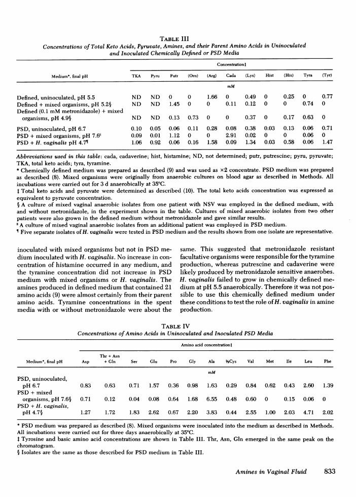
TABLE IIIConcentrations of Total Keto Acids, Pyruvate, Amines, and their Parent Amino Acids in Uninoculated
and Inoculated Chemically Defined or PSDMedia
Concentration t
Medium*, final pH TKA Pyru Putr (Orn) (Arg) Cada (Lys) Hist (His) Tyra (Tyr)
mM
Defined, uninoculated, pH 5.5 ND ND 0 0 1.66 0 0.49 0 0.25 0 0.77Defined + mixed organisms, pH 5.2§ ND ND 1.45 0 0 0.11 0.12 0 0 0.74 0Defined (0.1 mMmetronidazole) + mixed
organisms, pH 4.95 ND ND 0.13 0.73 0 0 0.37 0 0.17 0.63 0
PSD, uninoculated, pH 6.7 0.10 0.05 0.06 0.11 0.28 0.08 0.38 0.03 0.13 0.06 0.71PSD+ mixed organisms, pH 7.611 0.09 0.01 1.12 0 0 2.91 0.02 0 0 0.06 0PSD+ H. vaginalis pH 4.7 1.06 0.92 0.06 0.16 1.58 0.09 1.34 0.03 0.58 0.06 1.47
Abbreviations used in this table: cada, cadaverine; hist, histamine; ND, not determined; putr, putrescine; pyru, pyruvate;TKA, total keto acids; tyra, tyramine.* Chemically defined medium was prepared as described (9) and was used as x2 concentrate. PSD medium was preparedas described (8). Mixed organisms were originally from anaerobic cultures on blood agar as described in Methods. Allincubations were carried out for 3 d anaerobically at 350C.t Total keto acids and pyruvate were determined as described (10). The total keto acids concentration was expressed asequivalent to pyruvate concentration.§ A culture of mixed vaginal anaerobic isolates from one patient with NSV was employed in the defined medium, withand without metronidazole, in the experiment shown in the table. Cultures of mixed anaerobic isolates from two otherpatients were also grown in the defined medium without metronidazole and gave similar results."A culture of mixed vaginal anaerobic isolates from an additional patient was employed in PSDmedium.¶ Five separate isolates of H. vaginalis were tested in PSDmedium and the results shown from one isolate are representative.
inoculated with mixed organisms but not in PSDme-dium inoculated with H. vaginalis. No increase in con-centration of histamine occurred in any medium, andthe tyramine concentration did not increase in PSDmedium with mixed organisms or H. vaginalis. Theamines produced in defined medium that contained 21amino acids (9) were almost certainly from their parentamino acids. Tyramine concentrations in the spentmedia with or without metronidazole were about the
same. This suggested that metronidazole resistantfacultative organisms were responsible for the tyramineproduction, whereas putrescine and cadaverine werelikely produced by metronidazole sensitive anaerobes.H. vaginalis failed to grow in chemically defined me-dium at pH 5.5 anaerobically. Therefore it was not pos-sible to use this chemically defined medium underthese conditions to test the role of H. vaginalis in amineproduction.
TABLE IVConcentrations of Amino Acids in Uninoculated and Inoculated PSDMedia
Amino acid concentrationt
Thr + AsnMedium*, final pH Asp + Gln Ser Glu Pro Gly Ala 'Cys Val Met Ile Leu Phe
mMPSD, uninoculated,
pH 6.7 0.83 0.63 0.71 1.57 0.36 0.98 1.63 0.29 0.84 0.62 0.43 2.60 1.39PSD + mixed
organisms, pH 7.6§ 0.71 0.12 0.04 0.08 0.64 1.68 6.55 0.48 0.60 0 0.15 0.06 0PSD + H. vaginalis,
pH 4.7§ 1.27 1.72 1.83 2.62 0.67 2.20 3.83 0.44 2.55 1.00 2.03 4.71 2.02
* PSDmedium was prepared as described (8). Mixed organisms were inoculated into the medium as described in Methods.All incubations were carried out for three days anaerobically at 35°C.t Tyrosine and basic amino acid concentrations are shown in Table III. Thr, Asn, Gln emerged in the same peak on thechromatogram.5 Isolates are the same as those described for PSDmedium in Table III.
Amines in Vaginal Fluid 833

Possible symbiotic relationship between H. vaginalisand other mixed organisms. As shown in Tables IIIand IV, when mixed organisms grew in PSDmedium,amino acids were consumed by the organisms fromthe medium to a greater extent than they were proteolyzedfrom the medium or were biosynthesized by the organ-isms, except for proline, glycine, and alanine. There-fore, free amino acids in the medium could play animportant role for growth. In contrast, when H. vaginalisgrew in PSDmedium, generation of free amino acidsexceeded consumption. The final pH of the PSDspentmedium was found to be 4.3-5.0 (pH of the unin-oculated medium was 6.7) when pure cultures of H.vaginalis from five clinical isolates were grown in PSDmedium for 3 d. This low pH may result from the pro-duction of a high concentration of pyruvic acid (TableIII), which has a low pKa of 2.5 in addition to aceticacid (pKa = 4.74) which is known to be produced byH. vaginalis (13, 14). These acids may contribute todeath of the organisms (15).
Thus, in patients with H. vaginalis vaginal infection,a symbiotic relationship may exist between H. vaginalisand other yet undefined anaerobic vaginal organisms,in which H. vaginalis generates amino acids and ketoacids, especially pyruvic acid, which are utilized byanaerobic members of the vaginal flora, whereasanaerobic organisms relieve excess acidity by produc-ing amines and by utilizing the generated keto acids.
DISCUSSION
As reported by Pheifer et al. (1), an amine-like odoris liberated when 10% KOHis added to vaginal dis-charges from patients with NSV. High-voltage electro-phoresis of vaginal washings confirms the presence ofamines in such secretions. This technique is rapid (12min) and sensitive (1 nmol of diamines can be de-tected), and provides an objective method for evaluat-ing response to anti-microbial treatment.
Quantitative analysis of amines in vaginal secretionsmay provide a molecular explanation for at least someof the symptoms of NSV. Cadaverine and phenethyl-amine are known to be skin irritants and possiblesensitizers (16). Methylamine is also a skin irritant(16). Isobutylamine can cause erythema and blisteringin skin (16). Histamine has many actions, includingdilation and increased permeability of the microcircu-lation. These various amines might contribute to theepithelial cell shedding and the discharge of some pa-tients as well as to the characteristic odor of NSV.
As shown in Fig. 3, the pH range of vaginal wash-ings from untreated patients is from 4.7 to 6.5. Anyamines present in this pH range exist in the protonatedform (salt) and are not volatile. However, after addi-tion of 10% KOH(1), the amines are converted to anunprotonated form (free base) and become volatile and
thus odorous. During and after intercourse, the alkalineprostatic fluid may convert part of the amines to theunprotonated form and thus cause the characteristicfishy odor.
H. vaginalis has been reported as the most commonorganism among vaginal flora in patients with NSV(1, 17). It is not certain whether this organism repre-sents simply a marker for the syndrome as an innocentbystander, or whether it contributes to the patho-genesis of NSV. It is possible that other organisms, byproducing decarboxylases that convert amino acids toamines, and perhaps by other mechanisms such asfurther metabolizing acids produced by H. vaginalisraise the pH of vaginal secretions to a level that en-hances growth of the H. vaginalis.
Putrescine is known to be an essential growth factorfor some organisms (18-21), and may be essential forgrowth of some organisms in vaginal flora. As shown inTable III, putrescine was produced in vitro by anaer-obe(s) in the vaginal flora of patients with NSV.
Gardner and Dukes (17) inoculated 15 patients provenfree of H. vaginalis infection with fluid from thevaginal tract of patients with vaginitis associated withH. vaginalis. 11 of the 15 patients developed the symp-toms of NSV and H. vaginalis was recovered sub-sequently as the predominant bacterium from each ofthese 11 volunteers. However, a much smaller percent-age of healthy women (8%) inoculated with pure cul-tures of H. vaginalis developed H. vaginalis infection(17). Thus the presence of certain other vaginal organ-isms in NSVmay enhance infectivity and survival ofH. vaginalis. It is also possible that H. vaginalis some-how enhances the growth of those organisms that de-carboxylate amino acids, thus increasing the concentra-tions of amines in vaginal secretions and contributingto the pathogenesis of NSV. The specific organism(s)responsible for decarboxylation of amino acids invaginal secretions remains to be determined.
ACKNOWLEDGMENTSWe thank Peter Klein, Pamella R. Davick, and Stuart W.Snyder for excellent technical assistance. Wealso thank Dr.Terrence Pheifer and Dr. Marcia Durfee for performing theclinical evaluation of many of the patients studied.
This study was supported by U. S. Public Health Servicegrant 5 S07 RR05714-07, by U. S. Public Health Service grantsRO1-Al-13149 and A1-12192 from the National Institute ofAllergy and Infectious Diseases, and by the Division of Hos-pitals and Clinics, Bureau of Medical Services under U. S.Public Health Service Project grant SEA 76-06-72.
REFERENCES1. Pheifer, T. A., P. S. Forsyth, M. A. Durfee, H. M. Pollock,
and K. K. Holmes. 1978. Nonspecific Vaginitis-Role ofHaemophilus vaginalis and treatment with metronidazole.N. Engi. J. Med. 298: 1429-1434.
834 K. C. S. Chen, P. S. Forsyth, T. M. Buchanan, and K. K. Holmes

2. Chen, K. C. S., and R. M. Krause. 1975. A peptide map-ping technique-a three map system. Anal. Biochem.69: 180-186.
3. Heilmann, J., J. Barrollier, and E. Watzke. 1957. BeitragZur aminosaurebestimung auf papierchromatogrammen.Hoppe-Seyler's Z. Physiol. Chem. 309: 219-220.
4. Stevenson, K. J. 1971. Use of visual dye markers on high-voltage paper ionophoresis. Anal. Biochem. 40, 29-34.
5. Gray, W. R. 1967. Dansyl chloride procedure. In Methodsin Enzymology. C. H. W. Hirs, editor. Academic Press,Inc., New York. 11: 139-151.
6. Woods, K. R., and K. T. Wang. 1967. Separation of dansyl-amino acids by polyamide layer chromatography. Bio-chim. Biophys. Acta. 133: 369-370.
7. Eschenbach, D. A., T. M. Buchanan, H. M. Pollock, P. S.Forsyth, E. R. Alexander, J. S. Lin, S. P. Wang, B. B.Wentworth, W. M. McCormack, and K. K. Holmes. 1975Polymicrobial etiology of acute pelvic inflammatory dis-ease. N. Engl. J. Med. 293: 166-177.
8. Dunkelberg, W. E., R. Skaggs, and D. S. Kellog. 1970.Method for isolation and identification of Corynebac-terium vaginale (Haemophilus vaginalis). Appl. Micro-biol. 19: 47-52.
9. Catlin, B. W. 1973. Nutritional profiles of Neisseria gonor-rhoeae. Neisseria meningitidis, and Neisseria lactamicain chemically defined media and the use of growth re-quirements for gonococcal typing. J. Infect. Dis. 128:178-194.
10. Friedemann, T. E., and G. E. Haugen. 1943. Pyruvic acidII. The determination of keto acids in blood and urine.
J. Biol. Chem. 147: 415-442.11. Chen, K. C. S., T. J. Kindt, and R. M. Krause. 1975. Pri-
mary Structure of the L chain from a rabbit homogeneous
antibody to streptococal carbohydrate 1. Purification ofantibody and sequence determination of peptides froma-chymotryptic and thermolytic digests. J. Biol. Chem.250: 3280-3288.
12. Bachrach, U. 1973. Biosynthesis of diamines. In Functionof Naturally Occurring Polyamines. Academic Press, Inc.,New York. 121-124.
13. Moss, C. W., and WV. E. Dunkelberg, Jr. 1969. Volatileand Cellular fatty acids of Haemophilus vaginalis. J.Bacteriol. 100: 544-546.
14. Akerlund, M., and Mardh P. A. 1974. Isolation and iden-tification of Corynebacterium vaginale (Haemophilusvaginalis) in women with infections of the lower genitaltract. Acta. Obstet. Gynecol. Scand. 53: 85-90.
15. Dunkelberg, W. E. 1977. Corynebacterium vaginale. SexTransm. Dis. 4: 69-75.
16. Stecher, P. G., editor. 1968. The Merck Index. Merck &Co., Inc., Rahway, N. J. 8th edition.
17. Gardner, H. L., and C. D. Dukes. 1955. Haemophilusvaginalis Vaginitis-A newly defined specific infectionpreviously classified "nonspecific" vaginitis. Am. J.Obstet. Gynecol. 69: 962-976.
18. Herbst, E. J., and E. E. Snell. 1948. Putrescine as a growthfactor for Haemophilus parainfluenzae. J. Biol. Chem.176: 989-990.
19. Martin, W. H., Jr., M. J. Pelczar, Jr., and P. A. Hansen.1952. Putrescine as a growth requirement for Neisseria.Science (Wash. D. C.). 116: 483-484.
20. Sneath, P. H. A. 1955. Putrescine as an essential growthfactor for a mutant of Aspergillus nidulans. Nature(Lond.). 175: 818.
21. Rogosa, M., and Bishop, F. S. 1964. The genus Veillonella.II. Nutritional studies. J. Bacteriol. 87: 574-580.
Amines in Vaginal Fluid 835