basophil production -...
TRANSCRIPT
Basophil Production
JUDAHA. DENBURG,MAUREENDAVISON, and JOHNBIENENSTOCK,Host Resistance Programme, Departments of Medicine and Pathology,McMaster University, Hamilton, Ontario, Canada, L8S 4J9
A B S T R A C T Factors influencing basophil produc-tion from the bone marrow of ovalbumin (OA)-sensitized guinea pigs have been examined in vitro.Autologous co-cultures of marrow and spleen cells fromOA-immune animals contained significantly highernumbers of basophils after 7 d of liquid culture inthe presence of OA, compared with control co-culturesor with marrow cultures alone (P < 0.005).
Basophils increased in co-culture as the number ofspleen cells added to a fixed number of marrowcells was increased from 0.10 to 2.5 x 10⁶/ml; at eachspleen cell concentration, the presence of OA signif-icantly enhanced basophil production in vitro whencompared with unstimulated co-cultures. There was nobasophil production from spleen cell suspensionscultured in the absence of autologous marrow cells.Conditioned media (CM) prepared from OA-stimu-lated spleen cells of OA-treated animals (CM-OA)caused a specific stimulation of basophil productionfrom normal guinea pig bone marrow cells in liquidcultures (P < 0.01). Phytohemagglutinin (PHA)- andpokeweed mitogen-stimulated CM (CM-PHA, CM-pokeweed mitogen) nonspecifically enhanced normalbasophilopoiesis, causing dose-dependent increases inbasophils and histamine in vitro. CM-OA and CM-PHA also preferentially stimulated formation ofneutrophil-macrophage colony-forming units in semi-solid methylcellulose cultures.
CM-PHAprepared from T cell-enriched splenic cellsuspensions contained basophil-stimulating activity,whereas T cell-depleted CM-PHA activity did notexceed control values (P < 0.01). Preliminary charac-terization of CM-PHA revealed that basophil-stimu-lating activity was predominantly heat stable andnondialyzable.
These results demonstrate OA-specific, as well asmitogen-dependent T-cell regulation of guinea pigbasophilopoiesis in vitro. The data are compatiblewith the existence of a specific "basophilopoietin"in CMderived from guinea pig splenic T cells.
Received for publication 15 May 1979 and in revised form8 September 1979.
390
INTRODUCTIONLymphomononuclear cell-derived factors that stimu-late the in vitro differentiation of bone marrow pre-cursor cells are well recognized. Colony-stimulatingactivity (CSA)' derived from lymphocytes for humanor murine neutrophil (1), monocyte-macrophage (2),eosinophil (3), megakaryocyte (4), and lymphoid (5)cell lines has been described. CSA with cross-species activity produced by mitogen-stimulated hu-man monocytes and lymphocytes has now beencharacterized (6), and T cells that enhance erythro-poiesis in semisolid media have recently beenreported (7).
There is no similar information, however, aboutthe factors necessary for the growth and differen-tiation of basophils. Despite the marked increase inthis cell type in the peripheral blood in some of thehuman myeloproliferative disorders, there is only onerecent study suggesting an increased capacity forbasophilopoiesis in long term liquid cultures of buffycoat cells from patients with chronic myeloidleukemia (8).
Metcalf and his co-workers (9, 10) have begun todescribe the morphology of human granulocytecolonies grown in semisolid media, demonstratingthat -3% of these colonies are comprised of pureeosinophils, and that eosinophil colony formation canbe enhanced by human placental lactogen or lympho-cyte-conditioned media. This suggests that committedmarrow precursor cells exist for each of the spe-cialized granulocytic types described. More recently,a specific eosinophil colony-stimulating factor hasbeen partially separated by isoelectric focusing ofpokeweed mitogen (PWM)-stimulated splenic lympho-cyte-conditioned media (11).
Little evidence exists for a committed basophilprecursor either in semisolid or liquid culture, al-
'Abbreviations used in this paper: BSI, basophil stimula-tion index; CFU-C, colony-forming units in culture; CM, con-ditioned medium; CSA, colony-stimulating activity; HSA,human serum albumin; OA, ovalbumin; PHA, phytohemag-glutinin; PWM, pokeweed mitogen; RSA, rabbit serumalbumin.
J. Clin. Invest. © The American Society for Clinical Investigation, Inc. 0021-9738/80/02/0390/10 $1.00Volume 65 February 1980 390-399
though human bone marrow colony-forming cells inculture (CFU-C) have been reported to give rise tosmall numbers of basophils in liquid culture (12).
The model of Basten and Beeson (13) for eosino-philia in Trichinella-infected mice clearly implicatesT lymphocytes to be necessary for eosinophil pro-duction. In both murine and human systems, antigen-and mitogen-dependent lymphocyte-derived stimu-lators of eosinophilopoiesis have now been de-scribed (14-17). Furthermore, eosinophilia may beclosely related to T-cell factors (14), which also havebeen postulated to be secreted in the course of aneosinophilic-delayed hypersensitivity response inmice (18). The control of basophil homeostasis withspecific reference to the immune response is muchless clear. Injected tumor cell extracts from ratswith basophilic leukemia were shown to cause in-creases in normal rat marrow basophil counts, butthe mechanism of this response was not sought (19).
Dvorak et al. (20) have developed models of cu-taneous and systemic basophilia in the guinea pig,and it has been demonstrated that some basophilhypersensitivity responses require the participation ofboth T cells and specific antibodies (21).
Previous work from this laboratory has demon-strated a thymic antigen to be present on rabbitbasophils (22), suggesting that T lymphocytes andbasophils are related in ontogeny. This concept hasfound further support in a recent study showing afunctional relationship between basophils and thethymus (23). We have used a modified protocol(20) of large amounts of intraperitoneal ovalbumin(OA) to induce basophilia in vivo to study in vitrobasophil production in the guinea pig. Our initialinvestigations of guinea pig basophilopoiesis demon-strate requirements for OAantigen, splenic T lympho-cytes, and mitogen-dependent soluble T-cell productsin the differentiation of marrow precursors tobasophils.
METHODS
Animals. Random bred, colored-hair guinea pigs (HighOak Farms, Goderich, Ontario, Canada), weighing 250-500 g,were used throughout.
Stimulation and estimation of peripheral blood and bonemarrow basophilia itn vivo. Animals were given 1-ml intra-peritoneal injections of lyophilized OA, grade III (GrandIsland Biological Co., Grand Island, N. Y.), dissolved in0.9% of NaCl at 50 mg/ml, or 1 ml of 0.9% NaCl by the sameroute, for 6 successive d, based on a protocol for promotionof basophilia developed by Dvorak et al. (20). Peripheralblood was obtained by serial 0.5-1.0-ml cardiac bleeds, andfemoral bone marrow aspirations (0.1-0.25 ml) performed inthe same animals via a pediatric 19-gauge Osgood biopsyneedle (Becton, Dickinson & Co., Rutherford, N. J.) underketamine anesthesia. Blood and bone marrow nucleated cellcounts using the Coulter Zb (Coulter Electronics, Inc.,Hialeah, Fla.) were performed serially in each guinea
pig (which thus served as its own control) before andduring the course of intraperitoneal injections, as well as upto 120 d after completion of treatments in some animals. Allsamples were taken into 2 mg/ml EDTA (disodium salt,Matheson, Colemani & Bell, East Rutherford, N. J.) understerile conditions. Peripheral blood and bone marrow smearswere made, air-dried, and stained with May-Gruinwald-Giemsa (Harleco, Hartman-Leddon, Philadelphia, Pa.) or0.05% toluidine blue-0 (Fisher Scientific Co., Pittsburgh,Pa.) and 500-1,000 cell-differential counts were performed.Smears of single cell suspensions of bone marrow preparedfor culture (see below) were made by cytocentrifuge andstained similarly for differential counts. Absolute basophilcounts were calculated from the total viable nucleated cellcounts and the differential percentage of basophils on smears.
The usual range of basophils counted was 0.1 to 3.0% ofperipheral blood or bone marrow nucleated cells (1-30 ina 1,000-cell differential). Highest values were seen in bonemarrow cell suspensions of OA-treated animals. Counts werereliable and reproducible; histamine values, used as anindependent criterion of basophilia, correlated well withcalculated differential basophil counts (see below). Attemptsat accurate assessment of absolute basophils by direct livestaining with toluidine blue were not routinely success-ful, even when guinea pig serum was used (20).
Preparation of bone marrow and spleen cellculturesBone marrow, liquid cultures. Guinea pig bone marrow
cell suspensions from untreated, saline-, or OA-treated animalswere prepared either from samples aspirated directly fromfemora (see below) or from the contents of femora and tibiaefreed of muscle and tendon attachments, cut at both ends,and flushed with sterile McCoy's 5A medium, with 15% fetalcalf serum, and 1% penicillin-streptomycin (all from GrandIsland Biological Co.). After resuspension, cell debris wasremoved by sedimentation at unit gravity and then at 400 gover 100% fetal calf serum, after methods described byShortman et al. (24). Single marrow cell suspensions inmedium were placed over a Ficoll-Hypaque (Ficoll-400,Pharmacia Fine Chemicals, Inc., Div. Pharmacia, Inc., Upp-sala, Sweden; Hypaque-sodium, 50%, Winthrop Labora-tories, New York), density gradient (specific gravity = 1.085,311 mosmol/kg) at a cell concentration of 10-20 x 106/mland centrifuged at room temperature for 40 min at 400 g.Interface cells were removed, resuspended in medium,washed three times and single cell suspensions made at 1x 10⁶/ml in the same medium. 5 ml of these suspensionswas placed in Falcon 25-cm2 plastic flasks (Falcon Labware,Div. of Becton, Dickinson & Co., Oxnard, Calif.), with orwithout co-cultured spleen cells, conditioned medium (CM)supplementation at 1-20% vol/vol, OA, or human (HSA,Fraction V, from Pentex Biochemical, Kankakee, Ill., MilesLaboratories, Inc., Montreal) or rabbit-serum albumin (RSA,Fraction V, Nutritional Biochemicals Corp., Cleveland, Ohio)at 10 ug/ml. All cultures were further supplemented with2-mercaptoethanol (Sigma Chemical Co., St. Louis, Mo.) at50 um final concentration. Cells were cultured at 37°C and5% CO2 in a moist incubator and harvested at 7 d; cytocen-trifuge smears and stains, 1,000 cell-differential counts, andcell-associated histamine assays were performed on all cellsuspensions.
Bone marrow, methylcellulose cultures. Semisolid cul-tures of 1-2 x 10⁵ cells/35 x 10 mmFalcon tissue culturePetri dish in Dow A4M Premium methylcellulose (2.2%;Dow Chemical Co., Midland,, Mich.) supplemented with20% fetal calf serum and 20% vol/vol medium/2-mercapto-
Stimulation by Factors Derived from Guinlea Pig Splenic T LymIphocytes 391

ethatnol or CNI/2-mercaptoethanol (diluted to appropriatefinal conieentrationi) vere performied by techni(lues pre-viouslv described (4, 7, 9). CFtU-C Nere counted on day7 bv inverted microscol)e in duiplicate or (lqladrul)licate foreach conidition. CSA was defined as the ability to stimulalteC(FU-C in methvicellulose culttures. Individual colonies werepicked by a 5-y1. micropipette uLnder microscopy and diltutedin three drops MIcCoy's 5A mediuim before prepatration ofcytocentrifuige smears. These smears were stai ned andcounted as above.
Spleen. Sple en cell suspensions wvere prepared by remov-ing spleens intaet unider sterile conditionis into mediumi,teasinig wvith wvooden applicator sticks, ind uising the pro-cedures of Shortmai tn et al. (24) for removal of fine tnd coarsecellula.r debris. To remove contaminiating erythrocytes. cellsw!ere exposed to 0.17 I NH4C aIt 4°C for 10 min. Cellsuspensions so obtained were washed three times, resus-pende(l at I x 106/ml, in(l cultured asi above or co-eulturedwith marrowv cells. Spleen cell b)asophil counllts rainged fromii0.05 to 0.2%; .90% of splee n cells wvere lymphomono-nuclear. Cell viabilities wvere assessed l)y Trypan blue (liar-leeo flItrtmain-Leddoni), dve excluision, and were always>95% for freshly prepared marrow or spleen suspenisions.
Preparation and assaytof spleen cell CM. Spleens wereremoved from animals siacrificed 2-4 mo after treatmtient witheither OA or saline. Single splenic cell suispenisionis at1 x 106/nml, with or without 10 Ag/ml OA, HSA, RSA,phvtolhemiagglutinin (PHA-M); (Difco Laboratories, Detroit,NMich.), or 0.1% P\VI (Grand Island Biological Co.) vereprepare(l as describe(d anid culture(d at 37°C and( 5% CO2in 100% humi(lity for 5 d. Cell-free supemnates obtaine(dfro-m these cultures were filtered throuigh 0.22-Amul Milli-pore filters (Millipore Corp., Bedford, Mass.) anid storedatt -20)C for ul) to 14 milo; these CNI wvere assayed it1-20% vol/vol in liqjuidi and in methylcellulose ctultures
sinlg normal, previously uintreatted, guinea l)ig inarrow celsas targets in 1)th systems.
A 'basophil stimutlationi index" (BSI) of CNI in liqtuidctulttures was calculated by the following ratio: A )asophils(fiomi day 0) in presence of CM\I A basophils (f'rom day 0) inabsence of CNM
Ilistam1inte alssay. Cell-associated hiistaminie assays wvereperformed on all cell suspensions 1b a modification of themetlhod described by Beaven (25). The teehniquie uised isl.tsed on the conversioni of histaimine to [3H]methvl-histamiinie by incumbation with histitain ine N-mnetlhy transferaseanid [ inethyl-3H]-S-aldenosvlmethionine.
Splenir T-cell separationi. Splenici T cells wvere separatedby their ability to formii rosettes with rabbit erythrocytes,according to methods described 1y WSilson and Gurner (26).Briefly, 5 ml of a :3% suspension of palplainizcel, sterile
xvashed rabbit ervthrocytes was mixed wvith ain equall volumeof' a twice-xwashed splenic cell suspension at 1.8 x 10⁷cells/ml. After at 5-mmin cenitrifugationi at 100 g, the pellet wasincublatedl for 1 h att roomii temperature. The lellet was gentlyresuusIpended, layered onto a Ficoll-Hlypaque density gradienit(see ab)ove), aind cenitrifluged for 30 min att 400g. Interfacecells (T-depleted) were removed and washed twice in 1%bovine serumiI albuinin in l)hosphate-buffered saline while thepellete(l (T-enriched) cells xve're subl)jected to hvpotonic lysisifor 30 s and watshed twice in the saime wav. E-rosettes wereenumerated in at hemocvtomceter, usiing (.1% toltuidine blue;200 cells vvere couintedl ainld only cells with three or morebouid ervthrocv tes wvere deemned "E-positive" (T cells). Cellsfrom eaclh fralctioni wvere theni incubated with at fluorescinatedgoat iainti-guiniiea. pig inimuinoglobhilin (Ig)G (w,khole) andcouinte(d for surface immunoglobhulin positivity. Percentagesof IgG-positive cells wvere thtus obtained for eaclch spleencell fratction.
Morphology. A cell was morphologically defined by lightmicroscop)y as at basophil if there was nuclear indentationor polymorphonuclearity, if it conitained grantules that stainedmletachlromlaticallv-, and if the grannules themsielves appearedovoid, considered a clharacteristic of gtineea pig-specificgranulattion. Other cells were classified by accepted hemiato-logicall criteriaI. Mast cells are very rarely, if aIt all, en-counteredl in the gtinleat pig; thev were niot seeni in ourlp)eripheral 1loo0(, spleen, or )one marrow ctulttures. When seeniby others, guineta pig maiiist cells have l)een distinguishalcefiomnbatsophils by size, location in acnd difficult extratctionfrom coinmective tissties. mnononuclearitv, aniid a p)artiallyvacuiolated granuile-containing cytoplasmn (27).
RESULTS
Peripheral blood and bonte mnarrow basophil re-sponses in vivo. The serial peripheral blood lbasophilresponise to 6 succiessive d of intraperitoneal injectionsin 24 guinea pigs is shown in Table I. Basophil countsincreatsed by1w meain of approximately fivefold in15 OA-treated animals when compared with niniesaline-treated controls. Increased numl)ers of circulat-inlg basophils were seen by day 3 of injections, re-mained significantly elevated at day 7 (1 d after ces-sation of injections), aind subsided within 10 d afterwvithdrawal of OA; day 14 counts in OA-treated animiiialswere not significantly different from untreated controlvalues. Serial total leukocyte counts did not differaimong animnal groups (OA, saline, or untreated) when
TABLE IPeripheral Blood1 Respotise to 6 Sticcessive d of Iutraperitotical OA hi the Guiiuea Pig
Treate(dUjintreated
Grotip niormiial Saline OA
Day of inijectionis 0 0 3 7 0 3 7Totall ntucleated cells 8.89±0.964 8.36±2.79 8.86±1.25 9.12±1.45 9.21±1.06 9.45±1.12 10.6±2.42Basophils 34.9+3.9 36.2±4.1 48.2±6.0 38.5±7.1 33.6±2.9 177±31.9 123±35.2
(24) (9) (9) (7) (15) (14) (8)
Mean total niucleated cells x 103/mm3±SE \Iean absolute basophils per mm3 SE. Numbers per group in parentheses.Day 3 atnd 7 OA-treated group basophils differ significantly from (lay 3 and day 7 saline-treated group basophils, respectively,and fromii normiial values, P < 0.05. No signifficant differences ill total nucleated cell counts betveeni subgroups or days.
392 J. A. Denburg, .M. D)avison, anddJ. Bienienistoc k
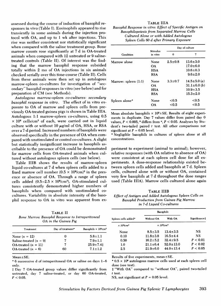
assessed during the course of induction of basophil re-sponses in vivo (Table I). Eosinophils appeared to risetransiently in some animals during the injection pro-tocol with OA, and up to 1 wk after injections. Thisrise was neither consistent nor statistically significantwhen compared with the saline treatment group. Bonemarrow counts rose significantly at 7 d in OA-treatedanimals when compared with 12 untreated or 9 saline-treated controls (Table II). Of interest was the find-ing that the marrow basophil response subsidedtotally within 2 mo of OA injections in six animalschecked serially over this time-course (Table II). Cellsfrom these animals were then set up in autologousmarrow-spleen co-cultures for investigation of "sec-ondary" basophil responses in vitro (see below) and forpreparation of CM(see Methods).
Autologous marrow-spleen co-cultures: secondarybasophil response in vitro. The effect of in vitro ex-posure to OA of marrow and spleen cells from pre-viously OA-treated guinea pigs is shown in Table IIIA.Autologous 1:1 marrow-spleen co-cultures, using 0.5x 10⁶ cells/cm3 of each, were carried out in liquidculture with or without 10 ,ug/ml of OA, HSA, or RSAover a 7-d period. Increased numbers of basophils wereobserved specifically in the presence of OAwhen com-pared with unstimulated or control co-cultures. A slightbut statistically insignificant increase in basophils as-cribable to the presence of OAcould be demonstratedfor marrow cells from OA-treated animals when cul-tured without autologous spleen cells (see below).
Table IIIB shows the results of marrow-spleenliquid co-cultures at 7 d when spleen cells added to afixed marrow cell number (0.5 x 10⁶/cm)3 in the pres-ence or absence of OA. Through a range of spleencells added (0.5-2.5 x 10⁶/cm3), OA-stimulated cul-tures consistently demonstrated higher numbers ofbasophils when compared with unstimulated co-cultures. Variability in absolute intensity of the baso-phil response to OA in vitro was apparent from ex-
TABLE IIBone Marrow Basophil Response to Intraperitoneal
OA in the Guinea Pig
Treatment Day of treatment* Basophils x 103/cm3
None (n = 12) 0 5.8±+1.1Saline-treated (n = 9) 7 7.9±+1.1OA-treated (n = 11) 7 25.9+7.4tOA-treated (n = 6) 60 7.1±+ 1.2
Mean-+SE.* 6 successive d of intraperitoneal OAor saline on days 1-6only.4 Day 7 OA-treated group values differ significantly fromuntreated, day 7 saline-treated, or day 60 OA-treated,P < 0.05.
TABLE IIIABasophil Response in vitro: Effect of Specific Antigen on
Basophilopoiesis from Separated Marrow CellsCultured Alone or with Added AutologousSpleen Cells 60 d after Primary Exposure
Day of cultureStimulus
Condition in vitro 0 7
Marrow alone None 2.5+0.8 13.6±3.0OA 17.0±6.0HSA 10.6±0.3RSA 9.6±2.0
Marrow: spleen (1:1) None 3.3±0.7 14.5±5.0 (a)OA 31.1±6.0 (b)HSA 10.9±3.5RSA 15.3±2.0
Spleen alone* None <0.5 <0.5OA <0.5 <0.5
Mean absolute basophils x 103±SE. Results of eight experi-ments in duplicate. Day 7 values differ from paired day 0values, P < 0.005, differs from a, p < 0.05. Analyses by Stu-dent's two-tailed paired t test. All other comparisons notsignificant at P = 0.05 level.* Negligible basophils in cultures of spleen alone at allconcentrations.
periment to experiment (animal to animal); however,relative responses (with OArelative to absence of OA)were consistent at each spleen cell dose for all ex-periments. A dose-response relationship existed be-tween spleen cells added and basophils at 7 d. Spleencells, cultured alone with or without OA, containedvery few basophils at 7 d throughout the dose rangesused (Table IIIA). Marrow cells cultured alone again
TABLE IIIBEffect of Antigen and Added Autologous Spleen Cells on
Basophil Production from Guinea Pig Marrotwin 7-d Liquid Co-cultures
Basophils
Spleen cells added* Without OA With OA Significancet
X 106Icm3 X 103/cm3
None 8.5±3.5 13.4±3.5 NS0.10 11.8±3.8 16.5±4.4 NS0.50 16.2±5.2 32.4±9.5 P < 0.011.0 21.1±6.4 52.0±13.0 P < 0.022.5 21.9±6.0 44.0±+13.4 P < 0.05
Results of five experiments, mean±SE.* 0.5 x 10⁶ autologous marrow cells used at each spleen celldose (see text).I "With OA" compared to "without OA", paired two-tailedt test.NS, not significant at P = 0.05 level.
Stitmulation by Factors Derived from Guinea Pig Splenic T Lymphocytes 393
showed a slight increase in basophils in the presenceof OA(Tables IIIA and B).
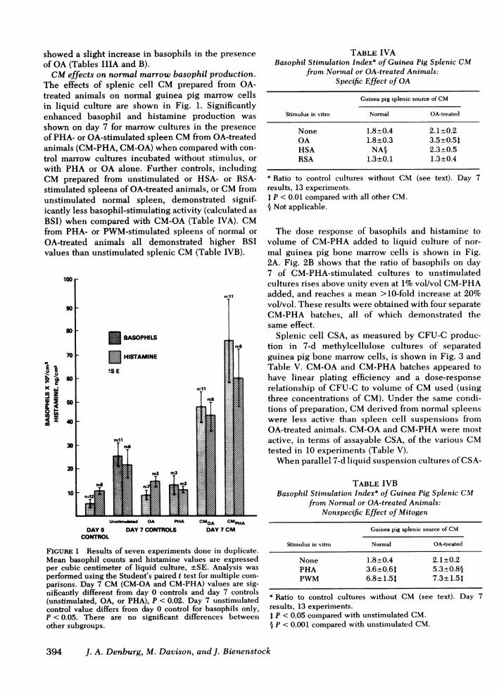
CMeffects on normal marrow basophil production.The effects of splenic cell CM prepared from OA-treated animals on normal guinea pig marrow cellsin liquid culture are shown in Fig. 1. Significantlyenhanced basophil and histamine production wasshown on day 7 for marrow cultures in the presenceof PHA- or OA-stimulated spleen CMfrom OA-treatedanimals (CM-PHA, CM-OA) when compared with con-trol marrow cultures incubated without stimulus, orwith PHA or OA alone. Further controls, includingCM prepared from unstimulated or HSA- or RSA-stimulated spleens of OA-treated animals, or CMfromunstimulated normal spleen, demonstrated signif-icantly less basophil-stimulating activity (calculated asBSI) when compared with CM-OA(Table IVA). CMfrom PHA- or PWM-stimulated spleens of rOA-treated animals all demonstrated higvalues than unstimulated splenic CM(Table
-
x uJ
53ZmI
@4
BASOPHILS
E HISTAMINE
tS E
noln=
I
rn11
10
DAY0CONTROIC
Un.timuImd OA PHA
DAY7 CONTROLS
TABLE IVABasophil Stimulation Index* of Guinea Pig Splenic CM
from Normal or OA-treated Animals:Specific Effect of OA
Guinea pig splenic source of CM
Stimulus in vitro Normal OA-treated
None 1.8+0.4 2.1±0.2OA 1.8±0.3 3.5±0.5tHSA NA§ 2.3±0.5RSA 1.3±0.1 1.3±0.4
* Ratio to control cultures without CMresults, 13 experiments.4 P < 0.01 compared with all other CM.§ Not applicable.
(see text). Day 7
iormal or The dose response of basophils and histamine torher BSI volume of CM-PHAadded to liquid culture of nor-IVB). mal guinea pig bone marrow cells is shown in Fig.
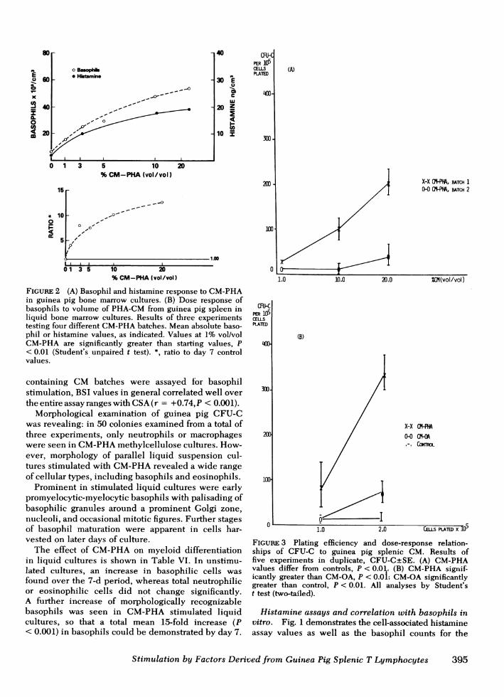
2A. Fig. 2B shows that the ratio of basophils on day7 of CM-PHA-stimulated cultures to unstimulatedcultures rises above unity even at 1%vol/vol CM-PHAadded, and reaches a mean >10-fold increase at 20%vol/vol. These results were obtained with four separateCM-PHA batches, all of which demonstrated thesame effect.
Splenic cell CSA, as measured by CFU-C produc-tion in 7-d methylcellulose cultures of separatedguinea pig bone marrow cells, is shown in Fig. 3 andTable V. CM-OAand CM-PHAbatches appeared tohave linear plating efficiency and a dose-responserelationship of CFU-C to volume of CMused (usingthree concentrations of CM). Under the same condi-tions of preparation, CMderived from normal spleenswere less active than spleen cell suspensions fromOA-treated animals. CM-OAand CM-PHAwere most
active, in terms of assayable CSA, of the various CMtested in 10 experiments (Table V).
Whenparallel 7-d liquid suspension cultures of CSA-
TABLE IVBBasophil Stimulation Index* of Guinea Pig Splenic CM
from Normal or OA-treated Animals:Nonspecific Effect of Mitogen
k"OA G"PHADAY7 CM
FIGURE 1 Results of seven experiments done in duplicate.Mean basophil counts and histamine values are expressedper cubic centimeter of liquid culture, +SE. Analysis wasperformed using the Student's paired t test for multiple com-parisons. Day 7 CM(CM-OA and CM-PHA) values are sig-nificantly different from day 0 controls and day 7 controls(unstimulated, OA, or PHA), P < 0.02. Day 7 unstimulatedcontrol value differs from day 0 control for basophils only,P < 0.05. There are no significant differences betweenother subgroups.
Guinea pig splenic source of CM
Stimulus in vitro Normal OA-treated
None 1.8+0.4 2.1+0.2PHA 3.6+0.64 5.3+0.8§PWM 6.8+1.54 7.3+1.54
* Ratio to control cultures without CM (see text). Day 7results, 13 experiments.t P < 0.05 compared with unstimulated CM.§ P < 0.001 compared with unstimulated CM.
394 J. A. Denburg, M. Davison, and J. Bienenstock
X
S
0
El
60-F
44
o Basophls* Hisumine
I-I0
[0 1 3 5 10
%CM-PHA(vol/vol)
15
* 100
5
40
30EU
CcouJ
20Z
10 Y
CF-HPER X05CELLSPLATED
400-
200
0'-_
01 3 5 10 20%CM-PHA(vol/vol)
FIGURE 2 (A) Basophil and histamine response to CM-PHAin guinea pig bone marrow cultures. (B) Dose response ofbasophils to volume of PHA-CM from guinea pig spleen inliquid bone marrow cultures. Results of three experimentstesting four different CM-PHAbatches. Mean absolute baso-phil or histamine values, as indicated. Values at 1% vol/volCM-PHA are significantly greater than starting values, P< 0.01 (Student's unpaired t test). *, ratio to day 7 controlvalues.
containing CM batches were assayed for basophilstimulation, BSI values in general correlated well overthe entire assay ranges with CSA(r = +0.74,P < 0.001).
Morphological examination of guinea pig CFU-Cwas revealing: in 50 colonies examined from a total ofthree experiments, only neutrophils or macrophageswere seen in CM-PHAmethylcellulose cultures. How-ever, morphology of parallel liquid suspension cul-tures stimulated with CM-PHArevealed a wide rangeof cellular types, including basophils and eosinophils.
Prominent in stimulated liquid cultures were earlypromyelocytic-myelocytic basophils with palisading ofbasophilic granules around a prominent Golgi zone,nucleoli, and occasional mitotic figures. Further stagesof basophil maturation were apparent in cells har-vested on later days of culture.
The effect of CM-PHAon myeloid differentiationin liquid cultures is shown in Table VI. In unstimu-lated cultures, an increase in basophilic cells wasfound over the 7-d period, whereas total neutrophilicor eosinophilic cells did not change significantly.A further increase of morphologically recognizablebasophils was seen in CM-PHA stimulated liquidcultures, so that a total mean 15-fold increase (P< 0.001) in basophils could be demonstrated by day 7.
mii-cPER 10CELLSPLATED
400.
300-
100-
(A)
X-X (?-PHA, BATCH 10-0 G-MPH, BATCH 2
1.0 10.0 20.0 ZO1(vol/vol)
(B)
X-X cl-fA0-0o.-. CTROL
I Io ~~~~~I
1.0 2.0 CELLS PLATED X 1
FIGuRE 3 Plating efficiency and dose-response relation-ships of CFU-C to guinea pig splenic CM. Results offive experiments in duplicate, CFU-CtSE. (A) CM-PHAvalues differ from controls, P < 0.0 l (B) CM-PHA signif-icantly greater than CM-OA, P < 0.0L CM-OAsignificantlygreater than control, P < 0.01. All analyses by Student'st test (two-tailed).
Histamine assays and correlation with basophils invitro. Fig. 1 demonstrates the cell-associated histamineassay values as well as the basophil counts for the
Stimulation by Factors Derived from Guinea Pig Splenic T Lymphocytes
z
)L
80 r
.10 ,O.,,I
0
t
m0
C
395
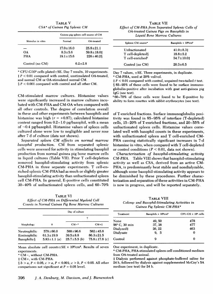
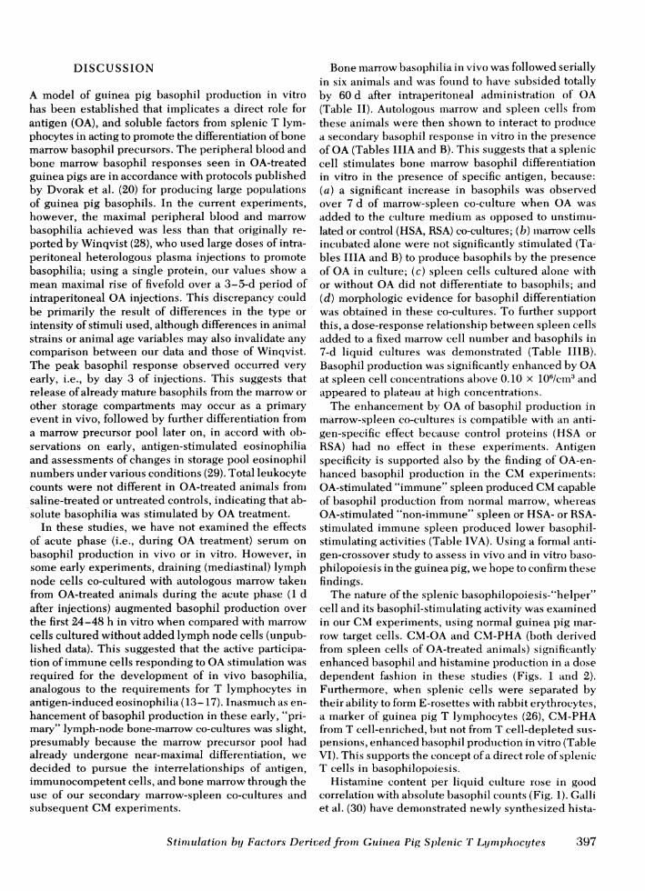
TABLE VCSA* of Guinea Pig Splenic CM
Guinea pig spleen cell source of CM
Stimulus in vitro Normal OA-treated
17.0±16.0 25.8±21.1OA 8.3±5.6 59.8±18.0tPHA 19.1±15.6 226±40.2§
Control (no CM) 6.2±2.8
* CFU-C/105 cells plated± SE. Day 7 results, 10 experiments.P < 0.01 compared with control, unstimulated OA-treated,
and normal CMor OA-stimulated normal CM.§ P < 0.001 compared with control and all other CM.
CM-stimulated marrow cultures. Histamine valueswere significantly increased in marrow cultures incu-bated with CM-PHAand CM-OAwhen compared withall other controls. The degree of correlation overallin these and other experiments between basophils andhistamine was high (r = +0.87); calculated histaminecontent ranged from 0.2-1.0 pg/basophil, with a meanof -0.4 pg/basophil. Histamine values of spleen cellscultured alone were low to negligible and never roseafter 7 d of culture (data not shown).
Separated spleen CM effects on normal marrowbasophil production. CM from separated spleniccells were assessed for activity in stimulating basophilproduction from normal guinea pig bone marrow cellsin liquid cultures (Table VII). Prior T cell-depletionremoved basophil-stimulating activity from splenicCM-PHA in three separate experiments. T cell-en-riched splenic CM-PHAhad as much or slightly greaterbasophil-stimulating activity than unfractionated spleencell CM-PHA. In general, E-positive cells constituted30-40% of unfractionated spleen cells, and 60-70%
TABLE VIEffect of CM-PHAon Differential Myeloid Cell
Counts in Normal Guinea Pig Bone Marrow Cultures
Day of culture
7
Morphology 0 CM-* CM+t
Neutrophilic 578±66.0 588±96.6 582±45.8Eosinophilic 61.3±19.9 39.5±8.8 86.3±21.5Basophilic§ 5.83±1.1 (a) 25.7±5.5 (b) 75.8+17.8 (c)
Mean absolute cell counts±SE x 103/cm3. Results of sevenexperiments.* CM-, without CM-PHA.4 CM+, with CM-PHA.§ b > a, P < 0.05; c > a, P < 0.001; c > b, P < 0.05. All othercomparisons not significant at P = 0.05 level.
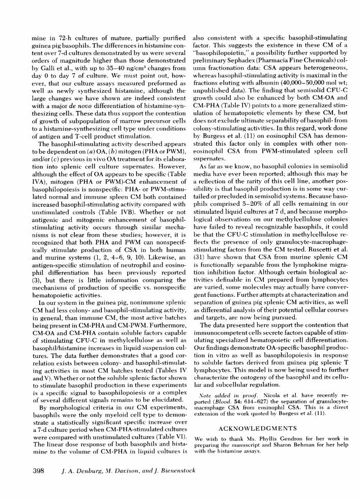
TABLE VIIEffect of CM-PHAfrom Separated Splenic Cells of
OA-treated Guinea Pigs on Basophils inLiquid Bone Marrow Cultures
Splenic CMsource* Basophils x 103/cm3
Unfractionated 41.0+8.34T cell-depleted§ 18.8+5.2T cell-enriched" 54.7± 10.0tControl (no CM) 20.3±6.5
Day 7 values, ±SE. Three experiments, in duplicate.* CM-PHA, used at 20% vol/vol.4 P < 0.01 compared with control, unpaired two-tailed t test.§ 85-95% of these cells were found to be surface immuno-globulin-positive after incubation with goat anti-guinea pigIgG (see text)."60-70% of these cells were found to be E-positive byability to form rosettes with rabbit erythrocytes (see text).
of T-enriched fractions. Surface immunoglobulin posi-tivity was found in 85-95% of interface (T-depleted)cells, 15-20% of T-enriched fractions, and 20-55% ofunfractionated spleen cells. Histamine values corre-lated well with basophil counts in these experiments,with unfractionated spleen and T cell-enriched CM-PHA causing statistically significant increases in 7-dhistamine in vitro, when compared with T cell-depletedor control conditions (P < 0.01, data not shown).
Characterization of basophil-stimulating activityCM-PHA. Table VIII shows that basophil-stimulatingactivity as well as CSA, derived from an active CM-PHA, is predominantly heat stable and nondialyzable,although some basophil-stimulating activity appears tobe diminished by these procedures. Further charac-terization and separation of these activities in CM-PHAis now in progress, and will be reported separately.
TABLE VIIIColony- and Basophil-Stimulating Activities in
Guinea Pig Splenic CM-PHA*
Treatment Basophils x 103/cm3 CFU-C/2 x 105 cells
None 49, 50 47856° C, 30 min 37, 38 477Dialyzedt 26, 22 463Dialysate 9, 5 0
Control 9 0
One experiment, in duplicate.* CM-PHA, PHA-stimulated spleen cell conditioned mediumfrom OA-treated animal.t Dialysis performed against phosphate-buffered saline for24 h, followed by dialysis against supplemented McCoy's 5Amedium (see text) for 24 h.
396 J. A. Denburg, M. Davison, and J. Bienenstock
DISCUSSION
A model of guinea pig basophil production in vitrohas been established that implicates a direct role forantigen (OA), and soluble factors from splenic T lym-phocytes in acting to promote the differentiation of bonemarrow basophil precursors. The peripheral blood andbone marrow basophil responses seen in OA-treatedguinea pigs are in accordance with protocols publishedby Dvorak et al. (20) for producing large populationsof guinea pig basophils. In the current experiments,however, the maximal peripheral blood and marrowbasophilia achieved was less than that originally re-ported by Winqvist (28), who used large doses of intra-peritoneal heterologous plasma injections to promotebasophilia; using a single protein, our values show amean maximal rise of fivefold over a 3-5-d period ofintraperitoneal OAinjections. This discrepancy couldbe primarily the result of differences in the type orintensity of stimuli used, although differences in animalstrains or animal age variables may also invalidate anycomparison between our data and those of Winqvist.The peak basophil response observed occurred veryearly, i.e., by day 3 of injections. This suggests thatrelease of already mature basophils from the marrow orother storage compartments may occur as a primaryevent in vivo, followed by further differentiation froma marrow precursor pool later on, in accord with ob-servations on early, antigen-stimulated eosinophiliaand assessments of changes in storage pool eosinophilnumbers under various conditions (29). Total leukocytecounts were not different in OA-treated animals fronisaline-treated or untreated controls, indicating that ab-solute basophilia was stimulated by OA treatment.
In these studies, we have not examined the effectsof acute phase (i.e., during OA treatment) serum onbasophil production in vivo or in vitro. However, insome early experiments, draining (mediastinal) lymphnode cells co-cultured with autologous marrow takenifrom OA-treated animals during the acute phase (1 dafter injections) augmented basophil production overthe first 24-48 h in vitro when compared with marrowcells cultured without added lymph node cells (unpub-lished data). This suggested that the active participa-tion of immune cells responding to OAstimulation wasrequired for the development of in vivo basophilia,analogous to the requirements for T lymphocytes inantigen-induced eosinophilia (13-17). Inasmuch as en-hancement of basophil production in these early, "pri-mary" lymph-node bone-marrow co-cultures was slight,presumably because the marrow precursor pool hadalready undergone near-maximal differentiation, wedecided to pursue the interrelationships of antigen,immunocompetent cells, and bone marrow through theuse of our secondary marrow-spleen co-cultures andsubsequent CMexperiments.
Bone marrow basophilia in vivo was followed seriallyin six animals and was found to have subsided totallyby 60 d after intraperitoneal administration of OA(Table II). Autologous marrow and spleen cells fromthese animals were then shown to interact to produicea secondary basophil response in vitro in the presenceof OA(Tables IIIA and B). This suggests that a spleniccell stimulates bone marrow basophil differentiationin vitro in the presence of specific antigen, because:(a) a significant increase in basophils was observedover 7 d of marrow-spleen co-culture when OA wasadded to the culture medium as opposed to unstimu-lated or control (HSA, RSA) co-cultures; (b) miarrow cellsincubated alone were not significantly stimulated (Ta-bles IIIA and B) to produce basophils by the presenceof OA in culture; (c) spleen cells cultured alone withor without OA did not differentiate to basophils; and(d) morphologic evidence for basophil differentiationwas obtained in these co-cultures. To further supportthis, a dose-response relationship between spleen cellsadded to a fixed marrow cell number and basophils in7-d liquid cultures was demonstrated (Table IIIB).Basophil production was significantly enhanced by OAat spleen cell conicentrations above 0.10 x 10⁶/cm3 andappeared to plateau at high concentrations.
The enhancement by OAof basophil production inmarrow-spleen co-cultures is compatible with an anti-gen-specific effect because control proteins (HSA orRSA) had no effect in these experiments. Antigenspecificity is supported also by the finding of OA-en-hanced basophil production in the CMexperiments:OA-stimulated "immune" spleen produced CMcapableof basophil production from normal marrow, whereasOA-stimulated "non-immune" spleen or HSA- or RSA-stimulated immune spleen produced lower basophil-stimulating activities (Table IVA). Using a formal anti-gen-crossover study to assess in vivo and in vitro baso-philopoiesis in the guinea pig, we hope to confirm thesefindings.
The nature of the splenic basophilopoiesis-"helper"cell and its basophil-stimulating activity was exaininedin our CMexperiments, using normal guinea pig mar-row target cells. CM-OAand CM-PHA(both derivedfrom spleen cells of OA-treated animals) significantlyenhanced basophil and histamine production in a dosedependent fashion in these studies (Figs. 1 atnd 2).Furthermore, when splenic cells were separated bytheir ability to form E-rosettes with rabbit erythrocytes,a marker of guinea pig T lymphocytes (26), CM-PHAfrom T cell-enriched, but not from T cell-depleted sus-pensions, enhanced basophil production in vitro (TableVI). This supports the concept of a direct role of spleniicT cells in basophilopoiesis.
Histamine content per liquid culture rose in goodcorrelation with absolute basophil counts (Fig. 1). Galliet al. (30) have demonstrated newly synthesized hista-
Stimulation by Factors Derived from Guinea Pig Splenic T Lymphocytes 397
mine in 72-h cultures of mature, partially purifiedguinea pig basophils. The differences in histamine con-tent over 7-d cultures demonstrated by us were severalorders of magnitude higher than those demonstratedby Galli et al., with up to 35-40 ng/cm3 changes fromday 0 to day 7 of culture. We must point out, how-ever, that our culture assays measured preformed aswell as newly synthesized histamine, although thelarge changes we have shown are indeed consistentwith a major de novo differentiation of histamine-syn-thesizing cells. These data thus support the contentionof growth of subpopulation of marrow precursor cellsto a histamine-synthesizing cell type under conditionsof antigen and T-cell product stimulation.
The basophil-stimulating activity described appearsto be dependent on (a) OA, (b) mitogen (PHA or PWM),and/or (c) previous in vivo OAtreatment for its elabora-tion into splenic cell culture supernates. However,although the effect of OAappears to be specific (TableIVA), mitogen (PHA or PWM)-CMenhancement ofbasophilopoiesis is nonspecific: PHA- or PWM-stimu-lated normal and immune spleen CMboth containedincreased basophil-stimulating activity compared withunstimulated controls (Table IVB). Whether or notantigenic and mitogenic enhancement of basophil-stimulating activity occurs through similar mecha-nisms is not clear from these studies; however, it isrecognized that both PHA and PWMcan nonspecif-ically stimulate production of CSA in both humanand murine systems (1, 2, 4-6, 9, 10). Likewise, anantigen-specific stimulation of neutrophil and eosino-phil differentiation has been previously reported(3), but there is little information comparing themechanisms of production of specific vs. nonspecifichematopoietic activities.
In our system in the guinea pig, nonimmune splenicCMhad less colony- and basophil-stimulating activity,in general, than immune CM, the most active batchesbeing present in CM-PHAand CM-PWM.Furthermore,CM-OAand CM-PHAcontain soluble factors capableof stimulating CFU-C in methylcellulose as well asbasophil/histamine increases in liquid suspension cul-tures. The data further demonstrates that a good cor-relation exists between colony- and basophil-stimulat-ing activities in most CMbatches tested (Tables IVand V). Whether or not the soluble splenic factor shownto stimulate basophil production in these experimentsis a specific signal to basophilopoiesis or a complexof several different signals remains to be elucidated.
By morphological criteria in our CMexperiments,basophils were the only myeloid cell type to demon-strate a statistically significaint specific increase overa 7-d culture period when CM-PHA-stimulated cultureswere compared with unstimulated cultures (Table VI).The linear dose response of both basophils and hista-mine to the volume of CM1-PHA in liquid cultures is
also consistent with a specific basophil-stimulatingfactor. This suggests the existence in these CMof a"basophilopoietin," a possibility further supported bypreliminary Sephadex (Pharmacia Fine Chemicals) col-umn fractionation data: CSA appears heterogeneous,whereas basophil-stimulating activity is maximal in thefractions eluting with albumin (40,000-50,000 mol xvt;unpublished data). The finding that semisolid CFU-Cgrowth could also be enhanced by both CM-OAandCM-PHA(Table IV) points to a more generalized stim-ulation of hematopoietic elements by these CM, butdoes not exclude ultimate separability of basophil- fromcolony-stimulating activities. In this regard, work donebv Burgess et al. (11) on eosinophil CSA has demon-strated this factor only in complex with other non-eosinophil CSA from PWM-stimulated spleen cellstipernates.
As far as we know, no basophil colonies in semisolidmedia have ever been reported; although this may bea reflection of the rarity of this cell line, another pos-sibility is that basophil production is in some way cur-tailed or precluded in semisolid systems. Because baso-phils comprised 5-20% of all cells remaining in ourstimulated liquid cultures at 7 d, and because morpho-logical observations on our methylcellulose colonieshave failed to reveal recognizable basophils, it couldbe that the CFU-C stimulation in methylcellulose re-flects the presence of only granulocyte-macrophage-stimulating factors from the CMtested. Ruscetti et al.(31) have shown that CSA from murine splenic CMis functionally separable from the lymphokine migra-tion inhibition factor. Although certain biological ac-tivities definable in CMprepared from lymphocytesare varied, some molecules may actually have conver-gent functions. Further attempts at characterization andseparation of guinea pig splenic CMactivities, as wellas differential analysis of their potential cellular coursesand targets, are now being pursued.
The data presented here support the contention thatimmunocompetent cells secrete factors capable of stim-ulating specialized hematopoietic cell differentiation.Our findings demonstrate OA-specific basophil produc-tion in vitro as well as basophilopoiesis in responseto soluble factors derived from guinea pig splenic Tlymphocytes. This model is now being used to furthercharacterize the ontogeny of the basophil and its cellu-lar and subcellular regulation.
Note adlded in proof. Nicola et al. have recently re-ported (Blood. 54: 614-627) the separation of granulocyte-imacrophage CSA from eosinophil CSA. This is a directextensioni of the vork (luoted by Burgess et al. (11).
ACKNOWLEDGMENTS
We wish to thank Ms. Phyllis Gendron for her work inpreparinig the manuiscript and Sharon Behman for her helpwith the histamine assays.
398 J. A. Detnbuirg, Ml. Davisoni, anid]J. Bienienstock
REFERENCES
1. Cline, M. J., and D. W. Golde. 1974. Production of colony-stimulating activity by human lymphocytes. Nature(Lond.). 248: 703.
2. Ruscetti, F. W., and P. A. Chervenick. 1975. Release ofcolony-stimulating activity from thymus-derived lympho-cytes. J. Clin. Invest. 55: 520-527.
3. Ruscetti, F. W., R. H. Cypess, and P. A. Chervenick. 1976.Specific release of neutrophilic- and eosinophilic-stim-ulating factors from sensitized lymphocytes. Blood. 47:757-765.
4. Metcalf, D., H. R. MacDonald, N. Odartchenko, and B.Sordat. 1975. Growth of mouse megakaryocyte coloniesin vitro. Proc. Natl. Acad. Sci. (U. S. A.). 72: 1744-1748.
5. Morgan, D. A., F. W. Ruscetti, and R. Gallo. 1976. Selec-tive in vitro growth of T-lymphocytes from normal humanbone marrows. Science (Wash. D. C.). 193: 1007-1008.
6. Shah, R. G., L. H. Caporale, and M. A. S. Moore. 1977.Characterization of colony-stimulating activity producedby human monocytes and phytohemagglutinin-stimulatedlymphocytes. Blood. 50: 811-821.
7. Nathan, D., L. Chess, D. G. Hillman, B. Clarke, J. Breard,E. Merler, and D. E. Housman. 1978. Human erythroidburst-forming unit: T-cell requirement for proliferationin vitro. J. Exp. Med. 147: 324-339.
8. Miyoshi, I., H. Uchida, T. Tsubota, I. Kubouishi, S. Hiraki,and K. Kitajima. 1977. Basophilic differentiation ofchronic myelogenous leukemia cells in vitro. Scand. J.Hematol. 19: 321-326.
9. Dao, C., D. Metcalf, and G. Bilski-Pasquier. 1977. Eosino-phil and neutrophil colony forming cells in culture. Blood.50: 833-839.
10. Dresch, C., G. R. Johnson, and D. Metcalf. 1977. Eosino-phil colony formation in semisolid cultures of human bonemarrow cells. Blood. 49: 835-844.
11. Burgess, A. W., D. Metcalf, and S. Russel. 1977. Regula-tion of hematopoietic differentiation and proliferation bycolony-stimulating factors. Cold Spring Harbor Sympo-sium on Differentiation of Normal and Neoplastic Hem-atopoietic Cells. 6-11 September. Abstract. 21.
12. Barker, B. E., P. Farnes, and P. E. Harrison. 1976. In vitrodifferentiation of basophil granulocytes from humanblood CFU-C. J. Cell Biol. 70: 100a. (Abstr.)
13. Basten, A., and P. B. Beeson. 1970. Mechanism of eosino-philia. II. Role of the lymphocyte. J. Exp. Med. 131:1288-1305.
14. Colley, D. G. 1976. Eosinophils and immune mechanisms.IV. Culture conditions, antigen requirements, productionkinetics and immunologic specificity of the lymphokineeosinophil stimulation promoter. Cell. Immunol. 24:328-335.
15. Greene, B. M., and D. G. Colley. 1976. Eosinophils andimmune mechanisms. III. Production of the lymphokineeosinophil stimulation promoter by mouse T lympho-cytes. J. Immunol. 116: 1078-1083.
16. Miller, A. M., and M. P. McGarry. 1976. A diffusible stim-ulator of eosinophilopoiesis produced by lymphoid cellsas demonstrated with diffusion chambers. Blood. 48:293-300.
17. Weller, P. F., J. A. Dvorak, and W. C. Whitehouse. 1978.Human eosinophil stimulating promoter lymphokine:production by antigen stimulated lymphocytes and assaywith a new electro-optical technique. Cell. Immunol. 40:91-102.
18. Parish, W. E., E. Luckhurst, and S. I. Cowan. 1977. Eosino-philia. V. Delayed hypersensitivity, blood and bone mar-row eosinophilia, induced in normal guinea pigs byadoptive transfer of lymphocytes from syngeneic donors.Clin. Exp. Immunol. 29: 75-83.
19. Eccleston, E., B. J. Leonard, J. S. Lowe, and H. J. Wel-ford. 1973. Basophilic leukemia in the albino rat and ademonstration of the basopoietin. Nat. New Biol. 244:73-76.
20. Dvorak, H. F., S. S. Selvaggio, A. M. Dvorak, R. B. Colvin,D. B. Lean, and J. Rypsyc. 1974. Purification of basophilicleukocytes from guinea pig blood and bone marrow. J.Immunol. 113: 1694-1702.
21. Askenase, P. W. 1977. Role of basophils, mast cells andvasoamines in hypersensitivity reactions with a delayedtime course. Prog. Allergy. 23: 199-320.
22. Day, R. P., D. P. Singal, and J. Bienenstock. 1976. Pres-ence of thymic antigen on rabbit basophils. J. Immunol.114: 1333-1336.
23. Schulte-Wissermann, H., M. S. Borzy, R. Albrecht, andR. Hong. 1979. Functional relationship of macrophagesand basophils to the thymus gland. Scand. J. Immunol.9: 45-52.
24. Shortman, K., N. Williams, and P. Adams. 1972. Theseparation of different cell classes from lymphoid organs.V. Simple procedures for the removal of cell debris,damaged cells and erythroid cells from lymphoid cell sus-pensions. J. Immunol. Methods. 1: 273-287.
25. Beaven, M. A., S. Jacobsen, and Z. Horakova. 1972. Modi-fication of the enzymatic isotopic assay of histamine andits application to measurement of histamine in tissues,serum and urine. Clin. Chim. Acta. 37: 91-103.
26. Wilson, A. B., and B. W. Gumer. 1975. Increased af-finity of guinea pig thymocytes and thymic-dependentlymphocytes for papain-treated rabbit erythrocytes com-pared to untreated erythrocytes. J. Immunol. Methods.7: 163-168.
27. Pearce, F. L., H. Behrendt, U. Blum, G. Poblete-Freundt,P. Pult, Ch. Stang-Voss, and W. Schmutzler. 1977. Isola-tion and study of functional mast cells from lung andmesentery of the guinea pig. Agents Actions. 7: 45-56.
28. Winqvist, G. 1960. Experimental production of basophilgranulocytes in the guinea pig. Exp. Cell. Res. 19: 7-12.
29. Beeson, P. B., and D. A. Bass. 1977. In The Eosinophil,Major Problems in Internal Medicine. W. B. SaundersCompany, Philadelphia. 14: 10-14.
30. Galli, S. J., A. S. Galli, A. M. Dvorak, and H. F. Dvorak.1976. Metabolic studies of guinea pig basophilic leuco-cytes in short-term tissue culture. I. Measurement of hista-mine-synthesizing capacity by using an isotopic-thin layerchromatographic assay. J. Immunol. 117: 1085-1092.
31. Ruscetti, F. W., J. Allalunis, and P. A. Chervenick. 1978.Granulocyte colony stimulating activity from lymphocytes:separation from lymphokines by cytochalasin B. Cell. Im-munol. 36: 388-392.
Stimulation by Factors Derived from Guinea Pig Splenic T Lymphocytes 399