altered morphology and function of the lacrimal...
TRANSCRIPT

Altered Morphology and Function of the LacrimalFunctional Unit in Protein Kinase C� Knockout Mice
Zhuo Chen,1,2 Zhijie Li,3 Surendra Basti,1,4 William J. Farley,1 and Stephen C. Pflugfelder1
PURPOSE. Protein kinase C (PKC) � plays a major role in theparasympathetic neural stimulation of lacrimal gland (LG) se-cretion. It also has been reported to have antiapoptotic prop-erties and to promote cell survival. Therefore, the hypothesisfor the present study was that PKC� knockout (�/�) mice haveimpaired ocular surface–lacrimal gland signaling, renderingthem susceptible to desiccating stress and impaired cornealepithelial wound healing. In this study, the lacrimal functionunit (LFU) and the stressed wound-healing response were ex-amined in PKC��/� mice.
METHODS. In PKC��/� control mice and PKC��/� mice, tearproduction, osmolarity, and clearance rate were evaluated beforeand after experimental desiccating stress. Histology and immuno-fluorescent staining of PKC and epidermal growth factor wereperformed in tissues of the LFU. Cornified envelope (CE) precur-sor protein expression and cell proliferation were evaluated. Thetime course of healing and degree of neutrophil infiltration wasevaluated after corneal epithelial wounding.
RESULTS. Compared with the PKC��/� mice, the PKC��/�
mice were noted to have significantly increased lacrimal glandweight, with enlarged, carbohydrate-rich, PAS-positive acinarcells; increased corneal epithelia permeability, with reducedCE expression; and larger conjunctival epithelial goblet cells.The PKC��/� mice showed more rapid corneal epithelial heal-ing, with less neutrophil infiltration and fewer proliferatingcells than did the PKC��/� mice.
CONCLUSIONS. The PKC��/� mice showed lower tear produc-tion, which appeared to be caused by impaired secretion bythe LG and conjunctival goblet cells. Despite their altered teardynamics, the PKC��/� mice demonstrated more rapid cor-neal epithelial wound healing, perhaps due to decreased neu-trophil infiltration. (Invest Ophthalmol Vis Sci. 2010;51:5592–5600) DOI:10.1167/iovs.09-4562
Protein kinase C (PKC) is an important intracellular signalingmolecule that is present in mammalian cells as a family of
at least 12 closely related isozymes with serine-threonine ki-
nase activity. The isoforms in this family are divided into threegroups: classic PKC�, -�I, -�II, and -�, which are activated byCa2� and diacylglycerol (DAG); newly identified PKC�, -�, -�,-�, and -�, which are activated by DAG only; and Ca2� andDAG-independent, atypical PKC/ and -�. PKC is involved inthe intracellular transduction of a variety of signals related toproliferation, differentiation, migration, adhesion, transforma-tion, and protection from apoptosis, that are triggered byguanine-nucleotide–binding, protein-coupled (G-protein) re-ceptors; tyrosine kinase receptors; and other membrane-signal-ing molecules.1 The PKC isoforms are activated in specificprocesses in a tissue-specific manner.
Lacrimal acinar cells have been reported to contain fivedifferent PKC isoforms: �, �, �, �, �, and /. With the excep-tion of /, all are activated by DAG, and some also requireelevation of cytosolic Ca2�.2 Both Ca2� and PKC� activateexocytic fusion of preformed secretory vesicles with the apicalplasma membrane, so that the content proteins are releasedinto the forming lacrimal gland fluid.3,4 It has been docu-mented that the PKC-� and -� isoforms control exocytosis inthe lacrimal gland.5,6 The lacrimal gland is innervated by para-sympathetic and sympathetic nerves, with the former predom-inating. Cholinergic agonists activate phospholipase C (PLC),to generate inositol 1,4,5-trisphosphate (IP3) and DAG. IP3releases intracellular Ca2� from IP3-sensitive stores and stimu-lates protein secretion by activating Ca2� calmodulin-depen-dent protein kinase and PKC�,5 whereas DAG stimulates pro-tein secretion by activating PKC�. Both the Ca2� and thePKC-dependent pathways are equally potent in stimulatingprotein secretion. Thus, in parasympathetic lacrimal secretion,PKC� accounts for more than 50% of cholinergic agonist–induced protein secretion.7 Acinar cells of the lacrimal glandsecrete water, electrolytes, protein, and mucin into tear fluid,mostly in response to neural stimulation. The tears secreted bylacrimal glands contain supportive and protective factors forthe ocular surface, including the cornea. In 1998, the ocularsurface (cornea, conjunctiva, and meibomian glands), the lac-rimal glands, and the neural network that connects them weredescribed as the lacrimal functional unit (LFU),8 which con-trols secretion of the three major components of the tear film.The overall purpose of the LFU is to maintain the clarity of thecornea and quality of the image projected onto the retina.
Conjunctival goblet cell mucin secretion is under neuralcontrol, often stimulated by activation of afferent sensorynerves in the cornea. The efferent arc of this neural reflex ismediated by autonomic parasympathetic and sympatheticnerves that surround the goblet cells or by antidromic stimu-lation of the sensory nerves in the conjunctiva and collateralsensory nerves from the cornea.9 It has been documented thatcholinergic muscarinic agonists, increased Ca2�, and PKC ac-tivation stimulate conjunctival goblet cell secretion.10,11 How-ever, the contributory role of the various PKC isozymespresent in the goblet cells remains to be determined.
PKC� is activated by a variety of stimuli, including ligandsbinding to G-protein receptors and to tyrosine kinase recep-
From the 1The Ocular Surface Center, Cullen Eye Institute, De-partment of Ophthalmology, Baylor College of Medicine, Houston,Texas; the 2Tianjin Eye Hospital, Tianjin, Peoples Republic of China;3The Key Laboratory for Regenerative Medicine and Department ofOphthalmology, Jinan University, Guangzhou, Peoples Republic ofChina; and the 4Department of Ophthalmology, Northwestern Univer-sity, Evanston, Illinois.
Supported by a grant from Fight for Sight; National Institutes ofHealth Grant EY011915 (SCP), National Eye Institute, Bethesda, MD;and an unrestricted grant from Research to Prevent Blindness.
Submitted for publication August 31, 2009 revised January 28 andMay 11, 2010; accepted May 18, 2010.
Disclosure: Z. Chen, None; Z. Li, None; S. Basti, None; W.J.Farley, None; S.C. Pflugfelder, None
Corresponding author: Stephen C. Pflugfelder, Cullen Eye Insti-tute, Department of Ophthalmology, Baylor College of Medicine, 6565Fannin Street, NC-205, Houston, TX 77030; [email protected].
Cornea
Investigative Ophthalmology & Visual Science, November 2010, Vol. 51, No. 115592 Copyright © Association for Research in Vision and Ophthalmology
Downloaded From: http://iovs.arvojournals.org/pdfaccess.ashx?url=/data/journals/iovs/932968/ on 05/15/2018

tors, and also by physical stresses such as hypoxia and mechan-ical strain. Therefore, this isotype plays an important role in thecontrol of major cellular functions, including proliferation,apoptosis, differentiation, and motility, depending on whereand when it is activated and what substrates it acts on.
The proliferative effect of PKC� is mediated via activationof the extracellular signal–regulated kinase/mitogen-activatedprotein kinase (ERK/MAPK) cascade. PKC� is also involved incell differentiation and cell migration and adhesion by interact-ing with the integrin �1, ezrin, radixin, and moesin (ERZ)proteins and the F-actin-binding proteins, which are involvedin the maintenance of cell survivor and extension.12 Somestudies have reported that the overexpression of PKC� inhuman skin keratinocytes has no effect on proliferation ordifferentiation.13 The contribution of PKC� to corneal epithe-lial cell proliferation and differentiation after wound healinghas not been evaluated.
Programmed cell death occurs by apoptosis and cornifica-tion in the epithelia.14–16 PKC has been reported to increaseexpression of the cornified envelope precursor gene, SPRR1,which may promote death by cornification.17
Besides its proliferative and cornification functions, PKC�has been noted to inhibit inflammation by negatively regulatingNFKB-induced cytokine expression—particularly, that of inter-leukin (IL)-1.18
We evaluated the morphology and secretory function of theLFU in PKC��/� mice. The effects of PKC� knockout on tearproduction and tear concentrations of lacrimal growth factors,such as epidermal growth factor (EGF), that support the ocularsurface were assessed after desiccating stress. Markers of pro-liferation, cornification, and apoptosis were compared inPKC�/� mice and PKC�/� wild-type mice. Furthermore, theeffects of PKC� deficiency on corneal epithelial wound healingwere evaluated. Specifically, corneal epithelial morphology,proliferation, inflammation, and healing rate after experimentalcorneal epithelial debridement, were compared in the twomouse strains.
MATERIALS AND METHODS
Animal and Genotype
PKC� gene–deficient mice were generated by homologous recombi-nation and were provide as a gift from Michael Leitges (Laboratory forLymphocyte Signaling, University of Cologne, Cologne, Germany).19
The PKC isoenzymes were adapted from the rat to the mouse. Targetedembryonic stem (ES) cells (PKC��/�) were injected into NMRI albinomouse blastocysts to generate chimeras. Germ line–transmitting malechimeras were crossed to 129/SV females to give rise to F1 heterozy-gous offspring on a pure 129 background. Intercrosses of these micewere used to establish a homozygous, PKC �-deficient (�/�) mouseline. Male mice from this line were compared with age-matched male129/SV PKC� control (�/�) mice.19 Coat color was used to detectgerm line transmission in mice homozygous for PKC� deficiency. Allstudies were performed in accordance with the ARVO Statement forthe Use of Animals in Ophthalmic and Vision Research, and all animalresearch protocols were approved by the Baylor College of MedicineCenter for Comparative Medicine.
Twenty PKC� gene knockout mice (PKC��/�) and 20 wild-typecontrol mice (PKC��/�) at age 2 months were used. Each set of 20mice contained 10 female and 10 male mice. Genotypes of 1-month-oldpups from homozygous parents were tested by using genomic DNAisolated from their tails (Genomic DNA Isolation kit; Sigma-Aldrich, St.Louis, MO). PCR amplification was performed on a thermal cycler(DNA Thermal Cycler 480; GeneAmp PCR kit; Applied Biosystems,Foster City, CA) with the Pkca/Neo gene primer pair, GGCGAACAGT-TCGGCTGCGCGAGCCCC and GAGCCCTTGGGTTTCAAGTATAGA,yielding a 520-bp fragment from homozygous PKC��/� mice and a
200-bp fragment from homozygous PKC��/� mice, with both bandsdetected in the heterozygotes (�/�) (Fig. 1). Further confirmation ofPKC deficiency was detected by the absence of PKC� protein expres-sion shown by immunofluorescent staining in the cornea and lacrimalglands of the PKC��/� mice (Fig. 2).
Experimental Dry Eye Model
A modification of a previously reported mouse model of experimentaldry eye (EDE) was used.20 Briefly, age and sex-matched mice wereplaced in a low-humidity (�35%) environment and exposed to an airdraft for 18 hours per day. They received SC injections of 0.5 mg/0.2mL scopolamine hydrobromide (Sigma-Aldrich, St. Louis, MO) in alter-nating hindquarters, four times per day (9 AM, Noon, 3 PM, and 6 PM)to inhibit tear secretion.
Aqueous Tear Production
Tear volumes were measured by using phenol red–impregnated cottonthreads (Quick Zone; Oasis, Glendora, CA), held in the tear meniscusof the lateral canthus for 20 seconds. The measured uptake of tear fluidin millimeters was compared with a standard curve prepared fromcotton threads of known uptake volumes of a stock basic solution(1500 mL of 0.9% saline and 5 mL of 5 N NaOH) over 20 seconds, withvolumes in the range that would be expected in mouse tears.
Fluorescein Clearance Test
The fluorescein clearance test is a measure of total tear production,tear spread, and tear drainage.21 This test was performed by instilling1 �L of 2% sodium fluorescein into the conjunctival sac. After 15minutes, 1 �L of phosphate-buffered saline (PBS) was instilled, and thefluorescein-stained tear fluid was collected atraumatically from thelateral tear meniscus for 20 seconds under a surgical microscope witha 1-�L volume glass capillary tube (Drummond Scientific Co., Broom-hall, PA). The tear sample collected from the capillary tube was elutedinto a 1.5-mL tube containing 99 �L PBS and centrifuged for 5 minutes.The solution was transferred to a single well in a 96-well plate. Thefluorescein concentration was measured with a fluorophotometer (Cyto-Fluor II; Perseptive Biosystems, Framingham, MA), using 485-nm exci-tation and 530-nm emission filters.
Corneal Permeability to AlexaFluor Dextran
The corneal uptake of 10-kDa AlexaFluor dextran (AFD; MolecularProbes, Eugene, OR) was measured by using a modification of apreviously reported technique.22 Briefly, 1 �L of 0.3% AFD was in-stilled onto the ocular surface 15 minutes before euthanatization.Excised corneas were rinsed four times with 200 �L balanced saltsolution (BSS; Alcon Laboratory, Inc., Fort Worth, TX) and placed in200 �L balanced salt solution. The solution containing the cornealtissue was protected from light and placed on an orbital shaker. Theconcentration of eluted AFD was measured with 485-nm excitation and530-nm emission filters at 10, 20, and 60 minutes on a fluorophotom-eter (CytoFluor II; Perseptive Biosystems).
FIGURE 1. Genotype determination by PCR of mouse tail genomicDNA using PKC�-specific primer pairs. The PCR products were ana-lyzed by 1.5% agarose gel electrophoresis. A 520-bp band was obtainedfrom the wild-type mice (�/�), a 200-bp band from the PKC� knock-out mice (�/�), and both bands from the heterozygotes (�/�).
IOVS, November 2010, Vol. 51, No. 11 Lacrimal Functional Unit of PKC� Knockout Mouse 5593
Downloaded From: http://iovs.arvojournals.org/pdfaccess.ashx?url=/data/journals/iovs/932968/ on 05/15/2018

Tear Collection
PBS (1.5 �L) containing 0.1% bovine serum albumin (BSA) was instilledinto the conjunctival sac. The tear fluid and buffer were collected witha 1-�L volume glass capillary tube (Drummond Scientific Co.) from thetear meniscus in the lateral canthus.
Collection of tear fluid to measure sodium concentration was per-formed before and after 3 days of desiccation stress. Briefly, 1 �L ofdistilled water was instilled into the conjunctival sac for 20 seconds.The diluted tear fluid was collected with a 0.5-�L glass capillary tube(Drummond Scientific Co.) from the tear meniscus in the lateral can-thus and immediately added to 4.5 �L of distilled water. The sampleswere then capped to prevent evaporation and frozen for further anal-ysis.
Tear Sodium and Osmolarity Measurements
Tear sodium and osmolarity measurements were performed by using apublished method,20 with modifications. Briefly, 5 �L of a red mito-chondrial probe (CoroNa Red, cat. no. C-24431; Molecular Probes,Eugene, OR) in DMSO was added to each sample. The samples wereplaced in individual wells of a round-bottomed, 96-well plate (cat. no.163320; Nunc, Rochester, NY). A multiwell plate reader (CytoFluor II;Perkin Elmer, Wellesley, MA) was then used to measure fluorescenceintensity. A standard curve was created with the fluorescence valuesfrom 500 mM of the mitochondrial dye in DMSO placed in a 10-�Lsample with a known sodium concentration. The sodium concentra-tion of the mouse tear sample was then calculated from the standardcurve, by using a dilution factor based on the previously measured tearvolume, to give the following formula: total tear sodium concentration(mM) � measured tear sodium concentration � (1 � tear volume)/tearvolume. In four different mice of each strain, tear sodium was mea-sured in both eyes.
The osmolarity of the mouse tear sample derived from sodiumconcentration was then calculated by comparing the sodium concen-tration in the tear sample with the osmolarity of a standard curveestablished from samples of known sodium concentrations, whoseosmolarity was determined by using a vapor pressure osmometer(model 3300; Advanced Instruments Inc., Norwood, MA) at the clinicalcore laboratory of the Department of Pathology at Methodist Hospital(Houston, TX).
The serum osmolarity of the two mouse strains was determined bytaking blood from the orbital venous sinus immediately after euthana-tization. The serum was separated from the blood by centrifuging at14,000 rpm (Microcentrifuge model 5417 C, 18,000g; Eppendorf, Fre-mont, CA) at room temperature for 10 minutes, and the osmolarity wasmeasured (Table 1).
Lacrimal Gland Weight
After euthanatization, both exorbital lacrimal glands were removedfrom PKC��/� and PKC��/� mice. The lacrimal glands’ weight andvolume and the mouse’s body weight were measured. The lacrimalgland density (weight/volume) and the lacrimal gland/body weightratio were compared between the two mouse strains.
Histology and Immunofluorescent Staining
After the mice were euthanatized, the eyeballs, together with theeyelids and conjunctiva, were excised; embedded in a mixture of 75%(vol/vol) OCT compound (Sakura Finetek USA., Inc., Torrance, CA)and 25% (vol/vol) aqueous mounting medium (Immu-Mount; Thermo-Shandon, Pittsburgh, PA); and fresh frozen in liquid nitrogen. Sections(10 �m thick) were cut and stained with hematoxylin and eosin (H&E)or periodic acid-Schiff (PAS), to detect the carbohydrate-rich cells,which stain for a high proportion of carbohydrate macromolecules(i.e., conjunctival goblet cells and granules in acinar cell). For immu-nofluorescent staining, sections were fixed with 100% methanol at 4°Cfor 10 minutes and blocked with 5% normal goat serum in PBS for 30minutes. The primary antibody was applied for 1 hour at room tem-perature. These monoclonal antibodies and goat polyclonal antiserawere reactive with mouse EGF (Monosan, Uden, Netherlands), PKC�(BD Transduction Laboratories, San Jose, CA), MUC-5AC (a gift fromMarcia Jumblatt, University of Louisville, Louisville, KY) and SPRR2(Apotech, Epalinges, Switzerland). After the sections were washedwith PBS, the species-specific secondary antibody (AlexaFluor-488 con-jugate; 1:100 dilution; Molecular Probes) was applied for 1 hour in adark incubation chamber. After another wash with PBS, the sectionswere mounted with antifade medium (Gel-Mount; Fisher, Atlanta, GA)containing 1 �g/mL Hoechst 33342 dye, and a coverslip was applied.The sections were examined and photographed with an epifluores-cence microscope (Eclipse 400; Nikon, Tokyo, Japan) and a digitalcamera (model DMX 1200; Nikon).
BrdU Incorporation
BrdU is a nucleoside that can be incorporated into DNA in place ofthymidine to measure DNA synthesis and cell proliferation. Mice wereinjected with 100 �g BrdU/g body weight intraperitoneally for 1 to 2hours before euthanatization. BrdU was detected by immunostainingfrozen sections that were fixed in cold methanol at 4°C for 10 minutes,
FIGURE 2. Immunofluorescent staining of PKC (arrow) in cornea(top) and lacrimal gland (bottom) from PKC��/� (left) and PKC��/�
mice (right).
TABLE 1. Comparison of Tear Sodium, Tear Osmolarity, and Serum Osmolarity in Two Strain Mice before and after 3 Days EDE Treatment
Tear Sodium (mM) Tear Osmolarity (mOsm/L) Serum Osmolarity (mOsm/L)
Baseline EDE Baseline EDE Baseline EDE
PKC��/� 171 � 60 264 � 89* 317 � 112 491 � 165* 324 � 3.9 330 � 2.7PKC��/� 194 � 42 303 � 71* 359 � 77 561 � 132* 324 � 5.4 331 � 7.3
Data are expressed as the mean � SD.* P � 0.05, Mann-Whitney test.
5594 Chen et al. IOVS, November 2010, Vol. 51, No. 11
Downloaded From: http://iovs.arvojournals.org/pdfaccess.ashx?url=/data/journals/iovs/932968/ on 05/15/2018
followed by incubation with 2 N HCl at 37°C for 1 hour, to denaturethe DNA, and neutralization in boric acid (pH 8.5) for 20 minutes.Anti-BrdU polyclonal antibody (1:100) was applied, followed by incu-bation with anti-rabbit secondary antibody (1:300) with 3,3�-diamino-benzidine (DAB) and hematoxylin counterstaining. The BrdU labelingindex was assessed by point-counting positively stained cells throughan inverted microscope (TE200; Nikon), equipped with a 40� objec-tive lens. A total of 500 to 951 nuclei were counted in six to eightrepresentative fields because this number was considered to be theminimum requirement to observe representative labeling.23 The label-ing index was expressed as the number of positively labeled nuclei/total number of nuclei � 100%.
Corneal Epithelial Wound Healing
A central corneal wound was made as previously described.24,25 Age-and sex-matched PKC��/� and PKC��/� mice were used in the ex-periments. Briefly, the central corneal epithelium was marked with a3-mm trephine and then debrided with a golf club–shaped spatula(Accutome, Malvern, PA) under a dissecting microscope. After thecorneal wound was created, the rate of closure was measured in digitalimages of fluorescein-stained corneas obtained every 6 hours.
Immunofluorescent Staining of WholeMount Corneas
A protocol described in another publication24 was used for morpho-metric analysis of the corneal response to injury. Immunofluorescentstaining using anti-Gr-1-FITC (PharMingen, San Diego, CA) to detectneutrophils and anti-tubulin (Sigma, St. Louis, MO) to detect cellsundergoing mitosis was performed in excised corneas with the limbusretained. Nuclear morphology and cell division were assessed by stain-ing with 1 �M 4�,6-diamidino-2-phenylindole (DAPI; Sigma-Aldrich, St.Louis, MO) in flat, whole mount corneas. The pattern of microscopicanalysis included counting inflammatory cells (i.e., neutrophils) orhealing (dividing epithelial cells) in nine microscopic fields (40�objective; field of view diameter, 0.53 mm) across the cornea fromlimbus to limbus. Digital images were captured and saved for analysis(DeltaVision; Applied Precision, Issaquah, WA). At least six corneaswere examined in each group, and the average number of cells in eachfield was calculated and used for statistical analysis. The limbus wasdefined as the intervening zone between the cornea and sclera, andwas considered the most peripheral field.
Statistical Analysis
Based on the normality of the data distribution, the t-test or Mann-Whitney test was used for statistical comparison of assay results be-tween groups. P � 0.05 was regarded as statistically significant.
RESULTS
Phenotype of PKC��/� Mice
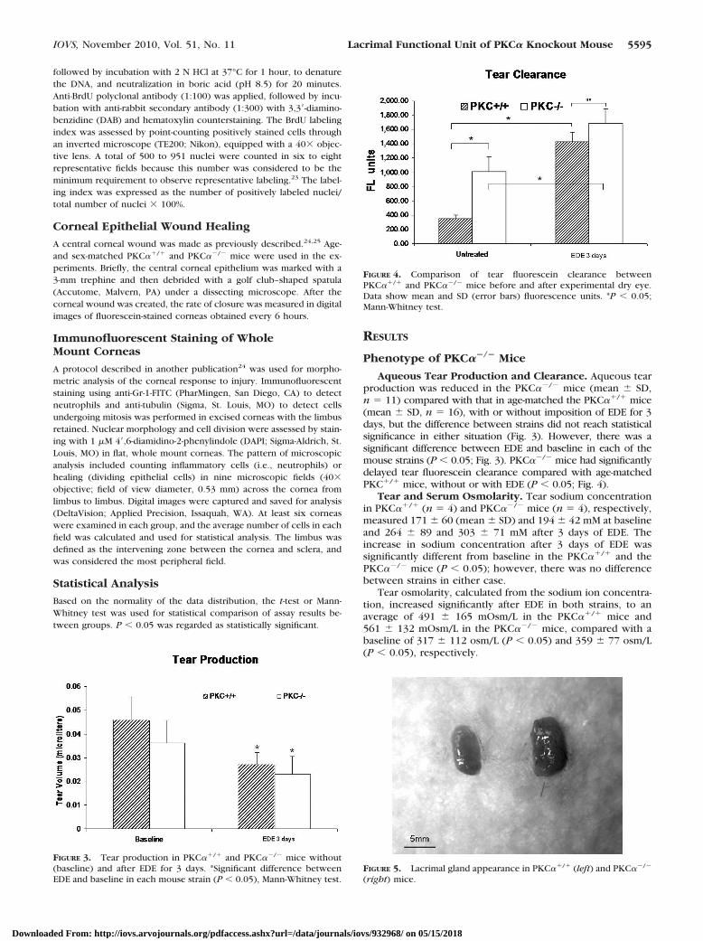
Aqueous Tear Production and Clearance. Aqueous tearproduction was reduced in the PKC��/� mice (mean � SD,n � 11) compared with that in age-matched the PKC��/� mice(mean � SD, n � 16), with or without imposition of EDE for 3days, but the difference between strains did not reach statisticalsignificance in either situation (Fig. 3). However, there was asignificant difference between EDE and baseline in each of themouse strains (P � 0.05; Fig. 3). PKC��/� mice had significantlydelayed tear fluorescein clearance compared with age-matchedPKC�/� mice, without or with EDE (P � 0.05; Fig. 4).
Tear and Serum Osmolarity. Tear sodium concentrationin PKC��/� (n � 4) and PKC��/� mice (n � 4), respectively,measured 171 � 60 (mean � SD) and 194 � 42 mM at baselineand 264 � 89 and 303 � 71 mM after 3 days of EDE. Theincrease in sodium concentration after 3 days of EDE wassignificantly different from baseline in the PKC��/� and thePKC��/� mice (P � 0.05); however, there was no differencebetween strains in either case.
Tear osmolarity, calculated from the sodium ion concentra-tion, increased significantly after EDE in both strains, to anaverage of 491 � 165 mOsm/L in the PKC��/� mice and561 � 132 mOsm/L in the PKC��/� mice, compared with abaseline of 317 � 112 osm/L (P � 0.05) and 359 � 77 osm/L(P � 0.05), respectively.
FIGURE 3. Tear production in PKC��/� and PKC��/� mice without(baseline) and after EDE for 3 days. *Significant difference betweenEDE and baseline in each mouse strain (P � 0.05), Mann-Whitney test.
FIGURE 4. Comparison of tear fluorescein clearance betweenPKC��/� and PKC��/� mice before and after experimental dry eye.Data show mean and SD (error bars) fluorescence units. *P � 0.05;Mann-Whitney test.
FIGURE 5. Lacrimal gland appearance in PKC��/� (left) and PKC��/�
(right) mice.
IOVS, November 2010, Vol. 51, No. 11 Lacrimal Functional Unit of PKC� Knockout Mouse 5595
Downloaded From: http://iovs.arvojournals.org/pdfaccess.ashx?url=/data/journals/iovs/932968/ on 05/15/2018
The serum osmolarity of the PKC��/� mice (n � 4) was324 � 3.9 mOsm/L at baseline and 330 � 2.7 mOsm/L after 3days of EDE, whereas the serum osmolarity of the PKC��/�
mice (n � 4) was 324 � 5.4 mOsm/L at baseline and 331 � 7.3mOsm/L after 3 days of EDE. There was no significant differ-ence between baseline and EDE measurements in either strain.
Lacrimal Gland Weight. The lacrimal glands of thePKC��/� mice weighed more (0.069 � 0.03 g, n � 4) thanthose of the age-matched PKC��/� mice (0.038 � 0.018 g, n �4, P � 0.05; Fig. 5). Similarly, lacrimal gland density (weight/volume) in the PKC��/� mice (2.76 � 0.6 mg/mm3) washigher than that in the age-matched PKC��/� mice (1.21 �
0.071, P � 0.05), as was the ratio of lacrimal gland to bodyweight (2.13 � 0.57 in the PKC��/� mice, 1.21 � 0.049 in thePKC��/� mice; P � 0.05).
Corneal Epithelial Permeability to AFD. Decreased cor-neal epithelial barrier function is a key feature of human dry eye.We assessed it by measuring corneal permeability to AFD. Therewas no difference in AFD permeability in the corneas of thePKC�/� and the age-matched PKC��/� mice as three males andtwo females at baseline. After EDE for 3 days, the corneas of thePKC��/� mice were significantly more permeable to AFD (475 �25 U, n � 5) than were those of the age-matched PKC��/� mice(300 � 25 U, n � 5, P � 0.05, by Mann-Whitney test). This finding
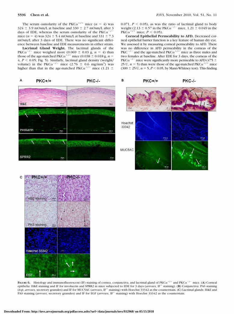
FIGURE 6. Histology and immunofluorescent (IF) staining of cornea, conjunctiva, and lacrimal gland of PKC��/� and PKC��/� mice. (A) Cornealepithelia: H&E staining and IF for involucrin and SPRR2 in mice subjected to EDE for 3 days (arrows, IF� staining). (B) Conjunctiva: PAS staining(top, arrows, secretory granules) and IF for MUC5AC (arrows, IF� staining) with Hoechst 33342 as the counterstain. (C) Lacrimal glands: H&E andPAS staining (arrows, secretory granules) and IF for EGF (arrows, IF� staining) with Hoechst 33342 as the counterstain.
5596 Chen et al. IOVS, November 2010, Vol. 51, No. 11
Downloaded From: http://iovs.arvojournals.org/pdfaccess.ashx?url=/data/journals/iovs/932968/ on 05/15/2018

suggests that PKC��/� mice are more susceptible to cornealbarrier disruption after experimental desiccating stress.
Morphology of the LFU. There was no difference in cor-neal epithelial morphology between the 2-month-old PKC��/�
mice and the age-matched wild-type mice (Fig. 6A). Immuno-fluorescent staining of mouse corneal frozen sections was per-formed to evaluate the expression by corneal epithelial cells ofthe cornification markers involucrin and SPRR2, which mayhave a role in corneal barrier functioning. The intensity of bothinvolucrin and SPRR2 immunofluorescent staining in the cor-neal epithelia of nonstressed mice was lower in the PKC��/�
than in the PKC��/� mice (Fig. 6A).In the conjunctiva, enlarged carbohydrate-rich goblet cells
with stronger PAS staining were observed in the PKC�/� micecompared with those in the PKC��/� mice at baseline (beforeEDE). Correspondingly, greater MUC5AC staining was detectedin the conjunctiva of the PKC��/� than in the PKC�/� mice(Fig. 6B).
Lacrimal gland histologic sections stained with H&E andPAS showed larger carbohydrate-rich acinar cells containingmore PAS-positive granules in samples from the PKC��/�
mice. The number of lacrimal gland acinar cells in thePKC��/� samples was similar to that in the wild-type, but theyappeared to be filled with secretory granules that were notbeing discharged into the lumen. Furthermore, the lacrimalglands of the PKC��/� mice had stronger staining for EGF, agrowth factor produced by the acinar cells and secreted intothe tears, than in the PKC�/� mice, but the difference waswithout statistical significance (immunofluorescent staining in-tensity: PKC�/� mice: 50.35 � 17.62; PKC��/� mice: 52.95 �18.17; P � 0.05; Fig. 6C).
BrdU Incorporation Index. BrdU incorporation was eval-uated as a marker of cell proliferation in tissues of the LFU. ThePKC��/� mice (n � 4) had a greater percentage of BrdU-positive cells in the cornea, conjunctival epithelia, and lac-rimal gland acinar cells (21.25% � 5.78%, 8.49% � 2.83%,and 11.11% � 4.37%, respectively) than did the PKC��/�
mice (n � 4, 10.85% � 3.92%, 2.83% � 0.95%, and 3.39% �1.49%, respectively; all P � 0.05%). These findings indicatethat PKC� is essential in initiating proliferation in the cellsof the LFU (Fig. 7).
Wound Healing
Effect of PKC� Deficiency on Corneal Re-epithelializa-tion after Wounding. A standard model of corneal woundhealing was established by removal of a 3-mm-diameter area ofepithelium from the center of the cornea. The epithelial heal-ing was monitored by fluorescein staining of the exposedstroma in the PKC��/� and PKC��/� mice. At 6, 12, and 18hours after wounding, the size of the wounded areas inPKC��/� and the PKC��/� mice were similar. At 24 and 36hours after creation of the epithelial defect, wound areas in thePKC��/� mice were reduced to 2% and 0% of the original area,respectively. In contrast, wound areas in the PKC��/� micewere reduced to 17% and 7% (Fig. 8). Complete epithelializa-tion was accelerated between 18 and 36 hours in the post-wound PKC��/� mice compared with that in the wild-typemice.
PKC� has been observed to have a regulatory role in cellproliferation that is crucial to corneal wound healing.26 Epithe-lial basal cell division was noted to be decreased in thePKC��/� mice at 18 and 30 hours after epithelial debridementcompared with that in the wild-type mice (Fig. 9).
Effect of PKC� Deficiency on Neutrophil Accumula-tion in the Cornea. Experimental neutropenia induced bydepletion of neutrophils has been noted to accelerate cornealepithelial wound closure.27,28 PKC� activation in keratinocytesis a crucial event that orchestrates cutaneous neutrophil re-sponses by increasing the concentration of two chemotacticfactors: cytokine-induced neutrophil chemoattractant and mac-rophage inflammatory protein (MIP)-2 in murine plasma.29–31
To determine whether the accelerated corneal epithelial heal-
FIGURE 7. BrdU incorporation (arrows) in corneal (top) and conjunc-tival (middle) epithelia and lacrimal gland (bottom) in PKC��/� andPKC� �/� mice (n � 4).
FIGURE 8. Accelerated corneal epi-thelial wound healing in PKC��/�
mice. (A) Fluorescein-stained cornealimages after corneal epithelial de-bridement in PKC��/� and PKC��/�
mice. (B) Percentage of the originalwound area is plotted over time andwas calculated from the area of fluo-rescein staining at each time point.Data are mean � SD (n � 6, **P �0.01).
IOVS, November 2010, Vol. 51, No. 11 Lacrimal Functional Unit of PKC� Knockout Mouse 5597
Downloaded From: http://iovs.arvojournals.org/pdfaccess.ashx?url=/data/journals/iovs/932968/ on 05/15/2018

ing observed in the PKC��/� mice is linked to reduced neu-trophil migration, we analyzed neutrophil accumulation at 18and 30 hours after epithelial debridement. Neutrophil migra-tion into the avascular cornea stroma in the PKC�-deficient andwild-type mice was similar at 18 hours (Fig. 10A). In contrast tothe response of the wild-type mice, the PKC��/� mice had amarkedly blunted accumulation of neutrophils in every field fromlimbus to limbus at 30 hours after wounding (P � 0.005, Fig.10B). These results demonstrate that neutrophil recruitment thatoccurs during the inflammatory phase after corneal epithelialwounding is markedly reduced in the absence of PKC�.
DISCUSSION
Effects of PKC� Deficiency on the Morphologyand Function of the Lacrimal LFUPKC� gene knockout was noted to affect the morphology andfunction of the LFU. These changes included reduced tear
production, secretory paralysis of the lacrimal gland acinar andconjunctival goblet cells, increased permeability of cornealepithelia after experimental desiccating stress, and decreasedcornification of the corneal epithelia.
Loss of PKC� appears to block the parasympathetic path-way of lacrimal gland acinar and conjunctival goblet cell secre-tion. This effect was signified by the enlarged cell size andstronger carbohydrate-rich cells stained in both tissues, and thefailure of exocytosis, due to the lack of the PKC� gene in theexocytic process of preformed secretory vesicles with an api-cal plasma membrane. The PKC��/� mice showed an elevatedocular surface response to experimental desiccating stress,with increased disruption of corneal epithelial barrier function.These changes may be directly due to the loss of the PKC�gene or may be secondary to the reduced secretion of thecorneal supportive factors by the lacrimal gland and conjunc-tival goblet cells. Secretion of one such factor, EGF, was notedto be decreased.
FIGURE 9. Effects of PKC� defi-ciency on dividing basal cells aftercorneal epithelial debridement. Di-viding basal epithelial cells in each re-gion of the cornea at 18 (A) and 30 (B)hours, respectively, after epithelial de-bridement. Representative photos of di-viding cells (arrow) stained by tubulin(green) in PKC��/� (C) and PKC��/�
mice (D) at 18 hours after wounding.DAPI (blue) was used for counterstain-ing. *P � 0.05 and **P � 0.01.
FIGURE 10. Neutrophil infiltrationin PKC�-deficient mice after epithe-lial debridement. Neutrophil migra-tion to corneal stroma in each regionof the cornea at 18 (A) and 30 (B)hours, respectively, after epithelialdebridement. *P � 0.005. Represen-tative photos of neutrophils (arrow)stained by anti-Gr-1-FITC in PKC��/�
(C) and PKC��/� mice (D) at 30hours after wounding.
5598 Chen et al. IOVS, November 2010, Vol. 51, No. 11
Downloaded From: http://iovs.arvojournals.org/pdfaccess.ashx?url=/data/journals/iovs/932968/ on 05/15/2018

Effect of PKC� Deficiency on Corneal EpithelialWound Healing and Inflammation
Traumatic corneal epithelial abrasion is a very common condi-tion encountered in clinical practice. To further characterizethe healing response after corneal epithelial debridement, weused our standardized epithelial wound model to evaluate thehealing rate, proliferation, and inflammatory cell infiltration ofthe corneal epithelium. PKCs are key intracellular signal trans-duction mediators that are involved in various cell functionsthroughout the body, including cell growth and differentiation.Their known functions suggest that they have a pivotal role incorneal wound healing. Studies have already established thepresence of PKC� in normal rabbit corneal epithelium.32–34 Itsexpression increased during epithelial wound healing. An oli-gonucleotide-targeting rabbit PKC� was found to inhibit thewound-healing process in vitro by more than 50%.34 In con-trast to the predictions made on the basis of these previouslyreported studies, we found that PKC��/� mice show acceler-ated corneal epithelial wound healing and faster re-epithelial-ization compared with PKC��/� mice.
The mechanism of these conflicting results is not clear. Onefactor that influences healing rate is the local inflammatoryresponse that may have a positive or negative impact on thehealing rate, as debated in a recent review.27 The neutrophil isthe predominant cell type in the acute phase of inflammation,soon followed by a second wave of monocyte infiltration.Although it is widely recognized that the infiltration of neutro-phils into injured tissue protects wounds from invading patho-gens, more recent studies suggest that neutrophils inhibit thewound repair process, including that in skin28 and cornealepithelium.25,29 The present study showed that neutrophilinfiltration into the injured cornea was markedly decreased inthe PKC��/� mice and is consistent with the finding of a studylinking neutrophil infiltration to PKC� activation in the epider-mis.30,31,35 Therefore, deficient neutrophil infiltration is one ofseveral possible explanations for the accelerated corneal epi-thelial re-epithelialization in the present study.
PKC�-modulated events in the cornea appear to havegreater effects on epithelial healing than PKC� deficiency hason lacrimal gland function. A reduced corneal epithelial heal-ing rate would be expected based on the reduced tear produc-tion that was observed in these mice. In summary, PKC� hasessential functions in regulating tear secretion by the LFU andthe ocular surface response to desiccating stress and cornealepithelial trauma. The details of the mechanism should befurther investigated.
References
1. Nishizuka Y. The molecular heterogeneity of protein kinase C andits implications for cellular regulation. Nature. 1988;334:661–665.
2. Zoukhri D, Hodges RR, Sergheraert C, Toker A, Dartt DA. Lacrimalgland PKC isoforms are differentially involved in agonist-inducedprotein secretion. Am J Physiol. 1997;272:C263–C269.
3. Sundermeier T, Matthews G, Brink PR, Walcott B. Calcium depen-dence of exocytosis in lacrimal gland acinar cells. Am J PhysiolCell Physiol. 2002;282:C360–C365.
4. Zoukhri D, Hodges RR, Willert S, Dartt DA. Immunolocalization oflacrimal gland PKC isoforms: effect of phorbol esters and cholin-ergic agonists on their cellular distribution. J Membr Biol. 1997;157:169–175.
5. Dartt DA. Regulation of tear secretion. Adv Exp Med Biol. 1994;350:1–9.
6. Zoukhri D, Hodges RR, Sergheraert C, Dartt DA. Protein kinase Cisoforms differentially control lacrimal gland functions. Adv ExpMed Biol. 1998;438:181–186.
7. Zoukhri D, Hodges RR, Dicker DM, Dartt DA. Role of proteinkinase C in cholinergic stimulation of lacrimal gland protein secre-tion. FEBS Lett. 1994;351:67–72.
8. Stern ME, Beuerman RW, Fox RI, Gao J, Mircheff AK, PflugfelderSC. The pathology of dry eye: the interaction between the ocularsurface and lacrimal glands. Cornea. 1998;17:584–589.
9. Dartt DA, Kessler TL, Chung EH, Zieske JD. Vasoactive intestinalpeptide-stimulated glycoconjugate secretion from conjunctivalgoblet cells. Exp Eye Res. 1996;63:27–34.
10. Rios JD, Zoukhri D, Rawe IM, Hodges RR, Zieske JD, Dartt DA.Immunolocalization of muscarinic and VIP receptor subtypes andtheir role in stimulating goblet cell secretion. Invest OphthalmolVis Sci. 1999;40:1102–1111.
11. Dartt DA, Rios JD, Kanno H, et al. Regulation of conjunctival gobletcell secretion by Ca(2�)and protein kinase C. Exp Eye Res. 2000;71:619–628.
12. Nakashima S. Protein kinase C alpha (PKC alpha): regulation andbiological function. J Biochem (Tokyo). 2002;132:669 – 675.
13. Ohba M, Ishino K, Kashiwagi M, et al. Induction of differentiationin normal human keratinocytes by adenovirus-mediated introduc-tion of the eta and delta isoforms of protein kinase C. Mol Cell Biol.1998;18:5199–5207.
14. Candi E, Schmidt R, Melino G. The cornified envelope: a model ofcell death in the skin. Nat Rev Mol Cell Biol. 2005;6:328–340.
15. Matassa AA, Kalkofen RL, Carpenter L, Biden TJ, Reyland ME.Inhibition of PKCalpha induces a PKCdelta-dependent apoptoticprogram in salivary epithelial cells. Cell Death Differ. 2003;10:269–277.
16. Ruvolo PP, Deng X, Carr BK, May WS. A functional role formitochondrial protein kinase Calpha in Bcl2 phosphorylationand suppression of apoptosis. J Biol Chem. 1998;273:25436 –25442.
17. Deng J, Chen Y, Wu R. Induction of cell cornification and en-hanced squamous-cell marker SPRR1 gene expression by phorbolester are regulated by different signaling pathways in human con-ducting airway epithelial cells. Am J Respir Cell Mol Biol. 2000;22:597–603.
18. Han Y, Meng T, Murray NR, Fields AP, Brasier AR. Interleukin-1-induced nuclear factor-kappaB-IkappaBalpha autoregulatory feed-back loop in hepatocytes: a role for protein kinase calpha inpost-transcriptional regulation of ikappabalpha resynthesis. J BiolChem. 1999;274:939–947.
19. Leitges M, Plomann M, Standaert ML, et al. Knockout of PKC alphaenhances insulin signaling through PI3K. Mol Endocrinol. 2002;16:847–858.
20. Stewart P, Chen Z, Farley W, Olmos L, Pflugfelder SC. Effect ofexperimental dry eye on tear sodium concentration in the mouse.Eye Contact Lens. 2005;31:175–178.
21. Afonso AA, Monroy D, Stern ME, Feuer WJ, Tseng SC, PflugfelderSC. Correlation of tear fluorescein clearance and Schirmer testscores with ocular irritation symptoms. Ophthalmology. 1999;106:803–810.
22. Dursun D, Wang M, Monroy D, et al. A mouse model of kerato-conjunctivitis sicca. Invest Ophthalmol Vis Sci. 2002;43:632–638.
23. Selvamurugan N, Kwok S, Alliston T, Reiss M, Partridge NC. Trans-forming growth factor-beta 1 regulation of collagenase-3 expres-sion in osteoblastic cells by cross-talk between the Smad andMAPK signaling pathways and their components, Smad2 andRunx2. J Biol Chem. 2004;279:19327–19334.
24. Li Z, Rumbaut RE, Burns AR, Smith CW. Platelet response tocorneal abrasion is necessary for acute inflammation and efficientre-epithelialization. Invest Ophthalmol Vis Sci. 2006;47:4794–4802.
25. Li Z, Burns AR, Smith CW. Lymphocyte function-associated anti-gen-1-dependent inhibition of corneal wound healing. Am JPathol. 2006;169:1590–1600.
26. Sharma GD, Ottino P, Bazan NG, Bazan HE. Epidermal and hepa-tocyte growth factors, but not keratinocyte growth factor, modu-late protein kinase Calpha translocation to the plasma membranethroughout 15(S)-hydroxyeicosatetraenoic acid synthesis. J BiolChem. 2005;280:7917–7924.
27. Martin P, Leibovich SJ. Inflammatory cells during wound repair:the good, the bad and the ugly. Trends Cell Biol. 2005;15:599–607.
IOVS, November 2010, Vol. 51, No. 11 Lacrimal Functional Unit of PKC� Knockout Mouse 5599
Downloaded From: http://iovs.arvojournals.org/pdfaccess.ashx?url=/data/journals/iovs/932968/ on 05/15/2018

28. Dovi JV, He LK, DiPietro LA. Accelerated wound closure in neu-trophil-depleted mice. J Leukoc Biol. 2003;73:448–455.
29. Ueno M, Lyons BL, Burzenski LM, et al. Accelerated wound healing ofalkali-burned corneas in MRL mice is associated with a reduced inflam-matory signature. Invest Ophthalmol Vis Sci. 2005;46:4097–4106.
30. Wang HQ, Smart RC. Overexpression of protein kinase C-alpha inthe epidermis of transgenic mice results in striking alterations inphorbol ester-induced inflammation and COX-2, MIP-2 and TNF-alpha expression but not tumor promotion. J Cell Sci. 1999;112:3497–3506.
31. Cataisson C, Joseloff E, Murillas R, et al. Activation of cutaneousprotein kinase C alpha induces keratinocyte apoptosis and in-traepidermal inflammation by independent signaling pathways.J Immunol. 2003;171:2703–2713.
32. Lin N, Bazan HE. Protein kinase C substrates in corneal epithe-lium during wound healing: the phosphorylation of growthassociated protein-43 (GAP-43). Exp Eye Res. 1995;61:451– 459.
33. Lin N, Bazan HE. Protein kinase C subspecies in rabbit cornealepithelium: increased activity of alpha subspecies duringwound healing. Curr Eye Res. 1992;11:899 –907.
34. Chandrasekher G, Bazan NG, Bazan HE. Selective changes inprotein kinase C (PKC) isoform expression in rabbit cornealepithelium during wound healing. Inhibition of corneal epithe-lial repair by PKCalpha antisense. Exp Eye Res. 1998;67:603–610.
35. Cataisson C, Pearson AJ, Tsien MZ, et al. CXCR2 ligands and G-CSFmediate PKCalpha-induced intraepidermal inflammation. J ClinInvest. 2006;116:2757–2766.
5600 Chen et al. IOVS, November 2010, Vol. 51, No. 11
Downloaded From: http://iovs.arvojournals.org/pdfaccess.ashx?url=/data/journals/iovs/932968/ on 05/15/2018