advances in shrimp biotechnologybiotec.or.th/shrinfo/documents/adv-shrimp-biotec.pdf · advances in...
TRANSCRIPT

Advances in ShrimpBiotechnology
Proceedings to theSpecial Session on Shrimp Biotechnology
5th Asian Fisheries ForumChiengmai, Thailand11-14 November 1998
Edited by
T.W. FlegelDepartment of Biotechnology
Faculty of Science, Mahidol UniversityRama 6 Road, Bangkok 10400, Thailand
BIOTECThe National Center for
Genetic Engineering and BiotechnologyThailand

2 Preface
Advances in Shrimp BiotechnologyProceedings to the special session on shrimp biotechnology5th Asian Fisheries Forum, 11-14 November 1998Chiengmai, Thailand
ISBN 974-7578-02-6
©BIOTECNational Center for Genetic Engineering and BiotechnologyNational Science and Technology Development AgencyNational Science and Technology Agency (NSTDA) Building73/1 Rama 6 Road, Bangkok 10400, Thailand
Tel. (66-2) 644-8150 to -8154Fax. (66-2) 644-8107www.biotec.or.th
Produced by Multimedia Asia Co. Ltd.979/37-41 Phaholyothin Rd., 18/F SM TowerBangkok 10400, Thailand
Tel. (66-2) 298-0646 to -0651Fax.(66-2) 298-0679e-mail: [email protected]

3Advances in shrimp biotechnology
Message from the EditorThis is an exciting time for the study of biotechnology related to shrimp rearing. Up to now,
basic knowledge regarding the major cultivated shrimp species has lagged far behind the technicalinnovations that have led to successful intensification of culture and to ever increasing world produc-tion. However, rearing problems and sometimes catastrophic farm losses have forced the realizationthat the lack of basic knowledge must be redressed if the causes of lost production are to be deter-mined and rectified so that high levels of production can be maintained. Furthermore, since shrimpfarming is a potentially self-polluting industry, it is well understood that sustaining high productionlevels will also require further innovation to minimize its adverse environmental impacts. Biotech-nology will play a central role in helping us to understand the shrimp and to improve all aspects ofrearing practice. The contributions in this volume show that an international effort in the field isalready yielding beneficial returns to the industry. The papers cover broad topics from environmentalissues, to shrimp domestication and larval rearing. Much of what is reported is new and in the earlystages of development and I am certain that many readers will be excited by the results and theirimplications. The National Center for Genetic Engineering and Biotechnology (BIOTEC), an imple-menting arm of the National Science and Technology Development Agency (NSTDA) of Thailand,was pleased to accept the invitation of the Asian Fisheries Society to organize this special session onshrimp biotechnology at the 5th Asian Fisheries Forum. Given the importance of shrimp exports to theThai economy, BIOTEC has had an active program to support and stimulate research on shrimp, andthis 5th AFF activity was considered an appropriate extension of that program. I am grateful to every-one for their enthusiastic support for the session and to the many colleagues who prepared presenta-tions for it. I was especially pleased that many were able to prepare full papers in advance of themeeting for publication in this volume. I would like to thank Dr. M. Shariff, President of the AsianFisheries Society, Dr. Piamsak Menasveta, the Chairman of the local organizing committee and theother members of the local organizing committee for their help in organizing the special shrimp ses-sion. I would also like to thank the staff of BIOTEC and especially Khun Roongthip Rojjananavinwhose assistance was so vitally important in making all the arrangements and handling all of thecorrespondence related to the session and to this publication. I am grateful to the staff at MultimediaAsia Co. Ltd. who introduced me to the intricacies of electronic publishing and who were so patientand helpful in getting all the necessary work done to have the CD ROM ready in time for the meeting.Finally, I would like to thank Mr. Ian H. MacRae for his vital assistance in overcoming the inevitableunexpected problems that were encountered in generating the ultimate portable document format(PDF) file for the CD ROM.
T.W. FlegelDirector, Shrimp Biotechnology ProgramBIOTEC

4 Preface

ContentsOverview
Sustainable Shrimp Culture Development: Biotechnological Issues and Challenges .......... 13Rohana P. Subasinghe, Devin M. Bartley, Sharon McGladdery, Uwe Barg
Alternatives in Shrimp Biowaste Processing ............................................................................ 19Willem F. Stevens, Preiradda Cheypratub, Sjeng Haiqing, Pranee Lertsutthiwong,Ng Chuen How, Suwalee Chandrkrachang
Maturation and GeneticsHormonal Control of Vitellogenesis in Penaeid Shrimp ......................................................... 29
I. Yano
Effect of Methyltestosterone and 17ααααα-hydroxyprogesterone on Spermatogenesis in theBlack Tiger Shrimp, Penaeus monodon Fab........................................................................ 33
R. Yashiro, P. Na-anant, V. Dumchum
The Effects of Cryoprotectants, Chilling and Freezing on Penaeus esculentus Embryos andNauplii ..................................................................................................................................... 37
N.P. Preston, F.E. Coman
Advances in Gene Mapping in Penaeid Shrimp ...................................................................... 45S.S. Moore, S.A. Lehnert
Development of Simple Sequence Length Polymorphisms in the Black Tiger Shrimp(Penaeus monodon) ................................................................................................................ 49
A. Vanavichit, S. Wuthimaethavee, D. Taechayinkphibool, P. S. Kruthoo,P. Lumubol, S. Tragoonrung
Genetic Variation, Population Differentiation and Gene Flow of the Giant Tiger Shrimp(Penaeus monodon) Inferred from mtDNA-RFLP Data..................................................... 51
Sirawut Klinbunga, David J. Penman, Brendan J. McAndrew, Anchalee Tassanakajon,Padermsak Jarayabhand
Quantitative Genetics and Genetic Transformation for the Selection of Pathogen-ResistantShrimp ..................................................................................................................................... 61
Virna Cedeño, Evelyne Bachère, Jenny Rodriguez, Edouard Bédier, Emmanuel Goyard,Viviane Boulo, Jean-Paul Cadoret, Eric Mialhe
Estimated Heritabilities for Early Growth Rate of the Black Tiger Prawn, Penaeusmonodon, Fabricius ................................................................................................................ 67
P. Jarayabhand, S. Uraiwan, S. Klinbunga, A. Tassanakajon, P. Srimukda,P. Pattanachan, R. Panakulchaiwit, P. Menasveta
Triploidy induction in chinese shrimp (Penaeus chinensis) with special reference to a newchemical inducer 6-DMAP .................................................................................................... 71
Jianhai Xiang, Linghua Zhou, Fuhua Li, Changgong Wu
Domestication and Selective Breeding of Penaeus monodon in Thailand ............................. 73Boonsirm Withyachumnarnkul, Vichai Boonsaeng, T.W. Flegel, Sakol Panyim,Chainarong Wongteerasupaya

6 Contents
Shrimp RearingStudy on the Impact of Intensive Marine Shrimp Farm Effluent on Sediment Quality in
Kung Krabaen Bay, Eastern Thailand ................................................................................. 81Siri Tookwinas, Chanin Sangrungruang, Osamu Matsuda
Some Recent Innovations in Marine Shrimp Pond Recycling Systems ................................. 87Arlo W. Fast, Piamsak Menasveta
Preservation of Water Quality in Shrimp Ponds by Ozone .................................................... 93M. Matsumura, V.P. Migo, D. Balobalo, H.K. Young, J.D. Albaladejo
Use of Probiotics for Improving Soil and Water Quality in Aquaculture Ponds................ 101Claude E. Boyd, Amit Gross
Application of Phytoplankters as Live Diet and Biocontrol in Improving the HatcheryProduction of Commercially Important Penaeid Shrimp in Asia ................................... 107
Jesse Dapon Ronquillo, Toshio Saisho, Shigehisa Yamasaki
Manipulation of Bacterial Populations in Shrimp Larval Cultures Fed Artificial Diets ... 109N. Misciattelli, D.A. Jones, N. Simoes, J.W. Latchford, P. Bridson
Effects of Astaxanthin on Larval Growth and Survival of the Giant Tiger Prawn, Penaeusmonodon ................................................................................................................................117
Jintana Darachai, Somkiat Piyatiratitivorakul, Prasat Kittakoop,Charoen Nitithamyong, Piamsak Menasveta
Low Salinity Culture of Penaeus monodon Fabricius and Its Effect on the Environment ............123Yont Musig, Sathit Boonnom
A Closed Recycle System for Sustainable Black Tiger Shrimp Culture in Freshwater Areas ......125Chalor Limsuwan, Pornlerd Chanratchakool
The Shrimp Defence SystemShrimp Immunity and Disease Control: An Integrated Approach ...................................... 129
Evelyne Bachère
Review of Crustacean Immunity ............................................................................................. 135Kenneth Söderhäll
Evidence for the haemocytic origin of lymphoidal spheroids in Penaeus monodon ...........137Margaretha Anggraeni, Leigh Owens
Immuno-physiology of the Black Tiger Shrimp (Penaeus monodon) .................................. 139Kidchakan Supamattaya
Studies on IgY for Passive Immunization of Shrimp Against White Spot Syndrome Virus141
Victoria Alday-Sanz, Sarayut Thaikua, A.N. Yousif, L.J. Albright, T.W. Flegel
Effect of Oxygen Depletion on Some Parameters of the Immune System in Black TigerShrimp (Penaeus monodon) ................................................................................................. 147
Sataporn Direkbusarakom, Yaowanit Danayadol

7Advances in shrimp biotechnology
Molecular Cloning and Characterization of Prophenoloxidase in the Black Tiger Shrimp,Penaeus monodon .................................................................................................................151
Kallaya Sritunyalucksana, Lage Cerenius, Kenneth Söderhäll
Comparison of Shrimp High Density Lipoprotein and Beta Glucan Binding Protein....... 153Gloria Yepiz-Plascencia, Francisco Vargas-Albores, Flor Jimenez-Vega,Lydia M. Ruiz-Verdugo, Gabriela Romo-Figueroa
Probiotics and ImmunostimulantsActivation of shrimp cellular defence functions by microbial products .............................. 161
Francisco Vargas-Albores, Jorge Hernández-López, Teresa Gollas-Galván,Karla Montaño-Pérez, Flor Jiménez-Vega, Gloria Yepiz-Plascencia
Immunostimulation of Shrimp Through Oral Administration of Vibrio Bacterin and YeastGlucan ................................................................................................................................... 167
T.N. Devaraja, S.K. Otta, G. Shubha, Indrani Karunasagar, P. Tauro,Iddya Karunasagar
Efficacy of Oral Administration of Fucoidan, a Sulfated Polysaccharide, in ControllingWhite Spot Syndrome in Kuruma Shrimp in Japan ........................................................ 171
Yukinori Takahashi, Kaori Uehara, Rikio Watanabe, Takekazu Okumura,Tetsuro Yamashita, Hiroshi Omura, Toshikatsu Yomo, Toshiro Kawano,Akio Kanemitsu, Hideo Narasaka, Nobutaka Suzuki, Toshiaki Itami1
Selection of Probiotic Bacteria for Use in Aquaculture ........................................................ 175Bruno Gomez-Gil, Ana Roque
Probiotics in Aquaculture: A Case Study of Probiotics for Larvae of the Black TigerShrimp (Penaeus monodon) ................................................................................................. 177
Sirirat Rengpipat, Sombat Rukpratanporn, Somkiat Piyatiratitivorakul,Piamsak Menasveta
Use of By-9 as a Probiotic Agent in the Larval Rearing of Penaeus monodon ...................183Ketut Sugama Haryanti, S. Tsumura
Will Microbial Manipulation Sustain the Ecological Balance in Shrimp (Penaeus monodon)Hatcheries? ........................................................................................................................... 185
C.R. Lavilla-Pitogo, L.J. Albright, M.G. Paner
Influence of LPS-Injection on Morphological, Antigenic and Functional Aspects ofHemocytes from Penaeus monodon ....................................................................................193
C.B.T. van de Braak, W.P.W. van der Knaap
Evaluation of a Booster Diet for the Nursery of Penaeid Shrimp ........................................ 195Greet Merchie, Marleen Dehasque, Piet Verstraete, Pete Bridson, David Jones
Bacterial Diseases and ToxinsStandardisation of Three Techniques for Experimental Vibrio Infections in the Marine
Shrimp Penaeus vannamei...................................................................................................199Ana Roque, Bruno Gomez-Gil, Ana Luisa Guerra Flores

8 Contents
Luminous Bacteria Associated with Shrimp Mortality ......................................................... 205Lila Ruangpan
Luminous Vibrio harveyi Associated with Tea Brown Gill Syndrome in Black Tiger Shrimp ......213Tirasak Pasharawipas, Siriporn Sriurairatana, Sataporn Direkbusarakom,Yaowanit Donayadol, Surayut Thaikua, Lila Ruangpan, T.W. Flegel
Genetic Diversity of Luminous Vibrio Isolated from Shrimp Larvae .................................. 217Antonius Suwanto, Munti Yuhana, Emi Herawaty, Sri Lestari Angka
Vibrionaceae Associated with the Larvae and Larval Rearing System of Macrobrachiumrosenbergii : Systematics and Pathogenicity ...................................................................... 225
Sarita G. Bhat, I.S. Bright Singh
Detection of Vibrio parahaemolyticus in Shrimp Haemolymph by DNA Hybridization andPCR Amplification ............................................................................................................... 227
Prasert Rojlorsakul, Vichai Boonsaeng, Watanalai Panbangred, Orasa Suthienkul,Tirasak Pasharawipas, T.W. Flegel
Successful use of Bacterial Bioremediation to Control Vibrio Populations in Shrimp Ponds .......235Jason J. David, C.T. Chu, S. Santhana Krishnan
Ultrastructure of a Mollicute Associated, Gut-node Disease of Penaeid Shrimp (Penaeuschinensis) ............................................................................................................................... 237
Yang Jifang
Toxiciological Studies with Juveniles of the Marine Shrimp Penaeus vannamei Using aFlow-Through System .......................................................................................................... 241
Miguel Betancourt-Lozano, Donald J Baird, Luz Maria Garcia-de al Parra,Ferdenando Gonzalez-Farias, Sylvia Morales-Gonzalez
Viral DiseasesActive Viral Accommodation: A New Concept for Crustacean Response to Viral Pathogens ......245
T.W. Flegel, T. Pasharawipas
Screening for Shrimp Viruses in the Philippines ................................................................... 251J.D. Albaladejo, L.M. Tapay, V.P. Migo, C.G. Alfafara, J.R. Somga,S.L. Mayo, R.C. Miranda, K. Natividad, F.O. Magbanua, T.Itami,M. Matsumura, E.C.B. Nadala, Jr., P.C. Loh
Recent Developments in Immunologically-Based and Cell Culture Protocols for the SpecificDetection of Shrimp Viral Pathogens ................................................................................. 255
Philip C. Loh, E. Cesar, B. Nadala Jr., Lourdes M. Tapay, Yuanan Lu
Genome Organization and Detection of Hepatopancreatic Parvovirus (HPV) from Penaeusmonodon in Thailand ........................................................................................................... 261
Wasana Sukhumsirichart, Chainarong Wongteerasupaya, Vichai Boonsaeng,Sakol Panyim, Siriporn Sriurairatana, Boonsirm Withyachumnarnkul, T.W. Flegel
The Emergence of Yellow Head-Related Viruses in Australia ............................................. 263Peter J. Walker, Jeff A. Cowley, Kirsten M. Spann, Christine M. Dimmock

9Advances in shrimp biotechnology
Multiplex PCR for Detection of Yellow-Head Virus and White Spot Syndrome Virus inPenaeus monodon .................................................................................................................265
Chainarong Wongteerasupaya, Vichai Boonsaeng, Wansika Tongchuea,Nusra Sitidilokratana, Panan Kanchanaphum, Ratchanee Klinputsorn,Sakol Panyim
Tissue distribution of white spot syndrome virus (WSSV) in shrimp and crabs ................ 267Guang-Hsiung Kou, Shao-En Peng, Ya-Lin Chiu, Chu-Fang Lo
Primary Shrimp Cell Culture: Applications for Studying White Spot Syndrome Virus(WSSV) .................................................................................................................................. 273
J. Kasornchandra, S. Boonyaratpalin
White Spot Virus Infective Properties Determined by a Single Tube Nested and Competi-tive PCR ................................................................................................................................ 277
Lee Kok Leong, Samson Soon, Tan Lee Tung, Mohd. Shariff Mohd. Din
One-step Nested PCR for Grading the Severity of White Spot Syndrome Virus Infections inPenaeus monodon .................................................................................................................279
Wansika Tongchuea, Vichai Boonsaeng, Chainarong Wongteerasupaya,Karnyupha Jittivadhna, Anchalee Tassanakajon, Sakol Panyim
PCR Monitoring of Cultured Shrimp for White Spot Syndrome Virus (WSSV) Infection inGrowout Ponds ..................................................................................................................... 281
Chu-Fang Lo, Yun-Shiang Chang, Chin-Te Cheng, Guang-Hsiung Kou
Application of PCR and Formalin Treatment to Prevent White Spot Disease in Shrimp .............287Pornlerd Chanratchakool, Chalor Limsuwan
Possible Prevention of White Spot Syndrome (WSS) in Kuruma Shrimp, Penaeusjaponicus, in Japan ............................................................................................................... 291
Toshiaki Itami, Minoru Maeda, Nobutaka Suzuki, Kazuo Tokushige,Atsushi Nakagawa, Oscar Hennig, Masakazu Kondo, Jiraporn Kasornchandra,Ikuo Hirono, Takashi Aoki, Riichi Kusuda, Yukinori Takahashi

10 Contents

Overview

12

Subasinghe RP, Bartley DM, McGladdery S, Barg U (1998) Sustainable shrimp culture develop-ment: biotechnological issues and challenges. In Flegel TW (ed) Advances in shrimp biotechnology.National Center for Genetic Engineering and Biotechnology, Bangkok.
Sustainable Shrimp Culture Development:Biotechnological Issues and Challenges
1Rohana P. Subasinghe, 1Devin M. Bartley, 2Sharon McGladdery, 1Uwe Barg
1Fisheries Department, Food and Agriculture Organization of the United Nations (FAO)Viale delle Terme di Caracalla, 00100 Rome, Italy
2Department of Fisheries and Oceans, Canada, Gulf Fisheries CentreP.O. Box 5030, Moncton, N.B., E1C 9B6 Canada
ABSTRACT: The shrimp farming sector has been growing at a compounded annual rate of about 16% overthe past decade, with much originating from low-income, food deficit, countries (LIFDCs). Continued devel-opment of the shrimp aquaculture sector, however, is constrained by i) recurrent disease epizootics withlimited prevention and control measures, ii) a lack of consistent broodstock and post-larvae quality iii) incon-sistent quality and limited choices of feed and iv) inadequate water control of water quality. Biotechnologicaladvances are now opening new opportunities for ameliorating these developmental constraints. Improvedsensitivity and accuracy of disease diagnosis is being developed using molecular detection techniques such asimmunoassays, PCR gene amplification and molecular probes. Such molecular techniques may also play arole in the eventual development of cell-lines for aquatic invertebrates. Definition of general health param-eters for shrimp are also being investigated using haematograms, enzyme analyses and quantification methodsfor serum proteins and non-protein defense molecules. Controlled breeding programs and genetic markers arebeing co-developed to enhance selection of genetic lines which are free of specific pathogens (specific patho-gen free - SPF) or resistant to the disease(s) they cause (specific pathogen resistant - SPR). Advances inunderstanding of shrimp immunity are also assisting development of immunostimulants and vaccines.Broodstock and post-larval quality are being improved through application of hormonal control of reproduc-tion and development of genetic tags to identify and produce pedigrees with optimal health and productivity.Feed enhancement using microencapsulation of nutrient supplements and probiotics may also play a role inenhancing quality of post-larvae, as well as reducing the need for antibiotic intervention. Bioremediation,recirculation and biofiltration technology all show promise for improving water quality control. The presentpaper attempts to outline the recent advamces in these fields and the potential contribution of modern biotech-nology to the sustainable development of shrimp aquaculture.
KEY WORDS: Biotechnology, bioremediation, bioencapsulation, probiotics, immunostimulants, vaccines,gene probes, PCR, microsatellite markers, SFP, SPR, shrimp, aquaculture, disease diagnosis, disease control,broodstock, larvae, quality and control.
INTRODUCTIONAt present, aquaculture is the world’s fastest growing
food-production sector, providing an acceptable, proteinrich supplement to, and substitute for, wild aquatic ani-mals and plants. Over the last decade, aquatic productionfrom capture fisheries and aquaculture has increased stead-ily, reaching 120.7 million mt in 1995, an increase ofaround 15.6 million mt since 1989. Much of this increaseis attributable to aquaculture. The proportion of totalaquatic production attributable to aquaculture (includingplants), increased from 14.4% in 1989 to 23.0% in 1995.By 1995, the total production of cultured finfish, shellfishand aquatic plants reached a record 27.8 million mt, val-ued at US$ 42.3 thousand million. Much of this increaseoriginated from low-income food deficit countries(LIFDCs), in particular China, and reflects the continuingtrend in these countries for increased use of aquatic re-sources to increase food production (FAO 1997).
The cultured shrimp and prawn subsector grew at an an-nual percent rate (APR) of 16.8 between 1984 and 1995.This increase was principally due to culture of penaeid shrimpspecies, which in 1995 accounted for 96.3% of all culturedshrimp and prawns. Penaeid production, notably of giant ti-ger prawn (Penaeus monodon) and other Penaeus species,increased from 31% or 54,000 mt and 12% or 21,000 mtrespectively, in 1984, to 54% or 503,000 mt and 18% or165,000 mt in 1995. However, production of the fleshy prawn(Penaeus chinensis), 99.5% of which was produced in Chinain 1995, has decreased substantially in terms of tonnage since1993. A decrease in the expansion rate of shrimp farmingduring 1990-1995 was also evident at a global level for eachof the top five penaeid species and species groups. This de-crease in expansion and production tonnage has been attrib-uted to environmental degradation, farm mismanagement andlosses due to disease, making disease and environmentalhealth the critical constraints to continued development of

14 Subasinghe et al.
sustainable shrimp aquaculture (FAO 1997). In addition,inadequate domestication (i.e., poor closed-recirculation sys-tem performance of broodstock with the resultant inadequatesupply of quality post-larvae), limited choice of efficient andeconomical live feed, and inadequate water quality manage-ment have been identified as further significant constraintsto the development of shrimp culture. There is, therefore,considerable scope for application of a wide range of thenewly developing biotechnologies in an effort to amelioratethese problems.
APPLICATIONS TO SHRIMP HEALTHAND DISEASE MANAGEMENT
Infectious disease is considered to be the single mostdevastating problem in shrimp culture. Conventional meth-ods for controlling aquatic animal pathogens, such as chemo-therapy, appear less effective in managing newly emergingpathogens. Thus, molecular biotechnology has an increas-ingly important role for application in screening and detec-tion of pathogens, elucidation of pathogenicity, developmentof effective control and preventive measures, and treatmentof diseases. The improvement of diagnostic methodologiesfor detection and identification of pathogens using immuno-logical and/or nucleic acid probes for prevention, manage-ment, and control of disease in cultured shrimp is one of themost important applications of biotechnology. Unlike tradi-tional chemotherapeutic methods which have been plaguedby development of pathogen resistance, these new technolo-gies provide an opportunity for prophylactic intervention tominimise disease outbreaks.
Enhanced pathogen detection sensitivityEarly, rapid and accurate diagnostics
A very promising and immediate use of molecular ge-netic technology in shrimp aquaculture is its application todisease diagnosis and screening. Ultra-sensitive DNA probescan detect the presence of minute quantities of DNA, ampli-fied by PCR, from pathogens such as viruses, fungi and bac-teria, before clinical symptoms of infection become evident.Commercially available molecular probes have already beendeveloped for IHHNV (Durand et. al. 1996), and probes forother viral pathogens, such as white spot syndrome virus(WSSV), MBV, TSV, HPV, YHV are in the process of beingdeveloped. PCR amplification of DNA sequences specificto the WSSV (DNA “fingerprints”) is being utilised withrelated DNA probes for early diagnosis of white spot dis-ease (Wongteerasupaya et. al. 1996). DNA probes and PCRare extremely sensitive diagnostic tools that can detect cryp-tic infections before they produce symptoms. In addition,these probes label DNA sequences which are highly sensi-tive to individual pathogen species. This greatly enhancesthe accuracy of diagnostic identifications and differentiationbetween opportunistic (usually managed by improved hus-bandry) and significant (contagious organisms which can-not be controlled easily through husbandry adjustments) in-fectious agents. Further development of biomolecular toolsfor screening and rapid detection of shrimp pathogens un-doubtedly will also reduce the current need for, and use of,antibiotics and antibacterials in shrimp aquaculture.
Diagnosis and research on intracellular pathogensSince the majority of severe pathogens of shrimp are vi-
ruses, the lack of self-replicating cell-line cultures for theirin vitro detection and isolation is a significant disease diag-nostic constraint. Considerable research has gone into thedevelopment and maintenance of crustacean cell-lines but,to date, success has been marginal. Some researchers havemanaged to develop primary cell cultures, but most of thesehave failed to achieve cell-division for subculture and conti-nuity of consistent cell characteristics (Le Groumellec et. al.1995). Thus, the need for viable crustacean (and other aquaticinvertebrate) cell-lines continues to be a significant challengefor disease research and management. Interestingly, molecu-lar biotechnology may also assist this area of research. Mo-lecular intervention, at the genetic level, has the potential topromote independent cell-division, overcoming the currentcell-line development barrier of limited longevity. Shouldthis occur, it would have significant positive consequencesfor economic application to shrimp aquaculture and diseasecontrol. Furthermore, cell-line based diagnostics have a well-established history and these techniques fall within the cur-rent capability of many aquatic pathology laboratories.
Disease risk assessment for transboundary transfers oflive shrimp
Transboundary movement of shrimp broodstock and post-larvae has been perceived as one of the main reasons for thespread of epizootic viral diseases of shrimp. Developmentof reliable and sensitive diagnostic tools which can be usedto screen shrimp for known, significant, pathogens, prior tomovement from one zone or country to another, will greatlyreduce the risk of inadvertent pathogen introduction to sus-ceptible populations. This is of particular importance, bear-ing in mind the difficulty in detecting sub-clinical (“healthy”)carriers with low levels of significant pathogens using someroutine diagnostics. Increased diagnostic sensitivity will, inturn, promote greater confidence in the shrimp culture in-dustry, which has been plagued by the shadow of viral pro-liferation and spread. It will also facilitate access to a widerinternational market.
Human pathogen detectionThe ultimate goal of all shrimp culture is optimal mar-
keting of the final product. Growing concerns about humanhealth aspects of aquatic animal products, as well as theemerging number of international trade and quality agree-ments and understandings, mean that improved diagnostictechnologies (i.e., DNA probes and PCR) to identify humanpathogens in aquaculture products is imperative. The sametechnologies now being applied to shrimp pathogens, areequally applicable to human gastro-intestinal pathogens, andmore research and development in this area is clearly neededto enhance consumer confidence and meet tightening inter-national trade requirements.
Assessment of general health statusBesides screening for pathogens, one of the most urgent
needs for health management in shrimp aquaculture is to es-tablish standard quantitative methods for accurate assess-ment of shrimp health. Biotechnological methods used forascertaining the health status of many aquatic organisms in-clude, haematocrit, leucocrit, blood cell differential counts,

15Biotechnology for shrimp culture
neutrophil counts, oxidative radical production,myeloperoxidase activity, phagocytic functions, etc.. Plasmasamples also show total protein, immunoglobulin, lysozyme,cortisol and ceruloplasmin levels. Methods such as aggluti-nation of precipitin gel tests used to assay antibody afterimmunisation can now be supplemented with FAT and ELISAimmunoassay techniques. Also samples of blood, orimmunopoietic organs, can be taken to determine which cellsare producing antibody (plaque-forming cells), e.g., by thehaemolytic plaque assay or by an enzyme labelled tag(ELISPOT). The latter is quantifiable and can show num-bers of immunoglobulin or non-specific antibody-secretingcells (Anderson 1995). Monoclonal antibodies have also beenprepared and are routinely used in immunodiagnosis, e.g.,immunofluorescence detection. Such diagnostic tools arerelatively new to aquaculture, but have enormous potential(Austin 1998). Although these may not all be applicable toshrimp culture, further research is clearly needed to ascer-tain their potential uses, especially in shrimp health man-agement.
Such techniques could also found the basis for develop-ment of simple and rapid diagnostic tests for use under fieldconditions by field health technicians or farmers themselves.There is also ample scope for developing methods to assessthe health status of shrimp using haemolymph parameters,such as cell counts, cell activities, enzyme levels, etc. Suchtests could be used as early warning indicators of impendingdisease outbreaks and, thus, enhance the early applicationof preventative measures (Flegel 1996).
Immunostimulants and vaccinesHarnessing the hosts’ specific and non-specific defence
mechanisms for controlling diseases has considerable po-tential for health management in shrimp aquaculture. Thiswill help reduce stress from handling (grading, manipulat-ing stocking densities, removing mortalities, etc.) and envi-ronmental manipulation (application of chemical treatments,pond drainage, etc.) in order to control disease expressionunder intensive culture conditions. Important biotechnologi-cal interventions are being developed in the field ofimmunostimulants and modulators in an effort to reduceshrimp susceptibility to disease. Immunostimulants and non-specific immune-enhancers are being incorporated into di-ets (see Feed Quality) to provide added protection to theanimals, even though our knowledge of shrimp immunity islimited at present, The large number of commercial“immunostimulants” available on the market reflects the in-terest of the industry in broadening the scope of tools avail-able to manage shrimp diseases However, the effectivenessof many of these products has yet to be established. Prelimi-nary results from biological trials appear highly variable.Further research and field trials are clearly essential to de-termine the precise mechanisms of the action of these prod-ucts and to evaluate their efficacy in commercial shrimp pro-duction (Flegel 1996, Subasinghe et. al. 1998).
Specific pathogen free (SPF) and specifcpathogen resistant (SPR) shrimp
Production of specific pathogen free (SPF) animals andthe development of specific pathogen resistant (SPR) strains
are two complementary approaches which are currently pos-sible using broodstock management programmes (seeBroodstock Development). SPF animals are produced byselecting animals free of known and detectable pathogensand raising them under controlled and strict sanitary condi-tions. The SPR animals are developed through selectivebreeding of animals known to be less susceptible to specificpathogens. These concepts are now being used in countrieslike the USA, Venezuela and French Polynesia with P.vannamei and P. stylirostris (Bedier 1998). The main ben-efit of this concept is production of high health (HH) post-larvae, free of, or resistant to, known pathogens. It should benoted, however, that many SPF stocks which have not beenexposed to pathogens (specific or general), perform poorlywhen pathogens are present (Browdy 1998). As with mostorganisms, a lack of exposure to infectious organisms in-creases clinical susceptibility. If the immune or physiologi-cal traits are heritable, this translates into performance im-provement at the farm level. The selection of lines with rein-forced non-specific defence or enhanced tolerance of exter-nal infection challenges, appears to be a promising option(Bedier 1998). Considering the major contribution of P.monodon to the global shrimp production and the economiclosses encountered due to disease outbreaks, it is appropri-ate and timely to concentrate further research on the devel-opment of SPF and SPR broodstock of this species. Researchin this area, through collaboration between scientists and pro-ducers from different regions, should, therefore, be givenhigh priority.
BROODSTOCK DEVELOPMENTHormonal control of reproduction
The successful expansion and sustainable developmentof shrimp culture depends largely on the consistent avail-ability of quality broodstock and post-larvae. Endocrine regu-lation of reproduction has been effectively applied in fishculture but is not yet at a practical stage of use in shrimpculture. Recent research has shown that inhibition of shrimpgonad inhibiting neurohormone (GIH) by chemical treatmentcan promote reproduction without the negative side effectsof eye stalk ablation. Shrimp GIH isolation techniques arestill under development but, by elucidating the structure andfunction of GIH, it should be possible to devise strategies tocounter the inhibitory effects of GIH. This will significantlyenhance consistent hatchery production of quality shrimppost-larvae. Further research in this direction is needed andcollaboration between researchers, shrimp aquaculturists andresource providers from different regions is essential in or-der to achieve this.
Genetic improvement, selective breeding andengineering techniques
The application of genetic principles to increase produc-tion from aquatic animals lags far behind that of the plantand livestock sectors. Only a small percentage of farmedaquatic animals have been subjected to genetic improvementprogrammes (Gjedrem 1997), and the application of genetictechniques to marine shrimp aquaculture is at an extremelyearly stage. Biotechnology and genetics have great potential

16 Subasinghe et al.
to increase production from shrimp farming and to help makeshrimp farming sustainable. However, Benzie (1998) notesthat there are still gaps in our basic knowledge of shrimpgenetics and physiology that must be addressed before ‘su-per-prawn biotechnology’ can be produced.
Genetic improvementTo date, several standard techniques for genetic improve-
ment have not yielded good results with marine shrimp. Forexample, additive genetic variance and hybridisation hasbeen complicated by pre-zygotic and post-zygotic reproduc-tive isolation problems. When accomplished it has not con-sistently produced heterosis in the F1 generation for eitherincreased growth rate or disease resistance. Nor has hybridi-sation been an effective means of combining desirable traitsfrom different species (Benzie et al. 1995). Chromosomemanipulation (polyploidy) has not been practical, and selec-tive breeding programmes have been hindered by difficul-ties with reproduction of key species, such as P. monodon,marking individuals, low heritability of growth-related traits(i.e., environmental factors greatly influence growth) (Benzieet al. 1997), and by the generally low priority afforded togenetic research by private industry (Wang 1998). However,it is interesting to note that more and more collaborationsare being established between scientists, researchers, labo-ratories, regional and international organisations, and thedonor community to further research efforts on shrimp ge-netics.
Selective breedingIn order to minimise environmental impacts on wild
populations and to fully realise the value of genetic selec-tion and diversity, shrimp aquaculture must break its reli-ance on wild post-larvae (Wang 1998). Although wild-caughtpost-larvae may currently perform better than some hatch-ery produced post-larvae, it inevitably involves the risk ofintroducing pathogens into the culture environment, and maypose a threat to aquatic biodiversity through removal of eggand larval stages of other species as a by-catch. Although,there is no direct evidence to strengthen the latter, hatcheryproduction of post-larvae will facilitate a year-round unin-terrupted supply of healthy post-larvae.
Tracking genetic lines and heritabilityModern molecular techniques also show promise for
shrimp hatchery production, in that they can provide accu-rate information on the genetic diversity of natural stocks,enhance selective breeding programmes, and allow genetictagging of shrimp to facilitate selection of individuals withthe characteristics which meet various market needs (size,coloration, rate of growth, time of spawning, etc.). Molecu-lar markers have already been developed to identify geneti-cally discrete natural populations and to provide family iden-tification and pedigree information in some selective breed-ing programmes. Identification of individual animals allowstracking of their reproductive performance and the traits ex-pressed in their offspring, which, in turn produces abroodstock line with a defined pedigree. Physical tagging ofearly life-history stages of many aquatic species is difficult,especially with marine shrimp. Genetic markers usingmicrosatellite DNA, and AFLP’s (amplified fragment lengthpolymorphisms) are beginning to be produced which pro-
vide a non-invasive mechanism of tracking broodstock andtheir offspring, in order to produce a well-documented pedi-gree. Molecular genetic markers are also being used to mapthe genome of commercially important shrimp species suchas P. japonicus and P. monodon. This will improve identifi-cation of loci, genes, and gene complexes, that improve cul-ture performance, product quality and profit. These markerscan also be used to map genetically linked characteristicsand identify loci with quantifiable traits (Garcia et. al. 1996,Benzie 1998, S. Moore, pers. comm). These advances willmake shrimp breeding programmes more efficient and prof-itable.
Genetic engineeringProduction of transgenic (genetically modified) shrimp
has been reported (Mialhe et al. 1995). However, this hasnot yet been achieved at a commercial scale (Benzie 1998).At present, the use of transgenic animals for aquaculture iscontroversial and it is not well accepted by the industry orconsumers for any shrimp species
LARVAL CULTURE AND LIVE FEEDSDependable availability of quality fry to stock grow-out
production systems has been one of the most critical factorsin the commercial success of industrial production of fishand shellfish (Sorgeloos 1995). More attention to the “qual-ity” and the “competence” of hatchery-produced post-lar-vae is needed, to ensure better performance under grow-outconditions, with particular focus on bioaugmentation andmicrobial management of the various culture steps in thehatchery. Although nutritional and dietary requirements ofmany fish and shellfish species have been identified, large-scale hatchery production of most aquatic invertebrates stilldepends on live feed, such as selected species of microalgae,the rotifer Brachionus and the brine shrimp Artemia.
Consistency and quality of shrimp feedMore than 15 species of diatoms and green algae are used
in first-feeding of hatchery produced shrimp larvae (zoeastage). Selection of these species has been based on theirdigestibility, as determined by trial and error rather than byscientific analysis. The systems used in most developingcountries are still labour intensive. This is not cost effectiveand poses many problems for mass production, includinginconsistent nutritional quality and microbial contaminationleading to decreased or lost production. This has created awhole new area of biotechnological research to find cost ef-fective and efficient supplements to live microalgae, Thisincludes research into commercial production of freeze-driedalgae, microencapsulated diets, and manipulated yeasts. Thisarea of biotechnology, however, requires further research inan effort to reduce current reliance on mass production oflive feed in shrimp hatcheries, with all the handling compli-cations and costs this entails.
Artemia nauplii are the most widely used live feed inshrimp aquaculture (Sorgeloos & Leger 1992). Over the yearsconsiderable progress has been made in improving their di-etary value through batch selection of traits including effi-cient cyst disinfection and decapsulation, success of naupliushatching, and cold storage (Sorgeloos 1995). In addition,

17Biotechnology for shrimp culture
improvement of the nutritional quality of Artemia throughbioencapsulation (enrichment), especially with highly unsatu-rated fatty acids and vitamins, has improved larviculture out-puts in terms of quality, survival, growth, and stress resist-ance.
Dietary nutrients and medical supplementationBioencapsulation has also been applied for oral delivery
of vaccines, vitamins, and chemotherapeutants (Lavens et.al. 1995). These positive results mean that continued researchinto bioencapsulation and use of live feed as a means of oraldelivery of dietary supplements and/or medication to shrimp,and other crustaceans, should be given high priority.
AQUATIC ENVIRONMENTAL QUALITYMANAGEMENT
Probiotic application to shrimp cultureProbiotics are microbes (usually bacteria) selected for
their ability to outcompete and displace potentially harmfulsympatric species. They are usually administered as feed sup-plements to improve the intestinal microbial balance. A sta-ble gut microflora helps digestion efficiency and the abilityof animals to resist pathogenic infections, particularly of thegastro-intestinal tract. Use of antibiotics, by definition, re-duces the level of the gut microflora. Restoration of this es-sential microflora can be accelerated through the use ofprobiotic-enhanced feeds designed to boost colonisation bythe most efficient digestive bacterial species. Probiotics arewidely used in terrestrial animal husbandry, but their use inaquaculture is still in its infancy. Reports on the potential ofprobiotics in shrimp aquaculture are, however, on the in-crease. In some countries it is reported that probiotic use hassignificantly reduced antibiotic use in shrimp hatcheries.Probiotics have been used to suppress the growth of patho-genic Vibrio spp. in many shrimp hatcheries by introducing(inoculating) non-pathogenic strains or species. This proce-dure appears effective and economical and demonstrates aclear need for further research into identifying potentialprobiotic strains of micro-organisms and evaluating their ef-ficacy under field/farm conditions.
Bioremediation techniquesBioremediation is another promising biotechnological
approach, involving degradation of hazardous waste to en-vironmentally safe levels by use of selected micro-organ-isms, bivalves, algae, etc. (Srinivasa Rao & Sudha 1996).Although bioremediation has been well-used in situationssuch as sewage treatment, its application to shrimpaquaculture is fairly novel. Many commercial products areavailable in the market, mainly bacterial preparations, al-though, as with immunostimulants, their mode of action andefficacy has yet to be scientifically ascertained. In additionto microbes, bivalves, seaweed, holothurians, etc., have beentested and used to reduce organic loading and excess nutri-ents in shrimp ponds (polyculture systems or reservoir pondsor “bio ponds”). Various preparations have also been devel-oped with the aim of removal of nitrogenous and other or-ganic waste from the water and bottom sludge. This improvesthe pond environment and reduces physiological stress onshrimp. More products will, undoubtedly, emerge as research
continues However, controlled field trials are urgently neededto determine the cost benefits and effectiveness of these un-der commercial culture conditions.
Recirculation techniques for water quality controlImproved water management techniques such as partial
and complete recirculation of water, with the view to avoidintroduction of sub-clinically infected wild carriers (e.g.,WSSV carriers), has been effective in controlling the recentviral disease epizootics which have spread throughout Asia.In addition, this technique has also proven effective in re-ducing proliferation of opportunistic pathogens. As a largenumber of aquatic organisms have been identified as poten-tial carries of WSSV, avoidance of contamination throughwater exchange is now considered as one of the most effec-tive preventive measures against this viral epizootic. Creat-ing an ecologically sound environment, which is less stress-ful to shrimp, through water recirculation and avoidance offrequent water exchange, is a challenge for the future. De-velopment of water management options through the in-creased use of biological agents and biotechnological means,to reduce excess nutrients and to maintain optimal physico-chemical and microbial quality of water, will continue to bea challenge for environmental scientists, and will certainlycontribute to the success of shrimp aquaculture.
CONCLUSIONSAquaculture biotechnology can be described as the sci-
entific application of biological concepts that enhance theproductivity and economic viability of its industrial sectors(Liao & Chao 1997). Such technology is certainly emergingas one of the most rapidly growing “new frontiers” in re-search, using a knowledge-intensive and advantage-orientedapproach. These developments are helping to diversifyaquaculture studies, potential investment, and internationalexchange. Continued development of biotechnology inshrimp aquaculture has the potential to provide a means ofproducing “super shrimp”, healthy and fast growing, throughenvironmentally friendly means. This development, however,depends on the desire and willingness of the producers towork hand-in-hand with scientists and the international do-nor community to assist all countries, and especially LIFDC’s,in this research. This applies particularly to capacity build-ing and infrastructure development. Improved exchange ofinformation and discussion of problems and achievementsbetween scientists, researchers, and producers from differ-ent regions is also essential to help this important food pro-duction sector further develop globally sustainable produc-tion of healthy shrimp. It should be noted that immediateand preliminary use of most of these new biotechnologieswill likely be restricted to specialised users and laborato-ries. However, this is normal for all technological advance-ments, as has been previously demonstrated in this field withfluorescence and electron microscopy, molecular electro-phoresis and polyclonal antibody immunodiagnostics. It is,therefore, expected that industry and diagnostic demands willurge rapid refinement of these newer technologies for moregeneral use within the next decade. The initiative is clearlyestablished, it just requires maintenance of the developmen-tal momentum.

18 Subasinghe et al.
LITERATURE CITED
Anderson DP (1995) Novel techniques in fish disease diagnosis.In: Shariff M, Arthur, JR, Subasinghe RP (eds) Diseases inAsian Aquaculture, Vol II, Fish Health Section of the AsianFisheries Society, Manila, Philippines, p 27-42
Austin, B (1998) Biotechnology and diagnosis and control of fishpathogens. J Mar Biotech 6:1-2
FAO (1997) FAO Fisheries Circular No 886, Rev 1, Rome, FAO.1997
Bedier E, Cochard JC, Le Moullac G, Patrois J, Aquacop (1998)Selective breeding and pathology in penaeid shrimp culture:the genetic approach to pathogen resistance. World Aquaculture29(2):46-51
Benzie JAH (1998) Penaeid genetics and biotechnology.Aquaculture 164: 23-47
Browdy, CL (1998) Recent developments in penaeid broodstockand seed production technologies: improving the outlook forsuperior captive stocks. Aquaculture 164: 3-21
Durand -S, Lightner DV, Nunan LM, Redman RM, Mari -J, BonamiJ-R (1996) Application of gene probes as diagnostic tools forwhite spot baculovirus (WSBV) of penaeid shrimp. Dis aquatOrg 27(1):59-66
Flegal TW (1996) A turning point for sustainable aquaculture: thewhite spot virus crisis in Asian shrimp culture. AquacultureAsia, July-September 1996. NACA, Bangkok, Thailand, p 29-34
Garcia DK, Dhar AK, Alciva-Warren A (1996) Molecular analysisof PAPD marker (B20) reveals two microsatellite and differ-ential mRNA expression in Penaeus vannemei. Mol Mar BiolBiotech 5:1-83
Gjedrem T (1997) Selective breeding to improve aquaculture pro-duction. World Aquaculture 28:33-45
Gjerde B, Rye M (1998) Design of breeding programmes inaquaculture species: possibilities and constraints. In: BartleyD, Basurco B (eds) Genetics and breeding of Mediterraneanaquaculture species. Cahiers OPTIONS Mèditerranèennes, Vol34. Zaragoza, Spain. p 181-192
Le Groumellec M, Martin C, Haffner P, Martin B, Aquacop (1995)Cell culture from tropical shrimp. J Aquacult Trop 10: 277-286
Lavens P, Sorgeloos P, Dhert P, Devresse,D (1995) Larval foodsIn: Bromage NR, Roberts JR (eds) Broodstock managementand egg and larval quality. Blackwell Science Limited, Ox-ford, p 373
Liao IC, Chao NH (1997) Developments in aquaculture biotech-nology in Taiwan. J Mar Biotech 5:16-23
Mialhe E, Bachere E, Boulo V, Cadoret JP, Rousseau C, CedenoV, Saraiva E, Carrera L, Colwell RR (1995) Future of biotech-nology-based control of disease in marine invertebrates. MolMar Biotechnol 4: 275-283
Sorgeloos P (1995) Bioengineering of hatcheries for marine fishand shellfish. J Mar Biotech 3:42-45
Sorgeloos P, Leger P (1992) Improved larviculture outputs of ma-rine fish, shrimp, and prawn. J World Aquacult Soc 23(4): 251-264
Sriniwasa Rao PS, Sudha PM (1996) Emerging trends in shrimpfarming. Fishing Chimes 16(3):25-26
Subasinghe RP, Barg U, Phillips MJ, Bartley D, Tacon A (In Press)Aquatic animal health management: investment opportunitieswithin developing countries. J Appl Ichth 14(3)
Wang Y (1998) Utilization of genetic resources in aquaculture: afarmer’s view for sustainable development. Bellagio Confer-ence, Towards Policies for Conservation and Sustainable Useof Aquatic Genetic Resources. FAO/ICLARM, Bellagio, Italy,14-18 April, 1998
Wongteerasupaya C, Wongwisansri S, Boonsaeng V, Panyim S,Pratanpipat P, Nash GL, Withyachumnarnkul B, Flegel TW(1996) DNA fragment of Penaeus monodon baculovirusPmNOBII gives positive in situ hybridization with white spotviral infections in six penaeid shrimp species. Aquaculture 143:23-32

Stevens WF, Cheypratub P, Haiqing S, Lertsutthiwong P, How NC, Chandrkrachang S (1998) Alter-natives in shrimp biowaste processing. In Flegel TW (ed) Advances in shrimp biotechnology. Na-tional Center for Genetic Engineering and Biotechnology, Bangkok.
Alternatives in Shrimp Biowaste Processing
Willem F. Stevens, Preiradda Cheypratub, Sjeng HaiqingPranee Lertsutthiwong, Ng Chuen How, Suwalee Chandrkrachang
Bioprocess Technology Program, Asian Institute of Technology, Bangkok, Thailand
ABSTRACT: In marine food processing, about half of shrimp weight is biowaste. Without further processing,industrial quantities of this waste are a severe burden for the environment. Decay starts in a few hours leading toa bad smell and, after disposal, to a high load in terms of COD and BOD. Shrimp biowaste can be valorized bydrying and mixing with other raw materials to compose animal feed. As an alternative, shrimp biowaste can betreated to isolate chitin, the aminocellulose component in the exoskeleton, and its valuable derivative chitosan.Steamed biowaste is applied in large quantities in animal feed. The process is highly efficient and automated. Allcomponents of the waste including the pigment contribute to the nutritional value of the end product. Thedisadvantage of the process is the low added value. Moreover, in small scale operations there is the risk ofinsufficient inactivation of shrimp pathogens present in the waste that can lead to a feed born reinfection inshrimp aquaculture. Experimental data will be discussed to estimate the significance of this argument. Forseparation of shrimp protein and chitin components, usually chemical extraction is applied. The common indus-trial process is efficient but uses chemicals including hot 50% alkali to produce deacetylated chitin or chitosan.However, the chemical treatment causes hydrolysis of the chitosan product and corrosion of the equipment. Inaddition it leads to generation of a hazardous high alkali, high protein waste. Systematic research of the chemicalprocessing has resulted in an improved system for production of chitin with a high degree of purity and with a10-30 fold higher viscosity. Also the quality of the waste water has been improved considerably. On the basis ofthe recent technology, high quality chitin and chitosan can be produced in a sustainable way. Biotechnology hasopened other alternatives to isolate chitin and chitosan. In this approach the waste is stabilized firstly by anensilage process using partial fermentation by Lactobacillus bacteria. The fermentation leads to production oflactic acid. This will decrease the pH of the medium (to pH 4-5) and inhibit the growth of other bacteria. It alsoprevents the formation of off-flavor amines. After fermentation, most of the shrimp protein is detached from thesolid component and can be isolated easily and turned into a protein preparation suitable for human food. Forthe isolation of chitin, only simple, further chemical treatment is required. Chitin can be deacetylated intochitosan using a fungal enzyme, chitin deacetylase (CDA). The CDA enzyme has been isolated from the fungusAbsidia cocruleus and characterized. Although with excess solid chitin as a substrate the enzyme activity ishigh, only a small percentage of the substrate is converted. Due to the high crystallinity of the natural substrate,only the chitin acetyl groups on the outside of chitin particles might be accessible to the enzyme. Therefore,larger scale production of chitosan using enzymes will only be successful if the chitin can be decrystallized andthe acetyl groups exposed to the enzyme. In this report a number of experiments with decrystallized amorphouschitin will be discussed. Either chemically or biotechnologically or in mixed procedures, cleaner and more intactchitosan can be produced. The high quality chitosan has many industrial applications including manufacture ofpharmaceutical and medical products, drugs and food additives. It is also used in paper and textile manufactur-ing and in other industrial processing. Progress in chitin applications in a few selected areas will be summarized.The biotechnological methods have the advantage of the production of human food protein. The separation ofshrimp waste into its components leads not only to high value products but also to safe food product cycles withregard to the chances of shrimp reinfection through feed containing shrimp waste.
KEY WORDS: shrimp waste, chitin, chitosan, Lactobacillus, Absidia cocruleus, chitin deacetylase,fermentation
INTRODUCTIONThe major product of the shrimp marine food industry is
shrimp meat. However, the meat constitutes only about 50%to the shrimp wet body weight. The other half, the residualbiomaterial or biowaste, contains many valuable compoundsthat after appropriate processing can add substantially tooverall profitability. This shrimp biomateral can be valor-ized without fractionation. Usually it is applied as such forfeeding in veterinary practice and aquaculture. Medium andlarge scale processing has been developed to dry the waste
and to mix it with other agricultural raw materials to pro-duce animal feed.
The shrimp biomaterial can be valorized further byfractionation into its major components: protein, mineralsand chitin. The protein has high potential as a human foodadditive and food supplement while the chitin is usually con-verted into its deacetylated product, chitosan. Chitin andchitosan are N containing polysaccharides which have a struc-ture similar to cellulose. Chitosan, in particular, has many

20 Stevens et al.
applications in foods, medicines, cosmetics and water puri-fication and as an ionic biopolymer for paper and textile fin-ishing and for flocculation processes. Various other valu-able products can be extracted from the biomaterial. Theseinclude shrimp flavor components and the natural dyeasthaxanthin that have many applications in the food andfeed industry.
A major problem with shrimp biomaterial valorization isthe high perishability of the material. Under tropical climaticconditions, decay starts within an hour after processing andleads to the production of biogenic amines with a very of-fensive smell. If this decay is unavoidable or not prevented,the biomaterial turns into real waste and due to its high pro-tein content, it becomes a real threat to the environment anda financial burden if discarded properly.
It is obvious for both environmental and economic rea-sons that, wherever possible, appropriate technology shouldbe applied to prevent decay and to convert the biomaterialinto valuable products. Technology should provide systemsfor the delay or prevention of decay and procedures forfractionation. In this paper alternative technologies for con-servation and valorization of shrimp biomaterial are discussedand evaluated.
USE OF NON FRACTIONATEDBIOMATERIAL
In areas with a low technical infrastructure, where fish-ermen individually catch and clean small amounts, most ofthe biowaste is used as feed for livestock without any treat-ment. In these cases, decay of a part of the waste will beunavoidable, leading to pollution of land and water. At loca-tions where large amounts of shrimp are landed or producedin aquaculture, processing infrastructure to handle the wasteis usually developed. The waste is collected at certain placesand dried. At most times of the year, solar drying is not suf-ficient to prevent decay and odor formation. Heat, usuallysteam, is used to preserve the waste and to dry it quickly intodry shell material and nearly dry protein. Due to the heat,many valuable compounds in the waste are lost. The driedmaterial is ground and sold as shrimp meal, to be used as aprotein rich component in fish feed.
In areas where massive amounts of shrimp biowaste aregenerated, large industrial facilities process it in mixtureswith soy products, with cheap carbohydrate products likecassava and potato and with minerals. After drying and grind-ing into a dry meal it is sold in large quantities in the interna-tional market. These forms of direct treatment do not re-quire fractionation and can be carried out in a straightfor-ward manner. However the added value of approximately10 cents US (4 Thai baht) per kg of non fractionated waste islow.
CHEMICAL FRACTIONATIONA much higher economic value can be added to the
biowaste through fractionation. The most common proce-dure is chemically very simple: treatment of the biowastewith 4% alkali to separate the protein and treatment with 4%acid to remove the calcium carbonate. The resulting chitin
product can be further deacetylated by concentrated 50%alkali to produce chitosan. The individual steps are outlinedin the following paragraphs.
DeproteinationIn the first step, the waste is treated with 4% sodium hy-
droxide (NaOH) at elevated temperatures (70-120oC). Un-der these conditions the protein becomes detached from thesolid component in the shrimp biowaste. To prevent oxida-tion of the products, the process is usually carried out in anitrogen atmosphere and in the presence of sodiumborohydride (NaBH
4). After completion of the deproteination
step, the protein hydrolysate is removed easily by separa-tion of the solids from the protein slurry by filtration. Theprotein hydrolysate can be dried and used in the form of acake or powder as a protein supplement in feed. This proteinhydrolysate also contains most of the shrimp flavor. The solidfraction consists mainly of chitin and calcium carbonate. Italso contains most of the pigment.
DemineralizationIn the next step, the solid fraction is treated with 4% hy-
drochloric acid (HCl) which converts the insoluble calciumcarbonate into soluble calcium chloride that can subsequentlybe removed by washing. With appropriate deproteination anddemineralization, the remaining product consists mainly ofchitin with minor amounts of protein and calcium, that canbe judged from a weak Biuret reaction and a low weightafter ashing, respectively. The chitin product should be white.It is insoluble in alkali and in most acids and organic sol-vents. Due to its low reactivity, chitin is usually deacetylatedto chitosan.
Deacetylation.The chemical deacetylation of chitin into chitosan re-
quires strong chemical conditions: 50% NaOH and elevatedtemperatures as high as 70-90oC. The highest degree ofdeacetylation possible is desired and several treatments areusually required to reach a sufficient degree to obtain a mar-ketable product (see Fig. 1). Chitin deacetylated by 70-90%(also referred to as 30-10% acetylated chitosan) is consid-ered to be a good end product. The material should be lowin protein and ash. Chitosan can be dissolved in 1-2% aceticacid and high viscosity of this solution is indicative of a wellprepared chitosan. If too rigorous conditions are appliedduring deacetylation, the main chain of the chitin breaks andthis results in low viscosity of chitosan dissolved in aceticacid. In addition the broken molecules cause discolorationand condensation, resulting in reduced transparency and solu-bility. A good chitosan preparation has a low ash content (<1%) and dissolves well in acetic acid giving high transpar-ency (>90% Transmission) and a viscosity of at least 300centipoise.
One might wonder why chitin cannot be deacetylated by90% in one step since the reagent, NaOH, is in excess, theacetate formed does not inhibit the deacetylation and theconditions are quite aggressive. It seems that there is a trans-port problem. The chitosan formed at the outside of the chitinparticle seems to inhibit entry of NaOH into the particle.After washing with water this barrier is removed and theparticle is again open so that the process of deacetylation
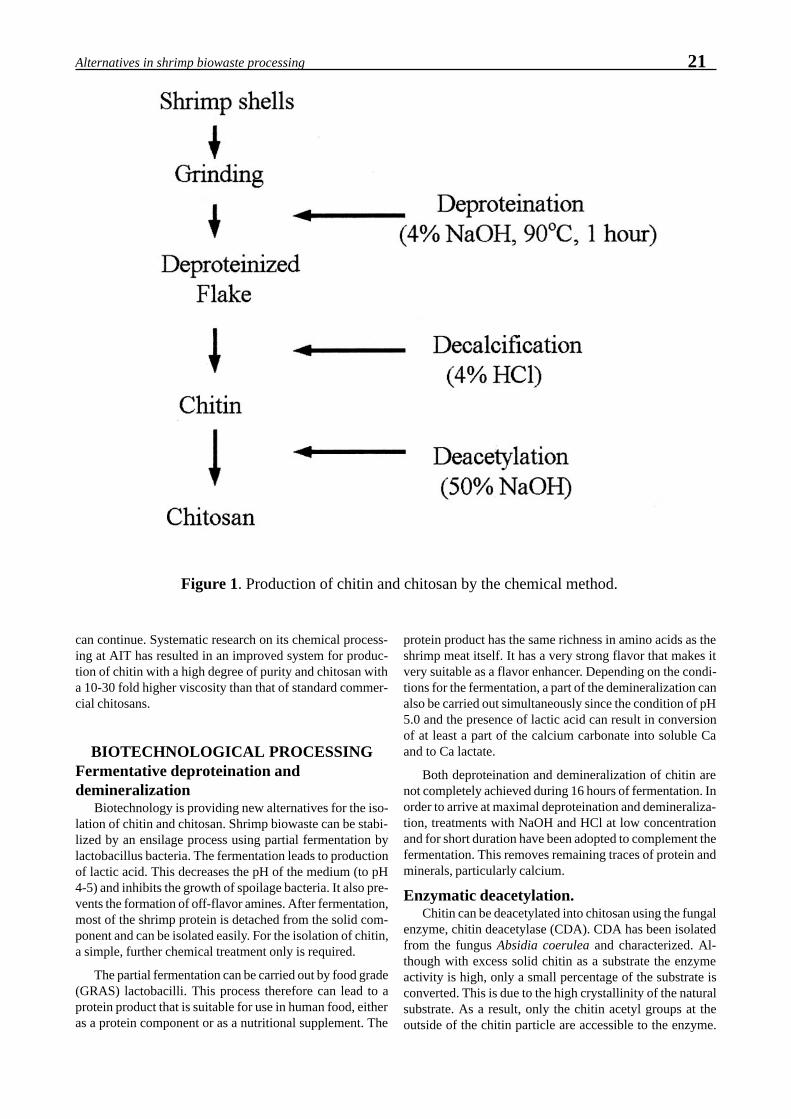
21Alternatives in shrimp biowaste processing
can continue. Systematic research on its chemical process-ing at AIT has resulted in an improved system for produc-tion of chitin with a high degree of purity and chitosan witha 10-30 fold higher viscosity than that of standard commer-cial chitosans.
BIOTECHNOLOGICAL PROCESSINGFermentative deproteination anddemineralization
Biotechnology is providing new alternatives for the iso-lation of chitin and chitosan. Shrimp biowaste can be stabi-lized by an ensilage process using partial fermentation bylactobacillus bacteria. The fermentation leads to productionof lactic acid. This decreases the pH of the medium (to pH4-5) and inhibits the growth of spoilage bacteria. It also pre-vents the formation of off-flavor amines. After fermentation,most of the shrimp protein is detached from the solid com-ponent and can be isolated easily. For the isolation of chitin,a simple, further chemical treatment only is required.
The partial fermentation can be carried out by food grade(GRAS) lactobacilli. This process therefore can lead to aprotein product that is suitable for use in human food, eitheras a protein component or as a nutritional supplement. The
protein product has the same richness in amino acids as theshrimp meat itself. It has a very strong flavor that makes itvery suitable as a flavor enhancer. Depending on the condi-tions for the fermentation, a part of the demineralization canalso be carried out simultaneously since the condition of pH5.0 and the presence of lactic acid can result in conversionof at least a part of the calcium carbonate into soluble Caand to Ca lactate.
Both deproteination and demineralization of chitin arenot completely achieved during 16 hours of fermentation. Inorder to arrive at maximal deproteination and demineraliza-tion, treatments with NaOH and HCl at low concentrationand for short duration have been adopted to complement thefermentation. This removes remaining traces of protein andminerals, particularly calcium.
Enzymatic deacetylation.Chitin can be deacetylated into chitosan using the fungal
enzyme, chitin deacetylase (CDA). CDA has been isolatedfrom the fungus Absidia coerulea and characterized. Al-though with excess solid chitin as a substrate the enzymeactivity is high, only a small percentage of the substrate isconverted. This is due to the high crystallinity of the naturalsubstrate. As a result, only the chitin acetyl groups at theoutside of the chitin particle are accessible to the enzyme.
Figure 1. Production of chitin and chitosan by the chemical method.

22 Stevens et al.
Therefore larger scale production of chitosan using enzymeswill only be successful if the chitin can be decrystallized andthe acetyl groups exposed to the enzyme.
A number of experiments have been carried out in orderto decrease the crystallinity of the chitin and make it a moreaccessible CDA substrate. Physical methods like heating andultrasonic treatment did not improve the chitin as a substratefor CDA. Limited steam explosion (i.e., heating to 120oC athigh pressure followed by sudden pressure release) did notalter its properties. However, chemical treatment using strongacids did create a chitin with a more open structure. Thisproblem of substrate accessibility has not been solved.
EVALUATION OF CHEMICAL ANDBIOCHEMICAL METHODS
In cases where proper conditions are applied and the tem-perature is kept below 70oC, chitin and chitosan can be pre-pared very well by chemical methods. Chitin is resistant toalkali and quite resistant to 4% hydrochloric acid. Chitosanis very resistant to 50% alkali. The major drawback of thechemical method is the non sustainability of the process. Thealkaline deproteination leads to a voluminous 4% alkalinewaste containing a high amount of protein per liter. The 50%alkaline condition for deacetylation is also highly corrosive
for the equipment used, especially at elevated temperatures.The waste after this step is highly alkaline and must be dis-carded properly, not released into the environment. The al-kaline treatment has also a drawback for the protein and thepigment. The protein once treated at elevated temperaturewith 4% alkali or acid is not considered to be food grade.The carotenoid pigment also disintegrates under conditionsof high temperature and light.
Processing of the shrimp protein by partial fermentationand (in future) by enzymatic deacetylation (Fig. 2) has twoadvantages. The protein hydrolysate remains a valuable prod-uct with a high nutritional value for human purposes. Fur-thermore, the process does not result in an alkalineproteineous waste water stream that is difficult to clean. Onthe other hand, the biotechnological process also has disad-vantages. It is slightly more complicated as the chemical proc-ess and the product quality might decrease if chitin andchitosan degrading enzymes are present under the proceduresused. However, on the basis of our experience up to now,this has not been a problem.
Nearly all chitin and chitosan is currently producedthrough chemical methods. The apparent ease of makingchitin and chitosan has encouraged many small firms to try,but the resistance of the biomaterial to treatment and prob-lems of scale have caused many unexpected problems. In
Figure 2. Total biochemical conversion of shrimp biowaste into chitin and chitosan.

23Alternatives in shrimp biowaste processing
some cases, the use of high temperatures has caused muchchemical damage and resulted in end products of low qual-ity. Since the physico chemical qualitative assessment ofbiopolymers requires specific expertise, many low qualitychitosans have entered the market. There is a need for inter-national criteria for chitin quality and for a reference centrethat can check for compliance with these criteria. (see Fig. 3for an example of a possible quality report). The BioprocessTechnology Program at the Asian Institute of Technologyhas the objective to realize such a center for the S. E. Asianregion.
Research on the prevention of proteineous waste waterduring chemical treatment has lead to a significant reduc-tion in released BOD. On the basis of the present technol-ogy, high quality chitin and chitosan can be produced chemi-cally in a sustainable way. In the biotechnological method,there is actually no waste left, the protein hydrolysate is col-lected as a human food supplement and the chitin/calciumcarbonate is used to make chitosan. If in future a technicalprocess becomes available for biocatalytic deacetylation, thewhole process would be truly sustainable.
Figure 3. Model for a chitin and chitosan quality assurance sheet.
CHITIN AND CHITOSAN QUALITY ASSURANCE FORM
Chitin ü Chitosan(Tick whatever appropriate) Grade GRAND
Parameters Mean Value Standard Deviation% Deacetylation 85 0.5Apparent Viscosity (cps) 1500 50Intrinsic (dL/g) 14.5 1.0Viscosity Average Molecular Weight (106) 1.8 0.2% Ash 0.45 0.05% Moisture 10.0 0.1% Protein 0.1 0.03Turbidity (NTU) 2 0.1% Insoluble 0Solid (Qualitative Observation) - Sample size (mesh) 14~20 - Color Milky white
Heavy Metals - Cu (ppm) 0.02 0.00 - Fe (ppm) 0.08 0.00 - Cd (ppm) 0.02 0.00 - Ni (ppm) 0.01 0.00 - Cr (ppm) 0.02 0.00 - Zn (ppm) 0.02 0.00 - Hg (ppb) < 2 - - As (ppb) < 2 -
Coliform groups None
Comments :Prepared by : Ng Chuen How Date : 25 Sept 1998Approved by : Prof. Willem Stevens Date : 26 Sept 1998
AVAILABILITY OF BIOWASTEThe amounts of chitin in the world are immense. It has
been estimated that the total amount of chitin equals or evenexceeds the amount of wood cellulose. Chitin is an impor-tant component of fungal cell walls, it is the building mate-rial for insect exoskeletons and together with calcium car-bonate, it forms the rigid structure of the crustacean shell.However, in comparison to cellulose, the possibilities forcollection of chitin are much more limited. The fungi andinsects are highly dispersed in nature and most of the crusta-ceans are in the deep sea. One source for chitin might be themycelium of industrially produced fungi, but recovery fromthat source does not seem very promising at this time.
The most attractive sources are the crab and shrimp seafood industries. At places where large volumes of crab andshrimp meat are processed, nearly equal amounts of biowasteare generated. For the time being only a small fraction oftotal crab and shrimp biowaste is being processed into chitin.Most of it is discarded if not used for food. The logical placesto produce chitin and chitosan are areas where the biowaste

24 Stevens et al.
is being produced in large quantities. Examples are alongthe coastal zones in Thailand and India and the landingharbors for crabs and shrimp in northern Europe and America.Seafood and shrimp factories that do not presently valorizetheir chitin containing wastes might consider ways of get-ting more value from the waste by extracting chitin, proteinand pigment.
APPLICATIONSProcessing the biowaste cannot be discussed apart from
the applications of the chitin, chitosan and shrimp waste prod-ucts. In this paragraph only a few examples of applicationare presented (Table 1). The application of non fractionatedbiowaste in aquaculture was mentioned at the start of thispaper. In addition, preparations of the pigment asthaxanthinare used.
Table 2. Shrimp mortality from injected WSSV after various treatments. Thetable shows daily morality and cumulative mortality after injection of heattreated viral preparations, an untreated viral preparation (positive control) andcarrier solution (negative control).
Day post injection of viral preparationsTreatment 1 2 3 4 5 6 7 8 9 10 Cumulative
% MortalityNeg. control 0 0 0 0 0 0 0 0 0 0 0100oc 0 0 3 0 0 0 0 0 0 0 2580oc 0 0 0 0 0 0 0 0 0 0 060oc 0 0 1 2 2 1 0 1 0 0 58.340oc 0 0 8 4 0 0 0 0 0 0 100Pos. control 0 1 6 5 0 0 0 0 0 0 100
Chitin itself has only limited applications. This is due toits insolubility in water and common organic solvents andits limited chemical reactivity. Chitin is being applied suc-cessfully in cultivation of mushrooms for instance. In soil itis broken down by chitinases and acts as a slow release sys-tem to provide organic nitrogen. The same amount of nitro-gen supplied as urea is toxic.
Chitosan has a free amino group and therefore has muchhigher chemical reactivity and solubility than chitin. It canbe produced as a powder, beads, flakes or as membranes. Itcan also be re-acetylated to chitin to regain the insolubilityand chemical resistance of chitin. Thus, chitin can be con-verted to chitosan, cast into a membrane and transformedback into a chitin membrane.
Table 1. Estimated consumption of chitin, chitosan and theirderivatives in the Japanese market in 1994.
Uses Consumptiona (tons/year)Cationic flocculating agents 350 Living wastewater treatment (200) Food manufacturing wastewater treatment (100) Sugar manufacturing (50)Food additives 125 Food processing (45) Functional health foods (80)Agricultural materials 120 (e.g., plant seed coating, fertilizers)Feed additives for pets, fishes and animals, etc 60Textiles and fabrics 50Cosmetic ingredient for hair and skin cares 40D-glucosamine and oligosaccharides 13Biomedical materials 20 (e.g., adsorbable suture, wound dressings)Paint and dyeing 10Thickeners 10Membranes 1Chromatographic media and reagents 1 (e.g., colloid titration, enzyme substrates, etc)aEstimated as chitosan

25Alternatives in shrimp biowaste processing
The most important applications of technical gradechitosan are in environmental applications concerned mainlywith water and waste water purification. Chitosan is an effi-cient aid in flocculation and a very effective binding mate-rial for metals like copper and chromium.
Technical chitosan is also being used on a large scale asan antifungicide and as a binder in the textile industry. Papertreated with chitosan becomes stronger and non wettable.
Pure chitosan is used in food, cosmetics, drugs and medi-cal products. The human consumption of chitosan hasboomed during last years as a dietary health food that bindsbile acids and so reduces uptake of cholesterol and lipids inthe body. In cosmetics, chitosan has been used for many yearsto enhance binding of cosmetics to negatively charged skinand hair. In pharmacy, chitosan can be applied as slow re-lease matrix for a variety of drugs and peptides. Medicalproducts include chitin surgical threads and transparent band-ages for wound healing.
At present there is a large imbalance between scale ofpossible applications of chitin and chitosan and the limitedamounts actually being used. The reason for this imbalanceconcerns the low quality of some of the chitins offered onthe world market and the problems in establishing clear cri-teria for chitin/chitosan quality. In the next decade, the de-mand for chitosan will rise further and so the valorization ofshrimp and crab biowaste should be of major interest of theshrimp and crab industries.
CONSTRAINTS ON THE USE OF SHRIMPBIOMATERIAL IN AQUACULTURE
The use of shrimp biowaste in shrimp meal and fish mealneeds to be considered further due to the high incidence ofvirus infections in shrimp aquaculture. Several viruses highlypathogenic for shrimp have been identified and studied indetail. Advanced methods for virus diagnosis have been de-veloped. However, the mechanism of infection, distributionand spread of these viruses is still poorly understood. It issupposed that shrimp viruses have a complicated structureand are rather labile, but in most cases, this has not beenscientifically confirmed.
Shrimp infected with viral pathogens cannot be excludedfrom processing plants. In the large amounts of biowasteprocessed, the chances are high that some processed shrimpshell may still contain viable shrimp viruses. Therefore, theprocessing of shrimp biowaste for shrimp meal should becarried out in a manner that can guarantee that the resultingfeed is free of shrimp pathogens. Inspection of large and amedium size production plants for shrimp and fish meal inThailand revealed that the chances for shrimp viruses to sur-vive the drying process are extremely small. In the large scaleoperations, the shrimp waste was heated together with otheringredients in a fluidized bed reactor to140oC for 30 min. Inthe medium size operations, the shrimp biowaste was steamedand then dried with constant mixing over a period of aboutone hour. Presumably labile viruses would not survive ei-ther treatment, even when somewhat protected inside shrimptissues. However, some viruses may have high heat stabilityand have more chance to survive.
The heat inactivation of white spot syndrome virus(WSSV) has been investigated in order to check the effi-ciency of heat in killing. The system designed for the testscould be used to study the inactivation of other viruses aswell. WSSV was subjected to a temperature treatment (40 to100oC for 1 hour) and then the infectivity of the heated ma-terial was tested by injection into live shrimp. To check thecause of any mortality, PCR tests were performed on theshrimp haemolymph after injection. As may be concludedfrom preliminary data shown in Table 2, WSSV survivedtreatment for 1 hour at 40 and 60oC but was inactivated attemperatures of 80oC or higher. PCR data confirmed thatdeath for the low temperature treatments was caused byWSSV. Some shrimp did die after receiving virus treated at100oC, but they were negative for WSSV by PCR and musthave died from some other cause. In this preliminary study,heat treatment was carried out on extracted virus and not onvirus in shrimp tissues. Whether virus in tissue would havebetter survival at high temperatures is presently under inves-tigation. Other pathogens, including a Vibrio bacteriophageimplicated in shrimp mortality, will be studied using this sys-tem.
In general, one may comment that it is risky to feed ananimal species with biowaste of the same species. Feedingchicks with residuals from chicken processing enhances thechance for chicken disease. Shrimp should not be fed withshrimp, especially when other economic alternatives areavailable. The production of chitosan, protein and pigmentfrom shrimp should also be considered in this perspective.
OUTLOOK AND RECOMMENDATIONSThe demand for high quality chitosan, preferentially
decrystallized will increase in the coming decade. As men-tioned, sharp growth is expected for application of chitincompounds in food, natural fungicides, waste water treat-ment, textile coating and cosmetics and medicines. Chitinand chitosan should be produced in various grades of purityand consistency depending on the requirements for a par-ticular type of application. Due to process improvements,the defined quality will go up and the price will change de-pending on the use and the quality. Overall profitability willincrease. Emphasis should be given to local systems for treat-ment of biowaste and to the organization of a multi tier sys-tem for chitin valorization. In the interim, shrimp drying pro-cedures should be monitored for their efficiency in killingshrimp pathogens.

26 Stevens et al.

Maturation and Genetics

28

Yano I (1998) Hormonal control of vitellogenesis in penaeid shrimp. In Flegel TW (ed) Advances inshrimp biotechnology. National Center for Genetic Engineering and Biotechnology, Bangkok.
e-mail: [email protected]
Hormonal Control of Vitellogenesis in Penaeid Shrimp
I. Yano
Department of Marine Bioscience, Fukui Prefectural University, Gakuen-cho 1-1, Obama, 917 Fukui, Japan
ABSTRACT: Control of maturation is a major problem in the development of commercial aquaculture programsfor penaeid shrimp. In penaeid shrimp, maturation involves two main processes, vitellogenesis and final matu-ration of oocytes. In vitellogenesis of penaeid shrimp, two phases are involved: vitellogenin (Vg, a precursor ofegg yolk protein) synthesis in the ovary and accumulation of vitellin in oocytes. Vg synthesis in ovarian tissuepieces can be stimulated by thoracic ganglion extract prepared from vitellogenic females. Vitellogenin synthesisin the ovary is stimulated by vitellogenesis-stimulating hormone (VSH), a neuropeptide of approximately 10000Da, secreted from the thoracic ganglion in the kuruma prawn, P. japonicus. Vg synthesis in incubatedprevitellogenic ovarian pieces can be stimulated by estradiol-17ß. Oil globule stage oocytes, which are theoocytes at the initial stage of primary vitellognesis, were found in incubated previtellogenic ovarian pieces aftertreatment with estradiol-17ß in vitro. These results suggested that estradiol-17ß, probably secreted from theovary, stimulated Vg synthesis in the ovary as a Vg-stimuating ovarian hormone in penaeid shrimp. Unilateraleyestalk ablation is effective in increasing serum Vg in the kuruma prawn. Vg synthesis can be induced slightlyin isolated previtellogenic ovarian pieces, which are not affected directly by hormones from eyestalks in vitro inthe kuruma prawn. This suggests that Vg synthesis in the ovary may be inhibited directly by vitellogenesis-inhibiting hormone (VIH) secreted from eyestalks. On the other hand, Vg synthesis in ovarian pieces incubatedwith thoracic ganglion pieces prepared from vitellogenic females could be stimulated by serotonin. However,serotonin was not effective in stimulating Vg synthesis in ovarian pieces incubated without thoracic ganglionpieces. These results suggested that secretion of vitellogenesis-stimulating hormone from the thoracic ganglionmay be induced by serotonin.
KEY WORDS: neurohormone, estradiol-17ß, vitellogenin synthesis, penaeid shrimp
INTRODUCTIONControl of maturation and spawning is a major problem
in the development of commercial aquaculture programs forpenaeid shrimp. Controlling maturation and spawning in cap-tivity could help to provide a reliable year-round supply ofjuveniles, serve in developing selective breeding programs,and be generally useful for obtaining disease-free spawners.Eyestalk ablation has been used to mature and spawn femaleshrimp in captivity in conjunction with management of wa-ter temperature, photoperiod, light intensity, density, sex ra-tio, and nutrition (Yano 1993). Knowledge of hormonal con-trol of maturation and spawning in crustaceans, however, isfragmentary. Since the pioneering work of the 1940s(Panouse 1943, 1944), many observations of endocrine sys-tems have been made with respect to the inhibition of matu-ration and spawning by eyestalk hormone(s). However, re-cent research has focused mostly on organs (brain, thoracicganglion and ovary) and their functions which are closelyrelated to the release of vitellogenesis-stimulalting hormonesand ovarian hormone (Yano 1992,1993). This paper sum-marizes our present knowledge of the endocrine systems ofpenaeid shrimp and how they control vitellogenesis.
THORACIC GANGLION HORMONEEFFECTS ON VITELLOGENESIS
In penaeid shrimp, maturation involves two main proc-esses, vitellogenesis (Yano et al. 1996) and final maturationwith germinal vesicle breakdown (GVBD) and ovulation in
oocytes (Yano 1988, 1995). Vitellogenesis occurs in hard-shelled shrimp at the intermolt stage C
4, shortly after molting
and continues until immediately before final maturation inpenaeid shrimp. Smaller size P. vannamei did not initiatevitellogenesis even after eyestalk ablation (Yano, unpublisheddata). Thus, vitellogenesis is controlled by two factors, onewhich inhibits and the other which stimulates. Inprevitellogenic (immature) females, the vitellogenesis-stimu-lating principle is absent or not yet functioning. Yano et al.(1988) demonstrated that vitellogenesis in shrimp can bestimulated by implantation of pieces of thoracic gangliontissue prepared from the female lobster, Homarusamericanus, with vitellogenic ovaries. This result indicatedthat vitellogenesis could be stimulated by a vitellogenesis-stimulating hormone (VSH) secreted by neurosecretory cellsof the thoracic ganglion of vitellogenic females and that lob-ster VSH was not species-specific in activity. Injection ofthoracic ganglion extract prepared from vitellogenic femalesis effective in increasing serum Vg in the kuruma prawn, P.japonicus. On the other hand, Vg (a necessary prerequisitefor ovarian oocytes to reach full vitellogenesis) is synthe-sized in the ovary in penaeid shrimp (Yano & Chinzei, 1987).Yano and Itakura found that Vg synthesis in incubated ovar-ian pieces can be stimulated by thoracic ganglion extract inkuruma prawn (unpublished data). Thoracic ganglion extract,prepared from vitellogenic kuruma prawn females, wasfractioned by gel filtration high-performance liquid chroma-tography, and high Vg-stimulating activity was detected in

30 Yano
the fraction corresponding to a molecular weight of 10,000(Yano, unpublished data). This fraction was inactivated bytrypsin. Therefore, the bioactive factor, VSH, could be char-acterized as a peptide hormone. The results indicated thatVSH secreted from the thoracic ganglion stimulated Vg syn-thesis in the ovary and vitellin (egg yolk protein) accumula-tion in ovarian oocytes in penaeid shrimp (Fig. 1).
EYESTALK HORMONE DIRECTLYINHIBITS VITELLOGENIN SYNTHESIS
Unilateral eyestalk ablation is effective in increasing se-rum Vg in the kuruma prawn (Yano 1992). Also for this spe-cies, Vg synthesis can be induced in isolated previtellogenicovarian pieces in vitro. However, they are not affected di-rectly by hormone from eyestalks in vitro. These results sug-gest that Vg synthesis in the ovary may be inhibited directlyby vitellogenesis-inhibiting hormone (VIH) secreted fromeyestalks (Fig. 1). Unilateral eyestalk ablation is not effec-tive in increasing serum Vg in vitellogenic female kurumaprawn. This suggests that VIH levels decrease quickly im-mediately before the initiation of vitellogenesis and stay at alow level until after vitellogenesisis is completed. Therefore,eyestalk ablation may not be effective in regulating the pro-duction of VIH after vitellogenesis has been initiated.
INDUCTION OF VITELLOGENINSYNTHESIS IN OVARY
BY ESTRADIOL-17ß Injection of estradiol-17ß is effective in increasing se-
rum Vg in the kuruma prawn, (Yano, unpublished data). Yanoand Hoshino found that Vg synthesis in incubated ovarianpieces can be stimulated by estradiol-17ß (unpublished data).In addition, oil globule stage oocytes (i.e., the oocytes atinitial stages of primary vitellognesis) were found in incu-bated previtellogenic ovarian pieces after treatment ofestradiol-17ß in vitro. Evidence has been presented to showthat 17α-hydroxy-progesterone is effective in increasing se-rum Vg in kuruma prawn (Yano, 1987). The hormoneestradiol-17ß or 17α-hydroxy-progesterone is generally dis-tributed in the ovary of crustaceans (Kanazawa & Teshima1971, Teshima & kanazawa 1971, Jeng et al. 1978, Couch etal. 1987, Faires et al. 1989). Considering these observations,17α-hydroxy-progesterone may be worked into the Vg syn-thesis mechanism in kuruma prawn as a precursor ofestradiol-17ß. It is suggested that estradiol-17ß, probablysecreted from ovarian follicle cells, induces Vg synthesis inthe ovary as a Vg-stimuating ovarian hormone in penaeidshrimp (Fig. 1).
Figure 1. Proposed model for the hormonal control of vitellogenesis in penaeid shrimp. VSH-RH, vitellogenesis-stimulat-ing hormone-releasing hormone; VSH, vitellogenesis-stimulating hormone;VIH, vitellogenesis-inhibiting hormone.

31Hormonal control of vitellogenesis in penaeid shrimp
SEROTONIN STIMULATES THERELEASE OF VITELLOGENESIS-
STIMULATING HORMONE SECRETEDFROM THE THORACIC GANGLIONVg synthesis in ovarian pieces incubated with thoracic
ganglion pieces prepared from vitellogenic females can bestimulated by serotonin (Yano & Itakura, unpublished data).However, serotonin was not effective in stimulating Vg syn-thesis in ovarian pieces incubated without thoracic ganglionpieces. Serotonin is found widely distributed throughout thenervous system in decapod crustaceans (Fingerman et al.1994). These facts suggest that the release of vitellogenesis-stimulating hormone from the thoracic ganglion may be in-duced by serotonin present in the central nervous system.Therefore, neurohormonal serotonin is nominated as a vitel-logenesis-stimulating hormone-releasing hormone (VSH-RH) responsible for ovarian Vg synthesis in penaeid shrimp.
LITERATURE CITEDCouch EF, Hagino N, Lee JW (1987) Changes in estradiol and
progesterone immunoreactivity in tissues of the lobster(Homarus americanus) with developing and immature ova-ries. Comp Biochem Physiol 87:765-770
Faires NJ, Evershed RP, Quinlan PT, Goad LJ (1989) Detection ofunconjugated and conjugated steroids in the ovary, egg, andhemolymph of the Decapod crustacean, Nephrops norvegicus.Gen Comp Endocrinol 4:199-208
Fingerman M, Nagabhushanam R, Sarojini R, Reddy PS (1994)Biogenic amines in crustaceans: Identification, localization, androles. J Crust Biol 14: 413-437
Jeng SS, Wan WCM, Chang CF (1978) Existence of an estrogen-like compound in the ovary of the shrimp Parapenaeus fissurus.Gen Comp Endocrinol 36:211-214
Kanazawa A, Teshima S (1971) In vivo conversion of cholesterolto steroid hormones in the spiny lobster, Panulirus japonicus.Bull Jpn Soc Sci Fish 37:891-898
Panouse JB (1943) Influence de l’ablation du predoncule sur lacroissance de l’ovaire chez la crevette Leander serratus. C RAcad Sci , Paris 217: 553-555.
Panouse JB (1944) L’action de la glande du sinus sur l’ovaire chezla crevette Leander. C R Acad Sci, Paris 218: 293-294.
Teshima S, kanazawa A (1971) Bioconversion of progesterone bythe ovaries of crab, Portunus trituberculatus. Gen CompEndocrinol 17:152-157
Yano I (1987) Effect of 17b-hydroxy-progesterone on vitellogeninsecretion in kuruma prawn, Penaeus japonicus. Aquaculture61:49-57.
Yano I (1988) Oocyte development in the kuruma prawn Penaeusjaponicus. Mar Biol 99:547-553
Yano I (1992) Hormonal control of reproductive maturation inpenaeid shrimp. NOAA Tech Rep NMFS 106:7-14
Yano I (1993) Ultraintensive culture and maturation in captivity ofpenaeid shrimp. In: McVey JP (ed). Mariculture, vol 1. CRCPress, Boca Raton, pp 289-313
Yano I (1995) Final oocyte maturation, spawning and mating inpenaeid shrimp. J Exp Mar Biol Ecol 193: 113-118
Yano I, Chinzei Y (1987) Ovary is the site of vitellogenin synthe-sis in kuruma prawn, Penaeus japonicus. Comp BiochemPhysiol 86:213-218
Yano I, Tsukimura B, Sweeney JN, Wyban JA (1988) Induced ovar-ian maturation of Penaeus vannamei by implantation of lob-ster ganglion. J World Aquacult Soc 19:204-209
Yano I, Krol RM, Overstreet RM, Hawkins WE (1996) Route ofegg yolk protein uptake in the oocytes of kuruma prawn,Penaeus japonicus. Mar Biol 125: 773-781

32 Yano

Yashiro R, Na-anant P, Dumchum V (1998) Effect of methyltestosterone and 17α-hydroxy-progesterone onspermatogenesis in the black tiger shrimp, Penaeus monodon Fab.In Flegel TW (ed) Advances in shrimp bio-technology. National Center for Genetic Engineering and Biotechnology, Bangkok.
*e-mail: [email protected]
Effect of Methyltestosterone and 17α17α17α17α17α-hydroxyprogesteroneon Spermatogenesis in the
Black Tiger Shrimp, Penaeus monodon Fab.
*R. Yashiro, P. Na-anant, V. Dumchum
Shrimp Culture Research Institute, Tasa-annt, Songkhla, Thailand
ABSTRACT: Methyltestosterone (MT) and 17α-hydroxyprogesterone(17α-HP) were injected into the abdomi-nal muscle of male Penaeus monodon in two experiments at various concentrations (100 and 200ng/g BW in thefirst and 50 and 100ng/g BW in the second). The shrimp used were earthen pond reared (260 days old) and hadan average body weight of 95.6 ±11.6 g. The sperm sacs at the bases of the 5th walking legs were checked at thebeginning and removed prior to hormone injection. After injection, they were checked every 7-10 days for atotal period of 40 days. Three developmental definitions were set:- 1) Productive : both sperm sacs with fullsperm masses (whitish) or one sperm sac full and one half-full. 2) Semi-productive: both sperm sacs half-filledwith sperm mass or one full and one empty (transparent). 3) Non-productive: both sperm sacs empty or one sidehalf-filled and one side empty. The results from the first experiment showed that 100% productive sperm sacswere obtained by day 25 after injection with MT 100ng/g BW. By contrast, 100% productive sperm sacs wereobtained by day 33 after injection with MT 200ng/g BW, 17α-HP 200ng/g BW and control solution (a mixtureof distilled water and ethanol). The results from the second experiment showed that 100% productive sperm sacswere obtained by day 22 after injection with MT 100ng/g BW. On the same day, the other treatments (MT 50ng/g BW, 17α-HP 50, 100ng/gBW and control solution) gave 75%, 50%, 50% and 20% productive sperm sacs,respectively. Thus, MT 100ng/g BW was the best among all treatments for enhancing spermatogenesis of maleblack tiger shrimp, yielding full sacs from empty sacs within 22-25 days. The sperm sacs in the productive grouphad an average weight of 45.8 ±17.8 mg and contained 8.17 ± 13.62 x 10⁶ cells/ml.
KEY WORDS: Penaeus monodon, spermatogenesis, steroid hormone
INTRODUCTIONSteroid hormone levels in crustaceans have been studied
and reported for many species such as the American lobsterHomarus americanus (Burns et al. 1984, Couch et al. 1987)brine shrimp Artemia spp. (Van Beek & De Loof 1988), thecrab Cacinus mianus (Hazel 1986), the crayfish Astacusleptodactylus (Ollevier et al. 1986) and the black tiger shrimpPenaeus monodon (Fairs et al. 1990, Yashiro & Chaisriha1991). Testosterone levels were significantly higher in ma-ture male P. monodon than in females from all maturationstages. Progesterone was also reported to play an importantrole in ovarian maturation of female P. monodon (Yashiro &Chaisriha 1991). 17α-hydroxyprogesterone (17α-HP) hasbeen shown to promote ovarian maturation in penaeid shrimp(Kulkani et al. 1979, Nagabhushanum et al. 1980, Yano 1985,1987, Yashiro 1992). Therefore, there is a possibility for useof methyltestosterone (MT) and 17α-HP for enhancing sper-matogenesis in mature male penaeid shrimp. These experi-ments aimed to test the effect of MT and 17α-HP on sper-matogenesis of P. monodon from earthen pond culture byexamination of the degree of development of external malereproductive structures. This can be achieved by observingthe enlarged distal vas deferens or terminal ampoules locatedat the bases of the fifth pair of periopods.
MATERIALS AND METHODSExperimental shrimp and facilities
Male P. monodon about 9 months or 260 days old fromearthen culture ponds were used for the experiments. Theiraverage body weight was 95.6±11.6g and average bodylength was 18.6±0.73cm. Two experiments were carried out.The first experiment tested 50 and 100ng/g-body weight overa total period of 53 days. The second experiment tested 100and 200ng/g-body weight over a total period of 40 days.The sperm sacs at the bases of the 5th periopods (Motoh 1979)were checked at the beginning of experiment and extractedprior injection of the hormone. They were then checked every7-10 days after injection. There were 5-10 shrimp in eachtreatment and control group. The percentage productivityfrom each group was recorded and compared statistically.Weight of extracted sperm sacs was also recorded and com-pared.
Criteria for examination of spermatogenesisThree criteria for were set examination of sperm sacs at
the terminal ampoule (Fig. 1). These were 1) productive withboth sperm sacs full (i.e., with a full, whitish sperm mass) orone sperm sac full and the other half-full, 2) semi-produc-tive with both sperm sacs half-full or one full and the otherempty (transparent), 3) non-productive with both sperm sacsempty or one half-full and the other empty.

34 Yashiro et al.
Hormone preparation and administration MT and 17-aHP were prepared in the same way by dis-
solving in ethanol first followed by dilution with distilledwater. The ratio of ethanol: distilled water was 1:4 in thefirst experiment and 1:8 in the second experiment. Hormonewas prepared immediately prior to use and injectedintramuscularly between the first and the second abdominalsegments using a G23 needle. The needle had to be care-fully inserted under the exoskeleton before injection into themuscle and the hormone mixture had to be slowly released.
Experimental designThe experiment employed a complete randomized de-
sign (CRD) to compare the effect of MT and 17α-HP at 50,100, 200ng/g body weight to the untreated control shrimpfor spermatogenesis of male P. monodon broodstock rearedin earthen ponds. The percentages of productivity from eachexperiment was recorded and means were compared using aone way analysis of variance (ANOVA). Mean percentagesof sperm sac productivity from groups that showed signifi-cant differences (P<0.05) were compared using Duncan’snew multiple range tests.
RESULTSEffect of hormones on sperm sac productivity
The results from the first experiment showed that 100%productive sperm sacs were obtained by the 25th day afterinjection of MT at 100ng/g BW. With MT 200ng/g BW, 17α-HP 200ng/g BW and control treatment (i.e., injected with amixture of distilled water and ethanol), 100% productivesperm sacs were obtained by the 33rd day after injection. Theresults from injection of MT 100ng and MT and 17α-HP200ng/g BW were not significantly different (P>0.05), butthe treatment with MT 100ng/g BW gave 100% productiv-ity sooner. Comparison of productivity over time for eachgroup revealed that productivity tended to increase with in-creasing time regardless of the treatment. The percentage ofproductivity before treatment was comparable to 19, 25 and33 days for the respective treatments. The productivity onday eight (Table 1) was significantly low due to extractionof sperm sacs before treatment.
The results from the second experiment showed that,100% productive sperm sacs were obtained on the 22nd dayafter injection with MT 100ng/g BW. The other treatments
Table 1. Percentage productivity for each group from 2 experiments on the effect ofmethyltestosterone (MT) and 17α-hydroxyprogesterone (17α-HP) on spermatogenesis in malePenaeus monodon (nd = no data, day –1 = day before extraction of sperm sacs) Numbers in thesame row with the same superscript letter were not significantly different (P>0.05).
EXPERIMENT 1 Period (Days post injection)
Treatment -1 0 8 19 25 33 40MT-100 ng/gBW 80.0 ab 0 44.4 b 75.0 ab 100.0 a 85.7 a 100.0 a
MT-200 ng/gBW 44.4 c 0 50.0 b 66.7 ab 83.3 a 100.0 a 100.0 a
17α-HP 200 ng/gBW 88.9 a 0 12.5 d 83.3 ab 83.3 a 100.0 a 100.0 a
Ethanol+distilled water 100.0 a 0 28.6 bd 83.3 ab 80.0 a 100.0 a 66.7 a
EXPERIMENT 2 Period (Days post injection)
Treatment -1 0 10 22 36 53MT-50 ng/gBW 50.0 0 25.0 75.0 66.7 0.0MT-100 ng/gBW 75.0 0 25.0 100.0 50.0 100.017α-HP 50 ng/gBW 75.0 0 0.0 50.0 33.3 nd17α-HP 100 ng/gBW 75.0 0 50.0 50.0 75.0 66.7Distilled water 50.0 0 25.0 20.0 nd nd
Figure 1. Male reproductive system of P. monodon. The sperm mass in the sacs was used for determination of productivitydegree. a. productive, b. semi-productive and c. non-productive (Modified from Motoh 1979).

35Spermatogenesis in the black tiger shrimp
(i.e., MT 50ng/g BW, 17α-HP 50 and 100ng/g BW and thecontrol without hormone) gave 75%, 50%, 50% and 20%productivity, respectively, on the same checking date. Thepercentage of semi-productive shrimp before hormone in-jection seemed to be higher than it was after the treatmenthad begun (Table 2) which opposite to the non-productivegroup (Table 3).
Weight of sperm sacs and number of spermcontained
The sperm sacs in the productive group had an averageweight of 45.8 ±17.8 mg and 8.17 ± 13.62 x 10⁶ cells/ml andover 90% was alive cell.
DISCUSSION AND CONCLUSIONSThe steroid hormones MT at 100ng/g and 200ng/g BW
and 17α-HP at 200ng/g BW gave no significantly different
percentages of productive shrimp at any date post injection.However, the best result for enhancing spermatogenesis ofmale black tiger shrimp from empty to full sacs within 22-25days seemed to be MT at 100ng/g BW. In female P. monodonand other penaeid shrimp, the steroid hormones progester-one and 17α-hydroxyprogesterone were reported to playimportant roles in promotion of ovarian maturation (Kulkaniet al. 1979, Nagabhushanum et al. 1980, Yano 1985, 1987,Yashiro & Chaisriha 1991, Yashiro 1992). The results fromthe experiments here showed that MT has some effect forenhancing spermatogenesis in male P. monodon. Suitabledoses of MT for this stimulation should be between 50-100ng/g BW. The effect of MT seems to be more positive thanthat of 17-aHP. However, 17α-HP at 200 ng/g BW seemedto be more effective than lower doses. MT has also beenreported to alter the ratio of female: male P. monodon from1:1 to 1:1.5 when feed to PL
15 for 126 days (Yashiro et al.
1995). The average weight of sperm sacs in the productive
Table 2. Percentage of semi-productive shrimp from each group in 2 experimentson the effect of methyltestosterone (MT) and 17α-hydroxyprogesterone (17α-HP) on spermatogenesis of male Penaeus monodon (nd = no data, day –1 =before extraction of sperm sacs).
EXPERIMENT 1 Period (Days post injection)
Treatment -1 0 8 19 25 33 40MT-100 ng/gBW 10.0 0 33.3 25.0 0.0 14.3 0.0MT-200 ng/gBW 55.6 0 50.0 33.3 0.0 0.0 0.017α-HP 200 ng/gBW 11.1 0 37.5 16.7 16.7 0.0 0.0Ethanol+distilled water 0.0 0 0.0 0.0 20.0 0.0 33.3EXPERIMENT 2 Period (Days post injection)
Treatment -1 0 10 22 36 53MT-50 ng/gBW 50.0 0 25.0 25.0 33.3 0.0MT-100 ng/gBW 25.0 0 0.0 0.0 25.0 0.017α-HP 50 ng/gBW 25.0 0 0.0 50.0 66.7 0.017α-HP 100 ng/gBW 25.0 0 25.0 50.0 25.0 0.0Distilled water 50.0 0 0.0 40.0 0.0 0.0
Table 3. Percentage of non-productive shrimp from each group in 2 experiments onthe effect of methyltestosterone (MT) and 17α-hydroxyprogesterone (17α-HP)on spermatogenesis of male Penaeus monodon (nd = no data, day –1 = beforeextraction of sperm sacs).
EXPERIMENT 1 Period (Days post injection)
Treatment -1 0 8 19 25 33 40MT-100 ng/gBW 10.0 0 22.2 0.0 0.0 0.0 0.0MT-200 ng/gBW 0.0 0 0.0 0.0 16.7 0.0 0.017α-HP 200 ng/gBW 0.0 0 50.0 0.0 0.0 0.0 0.0Ethanol+distilled water 0.0 0 71.4 0.0 0.0 0.0 0.0EXPERIMENT 2 Period (Days post injection)
Treatment -1 0 10 22 36 53MT-50 ng/gBW 0.0 0 50.0 0.0 0.0 100.0MT-100 ng/gBW 0.0 0 75.0 0.0 25.0 0.017α-HP 50 ng/gBW 0.0 0 100.0 0.0 0.0 0.017α-HP 100 ng/gBW 0.0 0 25.0 0.0 0.0 33.3Distilled water 0.0 0 75.0 40.0 0.0 0.0

36 Yashiro et al.
group was 45.8±17.8 mg with an average sperm count of8.17±13.62 x 10⁶ cells/ml. This did not differ much from 13month old and wild males (Yashiro & Charoensri 1996).
Primavera (1978) reported the presence of mature spermin 10 month old, 40 g males from both ponds and the wild.Male shrimp in our experiments were cultured in earthenponds for a total of 260 days or about 9 months. Gonadalmaturation in male penaeid shrimp is indicated by the pres-ence of fully developed spermatozoa with spikes (Clark etal. 1973), as observed with a microscope. In our observa-tion of the sperm sacs at the base of the fifth pair of periopods,shrimp in the productive and semi-productive groups (twoof our defined stages) had mature spermatozoa with spikes.
LITERATURE CITEDBurns BG, Saugalang GB, Freeman HC, Mc Menemy MB (1984)
Isolation and identification of testosterone from the serum andtestes of the American lobster (Homarus america- nus). GenComp Endocrinol 54:429-432
Couch ER, Hagino N, Lee JW (1987) Changes in estradiol andprogesterone immunoreactivity of the lobster Homarusamericanus with developing and immature ovaries. CompBiochem Physiol 85A: 765-770
Fairs NJ, Quinlan PT, Goad LJ (1990) Changes in ovarianunconjugated and conjugated steroid titers during vitel-logenesis in Penaeus monodon Aquaculture 89:83-99
Fingerman M (1987) The Endocrine Mechanisms of CrustaceansJ Crust Biol 7(1):1-24
Hazel CM (1986) Steroidogenesis in the female crab Cacinusmaenas Ph.D. Thesis University of Liverpool. pp 81-121
Kulkarni GK, Nagabhushanam R, Joshi PK (1979) Effect of pro-gesterone on ovarian maturation in a marine penaeid prawnParapenaeopsis hardwickii. Ind J Exp Biol 17:986-987
Motoh H (1981) Studies on the fisheries biology of the giant tigerprawn Penaeus monodon in the Philippines. Tech Rep No 7,SEAFDEC Aquaculture Dept., 128 pp
Motoh H (1979) Preliminary study on the internal genital organ ofthe giant tiger prawn, Penaeus monodon. SEAFDEC, Quar-terly Res Rpt 3(1):15-18
Nagabhushanam R, Joshi PK, Kulkarni GK (1980) Induced spawn-ing in the prawn Parapenaeopsis stylifera (H Milne Edwards)using a steroid hormone 1-hydroxyprogesterone. Ind J Mar Sci9:227
Primavera JH (1978) Induced maturation and spawning in five-month-old Penaeus monodon Fabricius by eyestalk ablation.Aquaculture 13: 355-359
Primavera JH (1984) A review of maturation and rematuration inclosed thelycum penaeids. In: Proceedings of the First Inter-national Conference on the Culture of P e n a e i dPrawns/Shrimps. Iloilo City Philippines. P. 47-64
Sarojini R, Jayalakshimi K, Sambashivarao S (1986) Effect ofexternal steroids on ovarian development in freshwater prawnMacrobrachium lamerrii. J Adv Zool 7:50-53.
Van Beek E, De Loof A (1988) Radioimmunological determina-tion of concentration of sex C
12, C
19 and C
18 steroids during the
reproductive cycle of Artemia sp Comp Biochem Physilo.89A:595-599
Yano I (1985) Induced ovarian Maturation and spawning ingreasyback shrimp Metapenaeus ensis by progesterone.Aquaculture. 47:223-229
Yano I (1987) Effect of 17α-hydroxyprogesterone on Vitellogeninsecretion in kuruma prawn Penaeus japonicus. Aquaculture.61:49-57
Yashiro R, Chaisriha U (1991) Effect of Injection Steroid Hor-mones on Gonad Development of Penaeus monodon I. SexSteroid Levels in Broodstock of Penaeus monodon. TechnicalPaper No. 9/1991 National Institute of Coastal AquacultureSongkhla Thailand. (In Thai).
Yashiro R (1992) Effect of Injection Steroid Hormones on GonadDevelopment of Penaeus monodon II. Effects of Some Exog-enous Hormones and Thoracic Ganglion on Ovarian Matura-tion of Penaeus monodon. Technical Paper No. 15/1992 Na-tional Institute of Coastal Aquaculture Songkhla Thailand. (inThai).
Yashiro R, Onlamai P, Charoensri C (1995) Effects of sex sterridon variation o f sex ratio in P . monodon. P.568–570 In : Pro-ceeding of the seminar on fisheries 1995. Department of Fish-eries, Thailand
Yashiro R, Charoensri C (1996) Morphology, weight, number anddevelopment of spermatozoa and sperm sac in Penaeusmonodon Fab. In: Book of Abstract, Proceeding of the semi-nar on fisheries 1995. Department of Fisheries, Thailand

Preston NP, Coman FE (1998) The effects of cryoprotectants, chilling and freezing on Penaeusesculentus embryos and nauplii. In Flegel TW (ed) Advances in shrimp biotechnology. NationalCenter for Genetic Engineering and Biotechnology, Bangkok.
*e-mail: [email protected]
The Effects of Cryoprotectants, Chilling and Freezing onPenaeus esculentus Embryos and Nauplii
*N.P. Preston, F.E. Coman
CSIRO Division of Marine Research, P.O. Box 120, Cleveland, Qld 4163, Australia.
ABSTRACT: The responses of Penaeus esculentus embryos to chilling, to a range of penetrating and non-penetrating cryoprotectants and various freeze-thaw protocols were investigated. The results revealed severalbarriers to the successful cryopreservation of penaeid prawn embryos. Both early-stage and late-stage embryoswere very sensitive to chilling and to osmotic change: their tolerance of -1oC was seldom more than 20 minutes.Rapid exposure to hyperosmotic or hypo-osmotic conditions was lethal to both stages. Tolerance to cryoprotectantsvaried according to the molecular weight and concentration of the cryoprotectant. All the cryoprotectants testedwere non-toxic at low concentrations, but none entirely prevented the formation of ice. Our results have con-firmed previous observations that, in the absence of ice, prawn embryos and nauplii can temporarily withstandsub-zero temperatures. However, the formation of intracellular or extracellular ice was lethal to embryos atevery cryoprotectant/freeze-thaw protocol tested. We suggest that the sensitivity of prawn embryos to ice for-mation is partly due to their high yolk content.
KEY WORDS: penaeid prawn embryos, cryopreservation, cryoprotectants, chilling, freeze-thawprotocols.
INTRODUCTIONCryopreservation of the embryos of penaeid prawns
would be of considerable value to the prawn farming indus-try and to researchers. The ability to stockpile and storeembryos for extended periods would make them availableon demand. Cryopreservation would also provide the meansto conserve natural gene pools and to store, indefinitely, suchdesirable traits as rapid growth and disease resistance. Be-cause of the poor viability of prawn tissue cultures,cryopreserved embryos could provide an alternative sourceof cells for testing responses to toxins or pathogens.
Compared to the long-established success with thecryopreservation of mammalian embryos (e.g. Whittinghamet al. 1972) and the more recent ones for insect embryos(Mazur et al. 1992), efforts to cryopreserve the embryos ofmarine invertebrates have been less successful. Limited tol-erance to freezing has been demonstrated in late-stage em-bryos of mussels (Toledo et al. 1989) and early-stage em-bryos of oysters (Renard 1991). While late-stage embryosof sea urchins can survive freezing, in the presence of suit-able cryoprotectants, their post-thaw development is oftenabnormal (Asahina & Takahashi 1978).
Cryopreservation has been achieved for some aquaticinvertebrates, such as rotifers, which are naturally able towithstand desiccation (King et al. 1983; Toledo & Kurokura1990; Toledo et al. 1991). Of the marine arthropods, onlyArtemia larvae have been reported to toleratecryopreservation (Baust & Lawrence 1980), but Artemiacysts provide a simpler method of storing embryos.
Previous studies have shown that penaeid prawn embryoscan tolerate exposure to some cryoprotectants (Robertson &Lawrence 1987). It has also been demonstrated that, in thepresence of suitable cryprotectants, embryos of Penaeussemisulcatus can tolerate short term exposure (up to 6 h) totemperatures as low as –10 °C (Diwan & Kandasami 1997)and nauplli of P. indicus can briefly tolerate exposure to –40°C (Subramoiam & Newton 1993, Subramoniam 1994).However, the capacity to successfully cryopreserve penaeidembryos or nauplii for subsequent use in research or com-merce has yet to be demonstrated.
The objective of our study was to examine the potentialfor successful cryopreservation techniques that would per-mit storage of penaeid prawn embryos or larvae for extendedperiods for subsequent use in research or commerce. In or-der to do this we followed the general research protocol sug-gested by Leopold (1992) and examined responses to chill-ing, to different solute concentrations, and to exposure tovarious cryoprotectants and freeze-thaw protocols for em-bryos and nauplii of the subtropical prawn P. esculentus.
MATERIALS AND METHODSShrimp and broodstock
Adult P. esculentus were trawled from Albatross Bay,Gulf of Carpentaria, Australia and air-freighted to the Ma-rine Laboratory in Cleveland, Brisbane. They completed theirovarian development in 10,000 L maturation tanks (tempera-ture range 25°C to 28°C and salinity range 30 to 32 ppt),

38 Preston & Coman
and were then transferred to individual 90 L spawning tanksmaintained at the same temperature and salinity as the matu-ration tanks. After spawning, embryos or nauplii were col-lected from the spawning tanks by siphoning into 2 L glassbeakers.
Chill toleranceThe effects of chill tolerance were examined at two stages
of embryo development: early embryos (1 to 2 h after spawn-ing, at the two-cell stage) and late embryos (6 to 8 h afterspawning, at the multicellular stage with limb developmentwell advanced). The effects of chilling on hatching successwas determined by exposing embryos from three separatesingle spawnings to seawater pre-cooled to 16°C, 11°C, 6°Cand 1°C. At each of these temperatures the periods of expo-sure tested were: 0.5, 1, 1.5, 2, 2.5 and 3 h. For each tem-perature, three replicates of approximately 100 embryos weremaintained in separate beakers of 500 mL of seawater. Afterthe required exposure time, the embryos were returned di-rectly to 28°C and incubated through to hatching. Toleranceof sub-zero chilling was examined by pipetting embryos di-
rectly into seawater pre-chilled to -1°C. The embryos wereincubated for 10, 20, 30, 40 or 50 min and then warmedrapidly (80°C /min) by returning them directly to ambientspawning temperatures.
Changes in solute concentrationThe effects of hyperosmotic and hypo-osmotic solutions
were determined by measuring the changes in the diameterof the hatching membrane of two-celled embryos and thehatch rates of two-cell and multicell embryos in response tochanges in salinity. The diameters were measured using anocular micrometer and camera lucida. The salinity treatmentswere: 0, 16, 32, 48, 63 and 126 ppt corresponding to 0, 0.25,0.5, 0.75, 1.0 and 2.0 molar solutions of filtered (1 mm)natural seawater. For each treatment there were three repli-cates of approximately 100 embryos each in 500 mL ofseawater. After exposure to one of the salinities for 20 min,subsets of 10 embryos from each treatment were removedfor measurement; the remainder were returned to their pre-treatment salinity (30.5 ppt). Twenty minutes later a furthersubset of 10 embryos were removed from each treatment
Table 1. The response of embryos (E) and nauplii (N) of Penaeus esculentus tocryoprotectants and temperature. + denotes successful hatching of embryos orsuccessful development of nauplii to protozoea 1. 0 denotes zero hatching ordevelopment of nauplii. n denotes no sample.
Cryoprotectant M.W. Ambient 28oC
Chilled -1oC
Super cooled -30oC
Frozen -196oC
E N E N E N E NPenetrating compoundsMethanol 32 + + + + + + 0 0Ethylene glycol 62 + + + + + + 0 0Propylene glycol (P.G.) 76 + + + + + + 0 0Dimethyl sulphoxide (DMSO) 78 + + + + + + 0 0Glycerol 92 + + + + + + 0 0Acetamide 59 0 0 n n n n n n
Non-penetrating compoundsL- Proline 115 + + + + 0 0 0 0Galactose 180 + + + + 0 0 0 0Glucose 181 + + + + 0 0 0 0Sucrose 342 + + + + 0 0 0 0Trehalose 342 + + + + 0 0 0 0Polyethylene glycol (P.E.G.) 6000 + + + + 0 0 0 0Polyvinylpyrrolidone (P.V.P.) 1000 + + + + 0 0 0 0
Combined compoundsD.M.S.O. & P.V.P. 78+1000 + + + + 0 0 0 0D.M.S.O. & P.E.G. 78+6000 + + + + 0 0 0 0D.M.S.O. & Sucrose 78+342 + + + + 0 0 0 0Glycerol & Glucose 92+181 + + + + 0 0 0 0Glycerol & P.V.P. 92+1000 + + + + 0 0 0 0Glycerol & P.E.G. 92+6000 + + + + 0 0 0 0P.G. & P.V.P. 76+1000 + + + + 0 0 0 0P.G. & P.E.G. 76+6000 + + + + 0 0 0 0P.G. & Sucrose 76+342 + + + + 0 0 0 0D.M.S.O. & Proline 78+115 + + + + 0 0 0 0
Vitrification compoundsVSI (Steponkus et al., 1990) 0 0 0 0 0 0 0 0

39Cryopreservation of shrimp embryos and nauplii
and the diameter of their hatching membranes measured. Thepercentage of embryos that hatched successfully in each treat-ment was recorded.
Cryopreservation protocolsInitially we tested the tolerance of embryos and nauplii
to a total of 6 low molecular weight (penetrating)cryoprotectants and 6 high molecular weight (non-penetrat-ing) cryoprotectants, plus ten combinations each comprisedof one penetrating and one non-penetrating cryoprotectant(Table 1). We also tested the tolerance of embryos and naupliito one vitrification compound. The concentrations of pen-etrating compounds tested were 0.1, 0.5, 1.0, 2.0, 3.0 and4.0 M. The concentrations of the non-penetrating compoundstested were 0.1 and 0.2 M for those with molecular weightsless than 350, and 5% by volume for those with molecularweights greater than 350 (Table 1).
Cryoprotectants at concentrations that were not lethal toembryos or nauplii were then used in a second stage of screen-ing in which embryos and nauplii were subjected to threedifferent low temperature regimes; chilling to -1°C, super-cooling (-30°C without ice formation) and freezing (-196°Cwith ice formed). The protocol used for these was the rela-tively slow freezing method of Toledo et al. (1989). Em-bryos or nauplii were added to cryoprotectants at ambienttemperature (28°C). A programmable, controlled-rate freez-ing chamber (AGTEC - Embryo Freezer) was then used tolower the temperature using the method of Toledo et al.(1989). In this protocol temperature was reduced to -1°C at5°C /min; held for 5 minutes; cooled to -80°C at 0.5 °C /min; held for 5 min. The embryos were then plunged intoliquid nitrogen (-196°C) for 30 min. Subsets (vials) of em-bryos or nauplii were removed when the temperature in thefreezer reached –1°C, -30°C and –196°C. The vials werethen warmed rapidly by immersing them in seawater at 28°C.The embryos or nauplii were then flushed with seawater at28°C to remove any cryoprotectant and the subsequent hatchrates/survival recorded.
Based on the results of this initial screening (see resultssection) we selected subsets of cryoprotectants and concen-trations together with two freeze/thaw protocols (slow andrapid) for further investigation.
CryoprotectantsThe subset of the cryoprotectants
selected for further testing are listed inTable 2 in the results section. In theseexperiments early stage embryos (1 to 2hours after spawning) and late stage em-bryos (6 to 8 hours after spawning) wereexposed for 20 minutes to the variousconcentrations of cryoprotectant at 28°C.There were three replicates for eachtreatment, each with 100 embryos in 100mL of cryoprotectant solution. After ex-posure, the embryos were flushed imme-diately with seawater at 28°C and theirsubsequent hatch rates recorded in rela-tion to the hatch rates of controls.
The rate of cooling during freezingThe response of embryos to variations in freeze/thaw
protocols was tested in a programmable, controlled-ratefreezing chamber (AGTEC - Embryo Freezer). Two freeze-thaw protocols were tested for each type and concentrationof cryoprotectant. The experiments at each cooling rate wererepeated three times.
The first protocol was the relatively slow freezing methodof Toledo et al. (1989) in which the temperature was reducedto -5°C at 5°C /min; held for 5 minutes; cooled to -80°C at0.5 °C /min; held for 5 min. The embryos are then plungedinto liquid nitrogen (-196°C) for 30 min and thawed rapidlyby immersing the cryovials in seawater at 28°C. The thawedsamples are then flushed with seawater at 28°C to removeany cryoprotectant.
The second protocol was the relatively rapid freezingmethod described by Asahina and Takahashi (1978). Withthis protocol the cooling rate was 8°C /min from 28°C to -80°C. The embryos were then held at -80°C for 5 min beforebeing plunged into liquid nitrogen for 5 min. After freezing,the embryos were thawed by warming to 28°C at 35°C /min.The cryovials were then flushed with seawater at 28°C.
The temperature and time at which freezing occurred weredetermined with a cryomicroscope which permitted directobservations of embryos or nauplii during the experiments(details about the construction of the cryomicroscope areavailable from the authors). Damage to embryos was re-corded on photomicrographs. Post-thaw hatch rates wererecorded for each freeze-thaw protocol.
Statistical analysesThe results of the experiments were analysed using analy-
sis of variance (ANOVA). Cochran’s test was used to deter-mine the homogeneity of variances in these analyses. Wheresignificant interaction terms were detected, further analysiswas done using the Student-Newman-Keuls (SNK) proce-dure (Winer 1971).
RESULTSChill tolerance
The results of chill tolerance experiments demonstratedthat P. esculentus embryos were very sensitive to chilling.Both early-stage and late-stage embryos withstood exposure
Table 2. Molecular weight (MW) and concentration (Molarity) ofcryoprotectants tested. The chemicals were diluted withseawater (35 ppt).
Compounds MW Concentration (M)Penetrating compoundsMethanol 32 0.1 - 0.5 1.0 2.0Ethylene glycol 62 0.1 - 0.5 1.0 2.0Dimethyl Sulphoxide (DMSO) 78 0.1 - 0.5 1.0 2.0
Non-penetrating compoundsL-Proline 115 0.1 0.2 0.5 - -Sucrose 342 0.1 0.2 0.5 - -Trehalose 0.1 0.2 0.5 - -

40 Preston & Coman
to 16°C for 2 h; longer exposure resulted in poor hatchingsuccess (Fig. 1A,B). With each 5°C reduction in tempera-ture below 16°C there was a significant reduction in hatch-ing success. Exposure to any temperature below 16°C for 2h was lethal to most embryos. At the lowest temperature tested(-1oC) 20 min was the limit for most embryos, irrespectiveof stage of development. Longer exposure resulted in a pro-nounced decline in hatch rates. However, late-stage embryoswere significantly more tolerant to this temperature than earlyembryos (Fig. 1C, D).
Changes in solute concentrationEarly-stage and late-stage embryos were very sensitive
to changes in solute concentration (Table 3). There was nosignificant difference in the response of the two stages. Rapidexposure of embryos to concentrations above or below 31.6ppt (0.5 M) were lethal. Exposure to these hyperosmotic orhypo-osmotic conditions produced rapid changes in the di-ameter of the hatching envelope, which was most pronouncedwhen the embryos swelled in fresh water. On return to ambi-ent salinity, the diameters all returned to approximately thesame size except for those exposed to 31.6 ppt, which werelarger.
Cryopreservation protocolsAmong the penetrating cryoprotectants tested, embryos
and nauplii did not tolerate acetamide at any temperature(Table 1). Otherwise embryos and nauplii survived chillingto –30oC in the presence of at least one tested concentrationof methanol, ethylene glycol, propylene glycol, glycerol anddimethyl sulphoxide. However, at lower temperatures, whenice formation occurred, none of the embryos or nauplii sur-vived.
Embryos and nauplii tolerated exposure to -1°C in thepresence of at least one concentration of every non-penetrat-ing compounds tested (Table 1). However no embryos ornauplii tolerated exposure to -30°C in the presence of any ofthese compounds. Similar results were obtained with thecombinations of penetrating and non-penetrating compoundstested. Embryos and nauplii did not tolerate exposure to -30°C in presence of any combination of penetrating and non-penetrating compounds.
The vitrification compounds of Steponkus et al. (1990)were lethal to embryos and nauplii.
CryoprotectantsBased on the results of this initial screening, we selected
the cryoprotectants and concentrations shown in Table 2 forfurther testing. None of these cryoprotectants were lethal toembryos at the lower concentrations tested (Fig. 2). At 0.1
ll
ll
l
l
t
t
t
tt t
n
n
n
n n n
uu
uu u u
0.5 1 1.5 2 2.5 30
20
40
60
80
100
Time (hours)
10 20 30 40 500
20
40
60
80
100
Time (minutes)
t
tt t
t
t
t
t
t
t
t t
t
t
t
tt t
t
t
t
t t t
0.5 1 1.5 2 2.5 30
20
40
60
80
100
Time (hours)
10 20 30 40 500
20
40
60
80
100
Time (minutes)
Figure 1. The effects of temperature and length of exposure on the percenthatching success of embryos of Penaeus esculentus. Hatch rates areshown for (A) early-stage and (B) late-stage embryos exposed toseawater pre-chilled to 16°C, 11°C, 6°C and 1°C; and (C) early-stageand (D) late-stage embryos exposed to seawater pre-chilled to -1°C.
Per
cent
Hat
ched
Per
cent
Hat
ched
A B
C D

41Cryopreservation of shrimp embryos and nauplii
and 0.5 M of the three penetratingcryoprotectants, the hatching successof both stages was not significantlydifferent from seawater controls(mean hatch rate = 82% ). However,at 1 or 2 M ethylene glycol and dime-thyl sulphoxide gave significantlylower hatching success. Both stagesof embryos were significantly moretolerant to methanol at 1 and 2 M, al-though late-stage embryos were sig-nificantly more tolerant to 2 M metha-nol than early-stage embryos.
Tolerance to non-penetratingcryoprotectants varied with stage ofembryonic development as well as thetype and concentration ofcryoprotectant (Fig. 2C, D). At thelowest concentration tested (0.1 M) early-stage embryosshowed significantly greater tolerance to L-proline and su-crose than to trehalose. At higher concentrations (0.2, 0.5M) early-stage embryos were significantly more tolerant ofproline than either trehalose or sucrose. Late-stage embryoswere significantly more tolerant than early-stage embryos tomedium concentrations (0.2 M) of each of the penetratingcryoprotectants. At the highest concentration tested (0.5 M)very few early-stage or late-stage embryos survived expo-sure to sucrose or trehalose and there was no significant dif-ference in the tolerance of the two stages. Both early- andlate-stage embryos showed significantly higher tolerance toL-proline.
The rate of cooling (during freezing)Neither of the freeze-thaw protocols, nor any of the types
or concentrations of cryoprotectants used entirely preventedthe formation of either intracellular or extracellular ice. Withor without cryoprotectants, the relatively slow freezing pro-tocol of Toledo et al. (1989) resulted in the formation ofextracellular ice. The results were similar at the relativelyhigher cooling rate of Asahina and Takahashi (1978), butintracellular ice was also formed particularly at lowcryoprotectant concentrations. In all cases the formation ofextracellular or intracellular ice was lethal to embryos.
DISCUSSIONThe results of this study support previous observations
that penaeid prawn embryos and nauplii can tolerate expo-sure sub-zero temperatures. Subramoiam and Newton (1993)and Subramoniam (1994) observed motility of P. indicusnauplii after exposure to –40oC, although they did not reportthe duration of exposure, or whether nauplii were subse-quently able to complete normal larval development. Morerecently, Diwan and Kandasami (1997) found that nauplii ofP. semisulcatus tolerated exposure to -10oC for 6 h in thepresence of dual cryoprotectants (DMSO + glycerol). How-ever, it has yet to be demonstrated that embryos or naupliican tolerate conditions that permit cryopreservation for anysignificant length of time beyond a few hours.
The results of our study showed that P. esculentus em-bryos only tolerated sub-zero temperatures in the absence ofice. The formation of intracellular or extracellular ice waslethal to embryos and nauplii at every cryoprotectant/freeze-thaw protocol that we tested. Our results indicate that theprincipal barriers to the successful cryopreservation of prawnembryos are similar to those encountered with Drosophilaembryos (Mazur et al. 1992).
Early- and late-stage prawn embryos proved sensitive tochilling and extremely sensitive to osmotic change. This con-firms previous studies demonstrating their low tolerance tochanges in salinity (Preston 1985 a,b). Their sensitivity tochilling and osmotic change are major barriers the use ofslow freezing techniques, although there is evidence thatprawn embryos might have some cellular capacity to adaptto the latter (Preston 1985 b). Tolerance to changes in os-motic concentration might be improved if the changes wereslow. Likewise, it is possible that the sensitivity of prawnembryos to chilling could be improved by acclimation. Itwould also be of interest to determine whether prawns haverelatively chill-tolerant stages during their embryonic devel-opment, as do Drosophila embryos (Mazur et al. 1992).
One of the conventional approaches to cryopreservationis to cool cells sufficiently slowly for intracellular water tobe removed by osmosis, thus reducing the amount of intrac-ellular ice formed when the cells freeze (Ashwood-Smith1986). But these slow cooling rates (<1oC/min) are lethal toearly Drosophila embryos (Mazur et al. 1992) and to P.esculentus embryos, which appear to be more sensitive tochilling than Drosophila embryos.
One successful strategy to overcome chilling injury, atleast in some cell types, is to cool and warm very rapidly(Fahy et al. 1984). This requires high concentrations ofpermeating cryoprotectants (5 to 9 molar) to induce vitrifi-cation and thus avoid the formation of intracellular ice (Fahyet al. 1984). We found that prawn embryos could not toler-ate such high concentrations of cryoprotectants. For exam-ple, exposure of prawn embryos to a 2 molar concentrationof ethylene glycol reduced hatching rates to <5% at the one-cell stage and <20% at the late embryo stage. By compari-
Table 3. Changes in the mean diameter (mm ± s.e.) and hatch rates of two-celled and late-stage embryos of Penaeus esculentus in response tochanges in salinity. The initial mean diameter of embryos at theambient salinity (30.5 ppt) was 0.320 (± 0.002) mm. The results areshown following exposure to each treatment and following thereturn of embryos to ambient salinity.
Salinity(ppt)
Molarity Diameter after 20minutes exposure
Diameter on returnto ambient salinity
% hatched(s.e.)
0.0 0.0 0.361 (0.012) 0.317 (0.006) 015.8 0.25 0.336 (0.013) 0.316 (0.007) 031.6 0.50 0.319 (0.003) 0.320 (0.004) 85 (6.2)47.4 0.75 0.313 (0.009) 0.316 (0007) 063.2 1.00 0.310 (0.016) 0.316 (0.006) 0126.4 2.00 0.310 (0.020) 0.316 (0.008) 0

42 Preston & Coman
son, successful vitrification of Drosophila embryos requiresa concentration of 8.5 molar ethylene glycol (Steponkus etal. 1990). Future attempts to develop a vitrification tech-nique for prawn embryos will have to overcome their sensi-tivity to changes in solute concentration. It also remains tobe determined whether the harmful effects of changes in sol-ute concentration are due to electrolytes reaching criticallevels (Lovelock 1953), osmotic shrinkage (Meryman 1974)or loss of plasma membrane integrity (Steponkus 1984).
In this study we tested a variety of penetrating and non-penetrating cryoprotectants. Penetrating compounds are gen-erally used to protect cells during slow cooling and freezing.They act by reducing cell dehydration, maintaining electro-lyte balance and increasing the amount of water remainingin extracellular spaces during freezing (Meryman 1971).Non-penetrating cryoprotectants such as sugars and aminoacids are generally used to protect cells during rapidcryopreservation; they appear to protect plasma membranestructure under the stress of freezing and thawing (Rudolph& Crowe 1985).
Prawn embryos were able to tolerate up to a 2 molar levelof penetrating cryoprotectants and submolar levels of non-penetrating cryoprotectants. However, we were unable to find
any combination of cryoprotectant(s) and freeze/thaw regimethat reduced the formation of intracellular or extracellularice to sub-lethal levels.
The acute sensitivity of prawn embryos to the formationof ice may be due, in part, to their high content of yolk. Theyolk provides nutrients for the growth and development ofthe non-feeding nauplius larvae. Lipovitellin, a major com-ponent of prawn egg yolk disassociates upon freezing (Lea& Hawke 1952). Lipid-protein complexes are thought to beparticularly vulnerable to chilling and freezing because theyare held together by weak associations rather than covalentbonding (Lovelock 1953).
There is some evidence that prawn embryos are less tol-erant to chilling than nauplii (Subramoniam 1994; Diwan &Kandasami 1997) which supports the suggestion bySubramoniam (1994) that problems associated with the yolkcontent of embryos could be partly alleviated as yolk re-serves are used up. In this respect the potential for thecryopreservation of post-hatch nauplii warrants further in-vestigation.
In summary, we have demonstrated that the sensitivity ofprawn embryos to the effects of chilling, changes in soluteconcentration, cryoprotectants and the formation ice present
ll
l
l
¡¡
¡
¡
n
n
n
n
0.1 0.5 1 20
20
40
60
80
100
l Methanol
¡ Ethylene glycol
n DMSO
ll
l
l
mm
m
m
nn
n
n
0.1 0.5 1 20
20
40
60
80
100
l
l
l
¡
¡
¡
n
n
n
0.1 0.2 0.50
20
40
60
80
100
Concentration (molarity)
l L-Proline
¡ Sucrose
n Trehalose
l
l
l
¡
¡
¡
n
n
n
0.1 0.2 0.50
20
40
60
80
100
Concentration (molarity)
Figure 2. Percent hatching success for Penaeus esculentus. (A) early-stage and (B)late-stage embryos exposed to different concentrations of the penetratingcryoprotectants methanol, ethylene glycol and dimethylsulfhoxide (DMSO);and hatch rates of (C) early-stage and (D) late-stage embryos exposed todifferent concentrations of the non-penetrating cryoprotectants L-proline,sucrose and trehalose.
Per
cent
Egg
s H
atch
edP
erce
nt E
ggs
Hat
ched
A B
C D
43Cryopreservation of shrimp embryos and nauplii
formidable obstacles that will have to be overcome ifcryopreservation is to be achieved. Whilst it is encouragingthat similar problems have been overcome in Drosophila,the results of our study suggest that prawn embryos presentan even more challenging task to cryobiologists.
Acknowledgments. This project was funded by the RuralBank of Australia, the Australian Fisheries Research and De-velopment Corporation (project 89/50) and a CSIRO post-doctoral fellowship. Bob Pendry constructed thecryomicroscope. Anne Leitch and Adrian Just provided tech-nical assistance. Dr R.A. Leopold assisted in revising thismanuscript. Our thanks to all.
LITERATURE CITEDAsahina E, Takahashi T (1978) Freezing tolerance in embryos and
spermatozoa of the sea urchin. Cryobiol 15: 122-127Ashwood-Smith MJ (1986) The cryopreservation of human em-
bryos. Human Repro 1: 319-332Baust JG, Lawrence AL (1980) Freezing tolerance in larval Artemia.
In: Persoone G , Sorgeloos P, Roels O, Jaspers E (eds) Thebrine shrimp Artemia. Physiology, Biochemistry and Molecu-lar Biology. Universal Press, Wetteren, Belgium. Vol 2: 115-122
Diwan AD, Kandasami K (1997) Freezing of viable embryos andlarvae of marine shrimp, Penaeus semisulcatus de Haan.Aquaculture research 28: 947-950
Fahy GM, MacFarlane DR, Angell CA, Meryman HT (1984) Vit-rification as an approach to cryopreservation. Cryobiol 21: 407-426
King CE, Berkeley Bayne H, Cannon TK, King AE (1983)Cryopreservation of monogonont rotifers. Hydrobiol 104: 85-88
Lea CH, Hawke JC (1952) The influence of water on the stabilityof lipovitellin and the effects of freezing and drying. BiochemJ 52:105
Leopold RA (1992) Cryopreservation of Insect germplasm: cells,tissues and organisms. In: Lee RE, Denlinger D (eds), Insectsat Low Temperature. Chapman and Hall, New York, pp. 379-407
Lovelock JE (1953) The mechanism of the cryoprotective actionof glycerol against haemolysis by freezing and thawing.Biochim Biophys Acta 11: 28-36
Mazur P, Cole KW, Hall JW, Schreuders PD, Mahowald AP (1992)Cryobiological preservation of Drosophila embryos. Science258: 1932-1935
Meryman HT (1971) Cryoprotective agents. Cryobiol 8: 173-183Meryman HT (1974) Freezing injury and its prevention in living
cells. Annu Rev Biophys 3: 341-363Preston NP (1985a) The combined effects of temperature and sa-
linity on hatching success and the survival, growth and devel-opment of the larval stages of Metapenaeus bennettae (Racek& Dall). J Exp Mar Biol Ecol 85: 57-74
Preston NP (1985b) The combined effects of temperature and sa-linity on survival and growth of larval Penaeus plebejus,Metapenaeus macleayi and M. bennettae. In: Rothlisberg PC,Hill BJ, Staples DJ (eds) Second Australian Prawn Seminar.NPS2, Cleveland, Queensland, Australia, pp. 125-136
Renard P (1991) Cooling and freezing tolerances in embryos ofthe Pacific oyster, Crassostrea gigas: methanol and sucroseeffects. Aquaculture 92: 43-57
Rudolph AS, Crowe JH (1985) Membrane stabilisation duringfreezing: the role of two natural cryoprotectants, trehalose andproline. Cryobiol 22: 367-377
Steponkus PL (1984) Role of the plasma membrane in freezinginjury and cold acclimation. Ann Rev Plant Physiol 35: 534-584
Steponkus PL, Myers SP, Lynch DV, Gardner L, Bronshteyn V,Leibo SP, Rall WF, Pitts RE, Linn TT, MacIntyre RJ (1990)Cryopreservation of Drosophila melanogaster embryos. Na-ture 345: 170-172
Subramoniam T (1994) Cryopreservation of crustacean gametesand embryos. Proceedings Indian National Science AcademyB 60(3) 229-236
Subramoniam T, Newton SS (1993) Cryopreservation of the em-bryos of the penaeid prawn Penaeus indicus. Current Science65: 176-177
Toledo JD, Kurokura H, Nakagawa H (1991) Cryopreservation ofdifferent strains of the euryhaline rotifer Brachionus plicatilis.Nippon Suisan Gakkaishi 57: 1347-1350
Toledo JD, Kurokura H (1990) Cryopreservation of the euryhalinerotifer Brachionus plicatilis. Aquaculture 91: 385-394
Toledo JD, Kurokura H, Kasahara S (1989) Preliminary studies onthe cryopreservation of blue mussels. Nippon Suisan Gakkaishi55: 1661
Whittingham DG, Leibo SP, Mazur P (1972) Survival of mouseembryos frozen to -196oC and -269oC. Science 178: 414
Winer BJ (1971) Statistical principles in experimental design, 2nd
edn. McGraw-Hill, Kogskusha, Tokyo

44 Preston & Coman

Moore SS, Lehnert SA (1998) Advances in gene mapping in penaeid shrimp. In Flegel TW (ed)Advances in shrimp biotechnology. National Center for Genetic Engineering and Biotechnology,Bangkok.
*e-mail: [email protected]
Advances in Gene Mapping in Penaeid Shrimp
*S.S. Moore, S.A. Lehnert
CSIRO, Tropical Agriculture, Molecular Animal Genetics CentreBrisbane, Australia
ABSTRACT: PCR-based gene mapping technologies are being applied to P. japonicus and P. monodon. Ge-netic linkage maps based on amplified fragment length polymorphisms (AFLP) will enable quantitative trait loci(QTL) influencing economically important traits to be identified and marker assisted breeding programs devel-oped. However, maps based on Type II anonymous markers will be specific to each species of shrimp. Type Ianchor loci are being developed allowing integration of genetic maps. Such markers will facilitate transfer ofinformation between species and open the possibility of comparative mapping with model organisms such asDrosophila melanogaster.
KEY WORDS: Penaeid shrimp, gene mapping, AFLP, microsatellite, comparative mapping
INTRODUCTIONGene mapping technologies have developed rapidly over
the past decade, to the point where gene maps are now avail-able in many species where this was not previously consid-ered feasible. The first human genetic linkage map (Donnis-Keller et al. 1987) based on restriction fragment lengthpolymorphisms (RFLPs), was followed by the developmentof much less expensive technologies. These are based on thepolymerase chain reaction (PCR) (Saiki et al. 1985) andhypervariable loci such as variable number of tandem repeat(VNTR) (Nakamura et al. 1987) and the simple tandem re-peat (STR) in microsatellite loci (Weber & May, 1989).These techniques reduce cost and increase throughput to thestage that most livestock species such as cattle (Barendse etal. 1997, Kappes et al. 1997) and sheep (Crawford et al.1995) now have moderately dense genetic maps available.
Initial efforts to develop genetic maps in penaeid shrimphave been hampered by the difficulty in characterisingmicrosatellite markers from these species (Moore et al. 1998,GIS report 1997). Application of new technology based onidentification and PCR amplification of restriction site lengthpolymorphisms (Vos et al. 1995) has meant that genetic link-age maps can now be developed rapidly and inexpensivelyfor all species of penaeid shrimp.
In order to optimise the information resulting from suchmapping studies, integrated genetic maps will be requiredwhich incorporate information from multiple species. Tothis end, Type I markers (O’Brien et al. 1993) are being de-veloped to “anchor” maps from different penaeid specieswith each other and with maps from model organisms suchas Drosophila melanogaster.
TECHNIQUESGene mapping technologies include genetic, physical and
comparative mapping. Together these assign location, or-
der and spacing of genes or genetic markers on chromosomes.Such knowledge can be used to identify genes influencingphenotypic variation, either directly through genetic map-ping or indirectly, using information from other speciesthrough comparative mapping approaches.
Genetic mapsGenetic maps assign order and spacing of genes or ge-
netic markers based on recombination units. The predomi-nant markers on such maps are usually Type II (e.g.,microsatellites or more recently AFLPs). These maps areparticularly useful in identifying genes influencing traitswhich exhibit continuous variaton (QTL). Such informa-tion can be used in selective breeding programs where mark-ers associated with economically important traits can be as-sessed in broodstock (Davis & DeNise, 1998).
Physical mapsPhysical maps assign order and spacing of genes on chro-
mosomes, chromosomal regions or syntenic groups (groupsof genes residing on a chromosome of unknown identity).The development of physical maps relies on a number oftechniques. These including somatic hybrid cell line analy-sis (Womack & Moll, 1986), fluorescent in situ hybridisa-tion (Trask, 1991) and large insert-size genomic librariessuch as yeast artificial chromosome (YAC) (Burke et al. 1987)and bacterial artificial chromosome (BAC) (Shizuya et al.1992) libraries. The first two techniques rely on availabilityof cell lines in the organisms of interest.
Comparative mapsComparative gene mapping aims to map homologous
genes in multiple species (O’Brien et al. 1993). This allowsan understanding of chromosomal evolution which accom-panies speciation. It also allows gene mapping informationfrom one species to be extrapolated to other species. Whenone species has been extensively studied (e.g., as with modelorganisms in the human genome project), it can provide large

46 Moore & Lehnert
amounts of useful information economically for less charac-terised species.
Genetic markersGenetic mapping efforts have benefited greatly from tech-
nological developments in DNA marker identification andanalysis. Early genetic maps were based on RFLP markers(Donnis-Keller et al. 1987). This technique is expensive andrequires relatively large quantities of DNA. More recent mapsare based on hypervariable sequences such as variablenumber of tandem tepeat (VNTR) (Nakamura et al. 1987)and microsatellites (Weber & May 1989). These markerscan be analysed utilising PCR-based methods, significantlyreducing the effort and expense of processing. These tech-niques have allowed the rapid development of genetic mapsfor most livestock species.
More recently PCR based methods have been developedto identify multiple restriction fragment length variation. TheAFLP technique (Vos et al. 1995) reduces the effort requiredfor genetic mapping further, to the extent where it is nowfeasible to construct maps for particular resource populationsde novo . The disadvantage is that although polymorphicAFLP bands are conserved across families of the same spe-cies, the resulting maps may still be difficult in practice totransfer across families. However, the inclusion of othermarkers such as microsatellites or Type I markers will re-solve this issue.
Recently a genetic linkage map for Tilapia (Oreochromisniloticus) has been reported using a combination ofmicrosatellite and AFLP markers (Kocher et al. 1998). Thefirst genetic linkage map for a penaeid species (Moore et al.1998) has also been developed using AFLP markers.
THE STATUS OF GENE MAPPING INPENAEID SHRIMP
International efforts are now underway to develop genemaps in a number of penaeid species (Alcivar-Warren 1997),including P. vanameii , P. monodon and P. japonicus. Earlyefforts in DNA marker development were based on DNAmicrosatellites, and this work continues. Such markers havesuccessfully been isolated in small numbers from the above-mentioned species of prawn (GIS report 1997,Tiptawonnukul et al. 1997, Wolfus et al. 1997, Moore et al.1998).
DNA marker isolationType II loci
Efforts to isolate the large numbers of markers necessaryfor genetic mappings have been hampered by large numbersof very long simple repeat arrays present in the penaeid ge-nome. Although simple repeat sequences are abundant, thelength and complexity of these repeats makes unique flank-ing sequences suitable for PCR primer design difficult toobtain (Moore et al. 1998).
Microsatellites developed from P. japonicus and P.esculentus have been shown to be species specific (Mooreet al. 1998). Combined with the difficulty in characterisingusable microsatellites, this makes these markers less attrac-tive as a primary tool for genetic mapping. However,
microsatellites are highly effective when used for pedigreeverification or to monitor inbreeding in selection lines (Mooreet al. 1998). Levels of variation in wild populations of penaeidshrimp are such that heterozygosities at microsatellite markerloci approach 100% in some instances (Moore et al. 1998,Alcivar-Warren personal communication).
AFLP markers (Vos et al. 1995) have been developedfor P. japonicus (Moore et al. 1998). Although this method-ology reveals RFLPs with individually low levels of poly-morphism throughout a population, PCR primer combina-tions can be used to rapidly define large numbers ofpolymorphisms. A total of 570 polymorphic loci were de-fined using combinations of 5 forward and 8 reverse prim-ers.
Type I lociAs most anonymous markers such as the hypervariable
microsatellites or AFLPs will be species-specific, strategiesneed to be put into place to develop Type I anchor loci(O’Brien et al. 1993) for use in penaeid shrimp. These locican integrate genetic maps both within and across species.Furthermore the use of such markers has the potential to al-low comparative maps to be developed between penaeidsand arthropods. Thus, the wealth of information developedfor model organisms such as D. melanogaseter could be uti-lised in penaeids. One limiting factor is the dearth of infor-mation available in DNA databases for any penaeid species.Our laboratory has begun random sequencing from a P.monodon cephalothorax cDNA library in order to establisha modest expressed sequence tag (EST) database. Sequencingof 115 clones resulted in characterisation of 87 unique se-quences, 35 of which could be putatively identified throughBLAST homology searches of the Genbank database (Table1). Polymorphisms are being identified using single strandedconformational analysis (SSCP) or exon primed intron cen-tred (EPIC) PCR (Slade et al. 1993, Palumbi et al. 1996).Presently 10 polymorphic loci have been defined using thesemethods.
Linkage mapsA genetic linkage map for P. japonicus has been con-
structed using AFLPs (Moore et al. 1998). Using 15 selec-tive primer pairs, 246 polymorphic loci were analysed in asingle family of 42 F2 progeny. Genome coverage was esti-mated at 57% with 129 loci linked to at least one other. Thismap has recently been extended by use of four families eachwith 46 F2 progeny genotyped using the same 15 primercombinations. Four hundred polymorphic loci were analysed,of which 191 were linked to at least one other locus in 51linkage groups. This genetic map is being utilised to mapQTL for growth and morphology traits in these families.
Three generation family DNA is being made available ina joint CSIRO/AIMS initiative aimed at developing a ge-netic map for P. monodon. The map will form a referenceresource for other gene mapping studies in this species. In-ternational participation is invited in the development of themap. To date, ten of a proposed 15 AFLP primer pairs havebeen run across these families at CSIRO, Tropical Agricul-ture. Further to these proposed 15 primer pairs, it is envis-aged that microsatellite and Type I markers will be added tothe map as they become available.

47Advances in gene mapping in penaeid shrimp
Physical mapsUntil such time as cell lines become available (Tong &
Miao 1996), physical mapping strategies based on in situhybridisation techniques or using hybrid somatic cell linesare not feasible for penaeids.
Large insert sized DNA libraries such as YAC (Burke etal. 1987) and BAC (Shizuya et al. 1992) libraries offer the
ability to physically map genes, at least at a regional level. AP. monodon BAC library is being developed at CSIRO, Tropi-cal Agriculture. This library will be utilised to characterisechromosomal segments around important genes.
Comparative mapsComparative maps will depend on the characterisation
of large numbers of specific gene sequences in penaeids.
Table 1. Homologous genes identified for randomly sequenced P. monodon cDNA clones.
CloneID
Seq.length
Gene GenBankaccession
Species with closesthomology
Probability Score
SAL109 825 acidic ribosomalphosphoprotein
M17885 human 1.0e-39 394
SAL006 300 actin (cytoplasmic) U09635 sea urchin 3.0e-70 775SAL114 787 aldose reductase M59754 cow 5.0e-55 787SAL048 677 arginine kinase X68703 lobster: H.vulgaris 1.1e-36 513SAL040 509 assembly protein 180 (AP
180)X68878 rat 9.3e-16 313
SAL099 754 cathepsin L-like cysteineproteinase
X85127 shrimp: P.vannamei 5.5e-163 1274
SAL068 674 cysteine string protein S81917 rat 7.8e-42 296SAL078 785 elongation factor (EF2) M86959 nematode 5.1e-84 449SAL117 845 GTP binding protein M33141 cow 8.6e-86 1106SAL081 854 GTP binding protein D10715 mouse 8.6e-77 797SAL010 295 heat shock protein hsp 60 X99341 fruit fly 1.4e-13 283SAL046 644 hemocyanin X82502 shrimp: P.vannamei 2.4e-117 1148SAL042 541 mitochondrial ATP
synthaseY07894 fruit fly 3.2e-34 535
SAL067 570 muscle actin D87740 fish 1.3e-96 1285SAL053 817 muscle LIM protein X81192 fruit fly 1.9e-41 624SAL033 697 myosin heavy chain M61229 fruit fly 3.0e-108 1427SAL096 890 myosin light chain L08051 fruit fly 8.3e-51 737SAL026 693 opsin X71665 mantis 1.9e-54 780SAL043 684 putative transcriptional
regulator (CON7)AF015771
fungus 3.1e-08 224
SAL028 707 pyruvate kinase X15800 rat 1.1e-11 266SAL005 414 receptor for activated
kinase (RACK1)AF025331
fish 9.7e-25 194
SAL037 500 ribosomal protein L7a M36072 human 7.8e-08 127SAL111 861 ribosomal protein L7a X62640 chicken 1.3e-71 597SAL012 538 ribosomal protein L27a U66358 fruit fly 9.1e-58 782SAL039 505 ribosomal protein S1a X57322 frog 1.1e-65 913SAL084 857 ribosomal protein S2 U01334 fruit fly 2.5e-120 1386SAL063 631 ribosomal protein S7 L20096 tobacco hornworm 9.8e-33 442SAL088 845 sarco/endoplasmic
reticulum Ca2+ ATPase(SERCA)
AF025848
crayfish:Procambarus clarkii
2.2e-107 1417
SAL072 631 thrombospondin-1 U76994 chicken 1.4e-13 289SAL082 834 thrombospondin-4 Z19585 human 1.5e-37 369SAL097 894 tropomyosin, fast isoform AF03495
4lobster:H.americanus
1.4e-117 1540
SAL110 878 tropomyosin, slow isoform U08008 shrimp:Metapenaeus ensis
4.8e-106 818
SAL107 809 trypsin X86369 shrimp: P.vannamei 2.8e-122 875SAL008 311 U2 snRNP-specific
A’protein...X13482 human 8.8e-36 539
SAL087 873 ubiquitin carbohydroxyl-terminal reductase
M30496 human 4.1e-15 132

48 Moore & Lehnert
Random cDNA sequencing has already yielded sequenceshomologous to genes in a number of species including ar-thropods and vertebrates. PCR primers generated in the3’UTR and EPIC primers developed in coding sequence haveboth been shown to work across various species of penaeid.The challenge will be to develop the number of polymor-phic loci required to link the various penaeid species maps.
CONCLUSIONSGene mapping strategies are developing rapidly. These
developments allow medium density genetic maps to be con-structed for all economically important species of farmedpenaeids. To maximise returns for efforts in map construc-tion, an international effort is required to develop and mapmarkers that link the various species maps. Further tools suchas BAC libraries and EST databases are under developmentat CSIRO, Tropical Agriculture to achieve this end.
LITERATURE CITED:Alcivar-Warren A, Dunham R, Gaffney P, Kocher T, Thorgaard, G
(1997) First aquaculture species genome mapping workshop.Animal Genetics 28: 451-452
Barendse W, Vaiman D, Kemp SJ, Sugimoto Y, Armitage SM(1997) A medium-density genetic linkage map of the bovinegenome. Mammalian Genome 8: 21-28
Burke DT, Carle GF, Olson MV (1987) Cloning large segments ofexogenous DNA into yeast by means of artificial chromosomevectors. Science 236: 806-812
Crawford AM, Dodds KG, Ede AJ, Pierson CA, Montgomery GW(1995) An autosomal genetic linkage map of the sheep genome.Genetics 140, 703-724
Davis GP, DeNise SK (1998) A review of the impact of geneticmarkers on selection and how to evaluate their economic im-pact. J Animal Science in press
Donis-Keller H, Green P, Helms C, Cartinhour S, Weiffenbach B(1987) A genetic linkage map of the human genome. Cell 51:319-337
GIS (1997) Developing genetic markers in Penaeus vanameii foruse in shrimp aquaculture. Final report submitted to UnitedStates Department of Agriculture Small Business InnovativeResearch Program. USDA grant 97-33610-4084. Genetic Iden-tification Inc. 9552 Topanga Canyon Blvd. Chatsworth, Cali-fornia 91311
Kappes SM, Keele JW, McGraw RA, Sonstegard TS, Smith TPL,Lopex-Corrales NL, Beattie CW (1997) A second-generationlinkage map of the bovine genome. Genome Research 7: 235-249
Kocher TD, Lee W-J, Sobolewska H, Penman D, McAndrew B(1998) A genetic linkage map of a cichlid fish, the tilapia(Oreochromis niloticus). Genetics 148: 1225-1232
Moore SS, Whan V, Davis GP, Byrne K, Hetzel DJS, Preston N(1998) The development and application of genetic markersfor the Kuruma prawn Penaeus japonicus. Aquaculture in press
Nakamura Y, Leppert M, O’Connell P, Wolff R, Holm T Culver,M Martin C, Fujimoto E, Hoff M, Kumlin E, White R (1987)Variable number of tandem repeat (VNTR) makrers for humangene mapping. Science 235, 1616-1622
O’Brien SJ, Womack JE, Lyons LA, Moore KJ, Jenkins NA,Copeland NG (1993) Anchored reference loci for comparativegenome mapping in mammals. Natute Genet 3: 103-112
Palumbi S, Hillis D, Moritz C (1996) Molecular Systematics.Sinauer MS. USA
Saiki RK, Scharf S, Faloona F, Mullis KB, Horn GT, Erlich HA,Arnheim N (1985) Enzymatic amplification of b-globin ge-nomic sequences and restriction site analysis for diagnosis ofsickle cell anemia Science 230: 1350-1354
Shizuya H, Birren B, Kim U-J, Mancino V, Slepak T, Tachiiri Y,Simon, M (1992) Cloning and stable maintenance of 300-kilobase-pair fragments of human DNA in Escherichia coliusing F-factor-based vector. Proc Natl Acad Sci USA 89:8794-8797
Slade R, Moritz C, Heideman A, Hale PT (1993) Rapid assess-ment of single-copy nuclear DNA variation in diverse species.Molecular Ecology: 359-373
Tiptawonnukul A, Cook D, Maunprasitporn C, RimphanitchayakitV, Jarayabhand P, Boonsaeng V, Tassanakajon A (1997)Microsatellite sequences in the black tiger prawn (P. monodon).Proceeding of the Sixth International Symposium on Geneticsin Aquaculture. Sterling, Scotland
Tong S-l, Miao H-Z (1996) Attempts to initiate cell cultures fromPenaeus chinensis tissues. Aquaculture 147: 151-157
Trask BJ (1991) Fluorescent in situ hybridisation : applications incytogenetics and gene mapping. Trends in Genetics 7:149-154
Vos P, Hogers R, Bleeker M, Reijans M, van de Lee T, Hornes M,Frijters A, Pot J, Peleman J, Kuiper M, Zabeau M (1995) AFLP:a new technique for DNA fingerprinting. Nucleic Acids Re-search, 23, 4407-4414
Weber JL, May PE (1989) Abundant class of human DNApolymorphisms which can be typed using the polymerase chainreaction. Am J Hum Genet 44: 388-396
Womack JE, Moll YD (1986) Gene map of the cow: conservationof linkage with mouse and man. J Hered 77: 2-7
Wolfus GM, Garcia DK, Alcivar-Warren A (1997) Application ofthe microsatellite technique for analysing genetic diversity inshrimp breeding programs. Aquaculture 152: 35-47

Vanavichit A, Wuthimaethavee S, Taechayinkphibool D, Kruthoo PS, Lumubol P, Tragoonrung S(1998) Development of simple sequence length polymorphisms in the black tiger shrimp (Penaeusmonodon). In Flegel TW (ed) Advances in shrimp biotechnology. National Center for Genetic Engi-neering and Biotechnology, Bangkok.
Development of Simple Sequence Length Polymorphismsin the Black Tiger Shrimp (Penaeus monodon)
1A. Vanavichit, 1S. Wuthimaethavee, 1D. Taechayinkphibool, 1P. S. Kruthoo2P. Lumubol, 1S. Tragoonrung
1DNA Fingerprinting Unit, National Center for Genetic Engineering and BiotechnologyRama 6 Road, Bangkok, Thailand
2Faculty of Fisheries, Kasetsart Univesity, Bangkaen, Bangkok, Thailand
ABSTRACT: Microsatellite markers are ideal for shrimp genome study and broodstock management. Genomiclibraries containing di, tri and tetra nucleotide repeats were enriched by different methods. Enriched librariescontained 34% more clones containing microsatellite repeats than controls. The dinucleotide repeats, TG, TC,TA and GC, represent the most abundant microsatellites in the shrimp genome. Several clones contained mix-tures of di, tri, and tetra nucleotide repeats. Of 52 dinucleotide-containing clones, primers could be designed foronly half. These primers were polymorphic, and PCR products were generated ranging from 5-25 alleles perprimer. These loci provided sufficient resolution to distinguish parentage of half-sib progenies raised in thesame pond and, thus, facilitated broodstock evaluation. However, a high degree of artefactual bands made alleleidentification difficult. Moreover, some primers were not specific to P. monodon. Several genomic librariesenriched for the trinucleotide repeats, GAT, ATC, GAA, CAG, and the tetranucleotide repeats, CATA, GATA,and TCAG were contructed and nucleotide sequences of flanking repeat motifs of positive clones have nowbeen determined. These primers generated unique banding patterns and could be used in genome mapping of P.monodon.
KEY WORDS: Penaeus monodon, microsatellites, genomic libraries, broodstock evaluation

50 Author et al.

Klinbunga S, Penman DJ, McAndrew BJ, Tassanakajon A, Jarayabhand P (1998) Genetic variation,population differentiation and gene flow of the giant tiger shrimp (Penaeus monodon) inferred frommtDNA-RFLP data. In Flegel TW (ed) Advances in shrimp biotechnology. National Center for Ge-netic Engineering and Biotechnology, Bangkok.
*e-mail: [email protected]
Genetic Variation, Population Differentiation and Gene Flowof the Giant Tiger Shrimp (Penaeus monodon)
Inferred from mtDNA-RFLP Data
*1Sirawut Klinbunga, 2David J. Penman, 2Brendan J. McAndrew3Anchalee Tassanakajon, 4,5Padermsak Jarayabhand
1Marine Biotechnology Research Unit, National Center for Genetic Engineering and Biotechnology, NationalScience and Development Agency, Bangkok, 10400, Thailand
2Institute of Aquaculture, University of Stirling, Stirling, FK9 4LA, UK3Department of Biochemistry, Faculty of Science, Chulalongkorn University, Bangkok, 10330, Thailand
4Aquatic Resources Research Institute, Chulalongkorn University, Bangkok, 10330, Thailand5Angsila Marine Biological Research Station, Department of Marine Science
Chulalongkorn University, Chonburi, 20210, Thailand
ABSTRACT: Genetic diversity, population differentiation, and gene flow level of Penaeus monodon were ex-amined using restriction analysis of the entire mitochondrial DNA (mtDNA) of two hundred and six individualscollected from 10 geographic locales (Dungun, Kedah, Lamu, Lingayen, Medan, North Java, South Java, Satun,Surat and Trat) using 5 restriction endonucleases (Bam HI, Sac I, Eco RV, Pvu II and Hind III). Nineteencomposite haplotypes generated from twenty-two restriction profiles were observed from all investigated indi-viduals. The mitochondrial genome size of P. monodon was estimated to be 15.858 ± 0.138. The average haplo-type diversity of P. monodon mtDNA was 0.537 ± 0.006. The average nucleotide diversity within and betweensamples were 3.341 ± 0.001% and 4.676 ± 0.000%, respectively, whereas the average nucleotide divergencebetween all pairs of samples was 1.335 ± 0.000%. The overall gene flow level was 2.6 female migrants pergeneration whereas at the microgeographic level (within the Southeast Asian region), the level of gene flow wasgreater (4.8 female migrants). Based on analysis of geographic heterogeneity, phylogenetic reconstruction andthe variance of allele frequencies among samples, the wild P. monodon samples in the Southeast Asia could beallocated to 3 different stocks: A (Trat, Surat, Dungun, North and South Java from the east of the Malaysianpeninsula), B (Kedah Medan and Satun from the Andaman Sea), and C (Lingayen from the Philippines). Addi-tionally, P. monodon from Lamu (Kenya) should be recognised as a different stock from those of Southeast Asia.
KEY WORDS: mtDNA-RFLP, shrimp genetics, populations, Penaeus monodon, gene flow
INTRODUCTIONGiant tiger shrimp, Penaeus monodon, a member of
Penaeidae, is an economically important species. The cul-ture of this species has shown rapid development during thelast decade (Suraswadi 1995). However, the farming of thisspecies relies entirely on wild females for the supply of ju-veniles. This may result in overexpliotation of the femalebroodstock in natural populations. One of the primary ob-jectives of fishery management is to understand the popula-tion structure of any exploited species. The recognition ofreproductively isolated and genetically differentiatedpopulations within a species is of importance for their man-agement. In the long term, the most important goal of fish-ery management is to conserve the existing resources of ex-ploited species to ensure sustainable yields (Allendorf et al.1987, Ryman 1991). However, relatively little is known aboutthe population structure and dynamics of P. monodon, infor-mation which is extremely important for management of thewild broodstock.
At present, mtDNA RFLP is commonly used for deter-mination of intraspecific population structure. The reasonfor this is that two properties of the mitochondrial genome,rapid evolution without major rearrangement and predomi-nantly maternal inheritance in most species, enable femalelineages to be tracked over relatively short time periods(Avise 1994). Due to its haploid nature and mode of inherit-ance, the effective population size estimated from mtDNAis smaller than that of nuclear genes, unless there is exten-sive heteroplasmy or the sex ratio is heavily biased in favourof females. This increases its susceptibility to genetic drift,inbreeding and bottleneck events (Birky et al. 1989, Ward& Grewe 1994).
Several factors are responsible for intraspecific differ-entiation: migration or gene flow, random genetic drift, modesof natural selection, mutation and genetic recombinationthrough mating systems. In addition to such general catego-ries, biological factors related to individual groups of organ-

52 Klinbunga et al.
isms, for instance ecological factors and life history, alsoplay a partitioning role in population differentiation (Avise1994). Based on a comparative summary of literature frommore than 300 animal species, it was concluded that mobil-ity is the most important factor reflecting the apparent mag-nitude of intraspecific subdivisions. Therefore, it is not sur-prising that vagile organisms (high degree of gene flow) forexample insects and birds, have lower levels of populationdifferentiation than do relatively sedentary species (Ward etal. 1992).
Gene flow is the exchange of genetic material betweenpopulations caused by movement of individuals or their suc-cessfully fertilising gametes. Basically, it is possible to esti-mate only the parameter N
em. This is interpreted as the ab-
solute number of successfully reproducing individuals ex-changed between populations per generation (Ovenden 1990,Avise 1994). Generally, a long-duration planktonic larvalstage influences the opportunity for a high degree of geneflow as evidenced by a near absence or lack of genetic dif-ferentiation over vast geographic areas in several taxa (e.g.,the sea urchins, S. purpuratus and Hediocedaris tuberculataand the red rock lobster, Jasus edwardsii) (Palumbi & Wilson1990, McMillan et al. 1992, Ovenden et al. 1992).
The genetic structure of penaeid shrimps inferred fromallozyme analysis has been reported from various geographiclocales in Australia. Population structure was illustrated inM. bennettae, M. endeavouri and P. latisulcatus. On the other
hand, a lack of geographical subdivision was shown in M.ensis, P. esculentus, P. merguiensis, P. plebejus and P.semisulcatus (Mulley & Latter 1981a, 1981b).
There have been a few publications concerning geneticstructure based on mtDNA variability of the tiger shrimp, P.monodon. The first publication on this species was by Benzieet al. (1993) who determined population structure of P.monodon in Australia collected from Cairns (n=6) andTownsville (n=6) from the east and De Grey River (n=3)from the west coasts. The entire mitochondrial DNA wasisolated and analysed with Bam HI, Eco RV, Sac I and EcoO109. The mtDNA genotype frequencies were significantlydifferent between the eastern and western populations (P <0.05). Subsequently, a publication on genetic variation inlaboratory strains originating from Fiji and Australia/Ma-laysia of this species was reported (Bouchon et al. 1994).However, the mtDNA RFLP data from both studies wereobtained from rather small sample sizes. The objective ofthis study was to investigate genetic variation, the popula-tion genetic structure and the gene flow level of P. monodonin the South East Asian region with a large sample size usingmtDNA-RFLPs.
MATERIALS AND METHODSSampling
Two hundred and six wild individuals of P. monodon were
Figure 1. Map of South East Asia showing collection sites for P. monodon samples used in this study. (I = Indonesia; M =Malaysia; P = Philippines, T = Thailand; DN = Dungun; LY = Lingayen, KD = Kedah; ME = Medan, NJ = northernJava, SJ = southern Java; SR = Surat, ST = Satun, TR = Trat) Note that Lamu (Kenya) is not on this map.

53Genetic analysis of P. monodon mtDNA
To determine whether there was significant heterogene-ity in the distribution of mtDNA composite haplotypes amongdifferent geographic samples, a χ2 test based on Monte Carlosimulations was carried out ten thousand times (Roff &Bentzen 1989) using REAP (McElroy et al. 1991). The re-sults were expressed as the probability of homogeneity be-tween compared samples or regions. The degrees of popula-tion subdivision at the haplotype and nucleotide levels wereestimated using F
ST and N
ST (Weir & Cockerham 1984, Lynch
& Crease 1990). The FST
and NST
estimates of 0 and 1 indi-cate zero and complete population subdivision, respectively.Levels of female gene flow (N
e(f)m) among pairs of samples
or regions were estimated using the following equation:
FST
≅ 1/(1+2 Ne(f)
m)
RESULTSDigestion of the entire P. monodon mtDNA with Bam
HI, Sac I, Eco RV, Pvu II and Hind III generated 2, 3, 5, 6and 6 restriction profiles, respectively (Fig. 2 and Table 1).A total of 47 restriction fragments were observed from allrestriction enzyme digestions with an average of 9.4 investi-gated fragments per enzyme. Nineteen composite haplotypeswere generated (Table 2). Nevertheless, only three compos-ite haplotypes (I, AAAAA; II, ABBAC and V, BBBBB) were
Table 1. Restriction fragment sizes (indicated inkilobases) of the digestion patterns observed fromdigestion of P. monodon mtDNA with each of therestriction endonucleases. Numbers in parenthesisindicate missing bands for which a single band oftheir sum was inferred.
Enzyme Restriction patterns observedBam HI A: 9.25, 6.9
B: 16.0Sac I A: 15.8
B: 13.0, 2.675C: 12.0, 3.9
Eco RV A: 16.0B: 9.4, 6.3C: 11.25, 4.6D: 9.4, 5.4, (0.9)E: 9.4, 4.9, (1.4)
Pvu II A: 8.2, 7.6B: 15.75C: 8.2, 4.9, 2.6D: 7.6, 6.6, 1.575E: 13.25, 2.6F: 9.4, 6.6
Hind III A: 3.8, 3.2, 2.65, 1.875, 1.6, 1.5, 0.93, 0.365B: 4.9, 4.9, 3.2, 2.45, 0.365C: 3.8, 2.725, 2.65, 1.875, 1.6, 1.5, 0.93, 0.46,0.365D: 4.15, 3.8, 3.2, 1.8, 1.5, 0.93, 0.365, (0.075)E: 4.9, 4.9, 2.45, 1.7, 1.475, 0.365F: 3.8, 3.2, 2.65, 1.6, 1.5, 1.25, 0.93, 0.66, 0.365Average mitochondrial genome size = 15.858 ±0.138
captured alive from ten geographic locations (Fig. 1). Thesecompirsed three locations from Indonesia (Medan, southernand northern Java), one site each from Lamu (Kenya) andLingayen (Philippines), two locations from Malaysia (Kedahand Dungun) and three locations from Thailand (Trat, Suratand Satun). The specimens were transported back to a labo-ratory at the Institute of Aquaculture, the University of Stir-ling, UK on dry ice and stored in a -70° C freezer until re-quired.
DNA extraction and preparationTotal DNA was extracted from the largest proximal sec-
tion of frozen pleopods of P. monodon according toKlinbunga et al. (1996b). MtDNA was isolated from abdomi-nal pleopods of P. monodon using a modification of themethod of Chapman and Powers (1984) as described inKlinbunga (1996a). The mtDNA probe was prepared usingthe hexanucleotide random primed method (Feinberg &Vogelstein 1983).
Restriction endonuclease analysis, Southerntransfer and hybridisation
Two µg of total DNA isolated from each individual wassingly restricted with a battery of five restriction enzymes;Bam HI (G/GATCC), Eco RV (GAT/ATC), Hind III (A/AGCTT), Pvu II (CAG/CTG) and Sac I (GAGC/TC) usingthe condition recommended by the manufacturer (Promega).The restricted DNA was electrophoresed through a 0.8%agarose gel before alkaline-transfer onto a nylon membrane(Hybond N+, Amersham). The membrane was prehybridisedand hybridised using the method described by Reed andMann (1985). Each membrane was then subjected to autora-diography detection for 16-24 h at -70°C with an intensify-ing screen before developing and fixing of the film. Sizeestimations of fragments were calculated by comparing theirelectrophoretic mobility with those of λ-Hind III and/or KBmarkers.
Data analysisRestriction patterns obtained with each enzyme were al-
phabetically assigned as A, B, C etc, in order of frequency.The composite haplotype of each P. monodon individual wasgenerated as a five letter code arranged according to the en-zyme generating the lowest number of restriction patterns(Bam HI) to that generating the highest number of restric-tion patterns (Hind III).
The genetic distance (percent sequence divergence) be-tween mtDNA composite haplotypes was estimated follow-ing Nei and Li (1979). The haplotype diversity was calcu-lated after Nei and Tajima (1981). The nucleotide diversityper sample (the average number of nucleotide substitutionswithin a geographic sample), nucleotide diversity betweensamples (the average number of nucleotide substitutions be-tween DNA haplotypes from samples X and Y) and nucle-otide divergence (the average number of nucleotide substi-tutions per site where the effect of within sample polymor-phism has been subtracted) were calculated according to Neiand Tajima (1983). Phenograms based on interpopulationnucleotide divergence (d
A) were constructed using
unweighted pair-group method with arithmetic mean(UPGMA) implemented in Phylip 3.5c (Felsenstein 1993).

54 Klinbunga et al.
Figure 2. Autoradiographs showing mtDNA-RFLP pat-terns of P. monodon generated from Bam HI (A), SacI (B), Eco RV (C), Pvu II (D) and Hind III (E) diges-tions. Two, three, five, six and six restriction profileswere observed, respectively. Note that only commonpatterns from each enzyme digestion are illustrated.
A B
C D
E

55Genetic analysis of P. monodon mtDNA
possessed by at least 2% of investigated individuals. Thesecommon haplotypes were not population-specific butoverlappingly distributed across vast geographic regions. Themost common haplotype I (AAAAA) was found in all South-east Asian samples with geographic distribution frequen-cies between 25.00% (Medan) to 83.33% (Lingayen). Gen-erally, the frequencies of this haplotype in each of the eastcoast samples were higher than that of the Andaman Sea .The other common haplotype V (BBABB) was observed inall but Lingayen. The distribution frequencies of this haplo-type were in the opposite direction to that of haplotype I.Haplotype VIII (ABBDC) was found only in specimensoriginating from the Java Sea. A total of eleven compositehaplotypes were private (observed in only one geographicsample).
Haplotype diversity was high in all samples except Lamu(0.000 ± 0.000), Lingayen (0.301 ± 0.083) and Dungun(0.381 ± 0.112). The average haplotype diversity for overallsamples was 0.537 ± 0.006. The nucleotide diversity withina geographic sample of the Southeast Asian P. monodon var-ied from 0.788 (Lingayen) to 5.090 (N. Java), whereas thediversity of Lamu was 0.000. The average nucleotide diver-sity within samples was 3.341 ± 0.003% (Table 3). The nu-cleotide diversity between a pair of samples ranged from1.819 (Dungun - Lingayen) to 8.538 (Lamu - Lingayen).The amount of nucleotide divergence between pairs of sam-ples was less than zero for seven out of forty-five possiblepairwise comparisons. The mean nucleotide divergence forall pairwise combinations was 1.335 ± 0.000% providingan indication of genetic structuring between different geo-graphic P. monodon (Table 4).
Additionally, the nucleotide divergence between all pairsof samples was subjected to phylogenetic reconstruction us-ing UPGMA (Fig. 3). Apparently, there were at least threephylogenetically different P. monodon lineages in all thesamples from the east of the peninsula and Kedah, Satunand Medan from the Andaman Sea and Lamu from Kenya.
Based on geographic heterogeneity analysis using MonteCarlo simulations, the distributions of haplotype frequen-cies for overall samples and overall Southeast Asian sam-ples were significantly different (P = 0.0000). This indicatedthat population differentiation among P. monodon originat-ing from different geographic locales does exist. This wasfurther supported by hierarchical analysis of populationstructure using F
st and N
st (Table 5). Moreover, P. monodon
from the Andaman Sea had significantly different haplo-type frequencies to that from the east of the Malaysian Pe-
Table 3. Genetic variability of P. monodon mtDNAindicated by haplotype and percent nucleotide diversitywithin samples.
Geographiclocation
No. ofindividuals
Haplotype diversity(± SE)
Nucleotidediversity x100
Lingayen 24 0.301 ± 0.083 0.788Surat 24 0.716 ± 0.048 4.163Satun 24 0.653 ± 0.042 4.670Trat 24 0.684 ± 0.052 4.102Kedah 20 0.677 ± 0.047 4.208Dungun 14 0.381 ± 0.112 2.716North Java 16 0.742 ± 0.056 5.090South Java 24 0.610 ± 0.064 2.987Medan 24 0.603 ± 0.063 4.681Lamu 12 0.000± 0.000 0.000Average 20.6 0.537 ± 0.006 3.341 ± 0.003
Table 2. Geographic distribution of 19 composite haplotypes from 10 samples ofP. monodon based on 5 restriction enzymes (Bam HI, Sac I, Eco RV, Pvu IIand Hind III).
Geographic distributionHaplotype LY SR ST TR KD DN NJ SJ ME LM Total
I AAAAA 20 11 9 12 9 11 7 14 6 0 99II ABBAC 2 6 0 5 1 0 0 5 1 0 20III BBCCD 1 0 0 2 0 0 1 0 0 0 4IV AABAA 1 0 0 0 0 0 0 0 0 0 1V BBABB 0 3 11 4 7 1 4 3 14 12 59VI ACAAA 0 0 1 0 2 0 0 0 0 0 3VII ABAAA 0 0 0 0 1 0 0 0 0 0 1VIII ABBDC 0 0 0 0 0 0 2 1 0 0 3IX BBECD 0 0 0 0 0 0 0 1 0 0 1X BBDBB 0 0 0 1 0 0 0 0 0 0 1XI BBAEB 0 0 1 0 0 0 0 0 0 0 1XII BBABE 0 0 2 0 0 1 1 0 0 0 4XIII AACAA 0 0 0 0 0 0 1 0 0 0 1XIV BBAFB 0 0 0 0 0 0 0 0 1 0 1XV BBCBB 0 1 0 0 0 0 0 0 1 0 2XVI AABAC 0 0 0 0 0 0 0 0 1 0 1XVII BBBBB 0 2 0 0 0 0 0 0 0 0 2XVIII ABBAA 0 1 0 0 0 0 0 0 0 0 1XIX AAAAF 0 0 0 0 0 1 0 0 0 0 1Total 24 24 24 24 20 14 16 24 24 12 206

56 Klinbunga et al.
were not significantly different. In contrast, statistically sig-nificant differences of the F
st estimates between Lingayen-
the South China Sea, Lingayen-the Java Sea were, however,observed (P < 0.05) but not between the South China andJava Seas (P > 0.05).
Estimation of population structure at haplotype (FST
) andat nucleotide levels (N
ST) was generally in agreement. The
overall FST
and NST
were 0.160 and 0.286, respectively. Thesevalues indicated that 84.0% and 71.4% of the mtDNA vari-ation was from within samples. Gene flow levels betweenLamu and each of the Southeast Asian samples were low(0.1 - 1.6 female migrants per generation). The mean geneflow level for all samples was 2.6 female individuals. Thisvalue was, however, approximately twice greater (4.8 fe-male individuals) when Lamu was excluded from the analy-sis. Based on all analyses described above, P. monodon inthe Southeast Asian region could be regarded as 3 differentpopulations (stocks) composing of A (Trat Surat, Dungun,North and South Java), B (Kedah Medan and Satun), and C(Lingayen). Although a geographic heterogeneity test didnot indicate significant differences in haplotype frequenciesbetween P. monodon from Lamu (Kenya) and the Andamansamples (P = 0.2723), F
ST and N
ST showed population dif-
ferentiation between these conspecific samples (FST
= 0.238,P < 0.01 and N
ST = 0.233). Further investigation using six
additional restriction enzymes (Ava II, Bgl II, Cla I, Dra I,Sca I and Xba I) showed that the Lamu P. monodon signifi-cantly differed from the Southeast Asian P. monodon (P =0.0000). Therefore, it should be recognised as a differentpopulation.
DISCUSSIONThe mitochondrial genome size of P. monodon was esti-
mated to be 15.858 ± 0.138 kb (average from all restrictionpatterns observed), This estimate generally agrees with thosefrom other invertebrates. For example, sizes are 17.7 ± 0.3kb and 18.7 ± 0.2 kb in the Japanese oyster, Crassostreagigas (Oohara & Mori 1989, Boom et al. 1994), 16.68 ±0.44 kb in P. japonicus (Bouchon et al. 1994), and 16.920 ±
Lingayen
Dungun
South Java
Trat
Surat
Kedah
North Java
Lamu
Medan
Satun
Percent nucleotide divergence
01.50 0.501.00
Figure 3. A UPGMA phenogram illustrating thephylogenetic relationships among 10 geographic sam-ples of P. monodon based on percent nucleotide diver-gence.
Table 4. Percent nucleotide divergence (below diagonal) and percent nucleotidediversity (above diagonal) estimates between pairs of P. monodon samples.
- LY SR ST TR KD DN NJ SJ ME LMLY - 3.125 5.419 2.918 3.409 1.819 3.733 2.035 6.047 8.538SR 0.650 - 5.290 4.005 4.273 3.706 4.493 3.581 5.540 6.595ST 2.690 0.873 - 5.232 4.677 5.143 5.181 5.330 4.522 3.647TR 0.474 -0.128 0.846 - 4.165 3.492 4.423 3.498 5.551 6.679KD 0.911 0.088 0.238 0.011 - 3.658 4.444 3.871 4.965 5.417DN 0.067 0.266 1.450 0.083 0.196 - 4.075 2.811 5.659 7.337NJ 0.794 -0.134 0.301 -0.169 -0.205 0.172 - 4.140 5.402 6.014SJ 0.147 0.005 1.501 -0.047 0.273 -0.041 0.101 - 5.786 7.505ME 3.312 1.118 -0.153 1.160 0.521 1.961 0.516 1.952 - 3.263LM 8.144 4.514 1.311 4.628 3.313 5.979 3.469 6.012 0.923 -
Average percent nucleotide diversity between all pairs of samples = 4.676 ± 0.000Average percent nucleotide divergence between all pairs of samples = 1.335 ± 0.001
ninsula (P = 0.0001 to Lingayen, P = 0.0000 to the SouthChina and P = 0.0008 to Java Seas). Within the east coastsamples, pairwise comparisons for distribution of haplotypefrequencies among Lingayen, the South China and Java Seas

57Genetic analysis of P. monodon mtDNA
0.254 in the banana shrimp, P. merguiensis (Daud 1995).Length heteroplasmy of the mitochondrial genome was notobserved in this species.
Haplotype and nucleotide diversity in P. monodon werehigh compared with those previously reported in other ma-rine invertebrates. For instance, they are 0.57 and 0.14%,respectively, for C. virginica collected from the AtlanticOcean (Reeb & Avise 1990) and 0.66 and 0.16%, respec-tively, for the Japanese scallop (Patinopecten yessoensis)(Boulding et al. 1993). Nevertheless, the estimates withinLamu and Lingayen were lower than they were for the otherconspecific samples. This result is in accord with that fromallozyme analysis of P. monodon from the same samplinglocations using 46 allozyme loci. The mean heterozygosityof Lamu (0.021) and Lingayen (0.020) was lower than theaverage heterozygosity of all the investigated samples fromSoutheast Asia (0.028) (Sodsuk 1996).
The reason for limited genetic diversity in these P.monodon samples is possibly due to a reduction in their ef-fective population sizes as a result of a bottleneck effect.Additionally, historical biogeographic barriers that resultedfrom changes of sea level that occurred approximately 10,000- 15,000 years ago separated Lamu and Lingayen from otherconspecific samples for a period of time. Therefore they pos-sibly suffered from reduced gene flow (Nei 1987, Dall et al.1990, Sodsuk 1996). Nevertheless, the most likely explana-tion for lack of Lamu genetic variation is that the samplewas established by a founder population having low mtDNAvariability (Phillips 1994). Further investigation of geneticdiversity levels of P. monodon from Lamu and Lingayenusing nuclear DNA markers such as microsatellite loci willprovide additional data for a clearer conclusion.
Theoretically, it is assumed that populations of nearlyall species exhibit at least some degree of genetic differen-tiation among geographic locales (Ehrlich & Raven 1969).The establishment of stock composition of a given speciescan be revealed by the significance of an unbiased χ2 analy-
sis (such as a Monte Carlo simulation in this study) and byquantitative estimates of nucleotide diversity. If the sam-ples show genetic differentiation, the amount of nucleotidediversity within samples would be less than that of nucle-otide diversity between samples (Roff & Bentzen 1989,Ovenden 1990, Neish 1993). Generally, large genetic dif-ferences were observed between Lamu and all SoutheastAsian samples. The average nucleotide diversity betweenpairs of samples (4.676 ± 0.000) was greater than that withinsamples (3.341 ± 0.003) implying population level geneticdifferentiation in P. monodon. The average nucleotide di-vergence between all pairwise comparisons of P. monodonwas higher than that in other marine invertebrates. For in-stance, they are 0.011 in the Pacific oyster, C. gigas (Boom1994) and 0.11% and 0.48% in Stronglylocentrotusdroebachensis and S. purpuratus, respectively (Palumbi &Wilson 1990). This indicates that degree of population dif-ferentiation in P. monodon is greater than in those species.
Population subdivision in P. monodon may reflecthistolical biogeographic barriers around the Malaysian ar-chipelago and ocean current during the last glacial epoch.These effectively could have cut off P. monodon from Lamu(Kenya) and Lingayen (Philippines). At that period, the SouthChina Sea was narrower than at present and the Java Seastill did not exist. As a result, Lamu and Lingayen may rep-resent two isolated groups, with subsequent mixing in SouthEast Asia giving rise to the recently established populations.However, the Malaysian archipelago still provides enoughof a barrier to create a “genetic break” in haplotype frequen-cies between the east and west coast samples (Dall et al 1990,Benzie et al. 1992, Daud 1995, Sodsuk 1996).
Surprisingly, Kedah from the west coast phylogeneticallyclustered with North Java, situated approximately 900 kmaway and in a different coastal location rather than withproximal geographic samples such as those from Medan orSatun. This may be a natural phenomena related to a relicpopulation that migrated into the area or it may have been
Table 5. Estimates of geographic heterogeneity (χ2), populationdifferentiation at haplotype (FST) and nucleotide levels (NST) and femalegene flow (Ne(f)m) of P. monodon.
Geographic location FST NST χ2 Ne(f)ma
Overall samples 0.160*** 0.286 P = 0.0000 2.6Overall South-East Asian samples 0.095*** 0.115 P = 0.0000 4.8The Andaman v Lingayen 0.274*** 0.259 P = 0.0001 1.3The Andaman v the South China Seas 0.110*** 0.122 P = 0.0000 4.0The Andaman v the Java Seas 0.076** 0.104 P = 0.0008 6.0The Andaman v Lamu 0.238** 0.233 P = 0.2723 1.6Lingayen v The South China Sea 0.065* 0.074 P = 0.2913 7.2Lingayen v the Java Sea 0.084* 0.106 P = 0.0899 5.5Lingayen v Lamu 0.801*** 0.954 P = 0.0000 0.1The South China v Java Seas -0.010ns 0.009 P = 0.4101 nd
The South China v Lamu 0.482*** 0.408 P = 0.0000 0.5The Java Sea v Lamu 0.462*** 0.543 P = 0.0000 0.6* = P < 0.05, ** = P < 0.01, *** = P < 0.001, ns = not significant, nd = not determined
acalculated from the equation: FST ≅ 1/(1 + 2Ne(f) m)

58 Klinbunga et al.
caused by man-made disturbances related to farming of P.monodon there (Klinbunga 1996a).
Gene flow or genetic exchange between samples wascalculated at the haplotype (F
ST) level. Nevertheless, the es-
timated values were interpreted as a quantitative guideline,not a precise estimate, as to whether populations experiencehigh, moderate or restricted gene flow. In the case of datainferred from nuclear genes, Slatkin (1987) and Avise (1994)proposed that an average exchange of one individual pergeneration (i.e., a value of N
em = 1 corresponding to an av-
erage value of Wright’s FST
= 0.2) between populations, ir-respective of deme size, is marginally sufficient to preventdramatic genetic differentiation by genetic drift alone.
Geographical differentiation and levels of gene flow ofthe tiger shrimp, P. monodon, based on allozyme analysiswas first established from populations in Australia (Benzieet al. 1992). The P. monodon samples obtained from sevenlocations throughout the species’s range in Australia weresurveyed electrophoretically at eight polymorphic loci. Geo-graphic differentiation between western compared with north-ern and eastern populations was evident (P < 0.001). Theaverage number of migrants per generation amongpopulations was estimated to be 7.8 (F
ST = 0.0311 ± 0.0036)
if the west coast sample (De Grey river) was included. Nev-ertheless, the values were approximately 8 times higher (52.9individuals, F
ST = 0.0047 ± 0.003) when that sample was
excluded.
Ours is the first report of female gene flow in P. monodonestimated from mtDNA-RFLP data. Restricted female geneflow was observed between Lamu and samples from the eastof the peninsula (Lingayen, South China Sea and Java Sea)while greater numbers of migrants among geographic sam-ples within Southeast Asian P. monodon were observed.Nonetheless, these female gene flow levels were not highenough to homogenise the populations and differentiationof P. monodon was revealed by significant differences ingeographic heterogeneity (P = 0.0000) and F-statistics (P <0.001) among different geographical samples.
The planktonic larval stages of P. monodon last 10-20days (Bailey-Brock & Moss 1992). The larvae of this spe-cies would, therefore, be assumed to be carried passively bywind-induced currents. The reason why high dispersal abil-ity of P. monodon larvae has not resulted in genetic homoge-neity may be explained by the surface currents of the SouthChina Sea (Dall 1956). The prevailing current may inhibitgene flow between the east of the Malaysian peninsula andthe Andaman samples, resulting in reproductively isolatedpopulations observed in the Andaman P. monodon.
Based on all analyses above, the Southeast Asian P.monodon samples were allocated into 3 different stocks com-posed of A (Trat, Surat, North Java, South Java and Dungun),B (Kedah, Medan and Satun) and C (Lingayen). The Lamusample was also different from the Southeast Asian P.monodon and should be regarded as a different population.The genetic diversity and population differentiation foundfrom this study yield critical information for an effectivemanagement programme. Therefore, geographically and ge-netically different populations of P. monodon should be rec-
ognised as different units and must be managed separately(Carvalho & Hauser 1994).
Acknowledgments. This work would not have been possiblewithout the assistance from several scientists who help usto collect P. monodon specimens around Southeast Asia andfrom Kenya. The authors would also like to thank Drs. J.Brown, S. Smith, M. Briggs and A. Roque for their kindnessin providing P. monodon postlarvae during the early stagesof the experiments. This research was subsidised by the UKOverseas Development Administration (project R4802)awarded to BJM and DJP and by a Thai Government Schol-arship to SK.
LITERATURE CITEDAllendorf FW and Ryman N (1987) Genetic management of hatch-
ery stocks, In: Ryman N, Utter N (eds.). Population Genetics& Fishery Management. Washington Sea Grant Program, Uni-versity of Washington Press, Seattle, p.141-159
Avise JC (1994) Molecular markers, Natural history and Evolu-tion. Chapman and Hall, London
Bailey-Brock JH, Moss SM (1992) Penaeid taxonomy, biology andzoogeography, In: Fast AW, Lester LJ (eds.). Marine ShrimpCulture: Principles and Practices, Elsevier Science Publishers,Amsterdam, p 9-23
Benzie JAH, Ballment E, Frusher S (1993) Genetic structure ofPenaeus monodon in Australia: concordant results frommtDNA and allozymes. Aquaculture 111: 89-93
Benzie JAH, Frusher S, Ballment E (1992) Geographical variationin allozyme frequencies of populations of Penaeus monodon(Crustacea: Decapoda) in Australia. Aust J Mar FreshwaterRes 43: 715-725
Birky CW Jr, Furest P, Maruyama (1989) Organelle gene diversityunder migration, mutation and drift: equilibrium expectations,approach to equilibrium, effects of heteroplasmic cells, andcomparison to nuclear genes. Genetics 121: 613-627
Boom JDG, Boulding EG, Beckenbach AT (1994) MitochondrialDNA variation in introduced populations of Pacific oyster,Crassostrea gigas, in British Columbia. Can J Fish Aqua Sci51: 1608-1614
Bouchon D, Souty-Grosset C, Raimond R (1994) MitochondrialDNA variation and markers of species identity in two penaeidshrimp species: Penaeus monodon Fabricius and P. japonicusBate. Aquaculture 127: 131-144
Boulding EG, Boom JDG, Bechenbach AT (1993) Genetic varia-tion in one bottlenecked and two wild populations of the Japa-nese scallop (Patinopecten yessoensis): empirical parameterestimates from coding regions of mitochondrial DNA. Can JFish Aquat Sci 50: 1147-1157
Carvalho GR, Hauser L (1994) Molecular genetics and the stockconcept in fisheries Reviews in Fish Biology and Fisheries 4:326-350
Chapman RW, Powers DA (1984) A method for the rapid isolationof mitochondrial DNA from fishes. Maryland Sea Grant Col-lege, University of Maryland, Publication number UM-SG-TS-84-05
Dall W, Hill BJ, Rothlisberg PC, Sharples DJ (1990) Marine Biol-ogy vol. 27, The Biology of the Penaeidae. Academic Press,London
Dall WL (1956) Wind and drift currents in the South China Sea.The Malaysian J Tropical Geography 8: 1-31
Daud SK (1995) Population Genetics of Penaeus monodonFabricius and Penaeus merguiensis De Man in Malaysia, PhD.Thesis, University of Stirling, UK. 327 pp
Ehrlich PR, Raven PH (1969) Differentiation of populations. Sci-ence 165: 1228-1232

59Genetic analysis of P. monodon mtDNAFeinberg AP, Vogelstein B (1983) A technique for radiolabelling
DNA restriction endonuclease to high specific activity. AnalBiochem 132: 6-13
Felsenstein J (1993) Phylip (Phylogenetic Inference Package) ver-sion 3.5c. Distributed by the author, Department of Genetics,University of Washington, Seattle
Klinbunga S (1996a) Genetic variation and population structureof the giant tiger shrimp (Penaeus monodon) PhD thesis, Uni-versity of Stirling, UK, 356 pp
Klinbunga S, Penman DJ, McAndrew BJ (1996b) An improvedprotocol for total DNA isolation and visualisation of mtDNARFLP(s) in tiger prawn, Penaeus monodon. Thai J Aquat Sci3:36-41
Lynch M, Crease TJ (1990) The analysis of population survey dataon DNA sequence variation. Mol Biol Evol 7: 377-394
McElroy D, Moran P, Birmingham E, Kornfield I (1991) REAP(Restriction Enzyme Analysis Package) version 4.0, Univer-sity of Orono, Maine
McMillan WO, Raff RA, Palumbi SR (1992) Population geneticconsequences of developmental evolution in sea urchins (ge-nus Heliocidaris ). Evolution 46: 1299-1312
Mulley JC, Latter BDH (1981a) Geographic differentiation of East-ern Australian penaeid prawn populations Aust J Mar Fresh-water Res 32: 889-895
Mulley JC, Latter BDH (1981b) Geographic differentiation of tropi-cal Australian penaeid prawn populations. Aust J Mar Fresh-water Res 32: 897-906
Nei M (1987) Molecular evolutionary genetics. Columbia Univer-sity Press, New York
Nei M, Li WH (1979) Mathematical model for studying geneticvariation in terms of restriction endonuclease. Proc Natl AcadSci USA 76: 5269-5273
Nei M, Tajima F (1981) DNA polymorphism detectable by restric-tion endonucleases. Genetics 97: 145-163
Nei M, Tajima F (1983) Maximum likelihood estimation of thenumber of nucleotide substitutions from restriction sites data.Genetics 105: 207-217.
Naish KA, Carvalho GR, Pitcher TJ (1993) The genetic structureand microdistribution of shoals of Phoxinus phoxinus, the Eu-ropean minow. J Fish Biol 43, 75-89.
Oohara I, Mori K (1989) Cloning and characterization of mito-chondrial DNA from the Japanese oyster, Crassostrea gigas.Bull Natl Res Inst Aquaculture 16:89-99
Ovenden JR (1990) Mitochondrial DNA and marine stock assess-ment: a review. Aust J Mar Freshwater Res 41: 835-853
Ovenden JR, Brasher DJ, White RWG (1992) Mitochondrial DNAanalyses of the red rock lobster Jasus edwardsii supports anapparent absence of population subdivision throughout Aus-tralia. Marine Biol 112: 319-326
Palumbi SR, Wilson AC (1990) Mitochondrial DNA diversity inthe sea urchins Strongylogentrotus purpulatus and S.droebachiensis. Evolution 44: 403-415
Phillips CA (1994) Geographic distribution of mitochondrial DNAvariants and the historical biogeography of the spotted sala-mander, Ambystoma maculatum. Evolution 48: 597-607
Reeb CA, Avise JC (1990) A genetic discontinuity in a continu-ously distributed species: mitochondrial DNA in the Ameri-can oyster, Crassostrea virginica. Genetics 124: 397-406
Reed KC, Mann DA (1985) Rapid transfer of DNA from agarosegels to nylon membrane. Nucleic Acids Res 13: 7207-7221
Roff DA, Bentzen P (1989) The statistical analysis of mitochon-drial DNA polymorphisms: X 2 and the problems of small sam-ples. Mol Biol Evol 6: 539-545
Ryman N (1991) Conservation genetics consideration in fisherymanagement. J Fish Biol 39 (suppl. A): 211-224
Slatkin M (1987) Gene flow and geographic structure of naturalpopulations. Science 236: 787-792
Sodsuk S (1996) Genetic Population Structure of Penaeus monodonFabricius using Allozyme and Mitochondrial DNA Analysis.PhD thesis. University of Stirling, UK, 303 pp
Suraswadi P (1995) Use of biological filtration and closed systemsto reduce environmental impacts from coastal shrimp (Penaeusmonodon ) culture. MSc. Thesis. University of Stirling, UK,150 pp
Ward RD, Skibinski, DOF and Woodwark, M (1992) Protein het-erozygosity, protein structure, and taxonomic differentiation.Evol Biol 26: 73-159
Ward RD, Grewe PM (1994) Appraisal of molecular genetic tech-niques in fisheries. Reviews in Fish Biology and Fisheries 4:300-325
Weir BS, Cockerham CC (1984) Estimating F-statistics for theanalysis of population structure. Evolution 38: 1358-1370

60 Klinbunga et al.

Quantitative Genetics and Genetic Transformation for theSelection of Pathogen-Resistant Shrimp
1Virna Cedeño, 2Evelyne Bachère, 1Jenny Rodriguez, 3Edouard Bédier, 3EmmanuelGoyard, 2Viviane Boulo, 2Jean-Paul Cadoret, 1Eric Mialhe
1French-ecuadorian cooperation, Embajada de Francia, Quito, Ecuador 2IFREMER, Laboratoire DRIM (Défense et Résistance chez les Invertébrés Marins), Université Montpellier II,
Place Eugène Bataillon, 34000, France3IFREMER, COP, Centre Océanologique du Pacifique, Papeete, Tahiti
ABSTRACT: From its beginning, shrimp aquaculture has been based on animals obtained from the sea, either aspost-larvae for supplying farms, or as adults for producing post-larvae in hatcheries. Such animals are non-adapted to the artificial conditions of shrimp culture that leads to physiological and immunological stress with asubsequent increased sensitivity to pathogens. Moreover, these wild animals constitute a permanent introduc-tion of pathogens in hatcheries and farms, resulting in epidemics or endemics because of the high density ofanimals. In addition to economic losses resulting from infectious diseases, traditional shrimp aquaculture isthreatened by ecological damage related to the overexploitation of wild shrimp populations, the use of antibiot-ics leading to residues with the subsequent occurrence of resistant bacteria pathogenic for shrimp or humans,and the destruction of mangroves for moving farms to non-infected areas. All around the world, domesticationof shrimp and selection of pathogen-resistant strains appear as the chief priorities for economic and ecologicalsustainability of shrimp aquaculture. Considering the complex molecular basis of animal growth and pathogenresistance and the nature and variety of shrimp diseases, a double strategy has been developed for shrimp geneticselection that is based on quantitative genetics and genetic transformation. For domestication and selectionthrough quantitative genetics, several traits related to key immune effectors and pathogen resistance were con-sidered as essential criteria in addition to growth and fitness characters. Genetic transformation was consideredas a complementary way to obtain multi-resistant domesticated shrimp through the expression of homologous orheterologous immune genes and the expression of specific viral proteins or ribozyme sequences. Domesticationand breeding programs through quantitative genetics can now be undertaken by shrimp producers, first becauseshrimp maturation and larval culture are well established procedures, second because several assays have beendesigned for individual evaluation of several essential quantitative immune characters and third because chal-lenge tests have been optimized for family evaluation of resistance against highly pathogenic vibrios and vi-ruses. Moreover, some microsatellites have been identified in shrimp genomes and AP-PCR technology hasbeen shown to be suitable to characterize family or individual RAPD genetic patterns with DNA extracted fromsmall hemolymph samples. These genetic markers allow several families to be mixed in a common pond forreliable comparisons of performance. Such breeding programs are urgently needed, but to be successful, theyneed to be organised by teams of specialists in shrimp genetics, immunology and pathology, thus taking intoaccount the diversity of technologies and concepts that have to be managed. For genetic transformation ofshrimp, much important progress has been made in the development of gene transfer technology. Transfectioncan be achieved through biolistic, lipofection and/or viral vectors. Heterologous promoters have been shown tobe efficient to control gene expression. Research is currently focused both on the identification of specifictransposable elements to construct integration vectors and on the evaluation of heterologous viral integrationvectors.
KEY WORDS: shrimp, genetics, quantitative genetics, genetic transformation, breeding program,growth, pathogen resistance, immune characters, genetic markers
INTRODUCTION
Shrimp aquaculture is a crucial component in theeconomy of several developing countries, where there is acontinuous impetus for production increases in order to de-crease the fishing impact on overexploited wild populations.The productivity and the sustainability of shrimp aquacultureare, however, permanently menaced all around the world by
epizootics of endemic and exotic orgin. The annual lossescaused by infectious diseases on world shrimp productionhave been estimated at 3,000 million dollars per year, whereasthe value of annual production is approximately 3,700 mil-lion dollars (Lundin, pers. comm.).
Infectious shrimp diseases are caused mostly by bacteria(particularly vibrio species that produce toxins) and viruses
Cedeño V, Bachère E, Jenny Rodriguez J, Bédier E, Goyard E, Boulo V, Cadoret J-P, Mialhe E(1998) Quantitative genetics and genetic transformation for the selection of pathogen-resistant shrimp.In Flegel TW (ed) Advances in shrimp biotechnology, National Center for Genetic Engineering andBiotechnology, Bangkok.

62 Cedeño et al.
(the majority of which are related to highly pathogenic vi-ruses of insects). Diseases may occur as outbreaks after theintroduction of infected animals into hatcheries or farms.Many such animals come from local wild populations or fromdistant areas through international commercialization with-out sanitary regulations for pathogen screening. More fre-quently, diseases affect shrimp production as endemics, sincethe pathogens are present as reservoirs in continuously popu-lated shrimp hatcheries and farms. Pathogens persist throughthe continuous supply of shrimp and also increase in patho-genicity through genetic changes such as mutation, trans-duction, transposition or transformation. These changes mayinclude the generation of antibiotic resistant strains of bac-teria that results from the routine use of antibiotics at lowconcentrations. This, in turn, can lead to high levels of anti-biotic residues in shrimp farming areas. The occurrence andincrease in antibiotic resistance affects not only shrimp butalso human pathogens, creating high-risk situations for hu-man health in countries with underdeveloped medical care.
Infectious diseases are also facilitated because shrimpaquaculture is presently based on wild animals that areadapted to natural conditions and not to the artificial condi-tions of shrimp hatcheries and farms, where water quality,microbiological flora and nutrition are vastly different fromthose in the sea. Intensive rearing conditions are stressfulfor shrimp and lead to physiological disturbances or immuno-deficiencies that increase sensitivity to pathogens.
The impact of infectious diseases could be partially re-duced by use of epidemiological surveys with efficient diag-nostic methods (Mialhe et al. 1995), in parallel with use ofprobiotic bacteria that compete with pathogenic bacteria forcolonization of the digestive tract in larvae and adult shrimp.However, as shrimp cannot be efficiently protected by vac-cination because they lack an antibody-mediated immuneresponse, genetic selection of pathogen resistant shrimps ap-pears the only way to control shrimp mortalities simultane-ously with an improvement in growth based on better adap-tation of domesticated shrimp to rearing conditions.
Domestication may be easily achieved whatever theshrimp species. For example, some research centers havedeveloped reliable methodologies for broodstock matura-tion and larval culture that have been successfully appliedup to thirty generations (Ifremer COP). On the basis of thesemethodologies, a double strategy has been investigated forgenetic selection based on growth rate and pathogen resist-ance. This double selection involves quantitative geneticsand genetic transformation: shrimp first selected throughquantitative genetics will subsequently be transformed withhomologous or heterologous immune genes, viral proteingenes or viral ribozyme sequences.
QUANTITATIVE GENETICS ANDBREEDING PROGRAMME
Quantitative traits for individual and familyselection related to pathogen resistance
Whereas growth rate and fitness can be directly and eas-ily estimated, specific assays had to be developed for meas-
uring quantitative traits related to the key immune effectorsin live shrimp. Such immune traits correspond to originalcriteria for individual selection in shrimp breeding programs.Such programs must be conceived by considering simulta-neously pathogen-resistance traits with growth rate and fit-ness traits in order to avoid the selection of large individualswith genetic immunodeficiency. The mistake of selecting forgrowth only has frequently resulted in the production of do-mestic animals and plants that is dependent on treatment withvaccines and chemicals. Moreover, selecting only for growthrate leads generally to an intensification of culture, makingrearing conditions more stressful and generating a higher riskof disease.
During the last few years, the most relevant immune ef-fectors have been studied by Ifremer and evaluated as indi-vidual selection traits. Specific reagents and assays were de-veloped and optimized as tools for applied breeding pro-grams (Bachère et al. 1995a, 1995b).
Individuals with superior or specific pathogen resistancesurvive an experimental infection and can be identified andsubsequently used as broodstock in a breeding programbased, partly or totally, on individual selection. This con-cept is not relevant for viruses, since there is the risk of se-lecting apparently healthy animals that in fact carry the virusand can subsequently propagate it inside the facilities of thebreeding program. In the case of bacteria, the selection ofbroodstock animals that survive strong experimental infec-tions represents an acceptable strategy for individual selec-tion, since the animals can later be cleaned from pathogenicbacteria by antibiotic treatment. Consequently, the differentchallenge tests that have been developed and optimized forpathogenic bacteria or viruses will be preferentially consid-ered as quantitative traits for family selection.
Quantitative immune traits for individualselectionHemograms
The shrimp immune response to pathogens is based oncellular and humoral effectors. Hemocytes are the effectorsof the cellular immune response and they are also involvedin the synthesis of the majority of humoral effectors. Threemajor types of hemocytes have been identified (hyalinocytes,semi-granulocytes and granulocytes) that have specific func-tions (Bachère et al. 1995). Consequently, a hemogram (i.e.,the number and the proportion of each hemocyte type), wasconsidered as a potentially interesting trait for selection re-lated to resistance. It has been previously shown in oystersthat animals with higher numbers of hemocytes were signifi-cantly more resistant to an intracellular parasite (Hervio etal. 1995).
The individual and temporal variability of Penaeusvannamei hemograms were investigated on the basis of alarge scale analyses of individual shrimp hemolymph sam-ples. The shrimps were sampled at the same time during sev-eral molt cycles in order to avoid problems of intra-cyclefluctuations. An important variability of hemograms wasfound and some individuals with a stable and significantlyhigher number of hemocytes were identified. Consequently,

63Shrimp quantitative genetics
the hemogram constitutes an interesting quantitative trait fora breeding program aimed at pathogen resistance.
Hemocyte respiratory burstHemocytes, as cellular immune effectors, are directly in-
volved in the elimination of invading pathogens throughphagocytosis, associated with a process known as “respira-tory burst”. This consists of pathogen killing mediated byoxygen radicals produced by a set of hemocytic enzymes.Consequently, the phenomenon of respiratory burst can beconsidered as an immune oligogenic trait and that makes itan interesting candidate for selection of pathogen resistantshrimp. An in vitro miniaturized assay has been developedand optimized for individual quantification of shrimphemocyte respiratory burst (Bachère et al. 1995b). Then, asfor hemograms, the individual and temporal variability of P.vannamei respiratory burst were investigated based on a largescale analyses of shrimp considered at the same time duringseveral molt cycles in order to avoid intra-cycle fluctuations.An important variability of the phagocytic oxidative activity(respiratory burst) was observed, and some individuals werefound with a stable and significantly higher activity. Theytherefore had a higher potential capacity to eliminate andkill pathogens.
Plasma antibacterial activityPathogenic bacteria, in particular vibrios, are endemically
associated with shrimp at all life stages. Bacterial pathogenic-ity is essentially based on capacity to colonize the shrimpdigestive tract, kill host cells through toxin production andcause diarrhea. After the massive destruction of host cells,the disease frequently evolves as septicemia. Whereas thediarrhea can directly lead to death in larvae, evolution of thedisease in juveniles and adults depends on their capacity toeliminate and kill the bacteria invading the haemolymph.Besides phagocytosis and the oxidative defense mechanismmediated by hemocytes, shrimp can kill invading bacteriathrough bactericidal activity of peptides present in the plasma.Recently, some related peptides named penaeidins, have beenpurified and characterized biochemically, antigenically andgenetically (Destoumieux et al. 1997). A miniaturized as-say, based on spectrophotometric quantification of bacterialgrowth reduction in the presence of shrimp plasma, has beendeveloped, leading to the identification of individuals with astable and extremely high capability to kill bacteria.
ELISA for plasma agglutininAgglutinins are key circulating molecules of shrimp
plasma since their function is to recognize and to bind in-vading microorganisms. This binding process, known asopsonization, facilitates the subsequent phagocytosis byhemocytes (Bachère et al. 1995b). A monoclonal antibodyspecifically reacting with agglutinins of several shrimp spe-cies has been produced (Rodriguez et al. 1995) and an ELISAhas been developed for an accurate quantification of plasmaagglutinin. This humoral immune effector corresponds to amonogenic trait that makes it very attractive in terms of ge-netic selection.
The individual and temporal variability of P. vannameiplasmatic agglutinin concentration were investigated on thesame type of large scale shrimp analyses used for hemograms
and respiratory burst. Plasmatic concentration of agglutininwas shown to be highly variable. Among animals kept in thesame conditions, some were found with a stable and signifi-cantly higher concentration of agglutinin, thus making themmore efficient in the recognition and opsonization of invad-ing microorganisms.
ELISA for plasma coagulogenCoagulogen is another key circulating molecule of shrimp
plasma, since its function is to form a coagulum at the site ofwounds from which microorganisms can invade the shrimpbody. This process, known as coagulation, needs to be veryfast in order to reduce the volume of lost haemolymph andthe risk of pathogen invasion (Bachère et al. 1995b). Amonoclonal antibody specifically reacting with thecoagulogen of several shrimp species has been produced(Rodriguez et al. 1995) and an ELISA has been developedfor its accurate quantification. This humoral immune effec-tor corresponds to a monogenic trait, that makes it very at-tractive in terms of genetic selection.
The individual and temporal variability of P. vannameiplasmatic coagulogen concentration were investigated asbefore and individuals with a stable and significantly higherpotential capability for coagulation were identified.
Immune indexThe preceding quantitative immune traits can be consid-
ered together in order to determine an individual immuneindex. A preliminary step consists of expressing each one asa percentage by referring to the range of observed values.Thus, the minimal and the maximal observed values wereconsidered as 0% and 100%. For example, with hemogramsranging between 1 and 50 million hemocytes per mL, ananimal with 20 million hemocytes per mL would have anindex value of 40%. The simple addition of each percentageindex corresponds to an overall index where all the traitswere considered of equal importance in a breeding program.However, according to the breeding goal, some traits mightbe favored in particular lines. An immune index correspond-ing to the five quantitative traits previously described hasbeen applied to the identification of adult shrimp with supe-rior immune potential. These animals are candidates forbroodstock in a breeding program orientated towards theselection of pathogen resistant lines.
Quantitative pathogen-resistant traitsfor family selection
It is important to state that all the quantitative traits pre-viously examined for individual selection can also be con-sidered for a breeding program based partly or totally onfamily selection.
Challenge tests for resistance to pathogenic bacteriaThe larval production of shrimp is endemically affected
by bacteriosis worldwide. In the case of P. vannamei,endemics are known as “bolitas syndrome” or “zoea II syn-drome”. These syndromes were shown to be vibriosis bycomparison of the etiological microorganisms through bio-chemical and genetic analyses. Two vibrio isolates, Vibrioharveyi (S2) and V. vulnificus (E22), constitute useful refer-ence strains. Recently, other pathogenic strains have beenisolated, but were difficult to identify because of their inca-

64 Cedeño et al.
pacity to grow in most identification media. This suggests aprocess of specialization by the pathogenic bacteria for para-sitic life in the form of genetic mutations for some metabolicprocesses, a phenomenon well known in human medicinewhere non-cultivable, pathogenic bacteria have been identi-fied (Carrera Int., pers. comm.).
Among the bacteria that have been isolated from the nor-mal bacterial flora of P. vannamei larvae, one identified asV. alginolyticus “strain Ili” (Morales, pers. comm.) has beenintensively investigated as a competitor to pathogenic bac-teria for colonization of the shrimp digestive tract (SanMiguel, pers. comm.). This species can prevent infection bypathogenic bacteria and has been successfully used as aprobiotic in shrimp hatcheries and farms (Carrera Int., pers.comm.).
In the case of challenge tests performed to determinebacterial resistance of shrimp larvae, the pathogenic strainV. vulnificus (E22) and the probiotic strain V. alginolyticus(Ili) are being routinely used according to an optimized pro-tocol of experimental infection (San Miguel, pers. comm.).The reliability of this protocol results from the use of disin-fected Nauplius V larvae that allows the monocolonizationof the digestive tract, either with Ili or E22, without any prob-lem of mixed infections by unidentified bacteria previouslypresent in the digestive tract. Thus, by mono-infection withprobiotic bacteria, it is possible to estimate the control sur-vival rate whereas the mono-infection with pathogenic bac-teria permits the determination larval survival rate in com-parison to the control. According to this protocol, it is possi-ble to compare survival values amongst different families atany time and, for any family from one generation to another.This is essential for the efficiency and the reliability of abreeding program.
This protocol is not only useful for family selection butcould also be applied to individuals by selecting animals sur-viving an infection arising from an extremely high numberof pathogenic bacteria. In order to avoid the risk of patho-genic bacterial propagation, the selected animals would haveto be treated by antibiotics and probiotics.
Because of the strong impact of septicemic vibriosis dur-ing the first weeks of shrimp culture, it is also very interest-ing to consider survival rates from experimental infectionsas a quantitative trait for family selection oriented to patho-gen-resistance. The protocol of infection to reproduce septi-cemia in juveniles is much easier than the one just describedfor larvae, since it consists solely of the injection of patho-genic bacteria.
Challenge tests for resistance to virusesResistance to viruses could essentially result from im-
mune effectors or from mutations (e.g., in the gene of areceptor on the host cell membrane). If the resistance corre-sponds to infection without mortalities, the selection may behazardous since selected animals could permanently produceviruses that can mutate and become highly pathogenic. Ifthe resistance corresponds to a refractory state (e.g., result-ing from a receptor mutation) there would be a risk for theselected animals to be later infected by mutant viruses. Thus,selection for viral resistance in shrimp appears to be rela-
tively uncertain through quantitative genetics. However, be-cause of the terrible impact of viral epizootics, it is extremelyimportant to consider virus resistance as a quantitative traitfor family selection. Individual selection from animals sur-viving experimental infections could be considered, providedthat the selected animals were checked for the absence ofvirus by using highly sensitive diagnostic assays.
In comparison to bacteria, reliable challenge tests for vi-ral resistance are very difficult to perform because of thelack of shrimp cell lines for production and titration of vi-ruses. The best alternative would be to use viral suspensionspurified from infected animals and subsequently titeredthrough LD50 assays. Moreover, it would be adequate toprepare large amounts of purified viral suspensions and tokeep them frozen in order to analyze, with the same virusstock, the evolution of shrimp sensitivity to virus during thebreeding program. The use of non purified suspensions thatcan be contaminated with other pathogens may lead to erro-neous interpretations, in particular if comparisons betweengenerations are made by infecting shrimp with different vi-ral suspensions. Finally, challenge tests would have to beperformed by injection in preference to ingestion for betterreproducibility, making it necessary to obtain bacteria-freesuspensions to avoid septicemia.
Due to the present impossibility of producing and titrat-ing shrimp viruses, however, challenge tests for viruses suchas BPV, IHHNV, or TSV would have to be relatively rudi-mentary since they would have to be performed with manysamples during several years of a breeding program. Theresults of these challenges would have to be considered withextreme caution.
Genetic markers as useful toolsfor efficient breeding
Shrimp farmers know very well the extreme variabilitybetween ponds, tanks, seasons and years, in terms of waterquality, primary productivity, zooplankton, bacteria, etc. Anobjective comparison between lines during the course of abreeding program depends on the possibility of mixing linesinside ponds with the possibility of later identifying animalsbelonging to each line. Different tag systems have been de-veloped, in particular based on the injection of coloredelastomer (Godin et al. 1996). Such systems are suitable fortagging juveniles or adults but are inappropriate for post-larvae or for mass tagging operations.
Genetic markers have been considered as the necessaryalternative. Investigations have been made during the lastfew years in order to identify microsatellites and to evaluatethe technique of AP-PCR to determine RAPDs in differentshrimp species (Garcia and Benzie, 1995, Garcia et al. 1994,Tassanakajon et al. 1997). Thus, a few teams are presentlyable to characterize shrimp by RAPD, the unlimited avail-ability of primers allowing specific markers to be obtainedfor any family in the course of a breeding program. Thistechnology has been optimized for the evaluation of bothimmune traits and RAPD on the same hemolymph sample.

65Shrimp quantitative genetics
ORGANIZATION OF A BREEDINGPROGRAMME FOR IMPROVEMENT OF
GROWTH RATE, FITNESS ANDPATHOGEN RESISTANCE
As previously mentioned, a breeding program for im-provement of growth rate, fitness and pathogen resistance,depends on the organization of a multidisciplinary team thatincludes specialists working closely with producers. The spe-cialists should include those concerned with shrimp matura-tion, larval culture, pathology, immunology and genetics. Thesuccess of the selection program will depend on the qualityof the shrimp culture and on the reliability of the analysesfor quantifying selection traits and determining the geneticmarkers.
Base populationThe base population will include animals from several
geographic origins and individuals from “domesticated”stocks, corresponding generally to animals produced in cap-tivity for two or three generations .
Genotype-environment interactionsWhatever the species, shrimp aquaculture is carried out
in a wide variety of environmental conditions, which makesit necessary to consider the possible interactions betweengenotypes and environment. However, it is necessary to cre-ate a hierarchy of the various environmental parameters, sinceit is impossible to repeat each selection experiment in all thedifferent environments encountered for shrimp aquaculture.In a country like Ecuador, the most relevant environmentalcriterion corresponds to salinity, since farms can be locatedin area ranging from the sea to fresh water. Thus, a breedingprogram would have to consider at least these two environ-mental extremes.
Selection modeThe availability of assays to quantify traits for individual
and family selection will enable the breeding program to de-velop on the basis of combined selection, which is consid-ered unanimously to be the most efficient selection method.Thus, the best individuals (individual selection) from the bestfamilies (family selection) can be used as broodstock for thenext generation. Under combined selection, the differentfamilies must be kept in environmental conditions as similaras possible, making it necessary to produce all the larvae inthe same hatchery in as short a period as possible. The ani-mals would also have to be tagged and pooled as early aspossible. In order to estimate the genetic gain related to se-lection, it is important to include a control group.
Individual selection, which is relatively efficient for traitswith high heritabilities, is currently the most frequently ap-plied selection method for increased growth rate inaquaculture species. This could also be very useful for im-mune traits, some of which are possibly correlated and somemonogenic. Individual selection, however, can cause inbreed-ing problems that must be permanently controlled and man-aged in the course of a breeding program. An interestingapproach would be to establish homozygous lines throughinbreeding and a very strong process of multi-trait selection,in parallel with analysis of heterozygous progeny perform-
ances of crossed-lines. It is interesting to note that a highlevel of consanguinity is acceptable for shrimp, since thestrains kept at Ifremer-COP for many generations were es-tablished from a very small number of animals without anyproblems of growth or fitness.
GENETIC TRANSFORMATION
By referring to pathogen-resistant transgenic plants andvertebrates, genetic transformation has been considered forshrimp as the way to obtain multi-resistant strains againstbacteria, through the expression of homologous or heterolo-gous genes coding for antibacterial peptides. Strains resist-ant to viruses could be obtained through the expression ofribozymes corresponding to viral sequences. Moreover, incommon with other organisms, for instance the fruit fly, Dro-sophila melanogaster, or the nematode, Caenorhabditiselegans, genetic transformation will be extremely useful forbasic research on all the fields of shrimp biology.
Compared with quantitative genetics, shrimp genetictransformation is still in the research phase with investiga-tions related to the identification of candidate genes and otherstudies focused on the development of reliable gene transfertechnology. These topics have been recently reviewed(Mialhe et al. 1995, Mialhe et al. 1997, Bachère et al. 1997).In terms of selection of pathogen-resistant shrimps, quanti-tative genetics and genetic transformation must be consid-ered as two complementary approaches.
Much important progress has been made for the devel-opment of gene transfer technology. Transfection can beachieved through biolistic (Gendreau et al. 1995), lipofectionand/or viral vectors. Heterologous promoters have beenshown to be efficient in the control of gene expression. Re-search is currently focused both on the identification of spe-cific transposable elements to construct integration vectorsand on the evaluation of heterologous viral integration vec-tors.
LITERATURE CITEDBachère E, Mialhe E, Rodriguez J (1995a) Identification of defense
effectors in the haemolymph of crustaceans with particular ref-erence to the shrimp Penaeus japonicus (Bate): prospects andapplications. Fish Shellfish Immunol 5:597-612
Bachère E, Mialhe E, Noël D, Boulo V, Morvan A, Rodriguez J.(1995b) Knowledge and research prospects in marine molluscand crustacean immunology. Aquaculture 132:17-32
Bachère E, Cedeño V, Rousseau C, Destoumieux D, Boulo V,Cadoret JP, Mialhe E (1997) Transgenic crustaceans. WorldAquacult 28:51-55
Destoumieux D, Bulet P, Loew D, Van Dorselaer A, Rodriguez J,Bachère E (1997) Penaeidins, a new family of antimicrobialpeptides isolated from the shrimp Penaeus vannamei(Decapoda). J Biol Chem 272:28398-28406
García DK, Benzie JAH (1995) RAPD markers of potential use inpenaeid prawn (Penaeus monodon) breeding programs.Aquaculture 130:137-144
García DK, Faggart MA, Rhoades L, Alcívar-Warren AA (1994)Genetic diversity of cultured Penaeus vannamei shrimps us-ing three molecular genetic techniques. Mol mar BiolBiotechnol 3:270-280
Gendreau S, Lardans V, Cadoret JP, Mialhe E (1995) Transientexpression of a luciferase reporter gene after ballistic intro-

66 Cedeño et al.duction into Artemia franciscana (Crustacea) embryos.Aquaculture 133:199-205
Godin DM, Carr WH, Hagino G, Segura F, Sweeney JN,Blankenship L (1996) Evaluation of a fluorescent elastomerinternal tag in juvenile and adult shrimp Penaeus vannamei.Aquaculture 139:243-248
Hervio D, Bachère E, Boulo V, Cochennec N, Vuillemin V, LeCoguic Y, Cailletaux G, Mazurié J, Mialhe E (1995) Estab-lishment of an experimental infection protocol for the flat oys-ter, Ostrea edulis, with the intrahaemocytic protozoan para-site, Bonamia ostreae: application in the selection of parasite-resistant oysters. Aquaculture 132:183-194
Mialhe E, Bachère E, Boulo V, Cadoret JP, Rousseau C, CedeñoV, Saraiva E, Carrera L, Calderon J, Colwell RR (1995) Futureof biotechnology-based control of disease in marine inverte-brates. Mol mar Biol Biotechnol 4:275-283
Mialhe E, Boulo V, Cadoret JP, Cedeño V, Rousseau C, Motte E,Gendreau S, Bachère E (1997) Gene transfer technology inmarine invertebrates. In: Houdebine LM (ed) Transgenic ani-mals: generation and use, Amsterdam, Harwood Academic Pub-lishers, p151-155
Rodriguez J, Boulo V, Mialhe E, Bachère E (1995) Characteriza-tion of shrimp haemocytes and plasma components bymonoclonal antibodies. J Cell Sci 108:1043-1050
Tassanakajon A, Pongsomboon S, Rimphanitchayakit V,Jarayabhand P, Boonsaeng V (1997) Random amplified poly-morphic DNA (RAPD) markers for determination of geneticvariation in wild populations of the black tiger prawn (Penaeusmonodon) in Thailand. Mol mar Biol Biotechnol 6:110-115

Jarayabhand P, Uraiwan S, Klinbunga S, Tassanakajon A, Srimukda P, Pattanachan P, PanakulchaiwitR, Menasveta P (1998) Estimated heritabilities for early growth rate of the black tiger prawn, Penaeusmonodon, Fabricius. In Flegel TW (ed) Advances in shrimp biotechnology. National Center for Ge-netic Engineering and Biotechnology, Bangkok.
Estimated Heritabilities for Early Growth Rateof the Black Tiger Prawn, Penaeus monodon, Fabricius
1,3P. Jarayabhand, 2S. Uraiwan, 4S. Klinbunga, 5A. Tassanakajon6P. Srimukda, 1P. Pattanachan, 7R. Panakulchaiwit, 3,4,7P. Menasveta
1Angsila Marine Biological Research Station, Department of Marine Science, Faculty of ScienceChulalongkorn University, Chon Buri, 20210, Thaiand
2National Aquaculture Genetics Research Institute, Department of FisheriesMinistry of Agriculture and Cooperative, Patum Thani, 12120, Thailand
3Aquatic Resources Research Institute, Chulalongkorn University, Bangkok, 10330, Thailand4Marine Biotechnology Research Unit, National Center for Genetic Engineering and Biotechnology
Ministry of Science Technology and Environment, Phyathai Rd., Bangkok 10330, Thailand5Department of Biochemistry, Faculty of Science, Chulalongkorn University, Bangkok, 10330, Thailand
6Chanta Buri Coastal Aquaculture Center, Department of Fisheries, Chanta Buri, Thailand7Department of Fisheries Science, Faculty of Agricultural Technology, KMITL
Chalongkrung Rd., Bangkok, 10520, THAILAND
ABSTRACT: Heritabilities for growth of the black tiger prawn, Penaeus monodon, were estimated at the ages of25 (PL
15) and 65 (PL
55) days by a full-sibs mating design. An artificial insemination technique (male:female =
1:1) was used to create twenty-one full-sib families in three batches (i.e., batch no. 1 included 9 full-sibs, batchno. 2 included 5 full-sibs, and batch no. 3 included 7 full-sibs) over a one month period. Growth rates measuredas total length at 25 days were significantly affected by batch, rearing conditions and full-sibs family. Effectsfrom batch numbers and replicates were not significant with respect to growth rate at the age of 65 days, but theeffect from full-sibs families was. At the age of 25 days, heritability for growth rate measured as total length was0.153±0.060, whereas the heritabilities at the age of 65 days were 0.073±0.037 for total length and 0.053±0.029for wet weight. Decline in the estimated heritabilites from 25 days to 65 days was partly due to a confounding ofcommon environmental factors in full-sibs variance at the early stage of life which decreased as the animals gotolder. In any case, this experiment demonstrated a significant genetic component controlling growth rate in thespecies. Advantages and disadvantages of the method used in this experiment for heritabiliity estimation arediscussed. It was concluded that further investigations with a better experimenal design based on a full-sibsmating design were required to obtain better heritability estimates for growth rates of natural stocks of P. monodonin Thailand. Finally, a high phenotypic correlation between total length and wet weight in P. monodon meantthat either total length or wet weight could be used to represent growth rate for a selective breeding program.
KEY WORDS: Genetics, Penaeus monodon, growth rate, heritability, full-sibs mating design
INTRODUCTIONAquaculture of the black tiger prawn, Penaeus monodon,
has been practiced in Thailand for more than 2 decades withvery satisfactory results. Development of this type ofaquaculture has consistently evoled from a very extensivesystem to an intensive one. Up to the present, P. monodonculture is widely known and accepted as a very importantargicultural practice in Thailand. Despite the advanced pro-duction technology being practiced, the production cycle ofP. monodon is still open. In other words, all broodstock usedin nauplii production are captured from the wild. This prac-tice arose historically from the ready availability of inex-pensive captured broodscock and therefore, this peneaid spe-cies has not been domesticated. This practice makes it im-possible to apply any selective breeding program in order toimprove production efficiency by genetic manipulation.
Recently, due to many production problems related tosuch issues as disease, slow growth rate etc., the issues ofdomestication, genetic improvement and selective breedingfor P. monodon have been consistently raised by most con-cerned parties (Jarayabhand et al. 1997). In general, increasedgrowth rate is widely accepted as an important economictrait. With this aim, a three year reseach project was initiatedin Thailand in 1996 to develop an appropriate selective breed-ing program for P. monodon. Estimation of heritability forgrowth rate was part of this project.
To calculate heritability, many methods are available.These include sib analysis (both half-sibs and full-sibs), off-spring-parent regression and response to selection (Falcomer1989). Lester (1988) reported on heritabilities with large er-rors for growth rate at larval stages of P. stylirostris and P.

68 Jarayabhand et al.
vannamei. Strong influences of environmental factors on lar-val growth of these penaeid species were mentioned. In 1997,Benzie et al. reported on estimation of heritability using eight-een half-sib families of P.monodon. From this report, thestrong influence of environmental factors controlling phe-notype of this aquatic species was also emphasised. Thesefactors resulted in highly variable data which lowered esti-mated heritabilities in his experiment. In 1997, Fjalestad etal. reported that estimated heritability for harvest weight inP. vannamei based on maternal half-sibs and full-sibs were0.50±0.13 and 0.45±0.1, respectively. In the same year,Hetzel et al. (1997) also reported on realized heritability ofharvested weight (PL
185) in P. japonicus with a value of 0.298.
An asymmetry of realized heritabilities between high andlow selection lines was also recognized. Estimatedheritabilities using various calculation methods are shownin Table 1. Such information for local stocks of P. monodonin Thailand is still lacking. This paper describes our earlyattempt to estimate heritability for growth rate in a naturalstock of P. monodon in Thailand.
MATERIALS AND METHODSBroodstock
From Trad province situated on the eastern coast of theGulf of Thailand, broodstock of P. monodon were caught bytrawler and transported to a private hatchery. Males and fe-males were kept separately in two 50 m3 cement tanks pro-vided with chlorinated sea water at salinity 30 ppt and 28-30oC. Artificial insemination (Pratoomchat et al. 1993) wasaccomplished by transfer of spermatophores from males tonewly molted females (less than 24 hrs after molt) on a 1:1basis. Following, this the females were subjected to unilat-eral eye-abalation to induce spawning of full-sib families.The broodstock were fed with a combination of fresh foodincluding squid, polychaetes and clams until satiation. Fe-male maturation stage was monitored daily. Fully maturedfemales (stage IV) were transferred separately to 500-lspawning tanks filled with chlorinated sea water at salinity30 ppt and 28-30oC. Spawning usually took place at nightand newly hatched nauplii were obtained by the next evening.
Rearing of full-sib family larvaeNewly hatched nauplii of various full-sib families were
transported to the shrimp hatchery at Angsila Marine Bio-logical Research Station, Chon Buri. The nauplii from eachfull-sibs family were stocked into separate 500-l tanks. Ini-tial stocking densities were controlled at 15,000 nauplii/tank.Filtered, dechlorinated sea water at salinity 28-30 ppt wasused. Larvae of each full-sib family were reared separatelyin two replicate tanks according to our standard PL produc-tion protocol. Mixtures of Chaetoceros sp., cooked and freshbrine shrimp were given to the larvae at appropriate timesand in appropriate amounts. At the age of 25 days (PL
15), the
total length of 30 individuals from each tank was measured.Then, 150 individuals from each full-sib family were ran-domly sampled and put into 70x40x55 cm3 cages of 1 mmmesh size at density of 50 individuals/cage (three replicatesfor each full-sib family). These cages were hung in 0.8x9x1m3 cement troughs with a maximum of 18 cages/tank. Thetanks were continuously supplied with recirculated sand-fil-tered sea water in a closed system. The larvae were fed inexcess four times a day with a commercial formulated feed(No. 1). At the age of 65 days (PL
55), total length and wet
weight of all individuals in each cage were measured.
Statistical calculationsSYSTAT 6.0 for Windows (SPSS Inc. 1996) was used
for statistical analyses and calculation of intraclass correla-tion or heritability (Becker 1992). For unequal family sizes,adjustments were made (Sokal & Rohlf 1981).
Computational models25 DAYS Y
ijk = µ + α
i + β
j + γ
k(j) + ε
ijk (1)
where Yijk
is the dependent variableµ is a constantα
iis the fixed effect from rearing condition ik
βj
is the fixed effect from batch number jk
γk(j)
is the random effect from full-sibs family kε
ijk is the error term.
65 DAYS Yijk
= µ + αi + γ
i(j) + ε
ijk (2)
whereαi
is the random effect from batch number ik
γi(j)
is the fixed effect from replication jk nestedin full sibs family ik
εijk
is the error term
Table 1. Estimated heritabilities using various methods in penaeid shrimp species.
Species Heritability Methods Authors RemarkP. stylirostris 0-1 with large errors full-sib design Lester 1988 larvae and PLP. vannamei 0-1 with large errors full-sib design Lester and Lawson,
1990larvae and PL
P. monodon 0.1 (sire;lt and wt)0.5-0.6 (dam; lt and wt)
half-sib design Benzie et al. 1997 6 weeks
0.1 (sire; lt and wt)0.3-0.4 (dam; lt and wt)
half-sib design Benzie et al. 1997 10 weeks
P. vannamei 0.50±0.13 maternal half-sibs Fjalestad et al.1997
harvest weight
0.45±0.10 full-sibs Fjalestad et al.1997
harvest weight
P. japonicus 0.277±0.083 (wt) regression (offspring onmid parents)
Hertzel et al. 1997 PL185
0.298 (wt) response to selection Hertzel et al. 1997 PL185

69Growth rate heritibility of the black tiger prawn
RESULTSTwenty-one full-sib families of P. monodon were ob-
tained. Due to the limited number of spawners each day,nauplii available each day were brought into the station inthree batches over 1 month (i.e., 9 full-sibs in Batch 1, 5full-sibs in Batch 2, and 7 full-sibs in Batch 3). At the age of65 days, all the PL from Batch 3 died. Partition of variancecomponents used in the heritability estimation are shown inTable 2. At the age of 25 days, effects from batch numberand rearing conditions were significant. Therefore, these fac-tors were included in the calculation model. However, at theage of 65 days, the parameters batch number and replicationwere not significant and they were dropped from the calcu-lation model (equation 2). Partitions of variance used forcalculation of heritabilities at 25 and 65 days are shown inTable 2. At this age, an allometric equation between pheno-types, total length (TL; cm) and wet weight (WW; g) wasalso calculated and shown below.
WW = 0.00485TL3.099 (P-value<0.001; n=1114; r2=0.851)
DISCUSSIONAt the beginning of this experiment, a half-sibs mating
design was prepared as described by Benzie et al. (1997).However, this trial was not successful. The problems weredue to low success in artificial insemination. In this experi-ment, only about 55% of the inseminated females spawned.The percentage was even lower when considered as com-plete half-sibs family number produced. Thus, it was diffi-cult to calculate heritability by this design. Spawning syn-
chronization was also another obstruction. Therefore, a full-sibs mating design was used instead. By this design, manyfull-sib families were produced. Intraclass correlations orheritabilities based on full-sibs were calculated. Later at ma-ture age, these families will be reproduced and calculationof response to selection (realized heritability) will also beestimated.
It is known that heritability calculated by this method isconfounded by non-genetic variance components such ascommon environments and maternal effects. These may re-sult in overestimation of the obtained calculations. In thisexperiment, effects of some non-genetic variance compo-nents were significant. These variance components were toa certain extent removed from calculations, and this improvedthe accuracy of the obtained estimated heritabilities. In ad-dition, these types of variance components were expected todecrease as the animals grew older. Such depletion was dem-onstrated again in this experiment and might partly be anexplanation for reduction of the estimated heritabilities fromage 25 days to 65 days. Calculated heritabilities in this ex-periment were low (Table 3), especially when compared withthose calculated for P. vannamei (Fjalestad et al. 1997) andP. japonicus (Hertzel et al. 1997). However, they were in thesame range (Table 1) as those reported by Benzie et al. (1997)for the same species. In both cases, the % variance compo-nent of error terms was high. Adjustment for non-geneticeffects such as rearing conditions and batch at 25 days re-sulted in a reduction of the % variance component to 54%(Tabel 2), whereas error terms reported by Benzie et al.(1977) were 83.5-89%. Benzie et al. (1997) used a half-sibs
Table 2. Analysis of variance for partition of variance components used tocalculate heritabilities at 25 and 65 days age in P. monodon.
Source DF Meansquare
F-Ratio Variancecomponent
% variancecomponent
25 days: (total length)
Rearing conditionsBatch numberBetween matings(Full-sibs number)Within progenies
12
18
968
15.313247.229
8.669
1.136
13.476*217.571*
7.629*
0.0330.7670.161
1.136
1.5736.577.68
54.0
65 days: (total length)
Between matings(Full-sibs number)Within progenies
13
1100
269.860
67.751
3.983* 2.570
67.751
3.65
96
65 days: (wet weight)
Between matings(Full-sibs number)Within progenies
13
1100
0.148
0.047
3.166* 0.001
0.047
2.1
97.90
DF = degrees of freedom; * significant at p-value<0.001; At 65 days for both totallength and wet weight, effects from batch no. and replication were not significant (p-value>0.05) and they were dropped from the computational model.

70 Jarayabhand et al.
mating design, whereas a full-sibs mating design was usedin our experiment. The results obtained showed that the full-sibs design, if carefullly conducted, can also be used to cal-culate heritability for P. monodon. In addition, this experi-ment demonstrated a significant genetic component control-ling growth rate in the species at both 25 and 65 days age. Itshould be pointed out that the best use of estimated herit-ability would be when animals were at market size.
Based on our situation, the full-sibs mating design waspreferable to the half-sibs design. To minimize the error terms,further investigations need to be carried out with a betterdesign (i.e., more full-sibs, more replicates, and less timedifference between batches, etc.).
Finally, a very high phenotypic correlation between totallength and wet weight in P. monodon meant that either totallength or wet weight could be used to represent growth ratefor a selective breeding program.
Acknowledgements. We would like to thank DechaJantawong, Wiroj Hirunchaiyapuck, and Muay Prayoonsakfor their technical assistance and Santi Songpaiboon for pro-duction of nauplii for this experiment. This research wasfunded by the National Science and Technology Develop-ment Agency (NSTDA), National Center for Genetic Engi-neering and Biotechnology (NCGEB) grant number BT-39-06-ATI-06-05.
LITERATURE CITEDBecker WA (1992) Manual of Quantitative Genetics. Academic
Enterprises, Pullman,WA, USABenzie JAH, Kenway M, Trott L (1996) Estimates for the herit-
ability of size in juvenile Penaeus monodon prawns from half-sib matings. Aquaculture 152:49-53
Falconer DS (1981) Introduction of Quantitative Genetics. Sec-ond edition. Longman, London and New York, 340 pp
Fjalestad KT, Carr WH, Lotz J, Sweeney JN, Gjedrem T (1997)Genetic variation and selection response in body weight anddisease resistance in Pacific White Shrimp (Penaeus vannamei).An abstract of the sixth international Symposium on geneticsin aquaculture, 24-28 June 1997, Institute of Aquaculture, Uni-versity of Stirling, Scotland
Hertzel DJS, Crocos PJ, Davis GP, Moore SS, Preston N (1997)Response to selection for growth in the Kuruma prawn, Penaeusjaponicus. An abstract of the sixth international symposiumon genetics in aquaculture, 24-28 June 1997, Institute ofAquaculture, University of Stirling, Scotland
Jarayabhand P, Tassanakajon A, Klinbunga S, Menasveta P (1997)Domestication and Genetic Improvement:What are these andhow are these important to Black Tiger Shrimp, Penaeusmonodon Culture in Thailand? A paper presented at TechnicalSeminar on Shrimp Biotechnology. December 13, Room 101,NSTDA building, RAMA VI Road, Bangkok, Thailand (in Thaiwith English abstract) p.11-15
Lester LJ (1988) Difference in larval growth among families ofPenaeus stylirostris Stimpson and P. vannamei Boone,Aquacult. Fish. Manage 19:243-251
Lester LJ, Lawson KS (1990) Inheritance of size as estimated byprincipal component analysis at two temperatures in Penaeusvannamei. Aquaculture 85:323 (abstract)
Pratoomchat B, Piyatiratitivorakul S, Menasveta P (1993) Spermquality of pond reared and wild-caught Penaeus monodon inThailand. Journal of the World Aquaculture Society 24(4):530-540
Sokal RR and Rohlf FJ (1981) Biometry. Second edition. W.H.Freeman and Company, San Francisco, 859 pp
SPSS Inc. (1996) SYSTAT 6.0 for Windows. SPSS Inc., Chicago
Table 3. Heratabilities for total length (cm) and wet weight (g) ofP. monodon.
Mean CV h2 s.e. n# full-sibfamilies
25 daystotal length 0.96 0.119 0.153 0.060 480 21
65 daystotal length 3.148 0.266 0.073 0.037 1114 14wet weight 0.221 0.997 0.053 0.029 1114 14

Xiang J, Zhou L, Li F, Wu C (1998) Triploidy induction in chinese shrimp (Penaeus chinensis) withspecial reference to a new chemical inducer 6-DMAP. In Flegel TW (ed) Advances in shrimp bio-technology. National Center for Genetic Engineering and Biotechnology, Bangkok.
Triploidy induction in chinese shrimp (Penaeus chinensis)with special reference to a new chemical inducer 6-DMAP
Jianhai Xiang, Linghua Zhou, Fuhua Li, Changgong Wu
Institute of Oceanology, chinesc Academy of Scienccs Qingdao , 266071, China
ABSTRACT: The Chinese shrimp Penaeus chinensis is a very important commercial species for fisheries andaquaculture in China. Triploidy induction is one possible way of using genetic manipulation to make stockimprovements. Usually, physical treatments including cold shock, heat shock and high pressure, or chemicaltreatment with Cytochlasin B (CB) have been used for this purpose. CB is a carcinogen and also a relativelyexpensive chemical. By contrast, 6-dimethylaminopurine (6-DMAP) is a puromycin analog that is cheaper andless toxic. It was initially reported to block the extrusion of first or second polar bodies in oocytes of starfish.After washing out the water soluble drug, the eggs were able to resume meiosis. This paper describes the firstutilisation of 6-DMAP as an efficient inducer for triploidy in shrimp. Ploidy rates were detected by countmethods and flow cytometry. The experimental results showed that the induced triploidy rate depended on threetreatment factors: the timing of exposure post fertilization, the duration of exposure and the concentration of 6-DMAP. The highest triploidy rates achieved were over 75%.
Key words : Penaeus chinensis, 6-dimethylaminopurine, 6-DMAP, triploidy induction, genetics

72 Author et al.

Withyachumnarnkul B, Boonsaeng V, Flegel TW Panyim S, Wongteerasupaya C (1998) Domestica-tion and selective breeding of Penaeus monodon in Thailand. In Flegel TW (ed) Advances in shrimpbiotechnology. National Center for Genetic Engineering and Biotechnology, Bangkok.
Domestication and Selective Breedingof Penaeus monodon in Thailand
1Boonsirm Withyachumnarnkul, 2Vichai Boonsaeng, 3T.W. Flegel2Sakol Panyim, 4Chainarong Wongteerasupaya
Departments of 1Anatomy, 2Biochemistry and 3Biotechnology, Faculty of ScienceMahidol University, Rama 6 Rd.. Bangkok 10400, Thailand
4Department of Biochemistry, Sri Nakarintaraviroj University, Sukumvit Rd., Bangkok, Thailand
ABSTRACT: This report describes progress in an ongoing project, “Domestication and Selective Breeding ofPenaeus monodon in Thailand”. Fast-growing and white-spot syndrome virus (WSSV)-free shrimp were se-lected from commercial grow-out ponds. They were reared for one year to become F
0 pond-reared broodstock
which were then bred to produce F1 offspring. These F
1 offspring were screened to be free of WSSV before
being reared onward for another year to become F1 broodstock. From these broodstock the F
2 offspring presently
comprise nine full-sibling families. Fecundity of the F1 broodstock was lower than, but approaching that of
wild, captured broodstock. The improvement was sufficient to make commercial scale production of postlarvaefrom these broodstock foreseeable.
KEY WORDS: Penaeus monodon, breeding, domestication
INTRODUCTIONFarmed production of P. monodon in Thailand steadily
increased for eight years prior to the years 1995-1997 dur-ing which time there was a leveling-off and even a slightdrop in production. Crop loss has been due to several fac-tors, but the most prominent cause has been outbreaks ofyellow-head disease (YHD) and white spot syndrome (WSS)disease (Flegel et al. 1997). To a lesser degree, luminescentbacterial infections have contributed to the declining pro-duction. White-spot syndrome virus (WSSV), commonlycalled systemic ectodermal and mesodermal baculovirus(SEMBV) (Wongteerasapaya, et al. 1995) in Thailand, isthe infectious agent of WSS, and it can be detected usingpolymerase chain reaction (PCR) assays (Wongwisansri1996; Lo et al. 1996). Checking for sub-clinical WSSV in-fections of by PCR has been a common practice in Thailandto help farmers in screening out WSSV +ve PL before stock-ing. PLs that are positive by the PCR assay generally appearhealthy until one or two months after stocking in earthenponds, at which time 80% of the ponds develop clinical white-spot disease (Withyachumnarnkul, unpublished data). Thisfinding, and that by Lo et al. (1997) who found WSSV in theovaries of captured broodstock, leaves little doubt that thedisease can be vertically transmitted. A survey currentlyunderway in Thailand by the Shrimp Culture Research Center,Charoen Pokphand Group of Companies and the Thai De-partment of Fisheries (DOF), suggests that WSSV infectionsare widespread among wild broodstock. In addition to thedisease problem, a decline in the growth rate of shrimp pro-duced from currently available wild broodstock has also beenobserved.
Obviously, the use of wild broodstock as the only sourceof PL is not wise. As in farming of other agricultural spe-cies, domestication is a better alternative; better because itallows for stricter disease prevention and control, and betterbecause genetic selection may also be achieved. In most casesas time passes, domestication also appears to enhance ani-mal fecundity automatically. In shrimp farming, genetic se-lection for fast-growing and disease-resistant traits wouldbe most desirable, as both would decrease the risk of lossdue to infections. A good example is that of P. vannameidomestication. Domesticated stocks of this economic spe-cies of South, Central and North America have been avail-able for more than five years. They are free of the majorviral diseases that affect the species and especially infec-tious hypodermal and haematopoietic necrotic virus(IHHNV) which was formerly a serious threat to the indus-try. A selective breeding program for improved growth anddisease resistance is underway. The advent of specific patho-gen-free (SPF) or high-health stocks of P. vannamei couldbe considered a breakthrough, since farmers using them en-joyed more than doubled production during the interval 1992-1994 (Wyban et al. 1992). This SPF stock, however, turnedout to be more sensitive than wild shrimp stocks to Taurasyndrome virus (TSV)when it struck several shrimp farmsduring 1995-1996 (Wyban 1997). Thus, development ofTSV-resistant, as well as fast-growing stocks of P. vannameiis being attempted (Carr et al. 1997).
This report describes progress to date in a 6 year-projectaimed at the domestication of P. monodon for the economi-cal production of SPF broodstock and PL. The project will

74 Withyachumnarnkul et al.
lead into the development of SPR stocks and improvedgrowth performance through selective breeding.
MATERIALS AND METHODSSelection of F0 stocks
In successful P. monodon culture in normal Thai earthenponds, shrimp production is in the order of 6 tons per hec-tare. The survival rate is usually 50% or more, and the stand-ard target body weight (BW) at harvest after 4 months ofcultivation is approximately 30 g average per piece. How-ever, in many harvests, it is quite usual to find shrimp of ~65g BW or higher. It is not yet known whether these fast-grow-ing individuals result from genetic variation or other factors(an area of some interest in itself). Whatever the reason fortheir existence, these shrimp were considered to be goodcandidates for fast-growing stock in a selective breeding pro-gram. Thus, they were selected from a number of differentponds to constitute the F
0 generation.
These fast-growing shrimp were screened for the pres-ence of pathogens by histological examination and PCR. Onepercent (~20 pcs) of the shrimp was sacrificed for routinehistological examination by light microscopy and WSSV-PCR. Any evidence for the presence of WSSV infection byeither histology or PCR in any of the tested shrimp from apond was deemed sufficient grounds to reject all individualsfrom that pond for selection.
The fast-growing shrimp that passed these screening testswere stocked in 1600 m2 ponds (i.e., one “rai” in Thai landmeasure) in a quarantine area. Stocking density was 4-8 pc/m2 with males and females at the ratio of 1:3. Study in P.japonicus suggests that male shrimp produce pheromonesthat induce sexual maturation in females (Yano 1993). Thequarantine system was designed and constructed to preventthe introduction of diseases, especially of viral origin.
Stocking of F1 and F2When the female F
0 shrimp reached 150 g in BW and
male 100 g in BW, they were transferred to maturation tanksin the hatchery. Breeding was performed by artificial in-semination. The offspring (F
1) were reared for one year and
then bred to produce F2 offspring in the same manner. Spawn-
ing rate, fertilization rate, number of eggs per spawner andhatching rate were determined. The larvae from individualfemales were reared separately from nauplius to PL15. Eachcrop of PL15 was again screened for the presence of WSSV,by PCR and routine histology. If negative by this screen,they were stocked at a density of 18-25 pcs/m2 in anotherseparate quarantine area.
RESULTSThe 1% top-sized shrimp taken from commercial ponds
weighed around 50-70 g. These were fast-growing individu-als, a typical size distribution curve is shown in Figure 1.After being reared for 8 months in quarantine, they grew tothe range of 150-180 g BW for females and 80-110 g BWfor males. Survival rate during this 8 month rearing periodwas only 30%. After eyestalk ablation, 20% achieved ovar-ian maturation, reached Stage IV, and spawned. The remain-ing females either did not show any ovarian development ordeveloped to Stage II or Stage III, but then regressed. Fromthe spawners, the average number of eggs was approximately300,000 per individual and the hatching rate was approxi-mately 30%. The nauplii were subsequently reared in thehatchery to PL15 with an average survival rate of 30%.Therefore, PL15 were produced at the rate of approximately30,000 pcs per spawner.
The fry that passed the WSSV screen were stocked infour 1600 m2 earthen ponds (one family per pond) wherethey were reared for four months before being harvested.The ADG was 0.24-0.26, which was better than that of the
.Growth Distribution Curve
Penaeus monodon
0 5 10 15 20 25 30 35 40 45 50 55 60 65 70 75 80 85 90 95 1000
20
40
60
80
Num
ber
of S
hrim
p
Body Weight (g)
FarmPondNo.Age ... daysSource of Fry ...Number of Parents ...Date of Survey .................% SR...Total Number Sampled 463Means 37 gStandard Deviation 13Take 65 g+
grdist in D20
Figure 1. A typical histogram showing distribution of body weightsof P. monodon at harvest.

75Domestication and breeding of Penaeus monodon
parent stock (0.18-0.22). The 1% top-sized shrimp fromthese ponds were selected and reared on for a further 8months. These shrimp were designated the F
1 generation
and they were used for breeding as described previously.This time, the spawning rate was 50% (i.e., half of theeyestalk-ablated females became spawners). Figure 2 showsthe average size of the F
1 broodstock, and other parameters
related to fecundity and survival rate of the F2 PLs. The
length of the F1 spawners, measured from the base of the
eyestalk to the end of the telson, was 10 inches and the aver-age BW was 140 g. This size was comparable to that ofwild broodstock. The number of eggs per spawner was higherthan for the parental stock (approximately 700,000 eggs perspawner), and only slightly lower than the average 800,000obtained from wild broodstock. Some of the F
2 broodstock
produced more than one million eggs, and most spawned atleast twice. The fertilization rate was variable, ranging from0% to 95% and averaging approximately 45%. The hatch-ing rate was also variable and averaged at 38%. The aver-age number of nauplii produced was 300,000 pcs perspawner. As the survival rate from nauplius to PL14 wasslightly over 30%, the production of PL14 was approximately100,000 pcs per spawner.
The F2 generation of pond-reared broodstock, consisting
of nine full-sibling families, is now contained in a strict quar-antine area. At the time of writing, the days of culture rangedfrom 60 to 105 days. They have been screened and found tobe SEMBV free for two generations. Further screening forMBV, HPV and other viruses is under way.
DISCUSSIONTable 1 compares the performance of pond-reared P.
monodon broodstock for the different generations studied.There is a trend of increasing fecundity, most notably for thespawning rate and the number of eggs per spawner.Millamena et al. (1986) reported approximately 30% spawn-ing rate. The average number of eggs per spawner rangedfrom 120,000 to 333,000 and the hatching rate ranged from4% to 36%. Menasveta et al. (1991) tried to improve thereproductive performance of pond-reared broodstock in are-circulating water system and obtained slightly improvedfecundity. San Miguel Foods, Inc. in the Philippines recentlyreported successful breeding of four generations of P.monodon pond-reared broodstock from 1991 to 1995 (Castroet al. 1997). The average number of eggs per spawner wasimproved up to 587,000 with a 42.7% hatching rate.
The improvement in fecundity seen here was probablydue to improvements in nutrition and in the environment inwhich the broodstock were kept. Fresh, natural feeds con-taining high amounts of poly-unsaturated fatty acids (PUFAs),asthaxantin and cholesterol, were used. PUFAs like 20:4n6,20:5n3 and 226n3 are known to be essential for ovarian de-velopment (Shimma 1977; Millamena et al. 1985) and cod-liver oil, blood worms (Pereneresis nunta vallata), squid andother marine species contain high amounts of these fatty ac-ids (Millamena et al. 1986). In addition to these long-chainedfatty acids, certain shorter chain fatty acids like linolenicacid (C18) have also been shown to enhance ovarian matu-ration in P. japonicus (Kanazawa et al. 1979).
Table 1. Comparison of the performance of pond-reared and wild broodstock.
Wild Pond-Reared BroodstockBroodstock Millamena et al.
Menasveta et al.San
MiguelSCRD
(F1 data)Size of the Female (g) 90-200 50-100 80-160 150-200Spawning Rate (%) >80% ~30% ? ~50%No. of Eggs/Spawner >800,000 ~200,000 ~600,000 ~700,000Hatching Rate (%) ~80% ~20% ~40% ~40%Breeding Generation - F4 F2
T o t a l L e n g t h ( i n )
02468
1012
Number of Eggs/Spawner
0
200,000
400,000
600,000
800,000
1,000,000
Fertilization Rate (%)
0
10
20
30
40
50
60
Hatching Rate (%)
0
10
20
30
40
50
Number of Nauplii/Spawner
0
100000
200000
300000
400000
500000
Survival Rate From Nauplius to PL14 (%)
05
10152025303540
Figure 2. Data on the size, fecundityand offspring of the pond-reared F
1
spawners. The values are means ±S.E.

76 Withyachumnarnkul et al.
Water depth, salinity, temperature, photoperiod and lightintensity may also play roles in shirmp maturation (Quinitioet al. 1993; Yano 1993). Light-dark cycle is an importantfactor in controlling reproductive function of several spe-cies. In mammals, for example, the environmental light mes-sage is conveyed to internal organs via the pineal gland (forreview, see Reiter 1991). Crustaceans lack a pineal gland,but such messages might be carried through the optic lobe inthe eyestalk, which contains a pineal-equivalent structure.The effects of photoperiod on the optic lobe biochemistry ofthe freshwater crustacean, Macrobrachium rosenbergii deMan, and the black tiger prawn, P. monodon, have been re-ported (Withyachumnarnkul et al.1990, 1992, 1995). Yano(1993) reported that a light:dark ratio of 14:10 h, with anintensity of 3-50 lux was optimum for the promotion of ovar-ian growth of P. japonicus in maturation tanks.
The finding that the F1 generation grew 10% faster thanthe parental stock suggests the possibility of inheritance forgrowth performance of P. monodon. However, more dataare needed to draw definite conclusions, as the environmentalso plays an important role in growth.
This work showed an improvement in the performanceof the pond-reared broodstock, with an averaged productionof 100,000 PL15 per broodstock individual. This figure isabout one-third of that for wild broodstock. Although lower,production at this rate is commercially viable. Moreover,the fry produced from pond-reared broodstock are healthier(i.e., WSSV-free). In the future, it is hoped that genetic im-provement and better culture performance of the fry willcompensate for lower production numbers.
Acknowledgments. This work was supported by the ShrimpCulture Research and Development, Inc. (SCRD), Thailandand the National Fisheries Institute, USA.
LITERATURE CITEDCastro MSC, Empig LT, Pacatang AE, Cruz AA (1997) Commer-
cial production of Penaeus monodon nauplii and postlarvaefrom pond-reared broodstocks. In The Book of Abstracts ofThe Annual International conference & Exposition of the worldAquaculture Society. February 19-23, 1997, Seattle, Washing-ton, USA. pp 68-69
Carr WH, Fjalestad KT, Lotz JM, Oyama RN, Godin DM, ChangCPL, Segura FJ, Roddy SC, Sweeney JN (1997) Advances inselective (SPF) pacific white shrimp, breeding for growth per-formance and disease resistance in specific pathogen freePenaeus vannamei. In The Book of Abstracts of The AnnualInternational Conference & Exposition of the WorldAquaculture Society, p68. February 19-23, 1997, Seattle, Wash-ington, USA.
Flegel, T.W., Sitdhi Boonyaratpalin and BoonsirmWithyachumnarnkul. 1997. Current status of research on yel-low-head virus and white-spot virus in Thailand. In T.W. Flegeland I. MacRae (eds.) Diseases in Asian Aquaculture III. FishHealth Section, Asian Fisheries Soc, Manila, 285-296.
Kanazawa A, Teshima S, Ono K (1979) Relationship between es-sential fatty acid requirements of aquatic animals of linolenicacid to highly unsaturated fatty acids. Comp Biochem Physiol63B: 295-298
Lo CF, Leu JH, Ho CH, Chen CH, Peng SE, Chen YT, Chou CM,Yeh PY, Huang CJ, Chou HY, Wang CH, Kou GH (1996) De-
tection of baculovirus associated with white spot syndrome(WSBV) in penaeid shrimps using polymerase chain reaction.Dis Aquat Org 25: 133-141.
Lo CF, Ho CH, Chen CH, Liu KF, Chiu YL, Yeh PY, Peng SE,Hsu HC, Liu HC, Chang CF, Su MS, Wang CH, Kou GH (1997)Detection and tissue tropism of white spot syndromebaculovirus (WSBV) in captured brooders of Penaeus monodonwith a special emphasis on reproductive organs. Dis Aquat Org30: 53-72.
Menasveta P, Fast AW, Piyatirativorakul S, Rungsupa S (1991) Animproved, closed seawater recirculation maturation system forgiant tiger prawn (Penaeus monodon Fabricius). AquacultureEngineering 10: 173-181
Millamena OM, Pudadera RA, Catacutan MR (1985) Effect of dieton reproductive performance of wild ablated Penaeus monodonbroodstock. In Proceedings of the First International Confer-ence on the culture of Penaeid Prawns/Shrimps. AquacultureDepartment, Southeast Asian Fisheries Forum. Asian Fisher-ies Society. Taki Y, Primavera JH, Llobrera JA (eds) Manila,the Philippines. pp 178-179
Millamena OM, Primavera JH, Pudadera RA, Caballero RV (1986)The effect of diet on the reproductive performance of pond-reared Penaeus monodon Fabricius broodstock. In The FirstAsian Fisheries Forum. Asian Fisheries Society,. Maclean JL,Dizon LB, Hosillos LV (eds) Manila, the Philippines. pp 593-596
Quinitio ET, Caballers RM, Gustilo L (1993) Ovarian develop-ment in relation to changes in the external genitalia in captivePenaeus monodon. Aquaculture 114: 71-81
Reiter RJ (1991) Pineal Gland: Interphase between the photoperi-odic environment and the endocrine system, Trends inEndocrinol Metab 2: 13-19
Shimma Y (1977) On the lipids of adult carps raised on fresh mealand SCP feeds and hatchabilities of their eggs. Bull Freshwa-ter Fish Res Lab 27: 35-48
Withyachumnarnkul B, Poolsanguan B, Poolsanguan W (1990)continuous darkness stimulates body growth of the juvenilegiant freshwater prawn, Macrobrachium rosenbergii de Man.Chronobiology International 7(2): 92-97
Withyachumnarnkul B, Pongsa-Aswapaiboon A, Ajpru S,Siamwalla P, Trakulrungsi W, Samritthong C (1992) Continu-ous light increases N-acetyltransferase activity in the optic lobeof Macrobrachium rosenbergii de Man (Crustacea: Decapoda).Life Sci 51(19): 1470-1484
Withyachumnarnkul B, Pongtippatee P, Ajpru S (1995) N-Acetyltransferase, hydroxyindole-O-methyltransferase and me-latonin in the optic lobes of the giant tiger shrimp Penaeusmonodon. J Pin Res 18(4): 217-221
Wongteerasapaya C, Vickers JE, Sriurairatana S, Nash GL,Akarajamorn A, Boonsaeng V, Panyim S, Tassanakajon A,Withyachumnarnkul B, Flegel TW (1995) A non-occluded, sys-temic baculovirus that occurs in cells of ectodermal and meso-dermal origin and causes high mortality in the black tiger prawnPenaeus monodon. Dis Aquatic Org 21: 69-77
Wongwisarnsri S (1996) PCR-based method and in situ hybridiza-tion method for detection of white-spot virus in penaeid shrimp.Thesis for the Master Degree of Science, Faculty of Science,Mahidol University.
Wyban JA, Swingle JS, Sweeney JN, Pruder GD (1992) Develop-ment and commercial performance of high health shrimp us-ing specific pathogen-free (SPF) broodstock Penaeusvannamei. In Proceeding of the Special Session of Shrimp farm-ing. world Aquaculture society, Baton rouge, LA, USA. pp254-260
Wyban JA (1997) Recent progress in domestication of marineshrimp. In Abstracts of the 2nd Asia-Pacific Marine Biotech-

77Domestication and breeding of Penaeus monodonnology Conference and 3rd Asia-Pacific Conference on AlgalBiotechnology, 7-10 May, 1997, Phuket, Thailand. p 24
Yano I (1993) Ultraintensive culture and maturation in captivity ofPenaeid shrimp. In CRC Handbook of Mariculture, 2nd Edi-tion, Volume 1: Crustacean Aquaculture. McVey JP (ed) CRCPress, Boca Raton, pp 289-313

78 Withyachumnarnkul et al.

Shrimp Rearing

80

Tookwinas S, Sangrungruang C, Matsuda O (1998) Study on the impact of intensive marine shrimp farmeffluent on sediment quality in Kung Krabaen Bay, eastern Thailand. In Flegel TW (ed) Advances inshrimp biotechnology. National Center for Genetic Engineering and Biotechnology, Bangkok.
Study on the Impact ofIntensive Marine Shrimp Farm Effluent on
Sediment Quality in Kung Krabaen Bay, Eastern Thailand1Siri Tookwinas, 1Chanin Sangrungruang, 2Osamu Matsuda
1Department of Fisheries, Chatuchak, Bangkok2Department of Applied Biological Science, Hiroshima University, Japan
ABSTRACT: Investigations on the impact of shrimp farm effluent on bottom sediment quality in Kung KrabaenBay indicated that the most affected areas were wastewater canals. The impact decreased with increasing dis-tance form the bay shoreline. The organic matter in wastewater canals, water supply canals, the shoreline, and500 m and 1,000 m from shoreline were 7.15, 6.58, 5.10, 3.37 and 1.69%, respectively. From the analysis ofKendall correlation coefficients, every parameter (except available phosphorus) showed positive correlation.The decrease in organic matter content from wastewater canals to the center of the bay was significantly differ-ent (P<0.01). Soil texture analysis showed that the sand content in the bay was affected by the coastal seaoutside the bay during monsoon season while the silt content was affected by shrimp farm effluents duringshrimp harvesting periods, which were significantly different among the survey stations (P<0.01). The quantityof shrimp harvesting affected C:N ratios and percentages of clay content in sediments which were also signifi-cantly different among the survey stations (P<0.01).
KEY WORDS: intensive shrimp farm, effluent, sediment quality, environmental impact, Thailand
INTRODUCTIONKung Karbaen Bay is located at latitude 12.33 – 12.36
oN and longitude 101 53 – 101 55 oE in Chathaburi provincein Eastern Thailand. The bay is fringed in the inner parts bymangrove forest which is about 500 – 800 meters wide fromlandward to seaward edge (Raine 1992). Intensive shrimpfarm ponds are located around the bay behind the mangrovearea with a total pond area of 348 ponds and approximately142.76 ha. To understand the impact of shrimp farm efflu-ent around Kung Krabaen Bay on the surrounding environ-ment, it is important to assess the effect of these nutrientson sediment quality in the discharge canal and water receiv-ing the shrimp pond effluent (i.e., Kung Karbaen Bay). Thisstudy analysed sediment quality and sediment settling ratein water supply canals, water discharge canals and KungKrabaen Bay. The impacts of shrimp farm effluent on sur-rounding environment were then explored using Kendallcorrelation coefficients.
MATERIALS AND METHODSSediment from shrimp farms aroundKung Krabaen bay
Sediment was collected in water supply canals, waterdrainage canals, Kung Krabaen’s shoreline, 500 m fromshoreline and 1,000 m from shoreline (i.e., the bay’s center).The total number of sampling or survey stations was 23(Fig.1). The sediment was collected in three PVC bottles, 4inches in diameter and 25 cm long. Each bottle set was closed
and weighted by a metal frame and placed at the surveystation for 15 days per month. The sampling period was onceper month for 12 months from June 1994-May 1995. Thesettling matter or sediment (gm/m2/day) in the bottle bot-toms was calculated as sediment according to the followingequation:
S = A x Q x 365/1,000 x 1,000 ton
where S = sediment (ton), A = area (m2), Q = settlingmatter (sediment), Average per day (gm/m2/day). Textureand organic matter were analyzed by the Walkely and Blackmethod (Jackson 1958). Total nitrogen and available phos-phorus were analyzed by the Kjeldahl method and the methodof Murphy and Rilly, respectively (Anthanan et al. 1989). Amixture of soil and water (Soil : water = 1:1) was used tomeasure pH. The statistical analysis for quantity and qual-ity of sediment as done by One-Way ANOVA and Dumcam’snew Multiple Range Test (DMRT). The correlation coeffi-cients were analyzed by the Kendall correlation coefficientmethod (SPSS for Window Version 6.0) (Watanachayakul1989).
RESULTS AND DISCUSSIONThe sediment was analyzed over a 12 month period (June
1994–May 1995). It was found that the sediment in the pe-riod of the Southwest monsoon (Jun.– Sept. 1994), the pe-riod of Oct. 1994 – Jan 1995 and the period of Feb.– May1995 were significantly different (P<0.05). During the

82 Tookwinas et al.
monsoon period, the average value was 2296.73 + 348.82gm/m2/day. During the period of Oct. 1994 to Jan. 1995 andduring Feb – May 1995, the average values were 414.94 +60.88 and 843.43 + 170.71 gm/m2/day, respectively. In themonsoon season, the sediment was highest at the center ofthe bay (survey stations 21,22 and 23). The value decreasedsignificantly (P<0.05) with increasing distance from thecenter of the bay. The sediment values were 8638.65 +2076.16, 3236.66 + 708.51, 1334.90 + 213.53, 739.99 +180.13 and 583.51 + 106.68 gm/m2/day, respectively, at thecenter, at 500 m from bay shoreline, at the bay shoreline, atwater supply canals and at water drainage canals. (Table 1).
The sediment quality (pH, organic matter, total nitro-gen, C:N ratio, available phosphorus, BOD
5 and texture are
shown in detail in Tables 2 to 10. Organic matter in sedi-ment was highest during Oct. 1994 – Jan. 1995 (winter sea-son or shrimp harvesting period) and was significantly dif-ferent (P<0.05) among the survey stations. The average val-ues were 4.59 + 0.33, 3.91 + 0.31 and 3.45 + 0.24% during
the period of Oct. 1994 – Jan. 1995, the period of themonsoon and the summer period, respectively (Table 3). Theorganic matter was very high at areas near shrimp farms anddecreased with increasing distance from the shrimp farmareas. The total nitrogen was highest during shrimp harvest-ing (Oct. 1994 – Jan. 1995) (Table 4) when it was 0.165 +0.008%. The C : N ratio in the sediment varied over a widerange during the shrimp harvesting period, and was signifi-cantly different (P < 0.05) from other periods. The BOD
5
value for the sediments fluctuated similar to the values for
organic matter. It was highest during the shrimp harvestingperiod which was significantly different (P< 0.05) to otherseasons. The clay was highest in the period of shrimp har-vesting and this was significant different (P<0.05) to themonsoon season. The soil texture in water supply canals,water drainage canals and the bay shoreline consisted ofsilt, sandy loam, loam with silt 60 – 65%, clay 17 – 24% andsand loam 9 – 22%. The soil texture in the bay during themonsoon season was sandy loam.
Figure 1. Survey stations in Kung Kabaen Bay.

83Impact of intensive shrimp farm effluent
Tables 1-10. Sediment characteristics. The values in the same row that have a differentsuperscript are significantly different at P<0.05 Remark: WSC = Water Supply Canal, SL =Bay’s shoreline, WDC = Water Drainage Canal, 500 m., 1000 m. = Distance from the bay’sshoreline.
Table 1. Quantity of sediment (gm/m2/day).
Location Jun-Sept 94 Oct 94-Jan 95 Feb-May 95 Mean ± SDWSC 739.99±180.13c 361.70±130.51a 481.22±131.04b 545.65±92.36b
WDC 583.51±106.68c 428.58±67.49a 631.66±185.08b 547.92±73.61b
SL 1334.90±213.53c 504.49±113.16a 584.77±76.45b 822.49±93.10b
500 m. 3230.66±708.51b 257.93±250.14a 613.68±161.47b 1437.74±316.49b
1,000 m 8638.65±2076.16a 342.89±121.84a 2523.56±1142.50a 3246.46±886.50a
Mean ± SD 2296.73±348.82A 414.94±60.88B 843.43±170.71B 1197.87±143.19
Table 2. pH in sediment.
Location Jun-Sept 94 Oct 94-Jan 95 Feb-May 95 Mean ± SDWSC 7.22±0.19a 6.84±0.46d 7.25±0.20a 7.13±0.12a
WDC 7.14±0.17a 7.09±0.14d 7.22±0.24a 7.15±0.10a
SL 7.55±0.06a 7.43±0.07c 7.60±0.08a 7.53±0.04a
500 m. 7.86±0.08a 7.77±0.06b 7.33±0.49a 7.65±0.18a
1,000 m 8.24±0.52a 8.09±0.05a 7.42±0.74a 7.60±0.39a
Mean ± SD 7.62±0.10A 7.49±0.06A 7.45±0.15A 7.49±0.06
Table 3. Organic matter in sediment (%).
Location Jun-Sept 94 Oct 94-Jan 95 Feb-May 95 Mean ± SDWSC 6.09±0.88ab 6.58±1.40a 6.39±0.89a 6.33±0.57a
WDC 6.72±1.33a 7.15±1.19a 4.17±0.37b 6.02±0.64a
SL 4.28±0.39b 5.10±0.49a 3.79±0.35b 4.39±0.24b
500 m. 2.10±0.42c 3.37±0.31b 2.74±0.29b 2.71±0.21c
1,000 m 1.24±0.40c 1.69±0.22b 0.98±0.17c 1.27±0.16d
Mean ± SD 3.91±0.31AB 4.59±0.33A 3.45±0.24B 3.97±0.17
Table 4. Total nitrogen in sediment (%).
Location Jun-Sept 94 Oct 94-Jan 95 Feb-May 95 Mean ± SDWSC 0.219±0.017ab 0.185±0.036a 0.226±0.021a 0.211±0.014a
WDC 0.280±0.044a 0.196±0.023a 0.146±0.011b 0.208±0.020a
SL 0.183±0.013b 0.186±0.012a 0.166±0.011b 0.178±0.007a
500 m. 0.094±0.015c 0.145±0.011a 0.165±0.017b 0.133±0.009b
1,000 m 0.060±0.015c 0.085±0.009b 0.064±0.011c 0.068±0.007c
Mean ± SD 0.162±0.011A 0.165±0.008A 0.155±0.008A 0.161±0.005
Table 5. C:N ratio in sediment.
Location Jun-Sept 94 Oct 94-Jan 95 Feb-May 95 Mean ± SDWSC 15.83±1.08:1a 19.80±2.09:1a 16.25±1.32:1a 17.10±0.89:1a
WDC 14.98±2.51:1a 21.10±2.77:1a 16.99±1.42:1a 17.69±1.38:1a
SL 14.10±1.19:1a 15.30±0.72:1b 12.81±0.65:1b 14.08±0.53:1b
500 m. 11.69±0.89:1a 14.02±1.34:1b 9.84±1.11:1c 11.84±0.67:1c
1,000 m 10.80±1.62:1a 10.73±1.23:1c 8.37±1.13:1c 9.56±0.75:1c
Mean ± SD 13.51±0.71:1B 15.32±0.63:1A 12.31±0.53:1B 13.65±0.37:1
Table 6. Available phosphorus in sediment (mg/kg).
Location Jun-Sept 94 Oct 94-Jan 95 Feb-May 95 Mean ± SDWSC 64.28±6.40a 62.57±4.63b 69.84±4.25a 65.65±3.05ab WDC 67.99±6.20a 69.88±3.67ab 64.82±7.45a 67.57±3.33ab
SL 66.41±2.65a 69.58±1.53ab 69.72±2.07a 68.51±1.25ab
500 m. 73.49±2.81a 74.52±2.65a 67.62±5.89a 71.94±2.30a
1,000 m 64.74±9.17a 72.89±2.42ab 60.12±6.82a 63.90±3.77b
Mean ± SD 67.75±1.92A 70.55±1.12A 67.56±1.95A 68.34±0.99

84 Tookwinas et al.
Table 7. BOD5 in sediment (mg/g soil).
Location Jun-Sept 94 Oct 94-Jan 95 Feb-May 95 Mean ± SDWSC 4.61±0.22b 4.08±1.02ab 3.63±0.71ab 4.13±0.37b
WDC 6.91±1. 16a 5.49±1.35a 5.25±1.57a 5.88±0.77a
SL 3.84±0.32b 3.84±0.33ab 2.79±0.30b 3.51±0.19b
500 m. 1.97±0.29c 2.99±0.39b 2.69±0.33bc 2.53±0.20c
1,000 m 1.08±0.25c 2.31±0.29b 1.22±0.21c 1.53±0.18d
Mean ± SD 3.53±0.26AB 3.64±0.25A 2.87±0.25B 3.34±0.15
Table 8. Sand in sediment (%)
Location Jun-Sept 94 Oct 94-Jan 95 Feb-May 95 Mean ± SDWSC 7.64+1.96c 17.30+4.60b 9.71+2.00b 10.78+1.70c WDC 14.34+4.11c 22.05+3.70b 11. 61+1.88b 16.00+2.08c
SL 14.99±2.46c 15.77±2.01b 20.30±2.40b 17.14±1.37c
500 m. 37.38±6.57b 14.20±1.91b 22.29±3.53b 27.67±3.53b
1,000 m 71.82±6.89a 51.86±4.34a 64.16±4.60a 62.37±3.29a
Mean ± SD 24.31±2.85A 21.55±2.11A 24.42±2.31A 23.61±1.45
Table 9. Silt in sediment (%).
Location Jun-Sept 94 Oct 94-Jan 95 Feb-May 95 Mean ± SDWSC 65.71±4.74a 58.07±7.88a 71.10±2.99a 65.69±2.98a
WDC 63.08±5.68a 56.41±5.10a 71.33±2.63a 63.61±2.87a
SL 67.13±2.98a 61.68±2.15a 59.14±2.46b 62.83±1.53a 500 m. 45.61±6.50b 56.17±4.67a 54.24±2.29b 50.70±3.18b 1,000 m 16.62±6.32c 28.13±4.07b 16.93±3.74c 21.63±2.71c Mean ± SD 57.20±2.81A 55.35±2.18A 55.73±2.17A 56.16±1.42
Table 10. Clay in sediment (%).
Location Jun-Sept 94 Oct 94-Jan 95 Feb-May 95 Mean ± SDWSC 26.65±5.28a 24.04±3.69ab 19.19±1.75ab 23.39±2.39a
WDC 22.58±4.93ab 21.50±2.30b 17.06±1.85ab 20.38±1.90ab
SL 17.88±2.04ab 22.45±1.53ab 20.18±1.45ab 19.94±1.00ab
500 m. 17.00±2.47ab 29.63±3.58a 23.47±1.82a 21.64±1.60a
1,000 m 11.56±1.13b 20.02±2.49b 15.90±1.19b 16.00±1.15b
Mean ± SD 18.49±1.38B 22.88±1.09A 19.86±0.87AB 20.17±0.67
The relationships were analyzed by Kendall correlationcoefficients. pH and sand were significantly different(P<0.01) with increasing distance from shrimp farming sites,with Kendall correlation coefficients of 0.4674 and 0.3618,respectively (Table 11). Organic matter, total nitrogen, C:Nratio, BOD
5 and silt decreased with increasing distance from
shrimp farm sites, with Kendall correlation coefficients of –0.4467, - 0.3685, - 0.3367, - 0.3352 and - 0.3537, respec-tively. This indicated that the bay was impacted by organicmatter from shrimp farms. The impact decreased with in-creasing distance from a shrimp farm site (point source).Thecorrelation of pH in sediment and sand quantity were sig-nificantly different (P < 0.01). Organic matter, total nitro-gen, BOD
5, silt and clay had correlations significantly dif-
ferent to the quantity of sediment (P<0.01) . The high quan-tity of sediment in the monsoon season contained very loworganic matter and contained a very high portion of sand.The sand would have come from the coastal sea outside KungKrabaen Bay, rather than from shrimp farms which containvery high portions of silt and clay. The sediment from coastalareas in the sea outside the bay would be transported into
the bay by wave action during the monsoon season. How-ever, the shrimp harvesting period (Oct. 1994 – Jan 1995)had an impact on quantity of clay in the bay. Shrimp har-vesting also affected the C : N ratio which varied over avery wide range. It could make very low rate for organicmatter decomposition. Sediment quantity at the bay centerwas highest during the monsoon season and lowest duringthe shrimp harvesting period and summer season. Sedimentin water supply canals was higher than in water drainagecanals. During the shrimp harvesting period, the quantity ofsediment was highest at the bay shoreline. Praphruttham(1985) reported that decomposition in sediment would lib-erate hydrogen ions and decrease the pH. That contentionwas supported by our results. In the monsoon season, thepH in sediments was highest and it decreased during theperiod of shrimp harvesting. This was probably due to thehigh amount of organic matter in shrimp farm sedimentsand effluents.
The organic matter was very high in water drainage ca-nals and decreased with increasing distance from the shrimpfarm site. Tookwinas and Neumhom (1995) reported that
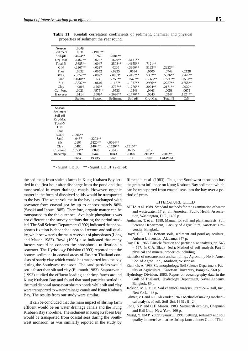
85Impact of intensive shrimp farm effluent
Table 11. Kendall correlation coefficients of sediment, chemical and physicalproperties of sediment the year round.
SeasonSedimentSoil-pHOrg-MatTotal-N
C:NPhos
BOD5SandSiltClay
Cul-PondHarvestp
.0049
.0631.4674**-.4467**-.3685**-.3367**
.0632-.3352**.3618**-.3537**-.0816.0021.0114
-.1906**.0262-.0267-.0047-.0327-.0052-.0922.0630-.0646.1269*
-.4975**.1089*
.2084**-.1679**-.2508**
.0220-.0235-.0963*.2259**-.1167*-.2797**-.0533
-.2690**
-.5131**-.4155**-.3800**
.0534-.4152**.2545**-.1937**-.1776**-.0548
-.1770**
.7121**
.5182**.0505
.5302**-.3565**.2956**.2094**.0463.0843
.2232**.0743
.5336**-.3598**.2757**.2171**.0058.0247
-.2128.2764**-.1531**.1658**.0932*.0675
.1324**Station Season Sediment Soil pH Org-Mat Total-N C:N
SeasonSedimentSoil-pHOrg-MatTotal-N
C:NPhos
BOD5SandSiltClay
Cul-PondHarvestp
.1094**-.0467.0167.0480
.1353**.0194
-.2293**.1820**.1404**.0828.0440
-.6564**-.1520**-.0840.0043
-.1910**.0715-.0807
.0012.2103** .2900**
Phos BOD5 Sand Silt Clay Cul-Pond
* - Signif. LE .05 ** - Signif. LE .01 (2-tailed)
the sediment from shrimp farms in Kung Krabaen Bay set-tled in the first hour after discharge from the pond and thatmost settled in water drainage canals. However, organicmatter in the form of dissolved solids would be transportedto the bay. The water volume in the bay is exchanged withseawater from coastal sea by up to approximately 86%(Sasaki and Inoue 1985). Therefore, organic matter can betransported to the the outer sea. Available phosphorus wasnot different at the survey stations during the period stud-ied. The Soil Science Department (1992) indicated that phos-phorus fixation is depended upon soil texture and soil qual-ity, while seawater is the main reservoir of phosphorus (Longand Mason 1983). Boyd (1995) also indicated that manyfactors would be concern the phosphorus utilization inseawater. The Hydrology Division (1993) reported that thebottom sediment in coastal areas of Eastern Thailand con-sists of sandy clay which would be transported into the bayduring the Southwest monsoon. The sand particles wouldsettle faster than silt and clay (Eiumnoh 1983). Stapornvanit(1993) studied the effluent loading at shrimp farms aroundKung Krabaen Bay and found that sand particles settled inthe mud disposal areas near shrimp ponds while silt and claywere transported to water drainage canals and Kung KrabaenBay. The results from our study were similar.
It can be concluded that the main impact of shrimp farmeffluent would be on water drainage canals and the KungKrabaen Bay shoreline. The sediment in Kung Krabaen Baywould be transported from coastal seas during the South-west monsoon, as was similarly repoted in the study by
Rimchala et al. (1983). Thus, the Southwest monsoon hasthe greatest influence on Kung Krabaen Bay sediment whichcan be transported from coastal seas into the bay over a pe-riod of years.
LITERATURE CITEDAPHA et al. 1989. Standard methods for the examination of water
and wastewater. 17 et al., American Public Health Associa-tion, Washington, D.C., 1430 p.
Anthanan, T. et al. 1989. Manual for soil and plant analysis, SoilScience Department, Faculty of Agriculture, Kasetsart Uni-versity, Bangkok.
Boyd, C.E. 1995 Bottom soils, sediment and pond aquaculture,Auburn University, Alabama. 347 p.
Day, P.R. 1965. Particle fraction and particle size analysis, pp. 545– 567. In C.A. Black (ed.); Method of soil analysis Part I,physical and mineral propertics including
statistics of measurement and sampling., Agronomy No 9, Amer.Soc. of Agron. Inc., Madison, Wisconsin.
Eiunnoh, A. 1983. Geomorphology, Soil Science Department, Fac-ulty of Agriculture, Kasetsart University, Bangkok, 560 p.
Hydrology Division. 1993. Report on oceanography data in theGulf of Thailand, Hydrology Department, Naval Acdemy,Bangkok, 89 p.
Jackson, M.L. 1958. Soil chemical analysis, Prentice – Hall, Inc.,NewYork, 498 p.
Kilmer, V.J. and L.T. Alexander. 1949. Method of making mechani-cal analysis of soil, Soil Sci. 1949 : 8 –24.
Long, S.P. and C.F. Mason. 1983. Saltmarsh ecology, Chapmanand Hall Ltd., New York. 160 p.
Musig, Y. and P. Yuthrutyanukul. 1991. Settling, sediment and soilquality in intensive marine shrimp farm at inner Gulf of Thai-

86 Tookwinas et al.land, Fisheries Science Journal 1 (1) : 47 – 55.
Praphruttham, P. 1985 Soil chemistry, Soil Science Department,Faculty of Agriculture, Kasetsart University, Bangkok, 502 p.
Raine, R.M. 1992. Land use patterns, environmentally sensitiveareas and changes in land use over time in the Coastal Zone ofChanthaburi Province, Thailand, 44 p.
Rimchala, P. et al. 1993. Report on soil survery in Kung KrabaenBay Royal Development and Study Center, Soil Survery Di-vision, Land Development Department, Ministry of Agricul-ture and Cooperative, Bangkok, 143 p.
Sasaki, T. and H. Inoue. 1985. Studies on fundamental environ-ments in the Kung Krabaen Bay, pp. 77 – 98. In Mangroveestuarine ecology in Thailand, Thai – Japanese CooperativeResearch Project on Mangrove Productivity and Development.
Soil Science Department. 1992. Introduction to soil science, Fac-ulty of Agriculture Kasetsart University, Bangkok, 730 p.
Stapornvanit, K. 1993. The environmental impact of shrimp farmeffluent, M.Sc. Thesis, AIT, Bangkok.
Tookwinas, S. and P. Neumhom 1995. Experiment on coagulationof an intensive marine shrimp farm effluent, International Semi-nar on Marine Fisheries Environment, DOF/JICA, 9 –10 March1995. Rayong, Thailand.
Wattanachayakul, S. 1989 Fundamental of statistical analysis,Prakaiyphrit Printing Co. Ltd., Bangkok, 297 p.

Fast AW, Menasveta P (1998) Some recent innovations in marine shrimp pond recycling systems. InFlegel TW (ed) Advances in shrimp biotechnology. National Center for Genetic Engineering andBiotechnology, Bangkok.
e-mail: [email protected]; [email protected]
Some Recent Innovations inMarine Shrimp Pond Recycling Systems
1Arlo W. Fast, 2Piamsak Menasveta
1Hawaii Institute of Marine Biology, University of Hawaii at ManoaP.O. Box 1346, Kaneohe, Hawaii 96744, U.S.A.
2Aquatic Resources Research Institute and Department of Marine ScienceChulalongkorn University, Bangkok 10330, Thailand
ABSTRACT: Abundant shrimp seed availability beginning during the 1970’s has resulted in numerous innova-tions and intensification with marine shrimp growout. Pond water recycling and recirculation systems are onesuch innovation which holds much promise for profitable and sustainable shrimp culture. These systems arebased on earlier work with smaller, conventional water treatment systems. Pond water recycling systems canresult in better disease control, less water use, higher water quality, crop diversification, and improved effluentquality. We review several types of existing and proposed pond water recycling designs for both large and smallshrimp farm applications.
KEY WORDS: shrimp, grow-out ponds, water recycling
INTRODUCTIONBefore abundant marine shrimp seed availability from
hatcheries and from wild captured seed, shrimp pond growouttechniques changed very little over the centuries. Shrimpwere mostly cultured coincidentally with fish and other crus-taceans in large, extensive pond systems characterized bylow productivity (Fast 1991, 1992a). These primitive sys-tems were basically catch, hold and harvest systems withlittle or no energy, feed, or material inputs. Shrimp yieldswere perhaps a few hundred kg/ha/yr. Growout was low-tech as judged by any standard.
After breakthroughs in large-scale, shrimp larviculturetechniques during the 1970’s, all aspects of shrimp cultureunderwent rapid and innovative changes. These changeswere motivated by abundant availability of shrimp seed forpond culture, limited amounts of captured oceanic shrimp,and high profits from pond cultured shrimp. These condi-tions resulted in numerous growout innovations. Pond yieldswith the more intensive shrimp culture systems increased to>10,000 kg/ha/yr.
Fast (1992b,c,d) described many innovations in pondgrowout techniques that occurred during the 1980’s and ear-lier. In our present paper, we describe some innovationswhich have occurred since then. Specifically, we reviewshrimp pond growout systems which use pond water recy-cling and recirculation.
POND WATER RECYCLING ANDRECIRCULALTION SYSTEMS
Intensive monoculture of marine shrimp is potentiallyunstable and risky. It requires large applications of organic
feed and mechanical energy per unit water volume. Theseapplications focus productivity from much larger land andoceanic areas within a smaller area of shrimp growout pond(Folke & Kautsky 1992). The pond becomes the “tip of thefunnel.” As a result, large amounts of uneaten feed, fecesand metabolic wastes accumulate in pond waters and pondsoils. These wastes are degraded through microbial and otherdecomposition processes to produce among other things;ammonia, nitrite, nitrate and phosphate. These mineralizednutrients stimulate algal growth and lead to dense blooms inthe pond. In addition to toxicity from some of these degra-dation products, collapse of algal populations can also causeshrimp stress and mortality through disease, oxygen deple-tion and increased metabolite toxicity. A conventional solu-tion to these problems has been increased water exchange.Excess metabolites and algae are thus removed from the pondand replaced with water of lower metabolite and algal con-centration and greater oxygen content. This water exchangeor flushing solution is, however, not without potential perilsof its own. In many cases, source waters for flushing con-tain disease organisms which infect the shrimp crop and causemassive mortalities. Industrial, domestic and agriculturalpollution of source waters can likewise cause massive shrimpmortalities. In addition, source waters often contain highconcentrations of suspended sediments which settle in thepond and cause shoaling of water depths that requires timeconsuming removal and disposal between crops. An alter-native to high rates of water exchange and flushing is waterrecycling and recirculation. This allows water to be reusedmany times and reconditioned between each use. This hasmany advantages which will be reviewed herein.
Aquatic animal culture systems which use waterrecirculation and treatment have been used for many years

88 Fast & Menasveta
(Allen & Kinney 1981; Timmons & Losordo 1994). Thesewater treatment methods have been used even longer fordomestic and industrial wastewater treatment. Until recently,aquaculture recirculation systems were used mostly for ex-perimental culture applications involving relatively smallwater volumes, or for relatively small commercial venturesinvolving high value crops such as ornamental fish (Anony-mous 1987; Adey & Loveland 1991; Moe 1992), fish andcrustacean seed for conventional growout or stock enhance-ment (Menasveta et al. 1989, 1991, Colt & White 1991, Wang1993, Tseng et al. 1998), or for research (Yang et al. 1989).These conventional aquaculture recirculation applicationstypically range from small aquarium (~200 l), to perhaps100 m3 culture tanks, silos or raceways (Table 1).
System components of conventional water recirculationsystems generally include provisions for: containment of theprimary crop; solids removal; nitrification; and means ofwater recirculation and aeration (Table 1). Additional, op-tional components include: temperature control;denitrification; foam fractionation; and biocidal treatmentssuch as ozonation and/or UV irradiation (Losordo 1998a,b).Daily water exchange rates, or flow-through rates typicallyrange from near zero with denitrification, to 10% or morewithout denitrification. Application of these conventionalrecirculation systems to marine shrimp culture includebroodstock maturation (Menasveta et al. 1991), larviculture(Huguenin & Colt 1989) and nursery operations (Sturmer etal. 1992).
During the past 15 years, recirculation systems have beenapplied commercially to marine shrimp growout on muchlarger scales than previously envisioned. Reasons for theseapplications relate mostly to: control of diseases from sourcewaters; control of organisms in source waters; control of waterquality problems with source waters; scarcity of high qual-ity source waters; improved growth performance due togreater control over water quality; and concerns about envi-ronmental degradation caused by shrimp pond effluents.
Serious shrimp diseases can come from source watersand/or organisms in source waters. Therefore, by limitinguse of source waters, by pre-treating these waters before useand by reconditioning effluent for recycle, chances of dis-ease are greatly reduced. These precautions combined withuse of specific pathogen free (SPF) or specific pathogen re-sistant (SPR) shrimp can greatly reduce disease incidenceduring growout. Recirculating pond systems can also re-duce sedimentation within ponds, and improve dischargewater quality to receiving waters.
A wide variety of shrimp pond recirculation schemes havebeen proposed and/or used. These share many characteris-tics with conventional recirculation culture systems, but also
differ in some respects (Table 1). In addition to sizedifferences, conventional and pond recirculation sys-tems perhaps differ most markedly with respect to pho-tosynthesis. With conventional systems, photosynthe-sis is typically absent. Aeration and ammonia con-version with conventional systems are achieved by me-chanical means and by bacteria. With pondrecirculation systems, photosynthesis plays a domi-nant role in oxygenation and ammonia conversion. Inaddition, energy and water exchange requirements aretypically much lower with pond recirculation systems.
Although no two pond recirculation systems de-scribed in the literature to date are identical in all re-spects, they share the same objectives of reducingwater exchange, reducing settlable solids, and reduc-ing metabolite toxicity (especially nitrogen com-pounds). Satisfactory achievement of these objectivesshould result in more successful shrimp culture withless environmental degradation.
Shigueno (1985) described one of the earliest wa-ter recycle systems for ultra-intensive culture of thekaruma prawn (P. japonicus). This system was devel-
oped during the 1970’s and used relatively small (~0.2 ha),outdoor tanks. These circular concrete tanks had “false”bottoms with a sand layer through which water percolatedand was collected for recirculation or discharge. The sandbottom functioned as a nitrification site (biofilter) and a placefor the shrimp to burrow. Water flow-through was very highat 300% to 400% per day. Because of high capital and oper-ating costs, these systems were unprofitable despite very highyields (>35,000 kg/ha/yr) and high prices for live shrimp(U.S.$80/kg) for the sushi market (Shigueno 1985. Fast1992d).
Another ultra-intensive culture system using partialrecirculation was developed in the U.S. and Mexico aboutthe same time as the Shigueno round culture tanks in Japan.This was the covered raceway system developed by the Uni-versity of Arizona and others (Colvin 1985, Liao 1986, Fast1992d). This system used plastic lined raceways with aera-tion and partial water recirculation. An inflated plastic coverhelped maintain optimal water temperatures. Yields of70,000 kg/ha/crop with Pacific blue shrimp (P. stylirostris)were occasionally realized, but the system was plagued bydisease problems of unknown origin and ultimately provedunprofitable.
Table 1. Comparisons between typical characteristicsof conventional recirculation aquaculturesystems, and commercial scale, marineshrimp recirculation pond culture systems.
Characteristic ConventionalRecirculationSystems
Shrimp PondRecirculationSystems
Size small (aquaria, tanks,silos, raceways)
large (ponds >1 ha)
System componentsCulture containment tanks pondsSolids removal settling; pressurized
mechanical filtrationsettling
Aeration mechanical photosynthesis andmechanical
Nitrification(NH3→NO3)
“biofilters” photosynthesis andpond surfaces
Denitrification (NO3→N2) requires anaerobicsub-component
pond mud
Ozonation/UV possible not likelyEnergy I/P intensive less intensiveSolar I/P none to small large and importantWater Replacement
(% daily flow-through)0 to 10% 0 to <5%

89Marine shrimp pond recycling systems
Photosynthetic activities were minimal or virtually non-existent with both of the above partial recirculation sys-tems, and they were operated primarily as flow-through sys-tems with only some recirculation.
More recently, Choroen Pakphand Group (CP) operateda recirculation pond system at their R&D center, MaeklongArea, Thailand (Anonymous 1996a). The system consistedof ten 0.5 ha shrimp culture ponds, two reservoir ponds, andfour water treatment ponds (Fig. 1). Source water from apublic canal was pumped into Reservoir Pond 1 where sol-ids settled. Water then flowed into Reservoir Pond 2 whichsupplied water to shrimp growout ponds, which in turn dis-charged into a drainage system. Water then either went todrain or was pumped into water treatment ponds. The firstversion of this system included an aeration pond (A1) fol-lowed by two solids removal ponds (A2, A3) with greenmussels (Mytilus smaragdinus) or oysters (Crassostrea sp.),and lastly a pond (A4) with sea weeds (Graciliaria sp.,Polycavernosa sp.) for nutrient stripping. Reclaimed waterthen flowed into Reservoir Pond 2 and returned to the shrimpgrowout ponds. This water treatment process reduced sus-pended organic solids by 30%, ammonia by 90% and ni-trites by 60%. CP later modified this recirculation system asshown in Figure 1 (Anonymous 1996b). The revised con-
figuration included green mussels on stakes in the drainagecanal, two sedimentation ponds (A2, A3) following the aera-tion pond (A1), and culture of sea bass (Lates calcarifer)stocked at 2.6 pcs/m2 in the final treatment pond (A4). Thisnew treatment system resulted in greater dissolved phospho-rus concentrations in pond waters and more stable algalblooms. Stable algal blooms are important since healthy,rapidly growing algae absorb NH
4 and CO
2, and create more
desirable water quality. Without adequate phosphorus andwith senescent algae, algal populations often crash resultingin NH
4 spikes, poor water quality and shrimp mortalities.
Using this recirculation system and P. monodon stocked atdensities of 30 PL/m2, yields averaged 8,267 kg/ha/crop af-ter 125 to 130 days culture. At the same time, sea bass yieldswere 6,288 kg/ha after 100 days culture.
In Indonesia, shrimp pond recirculation systems were de-veloped as a result of poor source water quality and result-ant negative impacts on shrimp yields. A typical Indonesianrecirculation system consists of 50% shrimp culture and 50%water reclamation (Fig. 2; Anonymous 1996c). Shrimp pondeffluent first flows through a sedimentation pond, followedby a fish/bivalve pond, and lastly through an aeration pondbefore return to the shrimp growout ponds. Milkfish (Chanoschanos), mullet (Mugil spp.) and/or green mussels or oys-
Figure 1. Recirculation shrimp pond system operated by Charoen Pokphand Group (CP) in Thailand (Anonymous 1996b).Source waters enter Reservoir Pond 1 for sedimentation and conditioning before flowing into Reservoir Pond 2, andthen into shrimp growout ponds (A5-A8). Effluent from shrimp growout ponds goes into the Drainage Canal fromwhence some or all of it is circulated through four water treatment ponds (A1-A4) for aeration, solids removal andreconditioning. Green mussels are placed on stakes in the Drainage Canal. Sea bass are reared in pond A4 to helpincrease dissolved phosphorus and stabilize phytoplankton populations. (Note that pump water volumes should be inm3 instead of m2 as shown in the diagram).

90 Fast & Menasveta
ters are commonly used. The system can be operated closed,or partly-closed. Using this system, average shrimp yieldsof 8,600 kg/ha/crop were reported after about 145 days cul-ture when stocked at 50 PL/m2. Milkfish stocking densitieswere 1,000 pcs/ha.
Menasveta and Jarayabhand (1995) have proposed anin-pond treatment system (Fig. 3). This system consists of afloating raft from which oysters are suspended on strings ortrays. Oysters would remove excess algae and other sus-pended solids, and provide an additional nitrificationsubstrate. Oysters are preferred to green mussels because oftheir greater range of salinity tolerance and higher market
value. Four paddlewheel aerators would move water in acircular motion, thus concentrating wastes in the center ofthe pond.
Another proposed system would consist of a small inten-sive shrimp culture pond nestled within a much larger exten-sive culture pond (Fig. 4; A.W. Fast, unpublished). Waterwould enter the intensive culture pond using low-energy watermovers (Rogers & Fast 1988), or more commonly used push-
Figure 2. Typical recirculatingshrimp farm system in Indone-sia (Anonymous 1996c).Shrimp pond effluent flows toa sedimentation pond, followedby treatment with fish and/orbivalves. Milkfish, mullet,green mussles and/or oystersare commonly used. Waters arethen aerated before returning tothe shrimp growout ponds.
Figure 3. In-pond treatment system using an oyster raft pro-posed by Menasveta and Jarayabhand (1995). Oystersare preferred to green mussles because of their greatersalinity tolerances and higher market value. Paddle wheelaerators would create circular water motions and con-centrate wastes in the pond center.
Figure 4. Proposed pond-in-pond culture system whereshrimp are cultured intensively in a small pond, whilesource water and effluents recycle with a much largerextensive pond. The extensive pond serves as a wastetreatment component and provides secondary cash crops.This system could easily be retrofited to existing exten-sive ponds withour creating new pond area.

91Marine shrimp pond recycling systems
pumps. The intensive pond would be deeper and have me-chanical aeration such as paddlewheels. Effluent from theintensive pond would return to the extensive pond wheresuspended solids would settle and nutrients would stimulateprimary and secondary productivity for the secondary crop.Shrimp would be cultured at high densities in the intensivepond, while milkfish, mullet, tilapia, bivalves and/or shrimpcould be cultured in the extensive pond at lower densities.A major advantage of this pond-in-pond system is that itcould be retrofitted to existing extensive ponds without needto create more new pond areas from land now in other use.
Recently, Brune (1998) and Drapcho (1993) describedtheir work at Clemson University with an algal based, parti-tioned aquaculture system (PAS) for freshwater channel cat-fish (Ictalurus punctatus). The PAS has been under devel-opment since 1988 and has produced catfish yields of 15,000to 22,000 kg/ha/yr, compared with about 5,500 kg/ha/yr withconventional, intensive culture of catfish. The PAS consistsof a fish culture raceway component which is designed tohave uniform, well mixed flow with high phytoplanktongrowth (Fig. 5). This provides high dissolved oxygen con-centrations while at the same time reduced ammonia. Fol-lowing the raceway is a settling area where wastes settle forremoval from the system. Tilapia (Oreochromis niloticus)
and/or other filter feeding fish and plants can also be cul-tured in this settling areas to further utilize algal productionand wastes, and to provide additional cash crops. Highestyields were realized when catfish were co-cultured withtilapia. Oxygen transfer and other control functions are ac-complished by computer control of the paddlewheel circu-lator. Advantages of the PAS include: low water use and nowater discharge; good disease control; good predator con-trol; high crop yields; simplified grading of crop by size;easy crop harvest; and multi-cropping of more than one spe-cies. The PAS system has potential application to marineshrimp culture, but has not yet been used for this purpose.
Chien & Liao (1995) describe a somewhat different con-ceptual farm. Their proposed system includes both watertreatment components (bivalves, seaweed, marsh, mangrovesand settling), as well as waste water recycling. The reusesystem includes first using reservoir or treated waters forintensive shrimp culture, followed by use in less intensiveshrimp culture ponds before treatment and recycling. Theyalso allow for a wide range of options, which are probablybeyond the scope of our present ability to operate optimally.
Figure 5. Schematic or Partitioned Aquaculture System (PAS) for pond culture under development at Clemson Univer-sity, South Carolina, U.S.A. The primary crop is cultured in the raceway section, while phytoplankton and other solidsare removed in the settling area where water velocities are less. Phytoplankton remove metabolic wastes and oxygen-ate the water. The system is computer controlled to maintain adequate oxygen concentrations and optimal algal growth.Figure courtesy of D.E. Brune (1998).

92 Fast & Menasveta
CONCLUSIONSClearly, shrimp pond water recycling and recirculation
are being used, and will continue to be used on a large scalefor all the reasons identified above. However, it is also clearfrom the review of existing literature that while operatingprincipals are well understood, there is little documentationof operating performance. More importantly perhaps, de-sign criteria needed to size a given pond recycling systemare largely lacking. This contrasts with conventionalrecirculation systems where these parameters are much bet-ter understood and where design criteria are described(Timmons & Losordo 1995). The reasons for this lack ofdetail for pond recirculation systems relates partly to uncer-tainties with performance predictability and partly to theirrelative newness. We anticipate much greater understand-ing of these pond systems over the next 10 years or so. Whileshrimp pond water recirculation and recycling will increase,it is not practical for all shrimp farms. A 1994 farm surveyby the CP Shrimp Feed Marketing Department revealed that70% of all intensive farms in Thailand were <1.6 ha (Anony-mous 1996d). Most of these farms do not have adequateland area or perhaps the farmers do not have adequate skillsto achieve meaningful water recycling. With these farms,some in-pond treatment methods seem more appropriate.
Acknowledgements. This study is financially supported byThailand Research Fund (TRF).
LITERATURE CITEDAdey WH, Loveland K (1991) Dynamic aquaria. Academic Press,
San Diego, CA, 643 p.Allen LJ, Kinney EC (1981) Proceedings of the bio-engineering
symposium for fish culture. American Fish. Soc., Bethesda,MD, 307 p.
Anonymous (1987) Practical koi keeping. Koi USA, Midway City,CA, 191 p.
Anonymous (1996a) The recirculation system, new technology forshrimp culture. In: Lin, CK, Nash, GL (compilers), AsianShrimp News Collected Volume 1989-1995. Asian ShrimpCulture Council, C.P. Tower, 313 Silom Rd., Bangkok 10500,p 44-46
Anonymous (1996b) The recycle system for shrimp culture. In:Ibid. p 52-54
Anonymous (1996c) Successful Indonesian experience in shrimp-fish integration in recycle system. In: Ibid. p 56-58
Anonymous (1996d) Update on shrimp farm size, distribution andfeed performance in Thailand. In: Ibid. p 91-92
Brune DE (1998) Recirculation in photosynthetic aquaculture sys-tems. Aquaculture Magazine 24(3):63-71
Chien YH, Liao IC (1995) Integrated approach to prawn growoutsystem design. In: Browdy CL, Hopkins JS (eds.), Swimmingthrough troubled waters. Proceed. Special Session on ShrimpFarming, Aquaculture ’95. World Aquacult. Soc., Baton Rouge,LA, p 167-182
Colt J, White RJ (1991) Fisheries bioengineering symposium.American Fish. Soc., Bethesda, MA
Colvin LB (1985) Intensive growout systems for shrimp. In: Cham-berlain GW, Halby MG, Midget RJ (eds.), Texas Shrimp Farm-ing Manual. Texas Agricult. Ext. Service, Texas A&M Univ.,College Station, TX, IV-1-IV-14 p
Drapcho CM (1993) Modeling of algal productivity and diel oxy-gen profiles in the partitioned aquaculture system. Ph.D. the-
sis, Dept. of Agri. & Biol. Engr. Dept., Clemson Univ.,Clemson, SC.
Fast AW (1991) Marine shrimp pond growout conditions and strat-egies: a review and prognosis. Reviews in Aquatic Sciences3:357-399
Fast AW (1992a) Penaeid growout systems: an overview. In: FastAW, Lester LJ (eds). Marine Shrimp Culture: Principles andPractices. Elsevier Science Publishers, Amsterdam. 347-356p
Fast AW (1992b) Penaeid semi-intensive growout systems. In:Ibid. 371-382 p
Fast AW (1992c) Penaeid intensive growout systems. In: Ibid.383-392 p
Fast AW (1992d) Penaeid ultra-intensive growout systems. In:Ibid. 393-402 p
Folke C, Kautsky N (1992) Ecological economic principles foraquaculture development. In: Cowey CB, Cho CY (eds), Feed-ing fish in our water: nutritional strategies in management ofaquaculture waste. Univ. of Guelph, Ontario, Canada
Huguenin J, Colt J (1989) Design and operation guide foraquaculture seawater systems. Elsevier, Amsterdam.
Liao IC (1986) General introduction to the prawn system in Tai-wan. Aquacultural Engineering 5:219-233.
Losordo TM (1998a) Recirculating aquaculture production sys-tems: the status and future. Aquaculture Magazine (Jan./Feb.):38-45.
Losordo TM (1998b) Recirculating production systems: the statusand future, part II. Aquaculture Magazine (March/April):45-53.
Menasveta P, Aranyakanonda P, Rungsupa S, Moree N (1991)Maturation and larviculture of penaeid shrimp in closedrecirculating seawater system. Aquacultural Engineering 8:357-368
Menasveta P, Fast AW, Piyatiratitivorakul S, Rungsupa S (1991)An improved, closed seawater recirculation maturation sys-tem for giant tiger prawn (Penaeus monodon Fabricius).Aquacultural Engineering 10:173-181.
Menasveta P, Jarayabhand P (1995) Environmental planning inthe coastal area for sustainable shrimp culture operation. ThaiJour. Aquatic Sci. 2(1):48-58.
Moe MA, Jr. (1992) The marine aquarium reference, systems andinvertebrates. Green Turtle Publications, Plantation, FL, 512 p
Rogers GL, Fast AW (1988) Potential benefits of low energy watercirculation in Hawaiian prawn ponds. J. Aquacultural Engi-neering 7:155-165.
Shigueno K (1985) Intensive culture and feed development inPenaeus japonicus. In: Taki Y, Primavera JH, Llobrera JA (eds),Proceed. First Internat. Confer. on the Culture of PenaeidPrawns/Shrimp. Southeast Asian Fish. Devel. Center, IloiloCity, Philippines, p 115-122
Sturmer LN, Samocha TM, Lawrence AL (1992) Intensificationof penaeid nursery systems. In: Fast AW, Lester LJ (eds).Marine Shrimp Culture: Principles and Practices. ElsevierScience Publishers, Amsterdam, p 321-344
Timmons MB, Losordo TM (1994) Aquaculture water reuse sys-tems: engineering design and management. Elsevier SciencePublishers, Amsterdam. 346 p
Tseng KF, Su HM, Su MS (1998) Culture of Penaeus monodon ina recirculating system. Aquacultural Engineering 17:138-147.
Wang JK (1993) Techniques for modern aquaculture. AmericanSoc. Agricultural Engineers. Niles, MI, 604 p
Yang WT, Halon RT, Lee PG, Turk PE (1989) Design and functionof closed seawater systems for culturing loliginid squid.Aquacultural Engineering 8:47-65.

Matsumura M, Migo VP, Balobalo D, Young HK, Albaladejo JD (1998) Preservation of water qual-ity in shrimp ponds by ozone. In Flegel TW (ed) Advances in shrimp biotechnology. National Centerfor Genetic Engineering and Biotechnology, Bangkok.
Preservation of Water Quality in Shrimp Ponds by Ozone1M. Matsumura, 2V.P. Migo, 3D. Balobalo, 3H.K. Young, 4J.D. Albaladejo
1Institute of Applied Biochemistry, University of Tsukuba, Tsukuba, Japan2National Institute of Molecular Biology and Biotechnology, University of the Philippines Los Banos
Laguna, Philippines3Aqua-Ecotechnology Research Inc., Caloocan, Philippines
4Bureau of Fisheries and Aquatic Resources, Quezon City, Philippines
ABSTRACT: A novel application system of ozone for disinfection and water quality improvement in shrimpgrow-out ponds was studied in this work. In the system a water-jet circulator was employed as an ozone distribu-tor. This water-jet circulator enhanced the liquid mixing in shrimp ponds and diminished the vertical distribu-tion of DO, which was also effective for sediment improvement. An appropriate ozone dosing rate for disinfec-tion was determined through actual shrimp cultivation in polluted ponds with pathogenic Vibrio.
KEY WORDS : prawn culture, disinfection ozone, water-jet circulator
INTRODUCTIONIn 1991, China attained the world record of 210,000 tons
prawn production. However, the following year productiondropped to 140,000 and finally to 30,000 tons in 1993. Thisvirtual collapse of the prawn farming industry was causedby the prevalence of white spot syndrome virus (WSSV).Almost all prawn farms in the Asia-Pacific region have simi-lar disease problems caused by pathogenic bacteria and vi-ruses. These diseases are the result of the interaction of host(shrimp), pathogens and environment parameters like waterand sediment quality. Therefore, to arrest the problems com-pletely, we should approach prawn diseases in an integratedmanner and develop the technologies for rapid detection ofpathogens, disinfection, management of sediment and wa-ter, immunostimulation and also selective breeding for dis-ease resistance. However, disinfection is now the most ur-gent requirement. Formalin and chlorine have been employedas disinfectants for pathogenic bacteria and viruses. Theemergence of these diseases is associated with aquatic envi-ronment degradation which is pronounced in intensive cul-ture ponds. Therefore, we have to focus our attention notonly on disinfection but also on the improvement of waterand sediment quality.
Ozone is one of the most powerful oxidizers known, andit has been utilized for disinfection, controlling taste and odorof water and removing color. Unlike chlorine and chlorinecompounds, it produces no harmful byproducts. Once it hascompleted its task as a purifier and sanitizer, residual ozonereverts to natural oxygen which helps to aerate the water andimprove the aquatic environment. Ozone is also very effec-tive in breaking down ammonia and ammonia compounds,as well as organic matter refractory to biodegradation. There-fore, ozone appears to have prospects for use in water qual-ity improvement of aquaculture ponds such as those usedfor prawn cultivation, where significant damage occurs dueto bacterial and viral diseases. The application of ozone in
intensive prawn farms is still limited in spite of its multiplefunctions as a disinfectant and oxidizer of toxic ammoniaand nitrite. This is probably due to the lack of suitable in situsystems for ozone application in large water bodies.
The present work aimed to study the characteristics ofozone reaction with bromide and chloride ion which are in-volved in seawater, and to evaluate the performance of anovel ozone distribution system using an energy-efficientwater-jet circulator.
BACKGROUND INFORMATIONOxidants formed by ozone in sea water
When ozone is supplied into freshwater, decompositionof ozone is initiated by the formation of hydroxyl radical.Then it causes several chain reactions to form different radi-cals and oxidants like hydroperoxyl radicals and hydrogenperoxide, respectively. The hydroxyl radical is, however,mainly responsible for the high oxidative potential of ozonein freshwater. In the case of seawater containing bromideion, the reaction is completely different from that in fresh-water. This is shown by the following equations together withtheir reaction constants:
O3 + Br- → O
2 + OBr- (k
1 = 160 M-1s-1) (1)
O3 + OBr- → 2O
2 + Br- (k
2 = 330 M-1s-1) (2)
2O3 + OBr- → 2O
2 + BrO
3- (k
3 = 100 M-1s-1) (3)
H+ + OBr- ↔ HBrO ( pKa = 8.68) (4)
As we can see from the reaction constant k1, ozone very
quickly produces hypobromite ion, and the hypobromite ioncan either be reduced back to bromide ion or oxidized tobromate ion as expressed by Eqs. (2) and (3), respectively.In the presence of bromide ion, ozone is catalytically de-stroyed, and dissolved free ozone is negligibly small. Ozone

94 Matsumura et al.
also reacts with chloride. Analogous reactions to Eqs. (1) -(3), involving chlorine in place of bromine, have been pro-posed with the following rate constants; k
1 = 0.003 M-1s-1, k
2
= 110 M-1s-1 and k3 = 30 M-1s-1. Because the ratio of k
1 values
for bromide ion to chloride ion is at least 5.3x10⁴, bromideion reacts with ozone preferentially to chloride ion. Oxidativeproducts of bromide ion are expected to dominate those ofchloride ion even in seawater, where the molar ratio of Br-/Cl- is 1.5x10⁻³. Even if hypochlorite ion forms, it reactsquickly with bromide ion to form hypobromite ion as shownby Eq. (5). Therefore, the oxidants produced from bromideion are mainly responsible for the high oxidative potentialof ozone in seawater.
OCl- + Br- → OBr- + Cl- (k4 = 6.77x10³ M-1s-1) (5)
Decomposition of harmful inorganic com-pounds
Shrimp are very sensitive to ammonia, nitrite and hydro-gen sulfide, and their concentrations should be held lowerthan 0.1 ppm, 0.1 ppm and 0.033 ppm for NH
3-N, NO
2-N
and H2S, respectively. Ozone can easily convert these toxic
inorganic compounds to harmless matter. The hypobromousacid formed by Eq. (4) reacts with ammonia to convert it tonitrogen gas and nitrate through the following reactions:
HBrO + NH3 → NH
2Br + H
2O (k
5 = 8x10⁷M-1s-1) (6)
HBrO + NH2Br → NHBr
2 + H
2O (7)
HBrO + NHBr2 → NBr
3 + H
2O (8)
NHBr2 + NBr
3 + 2H
2O → N
2 + 3Br- + 3H+ + 2HBrO (9)
NH2Br + 3O
3 → NO
3- + Br- + 2H+ + 3O
2 (k
6 = 40 M-1s-1) (10)
The ratio of nitrate formation to nitrogen gas formationhighly depends on the ratio of Br- to NH
4+-N. Under normal
conditions in a shrimp pond (Br- = 67 ppm, NH4+-N = 2
ppm, pH = 8, temp. = 28 oC), the Br-/NH4+-N ratio is about
34 and almost all ammonia would be converted to nitrogengas as shown in Fig.1. The conversion rate of ammonia tonitrogen gas is very fast, as can be seen from the rate con-stant of Eq. 6. Nitrite and hydrogen sulfide are also easilyconverted to nitrate and sulfate, respectively.
Inactivation of bacterial and viral pathogensWhile the precise mechanisms of oxidant disinfection are
unclear, a variety of mechanisms have been proposed. Cellwalls and membranes are the primary sites attacked by oxi-dants. These primary sites in bacteria are composed of li-popolysaccharide, lipoprotein, peptidoglycan, phospholipidand protein. Generally, these components contain doublebonds of unsaturated fatty acids, aromatic compounds andamino acids with thiol and amine groups which provide po-tential targets for oxidant action. Any alternation in lipidsand membrane proteins would result in structural and func-tional changes in the membrane. In the case of viruses, thecoat protein would be damaged first. This could denature itand destroy it ability to interact with host target moleculesrequired for attachment and cell entry. If oxidants diffusedinto the virus through the damaged coat protein, DNA andRNA could be damaged.
The inactivation of pathogens is affected by both totalresidual oxidant (TRO) concentration C and contact time t.This relationship has been expressed by the Chick-Watsonequation (K = Cn•Et). Figure 2 shows the effect of TROconcentration on inactivation of Vibrio harveyi pathogenicfor shrimp. Almost 1.5 log units and 4 log units of inactiva-tion occurred within a short contact time of 30 sec at an ini-tial oxidation concentration of 1 and 2.3 mg/l, respectively.Based on this experimental result, effective disinfection ofpathogenic bacteria in seawater coud be expected.
Figure 1. Ozonation of ammonium in sea water.
Figure 2. Inactivation of Vibrio harveyi in sea water(•••••=cell concentration; ̈ =Total residual oxidant).

95Ozonation of shrimp ponds
IN SITU APPLICATION OF OZONE INSHRIMP PONDS
Water-jet circulatorThere is a consensus among aquaculturists that water cir-
culation in ponds is beneficial. Water circulation preventsthermal and chemical stratification. This makes the entirepond volume habitable and it eliminates oxygen depletion atthe sediment-water interface. During daylight hours, surfacewater in ponds is often supersaturated with DO produced byphytoplankton, and water at greater depths may have lowDO concentration. The paddle wheel aerator is commonlyemployed in intensive culture ponds, because it blends sur-face water with subsurface water. By mixing pond water, auniform DO profile can be established, and the total DOcontent of a pond can be increased.
Figure 3 shows the 24-hour monitoring of DO at twodifferent sampling points (SP-1 and SP-2) in an intensiveculture pond with a surface area of 4500 m2 and 1.5 m depth.Four paddle wheel aerators were installed in the pond. Onehad 6 blades and the others had 3 blades. They were drivenby 1 horsepower motors. The monitoring was conducted onthe 150th day after stocking. The locations of sampling pointsSP-1 and SP-2 were the mid point between two paddle wheelaerators and the center of pond, respectively. At the sam-pling point SP-1, there was a liquid mixing enough to dimin-ish the vertical distribution of DO between the pond surfaceand the bottom. However, an obvious difference in DO be-tween the surface and the bottom could be seen at samplingpoint SP-2. This was caused by poor liquid mixing. It wasclear that uniform water quality in the pond could not beattained by the paddle wheel aerators. Therefore, we intro-duced a water-jet circulator to enhance liquid mixing.
The construction of the water-jet circulator was basedon a double stage ejector as shown in Fig. 4. It was installedat the bottom of the pond as shown in the system construc-tion. We could also introduce ozone mixed with air into thenacelle of the jet pump and make very fine bubbles which
were easily dispersed in the sea water. When the driving liq-uid was supplied into the jet pump through a narrow slit at avolumetric flow rate of Q
1, the liquid was sucked at a flow
rate of Q2. The driving force for the liquid suction is pro-
vided by the pressure drop of the driving liquid. Then, theliquid is discharged from the nacelle at a flow rate of Q
1+Q
2
. This discharged liquid acts as the driving liquid for thesecond ejector. The final discharged flow rate becomesQ
1+Q
2+Q
3. The design of the ejector has been well estab-
lished, and the energy efficiency of the jet pump ā is ex-pressed by the following equation:
η = [Q1/Q
2][(h
d - h
2)/(h
1 - h
d)] ≡ M·H (11)
where, h1, h
2, h
d are the hydrostatic head at the pump con-
nection points for drive line, suction line and discharge line,
Figure 3. Dissolved oxygen (DO) in a shrimp pondaerated with paddle wheels (∆=Top; ̈ =Middle;•••••=Bottom).
Figure 4. Schematic for construction of the water jet circulator.

96 Matsumura et al.
respectively. For a jet pump with a low discharge pressurebut a high suction flow rate, a low area ratio of about 0.2 isrecommended for the construction of the nozzle. In this case,M becomes 2.2, so the discharged liquid volume becomes3.2 times as much as that of the drive liquid. For the doublejet pump, this becomes 10.2 times.
Once the jet flow is established, the liquid surroundingthe jet stream is entrained into the stream. The volumetricflow rate of entrained liquid Q
E is given by Eq.(12):
QE = [0.203(x/d
o -1)]Q
d (12)
where Qd is the volumetric flow rate discharged from the
water-jet circulator (Q1+Q
2+Q
3), and d
o is the diameter of
open duct shown in Fig. 4. X is the maximum distance fromthe exit edge of the open duct where the jet flow exits. If x/d
o
is 40, QE in Eq.(12) becomes 8.2Q
d, and then Q
E/Q
1 is 83.5.
When a water-jet circulator with a driving liquid volume of0.75 m3/min is installed in a shrimp pond with a liquid vol-ume of 6,000 m3, the pond liquid passes through the circula-tor at a volumetric flow rate of 11,000 m3/d. Therefore, thecirculation time for the pond water becomes 0.54 d. A ratherhuge amount of liquid is entrained into the jet stream at avolumetric flow rate of 90,000 m3/d, and this liquid flowwill enhance the mixing of pond liquid. Figure 5 shows the24- hour monitoring of DO in the same pond shown in Fig.
3. In this case, one water-jet circulator (driving liquid vol-ume: 0.75 m3/min, energy consumption: 2.2 kW) was in-stalled together with two paddle wheel aerators. As you cansee, the liquid mixing was greatly improved, and the verticaldistribution of DO was diminished even at the center of thepond.
Effects of ozone on shrimp growthThe first experiment of shrimp cultivation using ozone
was conducted in a shrimp farm located at Calatagan in theLuzon Island (Philippines) where no problems caused bypathogenic bacteria and viruses occurred. The main purposeof this experiment was to investigate whether ozone had anyadverse effects on shrimp growth. An ultraviolet ozone gen-erator containing two 110 w lamps was used under an airflow rate of 100 l/min. The exit ozone concentration was100 ppm. The ozone dosing rate into the culture pond was0.22 mg-O3/h/m3.
Figure 6 shows the average body weight over time forshrimp cultivated in three different ponds, pond #3 (surfacearea; 4000 m2), #9 (4500 m2) and #15 (4000 m2). One water-jet circulator and two paddle wheels were installed in ponds#3 and #9 using sea water with an average salinity of 32 ppt,but ozone was employed only in pond #3. Pond #15 wasoperated with four conventional paddle wheels, and shrimp
Figure 5. Vertical dissolved oxygen (DO) distribution in a pond using a water-jet circulator (♦♦♦♦♦=SP-1 top;̈ =SP-1 bottom,
∆=SP-2 top; X=SP-2 bottom; ∗∗∗∗∗= SP-3 top; •••••=SP-3 bottom).
Figure 6. Average body weight of shrimp in ponds #3, #9 and #15 (♦♦♦♦♦=pond #3; ̈ =pond #9, ∆=pond #15).

97Ozonation of shrimp ponds
culture was conducted using brackish water with an averagesalinity of 19 ppt. The stocking in these three ponds wasstarted at the same time and their stocking ratio was about45 psc/cm2.
By comparing the results of ponds #3 and #9, it was clearthat an ozone dosing rate of 0.22 mg-O
3/h/m3 did not have
any adverse effects on shrimp growth. However, the salinityhad an obvious effect on shrimp growth. The survivals atharvest were about 60% in ponds #3 and #9, and 70% inpond #15. The concentration of chlorophyll a in ponds #3and #9 was around 40 mg/m3 (transparency: 20 - 30 cm),and in pond #15, 80 mg/m3 (transparency: 15 - 20 cm), andthe dominant algae were diatoms. One of the reasons whichfor the big difference in chlorophyll a concentration was con-sidered to be enhanced liquid mixing in the vertical direc-tion. It reduced the chance for phytoplankton to be exposedto the sun light. The liquid mixing was also effective in re-ducing the settling velocity of small organic particles likedetritus of plankton and residual feed. This enhanced theiroxidation in the water column. The sediment conditions inponds #3 and #9 were completely different from that of pond#15. Ponds #3 and #5 showed no mud accumulation, even atthe center.
Shrimp culture in polluted water with patho-genic Vibrio
In the Philippines, approximately 65% of the country’sprawn farms are situated in the Visayas island group, andparticularly on Negros Occidental. With recent disease out-breaks (luminous vibriosis caused by pathogenic Vibrio spe-cies like V. harveyi) only 30% of the prawn farms are opera-tional, and of these, only 17% experience optimum harvests.Our second cultivation experiment was conducted using twoponds on a farm located in Bacolod City (Negros Occidental).
In this experiment in pond # 21 (pond surface area; 5000m2), two water-jet circulators coupled with an ozone gen-erator with three 110 w UV lamps were installed togetherwith two paddle wheel aerators (2 blades) driven by 1 hpmotors. The control pond #15 (pond surface area 5400 m2)was prepared with two paddle wheel units, each with 2 bladesdriven by a 1 hp motor and two paddle wheel units with 4blades driven by 2 hp motors.
A pre-stocking experiment was done to determine howmany ozone lamps were necessary to reduce or eliminatebacterial pathogens. The total plate count (TPC), total Vi-brio count (TVC), luminous bacterial count (LBC), lumi-nous Vibrio count (LVC) and yellow and green colonies onTCBS agar were monitored. Figure 7 shows the results ofbacterial analysis (TOC and LVC) during the pre-stockingexperiment. The jump in TPC was observed just after theintroduction of water from a reservoir pond. Two UV lampswere turned on from the 15th day to the 34th day, four lampsfrom the 35th to 40th days, and 6 lamps from the 41st to 55th
days. The rate of decrease in TPC increased with increasingnumber of UV lamps, but a negative effect on the planktondensity was observed after 6 UV lamps were turned on. Af-ter this preliminary experiment, one water-jet circulator witha 2 UV lamp ozone generator was installed in the reservoirpond for pre-sterilization of water, and over-dosing of ozoneinto the grow-out pond was limited.
The stocking in control pond #15 and experimental pond#21 was done at the same time on August 3, 1997. The stock-ing ratio was 25 psc/m2. Figure 8 shows the percentage sur-vival of shrimp in these ponds. The percentage survival inthe control pond dropped quickly, and it became less than30% only one month after stocking. In pond #21, a fairlygood survival was maintained for two months, but after that
Figure 7. Effects of ozone dosing rate (i.e., number of generating lamps) on inactivation of bacteria(TPC = total plate count; LVC = luminous Vibrio count).

98 Matsumura et al.
it started to drop. In addition, its luminous Vibrio count (logCFU/ml) was held at a low level of around 1.3, and its DOwas highly improved as shown in Fig. 9. Unfortunately, rain-fall was very limited that year and an optimum salinity ofabout 20 ppt was not maintained. It started to increase twomonths after stocking and finally reached 39 ppt. The alka-linity of the pond water was around 30 ppm CaCO
3, which is
much lower than the ideal level (•„70 ppm). This low alka-linity induced rapid fluctuations in pH as shown in Fig. 10.Dolomite and agri-lime was added to both ponds to correct
the problem, but this did not increase the level of alkalinityto the optimum level. Water change also had a minimal ef-fect, since the source also had low alkalinity. The level ofchlorophyll a in these ponds was 20 - 25 mg/m3, and thetransparency was 50 -55 cm.. These values are far from ideallevels. The dominant alga was Chlorella, and diatoms werepresent at less than 10%. Especially, Skeletonema which se-cretes some anti-microbial compound was very rare. The lowalkalinity and low plankton bloom may have resulted fromthe nature of the sandy pond bottom.
Figure 8. Percent survivl of shrimpin ponds #15 and #21.
Figure 9. Dissolved oxygen (DO) at the centers of ponds #15 and #21over 24 hours.

99Ozonation of shrimp ponds
CONCLUSIONSThe goal of increasing pond productivity was not
achieved in the present experiments. However, we confirmedthat the water-jet circulator was effective for enhancing liq-uid mixing in grow-out ponds and that appropriate applica-tion of ozone is very useful for inactivation of pathogenicbacteria. We also learned many things through these experi-ments. For example, it is clear that the original nature ofpond soil and the algal bloom are very important in
Figure 6. Record of daily pH in pond #15 (♦♦♦♦♦=SP-1; ̈ =SP-2; ∆∆∆∆∆=SP-3).
maintianing stable environmental conditions. The ecosys-tem in the shrimp pond is very complicated. We need to ob-tain much more information, especially about the interac-tions between the algae and bacteria, and we need to de-velop the technology to make beneficial algae dominant. Weshould also develop an energy efficient method to improveDO at night, and work harder to develop effective feed addi-tives to enhance shrimp defense activity against pathogens.

100 Matsumura et al.

Use of Probiotics for Improving Soil and Water Qualityin Aquaculture Ponds*Claude E. Boyd, Amit Gross
Department of Fisheries and Allied AquaculturesAuburn University, Alabama USA 36849
ABSTRACT: A number of biological products including live bacterial inocula, enzyme preparations, and ex-tracts of plant products are being promoted for use as water and soil quality conditioners in aquaculture pondsand particularly in shrimp ponds. Although there is much anecdotal information about these products andconsiderable promotional material from vendors, little independent research has been conducted. Several stud-ies have shown no benefits of bacterial inocula, but one recent study at Auburn University demonstrated highersurvival of fish and thus greater net production in ponds treated frequently with live Bacillus of three speciesthan in control ponds. No improvements in water quality were noted, and the mechanism by which the bacteriaimproved survival is unknown. Laboratory studies of a bacterial inoculum demonstrated a greater rate of nitro-gen loss. Liming of laboratory systems tended to improve bacterial activity. Pond studies also showed thatapplications of an enzyme preparation tended to enhance microbial mineralization of organic matter, but noeffect on net fish production was observed. An extract of grapefruit seed caused greater survival of shrimp andhigher production resulted. Again, the mode of action is unknown because water quality was not measurablyimproved. These few studies suggest that probiotics possibly can be beneficial in aquaculture ponds. Too littleis known about their modes of action, the conditions under which they may be effective, and application ratesand methods for general recommendation of their use. Nevertheless, the products are safe to humans and theenvironment, and their use poses no hazards. Thus, commercial producers are encouraged to conduct trials withthese products, and researchers should conduct experiments with them.
INTRODUCTION
There is considerable interest in use of probiotics to im-prove conditions for production in pond aquaculture. Themost common probiotics are live bacterial inocula that some-times are supplemented by yeast extracts with extracellularenzymes. Some companies sell extracellular enzyme prepa-rations without live bacteria. Extracts of plant products alsoare used as probiotics. The most common are extracts ofcitrus seed and Yucca plants (Yucca schidigera). Claimsabout the potential benefits of probiotics in aquaculture pondsinclude: enhanced decomposition of organic matter; reduc-tion in nitrogen and phosphorus concentrations; better algalgrowth; greater availability of dissolved oxygen; lesscyanobacteria (blue-green algae); control of ammonia, ni-trite, and hydrogen sulfide; lower incidence of disease andgreater survival; greater shrimp and fish production. Ven-dors usually sell probiotics for prophylactic treatments toprotect against disease and to improve environmental con-ditions for culture. Few independent studies have been con-ducted on probiotics in aquaculture ponds. However, ven-dors have conducted trials on aquaculture farms and obtainedtestimonials from farmers. Results of trials and testimonialsare used as the basis for exalted claims of the benefits ofprobiotics. Farmers usually do not have sufficient knowl-edge of microbiology and water quality to evaluate the claims
of vendors. However, they are quite receptive to any newproduct that might enhance water and soil quality, reducethe incidence of disease, or increase production, and there isa brisk trade in probiotics for shrimp and fish farming.
BACTERIAL INOCULA
The argument for using bacterial inocula is thatpopulations of beneficial bacteria in ponds can be increasedby applying live bacteria or their propagules. Two types ofinocula have been used. One type consists of the spores orother resting bodies of one or more species in a mediumdesigned to prevent germination and retard growth. The me-dia may be a liquid or granular material coated with bacte-rial propagules. Relatively small amounts of these inoculaare placed in ponds with the assumption that propagules willquickly multiply and increase the abundance of the inocu-lated species. The second type of inoculum is a source ofpropagules that is inoculated into a nutrient solution and aculture of bacteria is produced for inoculating the pond. Ini-tially, bacterial inocula were applied to ponds at 2- to 4-week intervals. Today, most vendors recommend that in-ocula be applied at more frequent intervals or even daily.Species of Bacillus are most commonly used, but species of
KEYWORDS: probiotics, bacterial inocula, water conditioners
Boyd CE, Gross A (1998) Use of probiotics for improving soil and water quality in aquacultureponds. In Flegel TW (ed) Advances in shrimp biotechnology. National Center for Genetic Engineer-ing and Biotechnology, Bangkok.
*e-mail: [email protected]

102 Boyd & Goss
Nitrobacter, Pseudomonas, Enterobacter, Cellulomonas,Rhodopseudo-monas, and photosynthetic sulfur bacteria havebeen used as bacterial inocula (Boyd 1990). The number ofbacteria introduced per application varies greatly, but theinitial increase in the pond water normally ranges from 102
to 104 colony forming units (CFU) ml-1.
The mode of action of bacterial inocula is claimed to bethe enhancement of natural processes such as organic matterdegradation, nitrification, ammonia removal, denitrification,sulfide oxidation, and degradation of toxic pollutants. Somevendors state that by increasing the abundance of useful bac-teria, competitive exclusion of undesirable species occurs.They even declare that the bacterial inocula can reduce theincidence of fish and shrimp disease in ponds.
Ponds have a natural microbial flora capable of conduct-ing the entire range of organic matter and nutrient transfor-mation that occur in natural ecosystems. Bacterialpopulations respond to the availability of substrate. For ex-ample, if a pond has ammonia in the water, natural nitrifyingbacteria will oxidize the ammonia to nitrate. If ammoniaconcentration increases, the population of nitrifying bacte-ria will respond to the increase in substrate by rapidly in-creasing in abundance. When ammonia concentrations de-cline, the abundance of nitrifying bacteria will decline. Ni-trification rates are sensitive to ammonia concentration, butthey also are regulated by environmental variables such astemperature, dissolved oxygen concentration, and pH. Ifnitrification does not increase in response to increasing am-monia concentration, it is because some environmental vari-able is not satisfactory, and not because there is a shortageof nitrifying bacteria. The same argument may be made fororganic matter decomposition and other microbial processes.It does not seem likely that applications of bacterial inoculato most ponds would influence the rate of bacterially-medi-ated processes. Although some vague explanations of howapplication of beneficial bacteria can reduce the incidenceof undesirable bacteria have been advanced, no convincingexplanation of how bacterial inocula can reduce the frequencyof disease is available.
Boyd et al. (1984) treated four channel catfish ponds witha bacterial inoculum containing seven species of bacteria.Water quality and fish production were compared betweentreated and control ponds. The inoculum was claimed bythe vendor to contain more than 109 CFU ml-1, and 0.25 mgl-1 of inoculum was applied at monthly intervals between Juneand September. Concentrations of inorganic nitrogen, totalphosphorus, chemical oxygen demand, 5-day biochemical
oxygen demand, and chlorophyll a; numbers of bacteria andphytoplankters per milliliter; and percentages of blue-greenalgae did not differ significantly (p > 0.05) between treat-ments. On three sampling dates between mid July and midAugust, there were significantly higher (p < 0.05) dissolvedoxygen concentrations in ponds treated with the bacterialsuspension. Fish production did not differ between treat-ments (p > 0.1). Tucker and Lloyd (1985) conducted trialswith the same bacterial inoculum used by Boyd et al. (1984)with almost identical results.
Chiayvareesajja and Boyd (1993) treated laboratory mi-crocosms with up to 40 mg l-1 of a granular microbial inocu-lum, and no changes in total ammonia nitrogen concentra-tions related to concentrations in controls were observed (p> 0.05). Pond waters were treated with the inoculum at 5mg l-1 initially, 2.5 mg l-1 after week 1, and 0.5 mg l-1 for thenext two weeks. No reduction in ammonia concentrationrelative to the control ponds was noted (p > 0.05).
A commercial bacterial inoculum consisting of Bacillusspp. cultured on site was applied to three channel catfishIctalurus punctatus ponds at Auburn, Alabama, USA, threetimes per week from May until October 1996 (Queiroz &Boyd 1998). There were few significant differences (p <0.1) in concentrations of water quality variables betweenponds treated with bacteria and control ponds. In addition,bottom soil carbon and nitrogen did not differ between treatedand control ponds. However, survival and net production offish was significantly greater (p < 0.1) in ponds that receivedthe bacterial inoculum than in controls (Table 1). The mecha-nism by which the bacterial treatment influenced fish sur-vival cannot be explained from data collected in this study.
Because of the high variability encountered in water qual-ity among aquaculture ponds treated alike and the difficultyof establishing a given level of water quality impairment inproduction ponds, a laboratory study of a bacteria inoculumwas conducted at Auburn University. Pond soil-watermesocosms were established by placing 2 liters of pond soilenriched with 5 g of fish feed (32% crude protein) and 20liters of pond water enriched with nutrients for BOD dilu-tion water (Eaton et al. 1995). The following treatmentswere replicated four times in mesocosms: (1) control (soiland water with nutrients); (2) soil, water with nutrients, andbacterial inoculum; (3) soil, water and nutrients, calciumoxide, and bacterial inoculum; (4) autoclaved soil, water andnutrients, and bacterial inoculum. These mesocosms pro-vided conditions similar to those found in intensiveaquaculture ponds, i.e., high nutrient concentrations and or-
Table 1. Average fish production data (± SD) for channel catfish ponds treated and untreated (control)with bacterial inoculum.
Variable Treated ControlStocking rate (per hectare) 15,000 15,000Fish surviving until harvest (per hectare) 13,150 ± 2,007a 8,425 ±2,850b
Average harvest weight per fish (g) 311 ±63a 400 ±41a
Net production (kg ha-1) 4,020 ±438a 3,301 ±838b
Feed conversion ratio 1.5 ±0.07 1.82 ±0.36aMeans for the same variable were different at a probability level of 0.1

103Probiotics to improve soil and water in aquaculture ponds
ganically-enriched bottom soil. The treatment withautoclaved soil provided mesocosms with few bacteria atthe time of inoculation with bacterial product. The soil inmicrocosms had a pH of about 6, so a calcium oxide treat-ment was included to simulate liming.
The averages of water and soil analyses conducted overa 20-d period (Table 2) revealed bacteria abundance wasgreater in mesocosms treated with calcium oxide and inmesocosms with autoclaved soil than in the other treatments.Calcium oxide raised the pH to 8.34 and a pH of 8.0 to 8.5 isoptimum for bacterial activity (Boyd & Pipoppinyo 1994).Sterilization of the soil eliminated the natural bacterialpopulations. This may have reduced competition of naturalbacteria to provide a greater opportunity for growth for theinoculated bacteria.
Some differences (p< 0.05) were noted among treatments(Table 2). Total phosphorus and soluble reactive phospho-rus concentrations in water were less in mesocosms treatedwith calcium oxide than in the others. This resulted fromthe precipitation of phosphorus by calcium oxide and wasnot related to bacterial activity. Total ammonia nitrogen con-centrations were higher in waters of mesocosms containingautoclaved soil and control mesocosms than the other twotreatments. Lower total ammonia nitrogen in the calciumoxide-treatment microcosm was related to the higher pHcausing a greater loss of ammonia to the air by diffusion.The reason that application of bacteria to non-sterile soilresulted in lower total ammonia concentrations than foundin control mesocosms or in mesocosms with initially-steri-lized soils is not apparent. The chemical oxygen demandincreased in the unlimed, microbial-treated mesocosms rela-tive to the control. No differences in organic nitrogen, ni-trate-N, nitrite-N, biochemical oxygen demand, or dissolvedoxygen concentrations were noted among treatments.
This study failed to reveal a positive influence of the bac-teria inoculum on water quality in the mesocosms. The onlydifference among treatments was the lower concentrations
of total phosphorus, soluble reactive phosphorus, and totalammonia nitrogen in mesocosms treated with calcium ox-ide. Thus, liming to neutralize acidity in ponds with acidicsoil and low alkalinity water may have a much greater effecton water quality than can be expected from bacterial aug-mentation. In ponds with higher pH and alkalinity, neithertreatment is likely to enhance water quality.
ENZYMES
Reversible chemical reactions attain a state of equilib-rium when the velocities of forward and reverse reactionsbecome equal. The equilibrium state is influenced by tem-perature, pressure, concentrations of reactants, and catalysts.A catalyst can speed up both forward and reverse reactionsto allow equilibrium to be reached quickly. For moleculesto react, they must pass through a configuration known asthe activated state in which they have the activation energynecessary to react. Catalysts reduce the activation energy tofacilitate more rapid reaction of molecules. Catalysts arenot used up in reactions, and they can be used over and over.In biochemical reactions, catalysts are specialized proteinmolecules called enzymes that are very specific in the reac-tions that they catalyze. They occur in living cells, and ex-tracellular enzymes are produced and excreted by microor-ganisms. Enzymes are named for the reaction that theycatalyze. For example, cellulase catalyzes the breakdownof cellulose into smaller molecules, and oxidases catalyzeoxidations.
Bacteria excrete extracellular enzymes that degrade largemolecules into smaller particles that can be absorbed for fur-ther degradation by enzyme-catalyzed reactions within theircells. It should be obvious that enzyme additions cannotspeed up degradation of organic matter or toxic substancesunless bacteria are present. Extracellular enzymes are onlythe first step in the degradation process. In cases where thereis a high abundance of a substance, enzyme blocking mayoccur because of excess substrate.
Table 2. Grand means for soil-water mesocosms in which a bacterial inoculum was tested. Means withthe same letter did not differ at p = 0.05 (horizontal comparisons only).
Bacterial inoculum added
Control
Initially-sterilesoil
Un-sterilesoil
Un-sterile soiland calcium
oxideTotal phosphorus (mg l-1) 2.68a 3.50a 3.43a 1.36b
Soluble reactive phosphorus (mg l-1) 1.73a 1.83a 1.87a 0.72b
Total ammonia nitrogen (mg l-1) 4.50a 5.45a 3.32b 2.72b
Organic nitrogen (mg l-1) 2.70a 3.50a 2.04a 2.31a
Nitrate-nitrogen (mg l-1) 0.27a 0.01a 0.19a 0.11a
Nitrite-nitrogen (mg l-1) 0.37a 0.03a 0.60a 0.17a
Chemical oxygen demand (mg l-1) 31.7 b 42.2a 42.7a 34.9b
Biochemical oxygen demand (mg l-1) 9.0a 8.2a 7.5a 10.8a
Dissolved oxygen (mg l-1) 8.2a 7.8a 7.1a 7.9a
pH (standard units) 7.7a 7.6a 7.4a 8.3b
Bacteria (cfu ml-1) Water Soil
3.2 x 10⁵b
3.9 x 10⁶a2.9 x 106a
4.0 x 10⁶a2.5 x 105b
1.3 x 10⁶a2.3 x 106a
7.0 x 10⁶a

104 Boyd & Goss
A fermentation product that is rich in enzymes and con-tains stabilizers, nutrients, and minerals was applied to soilin laboratory respiration chambers (Boyd & Pippopinyo1994). There were no significant differences (p > 0.05) insoil respiration between controls and soils treated at 50 and200 mg kg-1 with the enzyme preparation. Queiroz et al.(1998) tested an enzyme preparation in channel catfish ponds.Although there were no significant differences (p > 0.1),ponds treated with the enzyme preparation tended to havehigher concentrations of dissolved oxygen during summermonths than control ponds. No differences in water qualityor soil condition were noted between enzyme-treated andcontrol ponds. However, there was a trend towards greaterorganic matter decomposition in soils treated with the en-zyme product, but because of the high variation, the differ-ence was not significant (p > 0.1). Fish survival was higherin the treated ponds (p > 0.1), but net fish production did notdiffer between treated and control ponds. Ponds used in thisstudy were stocked at a moderate rate (15,000 fish ha-1) andmaximum daily feeding rate was only 75 kg ha-1. Thus, wa-ter and soil quality were not severely impaired in the ponds.The enzyme product might produce greater benefits in pondswith higher stocking and feeding rates where the water andsoil quality deteriorate greatly during the production period.
PLANT EXTRACTS
There has been preliminary research on the use of natu-ral compounds extracted from plants for improving pondwater quality or for controlling blue-green algae. Some ofthese products contain substances that are toxic to bacteria,while others contain compounds that are toxic to plants andespecially to blue-green algae.
YuccaExtracts of the Yucca plant contain glycocomponents
which bind ammonia (Wacharonke 1994). Under labora-tory conditions, 1 mg l-1 of a commercial Yucca extract re-duced total ammonia nitrogen concentrations by 0.1-0.2 mgl-1. Because pH data were not provided, the removal of un-ionized ammonia nitrogen cannot be computed. Pond treat-ments were made at 15-d intervals with 0.3 mg l-1 of theYucca preparation per application. It was reported that am-monia concentrations were lower and shrimp survival betterin ponds treated with Yucca extract than in control ponds(Wacharonke 1994). In a trial conducted at Auburn Univer-
sity, the commercial Yucca extract was applied at 0.3 mg l-1
at 2-week intervals to channel catfish ponds. Concentra-tions of total ammonia nitrogen in treated ponds averaged0.1 mg l-1 lower than those of control ponds (p > 0.1). Fur-ther research on the use of Yucca extracts to reduce ammo-nia concentrations are needed to verify the benefits of thistreatment.
Citrus seed extractsThere has been considerable use of citrus extracts for
treating shrimp ponds in Ecuador to enhance soil and waterquality. The most popular one, KILOL, is an extract of grape-fruit seed. It is either applied directly to ponds or mixedwith lime and applied to ponds. It also can be mixed intoshrimp feed. KILOL has been approved by the United StatesFood and Drug Administration for use on foods and it doesnot cause environmental harm. One of the authors (CEB)designed and supervised an on-farm trial in Ecuador ofKILOL and KILOMAR (KILOL powder mixed with agri-cultural limestone).
Five ponds were treated with KILOL and KILOMARand KILOL was incorporated in feed at rates suggested bythe manufacturer, and four ponds served as untreated con-trol ponds. At the time of treatment, bottom soil conditionsand water quality were similar among ponds. Ponds werestocked with an average of 15 postlarval Penaeus vannamei/m2. Slightly more feed and fertilizer were applied to controlponds than to treated ponds. No large differences in soil andwater quality were observed between treated ponds and con-trol ponds, but on several dates, the color of water in treatedponds was yellowish brown, indicating large populations ofdiatoms, while waters of control ponds often were green incolor. At harvest, shrimp in treated ponds had an averagesurvival of 44.92%, a live weight production of 732 kg ha-1,and a feed conversion ration (FCR) of 1.26 (Table 3). Thecontrol ponds had a survival of 35.86%, a production of 596kg ha-1, and a FCR of 1.83. Thus, treated ponds performedbetter than control ponds. Shrimp size at harvest was notdifferent between control ponds and treated ponds. Thegreater production of shrimp in treated ponds was a result ofhigher shrimp survival in treated ponds than in control ponds.The farm manager reported fewer disease problems in treatedponds than in control ponds. However, it is not clear howKILOL and KILOMAR acted to reduce the incidence of dis-ease and enhance shrimp survival.
Table 3. Summary from a shrimp farm in Ecuador of shrimp production data for control ponds (untreated)and ponds treated with KILOL plus KILOMAR.
Variable Control KILOL and KILOMARStocking density (Postlarvae/m2) 15.0 14.7Feed applied (kg ha-1) 1,060 884Average culture period (days) 141 143Survival (%) 35.86 44.92Average size of shrimp at harvest (g) 11.08 11.19Production (kg ha-1) 596 732Feed conversion ratio (kg feed/kg shrimp) 1.83 1.26

105Probiotics to improve soil and water in aquaculture ponds
Potassium ricinoleatePotassium ricinoleate, a natural compound derived from
the saponification of castor oil and potassium hydroxide, wasreported to be selectively toxic toward blue-green algae (vanAller & Pessoney 1982). Ricinoleate is structurally similarto other allelopathic compounds isolated from the aquaticangiosperm Eleocharis microcarpa. Additional field test-ing revealed that the percentage of blue-green algae inphytoplankton communities in ponds receiving potassiumricinoleate was not reduced (Tucker & Lloyd 1987, Scott etal. 1989).
Tannic acidTannic acid has also been reported to inhibit the growth
of certain species of blue-green algae (Chung et al. 1995).However, Schrader et al. (1998) found that tannic acid wasneither extremely toxic nor selectively toxic towardsOscillatoria chalybea, but they identified several other natu-ral compounds that were selectively toxic towards O.chalybea in the laboratory. Also, decomposing barley strawhas been identified as a method for controlling blue-greenalgae growth in reservoirs (Everall & Lees 1996), andNewman and Barrett (1993) found decomposing barley strawto be inhibitory towards the blue-green algae Microcystisaeruginosa. Phenolic compounds released during the de-composition of barley straw may undergo oxidation to formquinones that are toxic towards blue-green algae (Everall &Lees 1997).
DISCUSSION
The material reviewed above reveals that relatively littleresearch has been done on probiotics in pond aquaculture.This is unfortunate because many companies are marketingthese products, and they advertise that probiotics can en-hance water and soil quality, improve production, and in-crease profits. The usual approach is to use low concentra-tions of probiotics for prevention and higher concentrationswhen a specific problem has been identified.
Results have shown very few positive benefits ofprobiotics to water or bottom soil quality. It may be thatprobiotics only produce improvements when specific prob-lems occur, and possibly, these conditions never occurred inthe ponds and laboratory trials reported above. Also, theadvertising literature often is based on laboratory studieswhere high concentrations of the probiotics were used. Inpractice, it is too expensive to use such high concentrations,so the vendor recommends much lower doses for ponds. Thisis a common problem with the adoption of amendments inpond management. If there is an effect that is known to beproduced by a certain agent, and if this effect would be goodin ponds, the agent may be adopted for use with no idea ofthe appropriate concentration or of the conditions underwhich the effect is produced. There have been a few casesof improved survival and production of shrimp and fishthrough application of probiotics. However, the mechanismof action of probiotics are not known and the conditions underwhich improvements may be expected cannot be identified.
Obviously, considerable research should be conductedon probiotics in pond aquaculture by independent partiesnot interested in sales of the products. This research needsto elucidate the effects of probiotics on aquatic ecosystems,identify conditions under which probiotics can be benefi-cial, and develop appropriate doses and methods of applica-tion. In addition, the economic benefits that can accrue fromprobiotic use need to be determined. Until these findingsare available, one should be cautious about probiotics. Al-though no damage to the fish or shrimp crop or to the envi-ronment should result from probiotics, one might spend con-siderable money on these products and receive little in re-turn. Nevertheless, if using probiotics in your pond will makeyou feel better and you are willing to risk the investment,there is no reason not to use them.
LITERATURE CITED
Boyd CE (1990) Water quality in ponds for aquaculture. AlabamaAgricultural Experiment Station, Auburn University, Alabama
Boyd CE, Pippopinyo S (1994) Factors affecting respiration in drypond bottom soils. Aquaculture 120:283-294
Boyd CE, Hollerman WD, Plumb JA, Saeed M (1984) Effect oftreatment with a commercial bacterial suspension on water qual-ity in channel catfish ponds. Prog Fish-Cult 46:36-40
Chiayvareesajja S, Boyd CE (1993) Effects of zeolite, formalin,bacterial augmentation, and aeration on total ammonia nitro-gen concentrations. Aquaculture 116:33-45
Chung K-T, Zhao G, Stevens, Jr. SE (1995) Inhibitory effects oftannic acid and its derivatives on growth of the cyanobacteriaNostoc sp. strain MAC and Agmenellum quadruplicatum PR-6. J Aquat Animal Health 7:341-344
Eaton AD, Clesceri LS, and Greenburg AE (1995) Standard meth-ods for the examination of water and wastewater, 19th edition.American Public Health Association, Washington, DC
Everall NC, Lees DR (1996) The use of barley-straw to controlgeneral and blue-green algal growth in a Derbyshire reservoir.Water Res 30:269-276
Everall NC, Lees DR (1997) The identification and significance ofchemicals released from decomposing barley straw during res-ervoir algal control. Water Res 31:614-620
Newman JR, Barrett PRF (1993) Control of Microcystis aeruginosaby decomposing barley straw. J Aquat Plant Man 31:203-206
Queiroz JF, Boyd CE (1998) Effects of a bacterial inoculum inchannel catfish ponds. J World Aquacult Soc 29:67-73
Queiroz JF, Boyd CE, Gross, A (1998) Evaluation of a bio-organiccatalyst in channel catfish, Ictalurus punctatus, ponds. J. ApplAquacult: in press
Schrader KK, DeRegt MO, Tidwell PD, Tucker CS, Duke SO(1998) Selective growth inhibition of the musty-odor produc-ing cyanobacterium Oscillatoria chalybea by natural com-pounds. Bull Environ Contam Toxicol 60:651-658
Scott JH, Bayne DR, Boyd CE (1989) Effects of potassium rici-noleate on water quality, phytoplankton, and off-flavor in chan-nel catfish ponds. J. Aquat Plant Man 27:26-31
Tucker CS, Lloyd SW (1985) Evaluation of a commercial bacte-rial amendment for improving water quality in channel catfishponds. Mississippi Agriculture and Forestry Experiment Sta-tion, Mississippi State University, Res Rpt 10:1-4
Tucker CS, Lloyd SW (1987) Evaluation of potassium ricinoleateas a selective blue-green algicide in channel catfish ponds.Aquaculture 65:141-148
van Aller RT, Pessoney GF (1982) U.S.M. algal research teammakes major off-flavor/water quality discovery. AquacultureMagazine 8:18-22

106 Boyd & GossWacharonke C (1994) Shrimp farming: a breakthrough in control-
ling nitrogen metabolism and minimizing water pollution. In:Lyons TP, Jacques KS (eds) Biotechnology in the feed indus-try. Nottingham University Press, Loughborough, United King-dom, p 247-252

Ronquillo JS, Saisho T, Yamasaki S (1998) Application of phytoplankters as live diet and biocontrolin improving the hatchery production of commercially important penaeid shrimp in Asia. In FlegelTW (ed) Advances in shrimp biotechnology. National Center for Genetic Engineering and Biotech-nology, Bangkok.
Application of Phytoplankters as Live Diet and Biocontrolin Improving the Hatchery Production of
Commercially Important Penaeid Shrimp in Asia1Jesse Dapon Ronquillo, 2Toshio Saisho, 2Shigehisa Yamasaki
1College of Fisheries, University of the Philippines in the Visayas, Miagao, Iloilo, Philippines 50232Faculty of Fisheries, Kagoshima University, Kagoshima City, Japan 890
ABSTRACT: Four genera and eight species of penaeid shrimp (Penaeus monodon, P. japonicus, P. semisulcatus,P. latisulcatus, P. chinensis, Metapenaeus ensis, Trachypenaeus curvirostris, and Metapenaeopsis barbata)found in the Indo-West Pacific region were spawned. The general objectives of the study were: (1) to developinnovative, more efficient and less expensive methods of producing healthy “seedlings” of penaeid shrimp; and(2) to develop rearing protocols for the larval production of other species of penaeid shrimp which are nottraditionally cultured. Live shrimp were bought from fishermen and spawned in the laboratory. The hatchednauplii were transferred to larval rearing tanks. The first protozoeae (PZ-1) were provided with phytoplankters(Chaetoceros calcitrans, C. gracilis, Tetraselmis tetrathele). The myses were likewise fed with phytoplanktersand/or zooplankters (Brachionus plicatilis and Artemia salina) until the first postlarval stage (PL-1). To analyzeand correlate the development of the feeding structures in every stage of larval development, the appendages andfeeding structures (maxillae, maxillules, maxillipeds, and mandibles) of the larvae were dissected by fine nee-dles, drawn using a camera lucida, and correlated with survival rate and developmental stage. Nutritional valuesand particle sizes of the different phytoplankters and zooplankters were evaluated. Mortality due to the presenceof harmful bacteria was also noted. Based on the inferred morphological function of the feeding structures,observation of larval feeding habits, faster rates of metamorphoses, and high survival rates on mixed phytoplankterdiets until the first postlarval strge (PL-1), the early larval stages of penaeid shrimp are filter feeders. However,when the postlarvae of Penaeus monodon, P. joponicus, P. chinensis, and P. semisulcatus metamorphosed intothe first postlarval stage, morphological changes were observed: the plumose setae on maxillae and maxillipedswere lost; endopods and exopods became vestigial; and inner margins of the first to third pereiopod chelaebecame serrated and functional. These morphological changes corresponded with a change in feeding habitsfrom filter feeding to raptorial omnivore feeding as the planktonic larvae adapted to benthic life. The postlarvaeof Penaeus latisulcatus and Metapenaeus ensis remained as filter feeders until about the fifth postlarval stage(PL-5), but those of Trachypenaeus curvirostris and Metapenaeopsis barbato were able to subsist onphytoplankters even at PL-15. These results question the practice of feeding the protozoeae and mysis substagesof penaeid shrimp with expensive Artemia nauplii when the early larval stages are still filter feeders and benefitmuch from cheaper but nutrilionally efficient nalural diets of phytoplankton. Diatoms and prasinophytes exhibitantagonistic effects on or inhibit the growth of virulent bacteria and are rich in nutrients, which are limiting inartificial diets and zooplankters (Artemia nauplii and Brachionus plicatilis). Thus, they are ideal live feeds forpenaeid shrimp larvae. The presently developed hatchery technique was also applicable to other species ofpenaeid shrimp which are not traditionally cultured. Adoption of this new technique in shrimp hatchery man-agement could boost the production of healthy “seedlings” and greatly reduce operating costs.
KEYWORDS : Prawn/shrimp hatchery; penaeid shrimps; larval rearing; biocontrol; live diet

108

Misciattelli N, Jones DA, Simoes N, Latchford JW, Bridson P (1998) Manipulation of bacterialpopulations in shrimp larval cultures fed artificial diets. In Flegel TW (ed) Advances in shrimp bio-technology. National Center for Genetic Engineering and Biotechnology, Bangkok.
Manipulation of Bacterial Populations inShrimp Larval Cultures Fed Artificial Diets
N. Misciattelli, D.A. Jones, N. Simoes, J.W. Latchford, P. Bridson
School of Ocean Sciences,University Wales Bangor, Menai BridgeAnglesey, N.Wales, UK.LL59 5EY, UK
ABSTRACT: While microencapsulated diets (MED) have been used to totally replace microalgae in penaeidhatcheries where 5 µm filtered ocean quality sea water is used, in coastal water subject to bacterial blooms totalalgal replacement results in variable larval survival levels. Penaeus indicus larval rearing trials conducted over2 years in the UK using 5 µm filtered sea water subject to seasonal bacterial blooms confirms the link betweenhigh bacterial populations and poor survival on MED. These trials also demonstrate that if a single dose of livealgae (SDLA) is given at the first protozoea (PZ1) stage, survival levels on MED are not significantly differentfrom controls fed live diet. Experiments reveal that the factor responsible in SDLA for improving survival inMED fed cultures is likely to be an algal metabolite or exudate which modifies the bacteria in the culture water.The action of this algal metabolite does not appear to be bacteriacidal, but reduces or prevents swarming behav-iour in V. harveyi and other species. Further experiments demonstrate that some micro and macro algae mayproduce N-acyl-homoserine lactone (AHL) mimics which prevent swarming and virulence in V. harveyi. It issuggested that these compounds may be used to control bacterial populations in MED fed penaeid larval cul-tures.
KEY WORDS: shrimp larval culture, Vibrio harveyi, AHL mimics, artificial diets, algae
INTRODUCTIONThe use of artificial feeds, either microencapsulated
(MED) or microparticulate (MPD), as partial replacementfor conventional microalgae is now universal through outthe penaeid hatchery industry (Wilkenfeld 1992). Howevercomplete replacement of microalgae by artificial feeds hasonly been achieved in hatcheries using 5 µm filtered oceanquality seawater retaining the natural bacterial community(Avalhe and Rothius 1990, Ottogali 1991, 1992). Alabi etal. (1997) also showed that successful total microalgal re-placement is dependent upon the establishment of a balancedbacterial community originating either from 5 µm filteredseawater or as in fish culture, the conditioning effect ofmicroalgae (Skjermo et al. 1997). As a result Jones et al.(1997) suggested the inoculation of a single dose of live al-gae (SDLA) before use of artificial feeds to condition hatch-ery water when it is taken from coastal water of variablebacterial quality.
The first part of this paper describes the variable sur-vival of Penaeus indicus Edwards larvae fed MED in a se-ries of trials at the School of Ocean Sciences (SOS) through-out 1996 using 5 µm filtered coastal sea water subject toboth algal and bacterial blooms (Blight et al. 1995). In 1997a similar series of trials demonstrated that the addition ofSDLA to MED fed larvae is sufficient to remove this vari-ability in survival. By contrast, presterilisation of culturewater proved ineffective in reducing the variability (Alabi etal. 1997). To identify the factors in SDLA responsible forimproving larval survival, a further series of experiments
were run in the summer of 1997. Results showed that sur-vival was related to the presence of a metabolite or exudatefrom the live microalgae.
Recent outbreaks of viral disease affecting P.monodonand P. indicus in India and P. monodon in Indonesia havebeen associated with vibriosis. These outbreaks were initi-ated by sudden changes in coastal water due to cyclonic ormonsoon rains (Prayatno & Latchford 1995, Krishna et al.1997). Species such as Vibrio harveyi are universally presentin coastal water and appearance of pathogenic or luminousforms is often associated with salinity, temperature or nutri-ent changes, some of which can be induced experimentally(Prayatno & Latchford 1995).
Robson et al. 1997 reported that this bacterium utilisesan N-acyl-homoserine lactone (AHL) signalling system toactivate the transformation to swarming phase which pro-duces luminescence, exoprotease and /or virulence. Theseauthors suggested that, as only a small number of genes areinvolved, it may be possible to block the AHL pathway byusing AHL mimics which will bind to the bacterial AHLreceptors, prevent inter-bacterial signaling and prevent theexpression of virulence.
One aim of the work reported here was to substantiateexisting evidence for antibacterial activity of algae (Hornsey& Hide 1974, Reichelt & Borowitzka 1984, Kellam & Walker1989). A second aim was to further investigate the potentialof algae for bacterial control in larval cultures fed MED.

110 Misciattelli et al.
MATERIALS AND METHODSLarval cultures
P.indicus larvae were derived from broodstock originat-ing from Saudi Arabia held in a recirculation system with 2round 4 metric ton black tanks. The larvae were stocked atprotozoea 1 (PZ1) at a density of 100 l –1 in 2 l round bottomglass flasks to assess survival and growth performance ondifferent live and artificial diets (From 3-8 replicates per treat-ment). Seawater from the Menai Strait (SOS) was filteredthrough 5 µm standard cartridge filters and temperaturemaintained at 28± 1 oC. Salinity varied between 30-35 ppt.A mixture of Skeletonema costatum and Tetraselmis chuiiwas used as the live diet control (50 cells ml –1) and the samemixture at 10-15 cells ml –1as a single dose of algae (SDLA)at PZ1. An experimental MED formulation (INVE Technolo-gies Belgium) was fed in all trials at 4 mg l–1, up to mysis 3(M3) stage. Newly hatched Artemia (EG INVE aquaculture)were also given to all trials at 2-5 Artemia ml–1 from PZ3onwards. Flasks were thoroughly washed and sterilised aftereach trial. Light followed a controlled 14 hr day and 10 hrnight cycle. Regular checks on pH and concentration of am-monia and nitrite were also performed following standardmethods (Parsons et al. 1984, Boyd & Tucker 1992), andlevels remained within acceptable limits for penaeid larvalculture (Chin & Chen 1987, Chen & Chin 1988).
Every other day the flasks were emptied and the larvaecounted and staged according to the method of Silas et al.(1978). All artificial diets were hydrated daily and introducedinto the larval culture at 08:00,12:00,20:00,24:00 hr. Algaewere grown in semi continuous culture and algal cell densitywas estimated using a haemocytometer.
Algal extracts The red alga Polysiphonia lanosa (Linnaeus) was col-
lected from Rosneigr (Anglesey. N. Wales) at low tide.Tetraselmis chuii was maintained in artificial culture and washarvested by centrifuging at 5000 g for 15 min. All algaewere freeze dried. The algae were extracted usingdichloromethane. The dichloromethane was rotary evapo-
rated and the extract resuspended in sterile seawater(autoclaved).
Effect of algal extracts on bacterial growth Bacteriacidal tests were conducted using specific media
(as advised by the National Collection of Industrial MarineBacteria or NCIMB, for 7 bacterial species. These were Bac-terium I, Bacterium II, Serratia liquefaciens, Pseudomonasfluorescens, Vibrio harveyi and Vibrio ordalii. Tetraselmischuii and Polysiphonia lanosa extracts were tested. The lat-ter species was chosen, as Hornsey and Hide (1974) reportedthat it had a strong anti-microbial activity. Growth inhibitionwas estimated using 5 mm diameter wells in agar as describedby Kellam & Walker (1989) and using 5 mm and 25 mmGFC milipore filterpaper discs following Duff et al. (1966).
Swarming motility inhibitionSwarming was examined using 2 bacteria isolated from
Menai Strait sediment and water column. These bacteria ex-hibited pronounced swarming ability. Swarming was exam-ined using sea water complex (SWC) agar plates supple-mented with various concentrations of algal extract. Theplates were dried in a laminar flow cabinet for 1h to ensurecomplete dryness of the surface of the plate. The bacteriawere grown for 24 hr in a shaking incubator (150 rpm) inSWC liquid medium at 28oC. Each plate was then stab in-oculated with 5 ml of bacterial culture into the middle of theplate. The swarming distance was measured using verniercallipers at set intervals.
Protease activityBacterial protease activity was assessed by use of
skimmed milk agar plates. Each plate was stab inoculatedwith 5 ml of bacteria into the middle of the plate and thediameter of the ‘halo’ measured.
Bacterial countsBacterial counts in the Menai Strait samples were done
using the DAPI fluorescent stain method (Porter & Feig,1980). The stain concentration was kept standard at 50 mg–
30 60 90 120 150 180 210 240 270 300 330
0
20
40
60
80
100
Live feeds MED only SDLA + MED
% s
urv
iva
l to
PL
1
T ime (days 1996)
0
2
4
6
8
Number of bacteria (1994 - 1998)
Ba
cte
ria
nu
mb
er
(106 c
ells
cm
-3)
Figure 1. Percentage survival ofPenaeus indicus larvae up to PLin trials fed on MED and live feedsin 5µm filtered seawater from theMenai Strait in 1996. Live feedsconsisted in 25 cells/ml ofTetraselmis chuii and 35 cells/mlof Skeletonema costatum to PZ3,followed by Artemia nauplii at 2-5/ml. MED was fed at 4mg/l dayto PZ3 followed by Artemia at 2-5/ml. MED + SDLA, was fed asbefore, but with single dose ofmixed live algae (10-15 cells/ml)given at PZ1 stage. Bacterialcounts taken from Menai Straitclose to intake water pipe (Blightet al. 1995, Dr. Rubina pers. com.,1996 and authors own data 1997/98).

111Bacterial populations in shrimp larval cultures
ml-1. In the algal bactericidal experiment bacteria counts weredone using total culturable counts (TCC) on SWC.
RESULTS AND DISCUSSIONLarval survival with and without SDLA
Fig. 1 plots the percentage survival to post larva 1 (PL1)
in a series of trials with P. indicus using larvae fed on stand-ard live feeds (microalgae to PZ3 followed by Artemia toPL
1). These trials ran throughout 1996, using Menai Strait
sea water filtered to 5 µm. There were strong seasonalchanges in the primary production in this water with an an-nual early summer Phaeocyctis bloom followed by a peak inbacterial numbers. Winter sea bacterial numbers averaged 1x 10³ ml –1 but rose sharply following the collapse of thealgal bloom. They ranged from 5 to 7 x 10⁶ ml –1 over theperiod 1994 – 1998. Data plotted (Fig. 1) is from Blight etal (1995) together with spot values for later years.
In 1996 larval survival on live feeds was high in wintermonths when coastal water bacterial numbers were low, butdeclined as bacterial numbers rose following the Phaeocyctisbloom. Overall fluctuations in survival ranged from 49 -66.6% for larvae fed algae. By contrast, survival ranged from0 – 80% when MED was substituted for microalgae, andculture collapse coincided with the period when bacteriallevels in the coastal water were elevated (Fig. 1). At timeswhen coastal bacterial levels were reduced, survival of lar-vae on MED was similar to that of larvae fed microalgae,but when SDLA was given in addition to MED at the firstfeeding PZ1 stage in the experiment (Fig. 1), survival im-proved to 89% as opposed to 62% on live feeds and 57% onMED.
Fig. 2 shows the results of a similar series of larval trialsrun throughout 1997. Again, these indicated higher survivalon live feeds during winter months. However, when MEDfed larvae were given SDLA in 5 µm filtered sea water intrials run at the same time as the live feed controls, there wasno significant difference in survival (W= 314.0, P= 0.56)between MED and live feed controls. Although lowest sur-vival on MED (56%) coincided with the period of high bac-
terial numbers in the Menai Strait, the larval cultures did notcollapse. This contrasted with the controls fed on MED alonewhich gave only 0 - 40% survival (Fig. 2).
In an attempt to isolate the factor in SDLA responsiblefor promoting survival, even in the presence of high bacte-rial numbers, a further series of experiments were conductedduring the summer period (Table 1). In Experiments 1 to 3,SDLA consisting of 8-10 cells ml -1 Skeletonema and 8-10cells ml -1of Tetraselmis were harvested as normal. How-ever, the algal cells were removed by filtration and only thealgal culture water (AW) was added to MED. Due to short-age of larvae, it was not always possible to run a full set ofcontrols. However, the results still clearly showed that addi-tion of algal water to MED gave significantly higher sur-vival than seen with MED alone (Table 1, Experiments 2and 3). In Experiment 1 (Table 1) addition of algal watergave similar survivals to those for MED plus SDLA and forlive feed controls, indicating that the algae in the SDLA didnot contribute to larval nutrition. Similarly, the high survivalrate on MED with algal water filtered to 0.2mm excludedany contribution by bacteria from the algal culture.
In Experiments 2 to 5 (Table 1), survival rates for MEDwithout SDLA were variable, ranging from 0 - 57%, as pre-viously seen when using 5 µm filtered sea water during thisperiod of the year (Fig. 1). Sterilisation of this sea waterbefore use (Table 1 Experiment 4) caused a total collapse ofthe larval culture. In contrast, Experiments 5 to 7 showedthe consistently higher survival for larval cultures fed onMED with SDLA. Results (Table 1) indicate that enhancedlarval survival, even in the presence of high bacterial num-bers, was probably due to a microalgal exudate or metabolite.Thus, a further series of experiments was designed to testthe effects of algal extracts on bacteria.
These results confirmed earlier suggestions (Jones et al.1997) that the addition of a single dose of live microalgae(10–15 cell ml–1) to MED fed penaeid larval cultures pre-vents culture collapse. Kumlu and Jones (1995) demonstratedthat the nutrition supplied by 10–15 algal cells ml–1 is insuf-ficient to support observed postlarval yields. This was con-firmed in the present work, where survival rates on MED
30 60 90 120 150 180 210 240 270 300 330 360
0
20
40
60
80
100 exponentia l inc rease in ch lorophil a due to thePhaesocystis sp. b loom in 1997 (C laudia et a l. 1998)
L ive feed M E D + S D LA M E D on ly
% s
urv
iva
l to
PL
1
T im e (days 1997)
0
2
4
6
8
average num ber o f bacte ria (1994 - 1998)
Ba
cte
ria
nu
mb
er
(10
6 ce
lls c
m-3)Figure 2. Percentage survival of
Penaeus indicus larvae toPL1 in trials fed on MED andlive feeds in 5µm filteredseawater from the MenaiStrait in 1997. Live feeds asFigure 1. MED as Figure 1plus single dose of mixed al-gae (10-15 cells/?l) given atthe PZ1 stage. Bacteria asFigure 1. Phaeocystisbloomed from day 110-210 in1997.

112 Misciattelli et al.
supplemented by algal water alone were similar to those forlive feed larval controls (Table 1). Similarly, a probiotic ef-fect caused by transferring the bacterial community associ-ated with the algal growth culture can be excluded since 0.2mm filtered algal culture water was equally successful inpromoting larval survival when added to cultures of MEDfed larvae (Table 1). As the positive effect of SDLA did notappear to be nutritional or due to probiotic bacteria, the pres-ence of a chemical in algal water was further explored.
Effect of algal extracts on bacteriaIn initial tests, compounds from the microalga Tetraselmis
chuii and the macroalga Polysiphonia lanosa were extractedwith dichloromethane and tested for antibacterial activityagainst a series of bacterial species (See materials and meth-ods). Based on the lack of inhibition zones using either agarwell or filter-paper disc methods, neither algal extract inhib-ited growth of any of the bacterial species tested. Bacteria Iand II were isolated from Menai Strait sea water/ sedimentand the other tested species were obtained from NCIMB.The absence of an inhibitory effect on bacterial growth wasconfirmed by inoculating Menai Strait sea water with an ex-tract of Polysiphonia. The extract actually enhanced bacte-rial growth.
As the extracts did not appear to be bacteriacidal, theywhere subsequently tested for effect on the swarming be-haviour of Bacteria I and II. Figs. 3a and 3b show the effectof Tetraselmis chuii extract at concentrations of 0.017 g (1ml) and 0.033 g (2 ml) dry wt algae plate-1 on the swarmingof Bacteria I and II. The average swarming distance of un-treated Bacterium I at 36 hr was 30 mm while it was reducedto 22 and 17 mm, respectively, by 1 ml and 2 ml of extracts.For untreated Bacterium II at 36 hr it was 35 mm while itwas reduced to 28 and 24 mm, respectively, by 1 ml and 2ml of extract. Over 50 % of the data was shown to be nor-mally distributed (A2 = 0.189-0.488, p=0.387-0.057) and
Table 1. Percentage survival of P. indicus at PL1 fed on different regimens and with differentwater treatments. The feeds were a live feed control (mixed microalgae to PZ3, Artemianauplii to PL1), total algal replacement by MED (4mg l-1day-1 to PZ3, Artemia nauplii toPL1) or MED in combination with a single dose of live algae at first feed (SDLA). Thewater treatments were 5µm filtered water or autoclaved water and no change of waterthroughout the culture period. There were 7 Trials, each done with 3-5 replicates. AW =water from algal culture. The 0.2µm filtered AW was filtered to exclude bacteria.
Feed Sea water P. indicus % survival to PL1 in experiments 1-7 Mean %treatment 1 2 3 4 5 6 7 survival
live feedcontrol
5µm filtered 63±5.9 68±2.6 88±10.1 61±6.5 62±6.7 84±6.2 76±6.5 72
MED + SDLA 5µm filtered 89±4.9 89±5.4 73±3.5 80±7.8 83
MED + AW 5µm filtered 89±12.2 49±2.2 76±4.9 71
MED + AWfiltered 0.2µm
autoclaved 83±3.2 83
MED 5µm filtered 0 3±1.3 43±10.7 57±6.6 34
MED autoclaved 0 0
05
1015202530354045
12 24 36Time in hours
Sw
arm
ing
dist
ance
in m
m
Control Tet 1mlTet 2 ml
Figure 3a. Shows the effect of Tetraselmis chuii extract inoculated at twodifferent concentrations (1ml; 2ml) on Bacterium I (Bact I) swarmingactivity. The experiment was conducted in triplicate taking triplicatemeasurements from each vessel. The results shown is the mean of the9 samples and the standard deviation.
05
1015202530354045
12 24 36
Tim e in hours
Sw
arm
ing
dist
ance
in m
m
Control Tet 1mlTet 2 ml
Figure 3b. Shows the effect of Tetraselmis chuii extract inoculated at twodifferent concentrations (1ml; 2ml) on Bacterium II ( Bact II) swarm-ing activity. The experiment was conducted in triplicate taking tripli-cate measurements from each vessel. The results shown are the meanof the 9 samples and the standard deviation.

113Bacterial populations in shrimp larval cultures
Figs. 4a and 4b show the effect of Polysiphonia lanosaextracts at concentrations of 0.017 g (1 ml) and 0.033 g (2ml) dry wt algae plate-1 on the swarming of Bacteria I and II.The average swarming distance of untreated Bacterium I was44 mm while it was 23 and 7 mm, respectively, with 1 mland 2 ml of extract. For untreated Bacterium II at 36 hr itwas 42 mm while it was 21 and 8 mm, respectively, with 1ml and 2 ml of extract. The data was found to be normallydistributed for Bacterium I (A2 =0.189-0.488, p =0.057-0.631). Heterogeneity of variance was established (Bartletttest = 5.38, p=0.068) and an ANOVA was therefore con-ducted (F = 106.11, p<0.001).The ANOVA showed that therewere a significant differences amongst treatments, andTukey’s pair-wise comparison indicated significant differ-ences between Polysiphonia 1 ml and 2 ml (mean difference= 15.67±7.89). Polysiphonia 1 ml and 2 ml were also sig-
nificantly different from the control (mean difference=37.34±7.89, 20.67±7.89, respectively). Bacterium II wasalso normally distributed (A2 = 0.197-0.204, p= 0.596-0.565)and heterogeneity of variance was also shown (Bartlett test= 0.174, p= 0.676). An ANOVA was conducted showingsignificant differences amongst the treatments ( F = 66.45,p< 0.001). Tukey’s pair-wise comparison showed significantdifferences between Polysiphonia 1 ml and 2 ml (mean dif-ference =15.67±8.89). Polysiphonia 1 ml and 2 ml were alsosignificantly different from the control (mean difference =37.34±8.89, 20.67±8.89, respectively)
As the initiation of swarming behaviour in bacteria isregulated by the AHL pathway (Robson et al. 1997), theextract from Polysiphonia was tested for effects on otherbacterial characteristics such as exoprotease and light pro-duction known to be controlled by this pathway. Polysiphoniawas used for these and other experiments, because it waseasier to collect in larger volumes than the microalgaTetraselmis and because it produced similar inhibitory be-haviour with the bacteria.
Fig. 5a and 5b show the effects of Polysiphonia lanosaextract at concentrations of 0.017 g (1 ml) and 0.033 g (2ml) dry wt algae plate-1 on the production of exoprotease byboth Vibrio harveyi and Listonella angullarum. The aver-age exoprotease hydrolysis zone for untreated Vibrio harveyiwas 4.67 mm at 48 hr while it was 3.33 and 2.0 mm, respec-tively, with 1 ml and 2 ml of Polysiphonia. The average
there was no significant heterogeneity of variance (Levenstest =2.032, p =0.128). A two way ANOVA using a generallinear model indicated that the mean swarming distances ofthe two bacteria (Bacteria I and II) were significantly differ-ent (F=8.25, p=0.011) and that the inclusion of Tetraselmisextract significantly reduced the swarming distance (F=13.06,p<0.001). The interaction term was not significant (F=0.03,p=0.974). Schaffe’s comparison indicated that the mean dif-ferences in the presence of 1or 2 ml of Tetraselmis extractwere not significantly different (mean difference = 4.67, 95% CI of diff = - 2.55 – 11.89).
02468
101214
0 24 48Tim e in hours
Pro
tiase
bre
akdo
wn
dsta
nce
in
mm
ControlPoly 1ml Poly 2 ml
-0.50.51.52.53.54.55.56.5
0 24 48Time in hours
Pro
teas
e cl
earin
g di
stan
ce in
mm
ControlPoly 1ml Poly 2 ml
Figure 5a. Shows the effect of Polysiphonia lanosa (Poly) extract inocu-lated at two different concentrations ( 1ml; 2ml) on Vibrio harveyiprotease production. The experiment was conducted in triplicate tak-ing triplicate measurements from each vessel. The results shown arethe mean of the 9 samples and the standard deviation.
Figure 5b. Shows the effect of Polysiphonia lanosa (Poly) extract inocu-lated at 2 different concentrations ( 1ml;2ml) on Listonella angullarumprotease production. The experiment was conducted in triplicate tak-ing triplicate measurements from each vessel. The results shown are
the mean of the 9 samples and the standard deviation .
0
5
10
15
20
25
30
35
40
45
50
5 16 22 26 36
T im e in h o u r s
swar
min
g di
stan
ce in
mm
Po ly 1 m l
Po ly 2 m l
C o n t r o l
Figure 4a. The effect of Polysiphonia lanosa (Poly) extract inoculated attwo different concentrations ( 1ml; 2ml) on Bacterium I (Bact I) swarm-ing activity. The experiment was conducted in triplicate taking tripli-cate measurements from each vessel. The results show the mean ofthe 9 samples and the standard deviation.
Figure 4b. Shows the effect of Polysiphonia lanosa (Poly) extract inocu-lated at two different concentrations ( 1ml; 2ml) on Bacteria II (BactII) swarming activity. The experiment was conducted in triplicate tak-ing triplicate measurements from each vessel. The results shown is
the mean of the 9 samples and the standard deviation.
05
1 01 52 02 53 03 54 04 5
5 1 6 2 2 2 6 3 6T im e in h o u r s
Sw
arm
ing
dist
ance
in m
m
Poly 1 mlPo ly 2 mlCo ntro l

114 Misciattelli et al.
exoprotease hydrolysis zone for untreated Listonellaangullarum at 48 hr was 11.33 mm while it was 6.33 and6.0, respectively, with 1 ml and 2 ml of extract. Normalitywas established for the Listonella angullarum data(A2=0.230-0.488, p= 0.0057-0.487) and heterogenicity ofvariance was also established (Bartlett test = 1.323, p=0.250).The ANOVA showed a significant difference amongst treat-ments (F=28.16, p=0.006). Tukey’s pair-wise comparisonestablished that Polysiphonia 1 ml was not significantly dif-ferent from Polysiphonia 2 ml (mean difference = 0.33, 95% CI of diff = -3.02-3.69) but that Polysiphonia 1 ml and 2ml were significantly different from the control (mean dif-ference -5.0±3.35, -5.33±3.35). Normality was also estab-lished for Vibrio harveyi data (A2=0.488, p= 0.0057), as washeterogeneity of variance (Bartlett test =0.500, p=0.630).The ANOVA showed a significant difference amongst treat-ments (F=8, p= 0.047) and Tukey’s pair-wise comparisonestablished that Polysiphonia 1 ml was not significantly dif-ferent from Polysiphonia 2 ml (mean difference =1.33?1.67,). However, both Polysiphonia 1 ml and 2 ml weresignificantly different from the control (mean difference=1.33±1.67, 2.66±1.67). Furthermore the algal extracts alsosignificantly interfered with bioluminescence in V. harveyi(data not shown).
Since bacterial swarming is often associated with bacte-rial virulence, the Polysiphonia extract was tested in a cul-ture experiment in which P. indicus larvae challenged withBP04, a V. harveyi strain isolated from Indonesian hatcher-ies (Prayatno & Latchford 1995). In this experiment P. indicuslarvae were cultured in 0.2 mm filtered seawater and fedMED at 4 mg l-1D-1. Control flasks (8) were challenged withBPO4 at 104 bacteria ml-1 and experimental flasks were chal-lenged with the same concentration of BPO4 together withPolysiphonia extract at a concentration of 0.01 g dry wt al-gae l-1. Fig. 6 shows that while average survival after 24 hr incontrol flasks was less than 2 %, survival in experimentalflasks was 83%. Normality of the data was established(A2=0.535, p=0.121, A2=0.629, p=0.069), as was heteroge-neity (Bartlett test = 7.533, p=0.006). A two sample T testindicated that the survival of larvae in the presence of theextract was significantly higher than that of the control(t=23.05, p<0.001, mean diff= 80.33±7.93).
The high survival of larvae in the BP04 challenge treatedwith algal extract indicated that bacterial swarming was re-duced together with virulence. Thus, we suggest that controlof swarming by the active factor in SDLA may contribute tothe success of larval cultures fed MED during periods ofbacterial blooms.
CONCLUSIONSSeason fluctuations in bacterial numbers occur on a regu-
lar annual basis following collapse of phytoplankton blooms(Blight et al. 1995). These fluctuations are typical of coastaltemperate regions, but are also similar to those which occurin tropical regions as a result of monsoons, cyclones or storms(Krishna et al. 1997). In all situations, these fluctuations maystimulate bacterial blooms which often contain virulent,swarming Vibrio strains, together with high numbers of viralparticles (Berg et al . 1989). Presterilisation of larval culturewater is not always effective in these circumstances, as op-portunistic pathogens rapidly recolonise cultures (Baticados& Pitogo 1990, Alabi et al 1997). While addition of probioticstrains of bacteria may alleviate the situation (Garriques &Arevalo 1995), both probiotic and pathogenic bacterialpopulations constantly change and thus probionts may pro-vide only a temporary solution.
Although the algal compound responsible for reductionin swarming has not yet been identified, it is likely that itwill be an AHL mimic exhibiting alloinhibition of the natu-ral AHL regulated pathway (Robson et al. 1997). It has beenshown that the marine alga Delisea pulchra inhibits foulingby the production of furanones which interfere with AHLmediated expression of bioluminescence, swarming motil-ity and exoenzyme synthesis in different bacterial species(Kjelleberg et al. 1997). However, the present work is thefirst to demonstrate both that the production of AHL ana-logues or mimics may be widespread amongst the algae andthat virulence in V. harveyi may be controlled using thismechanism.
The potential biotechnological application of this ap-proach to the control of virulent bacteria in shrimp and otheraquaculture systems is far reaching. Unlike antibiotics andbactericides which kill bacteria leading to the selection forresistance in any survivors, the suppression of a behaviouraltrait such as swarming and virulence may not induce resist-ance selection as bacterial numbers are not affected. In addi-tion, no new compounds would be introduced into marineecosystems since the present AHL mimics are derived frommarine algae.
Before bacterial swarming behaviour can be initiated,the AHL concentration in the medium must rise sufficientlyfor communication to occur between bacteria (Robson et al. 1997). An alternative approach to the control of virulencewould be to ensure dilution of either the medium or bacte-rial numbers so that this critical AHL concentration is neverattained. This may explain why presterilisation of culturewater is seldom successful, since rapid bacterial repopulationusually follows (Alabi et al. 1997). The potential forrecirculation of hatchery water with a continual removal ofbacteria and nutrients is currently under study.
0
20
40
60
80
100
Poly Control
Tre atm ents
% la
rval
sur
viva
l
Figure 6. Penaeus indicus larval experiment. Stocked atNauplius 6 inoculated with BPO4 (Vibrio harveyi) at aconcentration of 104.bacteria per ml plus 1 ml of bacterialnutrient media. Larvae fed immediately after metamor-phosis (zoea 1).

115Bacterial populations in shrimp larval cultures
Acknowledgements. This work was funded by INVE Tech-nologies NV. One of us (N.M.) is grateful for a grand fromthe Danis Research Academy.
LITERATURE CITEDAlabi AO, Yudiati E, Jones DA, Latchford JW (1997) Bacterial
levels in penaeid larval cultures. In: Flegel TW, MacRae IH(eds) Diseases in Asian aquaculture, fish health section. AsianFisheries Society, Manila
Avalhe O, Rothius AJ (1990) F.A.O. launches shrimp project inMadagascar Fish Farm Inter 17: 6-28
Baticados MCL, Pitogo CL (1990) Chlorination of seawater usedfor shrimp culture. Israeli Journal of Aquaculture-Bamidgen42 (4):128-130
Berg O, Borsheink KY, Bratbak G, Heldal M (1989) High abun-dance of viruses found in aquatic environments. Nature 340(6233): 467-468
Blight PS, Bantley TL Lefévre D, Robinson C, Rodrigues R,Rowlands J, Williams PB (1995) Phasing of autotrophic andheterotrophic plankton metabolism in a temperate coastal eco-system. Mar Ecol Prog Ser 128: 61-75
Boyd CE, Tucker CS (1992) Water quality and pond soil analysesfor aquaculture. Auburn University, Alabama. 183 pp
Chen JC, Chin TS (1988) Acute toxicity of nitrite to tiger prawn,Penaeus monodon, larvae. Aquaculture 69: 253-262
Chin TS, Chen JC (1987) Acute toxicity of ammonia to larvae oftiger prawn Penaeus monodon. Aquaculture 66: 247-253
Duff DCB, Bruce DL, Antia NJ (1966) The antibacterial activityof marine planktonic algae. Can J Microbiol 12: 877-884
Garriques D, Arevalo G (1995) An evaluation of the productionand use of a live bacteria isolate to manipulate the microbialflora in the commercial production of Penaeus vannameipostlarvae in Ecuador. In Browdy CL, Hopkins JS (eds ) Swim-ming through troubled water. Proceedings of the special ses-sion on shrimp farming.Aquaculture’95. World AquacultureSociety, Baton Rouge, Louisiana, pp 53-59
Hornsey IS, Hide D (1974) The production of antimicrobial com-pounds by British marine algae. I. Antibiotic- producing ma-rine algae. B Phycol J 9: 353-361
Jones DA, Freeman M, Latchford J, Misciattelli N, Ribeiro FALT,Rowlands SC (1997) Progress in the use of artificial feeds inpenaeid larval culture.2nd Asia-Pacific Marine BiotecnologyConf. And 3rd Asia-Pacific Conf. On Algal Biotchnology.Phuket Thailand.p 107 Abstract
Kellam SJ, Walker JM (1989) Antibacterial activity from marinemicroalgae in laboratory culture. B Phycol J 24: 191-194
Kjelleberg S, Steinberg P, Givskov MM, Gram L, Manefield M,deNys R (1997) Do marine natural products interfere withprokaryotic AHL regulatory systems. Aquatic Microbiol Ecol13 (1): 85-93
Krishna RR, Rao KG, Babu PH (1997) White spot disease. WorldAquacul 28: 1-19
Kumlu M, Jones DA (1995) The effect of live and artificial dietson growth, survival and trypsin activity of larvae of Penaeusindicus. J World Aquaculture Soc 26: 406-415
Ottogali L (1991) Total substitution of microparticles for algae forPenaeus stylirostris larval rearing in New Caledonia. J WorldAquaculture Soc 22: 46A
Ottogali L (1992) Nueva gestion del agua en las crias de peneidosen Nueva Caledonia Mem. In Calderon J, Sandoval V (eds)1er Congreso Ecuatoriano de Acuicultura. CENAIM,Guayaquil, Ecuador, pp. 87-91
Parsons TR, Maita Y, Lalli CM (1984) A manual of chemical andbiological methods for seawater analysis. Pergamon Press,173pp
Porter KG, Feig JS (1980) The use of DAPI for identifying andcounting aquatic microflora. Limnol Ocean 25 (5): 943-948
Prayatno SB, Latchford JW (1995) Experimental infections of crus-taceans with luminous bacteria related to Photobacterium andVibrio .Effect of salinity and pH on infectiosity. Aquaculture132: 105-112
Reichelt JL, Borowitzka MA (1984) Antimicrobial activity frommarine algae: Results of a large-scale screening programme.Hydrobiologia 116: 158-168.
Robson NDA, Cox RJ, McGowan SJ, Bycroft BW, Salmond GPC(1997) Bacterial N-Acyl- homoserine-lactone- dependent sig-nalling and its potential biotechnological application. TIBTECH15: 458-464
Silas EG, Muthu S, Pillai NN, George KV (1978) Larval develop-ment Penaeus indicus. CMFRI Bull 28: 2-12
Skjermo J, Salvesen I, Oie G, Olsen Y, Vadstein O (1997)Microbially matured water: a technique for selection of a non-opportunistic bacterial flora in water that may improve per-formance of marine larvae. Aquaculture International, 5: 13-28
Wilkenfield JS (1992) Commercial hatchery status report: an in-dustry panel viewpoint. In: Wyban J (ed) Proceedings SpecialSession on Shrimp Farming. World AquacultureSociety.Florida, USA, pp.71-86

116 Misciattelli et al.

Darachai J, Piyatiratitivorakul S, Kittakoop P, Nitithamyong C, Menasveta P (1998) Effects ofastaxanthin on larval growth and survival of the giant tiger prawn, Penaeus monodon. In Flegel TW(ed) Advances in shrimp biotechnology. National Center for Genetic Engineering and Biotechnol-ogy, Bangkok.
*e-mail: [email protected]
Effects of Astaxanthin on Larval Growth and Survivalof the Giant Tiger Prawn, Penaeus monodon
Jintana Darachai, *Somkiat Piyatiratitivorakul, Prasat KittakoopCharoen Nitithamyong, Piamsak Menasveta
Aquatic Resources Research Institute, Department of Marine Science andMarine Biotechnology Research Unit, Chulalongkorn University, Bangkok 10330, Thailand
ABSTRACT: The aim of this study was to evaluate effects of natural and synthetic astaxanthins on growth,survival and low salinity resistance of Penaeus monodon larvae. Four diets: algal astaxanthin-added diet (AAD),synthetic astaxanthin-added diet (SAD), non astaxanthin supplemented diet (NAD) and natural food (NF) wereprepared to feed three P. monodon larval stages (zoea, mysis and postlarvae). The results indicated that zoea fedAAD, NF and NAD survived significantly better than zoea fed SAD. For the mysis stage, larvae fed NF andAAD had better survival rates than those fed NAD and SAD. After 15 days of rearing the postlarval stage to PL-15, AAD showed the best survival rate and it was significantly higher than that of PL-15 that had been fed NF.PL-15 fed AAD and NF were significantly longer than those fed SAD and NAD (P<0.05). In low salinitychallenge tests, the postlarvae fed AAD showed higher tolerance than postlarvae fed other diets.
KEY WORDS: astaxanthin, Penaeus monodon, larvae, growth, survival, Haematococcus pluvialis
INTRODUCTIONLarviculture of marine shrimp has become increasingly
important due to an expansion of shrimp culture in coastalzones. Each year in Thailand, at least 40 billion postlarvaeof Penaeus monodon are needed to support shrimpaquaculture. In order to ensure good quality shrimppostlarvae, good quality food, either natural or artificial needsto be used. Since it is difficult or impossible to control natu-ral live feeds which vary in quality seasonally, larval feeddevelopments have focused on artificial feeds. These artifi-cial feeds are still in need of more intensive study. One ofthe components of artificial feeds is astaxanthin, the maincarotenoid found in the integument of shrimp. It is an or-ange to red carotenoid pigment that contributes to consumerappeal for foods in the marketplace. However, it is also im-portant for health, since it is involved in intra-cellular pro-tection by stabilizing cell membranes and improving healthand immunology through the removal of oxygen free radi-cals (Roche News 1993). Since animals lack the ability tosynthesize carotenoids, the pigments must be included in thierfeed. There is considerable interest in using natural sourcesof astaxanthin for feed, because synthetic astaxanthin is ex-pensive and difficult to prepare. Algae in the genusHaematococcus are known to produce astaxanthin. H.pluvialis has been especially studied because of its rapidgrowth and proficient production of astaxanthin (Johnson &An 1991). It can be used to produce the carotenoid in largequantity, free from toxicity. In the present experiments, naturalastaxanthin from H. pluvialis was produced and comparedwith synthetic astaxanthin for effects on shrimp larval health,growth and survival.
MATERIALS AND METHODSAlgal culture for astaxanthin
H. pluvialis NIES144 obtained from the National Insti-tute for Environmental Studies (NIES), Japan was culturedunder optimal conditions for growth (temperature 25oC, 10h in darkness and 14 h at 1.5 klux illumination). When theculture reached 106 cells per ml, the cells were continuouslystressed with 10 klux illumination to stimulate astaxanthinaccumulation.
Effect of astaxanthin on P. monodon larvaeThe present study was carried out at the Department of
Marine Science and the Marine Biotechnology ResearchUnit, Chulalongkorn University. A completely randomizeddesign was used for four diets with five replicates. The dietswere; natural food (NF: Chaetoceros sp. for zoea and thefirst mysis stages, and newly hatched Artemia salina for mysisII to postlarva-15), control diet (NAD: a semi-purified dietthat was astaxanthin-free), algal astaxanthin-added diet(AAD: a semi-purified diet with added astaxanthin from H.pluvialis) and a synthetic astaxanthin-added diet (SAD: asemi-purified diet with added synthetic astaxanthin). Theamount of astaxanthin added in the diet was controlled at200 mg/kg diet. The feeding experiments were carried out atthree larval stages; zoea I-III, mysis I-III and postlarvae 1-15.
P. monodon nauplii obtained from a commercial hatch-ery in Chonburi Province were used in this experiment.Nauplii parents were from Andaman Sea stock. The feedingtests were started at the beginning of zoea I, mysis I and

118 Darachai et al.
postlarva-1, respectively. The initial larval density for eachstage was 100 larvae/l for zoea, 80 larvae/l for mysis and 30larvae/l for postlarvae.
The larval rearing units consisted of conical shapedaquaria containing 4 l sea water. The rearing system wasstatic with partial water change (about 2/3 of total volume)every morning. Filtered and chlorinated seawater at 30 pptsalinity was used and water temperature was 27-30 oC. Allrearing units were provided with gentle, continuous aera-tion.
All larval stages were fed ad libitum with five feed addi-tions; 08:00, 11:00, 14:00, 17:00 and 20:00 h. The first feedfor zoea I in all treatment groups was Chaetoceros sp. andthen the designed diets were substituted as appropriate forthe later developmental stages. Sizes of the semi-purifieddiet particles used in the experiments were < 106 micronsfor zoea and mysis and 106-250 microns for postlarvae.
Experimental dietsThe basal diet in these experiments was in a semi-puri-
fied, microparticulate form. The diet was modified fromTeshima and Kanazawa (1984) and the composition is shownin Table 1. Astaxanthin sources were Carophyll Pink (syn-
thetic carotenoid, 8% astaxanthin, coated with gelatin, car-bohydrates and antioxidants and provided by Roche Ltd.,Switzerland) and algal astaxanthin (astaxanthin produced byH. pluvialis NIES144, 1.2% astaxanthin with 38% protein).The control diet contained no supplemental carotenoid. Theamount of wheat flour was altered to compensate forastaxanthin elimination and for added protein from the al-gae. The natural diet used consisted of Chaetoceros sp. andnewly hatched Artemia sp.
Salinity stress testsPostlarvae-15 fed different diets were challenged in stress
tests. Ten larvae from each treatment replication were im-mediately transferred from rearing salinity (30 ppt) to 2 pptsalinity. Then, the number of dead shrimp was recorded at10 min intervals for a period of two hours.
Data collectionSurvival of the larvae for each dietary treatment was re-
corded when the larvae reached the early phase of the nextdevelopmental stage. Survival rate was calculated as the fi-nal number surviving as a proportion of the initial number oflarvae in the test. For effect of diets on growth, ten postlarvae-15 from each replicate were randomly sampled for lengthmeasurement. The length was determined from the tip oftelson to rostrum under a x10 magnification projection mi-croscope. Water quality such as salinity and temperature wasdetermined daily, while ammonium, nitrate and pH weremonitored every 2 days. The astaxanthin content in H.pluvialis cysts, in synthetic astaxanthin, in diets and in shrimptissues was determined using the spectrophotometric tech-nique described by Boussiba and Vonshak (1991) and Davies(1976) and by HPLC (Weber 1990).
Statistical analysisAll experimental data were analyzed using the Statistic
Analysis System (SAS) Program (SAS 1985) or the SPSS-PC program for probit analysis. Means were compared us-ing Duncan’s new multiple-range test.
RESULTSCultivation of H. pluvialis NIES144
During cultivation for growth, the H. pluvialis cells weregreen, biflagellates swimmers with a size of approximately10µ. The specific growth rate (µ) and doubling time were0.26 and 2.67 days, respectively. After culturing up to 25days, the density of the cells reached 105-106 cells/ml. En-cystment was then promoted by increasing the light inten-sity to 10 klux. The cells lost their flagellae and formed per-sistent thick walled cysts. Simultaneously, they accumulatedmassive amounts of astaxanthin. Astaxanthin deposition firstoccurred around the nucleus and then proceeded radially untilthe entire protoplast turned red. The size of the cysts was 50µ (4-5 times larger than the vegetative cells). Fully maturedcysts only were harvested 10-12 days after initiating cellstress. The yield of dried cysts was approximate 0.2 g/l.Freeze dried cyst composition was protein 39.14%, fat 1%,carbohydrate 17.01%, ash 3.25% and moisture 7.45%. Theaverage astaxanthin quantity produced of H. pluvialis was1.44+0.35% of algal dry weight.
Proximate analysis of dietsProximate analysis of the artificial diets gave protein 47%,
fat 8%, fiber 1%, ash 7% and moisture 5%. The astaxanthincontent in the algal astaxanthin-added diet (AAD), the syn-thetic astaxanthin-added diet (SAD), and the control diet(NAD) was 188.90 + 4.84, 208.60 + 4.26 and 0 ppm, re-spectively.
Effect of astaxanthin on survivalSurvival of shrimp zoea, mysis and postlarvae fed differ-
ent diets are shown in Table 2. Survivals of zoea fed NF,NAD, and AAD were significantly higher than survival ofzoea fed SAD (P<0.05). At the mysis stage, the survival oflarvae fed AAD and NF was similar but significantly higherthan that of mysis fed NAD and SAD. At the postlarval stage,the best survival was obtained with AAD and this was sig-
Table 1. Percentage composition of the experimental diets.
Ingredients % Use NAD AAD SADCasein 56.0 56.0 54.7 56.0Wheat flour 15.0 15.0 15.0 15.0Fish oil (refined tuna) 6.0 6.0 6.0 6.0Na- citrate 0.3 0.3 0.3 0.3Na-succinate 0.3 0.3 0.3 0.3Glucosamine-HCl 0.8 0.8 0.8 0.8Mineral mix 9.4 9.4 9.4 9.4Vitamin mix* 3.9 3.9 3.9 3.9 Cholesterol 90%* 1.0 1.0 1.0 1.0Lecithin 2.0 2.0 2.0 2.0Rice bran 6.1 6.1 6.1 6.1Carrageenan 5.0 5.0 5.0 5.0Synthetic astaxanthin* 0.52 - - 0.52Cysts of H. pluvialis 3.5 - 3.5 -Wheat flour** - 0.89 - 0.37*Provided by Rovithai Ltd., Thailand.; **For proteincompensation; Mineral mix 100 g contains; K2HPO4 2.00 g,CaSO4 3.58 g, MgSO4.7H2O 3.04 g and Na2HPO4.2H20 0.79g; NAC = no added astaxanthin control diet; AAD = algalastaxanthin diet; SAD = synthetic astaxanthin diet

119Effects of astaxanthin on larval growth and survival
nificantly higher than that of postlarvae fed NF. However,there were no significant differences in survival amongst lar-val groups fed AAD, SAD and NAD.
Effect of astaxanthin on growthGrowth as measured in terms of total length of postlarva
15 fed different sources of astaxanthin diet are shown in Ta-ble 3. The mean lengths for larval groups fed NAD and SADwere significantly lower than those for larvae fed AAD andNF (P<0.05) but there was no significant difference betweenthe groups fed NF and AAD.
Low salinity toleranceLarvae from all of the diet treatment groups started to
die within 10 minutes of beginning the test. In the first hour,the highest cumulative mortality (CM) was for the larvalgroup fed NF (76%), followed by those fed NAD (68%),SAD (66%) and AAD (62%). Themortality rate declined after the firsthour. At 90 minutes, CM for larvaefed AAD and SAD was 76%, whileit was 80% and 86%, respectively forthose fed NF and NAD. After 2 h,remaining larvae from all the treat-ment groups had adapted to 2 pptsalinity and showed only slight mor-tality up to 140 minutes. Using probitanalysis, the effect of diet on time to50% CM for postlarva 15 was deter-mined and the results are shown inTable 4. They indicated that time to
50% CM for shrimp fed AAD was longer than that for shrimpfed SAD, NAD and NF.
Water quality The water quality of larval rearing units for all treat-
ment groups is shown in Table 5. Water quality parameterswere similar for the four dietary treatments and were in thenormal range for shrimp cultivation throughout the wholeperiod of the experiment.
Carotenoids in postlarvae fed different dietsBecause of their small size, carotenoid analysis of the
postlarva 15 was determined on a wet weight basis using aspectrophotometric method. The results of the carotenoidassay are shown in Table 6. Carotenoids in all the diet groupswere significantly different (P<0.05). The highest caroten-oid accumulation occurred in the shrimp fed NF, followedby the shrimp fed AAD, SAD and NAD, in order of highestto lowest accumulation.
Table 2. Percent survival of Penaeus monodon larvalstages fed different diets.
Survival of larval stage (%)Diets Zoea Mysis PostlarvaeNF* 82.0 ± 3.30a 76.7 ± 8.61a 55.2 ± 6.14b
NAD 74.0 ± 5.19a 57.3 ± 5.01b 68.6 ± 5.73ab
AAD 82.5 ± 3.53a 69.7 ± 12.05a 76.0 ± 9.46a
SAD 27.8 ± 4.01b 58.1 ± 0.29b 64.4 ± 11.86ab
Means in the same column with different superscripts aresignificantly different (P<0.05); *NF (Natural food) fromzoea I to mysis II was Chaetoceros sp. and for mysis II topostlarva-15 was newly hatched Artemia salina.
Table 3. Length of Penaeusmonodon postlarva-15 feddifferent diets.
Diets Length of PL 15 (cm)Mean ± SD
NF 0.9843 ± 0.073 ab
NAD 0.9674 ± 0.074 b
AAD 1.0019 ± 0.067 a
SAD 0.9652 ± 0.075 b
Means with different superscriptsare significantly different at P<0.05
Table 4. Time to 50% cumulative mortality(CM) in low salinity resistance test oflarvae fed different diets.
Diets Time to 50% CM (min)NF 27.99NAD 32.23AAD 44.86SAD 36.63
Table 5. Water quality of the rearing units for different stages ofPenaeus monodon larvae.
Diets Larvalstages
Ammonium(mg/l)
Nitrate(mg/l)
pH Temp.(oC)
Salinity(ppt)
Natural Z1-Z3 0.5-1 25 7.7 28-30 30diet M1-M3 0.5-1 25 7.7 28-29 30
PL1-PL15 0.5-1 10 7.7 27-28 30Artificial Z 1-Z3 1-2 10 7.4-7.7 28-30 30diets M1-M3 2-3 25 7.4-7.7 28-29 30
PL1-PL15 1-2 10 7.4-7.7 27-28 30
Table 6. Carotenoid concentration (µg/gbody wet weight) in PL-15 ofPenaeus monodon fed different diets.
Source feed Shrimp carotenoid content (ppm)NF 179.54 ± 0.65 a
NAD 97. 33 ± 3.42 b
AAD 122.57 ± 5.62 c
SAD 109.07 ± 0.47 d
Means with different superscripts are significantlydifferent at P<0.05

120 Darachai et al.
DISCUSSIONHaematococcus cultivation
H. pluvialis grew well under laboratory conditions at25oC. With high light intensity stress, it accumulatedastaxanthin and turned red. Astaxanthin esters are the pri-mary pigment in H. pluvialis cysts, typically ranging from60% to 80% by weight of the total pigment content (Spencer1989). Kobayashi et al. (1992) showed that H. pluvialisNIES144 accumulated astaxanthin mainly in esterified form[monoester (69%) and diester (20%)]. By contrast, freeastaxanthin is the major compound in synthetic astaxanthin.The predominant configuration isomer in H. pluvialis is 3S,3’S (Johnson & An 1991). This 3S, 3’S configuration is alsofound in lobster (Homarus gammarus) eggs and yeast(Phaffia rhodozyma).
Effect of dietary astaxanthin on shrimplarvae
From our results, the highest survival rate of zoea andmysis was obtained with shrimp fed AAD followed by NF,NAD and SAD, in descending order. This indicated thatshrimp larvae accept natural better than synthetic astaxanthin.Commonly, the early stages of shrimp larvae are fed withphytoplankton (e.g., Chaetoceros sp.) and zooplankton (e.g.,Artemia sp.) from which they derive natural carotenoids (andconvert them to astaxanthin). In the case of AAD, it con-tained H. pluvialis that had produced natural astaxanthin andaccumulated various nutrients (from the rich cultivation me-dium) and these could be utilized directly by the larvae. Bycontrast, the free form of astaxanthin in SAD may have beendifficult for early larval stages to utilize and this may haveaffected their survival. Indeed, the zoea fed SAD had thelowest survival.
In postlarval stages, there were no significant differencesin survival rate for larval groups fed AAD, SAD and NAD.The postlarvae fed NF had the lowest survival (significantlylower than the postlarvae on the AAD diet). This indicatedthat Artemia nauplii as the sole feed to postlarvae could notprovide sufficient nutrients for good survival. Chindamaikuland Phimonchinda (1990) reported that the survival of P.monodon from zoea to postlarva-15 was higher when theywere fed natural feed (Chaetoceros sp. and Artemia sp.) plusartificial feed than it was when they were fed only naturalfeed or only artificial feed.
Efficacy of astaxanthin was higher for postlarvae fedAAD than for those fed NF (Artrmia sp.). Tanaka et al. (1975)explained that Artemia sp. accumulated mainly canthaxanthinand that two biochemical steps were required for shrimp toconvert it to astaxanthin. By contrast, the astaxanthin in H.pluvialis cysts could be used directly. The source ofastaxanthin may affect survival rate of shrimp larvae differ-ently at different stages. For example, the survival of earlylarval stages fed the diet containing synthetic astaxanthin(SAD) was poor but survival was higher for larvae at thepostlarval stage. Utilization of free astaxanthin may be poorfor zoea and mysis but may be better for postlarvae. Bothzoeal and mysis stages naturally consume algae and smallliving organisms (e.g., rotifers and Artemia) which are natu-ral sources of astaxanthin.
The postlarva-15 fed natural diets containing naturalastaxanthin were larger than those fed diets containing syn-thetic astaxanthin or no astaxanthin. The best postlarvalgrowth was in the group fed AAD and this was significantlybetter than that for groups fed SAD. This indicated thatastaxanthin from H. pluvialis (mostly in esterified form) per-formed significantly better than free, synthetic astaxanthin.Brown et al. (1991) reported that shrimp fed a dry diet plusalgae grew faster and had better survival than shrimp fed adry diet only. This was probably because the algae containedessential nutrients that were lacking in the dry diet. Simi-larly, H. pluvialis NIES144 was cultured in a rich mediumcontaining thiamin as a growth factor and its dry cysts con-tained approximately 40% protein and 1% fat. These factorsmay explain why H. pluvialis cysts in the diet were moreadvantageous than synthetic pigments. Cohen (1986) re-ported that dietary xanthophylls from algae are incorporatedinto the exoskeleton of prawns and lobsters (as astaxanthin)where they play a major role in pigmentation. Other func-tions are poorly defined. Boonyaratpalin et al. (1994) foundthat addition of synthetic astaxanthin and canthaxanthin pig-ment to diets did not effect growth and feed efficiency of P.monodon juveniles.
The pigment content of shrimp fed pigment-free dietswas less than that of groups fed pigmented diets. This con-firmed that shrimp larvae can accumulate carotenoids (mainlyastaxanthin) from their diets. The astaxanthin content ofshrimp fed NF (Artemia sp.) was higher than that of othergroups. It may because Artemia sp. contains variouscarotenoids, such as astaxanthin, violaxanthin, zeaxanthin,echinenone, b-carotene, lutein in addition to its main pig-ment canthaxanthin. Some of these carotenoids can be con-verted to astaxanthin. Moreover, all carotenoids have simi-lar maximum absorption wavelengths that may overlap orinterfere during analysis by spectrophotometer. The larvaefed AAD contained higher amounts of carotenoids. This mayhave resulted for several reasons. For example, shrimp lar-vae may gain esterified astaxanthin and accumulate it in bodytissue better than free astaxanthin. Secondly, the larvae maybe more accustomed to natural astaxanthin than syntheticastaxanthin. Third, they may have accept AAD better thanSAD. However, the factors controlling pigment absorption,transportation and excretion among various shrimp tissuesare not known. Herring (1969) speculated that pigment ab-sorbed in excess an animal’s requirement is later excreted.
Determination of 50% cumulative mortality upon lowsalinity challenge showed that larvae fed AAD endured bet-ter than larvae fed NF, SAD and NAD. In addition, toler-ance of shrimp fed pigmented diets (AAD and SAD) washigher than those fed a pigment-free diet (NAD). Astaxanthinseemed to be helpful to the postlarvae and to prolong theirlife upon this acute environmental stress.
The esterified astaxanthin in AAD was a storage form ofthe pigment and it was probably accumulated in the lipidportion of the larval hepatopancreas before transfer to othersites in the body by haemolymph (Ghidalia 1985). This wasprobably also true for the free astaxanthin in SAD. How-ever, P. monodon has been reported to accumulate esterifiedastaxanthin (85.7%) more than free astaxanthin (14.3%)(Latscha 1989).

121Effects of astaxanthin on larval growth and survival
Because astaxanthins contain a long conjugated doublebond system, they are relatively unstable and usuallyscavange oxygen radicles in cells (Stanier et al. 1971). Whenthe shrimp were transferred from high to low salinity, theywere exposed to stress and needed to expend more energy tomaintain osmotic stability. There would have been the po-tential for generation abnormally high levels of oxygen radi-cles. However, the dynamics of this interaction and how itrelates to adequate or excess astaxanthin content in a cellwere not examined in this study. Although the mechanismby which astaxanthin improved the response to stress cannotbe explained, the information that natural astaxanthin (fromH. pluvialis) is more efficacious than synthetic astaxanthinfor growth, survival and stress resistance of shrimp larvaeshould be useful for further research on shrimp larval nutri-tion.
Acknowledgements. This research was supported by a grantfrom the Thai National Center for Genetic Engineering andBiotechnology. The authors would like to thank Mr. SariDonnau for his technical support.
LITERATURE CITEDBoonyaratpalin M, Wannagowat J, Borisut C, Boonchuay S (1994)
Effect of various dietary canthaxanthin and astaxanthin levelson pigmentation of giant tiger shrimp. Technical paper no.18/1994. National Institute of Coastal Aquaculture, Songkhla (inThai).
Boussiba S, Vonshak A (1991) Astaxanthin accumulation in thegreen alga Haematococcus pluvialis. Plant Cell Physiol. 32(7): 1077-1082
Brown MR, Jeffrey SW, Garland CD (1991) Nutritional aspects ofmicroalgae used in mariculture; A literature review. FishingIndustry Research and Development, Trust Account 86/81. De-partment of Primary Industry, Canberra, ACT.
Chindamaikul T, Phimonchinda T (1990) The studies on compari-son of nursing Penaeus monodon Fabricius with different feed.Technical paper No. 34/1990, Phuket Coastal Aquaculture,Department of Fisheries (in Thai).
Cohen Z (1986) Products from microalgae. Handbook of MicroalgalMass Culture. Richmond ed. Florida: CRC Press, pp. 421- 454
Davies BH (1976) Carotenoids. Chemistry and Biochemistry ofPlants Pigments, 2 nd ed, vol 2. London: Academic Press
Ghidalia W (1985) Structural and Biological Aspects of Pigment.In: Bliss DE, Mentel LH (eds). The Biology of Crustacea. vol9, Integument, Pigment and Hormonal Processes. London: Aca-demic Press
Herring P (1969) Pigmentation and carotenoid metabolism of themarine isopod Idotea metallica. Mar. Bio. Ass. UK. 49: 767-779
Johnson EA, An GH (1991) Astaxanthin from microbial sources.Critical Reviews in Biotechnology 11(4): 297-326
Kobayashi M, Kakizono T, Yamagushi K, Nishiro N, Nagai N(1992) Growth and astaxanthin formation of Haematococcuspluvialis in heterotrophic and mixotrophic conditions. Fermen-tation and Bioengineering 74(1): 17-20
Latscha T (1989) The role of astaxanthin in shrimp pigmentation.Advances in Tropical Aquaculture 319-325
Roche News (1993) Roche Aquaculture Center, Vol. 1/1993. Bang-kok.
SAS, 1985. The Statistic Analysis System, SAS Institute Inc. USA,95 pp
Spencer K (1989) Pigmentation supplements for animal feed com-position. US Patent No. 4,871,551
Stanier RY, Kunizawa MM, Cohen-Bazire G (1971) Purificationand property of unicellular blue-green algae (OrderChroococales). Bacterial Rev. 35: 171-201
Tanaka Y, Malsuguchi H, Katayama T, Simpson KL, ChichesterCO (1975) The biosynthesis of astaxanthin-XVIII. The me-tabolism of the carotenoids in the prawn Penaeus japonicusBate. Bull. Jpn. Soc. Sci. Fish. 42 (2): 197-202
Teshima S, Kanazawa A (1984) Effects of protein, lipid and carbo-hydrate levels in purified diets on growth and survival rates ofthe prawn larvae. Bull. Jpn Soc. Sci. Fish. 50 (10): 1709-1715
Weber S (1990) Determination of added stabilized astaxanthin infish feeds and premix with HPLC. Roche-publication, IndexNo. 2101, 59-61

122 Darachai et al.

Musig Y, Boonnom S (1998) Low salinity culture of Penaeus monodon Fabricius and its effect on theenvironment. In Flegel TW (ed) Advances in shrimp biotechnology. National Center for GeneticEngineering and Biotechnology, Bangkok.
Low Salinity Culture of Penaeus monodon Fabriciusand Its Effect on the Environment
*Yont Musig, Sathit Boonnom
Faculty of Fisheries, Kasetsart University, Bangkok 10900, Thailand
ABSTRACT: In 1997, a survey was made in the central part of Thailand of farming areas where low salinityculture of Penaeus monodon was being practised in fresh-water environments. The purpose of the study was todetermine the effect of such culture on the surrounding environment. This farming technique was being prac-tised over a total area of 11,504 ha in 12 central provinces. Culture areas could be categorised into 3 types: 1)freshwater areas with normal soil, 2) freshwater areas with saline soil, and 3) freshwater areas that become saltyduring the dry season due to the penetration of sea water upstream along waterways connected to the sea. Closedculture systems without water exchange were being used. In normal practice of this technique, water of 5 to 9 pptsalinity is prepared by mixing transported seawater brine with fresh water in earthen ponds that are filled toabout 30 to 50% of pond volume. After stocking with post larvae, fresh water is gradually added as requiredsuch that the salinity of water during the last month of culture falls to between 1-5 ppt. For small size farms (SF),the effluent is released directly into receiving waters. In some medium size and large farms (MLF), effluents aretreated and water is recirculated to rearing ponds. In MLF culture systems, no effluent is released into thesurrounding environment and dikes and ditches are built surrounding the farms to prevent the spread of saltwater. Analysis of pond effluents revealed that their average characteristics were 38.7 mg/l BOD
5, 0.63 mg/l of
inorganic nitrogen, 0.037 mg/l of total phosphorus, 182.4 mg/l of total suspended solids, and 0.2-5.0 ppt salin-ity. Analysis of electrical conductivity of soil in and around the farms indicated that most of the salt accumulatedin the top 0-15 cm layer of pond bottom soils. However, in some ponds, the salt penetrated down to depths of 70-100 cm. Analysis of soil samples surrounding the shrimp ponds indicated mostly that the salt spread for onlyshort distances from the ponds.
KEY WORDS: Penaeus monodon, low salinity culture, environmental impact, fresh water

124 Author et al.

Chalor C, Chanratchakool P (1998) A closed recycle system for sustainable black tiger shrimp cul-ture in freshwater areas. In Flegel TW (ed) Advances in shrimp biotechnology. National Center forGenetic Engineering and Biotechnology, Bangkok.
A Closed Recycle System forSustainable Black Tiger Shrimp Culture in Freshwater Areas
1Chalor Limsuwan, 2Pornlerd Chanratchakool
1Faculty of Fisheries, Kasetsart University, Bangkaen, Bangkok, Thailand2Aquatic Animal Health Research Institute, Bangkaen, Bangkok, Thailand
ABSTRACT: The culture of marine shrimp in inland freshwater areas of central Thailand was made possiblebecause farmers could purchase brine water from salt farms and transport it to their farms. Inland shrimp culturehas been so successful that the number of farms in Thailand has increased steadily since 1994. However, thisdevelopment has taken place without adequate research into the most appropriate and environmentally sustain-able culture systems. This is important, as the environment and surrounding farmland must be protected formany negative impacts brought on by the new activity. It is not just the environment that could be affected.Problems created by inland shrimp farming could result in social tensions and real economic losses. Thus,researchers feel there is a necessity and an urgency to carry out impact assessment surveys on inland shrimpculture. A typical closed recycle system for shrimp culture consisting of growout ponds, a reservoir, disposalponds and a canal surrounding the farm was used in this study. Shrimp were stocked in experimental ponds witheither 5, 7 or 10 ppt salinity during the summer production cycle and in ponds containing 3 or 5 ppt salinityduring the rainy season. Water from the summer production cycle was recycled and used again during the rainyseason period. The survival and growth rates from shrimp stocked in all ponds was compared. A salinity analysison soil samples from the bottom of each experimental pond was conducted every month after stocking theshrimp and once after harvest. The results revealed that closed recycle systems employing low salinity water areappropriate for inland shrimp culture. More details are discussed in the paper.
KEY WORDS: fresh water shrimp culture, sustainability, environmental impact

126 Author et al.

The Shrimp Defence System

128

Bachère E (1998) Shrimp immunity and disease control: an integrated approach. In Flegel TW (ed)Advances in shrimp biotechnology. National Center for Genetic Engineering and Biotechnology,Bangkok.
*Project supported by the European Commission, DG XII, in the programme International Cooperation withDeveloping Countries, INCO-DC ; Contract n° ERBIC18CT970209.
Shrimp Immunity and Disease Control:An Integrated Approach*
Evelyne Bachère
UMR 219, Défense et Résistance chez les Invertébrés Marins, IFREMER/CNRS, CC80Université Montpellier 2, 2 Place E. Bataillon, Montpellier 34095 FRANCE
ABSTRACT: The prevention and the control of diseases are now considered priorities for shrimp aquaculture inthe vast majority of shrimp producer countries. The maintenance and development of this industry are at stake asshrimp aquaculture faces increasingly significant ecological and pathological problems on a global scale. Theintensification of shrimp production based on progress in zootechnology occurred without significant progressin scientific knowledge on shrimp physiology. Within that field, research in immunology has been a subject ofonly minor interest, particularly when compared to similar research on other crustaceans and insects. In fact,shrimp immunology is a key element in establishing strategies for disease control in shrimp aquaculture. Thus,research on the development of assays for evaluating and monitoring the shrimp immune state must be empha-sized. Performing regular immune “check-ups” would permit not only the detection of shrimp immunodeficien-cies, but also the control and improvement of environmental quality. To do this, immune effectors must beidentified and characterised. However, the future sustainability of shrimp aquaculture will also depend greatlyon the selection of disease-resistant animals, making parallel research in immunology and genetics essential. Aresearch project, Shrimp Immunity & Disease Control, has been established with the primary aim of increasingbasic knowledge on penaeid immunity, by creating a collaborative network and by exchanging information andresults. By opening this network to other research areas related to shrimp pathology, physiology, genetics orenvironment, the project will ultimately contribute to the development of strategies for prophylaxis and controlof shrimp diseases.
KEY WORDS: penaeid shrimp, immunity, prophylaxis, selection, pathogen resistance, disease con-trol
INTRODUCTIONShrimp aquaculture started in the 1970s as an industrial
activity with a rapid development that has been linked tozootechnic progress and a huge increase in the number ofhatcheries and farms. Shrimp farming presently representsroughly 30% of the world shrimp market. This activity con-cerns about 50 inter-tropical countries in South East Asiaand Central and South America. The Eastern hemisphere isthe major producer of shrimp and Thailand was the world’sleading producer with 160,000 metric tons of Penaeusmonodon farmed in 1996. Ecuador is the second largest pro-ducer in the world, with a production estimated at 120,000metric tons of P. vannamei in 1996 (Rosenberry 1996). InEcuador, shrimp farming and processing generate about200,000 jobs and together they constitute the second largestactivity after the production of bananas.
The industry saw important growth during the 1980s, butnow shrimp production is regularly and dramatically affectedby a series of problems linked to environmental degradationand to infectious and non-infectious diseases. Together, theseproblems have caused a recent decrease of world produc-tion from 721,000 tons in 1992 to 609,000 tons in 1993(World Shrimp Farming 1993). The causative agents of in-fectious diseases in shrimp are mainly viruses and bacteriabelonging to Vibrionacea. All around the world, moreover,
non-infectious diseases are often suspected to be the conse-quence of environmental degradation.
Consequently, the control of diseases has become a glo-bal priority to assist in making shrimp production ecologi-cally and economically sustainable. To a large extent, thedurability of production is dependent on the equilibriumamongst (i) environmental quality, (ii) disease preventionby pathogen diagnosis and epidemiological surveys and (iii)the health status of the shrimp. Shrimp aquaculture is alsodependent on the selection of shrimp resistant to diseases.Therefore, the prevention and the control of shrimp diseaseswould appear to require an integrated approach. Knowledgein shrimp immunity must be improved while also consider-ing other research areas related to pathology, shrimp physi-ology, and genetics.
Recently, a collaborative research project “ Shrimp Im-munity & Disease Control ”(SI&DC) has been initiated andis now supported as a Concerted Action by the EuropeanCommission (DG XII) in the programme INCO-DC, Inter-national Cooperation with Developing Countries (projectn°IC18CT970209). Herein, we present the objectives andthe programme of this Concerted Action and we propose acollaborative approach aimed at the prevention and the con-trol of diseases in shrimp aquaculture.

130 Bachère
OBJECTIVES OFTHE SI&DC CONCERTED ACTION
There is an urgent need to organize and develop researchon penaeid shrimp if we are to ensure sustainable shrimpaquaculture by preventing and controlling disease. By doingthis, new methodologies and new concepts for shrimpaquaculture could be established and then developed bothwith and for producers. With this goal in mind, the SI&DCConcerted Action supported by the DGXII (European Com-mission) (Sept.1997-Sept.2000) is meant to increase basicknowledge on penaeid immunity by creating an open col-laborative network of research laboratories involved in theimmunity of shrimp and other invertebrate groups. In thisrelatively recent area, communication and collaboration mustalso be improved between ongoing research projects in dif-ferent parts of the world. The greatest benefits for collabo-rating countries, beyond the avoidance of redundancy, willlie in cooperation rather than competition and in exchangeof information. This will improve efficacy, which is the defi-nition of a concerted action (Loukopoulos 1995, in News-letter on Biomedical & Health Res., vol 6). Through theSI&DC project, it is expected that results and reagents willbe exchanged, that technologies and methodologies will betransferred and standardised, and that training and careerdevelopment of young scientists in producing countries willalso be improved.
Considering the importance of shrimp aquaculture andits limitations due to disease problems, the lack of special-ists in the fields of pathology, immunology and genetics ofpenaeids, particularly in the producing countries is surpris-ing. The crisis situation can be explained by the fact that theindustry developed rapidly with very little scientific knowl-edge to support the new farming techniques. The training oflocal scientists specialised in shrimp pathology, immunol-ogy and genetics is thus of prime importance.
The original project approved by the European Unionstarted with 10 scientists specialising in immunology. Theyare from European, Latin American and Asian countries andhave membership status with the SI&DC Concerted Action.They are:
Arala-Chaves M., ICBAS, Porto, Portugal([email protected])
Bachère E., IFREMER-CNRS, Montpellier, France([email protected])
Barraco M., Univ. Santa Catarina, Florianopolis, Brazil([email protected])
Bulet P., CNRS, Strasbourg, France ([email protected])
Cuellar-Anjel J., CENIACUA, Bogota, Colombia([email protected])
Jiravanichpaisal P., MBRU, Bangkok, Thailand([email protected])
Le Moullac, G., IFREMER, French Polynesia([email protected])
Rodriguez, J., CENAIM, Ecuador([email protected])
Saulnier, D., IFREMER, French Polynesia([email protected])
Söderhäll, K., Univ. Uppsala, Sweden([email protected])
Vargas-Albores F., CIAD, Hermosillo, Mexico([email protected])
The SI&DC project may initiate both new ideas and novelsupported projects. Moreover, in the framework of theSI&DC Concerted Action, one of the objectives is to trans-fer and to exchange information and results with other areas,not only on pathology and genetics, but also on nutrition,reproduction, environmental biology and ecotoxicology.Research domains such as these can be related with immu-nity in order to develop strategies for the prevention andcontrol of shrimp diseases. It is expected therefore, that otherscientists specialised in such domains will join the SI&DCintegrated approach as associate members.
EXPECTED OUTCOMES OFTHE SI&DC PROJECT
For the prevention of shrimp diseases, as for human healthand that of vertebrates generally, it is crucial to develop quan-titative assays for the evaluation and monitoring of immunestatus. The establishment of regular health controls will per-mit the detection of shrimp immunodeficiencies. In turn, thiswill help in the development of strategies to decrease dis-ease susceptibility and lead to the control and improvementof environmental quality (Bachère et al. 1995).
By reference to plant and animal genetics (Strittmatter &Wegener 1993, Müller & Brem 1994), a long term strategyto assist shrimp production would be to select disease resist-ant shrimp by developing parallel research in immunologyand genetics. The measurement of defence reactions as wellas the characterisation of immune genes could be exploitedin quantitative genetic selection for increased resistance todisease (Bachère et al. 1995, Cedeno et al. 1998, this issue).Genetic transformation may constitute another promisingstrategy for obtaining resistant strains. This might be accom-plished by modifying the expression of immune encodinggenes or by conferring new traits of resistance on shrimpthrough the expression of foreign genes characterised in otherspecies (Mialhe et al. 1995, Bachère et al. 1997, Mialhe etal. 1997).
PROGRAMME OFTHE SI&DC PROJECT
The programme concerns the main shrimp species usedin aquaculture, namely Penaeus monodon (South East Asia),P. vannamei and P. stylirostris (Latin America and Pacific),P. japonicus (Europe, Japan), P. paulensis (Brazil) and thecrayfish Pacifastacus leniusculus.
The scientific programme of the SI&DC project aims atthe characterisation of cellular and humoral defence effec-tors in shrimp, taking into account knowledge and experi-ence acquired in other arthropods. The innate defence mecha-nisms in arthropods are based on both cellular and humoralcomponents of the circulatory system which interplay fordetecting and eliminating foreign and potentially harmful mi-croorganisms and parasites. The immune response in arthro-pods can be subdivided into different phases: (i) an immedi-

131Shrimp immunity and disease control
ate and inductive stage corresponding to the recognition ofthe non-self factors, and to the initiation of immune reactiv-ity, (ii) a cellular and synthesis stage of effectors, and (iii) afinal humoral and cellular stage of recovery (as reviewed byRatcliffe 1993).
SPECIFIC ACTIONSWithin the overall framework of the SI&DC project, spe-
cific actions deal with functional, biochemical, antigenic andgenetic characterisation of effectors involved and expressedin response to pathological injuries or to stressful conditions(Figure 1). These actions are based on background knowl-edge already acquired in shrimps, as well as information fromother better studied crustacean species and other arthropods.
Action 1 – Non-self recognitionThe first immune process is the recognition of invading
microorganisms which is mediated both by the hemocytesand/or plasmatic proteins. There is little information aboutthe molecular mechanisms that mediate recognition. How-ever in crustaceans, several types of modulator proteins havebeen described that recognise cell wall components of mi-croorganisms.
In the freshwater crayfish, Pacifastacus leniusculus, a ß-1,3-glucan-binding protein has been characterized andcloned, and a hemocyte receptor has been partially charac-terised that binds the plasmatic glucan-binding protein afterthe latter has reacted with ß-1,3-glucan (reviewed bySöderhäll et al. 1996). In the shrimp, P. californiensis, a pro-tein which responds to b-1,3-glucan has been purified andcharacterised (Vargas-Albores et al. 1996). Recently, a pep-tidoglycan recognition protein capable of activating theproPO cascade has been purified from the hemolymph ofBombyx mori (Yoshida et al. 1996). Various lipopolysac-charide-binding proteins (LPS-BP) have been characterisedin different arthropods. For example, a lectin entraps bacte-ria within the hemolymph and participates in tissue regen-eration in the insect Periplaneta americana (Jomori & Natori1992, Natori & Kubo 1996); lipophorin detoxifies LPS fromBombyx mori hemolymph (Kato et al. 1994); and hemolin, acirculating protein with immunoglobulin-like domains char-acterised in the blood of some lepidopterans, can bind tobacteria and possibly function as an opsonin (Lanz-Mendozaet al. 1996). In addition, various proteins with LPS-bindingproperties have been characterised in the Chelicerate,Tachypleus tridentatus. These include lectins (tachylectins)and a serine proteinase (factor C) which induces a coagula-tion cascade (for review, see Iwanaga et al. 1998). In shrimpand crayfish plasma, LPS-binding proteins that are aggluti-nins have also been purifed, but their exact role remains tobe determined (Vargas-Albores et al. 1993b, Kopacek et al.1993a).
Considering the importance of bacterial diseases inshrimp, special attention must be devoted to the study of themolecules or mechanisms involved in the recognition of bac-teria belonging to the Vibrionacea group.
Action 2 – Circulating hemocytes andhematopoieitic cells
Recognition molecules may interact with and activate thehemocytes which play an important and central role in host
defence. In crustaceans, according to numerous works deal-ing with the identification of hemocyte cell types, a classifi-cation scheme has been commonly adopted which recog-nises three types of circulating haemocytes, namely hyaline,semigranular and granular cells (Bauchau A 1981, Martin &Graves 1985). Based on morphological and cytochemicalcharacterisation, some functions and involvement in differ-ent defence reactions have been attributed to the differentcell types. These include the involvement of hyaline cells incoagulation (Omori et al. 1989), and of granular and semi-granular cells in phagocytosis (Gargioni & Barracco 1998)and in the proPO system (Johansson & Söderhäll 1985). Flowcytometry analysis of the hemocyte populations of the shrimp,P. japonicus, also revealed three populations according tocell size, structure and granularity (Sequeira et al. 1995).,However, divergent results with hemogrammes and deter-mination of cell populations are still obtained by differentauthors. Such contradictory results are particularly evidentwhen analyses are based on the behaviour and involvementof hemocyte populations in response to immune stimulation.Recently, a characterisation of shrimp hemocytes based onspecific monoclonal antibodies has led to the identificationof proteins specifically expressed in different cell types(Rodriguez et al. 1995). New approaches relating flowcytometric analyses and molecular markers specific forhemocytic immune proteins will permit better determinationof the role and function of different cell populations. Theseapproaches should also help to establish cell lineage, a topicwhich is largely still debated in crustaceans.
Action 3 – Immediate defence systemsThe non-self recognition factors activate several imme-
diate defence systems mediated by the hemocytes. Theprophenoloxidase (proPO)-activating system is indubitablyone of the better studied system in crustaceans, particularlyin crayfish. This is an immediate defence process which leadsto the reaction of melanisation. Numerous components as-sociated with it have been characterised and functions cannow be proposed for most of them (Söderhäll et al. 1996,Söderhäll & Cerenius 1998). The activation (triggering) incrustacean hemolymph of a rapid and powerful coagulationreaction is closely related to activation of the proPO systemand it has been studied in some detail in crayfish. It not onlyprevents blood loss upon wounding but also performs animportant defence reaction by participating in the engulf-ment of foreign invading organisms (Kopacek et al. 1993b).In shrimp, some components of these defence reactions havebeen identified or have begun to be characterized. Theseinclude the proPO-activating system (Vargas-Albores et al.1993a, Perazzolo & Barracco 1997), a plasmatic clottingfactor and α2-Macroglobulin characterised by means of spe-cific monoclonal antibodies (Rodriguez et al. 1995, Bachèreet al. 1995). Defence reactions also include the hemocyticprocess of encapsulation, phagocytosis and production ofmicrobicidal, cytotoxic, reactive oxygen intermediates suchas those demonstrated in crabs (Bell & Smith 1993) andshrimp (Song & Hsieh 1994, Bachère et al. 1995). The roleand real function of these immediate defence reactions haveyet to be analysed in regard to pathological injuries, and it isnecessary initially to fully characterise their components inshrimp.

132B
ach
ère
Figure 1. Specific actions of the Shrimp Immunity and Disease Control (SI&DC) project and the expected outcomes for the preventionand the control of disease in penaeid shrimp.

133Shrimp immunity and disease control
Action 4 – Antimicrobial proteins or peptidesThe innate immune response of arthropods also relies on
the production of antimicrobial peptides which possess ac-tivity against a large range of pathogens. In the horseshoecrab (Chelicerata), endotoxin activation of hemocytes resultsin the release by exocytosis of the contents of two types ofgranules - clotting factors essential for hemolymph coagula-tion (e.g., proteases and tachylectins) and antimicrobialpeptides (e.g., tachyplesins, big defensin, tachycitin andtachystatins) (reviewed in Iwanaga et al. 1998). In insects,the synthesis of antimicrobial peptides induced by injury isthe hallmark of the immune response of higher insect or-ders. A septic injury induces the rapid and transient tran-scription of several genes encoding potent antibacterial andantifungal peptides that are released into the blood wherethey act to destroy the invading microorganisms. The acti-vation cascades that control the expression of the antimicro-bial peptide genes in higher insects show striking structuraland functional similarities with activation cascades involvedin cytokine-induced expression of acute phase proteins inmammals (Hultmark 1993, Hoffmann & Reichhart 1997).The production of antimicrobial peptides is in fact widespreadin the living kingdom, from bacteria to plants (Broekaert etal. 1995) and from vertebrates to invertebrates (for review,see (Hetru et al. 1994).
Surprisingly, in crustaceans, until now the innate defencereaction involving the synthesis of antimicrobial peptideshas been poorly studied. To date, constitutive hemocytic pro-teins have been isolated in the crab Carcinus maenas and a6.5 kDa antimicrobial peptide has been partially character-ised (Schnapp et al. 1996). In the penaeid shrimp, P.vannamei, three antimicrobial peptides have been purifiedfrom the hemocytes and the plasma. They have been fullycharacterised and their cDNA cloned. According to their bio-chemical and structural features, the three peptides couldnot be associated to other peptide families hitherto described,and they were named penaeidins after the genus Penaeus(Destoumieux et al. 1997). Research on the role of thepenaeidins in the immune response against pathogens is con-tinuing, as is research on the characterisation of other peptidesin shrimp.
Action 5 - Immune effector encoding genesand regulation of gene expression
Besides biochemical or antigenic characterisation anddetermination of the biological activities of immune effec-tors, one may focus on the cloning and characterisation oftheir encoding genes. Analyses of the expression of effectorgenes during the response to infections or to immune stimu-lation will lead to a better understanding of their involve-ment in defence reactions against specific harmful patho-gens. In this objective, determination of the mechanisms thatcontrol and regulate effector gene expression is of prime im-portance.
Action 6 – Health management and evalua-tion of immune responses
Compared to mammals, the availability and knowledgeof criteria for evaluating the health status of shrimp or otherinvertebrates are practically non-existent. The establishment
of health controls for shrimp firstly requires the develop-ment and standardisation of quantitative assays for both im-mune reactions and the expression of effectors. These as-says must be miniaturised for simultaneous individual analy-ses of different effectors. Furthermore, they have to beadapted for analysis of individually variable physiologicalparameters such as moult cycle, age, sex or breeding stage.The aim is to establish intervals of normality for immunefactors. In addition, the effect of defined physico-chemicalor environmental parameters needs to be considered. Withthese assays and standards, the effect of organic or chemicalpollutants and antibiotics used in aquaculture could be stud-ied. So also could the real effect of immunostimulants, whichare until now empirically used. For validation of health con-trols at the population level, immune responses of culturedshrimp should also be analysed under stressful conditionsand/or in the presence of pathological injuries.
CONCLUSIONSThe SI&DC integrated approach as proposed here would
greatly contribute to the improvement of knowledge aboutshrimp physiology. These findings could be exploited in ana-lysing the expression of immune effectors and in the evalua-tion of shrimp immune responses. Such knowledge could beused both for the establishment of prophylactic measuresand for further application in the genetic selection of diseaseresistant shrimp.
Litterature citedBachère E, Cedeno V, Rousseau C, Gendreau S, Robalino J,
Destoumieux D, Boulo V, Cadoret JP, Mialhe E. (1997)Transgenic crustaceans. World Aquaculture 28:51-55
Bachère E, Mialhe E, Rodriguez J (1995) Identification of defenceeffectors in the haemolymph of Crustaceans with particular ref-erence to the shrimp Penaeus japonicus (Bate): prospects andapplications. Fish & Shellfish Immunol 5:597-612
Bauchau A G (1981) Crustaceans. In: Rowley R (ed) Invertebrateblood cells, New-York, Academic Press, p 386-420
Bell KL, Smith VJ (1993) In vitro superoxide production by hya-line cells of the shore crab Carcinus maenas (L.). Dev CompImmunol 17:211-219
Broekaert WF, Terras FRG, Cammue BPA, Osborn RW (1995)Plant defensins: novel antimicrobial peptides as componentsof the host defense system. Plant Physiol 108:1353-1358
Destoumieux D, Bulet P, Loew D, Van Dorsselaer A, Rodriguez J,Bachère E (1997) Penaeidins: A new family of antimicrobialpeptides in the shrimp Penaeus vannamei (Decapoda). J BiolChem 272:28398-28406
Gargioni R, Barracco MA (1998) Hemocytes of the palaemonidsMacrobrachium rosenbergii and M. acanthurus, and of thePenaeid Penaeus paulensis. J Morphol 236:209-221
Hetru C, Bulet P, Cociancich S, Dimarcq JL, Hoffmann D,Hoffmann JA (1994) Antibacterial peptides/polypeptides in theinsect host defense: a comparison with vertebrate antibacterialpeptides/polypeptides. In: Hoffmann JA, Janeway CA, NatoriJS (eds) Phylogenetic perspectives in immunity: the insect hostdefense, Boca Raton, CRC Press, p 43-66
Hoffmann JA, Reichhart J-M (1997) Drosophila immunity. TrendsCell Biol 7:309-316
Hultmark D (1993) Immune reactions in Drosophila and other in-sects: a model for innate immunity. Trends Genet 9:178-183
Iwanaga S, Kawabata S-I, Muta T (1998) New types of clottingfactors and defense molecules found in Horseshoe Crab

134 Bachèrehemolymph: Their structures and functions. J Biochem 123:1-15
Johansson MW, Söderhäll K (1985) Exocytosis of theprophenoloxidase activating system from crayfish haemocytes.J Comp Physiol 156:175-181
Jomori T, Natori S (1992) Function of the lipopolysaccharide-bind-ing protein of Periplaneta americana as an opsonin. FEBS296:283-286
Kato Y, Motoi Y, Taniai K, Kadono-Okuda K, Yamamoto M,Higashino Y, Shimabukuro M, Chowdhury S, Xu J, SugiyamaM (1994) Lipopolysaccharide-lipophorin complex formationin insect hemolymph : a common pathway of lipopolysaccha-ride detoxification both in insects and in mammals. InsectBiochem Molec Biol 24:547-555
Kopacek P, Grubhoffer L, Söderhäll K (1993a) Isolation and char-acterization of a hemagglutinin with affinity forlipopolysaccharides from plasma of the crayfish Pacifastacusleniusculus. Dev Comp Immunol 17:407-418
Kopacek P, Hall M, Söderhäll K (1993b) Characterization of aclotting protein, isolated from plasma of the freshwater cray-fish Pacifastacus leniusculus. Eur J Biochem 213:591-597
Lanz-Mendoza H, Bettencourtn R, Fabbri M, Faye I (1996) Regu-lation of the insect immune response : the effect of hemolin oncellular immune mechanisme. Cell Immunol 169:47-54
Martin GG, Graves L (1985) Fine structure and classification ofshrimp hemocytes. J Morphol 185:339-348
Mialhe E, Bachère E, Boulo V, Cadoret JP, Saraiva E, Carrera L,Calderon J, Colwell R (1995) Biotechnology-based control ofdisease in marine invertebrates: development of molecularprobe diagnostics and disease-resistant transgenic shrimps andmolluscs. Mol Marine Biol Biotech 4:275-283
Mialhe E, Boulo V, Cadoret J-P, Cedeno V, Rousseau C, Motte E,Gendreau S, Bachère E (1997) Gene transfer technology inmarine invertebrates. In: Houdebine JM (ed) Transgenic ani-mals: generation and use, Amsterdam, Harwood Academic Pub-lishers, p 151-155
Müller M, Brem G (1994) Transgenic strategies to increase dis-ease resistance in livestock. Reprod Fertil Dev 6:605-613
Natori S, Kubo T (1996) Role of lectins in development and mor-phogenesis in insects. In: Söderhäll K, Iwanaga S, Vasta GR(eds) New Directions in Invertebrate Immunology, Fair Ha-ven, SOS publications, p 175-187
Omori SA, Martin GG, Hose JE (1989) Morphology of hemocytelysis and clotting in the ridgeback prawn, Sicyonia ingentis.Cell Tissue Res 255:117-123
Perazzolo LM, Barracco M (1997) The prophenoloxidase activat-ing system of the shrimp Penaeus paulensis and associatedfactors. Dev Comp Immunol 21:385-395
Ratcliffe NA (1993) Cellular responses of Insects: unresolved prob-lems. Parasites and Pathogens of Insects. Academic Press, Inc,p 267-304
Rodriguez J, Boulo V, Mialhe E, Bachère E (1995) Characterisa-tion of shrimp haemocytes and plasma components bymonoclonal antibodies. J Cell Sci 108:1043-1050
Rosenberry B (1996) World shrimp farming 1996. Rosenberry R,(ed) Shrimp News International. 167 p
Schnapp D, Kemp GD, Smith VJ (1996) Purification and charac-terization of a proline-rich antibacterial peptide, with sequencesimilarity to bactenecin-7, from the haemocytes of the shorecrab, Carcinus maenas. Eur J Biochem 240:532-539
Sequeira T, Vilanova M, Lobo-Da-Cunha A, Baldaia L, Arala-Chaves M (1995) Flow cytometric analysis of molt-relatedchanges in hemocytes types in male and female Penaeusjaponicus. Biol Bull 189:376-380
Söderhäll K, Cerenius L (1998) Role of the prophenoloxidase-activating system in invertebrate immunity. Cur OpinionImmunol 10:23-28
Söderhäll K, Cerenius L, Johansson MW (1996) Theprophenoloxidase activating system in invertebrates. In:Söderhäll K, Iwanaga S, Vasta GR (eds) New Directions inInvertebrate Immunology, Fair Haven, SOS publications, p 229-253
Song YL, Hsieh YT (1994) Immunostimulation of tiger shrimp(Penaeus monodon) hemocytes for generation of microbicidalsubstances: analysis of reactive oxygen species. Dev CompImmunol 18:201-209
Strittmatter G, Wegener D (1993) Genetic engineering of diseaseand pest resistance in plants: present state of the art. ZNaturforsch 48C:673-688
Vargas-Albores F, Guzman MA, Ochoa JL (1993a) An anticoagu-lant solution for haemolymph collection and prophenoloxidasestudies of penaeid shrimp (Penaeus californiensis). CompBiochem Physiol 106A:299-303
Vargas-Albores F, Guzman MA, Ochoa JL (1993b) A lipopolysac-charide-binding agglutinin isolated from Brown shrimp(Penaeus californiensis Holmes) haemolymph. Comp BiochemPhysiol 104B:407-413
Vargas-Albores F, Jimenez-Vega F, Söderhäll K (1996) A plasmaprotein isolated from brown shrimp (Penaeus califoriensis)which enhances the activation of prophenoloxidase system byb-1,3-glucan. Dev Comp Immunol 20:299-306
Yoshida H, Kinoshita K, Ashida M (1996) Purification of a pepti-doglycan recognition protein from hemolymph of the silkworm,Bombyx mori. J Biol Chem 271:13854-13860.

Söderhäll K (1998) Review of crustacean immunity. In Flegel TW (ed) Advances in shrimp biotech-nology. National Center for Genetic Engineering and Biotechnology, Bangkok.
Review of Crustacean Immunity
Kenneth Söderhäll
Department of Phsyiological Mycology, University of Uppsala, Villavägen 6, 752 36 Uppsala, Sweden
ABSTRACT: Crustaceans lack immune memory and therefore have to rely on innate immune reactions. Onesuch reaction is the clotting process which is very efficient and rapid. It is based on a clotting protein present inplasma and a transglutaminase in blood cells. The clotting protein has been cloned and belongs to the vitellogeninsuperfamily of proteins. Clotting proteins have also been purified from shrimp and they are very similar inproperties to those of crayfish. Another innate immune defence process is the socalled proPO-system which is anon-self recognition system. Upon activation by microbial products, it generates several factors which aid in theelimination of foreign particles or parasites. This system has been studied in greatest detail in freshwater crayfishand most of the proPO-components have been purified and cloned. Recently, we have also been able to clonesome proPO-componenets from a shrimp, Peneaus monodon. Cellular immune reactions are important in de-fence and two communicating proteins have been isolated and cloned. They are the beta-1,3-glucan bindingprotein and peroxinectin. Both of these proteins are associated with the proPO-system. Of great surprise was thefinding that peroxinectin had a functional peroxidase domain but that its peroxidase activity was not involved inits cell adhesion activity. Instead, a KGD motif was found to be of importance. Recently antibacterial peptideshave been characterised in shrimp and they have been named penaeidins. In this talk I will attempt to review ourrecent findings of the proPO-system in crayfish and shrimp and how it functions in crustacean defence.
KEY WORDS: crustaceans, immunity, shrimp, proPO, peroxynectin, peroxidase, transglutaminase,vitellogenin, penaeidin,

136 Author et al.

Anggraeni M, Owens L (1998) Evidence for the haemocytic origin of lymphoidal spheroids in Penaeusmonodon. In Flegel TW (ed) Advances in shrimp biotechnology. National Center for Genetic Engi-neering and Biotechnology, Bangkok.
*e-mail: [email protected]
Evidence for the haemocytic origin of lymphoidal spheroidsin Penaeus monodon
Margaretha Anggraeni, *Leigh Owens
Department of Microbiology and Immunology, James Cook University, Townsville, Queensland 4811, Australia
ABSTRACT: The lymphoid organ of penaeid prawns is believed to function within the immune system, but itsactual role is still debatable. The production of spheroids within the lymphoid organ has been associated withdisease states but again their role is unknown. We present evidence that the spheroids are made of accumulationsof large granular haemocytes in the haemal sinuses of the lymphoid organ. These spheroids are possibly re-moved via moulting as the animal grows. Within the groups of prawn haemocytes, hyalinocytes have neitherprophenoloxidase (proPO) nor peroxidase activity; small granular haemocytes have only proPO activity, whilstlarge granular haemocytes have both. Therefore, histological assays for proPO and peroxidase were conductedon lymphoid organs to discriminate the role of haemocytes in this organ. Only the spheroid cells were positivefor proPO and peroxidase. Occasionally a cell in the stromal matrix stained positive. The relative lack of smallgranular haemocytes was surprising. Staining with a universal crustacean, 18S rRNA gene probe showed thespheroid cells to be metabolically highly active relative to stromal matrix cells. A gene probe for spawner-isolated mortality virus (SMV), a probable ssDNA virus, mostly localised in the spheroid cells. Apoptosisassays demonstrated that the spheroid cells were highly apoptotic. This result must be treated with some cautionas the apoptosis assay would also bind to DNA strands of viruses such as SMV. Time-course experimentssuggested that the spheroids were periodically removed and it is hypothesised they were removed upon moult-ing. In summary, spheroid cells are most likely accumulations of exocytosed large granular haemocytes that areprobably at the end of their functional life. Coupled with the work of Martin and Hose, a function for thelymphoid organ is postulated. It seems likely that large granular haemocytes are the major viral removal mecha-nism for penaeid prawns and it may explain why so many viruses are visualised in the lymphoidal spheroids(e.g., lymphoidal parvovirus, lymphoid organ virus, lymphoid organ vacuolisation virus, gill-associated virus,rhabdovirus of penaeid shrimp, white spot syndrome virus, Taura virus, and spawner-isolated mortality virus).
KEY WORDS: Penaeus monodon, lymphoid organ, granular haemocytes, sphaeroids, apoptosis

138 Author et al.

Supamattaya K (1998) Immuno-physiology in the black tiger shrimp (Penaeus monodon). In FlegelTW (ed) Advances in shrimp biotechnology. National Center for Genetic Engineering and Biotech-nology, Bangkok.
Immuno-physiology of theBlack Tiger Shrimp (Penaeus monodon)
Kidchakan Supamattaya
Aquatic Animal Health Research Center, Faculty of Natural Resources, Prince of Songkla UniversityHad Yai, Songkhla 90110, Thailand
ABSTRACT: Basic immuno-physiological characteristics of the black tiger shrimp (Penaeus monodon) werestudied. These included numbers and types of haemocytes, phagocytic activity of hemocytes, and microscopicand ultrastructural morphology of hemocytes. Phenoloxidase activity in the cytoplasm of hemocytes, serumelectrolytes and osmolarity were also determined. These parameters were analyzed in healthy shrimp undernormal laboratory and normal pond culture conditions. The effects of molting cycle, size, sex, water quality andaquaculture chemicals on immuno-physiological functions of shrimp were also studied.
KEY WORDS: Penaeus monodon, immunity, immuno-physiology, phenoloxidase, haemocytes

140 Author et al.

Alday-Sanz V, Thaikua S, Yousif AN, Albright LJ, Flegel TW ( 1998) Studies on IgY for passive immunizationof shrimp against white spot syndrome virus. In Flegel TW (ed) Advances in shrimp biotechnology. NationalCenter for Genetic Engineering and Biotechnology, Bangkok.
*e-mail: [email protected]
Studies on IgY for Passive Immunization of ShrimpAgainst White Spot Syndrome Virus
*1Victoria Alday-Sanz, 1Sarayut Thaikua, 2A.N. Yousif2L.J. Albright, 1T.W. Flegel
1Department of Biotechnology, Faculty of Science, Mahidol University, Rama 6 Rd, Bangkok 10400, Thailand2Department of Biological Sciences, Simon Fraser University, Burnaby, B.C. V5A 1S6, Canada
ABSTRACT: In this study, we administered egg yolk immunoglobulins (IgY) against white spot syndrome virus(WSSV) to Penaeus monodon as a possible method of preventing or treating the disease. A semicontinousmethod for the production of WSSV was developed in the mud crab, Scylla serrata. The carapace was drilledover the heart and fitted with a rubber serum plug, providing direct access to the heart by hypodermic syringe.Crabs were infected with shrimp WSSV infected tissue extract (approximately 5x10⁵ viral particles/crab). After4 days allowed for viral replication, haemolymph was collected from the heart daily or on alternate days if thecrabs appeared somewhat weak. WSSV was purified by means of a sucrose gradient to a concentration of 109
viral particles/ml as determined by PCR tests of serial dilutions. The virus was inactivated with formaldehyde(0.6% final concentration) and used for the production of Ig in egg yolk (IgY). Hens were inoculated withinactivated WSSV and eggs were collected five weeks after the first inoculation. Egg yolks were washed withdistilled water to remove traces of albumin and mixed 1:3 with precooled (-20oC) isopropyl alcohol. The pre-cipitate was treated the same way another 3 times followed by the same treatment using acetone. The finalresidue was filtered, washed with a small amount of acetone and allowed to dry at room temperature. Thispowder was used as IgY. IgY were used for the development of an ELISA to detect WSSV and to detect anti-WSSV activity. Oral and injection routes for IgY administration were tested, and in vitro studies were carriedout to determine anti-WSSV IgY activity after exposure to the digestive enzymes and haemolymph. For oraladministration, pellets were coated with 2% IgY and fed to the shrimp twice daily for 7 days followed by feedingwith non-coated pellets. Haemolymph samples were collected from 3 shrimp 8 hr after the first feeding andevery 24 hr thereafter for 14 days. No IgY activity was detected in any of these haemolymph samples. After IgYinjection, haemolymph samples were collected from 3 shrimp at 15 min and thereafter for up to 17 days on 21occasions. IgY activity was detected in the samples collected after 15 and 30 min, but in no sample thereafter. Invitro, IgY were incubated with hepatopancreatic and stomach extracts diluted from 1:10 to 1:1000 from 15 minto 4 hr. The ELISA results showed a sharp decrease of IgY activity within the first 15 min and a subsequent slowbut steady decrease over the next 4 hr. Even IgY incubated with extracts diluted at 1:1000 showed some loss ofactivity. For in vitro study on the effect of haemolymph, IgY were incubated with haemolymph and activity waschecked on 10 occasions after 5 min and up to 5 hr thereafter. IgY activity was partially reduced within the first5 min of incubation but remained stable thereafter for the remainder of the 4 hr incubation time. The differencefrom results obtained in vivo may have resulted because heamolymph for the in vitro tests was diluted 1:2 inlobster haemolymph buffer anticoagulant. It was concluded that orally administered IgY was quickly inactivatedby digestive enzymes and that injected IgY was fully or partially inactivated by haemolymph. These resultssuggested that the use of IgY for passive immunization of shrimp would be unlikely to succeed.
KEY WORDS: IgY, egg yolk immunoglobulin, shrimp, passive immunization, white spot syndromevirus, WSSV
INTRODUCTIONThe WSSV epizootic in Asia has been the cause of seri-
ous losses for shrimp farmers and is largely responsible forlost production in the range of 3 billion dollars per year(Lundin 1997). Due to the wide range of potential hosts ofWSSV (Flegel 1997), control of the disease is extremelydifficult. Disease control by passive immunization has beenwidely used in mammals (Guarino et al. 1995) including fish(Nakamura et al. 1990, Gutierrez et al. 1993) with variableresults. The success of oral administration appears to be de-pendent on the gastric conditions of the animal, since a sig-nificant amount of IgY given orally is considered to be de-
graded and inactivated at this stage. Shimizu et al. (1988)and Ohtani et al. (1992), reported that the activity of IgYexamined by ELISA was quite stable on incubation withtrypsin or chymotrysin but it was sensitive to pepsin, espe-cially at pH lower than 4.5. Shrimp rely mainly in trypsinand to a lesser degree, chymiotripsin for protein digestion(Chuang et al. 1985, Yen & Tsen 1985, Glass et al. 1989,Lan & Pan 1991). Therefore, we considered that it might bepossible to succeed in using oral passive immunization toprotect shrimp from WSSV infection. Parenteral adminis-tration of Ig to shrimp has been reported to successfully pre-vent mortality after bacterial challenge for a 17 days period

142 Alday-Sanz et al.
(Lee et al. 1997). In this study, we administered egg yolkimmunoglobulins (IgY) against white spot syndrome virus(WSSV) to Penaeus monodon to assess their potential foruse in preventing or treating WSSV disease.
MATERIALS AND METHODSProduction of the virus
The initial WSSV sample was provided by the CharoenPokphand (CP) Shrimp Research Center from WSSV ex-perimentally infected Penaeus monodon shrimp. This sam-ple was amplified by injecting it in laboratory maintainedPenaeus monodon. Haemolymph and subcuticular epider-mis were subsequently collected from the moribund shrimpand homogenised (1:2) in lobster haemolymph buffer (LHB)(Boonyaratpalin, et al. 1993). This stock of WSSV was usedfor further production of the virus. Mud crabs (Scylla ser-rata) were used to produce WSSV, since they appear to tol-erate high levels of WSSV viremia without showing any signof disease (Supamattaya et al. 1998, Kanchanaphum et al.1998). Their carapace was drilled over the heart and fittedwith a rubber serum plug (Fig. 1), providing direct access tothe heart by hypodermic syringe. Crabs were injected with
shrimp WSSV tissue extract (containing approximately 5x10⁵
viral particles/crab). After allowing 4 days for viral replica-tion, 0.5ml of haemolymph (1:2 in LHB) was collected fromthe heart daily or on alternate days if the crabs appearedsomewhat weak. The haemolymph was stored at –70oC untilsufficient was collected for further processing.
Purification of the virusPurification was carried out following the modified
method described by Takahashi et al. (1996). Storedhaemolymph was pooled and thawed before centrifugationat 3000g for 30 minutes at 4oC. The supernatant was thenfiltered through a 0.45um membrane and the filtrateultracentrifuged at 100,000g for 1h. at 4oC. The pellet wasthen soaked in a small volume of TE buffer (10mM Tris-HCl, 1mM EDTA, pH 8.0) at 4oC overnight. The pellet wasthen gently resuspended and layered onto the top of a dis-continuous gradient of 10 and 50% (w/v) sucrose andultracentrifuged at 100,000g for 1h. at 4oC. The viral band,visible on top of the 50% sucrose solution, was collectedwith a Pasteur pipette and washed with TE buffer by centri-fuging at 100,000g for 1h. at 4oC. Finally the pellet wasresuspended in 50µl of TE buffer and frozen at -70oC.
Estimation of viral concentrationSerial dilutions of WSSV infected haemolymph were
used as the template for specific WSSV PCR according tothe protocol by Kanchanaphum et al. (1998), such that thelowest number of viral particles to give a clearly visibleamplicon band was considered approximately equal to 500viral genomes. This protocol was based on calculations de-rived from PCR assays of a known quantity of purified, seri-ally diluted WSSV DNA and assuming a viral genome sizeof approximately 160 kbp.
Inactivation of the virusWSSV was inactivated by adding formaldehyde to
achieve final concentration of 0.6% in the suspension. Thisinactivated viral suspension (IVS) (approximately 109 esti-mated viral particles per µl).
Anti-WSSV IgY productionHens were injected in the breast with 30 µl of IVS mixed
with twice the volume of complete Freud’s adjuvant. Injec-tions were repeated every 7 days for two weeks using thesame dose of antigen but mixed with incomplete Freud’sadjuvant. Eggs began to be collected five weeks after thefirst inoculation. They were cracked and the albumin wasremoved. The intact yolk was washed gently with distilledwater to remove as much albumin as possible. The yolk sacwas then cut open and the yolk material was mixed 1:3 withprecooled (-20oC) isopropyl alcohol at 5oC for 20-30 min.The precipitate was allowed to settle for 5 min before thesupernatant was decanted. This procedure was repeated threetimes with isopropyl alcohol and twice with precooled (-20oC) acetone, for complete removal of lipids. The final resi-due was filtered (Whatman filter paper no. 1, UK), washedwith a small amount of acetone, and left to dry at room tem-perature. The resultant powder was a mixture of yolk pro-teins, including anti-WSSV IgY, but for the purpose of thispaper it will be referred to simply as IgY. IgY were stored at
Figure 1. Mud crabs (Scylla serrata) prepared for semi-con-tinuous production of WSSV. (A) External view of a crabfitted with a rubber serum plug providing direct accessto the heart by hypodermic syringe. (B) View of internalanatomy after removal of the carapace (1 = heart , 2 =stomach, 3 = gills, 4 = hepatopancreas).
A
B

143IgY for passive immunization of shrimp
5oC until used. Eggs collected from the same hens prior toimmunization were used to extract the control IgY.
Development of an ELISAAn ELISA was developed using anti-WSSV IgY and
control IgY.
IgY oral administration trialCommercial shrimp pellets were coated with 2% anti-
WSSV IgY using fish oil and fed to 50 P. monodon juvenileshrimp twice daily for 7 days. Feeding then continued withuncoated pellets. Haemolymph samples were collected fromthe ventral sinus of the shrimp (1:2 LHB), 8 hr after the firstfeeding with coated pellets and every 24 hr thereafter for 14days. Three shrimp were sampled each time. Haemolymphof shrimp fed with untreated pellets was used as the control.Samples were checked for anti-WSSV IgY activity in thehaemolymph by ELISA. The experiment was repeated twice.
IgY injection trialIgY powder was resuspended in LHB (1g:20ml) and 50ml
of the suspension was injected intramuscularly at the 5th ab-dominal segment of 50 P. monodon juvenile shrimp.Haemolymph samples were collected from the ventral sinusof the shrimp (1:2 LHB) at 15 min, 30 min, 1 hr, 2 hr, 4 hr,24 hr and every 24 hr thereafter for 17 days after IgY injec-tion. Three shrimp were sampled at each time interval.Haemolymph of shrimp collected prior to the IgY injectionwas used as the control. Samples were checked for anti-WSSV IgY activity in the haemolymph by ELISA. The ex-periment was repeated twice.
In vitro digestibility of IgYHepatopancreases and stomachs were removed from 5
P. monodon juvenile shrimp and homogenized in four timestheir weight of 0.05M Tris buffer (pH 7.5). The homogenatewas ultrasonicated for 10 minutes in an ice bath and centri-fuged at 5000g at 4oC for 30 minutes. The supernatant wasfiltered (0.22µm) and regarded as crude enzyme extract (CE).IgY diluted 1:2000 in LHB was incubated at 28oC (1:1) withCE diluted 1:10, 1:20, 1:100. 1:500 and 1:1000. Sampleswere collected at 15 min, 30 min, 1 hr, 2 hr and 4 hr after thestart of incubation and further digestion was avoided by freez-ing at –20oC. Control samples were not exposed to the en-zyme extract. Samples were checked for anti-WSSV IgYactivity by ELISA . The experiment was repeated twice.
In vitro incubation of IgY and haemolymphHaemolymph collected from the ventral sinus (1:2 in
LHB) of 3 P. monodon juvenile shrimp, was incubated at28oC (1:1) with IgY diluted 1:2000 in LHB for 5 min, 10min, 15 min, 30 min, 1 hr, 2 hr and 4 hr. Control sampleswere not exposed to haemolymph. Samples were checkedfor anti-WSSV IgY activity by ELISA. The experiment wasrepeated twice.
RESULTSCrabs were bled for 15 to 20 days and gave an estimated
haemolymph concentration of WSSV over this period of 105-106 viral particles/µl. The purified virus suspension had anestimated concentration of 109 viral particles/µl . The devel-opment of the ELISA showed that dilution of the IgY pow-der at 1:2000 gave a strong specific reaction. The suspen-sion of WSSV in the antigen used for the ELISA had a con-centration of approximately 105 viral particles/ml. As thetitration of the virus could not be estimated more accurately,a stock of the virus suspension was prepared and aliquotedat the beginning of the study, so that every experiment couldbe carried out with the same viral suspension. This allowedfor comparison of results amongst the various tests. TheELISA could also be used to detect WSSV in infected ani-mals.
In the oral administration trial, no anti-WSSV IgY activ-ity was detected by ELISA in any of the haemolymph sam-ples. ELISA results from the injection trial showed anti-WSSV IgY activity in haemolymph samples collected at 15and 30 min after injection, but in no sample thereafter. ELISAresults on the in vitro digestibility of IgY showed a sharpdecrease in IgY activity within the first 15 min after mixingwith CE, followed by a slow but steady decrease over thenext 4 hr. IgY activity of samples incubated with CE diluted1:10 and 1:20 was almost nil after 4 h incubation and evenCE diluted at 1:1000 showed some loss of activity. ELISAvalues for the controls remained constant during 4 hr incu-bation at 28oC (Table 1). ELISA results from in vitro incu-bation of IgY and haemolymph showed a partial reductionin IgY activity within the first 5 min of incubation but a sta-ble activity thereafter for the remainder of the 4 hr incuba-tion time. ELISA values for the controls remained constantduring 4 hr incubation at 28oC (Table 2).
Table 1. ELISA results from the in vitro digestibility of IgY by crude enzyme extract (CE).OD450 data are shown for two replicates at each incubation time interval.
CE Incubation timeDilution 15 min 30 min 1 hr 2 hr 4 hr
None 0.009 0.006 0.003 0.029 0.076 0.054 0.017 0.033 0.047 0.0451:10 0.221 0.261 0.153 0.199 0.111 0.070 0.098 0.124 0.096 0.0731:20 0.486 03245 0.410 0.417 0.255 0.288 0.285 0.192 0.080 0.0791:100 0.898 0.841 0.870 0.986 0.819 0.692 0.718 0.715 0.517 0.4911:500 1.314 1.336 1.135 1.108 1.396 1.329 1.320 1.481 1.117 1.0271:1000 1.990 1.834 1.840 1.892 1.784 1.812 1.796 1.765 1.466 1.426Control 2.320 2.203 2.250 2.174 2.059 2.042 2.034 2.061 2.021 2.056Blank 0 0 0 0 0 0 0 0 0 0

144 Alday-Sanz et al.
DISCUSSIONThe use of crabs allowed for the semi-continous pro-
duction of WSSV. It proved to be a convinient method ofviral preparation since the crabs survived much longer thanWSSV infected shrimp. Chicken egg yolk was a good sourceof IgY. Lesli and Clem (1969) reported that more than 100-200 mg of antibody can be obtained from one egg. We ob-tained what we have referred as IgY (a mixture of IgY andother yolk proteins). Once diluted 1:2000, it could be usedas specific anti-WSSV IgY that may be developed into aninexpensive diagnostic test for WSSV in shrimp without fur-ther purification.
Two major difficulties were identified with respect topossible use of IgY for passive immunization in shrimp. First,orally administered IgY would be inactivated by digestiveenzymes, and second, any IgY absorbed into the haemolymphwould also be at least partially inactivated. By contrast, aprevious study carried out in gold fish (an agastric speciesnot producing pepsin) showed that orally administered rab-bit IgG was transported from the alimentary canal into bloodcirculation with preserved antigen-binding activity(Nakamura et al. 1990). Our result with the shrimp seems tocontradict the facts that shrimp apparently rely mainly intrypsin and to a lesser degree chymotripsin for protein di-gestion (Chuang et al. 1985, Yen & Tsen 1985, Glass et al.1989, Lan & Pan 1991) and that the activity of IgY exam-ined by ELISA has been reported to be quite stable on incu-bation with trypsin or chymotrysin (Shimizu et al. 1988,Ohtani et al. 1992). This suggests that another enzyme(s) inthe shrimp digestive extact degraded the IgY. It might alsobe argued that the IgY was pH sensitive. For example,Petschow and Talbott (1994) showed that some reduction ofthe neutralising activity of Ig occurred after gastric-phasedigestion, and that this was due to acidic pH and not proteo-lytic degradation. In the case of the shrimp, thehepatopancreas is the major site for proteolysis (Glass et al.1989) which is carried out in extracellular and intracellular
steps. The endopeptidases, trypsin and chimiotrypsin, areexcreted into the digestive tract and have highest activity atpH 7.5, the physiological pH of the hepatopancreas. Theexopeptidases carbopeptidase A and B and aminopeptidase,are intracellular and have highest activity at pH 4.0 (Al-Mohanna and Nott 1986). However, our in vitro digestibil-ity trials were carried out at pH 7.5, so there was no possi-bility of inactivation by low pH, and the results suggestedthat IgY inactivation was due to the effect of unidentifieddigestive enzymes. Whatever the reason, our oral adminis-tration trials showed that no anti-WSSV activity could bedetected in the haemolymph of shrimp fed with IgY coatedpellets. Perhaps any IgY that was absorbed was recognisedas foreign material and inactivated by unknown componentsin the haemolymph.
Lee et al. (1997) reported that parenteral administrationof Ig to shrimp successfully prevented mortality from bac-terial challenge for 17 days, although they did not test for Igactivity in the haemolymph. The results from our IgY injec-tion trial showed that anti-WSSV IgY activity could be de-tected for only 15 to 30 min after injection but not thereaf-ter. Thus, the IgY appeared to be rapidly neutralised afterinjection. By contrast, in vitro incubation of IgY withhaemolymph, showed rapid but only partial inactivation. Thishappened within the first 5 min of incubation, after whichactivity remained stable. The difference from the injectiontrial results may have resulted because the heamolymph forthe in vitro tests was diluted 1:2 in LHB anticoagulant. Thismay have diluted its capacity to inactivate the IgY. If so, itwould suggest that sufficient doses of IgY might be ablesaturate this neutralizing capability and achieve higher meas-urable levels.
Further work needs to be done before ruling out the pos-sible use of passive immunization in shrimp. Ultimately, vi-ral challenge tests would be required. However, the presentresults suggest that practical use of IgY for passive immu-
Table 2. ELISA results from the in vitro incubation of IgY with haemolymph.OD450 data are shown for two replicates at each incubation time interval.
Incubationtime Shrimp 1 Shrimp 2 Shrimp 3 Control Blank
5min 1.513 1.689 1.728 2.128 01.589 1.098 1.603 2.186 0
10min 1.528 1.598 1.635 2.028 01.680 1.603 1.654 2.256 0
15min 1.728 1.402 1.709 2.062 01.652 1.592 1.713 2.164 0
30min 1.629 1.098 1.667 2.222 01.603 1.032 1.543 2.088 0
1hr 1.418 1.467 1.566 2.067 01.523 1.574 1.666 2.058 0
2hr 1.527 1.488 1.532 2.066 01.534 1.383 1.654 2.102 0
4hr 1.510 1.341 1.761 2.022 01.530 1.282 1.698 2.091 0

145IgY for passive immunization of shrimp
nization of shrimp would first require the development ofadaptations to protect the reagent from digestion, promoteabsorption and to overcome haemolymph neutralization.
Acknowledgements. We would like to thank the NationalScience and Technology Development Agency (NSTDA) forsupport of a Post Doctoral Fellowship for Dr. Alday-Sanzand for funding to carry out this work. We would also like tothank Dr. Boonsirm Withyachumnarnkul and the CP ShrimpResearch Center for assistance in obtaining WSSV viral ex-tracts.
LITERATURE CITEDAl-Mohanna SY, Nott JA (1986) B-cells and digestion in the
hepatopancreas of Penaeus semisalcatus (Crustacea:Decapoda). Biol Assoc UK 66: 403-414
Boonyaratpalin S, Supamataya K, Kasornchandra J,Direkbusarakom S, Ekpanithanpong U, Chantanachookin C(1993) Non-occluded baculo-like virus the causative agent ofyellow-head disease in the black tiger shrimp Penaeusmonodon. Gyobo Kenkyu (Fish Pathol) 28:103-109
Chuang JL, Lee MF, Jenn JS (1985) Comparison of digestive en-zyme activities of five species of shrimp cultured in Taiwan. JFish Soc Taiwan 12: 43-53
Flegel TW (1997) Special topic review: Major viral diseases of theblack tiger prawn (Penaeus monodon) in Thailand. Wld JMicrobiol Biotechnol 13 (4): 433-442
Glass HJ, MacDonald NL, Moran RM, Stark JR (1989) Digestionof protein in different marine species. Comp Biochem Physiol94B (3): 607-611
Guarino A, Carnani RB, Russo S (1995) Developments in the treat-ment of rativurs gastroenteritis. Clinical Immunother 3 (6): 476-484
Gutierrez MA, Miyazaki T, Hatta H, Kim M (1993) Protective prop-erties of egg yolk IgY containing anti-Edwarsiella tarda anti-body against paracolo disease in the Japanese eel, Anguillajaponica. J Fish Dis 16 113-122
Kanchanaphum P, Wongteerasupaya C, Sitidilokratana N,Boonsaeng V, Panyim S, Tassanakajon A, WithyachumnarnkulB, Flegel TW (1998) Experimental transmission of white spotsyndrome virus (WSSV) from crabs to shrimp Penaeusmonodon. Dis Aquat Org (in press)
Lan CC, Pan BS (1991) Effect of substrates on proteolytic enzymeactivites of midgut gland polyhedrosis virus J Virol 27: 754-767
Lee KK, Liu PC, Kou GH, Chen SN (1997) Passive immunizationof the tiger prawn Penaeus monodon, using rabbit antisera toVibrio harveyi. Lett Appl Microbiol 25: 34-37
Lesli GA, Clem L W (1969) J Exper Medicine 130: 1337-1352Lundin CG (1997) Global attempts to address shrimp disease. Pro-
ceedings to the Second Asian Pacific Marine BiotechnologyConference, Phuket, Thailand, National Center for Genetic En-gineering and Biotechnology, Bangkok
Nakamura O, Kobayashi M, Suzuki Y, Aida K, Hanyu I (1990)Transport of orally administered rabbit IgG into blood circula-tion of goldfish. Nippon suisan Gakkaishi 56 (11) 1749-1753
Petchow, Talbott (1993) Reduction in virus neutralizing activity ofbovine colostrum immunoglobulin concentrate by gastric anddigestive enzymes. J Pediatric Gastro Nutr 19: 228-235
Shimizu M, Fitzsimmons RC, Nakai S (1988) J Food Sci 53: 1360-1366
Supamattaya K, Hoffmann RW, Boonyaratpalin S, KanchanaphumP (1998) Experimental transmission of white spot syndromevirus (WSSV) from balck tiger shrimp Penaeus monodon tothe sand crab Portunis pelagicus, mud crab Scylla serrata andkrill Acetes sp. Dis Aquat Org 32: 79-85
Ohtani H, Matsumoto K, Saeki A, Hosno A, Lebens M (1992)Lebensm. Wiss. Technol 24: 152-258
Yen CM, Tsen HY (1985) Application of swine blood meal forshrimp feed prepartion. A study on the relatioship between theproteolytic enzymes in the digestive tracts of grass prawn anddifferent feed protein. J Chinese Agricul Chem Soc 23 (3-4):328-338
Takahashi Y Itami T, Maeda M, Suzuki N, Kasornchandra J,Supamattaya K, Khongpradit R, Boonyaratpalin S, Kondo M,Kawai K, Kusuda R, Hirono I, Aoki T (1996) Polymerase chainreaction (PCR) amplification of bacilliform virus (RV-PJ) DNAin Penaeus japonicus Bate and systemic ectodermal and meso-dermal baculovirus (SEMBV) DNA in Penaeus monodonFabricius. J Fish Dis 19: 399-403

146 Alday-Sanz et al.

Direkbusarakom S, Danayadol Y (1998) Effect of oxygen depletion on some parameters of the im-mune system in black tiger shrimp (Penaeus monodon). In Flegel TW (ed) Advances in shrimp bio-technology. National Center for Genetic Engineering and Biotechnology, Bangkok.
INTRODUCTIONIntensive cultivation of the black tiger shrimp, Penaeus
monodon, began in Thailand in the mid 1980’s (Flegel et al.1995) and the production rose rapidly to reach a record ex-port quantity of 190,650 metric tonnes in 1994. Since 1995,the production gradually decreased to 130,728 metric tonesin 1997 (Department of Economics and Commerce 1997).Diseases are considered to be the most importanct causesfor this decrease. Outbreaks of yellow head disease were themost serious problem in central and southern Thailand dur-ing 1993-1994, while white spot disease has been the mostserious problem from 1994 to 1996. Also, from mid 1996until now, luminescent bacterial disease is increasingly con-sidered to be the cause for unsuccessful shrimp culture.
According to Snieszko’s (1974) well-known diagram,appropriate states of the host, pathogen, and environmentare necessary in order for disease outbreaks to occur. Oneimportant state of the environment is the presence of suffi-cient oxygen to maintain systems vital to the health of anaquatic ecosystem. Since all large aquatic organisms, plantor animal, depend on oxygen, its concentration in a pond isa good indicator of water quality. Low levels of oxygen arecommonly found in intensive shrimp ponds that are stockedat high density or where shrimp are being over fed.
Thus, this study aimed to investigate the effect of lowoxygen levels on the function of the immune system of theblack tiger shrimp, P monodon, in order to better understandthe cause of disease outbreaks in culture ponds.
MATERIALS AND METHODSShrimp and test protocol
The experiments were conducted in 2 treatments con-sisting of 10-15 g shrimp in a control group (DO about 6ppm) and a low oxygen group (DO about 1.8-2 ppm) in 40liter aquaria. The condition of low oxygen was generated bystopping the aeration for about 6 h. During these experimentswater temperature and pH were 29-30°C and 7.9-8.1, re-spectively. The haemolymph of the experimental shrimp wassampled to determine phagocytic activity and efficiency ofclearing luminescent bacteria.
Phagocytosis in vivoTwelve shrimp in each treatment group were injected with
0.1 ml of yeast suspension (Saccharomyces cerevisiae) con-taining 107 cells/ml in 0.85% NaCl. After 30 min, 0.2 ml ofhaemolymph was collected from the base of the 3th walkingleg and mixed with 0.4 ml of lobster haemolymph medium(LHM) containing 0.5% L-cystein. The haemocytes werewashed with LHM 3 times by centrifugation at 5000 rpm for2 min. Then the haemocyte suspension was adjusted to 106
cells/ml before it was spread on a glass slide and incubatedin a moist chamber for 45 min at room temperature. Finally,they were fixed with 2% glutaraldehyde for 10 min, washedwith LHM, stained with Giemsa for 30 min, and then ob-served under the light microscope. Two hundred haemocyteswere counted and the phagocytosis rate (PR) was calculatedaccording to the following formula:
PR = Phagocytic hemocytes x 100Total haemocytes
Effect of Oxygen Depletion on Some Parameters of theImmune System in Black Tiger Shrimp (Penaeus monodon)
Sataporn Direkbusarakom, Yaowanit Danayadol
National Institute of Coastal Aquaculture Songkhla Thailand 90000
ABSTRACT: Environmental stress (e.g., oxygen depletion) is a common risk in intensive shrimp culture pondsthat are stocked at a high density or where the shrimp are over fed. This stress can lead to the occurrence ofdisease. This study aimed to investigate the effect of oxygen depletion on the immune system in black tigershrimp, P. monodon, in order to better understand how it causes disease. The experiments consisted of 2 treat-ments (i.e., control groups at DO about 6 ppm and oxygen depleted groups at DO about 1.8-2.0 ppm) in 40 literaquaria using shrimp of 10-15 g. Oxygen depletion was achieved by stopping aeration for about 6 h. During theexperiment water temperature and pH were 29-30°C and 7.9-8.1, respectively. Haemolymph from the experi-mental shrimp was sampled to determine phagocytic activity and efficiency in clearing luminescent bacteria.Based on 5 trials, the results showed that phagocytic activity was 34.7% for control groups and 28.3% in oxygendepleted groups. The average clearance efficiency of the oxygen depleted groups was approximately 50% lessthan that of the control groups. These results suggested that low dissolved oxygen environmental stress can havea significant negative affect on the shrimp immune system and may therefore lead to disease outbreaks andmortality.
KEYWORDS: shrimp, oxygen, phagocytosis, clearance efficiency Introduction

148 Direkbusarakom & Danayadol
Clearance efficiencyFor these tests, each group contained 12 black tiger
shrimp (average size about 12.6-16.9 g) and tests were car-ried out with five replications. Each shrimp was injected with0.2 ml of V. harveyi (1.4 x 10¹⁰ CFU/ml) into the muscle ofthe sixth abdominal segment. After waiting 30 min, 0.2 mlof haemolymph was collected from the base of 3th walkingleg and mixed with 0.8 ml LHM. The number of bacteria inthe haemolymph was then counted by the spread plate methodusing BTB-teepol agar. Percentage inhibition (PI) was cal-culated according to Adams (1991) as follows:
PI = 100 - Mean of CFU in tested group x 100 Mean of CFU in control group
Effect of oxygen depletionFor these tests, each group contained 6 black tiger shrimps
of 12-15.6 g and tests were carried out with five replica-tions. In test groups, aeraters were stopped 6 h per day for 5consecutive days and mortality as observed over 7 days.
RESULTSPhagocytosis in control shrimp reared under normal con-
ditions ranged from 32 to 37%, while it was lower under lowoxygen conditions (25-31%). An ANOVA comparisonshowed a significant difference (P < 0.05) between the con-trol and test groups (Table 1).
The bacterial clearance efficiency of the oxygen depletedgroups was also less than that for normal control groups andranged from 12.45 to 143.29%. The clearance efficiencyamongst the 5 trials was highly variable and possibly de-pended on shrimp health. The shrimp used in Trials 2 and 3were weaker than those used in the other trials. Thus, clearand consistent results were not found between the test andcontrol groups (Table 2).
Mortality was observed only in the groups reared underlow oxygen conditions and cumulative mortality ranged from17-67% (Table 3).
DISCUSSIONDissolved oxygen is the one of the most important fac-
tors in aquaculture pond management. Oxygen depleted con-ditions cause high stress and mortality (Madenjian et al.1987). The optimal oxygen concentration for shrimp cultureponds is 5 ppm to saturation (Musig 1980) and the criticalconcentration for shrimp ponds is 3.7 ppm (Chen 1985).
Phagocytosis is an important cellular defense mechanismin crustaceans (Ratcliffe et al. 1985). An increase in the per-centage of active phagocytes in the freshwater crayfish,Parachaeraps bicarinatus, paralleled a rise in the protec-tive immunity of vaccinated animals (McKay & Jenkin 1970).In this study, the percentage of phagocytic activity in thelow oxygen test groups was lower than in the normal controlgroups. Moreover, the average of accumulative mortality was47% in the low oxygen groups after stopping aeration for 6h per day on 5 consecutive days. After stopping aeration, thedissolved oxygen gradually decreased from 6 ppm to 1.8-2.2 ppm. This low oxygen level depressed the immune sys-tem of the shrimp and it can lead to bacterial infections thatare usually the final cause of shrimp death (Flegel et al. 1995).Indeed, in our experience, luminescent bacteria isolated fromdiseased shrimp do not cause mortality in healthy shrimp. Inspite of this, luminescent bacteria have become the most se-rious problem for shrimp farmers in Thailand since late 1997.
Other stress factors that should be considered are tem-perature (higher than normal 1997-1998) and low quality ofshrimp feed pellets. Boyd (1989) reported that high tem-perature and high organic matter at the pond bottom are veryeffective in reducing the concentration of dissolved oxygenin the pond. Accumulated anaerobic sediments are an invi-tation to disease (Clifford 1992), and they have been widelyimplicated in outbreaks of various diseases caused by op-portunistic pathogens (Lightner 1993). This information sug-gests that oxygen depletion might often be the primary causeof shrimp mortality by opportunistic bacteria. If this is so,then one effective measure for control of bacterial diseasesin shrimp culture ponds would be to maintain high oxygenlevels in the water near the pond bottom. This is because itwould not only provide oxygen for shrimp but also promotethe removal of substances such as ammonia and prevent othersubstances such as iron, manganese and hydrogen sulfidefrom coming out of the mud (Boyd 1979).
Chamberlain (1988) reported that as much as 50%of the feed applied to a pond is not eaten by the shrimp, butbecomes part of the detritus. This detritus accumulates onthe pond bottom and can lead to anaerobic conditions. To
Table 1. Percentage of phagocytosis in black tiger shrimpreared normally or under low oxygen.
Treatment Trial 1 Trial 2 Trial 3 Trial 4 Trial 5 Avg.Control 37 32 34 35 35 35Low oxygen 31 26 29 25 31 28P-value 0.009 0.006 0.002 0.001 0.005
Table 2. Bacterial clearance efficiency in black tigershrimp reared under normal and low oxygenconditions.
Trial NumberTreatment 1 2 3 4 5 Avg.Control 0 0 0 0 0 0Low oxygen -37 -12 -15 -41 -143 -50
Table 3. Cumulative mortality of black tigershrimp reared under normal and lowoxygen conditions.
Trial NumberTreatment 1 2 3 4 5 Avg.Control 0 0 0 0 0 0Low oxygen 17 67 67 33 50 47

149 Effect of oxygen depletion on shrimp immunity
avoid overfeeding, the oxygen content at the pond bottomshould be continuously recorded and feeding tables mustconsider not only temperature and shrimp weight but alsosaturation of dissolved oxygen.
LITERATURE CITEDBoyd CE (1979) Water quakity in warmwater fish ponds. Agricul-
ture experimnt station, Auburn University, Auburn, Alabama.Boyd CE (1989) Water quality management and aeration in shrimp
farming. Fisheries and Allied Aquaculture Department SeriesNo. 2 Alabama Agriculture Experiment Station, Aubure Uni-versity, Alabama
Chamberlin GW (1988) Rethinking shrimp pond management,Coast. Aqua. 5(2):19.
Chen J (1985) Water quality criteria for farming the grass shrimp,Penaeus monodon. Proceeding of the First International Con-ference on the culture of Penacid prawns, SEAFDEC, Illoilo,Philippines, p 165
Clifford III HC (1992) Marine shrimp pond management; A re-view, In Wyban J (ed.) Proceedings of the special session onshrimp farming. World Aquaculture Society, Baton Rouge,Louisiania USA. p 110-137
Department of economic commerce (1997) Report of aquactic ex-port , Ministry of commerce.
Flegel TW, Fegan DF, Sriuratana S (1995) Environmental controlof infectious shrimp diseases in Thailand. In: Shariff M, ArthusJR, and Subasinghe RP (eds.) Diseases in Asian AquacultureII, Fish Health Section, Asian Fisheries Society, Manila p 65-79
Lightner DV (1993) Diseases of cultured penaeid shrimp. In McVeyJP (ed.) CRC handbook of mariculture, Crustacean aquaculture,Vol. 1 , CRC Press, Boca Ralton , Florida U.S.A. 394-486
Madenjian CP, Rogers GL and Fast AW (1987) Predicting mighttime dissolved oxygen loss in prawn pond of Hawii: Part IEvaluation of traditional method. Aqua. Eng. 6:191-208
McKay, D and Jenkin CR (1970) Immunity in the invertebrates :The role of serum factors in phagocytosis of erythrocytes byhaemocytes of th freshwater crayfish (Parachaerapsbicarinatus). Aust. J. Exp. Biol. Med. Sci. 48:139-150
Musig Y (1980) Water quality and mangment in marine shrimpculture. Seminar of shrimp culture and nutrition, Diethelmpharmachem Ltd. and Rovithai Ltd. Bangkok, p 1-9
Ratcliffe NA (1985) Invertebrate immunity: A primer for the non-specialist. Immunology letters 10: 253-270
Snieszko SF (1974) The effects of environmental stress on out-breaks of infectious diseases of fishes. J. Fish Biol. 6:197-208

150 Direkbusarakom & Danayadol

Sritunyalucksana K, Cerenius L, Söderhäll K (1998) Molecular cloning and characterization ofprophenoloxidase in the black tiger shrimp, Penaeus monodon. In Flegel TW (ed) Advances in shrimpbiotechnology. National Center for Genetic Engineering and Biotechnology, Bangkok.
Molecular Cloning and Characterization of Prophenoloxidasein the Black Tiger Shrimp, Penaeus monodon
Kallaya Sritunyalucksana, Lage Cerenius, Kenneth Söderhäll
Department of Physiological Botany, University of UppsalaVillavägen 6, S-75236, Uppsala, Sweden
ABSTRACT: A cDNA encoding shrimp, Penaeus monodon, prophenoloxidase (proPO) was obtained by screeninga hemocyte library by plaque hybridization using a proPO cDNA fragment probe from the freshwater crayfish,Pacifastacus leniusculus. The 3,069 bp cDNA contained an open reading frame of 2,121 bp and a 948 bp 3'-untranslated region. The molecular mass of the deduced amino acid sequence (688 amino acids) was 78,669 Dawith an estimated pI of 5.8. Two putative copper binding sites were present and were found to be highly con-served amongst arthropod proPOs. No signal peptide was detected in the shrimp proPO, as has been previouslyshown to be the case for the arthropod proPOs so far cloned. The cleavage site of zymogen activation is sug-gested to be between the amino acid positions No. 44 (Arg) and No. 45 (Val). A tentative complement-like motif(GCGWPQHM) was also observed. Northern blot analysis indicated a 3.2 kb-transcript and demonstrated thatshrimp proPO mRNA was synthesized in haemocytes, and not in the hepatopancreas. Phylogenetic analysisshowed that shrimp proPO is more closely related to another crustacean proPO (i.e., crayfish) than to insectproPOs.
KEY WORDS: Penaeus monodon, cDNA, proPO, gene, haemocytes

152 Author et al.

Comparison of Shrimp High Density Lipoproteinand Beta Glucan Binding Protein
*Gloria Yepiz-Plascencia, Francisco Vargas-Albores, Flor Jimenez-VegaLydia M. Ruiz-Verdugo, Gabriela Romo-Figueroa
Centro de Investigación en Alimentación y Desarrollo (CIAD)PO Box 1735, Hermosillo Son. 83000, Mexico
ABSTRACT: In the hemolymph of marine shrimp, a high density lipoprotein (HDL) and a ß-glucan bindingprotein (BGBP) have been found. These proteins are involved in the transport of lipids and in the recognition offoreign matter, respectively. Similarities between the color and molecular mass of these proteins were initiallydetected. To do a detailed comparison, HDL and BGBP were purified, and characterised from three shrimpspecies, white shrimp Penaeus vannamei, brown shrimp P. californiensis and blue shrimp P. stylirostris. HDLwas purified by two sequential density gradient ultracentrifugation steps while BGBP was purified by carbohy-drate (laminarin) affinity chromatography. Both proteins were monomeric with approximately the same molecu-lar mass by SDS-PAGE (~100-112 kDa). Both were recognized by ConA and WGA, indicating that they areglycoproteins. Since shrimp HDLs contain lipids, they have a lower density than the majority of shrimp plasmaproteins. This allowed their purification by density gradient ultracentrifugation. Purified HDL from P. vannameihad densities of 1.12-1.14 g/ml, while those from P. californiensis and P. stylirostris had average densities of1.139 and 1.137 g/ml, respectively. These proteins also had similar amino acid composition and high sequenceidentity in the N-terminus. Polyclonal antibodies were prepared against P. vannamei HDL (anti-HDL) and P.californiensis BGBP (anti-BGBP). Both antibodies recognized the six proteins: HDL and BGBP from P. vannamei,HDL and BGBP from P. californiensis and HDL and BGBP from P. stylirostris. Therefore, BGBP and HDLappear to be the same protein. This suggests a very close relationship amongst molecules of the immune systemin different shrimp species.
KEYWORDS: crustacea, BGBP, defense system, HDL, lipoprotein, plasma, shrimp
*e-mail: [email protected]
INTRODUCTIONCrustacean blood contains circulating cells and proteins,
including lipoproteins (Lps). These proteins are involved inoxygen transport, coagulation and in the defense system.Plasma Lps transport lipids from different tissues, includingpolyunsaturated fatty acids and cholesterol that are providedmainly by the diet (Teshima & Kanazawa 1971, Van denOord 1964, Zandee 1964). Similar to vertebrate Lps, crusta-cean Lps are classified according to their density as high,low and very low density Lps (Kanost et al. 1990, Stratakiset al. 1992), although to our knowledge, only HDL andVHDL have been detected (Hall et al. 1995, Komatsu &Ando 1992, Komatsu et al. 1993, Lee 1990, Spaziani et al.1995, Yepiz-Plascencia et al. 1995). The HDLs appear to bemore abundant than VHDLs and contain more lipid. There-fore, they may be more important as lipid transportation ve-hicles.
Two different types of HDLs have been reported in crus-taceans. One of them seems to be non sex-specific, while theother is found in ovigerous females and related to vitello-genesis (Lee 1990, Lubzens et al. 1997). The non sex-spe-cific HDL appears to be simpler in apolipoprotein composi-
tion than the female-specific HDL, since it contains only onehigh molecular mass subunit (100-112 kDa), whereasvitellogenin has several medium size apolipoproteins(Lubzens et al. 1997, Stratakis et al. 1992). The lipid moietyof crustacean Lps is composed mainly by phospholipids (Lee1990, Lubzens et al. 1997, Stratakis et al. 1992), in contrastto insect Lps that contain mainly diacylglycerol (Kanost etal. 1990); therefore, crustacean Lps differ from their insectcounterparts in their protein and lipid constituents.
Penaeid plasma non sex-specific HDL from Penaeusjaponicus (Teshima & Kanazawa 1980), P. semisulcatus(Tom et al. 1993) and P. vannamei (Yepiz-Plascencia et al.1995) have buoyant densities of 1.10 to 1.17 g/ml. Whiteshrimp P. vannamei has one apo-HDL of ~ 98 kDa in SDS-PAGE that is glycosylated with mannose and/or glucose(Yepiz-Plascencia et al. 1995); it also contains approximately50% lipids with phospholipid as the most abundant compo-nent (Ruiz-Verdugo et al. 1997).
Also present in crustacean hemolymph are two types ofproteins that have been proposed to be involved in the non-self recognition process: agglutinins that react with bacterial
Yepiz-Plascencia G, Vargas-Albores F, Jimenez-Vega F, Ruiz-Verdugo LM, Romo-Figueroa G (1998)Comparison of Shrimp High Density Lipoprotein and Beta Glucan Binding Protein. In Flegel TW(ed) Advances in shrimp biotechnology, National Center for Genetic Engineering and Biotechnol-ogy, Bangkok.

154 Yepiz-Plascencia et al.
lipopolysaccharides (LPS) (Kopacek et al. 1993, Vargas-Albores et al. 1993a) and the so-called ß-1,3-glucan bindingproteins (BGBP) (Duvic & Söderhäll 1990, Duvic &Söderhäll 1993, Thörnqvist et al. 1994, Vargas-Albores etal. 1996, Vargas-Albores et al. 1997). These proteins per seare unable to destroy foreign matter. Cellular defense sys-tems have to be involved (Barracco et al. 1991, Vargas-Albores 1995). Although ß-glucans can activate directly theprophenoloxidase (proPO) activating system of arthropods(Söderhäll 1982, Söderhäll 1992, Söderhäll et al. 1990,Söderhäll et al. 1988, Vargas-Albores 1995, Vargas-Alboreset al. 1993b), BGBP amplifies the activation.
BGBPs have been purified from insects (Ochiai & Ashida1988, Söderhäll et al. 1988) and crustaceans, including thefreshwater crayfish Pacifastacus leniusculus (Duvic &Söderhäll 1990) and two marine shrimp species P.californiensis (Vargas-Albores et al. 1996) and P. vannamei(Vargas-Albores et al. 1997). In addition, BGBP has beenidentified in other crustaceans (Duvic & Söderhäll 1993,Thörnqvist et al. 1994) by using a monospecific antiserumagainst crayfish BGBP (Duvic & Söderhäll 1990) that de-tected a similar protein.
Both, HDL and BGBP from penaeid shrimp plasma werestudied independently. Examination of their characteristicsrevealed striking similarities. They have similar pigmenta-tion, molecular mass and plasma concentration; in addition,their amino acid compositions and N-terminal sequenceswere compared. Furthermore, cross-recognition by antibod-ies prepared independently against the purified proteins wastested to determine whether they are the same protein par-ticipating in two different functions: i.e., lipid transport andactivation of the proPO system.
HDL PURIFICATION ANDCHARACTERIZATION
Density gradient ultracentrifugation has been used to iso-late lipoprotein from many organisms, including crustaceans.It relies on the density of the proteins due to their lipid com-ponents. Using this principle, two sequential KBr densitygradient ultracentrifugation steps at 400,000g for 2 hr yieldeda yellowish-orange non sex-specific lipoprotein from juve-nile male and females P. vannamei plasma (Ruiz-Verdugo etal. 1997). Approximately the same amount was obtained fromboth females and males (0.93 and 0.73% of the total plasmaprotein) (Yepiz-Plascencia et al. 1995). The lipoprotein hadan yellowish-orange color and a density of 1.1 to 1.17 g/mlwith a peak at 1.14 g/ml; this value classified the Lps as highdensity Lps. The protein concentration, density and neutrallipid content of the fractions obtained from the first densitygradient ultracentrifugation are shown in Fig. 1.
Reducing SDS-PAGE analysis revealed that P. vannameiHDL contained one apolipoprotein of approximately ~100kDa, and furthermore, that the native HDL was strongly rec-ognized by Concanavalin A. In addition, it gave a weak re-action with other lectins, indicating that it was a glycopro-tein (Yepiz-Plascencia et al. 1995).
The same methodology was successfully applied for thepurification of HDL from P. californienesis and P. stylirostris,revealing that these three penaeid HDLs had similar charac-teristics. This indicated that the same approach could prob-ably be used to purify other crustacean plasma HDLs.
The most abundant amino acids in the shrimp HDL wereaspartic acid/asparagine and glutamic acid/glutamine, with13 and 12 mol %, respectively. The methionine comtent wasvery low (Table 1). This was also the case for Potamon
A) B)
0
0.1
0.2
0.3
0.4
0.5
0.6
0.7
0.8
1.1198 1.1364 1.155 1.1717 1.1922
Density (g/ml)
200
45
33
1 2 3 4 5 6 7 8 9 10 11
66
11697
Figure 1.First density gradient ultracentrifugation step of P. vannamei plasma. Panel A), density of the fractions in g/ml,protein (____) and lipid (….) concentrations in mg/ml. Panel B, SDS-PAGE of the fractions from panel A. Arrow marks theposition of HDL apolipoprotein.

155Shrimp HDL and BGBP proteins
Table 1. Amino acid composition (mol%) of P. vannamei HDL andBGBP, P. californiensis BGBP, P. stylirostris BGBP and P.leniusculus BGBP.
Aminoacid
Penaeusvannamei
HDL
Penaeusvannamei
BGBP
Penaeuscaliforniensis
BGBP
Penaeusstylirostris
BGBP
Pacifastacusleniusculus
BGBPAla 5.43 6.53 7.14 6.98 5.74Arg 4.87 4.83 5.91 5.70 5.00Asx 13.19 12.36 12.87 15.34 12.22Cys 1.07 n.d. n.d. n.d. 0.54Glx 12.40 10.95 10.19 13.06 11.41Gly 10.99 8.92 10.22 9.30 7.52His 1.94 1.79 1.50 2.25 3.17Ile 5.96 6.08 5.90 5.60 6.03Leu 7.01 8.86 8.04 7.09 7.51Lys 5.90 6.37 6.66 7.35 6.84Met 1.54 0.95 0.79 0.58 1.18Phe 5.87 5.34 5.21 4.28 5.55Pro 2.92 3.56 3.14 1.92 3.20Ser 7.25 8.44 9.01 7.47 7.70Thr 6.14 6.31 5.91 5.02 6.23Tyr 3.00 2.98 2.79 3.32 3.37Val 4.53 5.62 4.55 4.74 6.78n.d. = not determined; Asx = asparagine + aspartic acid; Glx = glutamine +glutamic acid
potamios (Stratakis et al. 1992), Manduca sexta ApoLp-1(Shapiro et al. 1983) and human ApoA-1 (Ghiselli et al.1985). Furthermore, in all these cases the sums of the neu-tral amino acids Gly, Val, Ala, Ile and Leu are approximately34%, which may be related to hydrophobic regions that in-teract with lipids.
As previously mentioned, white shrimp, brown shrimpand blue shrimp HDLs have only one apolipoprotein with amolecular mass of approximately 100 kDa. When separatedby SDS-PAGE and transferred to nitrocelulose, theseapolipoproteins were recognized by the lectins ConA andWGA, indicating the presence of oligosaccharides.
ApoHDLs of approximately 100 kDa have also been re-ported from Callinectes sapidus (Lee & Puppione 1988) andLimulus polyphemus (Lee 1990). Furthermore, in Cancerantennarius the same apolipoprotein is present in three HDLsubclasses, where changes in lipid concentrations give riseto different density Lps (Spaziani et al. 1995).
BGBP PURIFICATION ANDCHARACTERIZATION
Similar to freshwater crayfish (Pacifastacus leniusculus)plasma (Duvic & Söderhäll 1990), dialysis of shrimp plasmaagainst a low ionic strength solution, resulted in the forma-tion of a yellowish-orange precipitate. This occurred with P.vannamei, P. californiensis and P. stylirostris plasma andthe precipitated material was rich in BGBP. P. californiensisBGBP was recognized by an anti-Pacifastacus leniusculusBGBP antibody, which was originally used to purify shrimpBGBP by immunoaffinity chromatography. The purified P.californiensis BGBP amplified activation of the proPO sys-tem (Vargas-Albores et al. 1996).
In the case of the shrimp P. vannamei, BGBP was puri-fied by laminarin-affinity chromatography, demonstrating theinteraction with ß-glucans as previously described for P.californiensis BGBP (Vargas-Albores et al. 1997)(Fig. 2).In addition, an antibody against P. vannamei BGBP was pre-pared and it recognized BGBP from the three shrimp spe-cies: P. vannamei, P. californiensis and P. stylirostris (Fig.2). In all cases, the shrimp BGBP was a monomeric proteinwith approximately 100 kDa molecular mass. The threeBGBPs were also recognized in protein blots by thebiotinylated lectins ConA and WGA, indicating the pres-ence of glucose and/or mannose residues and N-acetyl-glucosamine.
Purified BGBPs from the three marine shrimp speciesand from the freshwater crayfish P. leniusculus had similaramino acid compositions and N-terminal amino acid se-quences. By contrast, BGBP from the cockroach Blaberuscraniifer (Söderhäll et al. 1988) and the silkworm Bombyxmori (Ochiai & Ashida 1988) have different molecularmasses and amino acid compositions. There is a higher pro-portion of Gly, Glx and Ser in B. craniifer BGBP and a higherproportion of Pro in B. mori. Furthermore, they are smallerpolypeptides (a 62 kDa monomeric protein in B. mori and adimer of 63 and 52 kDa in B. craniifer).
In addition to freshwater crayfish (Duvic & Söderhäll1990), BGBP has been identified in the plasma of other fresh-water crustaceans (Procambarus clarkii and Astacus astacus)(Duvic & Söderhäll 1993) and one marine crustacean(Carcinus maenas) (Thörnqvist et al. 1994). BGBP appearsto be present in crustaceans as a monomeric protein of 95-110 kDa.

156 Yepiz-Plascencia et al.
COMPARISON OF SHRIMPHDL AND BGBP
Initial observations showed that HDL from P. vannameiand BGBP from P. californiensis were similar to Lps in thethree shrimp species studied. They are composed of a singleglycosylated polypeptide of ~100 kDa. To determineantigenic similarities, cross-reactions with independentlyprepared monospecific polyclonal antibodies were tested.The polyclonal antibodies against P. californiensis BGBPrecognized the HDLs and BGBPs from P. californiensis, P.vannamei and P. stylirostris. Similarly, the anti-P. vannamei-HDL antibodies recognized the HDLs and BGBPs from theother three shrimp species. These results indicated that similar
epitopes in all these proteins were recognized by the twoantibodies (Figs. 2 and 3).
Likewise, the amino acid composition of the shrimp pro-teins were very similar and also similar to that of P.leniusculus BGBP (Table 1). The same was true for N-ter-minal amino acid sequences, although only the N-terminalpart of the proteins has been obtained. Nevertheless, theseproteins appear to be highly conserved among marine andfreshwater crustaceans. There are only three amino acid dif-ferences between P. californiensis and P. vannamei HDL/BGBP and they correspond to conservative replacements.Moreover, there are 14 of 25 identical residues and 8 con-servative substitutions among the N-terminal sequences of
Figure 3. SDS-PAGE and west-ern blots of shrimp BGBP andHDL. A) SDS-PAGE, B) anti-BGBP recognition C) anti-HDLrecognition M, molecular mark-ers; 1, P. californiensis HDL, 2,P. californiensis BGBP; 3, P.vannamei HDL; 4, P. vannameiBGBP.
Fraction number
0 10 20 30 40
Op
tica
l De
nsi
ty a
t 5
60
nm
0.00
0.05
0.10
0.15
0.20
0.25
0.30
0.35
Figure 2. Purification of P. vannamei BGBP by laminarin affinity chromatography. Panel A) Chromatogram from laminarin-affinity column; arrow marks application point of Tris-HCl + 200 mM NaCL, pH 8.0, where BGBP begins to elute. PanelB) SDS-PAGE of affinity purified BGBP (lane 2); immunoblot of P. vannamei and P. stylirostris BGBP (lane 3 and 4,respectively), incubated with anti-P. californiensis BGBP antibodies.
BA

157Shrimp HDL and BGBP proteins
these molecules among the marine shrimp and the freshwa-ter crayfish P. leniusculus. Interestingly, residue number 7 isdifferent in brown shrimp and residue 24 is different in whiteshrimp (Fig. 4).
These results suggest that shrimp HDL and BGBP arethe same protein. At the least, they share the same molecularmass, contain linked oligosaccharides, have the same aminoacid composition and N-terminal sequence. Furthermore,polyclonal antibodies prepared independently to proteinspurified from two shrimp species recognized both, HDL andBGBP. Moreover, the polyclonal antibodies were raisedagainst two different protein conformations, the native HDLand the SDS-denatured BGBP. Both antibodies were able torecognize the four SDS-denatured proteins, suggesting thatat least some of the native protein epitopes remain availableafter SDS-denaturation of the proteins.
There must be some common epitopes also in other crus-tacean BGBPs, since the anti-crayfish BGBP antibody cross-reaction was initially used to purify brown shrimp BGBP(Vargas-Albores et al. 1996) and to detect BGBP in othercrustaceans such as Procambarus clarkii, Astacus astacus(Duvic & Söderhäll 1993) and Carcinus maenas (Thörnqvistet al. 1994). Recently, Hall et al. (1995) found similar re-sults in P. leniusculus. BGBP is a recognition protein in-volved in the shrimp defense system. It binds b-glucans andactivates the proPO system (Vargas-Albores 1995, Vargas-Albores et al. 1996, Vargas-Albores et al. 1997), and HDLis the major plasma lipoprotein (Ruiz-Verdugo et al. 1997,Yepiz-Plascencia et al. 1995). These results indicate a directrelationship between the transport of essential lipids pro-vided by the diet and a recognition protein component of theshrimp defense system. The effect of reduced dietary lipidin the shrimp defense system remains to be investigated.
Acknowledgemets. This research was partially supported byCONACyT (Mexico) grant O564P-N9506, and IFS grantA/2500-1. We are grateful to A. Sierra (CIBNOR, Mexico)for obtaining anti-brown shrimp BGBP, M. García Bañuelosfor preparing the anti-white shrimp HDL and R. SoteloMundo (Univ. Arizona) for searching and comparing pro-tein amino acid sequences.
LITERATURE CITEDBarracco MA, Duvic B, Söderhäll K (1991) The ß-1, 3-glucan-
binding protein from the crayfish Pacifastacus leniusculus,when reacted with a ß-1, 3-glucan, induces spreading and de-granulation of crayfish granular cells. Cell Tissue Res 266:491-497
Duvic B, Söderhäll K (1990) Purification and characterization of aß-1, 3-glucan binding protein from the plasma of the crayfishPacifastacus leniusculus. J Biol Chem 265:9332-9337
Duvic B, Söderhäll K (1993) ß-1,3-glucan-binding proteins fromplasma of the fresh-water crayfishes Astacus astacus andProcambarus clarkii. J Crust Biol 13:403-408
Ghiselli G, Rhode MF, Tannenbaum S, Krishnan S, Gotto AM(1985) Origin of apolipoprotein A-1 polymorphism in plasma.J Biol Chem 259:15662-15668
Hall M, Van Heusden MC, Söderhäll K (1995) Identification ofthe major lipoproteins in crayfish hemolymph as proteins in-volved in immune recognition and clotting. Biochem. Biophys.Res. Comm. 216:939-946
Kanost MR, Kawooya JK, Law HL, Ryan RO, Van Heusden MC,Ziegler R (1990) Insect hemolymph proteins. Adv InsectPhysiol 22:315-320
Komatsu M, Ando S (1992) A novel low density lipoprotein withlarge amounts of phospholipids found in the egg yolk ofcrustacea sand crayfish Ibascus ciliatus: its function as avitellogenin degrading proteinase. Biochem Biophys ResComm 186:498-502
Komatsu M, Ando S, Teshima S (1993) Comparison of hemolymphlipoproteins from four species of crustacea. J Exp Zool 266:257-265
Kopacek P, Grubhoffer L, Söderhäll K (1993) Isolation and char-acterization of a hemagglutinin with affinity forlipopolysaccharides from plasma of the crayfish Pacifastacusleniusculus. Develop Comp Immunol 17:407-418
Lee R (1990) Lipoproteins from the hemolymph and ovaries ofmarine invertebrates. Adv Comp Environ Physiol 7:187-207
Lee R, Puppione DL (1988) Lipoproteins I and II from thehemolymph of the blue crab Callinectes sapidus: lipoproteinII associated with vitellogenesis. J Exp Zool 248:278-289
Lubzens E, Ravid T, Khayat M, Daube N, Tietz A (1997) Isolationand characterization of the High-density lipoproteins from thehemolymph and ovary of the penaeid shrimp Penaeussemisulcatus (de Hann): apoproteins and lipids. J Exp Zool278:339-348
Ochiai M, Ashida M (1988) Purification of a ß-1, 3-glucan recog-nition protein in the prophenoloxidase activating system from
1 5 10 15 20 25
P. vannamei HDL D A G Q A S L A G N F N S L R
P. vannamei BGBP D A G Q A S L A G N F N S L R F N M K T P F V W A
P. californiensis BGBP D A G Q A S F A G N F N S L R F N M K T P F E R A
P. leniusculus HDL/BGBP D A G E A S L V T N F N S A K L H L K T P F A R A
∗ ∗ ∗ ∗ ∗ ∗ ∗ ∗
Figure 4. N-terminal amino acid sequence alignment of HDL and BGBP from marine shrimp and freshwater crayfish.Boxed residues are identical and (*) indicates conservative replacements.
158 Yepiz-Plascencia et al.hemolymph of the silkworm, Bombyx mori. J Biol Chem263:12056-12062
Ruiz-Verdugo LM, García-Bañuelos M, Vargas-Albores F, Higuera-Ciapara I, Yepiz-Plascencia GM (1997) Amino acids and lipidsof the plasma HDL from the white shrimp Penaeus vannameiBoone. Comp Biochem Physiol 118B:91-96
Shapiro JP, Keim PS, Law JH (1983) Structural studies onlipophorin, an insect lipoprotein. J Biol Chem 259:3680-3685
Söderhäll, K (1982) Prophenoloxidase activating system and mela-nization - a recognition system of arthropods? A review. De-velop Comp Immunol 6:601-611;
Söderhäll K (1992) Biochemical and molecular aspects of cellularcommunication in arthropods. Boll Zool 59:141-151
Söderhäll K, Aspán A, Duvic B (1990) The proPO system andassociated proteins -Role in cellular communication in arthro-pods. Res Immunol 141:896-907
Söderhäll K, Rögener W, Söderhäll I, Newton RP, Ratcliffe NA(1988) The properties and purification of a Blaberus craniiferplasma protein which enhances the activation of haemocyteprophenoloxidase by a ß-1, 3-glucan. Insect Biochem. 18:323-330
Spaziani E, Wang WL, Novy LA (1995) Serum high-densitylipoproteins in the crab Cancer antennarius- IV. Electrophoreticand immunological analyses of apolipoproteins and a questionof female-specific lipoproteins. Comp Biochem Physiol111B:265-276
Stratakis E, Fragkiadakis G, Carpeli-Moustazi E (1992) Isolationand characterization of a non sex-specific lipoprotein from thehemolymph of fresh water crab Potamon potamios. Biol ChemHoppe-Seyler 373:665-677
Teshima S, Kanazawa A (1971) Biosynthesis of sterols in the lob-ster, Panulirus japonica, the prawn, Penaeus japonicus, andthe crab, Portunus trituberculatus. Comp Biochem Physiol38B:597-602
Teshima S, Kanazawa A (1980) Lipid constituents of serumlipoproteins in the prawn. Bull Jap Soc Sci Fish 46:57-62
Thörnqvist PO, Johansson MW, Söderhäll K (1994) Opsonic ac-tivity of cell adhesion proteins and ß-1, 3-glucan binding pro-teins from two crustaceans. Develop Comp Immunol 18:3-12
Tom M, Shenker O, Ovadia M (1993) Partial characterization ofthree hemolymph proteins of Penaeus semisulcatus de Haan(crustacea, decapoda, penaeidae) and their specific antibodies.Comp Biochem Physiol 104B:811-816
Van den Oord AHA (1964) The absence of cholesterol synthesisin the crab, Cancer pagurus L. Comp Biochem Physiol104B:811-816
Vargas-Albores F (1995) The defense system of brown shrimp(Penaeus californiensis): Humoral recognition and cellular re-sponses. J Mar Biotechnol 3:153-156
Vargas-Albores F, Guzmán-Murillo A, Ochoa JL (1993a) A Li-popolysaccharide-binding agglutinin isolated from brownshrimp (Penaeus californiensis HOLMES) haemolymph. CompBiochem Physiol 104A:407-413
Vargas-Albores F, Guzmán-Murillo MA, Ochoa JL (1993b) Ananticoagulant solution for haemolymph collection andprophenoloxidase studies of Penaeid shrimp (Penaeuscaliforniensis). Comp Biochem Physiol 106A:299-303
Vargas-Albores F, Jiménez-Vega F, Söderhäll K (1996) A plasmaprotein isolated from brown shrimp (Penaeus californiensis)which enhances the activation of prophenoloxidase system byß-1, 3-glucan. Develop Comp Immunol 20:299-306
Vargas-Albores F, Jiménez-Vega F, Yepiz-Plascencia G (1997) Pu-rification and comparison of ß-1, 3-glucan binding protein fromwhite shrimp (Penaeus vannamei). Comp Biochem Physiol116B:453-458
Yepiz-Plascencia G, Sotelo-Mundo R, Vazquez-Moreno L, ZieglerR, Higuera-Ciapara I (1995) A non-sex-specific hemolymphlipoprotein from the white shrimp Penaeus vannamei Boone.Isolation and partial characterization. Comp Biochem Physiol111B:181-187
Zandee DI (1964) Absence of sterol synthesis in some arthropods.Nature 202:1335-1336

Probiotics andImmunostimulants

160

Vargas-Albores F, Jorge Hernández-López J, Gollas-Galván T, Montaño-Pérez K, Jiménez-Vega F,Yepiz-Plascencia G (1998) Activation of shrimp cellular defence functions by microbial products. InFlegel TW (ed) Advances in shrimp biotechnology. National Center for Genetic Engineering andBiotechnology, Bangkok.
*e-mail: [email protected]
Activation of shrimp cellular defence functionsby microbial products
*Francisco Vargas-Albores, Jorge Hernández-López, Teresa Gollas-Galván,Karla Montaño-Pérez, Flor Jiménez-Vega, Gloria Yepiz-Plascencia
CIAD, P.O. Box 1735, Hermosillo, Son., 83000, Mexico.
ABSTRACT: Despite their relatively short life and assumed lesser complexity, crustaceans have mechanisms todetect foreign matter. For example, they recognize characteristics such as lipopolysaccharides (LPS) and β-glucanspresent in bacterial and fungal surfaces. In shrimp, these microbial components can activate cellular defencefunctions such as phagocytosis, melanization, encapsulation and coagulation. In spite of a direct stimulation,two kinds of recognition proteins have been detected in shrimp plasma. The LPS-binding agglutinin acts like anopsonin to increase the phagocytic rate. After reaction with β-glucans, the beta glucan binding protein (BGBP)induces degranulation and the activation of pro-phenoloxidase (proPO). The activation of proPO triggers cellu-lar functions that inactivate, immobilise or destroy invaders. These plasma proteins stimulate cellular functiononly after reaction with LPS or β-glucans, in a manner resembling the secondary activities of vertebrate antibod-ies.
KEY WORDS: shrimp, cellular defence, proPO, BGBP
INTRODUCTIONSimilar to other crustaceans, penaeid shrimp have mol-
ecules, cells and systems involved in defensive mechanismsto prevent invasions by microorganisms. As with vertebrates,an efficient defence system is expected to include recogni-tion, effector and regulator components. Depending on theresponding immunological defence mechanisms have beendivided into delayed and immediate responses. As with mostother invertebrates, it has been very difficult to demonstratethat a delayed response can be established in shrimp. How-ever, some of their immediate response components havebeen studied and most of them have similar characteristicsto those found in vertebrates and in other invertebrates.
Another interesting question concerning invertebrates,including shrimp, is the specificity of the defence system.While vertebrates apparently recognise and establish an im-mune response against an unlimited number of chemicalstructures, the invertebrates have a poor, if any, spectrum.There are few examples of invertebrates responding to chal-lenge with proteins and other substances.
Besides other functions, the defence system in inverte-brates is strongly compromised in pathogen elimination.Thus, the most important foreign signals that they have torecognise are those localized on the pathogen surface. Inshrimp, two microbial compounds have been shown to beinvolved in the stimulation of cellular functions, i.e.,lipopolysaccharides (LPS) and β−glucans. Although the pri-mary interaction in both cases could be similar (i.e., directlyor through a plasma protein), the cell targets and the acti-vated systems are different. Microbial compounds are able
to activate cellular functions directly, but the participationof plasma proteins improves the efficiency of the immunesystem response. These proteins, defined as recognition pro-teins, react with microbial compounds and either prevent un-necessary cellular activation or, if necessary, increase theresponse of effector cells.
RECOGNITION PROTEINSIn shrimp, two kinds of proteins are involved in the rec-
ognition of microbial products, and their activation of cellu-lar functions has been described (Vargas-Albores 1995). Thefirst group is constitutes multivalent sugar-binding aggluti-nins, also named hemagglutinins or lectins. The second groupconstitutes molecules that are apparently monovalent anddo not induce agglutination, even though they are able tobind sugar residues. Agglutinating activities have been de-tected in plasma of Penaeus monodon (Ratanapo &Chulavatnatol 1990), Penaeus stylirostris (Vargas-Alboreset al. 1992), Penaeus californiensis (Vargas-Albores et al.1993a), P. japonicus (Bacheré et al. 1995) and Penaeusindicus (Maheswari et al. 1997). However, only the aggluti-nins from P. monodon and P. californiensis have been puri-fied and their main properties studied. From P. monodonplasma, a 420-kDa glycoprotein formed by identical 27-kDasubunits was isolated by affinity chromatography using afetuin-agarose column (Ratanapo & Chulavatnatol 1990).This lectin, named monodin, can react with NANA (N-acetylneuraminic acid) and other N-acetyl amino sugars (GalNAc,GlcNAc and ManNAc) as determined by inhibition studies.Monodin induces the agglutination of Vibrio vulnificus, amajor infective bacterium for prawns, and this agglutination

162 Vargas-Albores et al.
can be specifically inhibited by NANA (Ratanapo &Chulavatnatol 1992).
Similarly, the agglutinin isolated from P. californiensisplasma by affinity chromatography can be inhibited bymonosaccharides (GalNAc, GlcNAc, NANA) andglycoproteins (fetuin, submaxillary bovine mucin). Althoughspecificity has not been completely defined, this agglutinincan react with bacterial LPS and is also capable of aggluti-nating several Vibrios, including V. parahaemolyticus. Fur-thermore, the reaction can be inhibited by LPS (Vargas-Albores et al. 1993a). This shrimp LPS-binding agglutinin(LPS-BA) has an apparent molecular weight of 180-170 kDaand is formed of four 41-kDa subunits.
The second recognizing protein detected in shrimp plasmahas the capability to react with beta glucan, and therefore itis named beta glucan binding protein or BGBP. The firstcrustacean BGBP was reported in crayfish (Pacifastacusleniusculus) by Söderhäll and colleagues (Duvic & Söderhäll1990) who purified and characterized it exhaustively(Barracco et al. 1991, Duvic & Söderhäll 1992, Duvic &Söderhäll 1993, Thörnqvist et al. 1994). Antibodies againstcrayfish BGBP have been used to detect this protein in othercrustaceans, including shrimp (Duvic & Söderhäll 1993,Thörnqvist et al. 1994, Vargas-Albores et al. 1996).
BGBP has been purified from P. californiensis, P.vannamei and P. stylirostris plasma as a 100-kDa monomericprotein. As shown in Table 1, the amino acid content of theseshrimp BGBPs is nearly identical to the homologues fromthe freshwater crayfish, P. leniusculus. Major differencescould be noted when comparing BGBP from crustaceans andinsects, including differences in molecular masses and aminoacid composition. Bombyx mori BGBP has a molecular massof 62 kDa (Ochiai & Ashida 1988), significantly smaller thancrustacean BGBP. In Blaberus craniifer, BGBP was reportedas a 91 kDa protein that under reducing conditions split intosubunits of 63 and 52 kDa (Söderhäll & Hall 1988), but suchdimeric structure has not been reported in crustaceans.
When the known N-terminal amino acid sequence of crus-tacean BGBPs (P. californiensis, P. vannamei and P.leniusculus) are compared, high homology is observed. Cray-fish BGBP was recently cloned and sequenced (Cerenius etal. 1994), but until now no other crustacean or insect BGBPcomplete sequence is available. Eventhough amino acid sequence comparisonamong BGBP can only be done with theN-terminus of the protein, the sequenceappears to be highly conserved betweenshrimp and crayfish (Fig. 1). There areonly 3 different residues between whiteand yellowlegs shrimp BGBPs and nota-bly, all are substitutions by conservedamino acids. Whether this region corre-sponds to a specific domain important forprotein function or is representative of theentire protein conservation, remains to beanalysed once the complete sequence ofmore BGBPs are obtained. Although thesekinds of proteins have also been found in
insects (Ochiai & Ashida 1988, Söderhäll & Hall 1988),molecular masses and amino acid compositions are differ-ent to shrimp BGBP (Vargas-Albores et al. 1996). Unfortu-nately, studies on other beta glucan binding proteins thatstimulate the proPO system are not yet available.
THE SHRIMP PROPHENOLOXIDASE(PROPO) SYSTEM
In shrimp, like in all crustaceans, melanization is due tothe action of phenoloxidase (PO) which promoteshydroxylation of phenols and oxidation of o-phenols toquinones, in response to foreign matter invading thehemocoele and during wound healing (for reviews see(Ashida & Yamazaki 1990, Johansson & Söderhäll 1989,Söderhäll 1992, Söderhäll et al. 1994). These quinones aresubsequently transformed to melanin by non-enzymatic re-actions. Although a direct antimicrobial activity has beendescribed for melanin, the production of reactive oxygen
Table 1. Amino acid composition (mol %) ofβ-1,3-glucan binding proteins from the whiteshrimp (Vargas-Albores et al. 1997), brownshrimp (Vargas-Albores et al. 1996) and crayfish(Duvic & Söderhäll 1990).
Aminoacid
WhiteShrim
p
BrownShrimp
BlueShrimp
FreshwaterCrayfish
Ala 6.53 7.14 6.98 5.74Arg 4.83 5.91 5.70 5.00Asx 12.36 12.87 15.34 12.22Cys n.d. n.d. n.d. 0.54Glx 10.95 10.19 13.06 11.41Gly 8.92 10.22 9.30 7.52His 1.79 1.50 2.25 3.17Ile 6.08 5.90 5.60 6.03Leu 8.86 8.04 7.09 7.51Lys 6.37 6.66 7.35 6.84Met 0.95 0.79 0.58 1.18Phe 5.34 5.21 4.28 5.55Pro 3.56 3.14 1.92 3.20Ser 8.44 9.01 7.47 7.70Thr 6.31 5.91 5.02 6.23Tyr 2.98 2.79 3.32 3.37Val 5.62 4.55 4.74 6.78
n.d. not determined
Figure 1. Alignment of P. vannamei (Vargas-Albores et al. 1997), P.californiensis (Vargas-Albores et al. 1996) and P. leniusculus (Duvic &Söderhäll 1990) BGBP N-terminal sequences. Identical sequences are boxed.Plus signs indicate conservative replacement between white shrimp and cray-fish.

163Shrimp cellular responses
species such as superoxide anions and hydroxyl radicalsduring the generation of quinoids (Nappi et al. 1995, Song& Hsieh 1994) has also an important antimicrobial role. Inaddition, biological reactions like phagocytosis, encapsula-tion and nodulation are also activated.
Crustacean PO is located inside hemocytic granules asan inactive pro-enzyme called prophenoloxidase (proPO) andits transformation from proPO to PO involves several reac-tions known as the proPO activating system. As in other crus-taceans (Ashida et al. 1983, Smith & Söderhäll 1983,Söderhäll & Hall 1984, Söderhäll & Unestam 1979), theshrimp proPO system is specifically activated by β-1,3-glucans (Vargas-Albores 1995, Vargas-Albores et al. 1996,Vargas-Albores et al. 1997) and LPS (Gollas-Galván et al.1997, Hernández-López et al. 1996). Therefore, the crusta-cean proPO system has been considered as a recognitionsystem (Ashida et al. 1983, Ashida & Yamazaki 1990,Johansson & Söderhäll 1989, Johansson & Söderhäll 1992,Lanz et al. 1993, Ratcliffe 1985, Ratcliffe et al. 1991,Ratcliffe et al. 1985, Söderhäll 1992, Söderhäll et al. 1990,Söderhäll et al. 1994). In addition, the proPO system hasbeen proposed as the invertebrate counterpart of the verte-brate complement system since it can be activated by β-1,3-glucans (Ashida 1981, Leonard et al. 1985, Smith &Söderhäll 1983, Vargas-Albores et al. 1993b), has a cascadereaction, and involves proteinases (Aspán et al. 1990a,Söderhäll 1992, Söderhäll et al. 1994). However, other thanthese similarities, a direct lytic activity by the proPO systemhas not been detected.
Shrimp proPO can be obtained from hemocyte lysatesupernatant (HLS) or by recovering the granular content bycentrifuging the cells at 10000 g. However, neither HLS norgranular content could be directly activated by LPSs, includ-ing those from Escherichia coli, Vibrio cholerae, Pseu-domonas aeruginosa, Shigella flexneri, Klebsiellapneumoniae, Serratia marcencens and Salmonella thypi(Hernández-López et al. 1996) if Ca2+ was absent. On theother hand, by adding Ca2+ (5 mM or more), activation ofproPO is observed, but this activation occurs even in theabsence of LPS. Moreover, during incubation of wholeshrimp hemocytes with bacteria or laminarin, the activationof proPO can be detected only if Ca2+ (5 mM or more) ispresent. In absence of Ca2+, proPO (but not PO) could berecovered from the supernatant, indicating that laminarin orLPS induced hemocyte degranulation only, but did not acti-vate proPO.
The proposed proPO activation model for crustaceans(Johansson & Söderhäll 1989, Söderhäll 1992, Söderhäll etal. 1994) involves a proteolytic cleavage mediated by aserine-proteinase (Aspán et al. 1990a, Aspán et al. 1990b),namely proPO activating enzyme (PPAE). Although inshrimp, PPAE has been not purified, its presence and par-ticipation on proPO activation has been detected. ShrimpPPAE is contained as a zymogen inside the hemocyte gran-ules, together with proPO and is also released during micro-bial stimulus, centrifugation or cellular lysis. The activationof PPAE is Ca2+-dependent and the active enzyme can beinhibited by either melittin or soybean trypsin inhibitor (STI).
In shrimp, the activation of proPO involves in two steps.The first one is the degranulation that occurs when hemocytesare stimulated by bacteria, LPS or ß-glucans, and inactiveforms of both proPO and PPAE are released. The secondone requires the participation of Ca2+ for the conversion ofinactive PPAE to an active proteinase that, in turn, trans-forms proPO to active PO (Gollas-Galván et al. 1997). Thus,under in vivo conditions, PPAE is activated by plasmaticCa2+ after hemocyte degranulation, which is induced by ex-ternal stimuli (Gollas-Galván et al. 1997), like LPS andß-glucans.
THE COAGULATION SYSTEMAnother system activated by microbial products is co-
agulation. This is an essential defence response for crusta-ceans because it prevents both loss of hemolymph throughbreaks in the exoskeleton and the dissemination of bacteriathroughout the body (Martin et al. 1991). According to Tait(1911), shrimp posses type C coagulation (i.e., there is arequirement for plasma proteins and cellular components).The key plasma protein which constitutes the clot has beennamed clotting protein or CP. It appears to be present in rela-tively high concentrations in hemolymph.
CP has been purified and characterized from other crus-taceans, including the spiny lobster, Panulirus interruptus(Fuller & Doolittle 1971a), crayfish, P. leniusculus (Kopáceket al. 1993) and in the white shrimp, P. vannamei (Montaño-Pérez et al. 1998). In all cases, CP was reported as alipoglycoprotein with a molecular mass about 420 kDa, andwith two identical subunits linked by disulfide bridges. Inaddition, the amino acid composition of the white shrimpCP (Table 2) is similar to its homologues from crayfish(Kopácek et al. 1993), lobster (Fuller& Doolittle 1971a) andsand crab (Madaras et al. 1981), even though the latter spe-cies has a different type of coagulation (Ghidalia et al. 1981).
Table 2. Amino acid composition (mol %) of clottingprotein from white shrimp (Montaño-Pérez et al.1998), crayfish (Kop?cek et al. 1993), lobster (Fuller&Doolittle 1971a) and sand crab (Madaras et al. 1981).
Aminoacid
Penaeusvannamei
Pacifastacusleniusculus
Panulirusinterruptus
Ovalipesbipustulatus
Asx 10.39 9.05 9.90 11.94Thr 7.68 8.72 7.12 6.66Ser 7.92 7.65 8.19 6.75Gxl 11.61 12.30 10.91 12.22Pro 5.64 4.99 5.25 5.74Gly 7.17 5.84 6.17 7.40Ala 5.44 5.72 5.46 6.01Cys n.d. 1.53 1.31 1.27Val 6.97 6.48 6.90 6.66Met 1.38 1.49 1.69 1.75Ile 5.69 6.31 5.12 5.09Leu 8.79 8.32 9.60 8.14Tyr 3.28 3.13 3.18 2.31Phe 4.65 5.19 4.10 3.98His 3.77 3.18 4.16 3.61Lys 5.02 6.28 4.18 5.64Arg 4.56 3.84 4.75 4.81n.d. not determined

164 Vargas-Albores et al.
Apparently, cleavage of the N-terminus is not involvedin the crustacean event, since the same N-terminal sequenceis present before and after gelation (Fuller& Doolittle 1971b).However, the basis of coagulation is a cellulartransglutaminase (TGase) supplied by hemocytes (probablyhyaline cells) under external stimulus. The TGase-catalyzedreaction results in the formation of intermolecular ε−(γ-glutamyl)-lysine cross-links between the side chain of aglutamine residue on one polypeptide and the side chain of alysine residue on another polypeptide (Lorand & Conrad1984).
In shrimp, as well as in crayfish, polymerization of puri-fied shrimp CP occurs by adding guinea pig liver TGase inthe presence of Ca2+. Polymerization of P. vannamei CP wasalso possible with either P. californiensis or P. stylirostrishemocyte lysate, in the presence of Ca2+. Thus, consideringthat the reaction was also observed using guinea pig TGase,polymerization of CP is not species-specific. In addition,since polymerization of shrimp CP can be catalyzed by guineapig liver TGase, it is possible that other cellular factors arenot required for clot formation. Therefore, in spite of struc-tural or probable differences in origin, vertebrate and inver-tebrate coagulation systems have the same mechanisms, al-though different strategies. In both cases, the clot is formedby the action of a Ca2+-dependent TGase on a clottable plasmaprotein, fibrinogen or clotting protein, respectively. How-ever, in vertebrates, a previous proteolytic processing reac-tion is required (fibrinogen to fibrin) to obtain a substratefor the TGase which is found in plasma. By contrast, shrimpTGase is compartmentalised in hemocytes (as in some otherinvertebrates) and acts immediately after release. Consider-ing that prior proteolytic cleavage is not required, stimula-
tion of hyaline hemocyte with bacterial LPS appears to beenough to induce the release of TGase and lead to subse-quent plasma coagulation.
CELLULAR ACTIVATIONBoth LPS and ß-glucans are capable of stimulating shrimp
hemocytes to release cellular components. ß-glucans stimu-late the proPO activating system, whereas LPS can stimu-late both the proPO system and the coagulation system byactivating different hemocyte populations. In addition to di-rect activation, the participation of recognition proteins (LPS-BA and BGBP) is very important in shrimp both for pre-venting undesirable activation and for stimulating cell acti-vation. In despite of some differences, shrimp recognitionproteins share a characteristic with vertebrates antibodies:the activation of cellular mechanisms after reaction with an-tigens (Fig. 2).
When mice erythrocytes (used as target cells) were incu-bated with shrimp hemocytes and LPS-BA, a stimulation inphagocytosis was observed. In addition, when the erythro-cytes were previously incubated with sub-agglutinating dosesof LPS-BA, washed and followed by incubation with shrimphemocytes, similar stimulation of phagocytosis was recorded.Thus, LPS-BA acts as an opsonin rather than a direct activa-tor of shrimp hemocytes. These results help to explain whyin vivo spontaneous hemocyte agglutination does not occur,even though the agglutinin seems to bind to hemocyte mem-branes. Similarly, BGBP potentiates laminarin stimulationof the proPO system. However, the real stimulator is the com-plex BGBP-laminarin, as demonstrated in crayfish where acorresponding receptor on the hemocyte surface has been
Figure 2. b-glucans and LPS stimulate shrimp hemocytes. Granular cells are stimulated by both microbial compoundsproducing degranulation, whereas hyaline cells are stimulated by LPS (and tissue damage) releasing TGase.

165Shrimp cellular responses
reported. These results indicate that recognition proteins re-quire a prior reaction with ß-glucans before they can stimu-late cellular functions.
On the other hand, direct hyaline cell stimulation by LPSinduces the release of a polymerizing TGase-type enzymethat in turn causes clot formation. Thus, in shrimp severaldefensive responses are activated by microbial products (LPSand b-glucans). Even though these products can directly ac-tivate proPO and the coagulation system, plasma recogni-tion proteins that enhance the cellular defence mechanismalso play key roles. Paradoxically the signal to activate thedefence mechanisms is given by pathogens themselvesthrough their surface components like LPS or glucans.
LITERATURE CITEDAshida M (1981) A cane sugar factor suppresing activation of
prophenoloxidase in haemolymph of the silkworm, Bombyxmori. Insect Biochem 11:57-65
Ashida M, Ishizaki Y, Iwahana H (1983) Activation of pro-phenoloxidase by bacterial cell walls or β−1,3-glucans inplasma of the silkworm, Bombyx mori. Biochem Biophys ResCommun 113:562-568
Ashida M, Söderhäll K (1984) The prophenoloxidase activatingsystem in crayfish. Comp Biochem Physiol 77B:21-26
Ashida M, Yamazaki H (1990) Biochemistry of the phenoloxidasesystem in insects: with special reference to its activation. InOhnishi E, Ishizaki H (eds) Molting and metamorphosis.,Springer-Verlag: Berlin p 239-265
Aspán A, Hall M, Söderhäll K (1990a) The effect of endogeneousproteinase inhibitors on the prophenoloxidase activating en-zyme, a serine proteinase from crayfish haemocytes. InsectBiochem 20:485-492
Aspán A, Sturtevant J, Smith VJ, Söderhäll K (1990b) Purifica-tion and characterization of a prophenoloxidase activating en-zyme from crayfish blood cells. Insect Biochem 20:709-718
Bacheré E, Mialhe E, Rodriguez J (1995) Identification of defenceeffectors in the haemolymph of crustaceans with particular ref-erence to the shrimp Penaeus japonicus (bate) - prospects andapplications. Fish Shellfish Immunol 5:597-612
Barracco MA, Duvic B, Söderhäll K (1991) The β−1,3-glucan-binding protein from the crayfish Pacifastacus leniusculus,when reacted with a β−1,3-glucan, induces spreading and de-granulation of crayfish granular cells. Cell Tissue Res 266:491-497
Cerenius L, Liang Z, Duvic B, Keyser P, Hellman U, Tapio-PalvaE, Iwanaga S, Söderhäll K (1994) Structure and biological ac-tivity of a 1,3-β−D-glucan-binding protein in crustacean blood.J Biol Chem 269:29462-29467
Duvic B, Söderhäll K (1990) Purification and characterization of aβ−1,3-glucan binding protein from the plasma of the crayfishPacifastacus leniusculus. J Biol Chem 265:9332-9337
Duvic B, Söderhäll K (1992) Purification and partial characteriza-tion of a β−1,3-glucan-binding-protein membrane receptor fromblood cells of the crayfish Pacifastacus leniusculus. Eur JBiochem 207:223-228
Duvic B, Söderhäll K (1993) β−1,3-glucan-binding proteins fromplasma of the fresh-water crayfishes Astacus astacus andProcambarus clarkii. J Crust Biol 13:403-408
Fuller GM, Doolittle RF (1971a) Studies of invertebrate fibrino-gen. I. Purification and characterization of fibrinogen fromspiny lobster. Biochemistry 10:1305-1310
Fuller GM, Doolittle RF (1971b) Studies of invertebrate fibrino-gen. II. Transformation of lobster fibrinogen into fibrin. Bio-chemistry 10:1311-1315
Ghidalia W, Vendrely R, Montmory C, Coirault Y, Brouard MO(1981) Coagulation in decapod crustacea. Comparative stud-ies of the clotting process in species from group A, B and C. JComp Physiol 142:473-478
Gollas-Galván T, Hernández-López J, Vargas-Albores F (1997)Effect of calcium on the brown shrimp (Penaeus californiensis,Holmes) prophenol-oxidase system activation. Comp BiochemPhysiol 117A:419-425
Hernández-López J, Gollas-Galván T, Vargas-Albores F (1996)Activation of the prophenoloxidase system of the brown shrimp(Penaeus californiensis Holmes). Comp Biochem Physiol113C:61-66
Johansson MW, Söderhäll K (1989) Cellular immunity in crusta-ceans and the proPO system. Parasitol Today 5:171-176
Johansson MW, Söderhäll K (1992) Cellular defence and cell ad-hesion in crustaceans Anim Biol 1:97-107
Kopácek P, Hall M, Söderhäll K (1993) Characterization of a clot-ting protein, isolated from plasma of the freshwater crayfishPacifastacus leniusculus. Eur J Biochem 213:591-597
Lanz H, Hernández S, Garrido-Guerrero E, Tsutsumi V, Ar?chigaH (1993) Prophenoloxidase system activation in the crayfishProcambarus clarkii. Develop Comp Immunol 17:399-406
Leonard C, Ratcliffe NA, Rowley AF (1985) The role ofprophenoloxidase activation in non-self recognition and phago-cytosis by insect blood cells. J Insect Physiol 31:789-799
Lorand L, Conrad SM (1984) Transglutaminases. Mol CellBiochem 58:9-35
Madaras F, Chew MY, Parkin JD (1981) Purification and charac-terization of the sand crab (Ovalipes bipustulatuss) coagulogen(fibrinogen). Thrombos Haemostas 45:77-81
Maheswari R, Mullainadhan P, Arumugam M (1997) Characteri-sation of a natural haemagglutinin with affinity for acetylatedaminosugars in the serum of the marine prawn, Penaeus indicus(Edwards,H.Milne). Fish Shellfish Immunol 7:17-28
Martin GG, Hose JE, Omori S, Chong C, Hoodbhoy T, McKrell N(1991) Localization and roles of coagulogen andtransglutaminase in hemolymph coagulation in decapod crus-taceans. Comp Biochem Physiol 100B:517-522
Montaño-Pérez K, Yepiz-Plascencia G, Higuera-Ciapara I, Vargas-Albores F (1998) Purification and characterization of the clot-ting protein from the white shrimp (Penaeus vannamei). Com-parative Biochemistry & Physiology In press
Nappi A J, Vass E, Frey F, Carton Y (1995) Superoxide aniongeneration in Drosophila during melanotic encapsulation ofparasites. Eur J Cell Biol 68:450-456
Ochiai M, Ashida M (1988) Purification of a β-1,3-glucan recog-nition protein in the prophenoloxidase activating system fromhemolymph of the silkworm, Bombyx mori. J Biol Chem263:12056-12062
Ratanapo S, Chulavatnatol M (1990) Monodin, a new sialic acid-specific lectin from black tiger prawn (Penaeus monodon).Comp Biochem Physiol 97 B:515-520
Ratanapo S, Chulavatnatol M (1992) Monodin-induced aggluti-nation of Vibrio vulnificus, a major infective bacterium in blacktiger prawn (Penaeus monodon). Comp Biochem Physiol102B:855-859
Ratcliffe NA (1985) Invertebrate immunity - a primer for the non-specialist. Immunol Lett 10:253-270
Ratcliffe NA, Brookman JL, Rowley AF (1991) Activation of theprophenoloxidase cascade and initiation of nodule formationin locusts by bacterial lipopolysacharides. Develop CompImmunol 15:33-39
Ratcliffe NA, Rowley AF, Fitzgerald SW, Rhodes CP (1985) In-vertebrate immunity: Basic concepts and recent advances. IntRev Cytol 97:183-350

166 Vargas-Albores et al.Smith V J, Söderhäll K (1983) β−1,3 glucan activation of crusta-
cean hemocytes in vitro and in vivo. Biol Bull 164:299-314Söderhäll K (1992) Biochemical and molecular aspects of cellular
communication in arthropods. Boll Zool 59:141-151Söderhäll K, Aspán A, Duvic B (1990) The proPO system and
associated proteins -Role in cellular communication in arthro-pods. Res Immunol 141:896-907
Söderhäll K, Cerenius L, Johansson MW (1994) Theprophenoloxidase activating system and its role in invertebratedefense. Ann NY Acad Sci 712:155-161
Söderhäll K, Hall L (1984) Lipopolysaccharide-induced activa-tion of prophenoloxidase activating system in crayfish haemo-cyte lysate. Biochem Biophys Acta 797:99-104
Söderhäll K, R?gener W, Söderhäll I, Newton RP, Ratcliffe NA(1988) The properties and purification of a Blaberus craniiferplasma protein which enhances the activation of haemocyteprophenoloxidase by a β−1,3-glucan. Insect Biochem 18:323-330
Söderhäll K, Unestam T (1979) Activation of serumprophenoloxidase in arthropod immunity. The specificity ofcell wall glucan activation and activation by purified fungalglycoproteins of crayfish phenoloxidase. Can J Microbiol25:406-414
Song YL, Hsieh YT (1994) Immunostimulation of tiger shrimp(Penaeus monodon) hemocytes for generation of microbicidalsubstances: Analysis of reactive oxygen species. Developmentaland Comparative Immunology 18:201-209
Tait J (1911) Types of crustacean blood coagulation. J Mar BiolAssoc UK 9:191-198
Thörnqvist P-O, Johansson MW, Söderhäll K (1994) Opsonic ac-tivity of cell adhesion proteins and β−1,3-glucan binding pro-teins from two crustaceans. Develop Comp Immunol 18:3-12
Vargas-Albores F (1995) The defense system of brown shrimp(Penaeus californiensis): Humoral recognition and cellular re-sponses. J Mar Biotechnol 3:153-156
Vargas-Albores F, Guzman-Murillo A, Ochoa J L (1993a) A Li-popolysaccharide-binding agglutinin isolated from brownshrimp (Penaeus californiensis HOLMES) haemolymph. CompBiochem Physiol 104A:407-413
Vargas-Albores F, Guzman-Murillo MA, Ochoa J-L (1992) Size-dependent haemagglutinating activity in the haemolymph fromsub-adult blue shrimp (Penaeus stylirostris Stimpson). CompBiochem Physiol 103A:487-491
Vargas-Albores F, Guzman-Murillo MA, Ochoa J-L (1993b) Ananticoagulant solution for haemolymph collection andprophenoloxidase studies of Penaeid shrimp (Penaeuscaliforniensis). Comp Biochem Physiol 106A:299-303
Vargas-Albores F, Jiménez-Vega F, Söderhäll K (1996) A plasmaprotein isolated from brown shrimp (Penaeus californiensis)which enhances the activation of prophenoloxidase system byβ−1,3-glucan. Develop Comp Immunol 20:299-306
Vargas-Albores F, Jiménez-Vega F, Yepiz-Plascencia G (1997) Pu-rification and comparison of β−1,3-glucan binding protein fromwhite shrimp (Penaeus vannamei). Comp Biochem Physiol116B:453-458

Devaraja, TN, Otta SK, Shubha G, Karunasagar I, Tauro P, Karunasagar I (1998) Immunostimulation of shrimpthrough oral administration of Vibrio bacterin and yeast glucan. In Flegel TW (ed) Advances in shrimp biotech-nology. National Center for Genetic Engineering and Biotechnology, Bangkok.
*e-mail:[email protected]
Immunostimulation of Shrimp ThroughOral Administration of Vibrio Bacterin and Yeast Glucan
T.N. Devaraja, S.K. Otta, G. Shubha, *Indrani KarunasagarP. Tauro, Iddya Karunasagar
Department of Fishery Microbiology, University of Agricultural Sciences, College of FisheriesMangalore - 575 002, India.
ABSTRACT: The effect of oral administration of Vibrio bacterin and yeast glucan to black tiger shrimp, Penaeusmonodon was studied. Vibriocidal activity was detectable in hemolymph and hemocyte lysate. The activitypeaked at 48 h post treatment but persisted even at 72 h. Generation of reactive oxygen species in hemocytescould be detected by nitroblue tetrazolium (NBT) assay. Enhanced phenol oxidase activity was observed inhemocytes of treated shrimp. When a combination of bacterin and glucan was administered, the response wasmuch higher compared to individual treatments. The results suggest that immunostimulation of shrimp can beachieved through oral administration of a combination of Vibrio bacterin and yeast glucan.
KEY WORDS: Immunostimulants, Penaeus monodon, Vibrio bacterin, glucan.
INTRODUCTIONShrimp aquaculture has been facing serious disease prob-
lems and therefore, several investigators have considered thepossibility for immunoprophylaxis. Itami et al. (1989) notedthat immunised shrimps (Penaeus japonicus) were betterprotected against challenge. Adams (1991) observed thatbactericidins and other humoral factors were induced in P.monodon following treatment with vibrio bacterin. Sung etal. (1991) reported that P. monodon larvae treated with Vi-brio bacterin grew faster. Induction of resistance in P.monodon against challenge by Vibrio vulnificus after treat-ment with beta-glucan was reported by Sung et al. (1994).They further noted that beta-glucans stimulatedphenoloxidase activity in hemocytes of P. monodon. Acti-vation of hemocytes as indicated by increased respiratoryburst activity following treatment with beta-glucans has beenrecorded by Song and Hsieh (1994). Enhancement of thepro-phenol oxidase system (proPO) in hemocytes by treat-ment with beta-glucans was observed by Sung et al. (1994).However, most of these reports examined the response aftertreatment by immersion or injection. The preferred route ofdelivery in aquaculture systems would be the oral route. Fur-ther, interactions between immunostimulants have not beenexamined. In this study the response of black tiger shrimp,Penaeus monodon to oral administration ofimmunostimulants was evaluated and the combined effectof bacterin and glucan was also investigated.
MATERIALS AND METHODSShrimp
Healthy P. monodon weighing 22-27 g were obtainedfrom local farms. They were maintained in 300 l tanks con-
taining 150 l sea water and fed with commercial diet at 4%body weight three times a day.
Treatment with immunostimulants andassessment of immune response
Vibrio bacterin contained heat killed cells of V. harveyi(local strain, isolated from moribund shrimp) at a density of109/ml. Yeast B-1,3 glucan was a product obtained fromMangalore Biotech Laboratory. Immunostimulants wereadded to the feed at following levels before feeding: bac-terin, 5, 10, 15 and 20%; glucan, 0.1, 0.2, 0.3 and 0.4%. Thefollowing combinations of bacterin + glucan were also tried: 5+0.1, 10+0.2, 15+0.3 and 20+0.4%. Diet containingimmunostimulants was given at the rate of 4% body weightfor three successive feedings in one day at the start of thetests. Thereafter, normal diet was given. At 24, 48 and 72 hafter feeding with immunostimulant containing diet, the im-mune response was assayed. For immunological assays,hemolymph was collected from the sixth abdominal segmentusing a 26 gauge needle on a 2 ml syringe containing 0.2 mlanticoagulant (0.0l M Tris - HCl, 0.25 M sucrose, 0.1 Msodium citrate, pH 7.6). The hemolymph was centrifuged at300xg for 10 min at 4oC and the supernatant was used asplasma supernatant (PS). The pellet was resuspended inHanks Balanced salt solution (HBSS). Hemocyte lysatesupernatant (HLS) was prepared by a modification of theprotocol described by Sung et al. (1994). The hemocyteswere washed in HBSS (instead of cacodylate buffer) andlysed by sonication.
Vibriocidal activity was measured by modification of pro-cedure of Adams (1991). Briefly, V. harveyi was culturedovernight in tryptic soy broth (TSB) at 30oC. Bacteria were

168 Devaraja et al.
pelleted by centrifugation, washed in phosphate buffered sa-line (PBS) and resuspended in it to the original volume. To100µl PS or HLS, 1µl bacterial suspension was added andincubated for 1 h. Serial tenfold dilutions were then preparedin PBS and plated on tryptic soy agar (TSA). After 24 h at30°C, colonies were counted. Percentage inhibition was cal-culated as follows :
% inhibition =100 - cfu/ml in treated shrimp X 100
cfu/ml in untreated shrimp
Production of reactive oxygen species was measured bynitroblue tetrazolium (NBT) assay described by Song andHsieh (1994). Briefly 100µl aliquots of hemocyte suspen-sion in HBSS were transferred to flat bottom 96 wellmicrotitre plates and incubated at 37°C for 30 min. NBTsolution was removed and 100µl methanol was added to eachwell. Methanol was removed and wells washed thrice in 70%methanol, air dried and coated with a solution of 120µl KOH(2M) and 140µl DMSO to dissolve cytoplasmicformazan.The optical density was then measured at 630 nmusing a Precision Microplate Reader (Emax, Molecular De-vice, USA).
Phenoloxidase activity was measured as detailed by Sunget al. (1994). HLS (50µl) was transferred to a flat bottommicrotitre plate and 100µl L-DOPA (3-4 DihydroxyphenylL alanine, 1.6 mg/ml in HBSS) was added. After 10 min,OD was measured at 490 nm using a microplate reader. Pro-tein content of HLS was measured by Folin reaction (Lowryet al. 1951). Units of enzyme activity were calculated basedon the definition that increase in absorbance of 0.001/min/mg protein was one unit (Söderhäll & Unestam 1979). Allexperiments were repeated thrice and mean values are pre-sented. Friedman’s non-parametric test (Weber 1973) wasused to analyse the statistical significance of the results.
RESULTS AND DISCUSSIONResults in Table 1 show that vibriocidal activity was in-
duced in the plasma and hemocyte fractions of P. monodontreated orally with vibrio bacterin. Peak response was re-corded at 48 h after treatment and the activity persisted evenat 72 h. A maximum of 85.8% inhibition was recorded in theplasma of shrimp groups fed at 20% level. The maximuminhibition in hemocyte fractions was 79.2% in groups fed at10% bacterin. Even at 72 h after treatment, there was morethan 75% inhibition in all batches. Induction of bactericidalactivity in P. monodon following bacterin treatment has been
reported by Adams (1991) who administered bacterin by im-mersion and injection and by Sung et al. (1996) who admin-istered bacterin by immersion. The results presented hereshow that administration of Vibrio bacterin through feed isalso effective in inducing vibriocidal activity in P. monodonand that the activity persists for over 72h after treatment.
Treatment of P. monodon with yeast glucan at differentlevels also induced vibriocidal activity in plasma andhemocyte fractions (Table 2). Peak activity was noted at 48hand maximum activity was recorded in the groups treatedwith 0.4% glucan. Only marginally higher activity (75% in-hibition) was noted in the hemocyte lysate fraction whencompared to plasma (72.2 % inhibition). Results of Sung etal. (1996) indicated that the treatment of P. monodon withglucan by immersion brought about induction of anti-bacte-rial activity in the plasma which could be recorded up to 24h. Results presented here show that vibriocidal activity wasdetectable in both the plasma and the hemocyte fractions ofP. monodon treated with the glucan by the oral route andthat the activity persisted for more than 72 h.
Itami et al. (1994) suggested that oral administration ofbeta glucan enhanced disease resistance of Kuruma prawn,P .japonicus. They noted high phagocytic activity in thehemocytes of P. japonicus treated with 0.01% glucan for 3days or 0.005% glucan for 10 days. Results of the presentstudy indicate that oral administration of 0.1% glucan forone day can induce vibriocidal activity in both hemocytes aswell as plasma.
Results in Table 3 indicate the combined effect of bac-terin and glucan. It can be seen that in combination, the ac-tivity is significantly (p < 0.05) enhanced. Over 90% inhibi-tion was noticed in all batches at 48h and even at 72h, over83% inhibition was still recorded. This suggested that thebacterin and glucan combination was more effective thaneither of the components used individually.
Generation of reactive oxygen species as indicated byNBT assay has been used by a few investigators to measureimmunostimulation in shrimp. Song and Hsieh (1994) usedthe NBT assay to study the effect of treating shrimphemocytes with glucan in vitro. Sung et al. (1996) notedenhanced production of reactive oxygen (O
2-) in shrimp
treated by immersion with the Vibrio bacterin and glucan.However, they noted that levels dropped to baseline levelswithin 12 h. In the present study, treatment was by the oral
Table 1. Vibriocidal activity in plasma andhemocytes of P. monodon fed withVibrio bacterin at different levels.
Percentage inhibition of Vibrio harveyiTreatment(% bacterin)
Plasma Hemocyte lysatesupernatant
24h 48h 72h 24h 48h 72h5.0 70.1 80.9 79.6 65.5 76.5 75.510.0 69.5 82.9 81.8 67.2 79.2 79.015.0 72.0 84.9 82.8 70.1 78.7 76.320.0 71.9 85.8 83.9 69.5 76.9 76.0
Table 2. Vibriocidal activity in plasma andhemocytes of P. monodon fed withglucan at different levels.
Percentage inhibition of Vibrio harveyiTreatment(% bacterin)
Plasma Hemocyte lysatesupernatant
24h 48h 72h 24h 48h 72h0.1 55.2 67.8 66.1 60.8 69.1 67.80.2 60.2 66.2 65.9 63.3 70.9 70.10.3 58.8 71.1 68.2 64.2 73.0 69.50.4 61.9 72.2 67.9 63.2 75.0 69.3

169Immunostimulation of shrimp by bacterin and glucan
route and an enhanced response was observed in all groups,with a peak at 48 h (Table 4). Maximum response was notedat 5% bacterin and 0.2% glucan when they were used sepa-rately. Enhanced levels persisted even at 72h after the treat-ment. The longer duration of the response than that reportedby Sung et al. (1996) could have been due to differences inthe concentration of immunostimulants and/or the route ofadministration. Results in Table 4 further show that the treat-ment with a combination of 10% bacterin and 0.2% glucanresulted in much higher levels compared to individualimmunostimulants. These observations show a partially ad-ditive effect in stimulating the generation of reactive oxygenin hemocytes.
The pro-phenol oxidase (proPO) system has been con-sidered to play an important role in the defence system ofcrustaceans (Söderhäll & Cerenius 1992). Activation of theproPO system (measured in terms of the phenol oxidase ac-tivity) has been used by some investigators to measureimmunostimulation in shrimp (Sung et al.1994 1996). Theresults in Table 5 show significant enhancement (p < 0.05)in PO activity in the hemocytes from all the treated batches.
Peak activity in all the cases occurred 48h after treatmentand the elevated levels persisted up to 72h after treatment.In the case of the bacterin treatment, highest activity (33.4 u/min/mg protein) was in the groups treated with 15% bac-terin. Even at 72h after treatment, activity in this batch was10 times greater than that of the untreated control. Glucantreatment also brought about increased PO activity and thehighest activity (21.56 u/min/mg protein) was noticed with0.2% glucan. In general, the activity elicited by the glucanwas lower than that elicited by Vibrio bacterin. Sung et al.(1996) recorded PO activity in shrimp hemocytes treated byimmersion with the Vibrio bacterin and glucan. They notedpeak activity 3 h after treatment and a drop by 24 h. Wefound that treatment by the oral route resulted in a prolongedenhancement of PO activity. Further, our results showed thatbacterin and the glucan in combination were most effectivein eliciting PO activity. The highest level of 48.78 u/min/mgprotein was obtained with 15% bacterin + 0.3% glucan. Inthese treatment groups the PO activity was higher than thatof the untreated controls by a factor of 14, even 72 h aftertreatment.
The results of this study show that immunostimulationof shrimp, P. monodon, is possible through oral administra-tion of Vibrio bacterin and the yeast glucan. The results alsodemonstrate that a combination of these twoimmunostimulants is much more effective than either of themused individually.
Acknowledgements. This research was supported by grantsfrom the Department of Biotechnology, Govt. of India andthis financial assistance is gratefully acknowledged.
LITERATURE CITEDAdams A (1991) Response of penaeid shrimp to exposure to Vi-
brio species. Fish & Shellfish. Immunol 1: 59-70.Itami T, Takahashi Y, Tsuchihira E, Igusa H, Kondo M (1994) En-
Table 3. Vibriocidal activity in plasma andhemocytes of P. monodon fed with acombination of the Vibrio bacterin andyeast glucan.
Treatment Percentage inhibition of Vibrio harveyi(% bacterin+ % glucan)
Plasma Hemocyte lysatesupernatant
24h 48h 72h 24h 48h 72h5.0+0.1 77.5 90.6 87.4 78.2 90.0 83.110.0+0.2 80.0 92.9 87.4 80.6 92.7 84.815.0+0.3 78.1 92.4 83.8 77.1 92.7 83.120.0+0.4 75.6 91.8 83.1 76.4 92.0 87.5
Table 4. Respiratory burst activityin hemocytes of P. monodonmeasured by NBT assayafter feeding with orwithout immuno-stimulants.
OD630 at time post-treatmentTreatment 24h 48h 72hNil 0.5 0.5 0.5Bacterin 5 % 1.1 2.2 1.910% 1.1 2.2 1.615% 1.6 2.2 1.820% 1.4 1.2 1.8Glucan0.1% 0.8 1.2 1.10.2% 1.1 1.4 1.20.3% 1.2 1.4 1.40.4% 1.1 1.5 1.4Bacterin+Glucan 5 +0.1% 1.4 2.5 1.810+0.2% 1.5 2.8 2.015+0.3% 2.1 2.7 2.120+0.4% 1.9 2.6 2.0
Table 5. Phenol oxidase activity inhemocytes of P. monodon afterfeeding with or without immuno-stimulants.
Phenol oxidase activity(u/min/mg protein)
Treatment 24h 48h 72hNone 3.53 3.66 3.13Bacterin 5 % 22.21 25.33 22.6210% 28.65 32.41 30.1115% 28.71 33.38 31.1220% 26.69 32.74 30.47Glucan0.1% 12.98 17.12 13.930.2% 13.65 21.56 17.340.3% 12.71 19.18 15.230.4% 13.93 19.91 17.31Bacterin+Glucan 5 +0.1% 33.16 38.49 37.1810+0.2% 37.46 46.51 43.4615+0.3% 38.19 48.78 44.3920+0.4% 36.93 47.65 43.56
170 Devaraja et al.hancement of disease resistance of Kuruma prawn Penaeusjaponicus and increase in phagocytic activity of prawnhemocytes after oral administration of B 1-3 glucan(Schizophyllan) In: Chou LM, Munro AD, Lam TT, Chen TW,Leong LKK, Ding JK, Hooi KK, Khoo HW, Phang VPE, ShimKF C.H.Tan (eds) The Third Asian Fisheries Forum, AsianFish.Soc. Manila, p 375-378.
Itami T, Takahashi Y, Nakamura Y (1989) Efficacy of vaccinationagainst vibriosis in cultured Kuruma prawns Penaeusjaponicus. J Aquat Animal Health 1: 238-242
Lowry OH, Rosebrough NJ, Farr AL, Randall RJ (1951) Proteinmeasurement with Folin phenol reagent. J Biol Chem 193 :265 - 275
Söderhäll K , Unestam T (1991) Activation of serumprophenoloxidase in arthopod immunity. The specificity of cellwall glucan activation and activation by purified fungalglycoproteins of cray fish phenoloxidase. Can J Microbiol 25 :406 - 414
Söderhäll K, Cerenius L (1992) Crustacean immunity. Ann RevFish Dis 2: 3-23
Song Y, Hsieh Y ( 1994) Immunostimulation of tiger shrimp(Penaeus monodon) hemocytes for generation of microbicidalsubstances: analysis of reactive oxygen species. Dev CompImmunol 18: 201-209
Sung HH, Song YL, Kou GH (1991) Potential uses of bacterin toprevent shrimp vibriosis. Fish & Shellfish Immunol 1: 311-312
Sung HH, Kou GH, Song YL (1994) Vibriosis resistance inducedby glucan treatment in tiger shrimp (Penaeus monodon). FishPathol 29: 11-17
Sung HH, Yans YL, Song YLJ (1996) Enhancement of microbi-cidal activity in the tiger shrimp Penaeus monodon viaimmunostimulations. J Crustacean Biol 16: 278 -284
Waber E (1993) Non parametric methods. In : Delaunnois AL (ed)Biostatistics in Pharmacology - II. Pergamon Press Ltd. Ox-ford , p 937 - 942

Takahashi Y, Uehara K, Watanabe R, Okumura T, Yamashita T, Omura H, Yomo T, Kawano T, Kanemitsu A,Narasaka H, Suzuki N, Itami T (1998) Efficacy of oral administration of fucoidan, a sulfated polysaccharide, incontrolling white spot syndrome in kuruma shrimp in Japan. In Flegel TW (ed) Advances in shrimp biotechnol-ogy. National Center for Genetic Engineering and Biotechnology, Bangkok.
*e-mail: [email protected]
Efficacy of Oral Administration of Fucoidan, a SulfatedPolysaccharide, in Controlling White Spot Syndrome in
Kuruma Shrimp in Japan1Yukinori Takahashi, 2Kaori Uehara, 1Rikio Watanabe, 3Takekazu Okumura
3Tetsuro Yamashita, 3Hiroshi Omura, 2Toshikatsu Yomo, 2Toshiro Kawano2Akio Kanemitsu, 2Hideo Narasaka, 1Nobutaka Suzuki, *1Toshiaki Itami
1National Fisheries University, Shimonoseki, Yamaguchi, Japan2Miyako Kagaku Co., Ltd., Tokyo, Japan
3Yakult Central Institute for Microbiological Research, Kunitachi, Tokyo, Japan
ABSTRACT: Sulfated polysaccharides were previously shown to inhibit virus adsorption to the surfaces ofanimal cells. In this paper, we report that oral administration of fucoidan, a type of sulfated polysaccharide, toshrimp can prevent white spot syndrome (WSS). Partially purified fucoidan (PPF) was prepared from the brownalga “Okinawa-mozuku”, Cladosiphon okamuranus, and mixed with shrimp diet. PPF-mixed diet was fed tokuruma shrimp, Penaeus japonicus, at concentrations of 60 and 100 mg PPF/kg shrimp body weight/day for 15days. Four days after the initiation of PPF feeding, the shrimp were challenged with WSS virus (WSSV) byimmersion. Mortality was monitored for 10 days after the challenge. The average survivals of the PPF-fedshrimp were 77.0% in the 60-mg group and 76.2% in the 100-mg group. These two values were significantlyhigher than the values for the control groups (0%), which were fed with PPF-free diet (P<0.01). Therefore, weconclude that fucoidan inhibits the attachment of WSSV to kuruma shrimp cells, resulting in prevention ofWSSV infection.
KEY WORDS: Penaeid acute viremia, PRDV, shrimp, virus, baculovirus, fucoidan, Cladosiphonokamuranus
INTRODUCTIONPenaeid acute viremia (PAV) has been causing serious
problems to kuruma shrimp (Penaeus japonicus) farmers inJapan (Inouye et al, 1994; Takahashi et al, 1994). Its causa-tive agent is called penaeid rod-shaped DNA virus (PRDV)(Inouye et al, 1996). Infected shrimp show white spots in-side of the carapace and reddish discoloration of the body.Similar virus diseases were reported from other Asian coun-tries (Chou et al, 1995; Huang et al, 1995; Wongteerasupayaet al, 1995; Lightner, 1996) and the USA, where the diseasehas been named white spot syndrome (WSS) (Durand et al,1996). These diseases are commonly characterized by thepresence of white spots inside of the carapace, by hypertro-phied nuclei of infected cells and by the unique morphologyof the ovoid-shaped virions. Therefore, these two viruses,PRDV and WSS virus (WSSV), are thought to be closelyrelated, if not identical. Although PAV and PRDV are thestandardized names of this infection in Japan, in this paper,the names of WSS and WSSV are used instead since theyare recognized worldwide.
The inhibitory effects of sulfated polysaccharides (e.g.,fucoidan and dextran sulfate) on the replication of envel-oped viruses (herpes simplex virus, human cytomegalovirusand human immunodeficiency virus) have been proven (Baba
et al, 1988) and they are potent prophylactic agents againstinfections of these viruses. The mechanism of action of thesecompounds has been attributed to the inhibition of virus ad-sorption to host cells (Baba et al, 1988). Of these sulfatedpolysaccharides, fucoidan can be extracted from the brownalga “Okinawa-mozuku”, Cladosiphon okamuranus, by asimple process.
In this paper, we examined the efficacy of oral adminis-tration of fucoidan derived from Okinawa-mozuku for pre-vention of WSS in kuruma shrimp.
MATERIALS AND METHODSShrimp
Healthy juvenile kuruma shrimp were used for the feed-ing and challenge trails, and the experiments were done twiceusing different sizes of shrimp: 8.2 g in the first experimentand 12.3 g in the second experiment. The number of shrimpused in each group was 15-20. Water temperature in the hold-ing tanks was 21-22°C.
FucoidanFucoidan was extracted and partially purified from the
brown alga “Okinawa-mozuku”, Cladosiphon okamuranus.

172 Takahashi et al.
The concentration of fucoidan in this extract (partially puri-fied fucoidan: PPF) was 71.3%. PPF was mixed with com-ponents of the shrimp diet and dry pellets were prepared.The dosages of PPF were 60 and 100 mg/kg shrimp bodyweight/day. PPF-mixed diet was fed to shrimp every day for15 days. Four days after the initiation of feeding PPF diet,the shrimp were challenged with WSSV by the water-bornemethod. The same diet without added PPF was fed to thecontrol groups.
Challenge trialsThree heads of WSS-infected shrimp stored at -80°C were
used. Carapace, eyes and legs of the heads were removedand pooled heads were homogenized in 20 ml sterile sea
water. The homogenate was diluted 1/8,000 with sea water.Shrimp were immersed in this diluted homogenate for 2 h at22°C with aeration and then were transferred to holding tanks.Mortality was monitored for 10 days after the challenge andpolymerase chain reaction (PCR) assays were carried out todetect the virus in dead shrimp (Takahashi et al, 1996). Thestatistical significance was determined using a χ2 test.
RESULTS AND DISCUSSIONThe survival rates of shrimp challenged with WSSV are
shown in Fig. 1. The final survival rates in the first experi-ment (Fig. 1A) were 75.0% in the 60-mg PPF-fed group(n=20) and 70.0% in the 100-mg fed group (n=20). These
Figure 1. Survival of shrimp fed with partially purified fucoidan (PPF) compared with control shrimp after water-borne exposure to white spot syndrome virus. The experiments were repeated using different sizes of shrimp.(A) Survival of 8.2 g shrimp. (B) Survival of 12.2 g shrimp. n = fed 60 mg PPF per kg; ∇ = fed 100 mg PPFper kg; l = control group.

173Efficacy of fucoidan for prevention of WSS
values were significantly higher than the value in the controlgroup (0%, n=15, p<0.01). In the second experiment (Fig.1B), the final survival rates were 78.9% in the 60-mg PPF-fed group (n=19) and 82.4% in the 100-mg fed group (n=17).These values are significantly higher than the value in thecontrol group (0%, n=19, p<0.01). There were no signifi-cant differences between the 60-mg PPF-fed group and the100-mg fed group in either experiment. The shrimp that died3-4 days post-challenge showed the typical signs of WSS,and WSSV was detected by PCR in all the dead shrimp.These results revealed that the oral administration of PPFwas able to prevent WSS in shrimp, at as little as 60 mg/kgshrimp body weight/day.
In general, sulfated polysaccharides (e.g., fucoidan anddextran sulfate) inhibit the adsorption of enveloped virusesto host cells (Baba et al, 1988). The same mechanism ofaction of fucoidan is presumed to have occurred in the chal-lenged shrimp when fucoidan was administered orally. Fur-ther investigations are required to demonstrate the mode ofaction of fucoidan in the digestive tract by in vivo and invitro examination.
PPF, the preparation used in this experiment, was rathercostly and would not be cost-effective for use in aquaculture.In order to minimize the production cost of fucoidan for ap-plication to aquaculture industries, the potency of the crudeextract of this alga or other sources should be examined andevaluated. The optimum feeding schedule of the fucoidanmight also be determined with a view to reducing the totalcost. Our demonstration of the effectiveness of intermittentfeeding of peptidoglycan of Bifidobacterium thermophilum(Itami et al. 1998) is an example of such a reduced andoptimized feeding schedule. For long-term feeding, the ef-fect on growth performance with fucoidan should also beclarified.
LITERATURE CITEDBaba M, Snoeck R, Pauwels R, de Clercq E (1988) Sulfated
polysaccharides are potent and selective inhibitors of variousenveloped viruses, including herpes simplex virus, cytomega-lovirus, vesicular stomatitis virus, and human iumodeficiencyvirus. Antimicrobe Agents Chemother 32: 1742-1745
Chou H.-Y, Huang C-Y, Wang C-H, Chiang H-C, Lo C-F (1995)Pathogenicity of a baculovirus infection causing white spotsyndrome in cultured penaeid shrimp in Taiwan. Dis AquatOrg 23: 165-173
Durand S, Lightner DV, Nunan LM, Redman RM, Mari J, BonamiJ-R (1996) Application of gene probes as diagnostic tools forwhite spot baculovirus (WSBV) of penaeid shrimp. Dis AquatOrg 27: 59-66
Huang J, Song X, Yu J, Yang C (1995) Baculoviral hypodermaland hematopoietic necrosis - study on the pathogen and pa-thology of the explosive epidemic disease of shrimp. MarineFisheries Research 16: 1-10. (In Chinese)
Inouye K, Miwa S, Oseko N, Nakano H, Kimura T, MomoyamaK, Hiraoka M (1994) Mass mortality of cultured kuruma shrimpPenaeus japonicus in Japan in 1993: Electron microscopic evi-dence of the causative virus. Fish Pathol 29:149-158
Inouye K, Yamano K, Ikeda N, Kimura T, Nakano H, MomoyamaK, Kobayashi J, Miyajima S (1996) The penaeid rod-shapedDNA virus (PRDV), which causes penaeid acute viremia (PAV).Fish Pathol 31: 39-45
Itami T, Kubono K, Asano M, Tokushige K, Takeno N, NishimuraH, Kondo M, Takahashi Y (1998) Enhancement of disease re-sistance of kuruma shrimp, Penaeus japonicus, after oral ad-ministration of peptidoglycan derived from Bifidobacteriumthermophilum. Aquaculture 164: 277-288
Lightner, D. V. (1996) A handbook of shrimp pathology and diag-nostic procedures for diseases of culrured penaeid shrimp.World Aquaculture Society, Baton Rouge, Louisiana, USA.Section 3. 11
Maeda M, Itami T, Furumoto A, Hennig O, Imamura T, Kondo M,Hirono I, Aoki T, Takahashi Y (1998) Detection of penaeidrod-shaped dna virus (PRDV) in wild-caught shrimp and othercrustaceans. Fish Pathol, in press
Takahashi Y, Itami T, Kondo M, Maeda M, Fujii R, Tomonaga S,Supamattaya K, Boonyaratpalin S (1994) Electron microscopicevidence of bacilliform virus infection in kuruma shrimp(Penaeus japonicus). Fish Pathol 29: 121-125
Takahashi Y, Itami T, Maeda M, Suzuki N, Kasornchandra J,Supamattaya K, Khongpradit R, Boonyaratpalin S, Kondo M,Kawai K, Kusuda R, Hirono I, T. Aoki (1996) Polymerase chainreaction (PCR) amplification of bacilliform virus (RV-PJ) DNAin Penaeus japonicus Bate and systemic ectodermal and meso-dermal baculovirus (SEMBV) DNA in Penaeus monodonFabricius. J Fish Dis 19: 399-403
Wongteerasupaya, C, Vickers JE, Sriurairatana S, Nash G.L,Akrajamorn A, Boonsaeng V, Panyim S, Tassanakajon A,Withyachumnarnkul B, Flegel TW (1995) A non-occluded, sys-temic baculovirus that occurs in cells of ectodermal and meso-dermal origin and causes high mortality in the black tiger prawnPenaeus monodon. Dis Aquat Org 21: 69-77

174 Takahashi et al.

Gomez-Gil B, Roque A (1998) Selection of probiotic bacteria for use in aquaculture. In Flegel TW(ed) Advances in shrimp biotechnology. National Center for Genetic Engineering and Biotechnol-ogy, Bangkok.
Selection of Probiotic Bacteria for Use in Aquaculture
Bruno Gomez-Gil, Ana Roque
CIAD Mazatlán Unit for Aquaculture and Environmental ManagementAP. 711, Mazatlán, Sin. CP. 82000 Mexico
ABSTRACT: Selection of probiotic bacteria has usually been an empirical process based on limited scientificevidence. Many of the failures in probiotic research could be attributed to the selection of inappropriate micro-organisms. Selection steps have been defined, but they need to be adapted for different host species and environ-ments. General selection criteria are determined mainly by biosafety aspects, methods of production and processing,the method of administration of the probiotic and the location in the body where the microorganisms are ex-pected to be active. 1. Background information. The first two steps for the selection of potential probioticbacteria are a critical review of the literature available and the practice at hatcheries. 2. Acquisition of potentialprobiotic bacteria. For practical purposes the identification of the potential probiotic bacteria is not a primaryobjective; although proper identification is required for production quality control and patenting to protectcommercial interests. Important aspects are viability of the strain, resistance to antibiotics and ability to out-compete other strains. 3. Evaluating the ability of potential probiotic bacteria to out-compete pathogenic strains.The crucial step in selecting a probiotic bacterium may be its ability to out-compete potentially pathogenicstrains. This involves viability of the probiotic strain within larvae or in their environment, adherence to hostsurfaces, ability to colonise and to prevent the establishment of potentially pathogenic bacteria. If any of thesefeatures are absent it is difficult for a potentially probiotic strain to benefit the host for a reasonable period.Observing the growth of a selected potential probiont in a similar medium as the larval rearing environment canserve to predict its viability. 4. Evaluating the pathogenicity of selected strains. The next step would be toevaluate the pathogenicity of the strain for larvae. A bathchallenge with larvae would be preferable, but such chal-lenges are not yet reproducible. Alternative methods thatcan provide acceptable results are injection challenge ofjuvenile shrimp or bath challenge of disinfected Artemianauplii. If reliable results can be obtained from a larvalchallenge and if a highly positive correlation is estab-lished between mortalities observed in Artemia naupliitrials and results with shrimp larvae, then the Artemianauplii challenge might be used as a practical method toassess the pathogenicity of candidate strains. Neverthe-less, one has to be aware that nothing can replace theactual challenge with the desired target organisms. 5.Evaluating potentially probiotic bacteria with larvae. Aselected probiont must be tested with shrimp larvae. Testscan be carried out in an actual hatchery environment,but it is advisable first to perform laboratory trials. Posi-tive results might be not only higher survival but an im-provement in larval performance such as more rapiddevelopment, higher weight gains, improved resistanceto stress and lower incidence of potentially pathogenicbacteria. 6. Economic evaluation. Finally it is necessaryto perform an economic analysis to ascertain whethrimplementation of the probiotic treatment would beworth the investment.
KEY WORDS: Probiotics, aquaculture, shrimp
Lite ratureR e view
Prac tice atH a tcheries
Preselec tionLite rature
PO TEN T IALPR O BIO NT S
C O M PETITIO N O F ST R AIN SSO LID /LIQ U ID M ED IA
PAT H O G E NIC ITYIn je ction a nd bath cha lleng e
EF FE C T INT H E H AT C H ERY
EF FE C T INT H E LABO R ATO R Y
PROVENPROBIOTICBACTERIA
D E VELO PMEN TO F BR O TH
E con om icA na lysis
In ference
ECONOMICALLY
VIABLE
PROBIONT
C haracte r iza tionand Iden tifica tion
P ositive e ffectNegative effectNo effect
Fig ure 1. F low d ia g ram p ropose d to se lec t p rob io t ic m ic ro o rg an ism s for u sein the la rva l rea ring o f a qua tic o rgan ism s .
Sc reening ofs im ilar strains

176 Author et al.

Probiotics in Aquaculture: A Case Study of Probioticsfor Larvae of the Black Tiger Shrimp (Penaeus monodon)
* 1Sirirat Rengpipat, 1Sombat Rukpratanporn2Somkiat Piyatiratitivorakul, 2Piamsak Menasveta
1Department of Microbiology and 2Department of Marine Science, Faculty of ScienceChulalongkorn University, Bangkok 10330, Thailand
2Aquatic Resources Research Institute, Chulalongkorn University, Bangkok 10330, Thailand
ABSTRACT: The trend of using probiotics in aquaculture is increasing due to research results indicating theirability to increase production and prevent disease in farm animals. The development of suitable probiotics forbiocontrol in aquaculture would result in less reliance on chemicals and antibiotics and result in a better envi-ronment. In this investigation, a Thai Bacillus isolate (strain S11) was used as a probiotic bacterium by passagethrough Artemia sp. fed to the black tiger shrimp (Penaeus monodon). It was found that black tiger shrimplarvae reared using the Bacillus-fortified Artemia probiotic as a feed had significantly shorter developmenttimes and fewer disease problems than larvae reared without the probiotic.
Key words: Penaeus monodon, black tiger shrimp larvae, probiotics
INTRODUCTIONMicrobes and aquaculture
Microbes play both direct and indirect roles inaquaculture. They are important causes of diseases whichmay readily spread through water to aquatic animal hosts.As flora in soil and water, they also influence the aquaticenvironment by involvement in C, N, S and P cycles impor-tant for ecological balances. Other microbes live in and onaquatic plants and animals. These may be specific for indi-vidual organisms and important to their health. An inbalancein the microbial flora in the water or in these organisms of-ten leads to pathogenesis. For example, an inbalance in Vi-brio species in the rearing water or in the GI tracts of shrimpand fish can lead to pathogenesis (Rengpipat 1996).
Marine culture of shrimp, crabs, fish, oysters and mus-sels in Thailand provides good income for producers andproducts that are popular because of their good taste andreasonable price. The diminishing seafood from capture fish-eries has paved the way to industrial scale aquaculture. InThailand, particularly for the black tiger shrimp (Penaeusmonodon), most farmers build large ponds and raise shrimpintensively. Even though the production has been good forthe past 10 years (Thai Department of Business Economics1996), the trend for the last 2-3 years has been diminishedproduction due to viral diseases such as yellow head virusand white spot syndrome virus (WSSV) (also called systemicectodermal and mesodermal baculovirus or SEMBV) and toluminescent bacterial disease (Rengpipat 1996). These agentscan cause sudden death on a massive scale.
Probiotic microbes in aquacultureFor more than 50 years, beneficial microbes defined as
probiotics (Fuller, 1989, 1992 and 1997) have been usedsuccessfully for raising healthy and disease-tolerant farm ani-mals like swine ( Baird 1977; Pollman et al. 1980) and chick-ens (Dilworth & Day 1978; Miles et al.1981). Theseprobiotics are now widely used for enhancing production ofland animals and they have gained acceptance as being bet-ter, cheaper and more effective in promoting animal healththan administration of antibiotics or chemical substances.
More recently (within the past 10 years) researchers havesought beneficial microbes for aquaculture by attempting toisolate from seawater, sediments and GI tracts those capableof producing antibiotics and/or antimicrobial substances thatcan inhibit pathogens in vitro (Table 1). Bacteria and unicel-lular algae capable of inhibiting pathogenic bacteria havebeen found (Munro et al. 1995; Austin and Day 1990). Insetting criteria for the most suitable probiotics in aquaculture,one must be concerned with indirect effects on ecosystemcycles and food chains.
Douillet and Langdon (1994) used a commercial probioticbacterium (CA2) as a larval feed supplement to increase pro-duction in oysters. Other researchers have found thatprobiotics prevent diseases in salmon (Austin et al. 1992and 1995), larvae of scallop (Requelme et al. 1997) and blacktiger shrimp (Rengpipat et al. 1998 and unpublished data).Austin et al. (1992) stressed that probiotics control diseasesby prophylaxis, and that they are not meant to be used as
*e-mail: [email protected]
Rengpipat S, Rukpratanporn S, Piyatiratitivorakul S, Menasveta P (1998) Probiotics in Aquaculture:A Case Study of Probiotics for Larvae of the Black Tiger Shrimp (Penaeus monodon). In Flegel TW(ed) Advances in shrimp biotechnology. National Center for Genetic Engineering and Biotechnology,Bangkok.

178 Rengpipat et al.
therapeutics. For example, Lactobacillus plantarum inhibi-tory to the salmon pathogen Vibrio anguillarum could notbe used to treat fish infected with the lethal pathogen, A.salmonicida. However, lyophilized Lactobacillus plantarumfed to salmon was shown to be able to survive in the GI tract(Gildberg et al, 1995).
Probiotics and black tiger shrimp cultureRengpipat et al. (1998) isolated Bacillus strain S11 from
the GI tract of Penaeus monodon broodstock caught in theGulf of Thailand. It inhibited the luminescent disease bacte-rium, Vibrio harveyi, 100% and could promote better yieldsof black tiger shrimp. Mixed Lactobacillus species isolated
Table 1. Reduction of pathogens by microorganisms that possess probiotic properties at in vitro.
Aquatic animal Microorganisms Pathogens References
Fish Antibiotic-producingmarine bacteria (Obligatehalophilic bacteria)
Aeromonas hydrophila B-32A. salmonicida ATCC 14174
Dopazo et al. (1988)
Prawns Tetraselmissuecica(microalgae)
Vibrio alginolyticusV. anguillarumV. parahaemolyticusV. vulnificus
Austin and Day (1990)
Fish Planococcus Serratia liquefaciens Austin and Billaud (1990)Fish Gram -ve rod
Gram +ve rodV. anguillarum HI11345A. salmonicidaA. hydrophila
Westerdahl et al.(1991)
Fish(Turbot larvae)
Flavobacterium sp.V. fluvialisV. natvigensVibrio spp.
Pavlova lutheri(unicellular algae)
Munro et al. (1995)
Table 2. Probiotics and feed supplements used in aquaculture.
Aquatic animal Probiotic strain Challenge test with Results ReferencesSalmon Tetraselmis
suecicaA. salmonicidaA. hydrophilaLactobacillus spp.S. liquefaciensV. anguillarumV. salmonicidaYersinia ruckeri type Ι
-good control of diseases by Prophylaxis
Austin et al. (1992)
Oyster(larval culture)
CA2 N.D. -better yield Douillet and Langdon(1994)
Salmon V. alginolyticus A. salmonicidaV. anguillarumV. ordalii
-good control of disease
Austin et al. (1995)
Salmon L. plantarum(lyophilized form)could inhibitV. anguillarum
A. salmonicida -Lactobacillus colonized intestinal wall-could not controldisease
Gildberg et al. (1995)
Scallop(larval stage)
Vibrio spp.Pseudomonas sp.
V. anguillarumrelated (VAR)
- good control of disease
Riquelme et al. (1997)
Black tigershrimp
Bacillus strain S11
V. harveyi - better yield- good control of disease
Rengpipat et al.(1998)
Black tigershrimp
Lactobacillus spp. V. harveyi - better yield- good control of disease
Rengpipat et al.(unpublished data)

179Probiotics in aquaculture
from the GI tract of local Thai chickens was also used as afeed supplement to black tiger shrimp (Rengpipat et al., un-published data) and also resulted in higher shrimp produc-tion (Table 2).
Probiotics can be freshly prepared and mixed with theshrimp diet as described by Rengpipat et al. (1998). How-ever, this study was carried out to determine the effective-ness of feeding the Artemia encapsulated probiotic Bacillusstrain S11 for enhancing growth and survival of shrimp lar-vae.
MATERIALS AND METHODSThe present study was conducted at the aquaculture labo-
ratory, Department of Marine Science and at the Departmentof Microbiology, Faculty of Science, Chulalongkorn Uni-versity, Bangkok, Thailand.
Bacterial cultureBacillus strain S11 (Rengpipat et al. 1998) was stocked
on tryptic soy agar (TSA) (Difco) and cultured in tryptic soybroth (TSB) (Difco). Culture conditions were at 37oC in 2-Lflasks for 24 h, after which the cells were centrifuged andwashed in sterile normal saline solution (NSS) three timesimmediately before use.
Preparation of encapsulated probioticArtemia sp.
Artemia cysts (Great Lake Artemia, Salt Lake City, Utah,U.S.A.) were hatched (1 g of cysts per liter gently aerated 30ppt seawater) and harvested at 24 h. During harvesting thecysts and nauplii were seperated. The nauplii were then feddirectly to the shrimp postlarvae or kept at 4oC for furtheruse. For probiotic encapsulation, freshly prepared cell cul-tures of Bacillus strain S11 were added to Artemia culturesat the begining of the hatching process at a concentration of104 cells/ml. The Artemia were subsequently harvested at24 h and fed immediately to the shrimp postlarvae.
Experimental designPenaeus monodon postlarvae-10 were bought from a
backyard hatchery in Chonburi Province, Thailand. Thepostlarvae were from a single parent. After acclimatizationat the laboratory for 5 days, uniform size postlarvae wereselected for probiotic testing.
Experimental units for postlarvae rearing comprised 10-L cylindrical fiber glass tanks containing 7.5-L of 25 pptseawater and an inititial stocking of 50 postlarvae. Threereplicates were used for a total of 150 postlarvae per treat-ment. Each rearing unit was a closed recirculating systemwith self-contained filtering unit consisting of sand and oys-ter shells. Water was continuously pumped through the fil-tering unit by an air lift system. A small amount of dechlo-rinated tap water was added every two days to compensatefor evaporation and maintain a constant salinity.
Statistical comparisons of the control group (fed Artemiaonly) and the treated group (fed probiotic Artemia) werecarried out using a student t-test. The experiment was moni-tored for two weeks. Shrimp were fed three times daily at9:00, 13:00 and 18:00 h with an excess of Artemia nauplii.
All materials used for each experimental unit were sepa-rated to avoid any cross contamination.
Lengths and weights of 15 randomly selected shrimpfrom each tank were recorded weekly. Shrimp survival wasalso determined in each tank at the end of the first and sec-ond weeks. Weekly water samples of 100 ml were collectedfrom the center of each tank for two weeks. Water qualitywas monitored weekly and included the parameters of tem-perature, pH, salinity, dissolved oxygen, ammonium ion andphosphate ion measured using techniques described byStrickland and Parsons (1972).
Pathogen challenge testAfter feeding for two weeks, shrimp were challenged with
the luminous bacterium V. harveyi D331 which had beencultured and maintained using thiosulphate citrate bile saltTCBS broth and agar (Difco). Shrimp in the control andtreated groups (85 shrimp per treatment) were immersed ina suspension of V. harveyi D 331 at ~ 107 CFUml-1 accord-ing to Austin et al. (1995). Shrimp survival was determinedafter 4 days of challenge. At the same time, three shrimpfrom each treatment were randomly sampled. Each wholeshrimp was cut into small pieces using sterile surgical sciccorsand transferred to a sterile tube. Bacterial determinationswere then made using serial dilutions in NSS, followed byplating on TSA and TCBS agar. After 24-48 h of incubationat 37oC, colonies were counted and recorded. Microbialstrains from TSA were re-examined using Gram staining,spore staining and selected biochemical tests as describedby Sneath (1986). V. harveyi cultures isolated from shrimpwere purified and identified using Gram staining, an oxi-dase test and motility test and they were compared with theoriginal V. harveyi D331 culture. V. harveyi D331 culturewas kindly provided by the Shrimp Culture Research Center,Charoen Pokphand Feedmill Co. Ltd., Samutsakorn, Thai-land. We reconfirmed the identity of V. harveyi by follow-ing procedures described by Holt et al. (1986). Shrimp sur-vival was determined for each treatment after 4 days of thechallenge test.
RESULTS AND DISCUSSIONBacillus strain S11 showed no inhibitory effect on
Artemia hatching when compared to Artemia alone orArtemia fed with Saccharomyces cerevisiae. Artemia naupliiat ~1.84x10⁵ g-1 were counted after hatching for 24 h. Bacil-lus strain S11 on Artemia nauplii were found to be ~2x10², ~6.4x10⁴ and ~1x10² CFU g-1 (wet weight) at 0, 4 and 8 h,respectively, after hatching.
The raising of black tiger shrimp postlarvae usingArtemia encapsulated Bacillus strain S11 showed an increasein body weight and length (Table 3). No obvious effects ofBacillus strain S11 on water quality were found (Table 4).During the first week, however, ammonium increased to 1.67mgL-1 in one control group, but later decreased to near zero.At two weeks, Penaeus monodon survival was significantlydifferent between the control group (85%) and the treatedgroup (89%)(Figure 1).

180 Rengpipat et al.
When challenged with the luminous disease bacteriumVibrio harveyi, the shrimp treated with probiotics showed ahigher survival (13%) when compared to the control group(4%) (Figure 2). Vibrio harveyi was more virulent to youngerstages of shrimp. High numbers of Vibrio harveyi werepresent in both the rearing water and the shrimp themselveson the fourth day of this experiment (Table 5). However,Bacillus strain S11 was also detected in significant numbers in the rearing water and the shrimp, clearly showing that Artemia was an effective probiotic carrier.
Bacillus strain S11 is considered a saprophytic strainwhich is environmental-friendly and has been proven be-fore as a probiotic bacterium for black tiger shrimp(Rengpipat et al., 1998) when mixed with shrimp feed. There-fore, this investigation supported the previous work and alsoshowed that probiotics could be passed through Artemiawhich are routinely used to feed shrimp larvae. Our methodmay prove beneficial as an enhancement for hatcherypostlarvae or for improvement of young shrimp survival atthe initial stages of earthen pond culture.
Mechanisms of probiotic action in the host are not fullyunderstood. However, a user may select strains that are suit-able or specific for a particular host and environmentallysafe. The purpose of their use in aquaculture is to reduce thedependence on antibiotics and chemicals, thus improvingenvironmental safety. Use of local isolates is recommendedfor biosafety reasons and to avoid sudden changes in themicrobial flora of the ecosystem.
ACKNOWLEDGEMENTS: The authors are grateful to Dr.Hunsa Punnapayak for proof reading. This research was sup-ported by the Thailand Reseach Fund (TRF) and The RoyalThai Government Budget for Research.
LITERATURE CITED
Austin B, Billaud AC (1990) Inhibition of the fish pathogen, Ser-ratia liquefaciens, by an antibiotic-producing isolate ofPlanococcus recovered from sea water. J Fish Dis 13:553-556
Austin B, Day JG (1990) Inhibition of prawn pathogenic Vibriospp. by a commercial spray-dried preparation of Tetraselmissuecica. Aquaculture 90:389-392
Austin B, Baudet E, Stobie M (1992) Inhibition of bacterial fishpathogens by Tetraselmis suecica. J Fish Dis 15:55-61
Austin B, Stuckey LF, Robertson PAW, Effendi I, Griffith DRW(1995) A probiotic strain of Vibrio alginolyticus effective inreducing diseases caused by Aeromonas salmonicida, Vibrioanguillarum and Vibrio ordalii. J Fish Dis 18:93-96
Table 3. Average live weight and length ofPenaeus monodon cultured for 2weeks in two feed treatments
Parameters Control ProbioticsWeight (mg) 26.0* 43.8*Length (cm) 1.71b ± 0.20 1.83a ± 0.31
Control: shrimp with artemia; Probiotics: shrimp withBacillus strain S11- fed artemia; *Total weight divided by anumber of shrimp (43 shrimp); b,a Different superscripts in thesame row significantly different
Table 4. Range of water quality values in shrimp culturewater during 2 weeks of probiotic trial.
Range of water qualityvalues
Parameter Control ProbioticsTemperature (°C) 29.5 29.5 pH 7.79 - 8.23 7.78 - 8.22 Salinity (ppt) 25 25 Dissolved oxygen (mg L-1) 7.9 - 8.1 8.0 - 8.1 Ammonium (mg L-1) 0 - 1.67 0 - 0.5 Phosphate (mg L-1) 3 3Control : shrimp with artemiaProbiotics : shrimp with Bacillus strain S11- fed artemia
Time (weeks)
0 1 2
Sur
viva
l (%
)
75
80
85
90
95
100100 100
92
95
85
89
Control
Probiotic
Figure 1. Penaeus monodon survival after culture for 2weeks on control and probiotic feeds.
Sur
viva
l (%
)
0
5
10
15
4
13
Control
Probiotic
Figure 2. Percentage survival of Penaeusmonodon after challenge withVibrio harveyi D331.

181Probiotics in aquaculture
Baird DM (1977) Probiotics help boost feed efficiency. Feedstuffs49:11-12
Dilworth BC, Day EJ (1978) Lactobacillus cultures in brooder di-ets. Poultry Science 57:1101
Dopazo CP, Lemos ML, Lodeiros C, Bolinches J, Barja JL, ToranzoAE (1988) Inhibitory activity of antibiotic-producing marinebacteria against fish pathogens. J Appl Bacteriol 65:97-101
Douillet PA, Langdon CJ (1994) Use of a probiotic for the cultureof larvae of the Pacific oyster (Crassostrea gigas Thunberg).Aquaculture 119:25-40
Fuller R (1989) Probiotic in man and animals. J Appl Bacteriol 66:365-378
Fuller R (1992) Probiotics: The scientific basis. 1st ed. London:
Chapman & Hall 398 pp.Fuller R (1997) Probiotics 2 Applications and practical aspects. 1st
ed. London: Chapman & Hall 211 pp.Gildberg A, Johansen A, Bogwald J (1995) Growth and survival
of Atlantic salmon (Salmo salar) fry given diets supplementedwith fish protein hydrolysate and lactic acid bacteria during achallenge trial with Aeromonas salmonicida. Aquaculture138:23
Holt JG, Krieg NR, Sneath PHA, Staley JT, Williams ST (1986)Facultatively anaerobic Gram-negative rods. In: Bergey’smanual of determinative bacteriology. ninth edition,William&Wilkins, Baltimore, pp 175-289.
Miles RD, Arafa AS, Harms RH Carlson CW Reid BL, CrawfordJS (1981) Effects of a living non-freeze dried Lactobacillusacidophilus culture on performance, egg quality and gutmicroflora in commercial layers. Poultry Science 60;993-1004
Munro PD, McLean HA, Barbour A, Birkbeck TH (1995) Stimu-lation or inhibition of growth of the unicellular alga Pavlova
Table 5. Average bacterial counts in water and whole shrimp during challenged with V.harveyi D331 by immersion
V. harveyi (CFU ml-1 or g-1) Bacillus strain S11(CFU ml-1 or g-1)
Control Probiotics Control ProbioticsWater - day 1 1.28x10⁷ 1.31x10⁷ 0 75 - day 4 1.07x10⁶ 3.64x10⁵ 0 50Shrimp - day 1 6.36x10⁶ 6.09x10⁶ 0 1.20x10³
- day 4 TNTC TNTC 0 2.76x10²
Control : shrimp with artemia; Probiotics : shrimp with Bacillus strain S11- fed artemiaTNTC : Too numerous to count
lutheri by bacteria isolated from larval turbot culture systems.J Appl Bacteriol 79:519-524
Pollman DS, Danielson DM, Peo ER, (1980) Effect of microbialfeed additives on performance of starter and growing-finish-ing pigs. J Anim Sci 51:577-581
Rengpipat S, (1996) Microbes and black tiger shrimp culture. ThaiJ Aquat Sci 3:42-51
Rengpipat S, Phianpak W, Piyatiratitivorakul S, Menasveta P (1998)Effects of a probiotic bacterium on black tiger shrimp Penaeusmonodon survival and growth. Aquaculture (in press)
Rengpipat S, Phianpak W, Piyatiratitivorakul S, Menasveta P Uti-lization of Lactobacillus spp. as probiotics for feeding Penaeusmonodon, black tiger shrimp. ( unpublished data)
Riquelme C, Araya R, Vergara N, Rojas A, Guaita M, Candia M(1997) Potential probiotic strains in the culture of the Chileanscallop Argopecten purpuratus (Lamarck, 1819). Aquaculture154:17-26
Sneath, PHA (1986) Endospore-forming Gram-positive rods andcocci. In: PHA Sneath, NS Mair, ME Sharp and JG Holt (Edi-tors), Bergey’s manual of systematic bacteriology. Vol 2Baltimore:Williams&Wilkins, pp.1104-1105
Strickland JDH , Parsons TR (1972) A practical handbook ofseawater analysis. 2nd edition. Fisheries Research Board ofCanada Bull., 167. Alger Press, 310pp.
Thai Department of Business Economics (1996) Export value ofThai frozen shrimp 1986-1996. Shrimp Culture Newsletter96:1-4
Westerdahl A, Olsson JC, Kjelleberg S, Conway PL (1991) Isola-tion and characterisation of Turbot (Scophtalmus maximus)-associated bacteria with inhibitory effects agianst Vibrioanguillarum. Appl Environ Microbiol 57:2223-2228

182 Rengpipat et al.

Haryanti KS, Tsumura S (1998) Use of By-9 as a probiotic agent in the larval rearing of Penaeusmonodon. In Flegel TW (ed) Advances in shrimp biotechnology. National Center for Genetic Engi-neering and Biotechnology, Bangkok.
Use of By-9 as a Probiotic Agentin the Larval Rearing of Penaeus monodon
Ketut Sugama Haryanti, S. Tsumura
Gondol Research Station of Coastal FisheriesPO. Box. 140 Sin-araja 8 I 101 Bali, INDONESIA
ABSTRACT: Gondol Research Station for Coastal Fisheries Indonesia, has been testing the inoculaiton ofbacterial strains that promote the growth and survival of P. monodon. Batceria were required that would sup-press the growth of other pathogenic bacteria, especially Vibrio harveyi, but would not kill or inhibit usefulmicroflora. The present study reports the isolation, vibriostatic testing and mass culture of such bacteria, fol-lowed by application in the larval rearing of P. monodon to promote better growth and survival rates and also toprotect the larvae from pathogens without the use of antibiotics. Of 123 coastal bacterial isolates collected inEast Java and Bali and tested for vibriostatic activity, one strain coded BY-9 was confirmed to inhibit the growthof V. harveyi. BY-9 was produced on a large scale and several tests were conducted before it was used for massproduction of P. monodon larvae. The latest test was conducted in 18 ton larval rearing tanks where its densitywas maintained at 106 cfu/ml. The larvae were reared up to PL-10. The results showed that BY-9 inoculationgave lower Vibrio density and a higher survival rate (46.11 %) than uninoculated controls (10.57%). Theseresults suggested that BY-9 could be applied as a probiotic agent and as a biological control agent to promotebetter growth and survival in the larval rearing of P. monodon.
KEY WORDS: Penaeus monodon, Probiotic

184 Author et al.

Lavilla-Pitogo CR, Albright LJ, Paner MG (1998) Will microbial manipulation sustain the ecologicalbalance in shrimp (penaeus monodon) hatcheries? In Flegel TW (ed) Advances in shrimp biotech-nology. National Center for Genetic Engineering and Biotechnology, Bangkok.
*e-mail: [email protected]
Will Microbial Manipulation Sustain the Ecological Balancein Shrimp (Penaeus monodon) Hatcheries?
*1C.R. Lavilla-Pitogo, 2L.J. Albright, 1M.G. Paner
1Fish Health Section, Aquaculture Department, Southeast Asian Fisheries Development CenterTigbauan 5021, Iloilo, Philippines
2Department of Biological Sciences, Simon Fraser University, Burnaby, British Columbia, Canada
ABSTRACT: A shift in preferred methods employed to contain bacterial diseases in the hatchery phase ofshrimp culture has resulted largely from the unsuccessful control by and deleterious effects of chemotherapy.Manipulation of hatchery microbial ecology has gained popularity, but for successful implementation, this niche-filling approach requires a thorough understanding of the epidemiology of bacterial diseases in the hatchery.This study examined the responses of Vibrio harveyi populations, (associated with luminescent vibriosis inshrimp larvae) to various physico-chemical factors and various hatchery components. Results showed that V.harveyi had a wider range of tolerance to environmental parameters than larvae of Penaeus monodon, such thatcontrol measures based on manipulation of these parameters might not be feasible. However, it was evident fromthe results that there were components in the shrimp hatchery environment that could be manipulated to controlhigh populations of V. harveyi. The natural microflora of seawater, as well as the microbial flora associated withthe diatoms Skeletonema costatum and Chaetoceros calcitrans negatively affected the survival of V. harveyi inexperimental mixed cultures. The successful manipulation of such benign microbial components to competewith and exclude potential pathogens is necessary to sustain ecological balance in the shrimp hatchery environ-ment.
KEY WORDS: Vibrio harveyi, luminescent vibriosis, shrimp hatchery, Penaeus monodon
INTRODUCTIONLuminescent vibriosis due to the bacteria Vibrio harveyi
is one of the major causes of failure in shrimp, Penaeusmonodon, larval rearing in the Philippines (Lavilla-Pitogoet al. 1990). Chemical control of the disease based on effi-cacy of available drugs appears to be limited because of therestricted tolerance of shrimp larvae to drugs and because ofthe possible development of resistant strains of bacteria(Baticados et al. 1990). Preventive measures are, therefore,necessary. The effect of chlorine as water treatment againstV. harveyi was tested to establish effective levels for use inhatcheries (Baticados & Pitogo 1990). Furthermore, thesources of V. harveyi in shrimp hatcheries were investigatedto unravel part of its epidemiology (Lavilla-Pitogo et al.1992). A detailed analysis of the possible origin of lumines-cent bacterial disease outbreaks in the shrimp hatchery wasreviewed recently (Lavilla-Pitogo & dela Peña 1998) andfactors like shifting husbandry and feed practices leading toecological imbalance were pointed out. From successful ef-forts in land-based animal production industries, it is gener-ally recognized that environmental management to reducedisease impacts is more reliable for increasing profitabilitythan “pathogen” hunts for most diseases (Stoskopf 1996).
Armed with this view and with growing evidence for fea-sible microbial manipulation in the larval rearing environ-ment of various aquatic species (Dopazo et al. 1988, Nogami
& Maeda 1992, Austin et al. 1995, Garriques & Arevalo1995, Riquelme et al. 1997, Skjermo et al. 1997), some as-pects in the larval rearing of Penaeus monodon were exam-ined to give sound basis for effective control of bacterialpathogens in the shrimp hatchery.
The study measured the tolerance of Vibrio harveyi tovarious physico-chemical factors, determined the hatcheryconditions and components that could control its growth,and tested its growth in mix cultures with diatoms and otherbacteria. All these were aimed at identifying an ecologicallysound preventive practice against luminescent vibriosis. Thiswork was based on the premise that sound management ofthe microbial balance in the hatchery will prevent the crea-tion of a niche for opportunistically pathogenic bacteria.
MATERIALS AND METHODSGrowth response of Vibrio harveyi to differentsalinity, pH and temperature
The growth response of Vibrio harveyi to various tem-peratures, salinity, and pH levels were studied using nutrientbroth (NB) medium. Standardized bacterial inoculum con-tained an initial bacterial density of 101 colony forming units(cfu)/ml. For salinity and pH tolerance tests, cultures wereincubated at room temperature (27-29°C). Inoculated NB

186 Lavilla-Pitogo et al.
for temperature tolerance tests was incubated in a tempera-ture gradient incubator (Toyo Kagaku Sangyu Co. Ltd.) setat 10-39°C with minimum shaking. Viable cell counts weremonitored by the spread plate method on nutrient agar (NA)with 1.5% NaCl. Monitoring of bacterial populations wasdone daily for 7 consecutive days.
Growth response of Vibrio harveyi to varioushatchery components
The effect of various hatchery components on the sur-vival of Vibrio harveyi was determined. This experiment wasdone in 3.5 l glass jars containing 2 l autoclaved seawater. Atreatment using untreated seawater which was obtained di-rectly from SEAFDEC’s seawater line, was included to meas-ure the effects of natural components of seawater. Vibrioharveyi was added to the following: (a) autoclaved seawater,(b) untreated seawater, (c) mixed diatoms (Skeletonemacostatum and Chaetoceros calcitrans) in autoclavedseawater, (d) mixed diatoms and larvae in autoclavedseawater, and (e) larvae in autoclaved seawater. Diatoms wereobtained from peak cultures and added into the vessels toobtain initial cell densities of 30,000 - 50,000 cells/ml. Lar-vae were washed gently to remove surface-attached bacteriaand debris. Inoculated bacterial suspensions resulted to aninitial density of 104 cells/ml. The jars were aerated and in-cubated under static conditions at room temperature. Vibrioharveyi populations were monitored daily for 7 days by thespread plate method using NA with 1.5% NaCl.
Determination of the bacterial load ofSkeletonema costatum
One-liter samples from Skeletonema costatum culturesat different phases of growth were obtained, serially dilutedand spread plated on NA with 1.5% NaCl to determine thetotal bacterial count and luminescent bacterial count. To de-termine the number of presumptive Vibrio, samples wereplated on thiosulfate citrate bile sucrose (TCBS) agar, a se-lective medium for Vibrio. Plates were incubated at roomtemperature (28-30°C) and colonies were counted after 24h.
Mixed culture testsThe growth pattern of Vibrio harveyi in various mixed
cultures and in cell-free diatom filtrates was tested further todetermine the factors that influence its population. Vibrioharveyi cultures used in all tests were grown overnight onnutrient agar with 2.0% NaCl, harvested, and suspended insterile seawater. Initial densities in test containers were notmore than 102 cfu/ml in all cases. The following mixed cul-ture tests were done.
Skeletonema costatum and Chaetoceroscalcitrans populations
Two levels of Skeletonema and Chaetoceros were used:peak cell cultures and feeding levels. The test was done us-ing 500 ml sterile flasks with 250 ml of test mixture. Prepa-ration of feeding densities was accomplished by adding por-tions of peak cell cultures to sterilized seawater to obtain adensity of 30,000 - 50,000 cells/ml. Control flasks containedsterilized seawater only. Inoculation was done as describedabove.
Cell-free diatom filtrateTo know which component of the diatom culture had an
influence on populations of V. harveyi, peak cultures of C.calcitrans and S. costatum were centrifuged to remove thecells. The supernatants were sterilized by filtration using0.22µ Millipore filters. The filtrate was then distributed at250 ml/flask. Control flasks contained sterile seawater only.Vibrio harveyi was inoculated as previously described.
Commercial bacterial productsThe growth response of V. harveyi to the presence of two
commercially available bacterial products was tested. AquaBacta Aid (ABA, Argent Laboratories) is a suspension re-portedly containing 7 species of live complementary bacte-ria for use in hatcheries to improve water quality. NS SeriesHF is advertised as a scientifically blended concentration ofselected, adapted and cultured bacteria formulated with en-zymes and special buffers to help minimize problems inprawn hatcheries. Product ABA was available in liquid formwhile HF was available in granular form. Sterile seawaterwas used as the test medium and control. The manner ofpreparation of solutions and product concentration followedthe manufacturers’ directions. Vibrio harveyi was preparedand inoculated as previously described.
Mixed cultures were kept with minimum shaking through-out the tests. Bacterial counts was carried out upon inocula-tion of test containers and daily for 3 to 5 days thereafter,applying standard spread plate methods using nutrient agarwith 1.5 % NaCl. All cultures were maintained at room tem-perature (28-30oC). Luminescent colonies of V. harveyi werecounted in a dark room.
RESULTSThe growth patterns of Vibrio harveyi at various tem-
perature, pH, and salinity levels are shown in Figs. 1, 2, and3, respectively. Fig. 1 shows that V. harveyi can grow well atsalinity levels of 5 to 70 parts per thousand (ppt), although ittook two days for the bacteria to reach peak populations at 5and 70 ppt. High numbers of colony forming units were ob-tained at various salinity levels up to and beyond 7 days.However, bacteria inoculated into NB without NaCl and thoseat 80 ppt died within one day of inoculation.
Results of the pH tolerance test are shown in Fig. 2. Thegrowth patterns of Vibrio harveyi at pH 6 to 9 were verysimilar. However, at pH levels of 5 and 10, no colonies wererecovered after one day. The result of the temperature toler-ance test is shown in Fig. 3. The bacteria survived at tem-peratures of 14-37°C, but rapid growth occurred from 17-35°C (Fig. 3). High populations of bacteria remained viablein the tests even after 7 days of inoculation.
The growth patterns of Vibrio harveyi exposed to vari-ous components in the hatchery are shown in Fig. 4. Bacte-rial density in all treatments after inoculation was 104 cfu/ml. It is interesting to note that in the absence of competitorsin sterilized seawater, V. harveyi sustained high populationsof up to 106 cfu/ml, even after 7 days. By contrast, the popu-lation of V. harveyi inoculated into untreated seawater ob-tained directly from SEAFDEC’s seawater line started to

187Microbial manipulation in shrimp hatcheries
decline 3 days after inoculation and were undetectable after7 days. Vibrio harveyi counts in treatments with diatoms,larvae or a combination of both, started to decrease after 3days, finally reaching levels similar to those from near shoreseawater populations after 5 days. In treatment combinationswith larvae, V. harveyi populations were also reduced start-ing on the fourth day, but total larval mortality occurred within3 days.
Table 1 shows the mean bacterial populations (cfu/ml)associated with Skeletonema costatum at various phases ofculture. Total plate counts of bacteria on NA were constant
at 104 cfu/ml throughout the diatom culture period, whichnormally lasts for three days. Presumptive Vibrio coloniesdecreased from 7.5% on the first day of diatom culture to2% after three days. This period corresponds to the periodwhen peak Skeletonema cell density is attained. A similartrend was observed for Chaetoceros (Table 1).
Figs. 5a and 5b show the reduction of Vibrio harveyipopulations exposed to peak cell densities of Skeletonemacostatum (922,500 cells/ml) and Chaetoceros calcitrans(2,700,000 cells/ml). Effective reduction of V. harveyi to lev-els below 102 cfu/ml were achieved in one day in both mixed
Figure 1. Growth patterns of Vibrio harveyi at different NaCl concentrations.
10
100
1,000
10,000
100,000
1,000,000
10,000,000
100,000,000
1,000,000,000
1 2 3 4 5 6 7 8
Da ys
Bac
teria
l cel
ls (
cfu/
ml)
pH 6
pH 7
pH8
pH 9
Figure 2. Growth patterns of Vibrio harveyi at various pH levels.
10
100
1,000
10,000
100,000
1,000,000
10,000,000
100,000,000
1,000,000,000
1 2 3 4 5 6 7 8
Days
Bac
teria
l cel
ls (
cfu/
ml)
5 pp t
10 pp t
30 pp t
50 pp t
70 pp t

188 Lavilla-Pitogo et al.
10
100
1,000
10,000
100,000
1,000,000
10,000,000
100,000,000
1,000,000,000
10,000,000,000
1 2 3 4 5 6 7 8
Days
Bac
teria
l cel
ls (
cfu/
ml)
14 C
17 C
20 C
30 C
35 C
37 C
Table 1. Mean bacterial counts obtained from various phases of Skeletonema costatum andChaetoceros calcitrans cultures. Data for Chaetoceros calcitrans was obtained from Lavilla-Pitogo et al. (1992) and presented here for comparison.
Skeletonema ChaetocerosDay TPC* % Vibrio % V. harveyi TPC* % Vibrio % V. harveyi
0 2.5 X 10⁴ 7.5 0 1.2 X 10⁴ 5.0 01 3.2 X 10⁴ 6.0 0 1.4 X 10⁴ 3.5 02 3.4 X 10⁴ 4.5 0 3.7 X 10⁴ 3.0 03 3.7 X 10⁴ 2.0 0 3.8 X 10⁴ 0.9 0
*Total plate count
Figure3. Growth patterns of Vibrio harveyi at different temperatures.
Figure 4. Reduction in Vibrio harveyi populations exposed to various hatch-ery components (Autoclaved = sterilized sea water or SSW; Untreated =untreated sea water; D = diatoms; L = larvae).
1
10
100
1,000
10,000
100,000
1,000,000
10,000,000
1 2 3 4 5 6 7 8
Days
Bac
teria
l cel
ls (
cfu/
ml)
Autoclaved
Untreated
Larvae+SW
Diatoms
D+L+SSW

189Microbial manipulation in shrimp hatcheries
Figure 5a. Reduction ofVibrio harveyi popula-tion in mixed cultureswith Skeletonemacostatim.
Figure 5b. Reduction ofVibrio harveyi popula-tion in mixed cultureswith Chaetoceroscalcitrans.
Figure 6. Growth patterns of Vibrio harveyi inoculated into cell-free diatomfiltrates (CC = Chaetoceros calcitrans; SC = Skeletonema costatum).
0
200
400
600
800
1000
1 2 3 4Days
Bac
teria
l cel
ls (
cfu/
ml)
0
200,000
400,000
600,000
800,000
1,000,000
Dia
tom
den
sity
(c
ells
/ml)
V. harveyi
S. costatum
0
100
200
300
400
500
600
700
800
900
1 2 3 4
Days
Bac
teria
l den
sity
(cf
u/m
l)
0
500,000
1,000,000
1,500,000
2,000,000
2,500,000
3,000,000
Dia
tom
den
sity
(ce
lls/m
l)
V . harvey i
C. calc itrans
100
1,000
10,000
100,000
1,000,000
10,000,000
1 2 3 4 5 6
Days
Bac
teria
l cou
nt (
cfu/
ml)
CC filtrate
SC filtrate

190 Lavilla-Pitogo et al.
cultures. After 3 days with C. calcitrans and 4 days with S.costatum, V. harveyi reached undetectable levels. In cell-free filtrates of the same diatoms, however, the cells of V.harveyi maintained high populations even after five days (Fig.6). In mixed cultures with feeding-density levels of diatoms,no reduction in V. harveyi population was obtained and thepopulation maintained a plateau count at 103 cfu/ml for morethan three days. Vibrio harveyi populations in the controlreached more than 105 cfu/ml.
In mixed cultures with commercially available bacterialpreparations, Vibrio harveyi populations were not reducedto levels lower than the initial inoculum of 101 cfu/ml afterthree days, but they were lower than the population of 104
cfu/ml reached in the controls (Fig. 8).
DISCUSSIONThe responses of Vibrio harveyi to various salinity, pH
and temperature levels conform to those reported for the typestrain by Krieg and Holt (1984), and they show that thereare no constraining environmental conditions that Penaeusmonodon larvae can tolerate (Parado-Estepa et al. 1996).Control measures based on the manipulation of these pa-rameters would therefore be unlikely or impossible in the
hatchery. However, due to the halophilic nature of V. harveyi,luminescent vibriosis may not be expected in freshwater cul-ture facilities.
In the absence of competitors in seawater, Vibrio harveyisustained high populations for several days. This means thatthorough cleaning and disinfection of rearing water may havenegative effects, if added components like feed and larvaeare contaminated. Since sources of the bacterium have beenidentified (Lavilla-Pitogo et al. 1992), care must be taken toprevent the introduction of grossly contaminated materialseither during stocking, feeding, or water management, espe-cially when water treatment has been carried out thoroughlyto eradicate its microbial components. Re-population ofdechlorinated seawater has been found to occur very rapidly(Baticados & Pitogo 1990). Hence, recontamination mustbe avoided, especially with known V. harveyi-carrying com-ponents. Reduction in V. harveyi populations exposed to un-treated seawater after 96 h explains the benefits of agingseawater before use. Reported antibacterial activity ofseawater has been ascribed to physico-chemical effects andbiological factors, including predation, competition for nu-trients, bacteriophage activity, and antibiotics produced bysome marine organisms like the diatoms Skeletonema,Chaetoceros, Nitzchia, and Licmophora, among others
Figure 7. Growth patterns of Vibrioharveyi in mixed culture with diatomsat 30,000-50,000 cells/ml. (CC = V.harveyi with Chaetoceros calcitrans;SC = V. harveyi with Skeletonemacostatum).
Figure 8. Growth patterns of Vibrioharveyi in mixed culture with commer-cially available bacterial products(ABA = V. harveyi with ABA; HF = V.harveyi with HF).
1
10
100
1,000
10,000
100,000
1,000,000
1 2 3 4
Days
Bac
teria
l cou
nt (
cfu/
ml)
Control
CC
SC
1 0
1 0 0
1 ,0 0 0
1 0 ,0 0 0
1 0 0 ,0 0 0
1 2 3 4
Days
Bac
teria
l cou
nt (
cfu/
ml)
Control
ABA
HF

191Microbial manipulation in shrimp hatcheries
(Baslow 1969). The inhibition of bacterial pathogens by ex-tracts and supernatants derived from spray-dried prepara-tions of the microalga Tetraselmis suecica have been reported(Austin & Day 1990, Austin et al. 1992).
In the present study, effective reduction of Vibrio harveyipopulations in mixed culture with diatoms was influencedmainly by the associated microflora rather than the diatomsor their metabolic products, as shown by the high bacterialpopulation sustained in cell-free diatom filtrates. Sieburth(1968) found that high populations of the diatom Skeletonemacostatum could inhibit Vibrio. Interestingly, however, thesame study showed that dominant Flavobacterium associ-ated with the diatom did not show inhibitory action againstVibrio in cross-streak plates. In another study (Rico-Mora &Voltolina 1995a), Flavobacterium was also among the fivestrains of bacteria (including Plesiomonas, Aeromonas andtwo strains of Vibrio) found in non-axenic cultures of S.costatum. In that study, however, bacteriostatic and antibi-otic reactions among the strains were found. In another studyby the same authors (Rico-Mora & Voltolina 1995b), it wasshown that none of the S. costatum-associated bacteria werepathogenic to Artemia franciscana nauplii. Thorough analy-sis of bacterial associations is needed in order to exploit theirpotential. Aside from inhibitory activity, bacteria of choiceshould be benign to larval stages being cultured. In the presentstudy, although bacterial populations associated with shrimplarvae also effectively reduced V. harveyi, they also causedmortality, thus limiting their potential as disease controlagents.
Bacteria are among the most sought after marine organ-isms as potential sources of inhibitory compounds againstfish diseases (Dopazo et al. 1988, Lemos et al. 1991, Nogami& Maeda 1992, Austin et al. 1995, Garriques & Arevalo1995, Sugita et al. 1996, Gatesoupe 1997, Riquelme et al.1997). Depending on how the bacteria or their inhibitoryproducts are processed and applied, the action against targetpathogens may be that of antibiosis, growth inhibition, com-petition for available nutrients, colonization leading to com-petitive exclusion, or habitat and rearing water condition-ing. Probiotic application of defined or undefined bacterialpopulations has a long history of successful experimenta-tion and implementation in the poultry industry and it is gen-erally based on competitive exclusion of Salmonella (Stavric& D’Aoust 1993). Whether candidate probiotic marine bac-teria can be be effective as feed additives or simply added tothe water and whether they act as competitors or inhibitorsshould be studied further. There is no doubt that possibleagents of control are already in the hatchery, but careful studyand manipulation of their inherent control potential is neededto achieve the desired ecological balance in that system. Thefollowing items need consideration before bacteria or theirproducts are applied as biological agents for disease controlin the hatchery:
- The candidate bacteria must be benign
- They should provide antagonistic activity towards patho-gens and maintain them at manageable levels in therearing environment
- They should be able to re-establish rapidly in the rear-ing environment following regular water exchange
- To work as exclusion agents, they should be able toattach and divide on shrimp surfaces since V. harveyiis known to colonize and replicate on larval (Lavilla-Pitogo et al. 1990) and egg surfaces (Lavilla-Pitogo1995)
- They should be stable under conditions for optimumlarval rearing
The disease and pest problems that the aquaculture in-dustry is facing are mirror images of the travails faced byother animal industries when they were starting. Thus, muchcan be learned from disease management protocols adaptedby the poultry, cattle and hog industries. When formulatingbiological products and protocols for disease control, it isworthwhile to look beyond the present culture system andconsider areas that will make its implementation successfuland acceptable (Waage 1996).
Acknowledgements. We thank the International DevelopmentResearch Centre (IDRC) of Canada for funding the researchunder Project 3-P-88-1053-02. Sheila Mae Buen providedexcellent help with the graphs.
LITERATURE CITEDAustin B, Baudet E, Stobie M (1992) Inhibition of bacterial fish
pathogens by Tetraselmis suecica. J Fish Dis 15:55-61Austin B, Day JG (1990) Inhibition of prawn pathogenic Vibrio
spp. by a commercial spray-dried preparation of Tetraselmissuecica. Aquaculture 90: 389-392
Austin B, Stuckey LF, Robertson PAW, Effendi I, Griffith DRW(1995) A probiotic strain of Vibrio alginolyticus effective inreducing diseases caused by Aeromonas salmonicida, Vibrioanguillarum and Vibrio ordalii. J Fish Dis 18:93-96
Baslow MH (1969) Marine Pharmacology. The Williams andWilkins Co., Baltimore, MD, USA, 286 p
Baticados MCL, Lavilla-Pitogo CR, Cruz-Lacierda ER, dela PeñaLD, Suñaz NA (1990) Studies on the chemical control of lu-minous bacteria Vibrio harveyi and V. splendidus from dis-eased Penaeus monodon larvae and rearing water. Dis AquatOrg 9:133-140
Baticados MCL, Pitogo CL (1990) Chlorination of seawater usedfor shrimp culture. Israeli J Aquacult - Bamidgeh 42:128-130
Dopazo C, Lemos M, Lodieros C, Bolinches J, Barja J, Toranzo A(1988) Inhibitory activity of antibiotic-producing marine bac-teria against fish pathogens. J Appl Bacteriol 65: 97-101
Garriques D, Arevalo G (1995) An evaluation of the productionand use of live bacterial isolate to manipulate the microbialflora in the commercial production of Penaeus vannameipostlarvae in Ecuador. In: CL Browdy, JS Hopkins (eds) Pro-ceedings of the Special Session on Shrimp Farming.Aquaculture 95, World Aquaculture Society, Baton RougeLouisiana, USA, p 53-59
Gatesoupe F-J (1997) Siderophore production and probiotic effectof Vibrio sp. associated with turbot larvae, Scophthalmusmaximus. Aquat Living Resour 10: 239-246
Krieg NR, Holt JG (eds) (1984) Bergey’s Manual of SystematicBacteriology, vol. 1. Williams and Wilkins Co., Baltimore, MD,USA, 964 p
Lavilla-Pitogo CR (1995) Bacterial diseases of penaeid shrimps:an Asian view. In: Shariff M, Arthur JR, Subasinghe RP (eds)Diseases in Asian Aquaculture II. Fish Health Section, AsianFisheries Society, Manila, Philippines, p 107-121

192 Lavilla-Pitogo et al.Lavilla-Pitogo CR, Albright LJ, Paner MG and Suñaz NA (1992)
Studies on the sources of luminescent Vibrio harveyi in Penaeusmonodon hatcheries. In: Shariff M, Subasinghe RM, ArthurJR (eds) Diseases in Asian Aquaculture I. Fish Health Section,Asian Fisheries Society, Manila, Philippines, p 157-164
Lavilla-Pitogo CR, Baticados MCL, Cruz-Lacierda ER and de laPeña LD (1990) Occurrence of luminous bacterial disease ofPenaeus monodon larvae in the Philippines. Aquaculture 91:1-13
Lavilla-Pitogo CR, de la Peña LD (1998) Bacterial diseases inshrimp (Penaeus monodon) culture in the Philippines. FishPathol 33: (in press).
Lemos ML, Dopazo CP, Toranzo AE, and Barja JL (1991) Com-petitive dominance of antibiotic-producing marine bacteria inmixed cultures. J Appl Bacteriol 71:228-232
Parado-Estepa FD, Quinitio ET, Borlongan EL (1996) PrawnHatchery Operations. Aquaculture Extension Manual No 19.SEAFDEC Aquaculture Department, Tigbauan, Iloilo, Philip-pines. 45 p
Nogami K, Maeda M (1992) Bacteria as biocontrol agents for rear-ing larvae of the crab Portunus trituberculatus. Can J Fish AquatSci 49: 2373-2376
Rico-Mora R, Voltolina D (1995a) Bacterial interactions inSkeletonema costatum Cleve (Bacillariophyceae) cultures.Rivista Italiana Acquacoltura 30:105-109
Rico-Mora R, Voltolina D (1995b) Effects of bacterial isolates fromSkeletonema costatum cultures on the survival of Artemia
franciscana nauplii. J Invert Pathol 66:203-204Riquelme C, Araya R, Vergara N, Rojas A, Guaita M, Candia M
(1997) Potential probiotic strains in the culture of the Chileanscallop Argopecten purpuratus (Lamarck, 1819). Aquaculture154: 17-26
Seiburth JM (1968) The influence of algal antibiosis on the ecol-ogy of marine organisms. In: Droop MR and Ferguson WoodEJ (eds) Advances in Microbiology of the Sea. Academic Press,London, p 63-94
Skjermo J, Salvesen I, Oie G, Olsen Y, Vadstein O (1997)Microbially matured water - a technique for selection of a non-opportunistic bacterial flora in water that may improve per-formance of marine larvae. Aquacult International 5:13-28
Stavric S, D’Aoust J-Y (1993) Undefined and defined bacterialpreparations for the competitive exclusion of Salmonella inpoultry - a review. J Food Protect 56:173-180
Stoskopf MK (1996) Comparison of aquaculture and other animalproduction medicine strategies in the United States of America.In: Main K, Rosenfeld C (eds) Aquaculture Health Manage-ment Strategies for Marine Fishes. The Oceanic Institute, Hono-lulu, Hawaii, p 91-100
Sugita H, Shibuya K, Shimooka H, Deguchi Y (1996) Antibacte-rial abilities of intestinal bacteria in freshwater cultured fish.Aquaculture 145: 195-203
Waage J (1996) Yes, but does it work in the field - the challenge oftechnology transfer in biological control. Entomophaga 41: 315-332

van de Braak CBT, van der Knaap WPW (1998) Influence of LPS-injection on morphological, antigenicand functional aspects of hemocytes from Penaeus monodon. In Flegel TW (ed) Advances in shrimpbiotechnology. National Center for Genetic Engineering and Biotechnology, Bangkok.
*e-mail: [email protected]
Influence of LPS-Injection on Morphological, Antigenic andFunctional Aspects of Hemocytes from Penaeus monodon
*C.B.T. van de Braak, W.P.W. van der Knaap
Wageningen Institute of Animal Sciences, Fish Culture and Fisheries Group, Wageningen, The Netherlands
ABSTRACT: Mass culture of penaeid shrimp suffers severe losses caused by infectious diseases. Augmentingthe internal defense capacities of shrimp may help limit such losses. One potential way to increase the shrimpdefense potential may be immunostimulation, i.e., by confronting the defense system with pathogens or mol-ecules derived from them. In the present study, we focused on the hemocytes of Penaeus monodon. Numerical,morphological and ultrastructural aspects of the three main hemocyte types (hyaline, semi-granular and granularhemocytes) were studied. With the aid of eight monoclonal antibodies, a finer discrimination was made of sub-populations, using light and electron microscopy and facs analysis. Functional defense capacities were meas-ured by studying phagocytic activity, lysosomal enzyme activities, chemotactic responses, and mitotic activityof the hemocytes. All these parameters were studied in naive shrimp, and in shrimp at different time intervalsafter the injection of bacterial lipopolysaccharide (LPS).
KEY WORDS: Penaeus monodon, haemocytes, immunostimulation, lipopolysaccharide, monoclonalantibodies

194 Author et al.

Merchie G, Dehasque M, Verstraete P, Bridson P, Jones D (1998) Evaluation of a booster diet for thenursery of penaeid shrimp. In Flegel TW (ed) Advances in shrimp biotechnology. National Center forGenetic Engineering and Biotechnology, Bangkok.
Evaluation of a Booster Dietfor the Nursery of Penaeid Shrimp
Greet Merchie, Marleen Dehasque, Piet Verstraete, Pete Bridson, David Jones
INVE ASIA Ltd., Representative Office, 218/47-48, 4th Floor Geneva Building, Lakeview Condominium, MuangThong Thani, Amphur Pakkred, Nonthaburi 11120, Thailand
ABSTRACT: In view of the disease problems that are currently facing the shrimp farming sector, optimalquality postlarvae is recommended for stocking in grow-out ponds. In this respect, the significance of an ad-equate diet in maintaining animal health is widely recognized. Stresspak, a dietary supplement produced byINVE (Thailand) Ltd. for the shrimp nursery and early grow-out, aims at fortifying the animals before and justafter stocking in ponds. This specialty diet is enriched with vitamins (C, E, choline) and combined withimmunostimulants to prepare shrimp to deal with stress conditions. The use of this booster diet as a partialreplacement for a commercial diet was evaluated for the early postlarval stages of Penaeus indicus (PL1-PL15)in two consecutive lab-scale trials at the School of Ocean Sciences (University of Wales, Bangor-UK). In thefirst experiment, PL15-stage animals fed a 50% ration of Stresspak showed a significantly higher survival andfaster growth rate than shrimp on the control feed. A 30-% substitution gave intermediate results. Length wassimilar for the three groups during the first week; from day 8 onwards both treatments receiving Stresspakdisplayed faster growth. In the second trial, a similar significant effect on growth was noticed when feeding 50%of the booster diet, although survival for both test and control treatments was similar. In addition, feeding 50%Stresspak resulted in improved stress resistance of the postlarvae when exposed to low salinities. A preliminaryverification in a commercial tiger shrimp (P. monodon) operation demonstrated the practical relevance of thebooster diet. Stresspak was used in the hatchery for the molt of mysis to PL, again 24h prior to transfer of thePL7 to hatchery nursery ponds (25% of the diet), and again 24-48h before transfer of PL12 from hatcherynursery ponds to nursery ponds at a grow-out farm (10-15% of the diet). According to the production managerat the grow-out farm, the quality score of the postlarvae upon arrival at the farm increased from an average of100 out of 150 to 120 out of 150 when using Stresspak (score determined following internal farm procedures).
KEY WORDS: Penaeus indicus, Penaeus monodon, Stresspak, larvae, post larvae, immunostimulant

196 Author et al.

Bacterial Diseases and Toxins

198

Standardisation of Three Techniques forExperimental Vibrio Infections in the Marine Shrimp
Penaeus vannamei
*1Ana Roque, 1Bruno Gomez-Gil, 2Ana Luisa Guerra Flores
1Centro de Investigacion en Alimentacion y DesarrolloSabalo Cerritos SN Estero del Yugo CP 82010 AP 711 Mazatlán, Sin. MEXICO
2Facultad de Ciencias Marinas de la Universidad Autonoma de SinaloaPaseo Centenario CP 82000 Mazatlan, Sin. MEXICO
ABSTRACT: The need of a reproducible experimental challenge technique for shrimp bacterial diseases haslong been felt. In this study, two challenge techniques were standardised: bath and oral challenges and comparedwith the technique of injection challenge. The uses of each of these techniques is discussed.
KEY WORDS: Penaeus vannamei, Vibrio sp., challenge
*e-mail: [email protected]
INTRODUCTIONA reproducible experimental challenge technique for
shrimp bacterial diseases is required for a number of rea-sons such as assessing the virulence of bacterial isolates andtesting chemicals to be used for disease treatment or preven-tion. It would also help to determine which environmentalfactors affect the onset of vibriosis in cultured shrimp and togive a better understanding of the relationship between en-vironmental stressors and bacteria in the aetiology of dis-ease. In this study, challenge techniques by injection, bathand oral adminstration were standardised.
INJECTION CHALLENGEExperimental system
The experimental system used was a small recirculatingsystem with 16 experimental units (15 L glass tanks). Thetanks were individually aerated through an airline withairstones all connected to a high volume air blower. The waterconditions were: temperature 25 ± 1° C, salinity 35 ‰, pH8.0 ± 0.1.
Origin of bacterial isolates and inoculumpreparation
The Vibrio isolates used in this work were collected atshrimp farms from the state of Sinaloa, Mexico, either fromthe sea water or from shrimp. They all were identified withthe help of the BIOLOG system (Biolog inc., Hayward,USA). They were preserved in Protect Bacterial Preservers(Technical Service Consultant, Ltd Heywood, Lancs, UK)at -70° C and resuscitated into tryptone soya broth (TSB,Difco laboratories, Detroit, USA) with 2 % sodium chloride(NaCl) at the beginning of each experiment. They were grownin 10 ml of TSB + NaCl at 30° C for 24h and then centri-
fuged twice at 3500 rpm, 10°C for 15 min. Resuspensionswere made in sterile saline solution (2.5% NaCl). Beforebeing injected, the inoculum was brought to an optical den-sity of 1.0 at 610 nm using a spectrophotometer (HACH DR2010) and then diluted 1:1000. Positive control E.coli (ATCC25922) was preserved and prepared for injection in the samemanner as the Vibrio isolates.
BacteriologyThe density of inoculum used was monitored by the plate
count method in both TCBS (thiosulphate citrate bile su-crose agar, Difco Laboratories, Detroit, USA) and tryptonesoya agar (TSA, Difco) with 2.5% NaCl (Austin 1988). TSAplates inoculated with E. coli (positive control) and with ster-ile saline solution were also made to verify a similar densityof injected E. coli and the sterility of the saline solution.
Challenge protocolIn these experiments three treatments were used: 1) in-
jection with Vibrio HL57, 2) injection with Vibrio C14, 3)injection with E. coli, and 4) injection with sterile saline so-lution (2.5% NaCl). Each treatment had four randomly allo-cated replicates and each tank contained 10 shrimp.
Experimental procedureThe experimental system was set up with UV sterilised
sea water. After 1 day for stabilization, ten shrimp (approxi-mately 0.7 g or 4-6 weeks old) were introduced to each tank.They were allowed to adapt to this system for another threedays before trials were initiated. The shrimp from each tankwere removed and injected with 20 ml of the desired inocu-lum. A record of daily mortalities for each tank was kept forfive days. The experiment was performed several times toverify whether there consistently similar levels of mortalities.
Roque A, Gomez-Gil B, Guerra Flores AL (1998) Standardisation of three techniques for experi-mental Vibrio infections in the marine shrimp Penaeus vannamei. In Flegel TW (ed) Advances inshrimp biotechnology. National Center for Genetic Engineering and Biotechnology, Bangkok.

200 Roque et al.
ResultsFigure 1 shows the results of an experiment in which the
same bacteria were injected into shrimp on two different oc-casions. In Trial 1 all the bacteria were injected with a den-sity of 105 CFU/ml and in trial 2 they were injected with 104.For Trial 1, 100% of the shrimp injected with the two Vibriospecies died, whereas in Trial 2 the mortality levels for thetwo Vibrio species were significantly different.
DiscussionAlthough this technique is not new, it has not been stand-
ardised. In our facilities E. coli was used as a positive con-trol. This is an important feature to the experiment, becausethe results from Trial 1 might otherewise have been mis-leading if the deaths were solely attributed to cellular shocks.It is evident from both experiments that Vibrio HL57 is po-tentially more pathogenic to shrimp than Vibrio C14.
In all trials most mortalities occurred during the first 48h after injection. This may indicate that the shrimp were un-able to eliminate the inoculated bacteria before they wereable to multiply and produce lethal doses of toxins. The rapidclearance of bacteria by the defence mechanisms of theshrimp has been shown by Adams (1991), who demonstratedthat more than 99% of heat killed Vibrio alginolyticus werecleared from the haemolymph of injected P. monodon within4h.
The results presented in Fig. 1 suggest that this techniqueis only reliable to assess the virulence of an isolate if pre-liminary studies are performed to investigate what bacterialdensity causes mortality due to pathogenicity and not due toexcessive numbers of injected cells. In Trial 1 the shrimpseem to have died from cellular shock, as previously sug-gested in other studies (Adams 1991, Roque 1995). Con-versely, it may be possible that too few injected bacteriawould be easily and rapidly handled by shrimp. Anotherimportant necessity is to define the volume of injection ac-cordingly to the size of the shrimp used. Even though repro-
ducible, work done with this technique is limited due to itsartificiality and that is why 2 other techniques were devel-oped.
BATH CHALLENGEExperimental system
The experimental system used was a static system con-sisting of 24, 10 L glass tanks. Each tank was individuallyaerated by an airstone. The water conditions were: tempera-ture 25 ± 1° C, salinity 35 ‰, pH 8.0 ± 0.1.
Origin of Vibrio isolates andinoculum preparation
The Vibrio isolate used for this series of experi-ments was isolated from the haemolymph of a diseased ju-venile shrimp (P. vannamei) collected at a farm from thestate of Sinaloa, Mexico. The shrimp from that pond pre-sented gross signs of reddish coloration, anorexia and leth-argy. With the help of the BIOLOG system (Biolog Inc.,Hayward, USA) the isolate was identified as Vibrioparahaemolyticus, hereafter referred to as HL58. HL 58 waspreserved and resuscitated in the same manner as the iso-lates used in the injection challenges. It was grown in 10 mlof TSB + NaCl at 30° C for 24h and then transferred to 60ml of TSB + NaCl for a further 24 h before being used inthe experiments.
Wounding techniqueIn order to facilitate the induction of vibriosis, some of
the shrimp were artificially wounded. To achieve this, theshrimp were individually netted and a small incision (ap-proximately 2 mm long) was made through the cuticle andinto the muscle of the third abdominal segment by pushing ascalpel against the carapace until it penetrated.
BacteriologyThe concentration of inoculum used was also monitored
daily through the plate count method (Austin 1988). DailyVibrio spp. total counts from the water of each tank weremade using TCBS.
Challenge protocolDuring these series of trials four different treatments were
applied to the shrimp: 1) wounding and then challenge withHL58 (“wound+vibrio”), 2) wounding only (“wound”), 3)challenge with HL58 only (“vibrio”) and 4) neither wound-ing nor challenge (“control”). Each treatment comprised sixrandomly allocated replicates.
Experimental procedureThe tanks were filled with 3.0 L of UV-sterilised sea
water. The following day, ten shrimp (approximately 1 g or6-8 weeks old) were introduced to each tank. They were leftto adapt to this system for another two days before the trialwas initiated. On the first day of challenge 75% of the waterin each tank was replaced with UV sterilised sea water. The“vibrio” tanks were inoculated with 1.5 ml/L of HL58 cul-tured in TSB with 2% NaCl. The “wound” shrimp were re-moved from their tanks, wounded as described above andreplaced. For the “wound+vibrio” treatment the shrimp were
Treatment applied
c14 hl57 E.coli NaCl
cum
ulat
ive
mor
talit
ies
(%)
0
20
40
60
80
100
120
100,000 CFU/ml10,000 CFU/ml
Figure 1. Final mortalities after bacterial injection at 2densities. Error bars represent the standarderror of 4 replicates.

201Standardization of Vibrio infections in shrimp
removed from the tanks and the tanks were inoculated thesame volume of HL58 as the “vibrio” treatment tanks. Atthe same time, the shrimp were wounded and placed back inthe tanks. On the following days, 75% of the water in eachtank was replaced with UV-sterilised water and then the “vi-brio” and “wound+vibrio” tanks were re-inoculated with 1.5ml/L of HL58 culture. A record of daily mortalities for eachtank was kept for five days. This trial was repeated four timesto verify the reproducibility of the technique.
ResultsThe average bacterial numbers recorded on TCBS dur-
ing the trials differed for treatments where HL58 was inocu-lated (“wound+vibrio” and “vibrio”) (105 CFU/ml) and wasnot inoculated (“wound and control”) (102 CFU/ml) (T=155,n=10, P<0.0001 for all the trials). The types of colonies alsodiffered between the treatments. For the treatments“wound+vibrio” and “vibrio”, most of the colonies weregreen and their macroscopic features matched those of HL58(4 mm diameter colonies with complete borders, convex el-evation and a bright, smooth texture). Some yellow coloniesalso appeared, but they represented less than 20% of the to-tal count. For the treatments “wound” and “control” most ofTCBS colonies were yellow
All the mortalities occurred within four days of initialchallenge. Cumulative mortalities for each trial are presentedin Fig 2. For the “wound+vibrio” treatments, mortalities var-ied from 32-52%; for “wound” from 12-22%; for “vibrio”from 0-13% and for “control” from 0-3 %. The cumulativemortalities for the treatments “wound+vibrio” were signifi-cantly different from all the other treatments in each trial (P< 0.001).
Figure 2. Final shrimp mortalities after bath challenge with
bacteria. Each bar corresponds to one trial. The er-ror bars represent the standard error of the meanfor 6 replicates.
DiscussionThe objective of this study was to develop a bath chal-
lenge technique which would mimic, as far as possible, theconditions of a natural outbreak of vibriosis in a farm situa-tion.
Flegel et al. (1992) speculated that bacteria continuallyinvade shrimp. If this were so, high numbers of bacteria inthe water could overwhelm the shrimp haemocytes and evenif only a small proportion of the total count consisted of patho-
genic Vibrio sp., disease would result. Based on this hypoth-esis, the challenge model developed in this series of trialsused daily inoculation of bacteria in an attempt to maintainhigh levels of bacteria.
Because Vibrio spp. are opportunists, a stressor was con-sidered necessary to induce disease. Wounding was thoughtto be an appropriate stressor since physical damage has beenreferred as a primary risk in the development of vibriosis(Lightner 1988, Ruangpan & Kitao 1991). In addition tostressing the shrimp, physical damage may provide a routeof access for bacterial invasion.
In shrimp it has been shown that wounds require approxi-mately 16 days before they are completely sealed (Fontaine& Lightner 1973). By 48 h hours, there is a haemocytic con-centration at the ends of the wound. By 96 h the epidermisbegins to migrate into the wound, using the haemocytic net-work as a basal support. Thus, at this stage the wound isalready closed although not totally repaired. These observa-tions may explain why the mortalities ceased after 4 days,since after that time there was no easy bacterial access routefrom the water to the shrimp body. The brief duration ofmortality may suggest either that vibriosis occurs only whenthe shrimp are continually invaded by bacteria and/or thatthey rapidly die or recover from any established infection.
The specific cause of death was not verified in these ex-periments, but inferred by comparison with the controls. Thedead shrimp would have been invaded by any bacteria in thewater, invalidating bacterial analyses. In addition, autolysiswould have rendered them unsuitable for histology. It mayhave been possible to sample moribund animals for specificdiagnoses, but this would have required continuous moni-toring of the tanks. The same trial was repeated four times toensure the reproducibility of this technique and results sug-gested that it was a reliable bath challenge technique whichcould be used for a variety of studies.
ORAL CHALLENGEExperimental system
This static system employed six glass tanks divided intotwo experimental units of 5 L capacity each. Each experi-mental unit was individually aerated, and the water condi-tions were kept at 28 ± 0.5 °C, salinity 35 ‰ and pH 8.4 ±0.1.
ArtemiaSterile Artemia nauplii were obtained following the pro-
cedure described by Sogerloos et al. (1977) and modifiedby Gomez-Gil et al. (1998).
Bacterial isolateThe Vibrio species used in developing this technique was
the same as that used for the bath challenge (HL58). At thebeginning of each experiment, the isolate was resuscitatedin TSB + 2% NaCl. This culture was centrifuged at 4000g at20°C for 8 min. The supernatant fluid was discarded and thebacterial cells were rinsed in sterile saline solution (2.5%NaCl). These operations were repeated twice to ensure thecells were free of extracellular particles or exotoxins. The
Treatment applied
wound + vibrio wound vibrio control
Cu
mu
lativ
e m
ort
alit
ies
(%)
0
10
20
30
40
50
60
70
80
90
100
trial 1trial 2trial 3trial 4
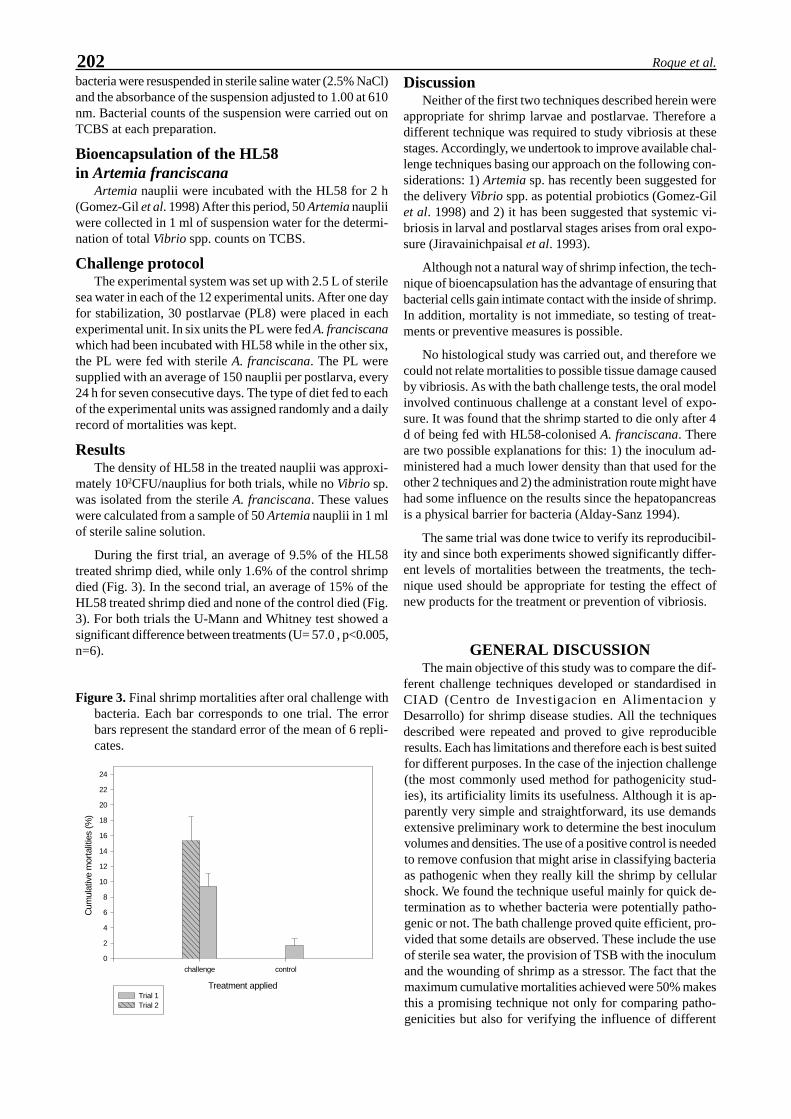
202 Roque et al.
bacteria were resuspended in sterile saline water (2.5% NaCl)and the absorbance of the suspension adjusted to 1.00 at 610nm. Bacterial counts of the suspension were carried out onTCBS at each preparation.
Bioencapsulation of the HL58in Artemia franciscana
Artemia nauplii were incubated with the HL58 for 2 h(Gomez-Gil et al. 1998) After this period, 50 Artemia naupliiwere collected in 1 ml of suspension water for the determi-nation of total Vibrio spp. counts on TCBS.
Challenge protocolThe experimental system was set up with 2.5 L of sterile
sea water in each of the 12 experimental units. After one dayfor stabilization, 30 postlarvae (PL8) were placed in eachexperimental unit. In six units the PL were fed A. franciscanawhich had been incubated with HL58 while in the other six,the PL were fed with sterile A. franciscana. The PL weresupplied with an average of 150 nauplii per postlarva, every24 h for seven consecutive days. The type of diet fed to eachof the experimental units was assigned randomly and a dailyrecord of mortalities was kept.
ResultsThe density of HL58 in the treated nauplii was approxi-
mately 102CFU/nauplius for both trials, while no Vibrio sp.was isolated from the sterile A. franciscana. These valueswere calculated from a sample of 50 Artemia nauplii in 1 mlof sterile saline solution.
During the first trial, an average of 9.5% of the HL58treated shrimp died, while only 1.6% of the control shrimpdied (Fig. 3). In the second trial, an average of 15% of theHL58 treated shrimp died and none of the control died (Fig.3). For both trials the U-Mann and Whitney test showed asignificant difference between treatments (U= 57.0 , p<0.005,n=6).
Figure 3. Final shrimp mortalities after oral challenge withbacteria. Each bar corresponds to one trial. The errorbars represent the standard error of the mean of 6 repli-cates.
DiscussionNeither of the first two techniques described herein were
appropriate for shrimp larvae and postlarvae. Therefore adifferent technique was required to study vibriosis at thesestages. Accordingly, we undertook to improve available chal-lenge techniques basing our approach on the following con-siderations: 1) Artemia sp. has recently been suggested forthe delivery Vibrio spp. as potential probiotics (Gomez-Gilet al. 1998) and 2) it has been suggested that systemic vi-briosis in larval and postlarval stages arises from oral expo-sure (Jiravainichpaisal et al. 1993).
Although not a natural way of shrimp infection, the tech-nique of bioencapsulation has the advantage of ensuring thatbacterial cells gain intimate contact with the inside of shrimp.In addition, mortality is not immediate, so testing of treat-ments or preventive measures is possible.
No histological study was carried out, and therefore wecould not relate mortalities to possible tissue damage causedby vibriosis. As with the bath challenge tests, the oral modelinvolved continuous challenge at a constant level of expo-sure. It was found that the shrimp started to die only after 4d of being fed with HL58-colonised A. franciscana. Thereare two possible explanations for this: 1) the inoculum ad-ministered had a much lower density than that used for theother 2 techniques and 2) the administration route might havehad some influence on the results since the hepatopancreasis a physical barrier for bacteria (Alday-Sanz 1994).
The same trial was done twice to verify its reproducibil-ity and since both experiments showed significantly differ-ent levels of mortalities between the treatments, the tech-nique used should be appropriate for testing the effect ofnew products for the treatment or prevention of vibriosis.
GENERAL DISCUSSIONThe main objective of this study was to compare the dif-
ferent challenge techniques developed or standardised inCIAD (Centro de Investigacion en Alimentacion yDesarrollo) for shrimp disease studies. All the techniquesdescribed were repeated and proved to give reproducibleresults. Each has limitations and therefore each is best suitedfor different purposes. In the case of the injection challenge(the most commonly used method for pathogenicity stud-ies), its artificiality limits its usefulness. Although it is ap-parently very simple and straightforward, its use demandsextensive preliminary work to determine the best inoculumvolumes and densities. The use of a positive control is neededto remove confusion that might arise in classifying bacteriaas pathogenic when they really kill the shrimp by cellularshock. We found the technique useful mainly for quick de-termination as to whether bacteria were potentially patho-genic or not. The bath challenge proved quite efficient, pro-vided that some details are observed. These include the useof sterile sea water, the provision of TSB with the inoculumand the wounding of shrimp as a stressor. The fact that themaximum cumulative mortalities achieved were 50% makesthis a promising technique not only for comparing patho-genicities but also for verifying the influence of different
Treatment applied
challenge control
Cum
ulat
ive
mor
talit
ies
(%)
0
2
4
6
8
10
12
14
16
18
20
22
24
Trial 1Trial 2

203Standardization of Vibrio infections in shrimp
water quality parameters on the onset of vibriosis. The factthat the mortalities took longer than 48 h to occur also makesfeasible studies on the the use of antibiotics or other poten-tial treatments, either therapeutic or prophylactic. The tech-nique was useless for larviculture studies because it is ex-tremely difficult to wound very small shrimp. Therefore,the oral challenge technique was developed to serve the samepurposes as the bath challenge. Table 1 summarises the mainfeatures of each of the techniques used in this study.
LITERATURE CITEDAdams A (1991) Response of penaeid shrimp to exposure to Vi-
brio species. Fish and shellfish immunology, 00: 1-12Alday-Sanz MV (1994) Studies on the pathogenicity of Vibrio sp.
in Penaeus monodon Fabricius. PhD thesis, Institute ofAquaculture, University of Stirling. UK.
Austin, B. (1988). Aquatic Microbiology. Cambridge press, 222pp.
Flegel TW, Fegan DF, Kongsom B, Vithikornudomkit D,Sriurairatana S, Boonyaratpalin S, Chanachookin C, VickersJE, Macdonald OD (1992 ) Occurrence diagnosis and treat-ment of shrimp diseases in Thailand In: W. Fulks, KL Main(eds) Diseases of Cultured Penaeid Shrimp in Asia and theUSA. Oceanic Institute Honlulu Hawaii. pp 57-112.
Fontaine CT, Lightner DV (1973) Observations on the process ofwound repair in penaeid shrimp. Journal of Invertebrate pa-thology, 22:23-33
Gomez-Gil B, Herrera Vega MA, Abreu Gorbois FA, Roque A,1998 Bioencapsulation of two strains of Vibrio spp. in Artemianauplii. Applied and Environmental Microbiology, 64: 2318-2322
Jiravanichpaisal P., Miyasaki T., Limsuwan C. andSomjetlerdchalern A. 1993. Comparative histopathology ofvibriosis in black tiger prawn Penaeus monodon. Diseases ofAsian Aquaculture II, Asian Fisheries Society, Fish HealthSection.
Lightner DV (1988) Vibrio disease. In: CJ Sinderman, DV Lightner(eds) Disease diagnosis and control in North American marineaquaculture , Developments in Aquaculture and fisheries sci-ence, 17:42-47. Sci. Publishing inc. New York USA
Roque A, 1995- The experimental induction of Vibrio spp. infec-tion in Penaeus monodon (Fabricius). PhD thesis, Institute ofAquaculture, University of Stirling United Kingdom.
Ruangpan L, Kitao T (1991) Vibrio bacteria isolated from blacktiger shrimp Penaeus monodon Fabricius Journal of fish dis-eases, 14: 383-388
Sorgeloos P, Bossuyt E, Lavina E, Baeza-Meza M, Persoone G(1977) Decapsulation of Artemia cysts: a simple technique forthe improvement of the use of brine shrimp in aquaculture.Aquaculture, 12: 311-315.
Table 1. Summary of main features and results for the techniques studied.
Challenge RouteFeature Injection Bath OralInoculum(CFU/ml)
10 4- 10 5 10 5 10 2
Challengefrequency
once daily daily
Time to lastdeath (d)
96 h 120 h 168 h
Withstress
no yes no
Repeatibility yes yes yesMaximumcumulativemortality/tank(%)
100 70 25
Uses quick pathogenicity tests;virulence factor tests
water quality parametertests; treatment tests;prophylaxis tests in juveniles
water quality parameter tests;treatment tests; prophylaxistests in mysis and postlarvae
Innovativechanges
volume according to shrimp;importance of inoculumpositive control density
daily challenge; sterileseawater; wound as stressor;death inductionreproducibility
sterile artemia; completelynew technique

204 Roque et al.

Ruangpan L (1998) Luminous bacteria associated with shrimp mortality. In Flegel TW (ed) Ad-vances in shrimp biotechnology. National Center for Genetic Engineering and Biotechnology, Bang-kok.
Luminous Bacteria Associated with Shrimp Mortality
Lila Ruangpan
Marine Shrimp Research and Development CenterPawong, Muang Songkhla 90100, Thailand
ABSTRACT : Luminous disease caused by luminous bacterial infection is currently a significant problem amongthe Asian countries where marine shrimp is the main aquaculture product. Luminous bacteria isolated from theshrimp farm environment include Vibrio fischeri, V. harveyi, V. cholerae biotype albensis, and Photobacteriumleiognathi. Of these, V. harveyi is claimed to be the causative agent associated with shrimp mortality. In hatch-eries, its pathogenicity is correlated with the developmental stage of the shrimp larvae. However, successfulprophylactic measures have been established. In grow-out ponds, luminous disease frequently causes mortalitywith 2-3 month old stock of Penaeus monodon and a bacteroiphage of V. harveyi has been implicated in mortal-ity of such shrimp exhibiting tea-brown gill syndrome (TBGS). Chemotherapeutics are more or less effective forcontrolling the disease but are not effective for curing it. Recent in vitro studies have demonstrated growthinhibition of this bacteria using biological control and the information gained paves the way for further investi-gations.
Key words : Luminous bacteria, Shrimp mortality, Vibrio harveyi
INTRODUCTIONWidespread interest in shrimp culture in Thailand has
developed only in the past 15 years. During that period, in-tensive farming systems have been introduced and the areasof shrimp farms number of farms has rapidly expanded. Inorder to reduce the introduction of high organic matter intothe coastal environment and thus avoid severe damage tothe production system (Nygaard et al. 1992), either closedpond systems or recycle systems are necessary. These closedand recycle systems increase the problems associated withbacterial pathogens. It is recognized that damage to shrimpstocks is frequently associated with bacterial diseases thatare mostly caused by luminous bacteria (Ruangpan 1987,Songserm et al. 1990, Ruangpan et al. 1997). Luminous bac-terial diseases have also been reported to cause economiclosses to the shrimp industry in the Philippines (Fernandez& Mayo 1994), Vietnam (Nguyen & Le Trong 1994), India(Raju 1994), and Indonesia (Sulasmi et al. 1994, Taslihan &Wijayati 1994). The problem seems to be common amongthe Asian countries where shrimp farming is the mainaquaculture activity. This presentation describes the spe-cies composition (incidence and intensity) of luminous bac-teria in shrimp farms and the coastal environment in Thai-land, their pathogenicity and recent studies on the chemo-therapy and biological control. It is hoped that the informa-tion will contribute towards further investigations leading tosuccessful resolution of the luminous disease problem.
SPECIES COMPOSITIONPreliminary studies on luminous bacterial species in Thai-
land have been conducted using numerical taxonomic analy-sis (Ruangpan et al. 1995). Media used for isolation includedthiosulfate citrate bile salts sucrose (TCBS, Difco) agar andmodified sea water complete (MSWC) media. A total of 180
luminous and nonluminous vibrios were isolated from dis-eased P. monodon cultivated in several areas of Thailand.Each strain was tested for 98 characters. Luminescence wasobserved on MSWC medium 18-24 h after isolation usingthe technique of Furniss et al. (1978). Based on unweighedaverage linkage analysis at 80% similarity, 15 phenotypicclusters were obtained. Among those, luminous vibrios rep-resented 4 clusters, according to their similarity in phenotypiccharacters, in guanine-cytosine (G + C) contents and to ref-erence cultures. Members in the first cluster comprised 21strains identified as V. harveyi. The second cluster contain-ing 2 strains identified as V. fischeri. The last 2 clusters con-taining 7 strains which could not be identified as any knownspecies. However, their characters and G+C contents werevery close to either V. harveyi or V. cholerae non-01 refer-ence cultures ATCC 14126 and NCTC 8021, respectively(Fig. 1).
Figure 1. Species composition of luminous and nonluminousvibrios from cultured black tiger shrimp. The exploded por-tion of the pie (16.7% of total strains) are subdivided intogroups by percentage of isolates.
Nonluminous vibrio
83.33%
70% V. herveyi
7.7% V. fisheri
23% Unidentified
LV

206 Ruangpan
0
0.1
0.2
0.3
0.4
0.5
0.6
0.7
0.8
0.9
0 5 10 15 20 25 30 35 40Salinity (ppt)
Lum
inou
s/N
onlu
min
ous
In 1996, identification of luminous bacteria isolated fromthe coastal water of Thailand was carried out by Sodthonkong(1996)(Table 1). Water samples were obtained from 128 sta-tions in 21 provinces located along the Andaman and Siamcoasts. Based on 49 phenotypic characters, a total of 210representative isolates were identified to V. harveyi, V.cholerae biotype albensis and Photo-bacterium leiognathi.
According to the results most of the V. harveyi isolatesexhibited green to yellow-green colonies on TCBS. Fiftyseven percent of these isolates grew on media supplementedwith 6% NaCl. All isolates had the ability to emit light whencultured in media containing 0.5-6% NaCl, although, 10%of them also grew and emitted light in media containing 8%NaCl.
V. cholerae biotype albensis exhibited yellow colonieson TCBS. All of its isolates grew on media without addedNaCl or with NaCl up to 3%. Forty seven percent grew onmedia containing 6% NaCl while only 7% grew on mediacontaining 8% NaCl. They gave high light when on mediacontaining 0-6% NaCl but low light with 8% NaCl.
P. leiognathi gave green colonies on TCBS. All its iso-lates grew and emited light in the media containing 0.5-3%NaCl but only 50% grew in 6% NaCl and no growth wasfound in the media containing 8% NaCl. Ruangpan et al.(1995) indicated that V. fischeri exhibited deep green colo-nies on TCBS, 100% of the isolates grew and emited lighton the media containing 1-8% NaCl.
INCIDENCE AND INTENSITYResults from the investigation of luminous and
nonluminous bacteria in Thai coastal water (Sodthongkong1996) revealed that luminous bacteria are normal compo-nents of the microbial flora of estuaries and brackish water
(Table 1). This finding is supported by farmer reports of lu-minous bacteria in the water and sediments of shrimp ponds,as well as the inlet and outlet water of mariculture opera-tions which depend totally on water supplied from the coastalenvironment (Sae-Oui et al. 1987, Songserm et al. 1990,Ruangpan et al. 1995 and 1997c).
The incidence of isolation from environmental water sam-ples was found to differ for species and sampling area. Iso-lates of V. harveyi were detected from 96 sampling stationswith 75% incidence. The incidence of V. cholerae biotypealbensis was 43% while that of P. leiognathi was 9%. Figure1 demonstrates the correlation between salinity and lumi-nous bacteria or vibrios. Interestingly, high incidences andintensities of both types were manifested at salinities between30 and 33 ppt. However, the ratios of these bacteria are shownfor estuarine water in the range of 1 ppt (Fig. 2).
Low intensities of luminous bacteria (0-4.2x10 cfu/ml)were found in the sampling stations located far fromaquaculture ponds and communities. The highest intensity(2.9x10³ cfu/ml) was recorded from aquaculture effluentwater (Sodthongkong 1996). The incidence of V. harveyi iso-lation from shrimp pond sediment was reported to be 80%,although its intensity was not mentioned (Prapassorn 1995).Very little information is available regarding its incidenceand intensity in shrimp. Most studies have focused on viru-lent isolates. Songserm et al. (1990) reported on the inci-dence from naupliar to postlarval stages of P. monodon (Ta-ble 2). Other investigators have reported Vibrio bacteriawhich included luminous and non-luminous isolates.Ruangpan et al. (1995) attempted to investigate the incidenceof luminous bacterial species found in diseased shrimp. The
Table 1. The percentage incidence and intensity ofluminous bacteria found in the coastalenvironment (shrimp ponds, pond effluent andhatcheries).
Sources Incidence (%) Intensity (cfu/m l)W ater : coastal environment 75 0.2 x 10 - 1.4 x 10 : shrimp ponds 84 9.1 x 10 - 1.2 x 10 2
: effluent from ponds 92 1.5 x 10 - 1.0 x 10 2
: hatcheries 67 0.2 x 10 - 2.9 x 10 3
: effluent from hatcheries 44 1.2 x 10 - 3.8 x 10 2
Sediment : shrimp pond 80 NDSources : Sodthongkong 1996, Ruangpan et al. 1997c
Table 2. Incidence of V. harveyi infection atdifferent developmental stages of P.monodon larvae.
% Incidence of infectionShrimpstock
Zoea Mysis Postlarvae Total
A 3.33 5.33 3.00 10.66B 4.00 4.66 2.66 11.33C 2.66 5.33 4.00 11.99D 5.33 6.00 3.00 13.33E 5.33 6.66 3.00 13.99Total 20.65 27.98 15.66 61.30 Source : Songserm et al. 1990
Figure 2. Variation in ratio ofliminous/nonluminous bacte-ria at various salinities.

207Luminous bacteria associated with shrimp mortality
results indicated that V. harveyi was predominant, account-ing for 70% of the isolates while V. fischeri and other lumi-nous species accounted for 6.7 and 23.3%, respectively (Fig.3).
PATHOGENESIS AND HOSTSUSCEPTIBILITY
Among the luminous bacterial species reported fromshrimp ponds, hatcheries and moribund shrimp, only V.harveyi has been confirmed to cause mortality of shrimp.The disease is widely known as “luminous disease” or “Kungruangsang” in Thai. Epizootics of this disease occur severaltimes a year and are expanding throughout shrimp farmingareas along the coast. A number of shrimp farmers have suf-fered severely from damage of long duration caused by thisdisease. Because of similar problems throughout Asia, sci-entists from many countries including the Philippines, Indo-nesia, Malaysia, India, Thailand and Taiwan are becomingincreasingly interested in this disease.
The first report on shrimp luminous disease in Thailandwas published in 1987 by Sae-oui and collaegues. The re-port indicated an outbreak of the disease in shrimp hatcher-ies in the central part of the country, which used to be thelargest productive area for marine shrimp. The bacterialpathogenesis resulted in mortalities up to 100% for naupliarto zoeal stages of P. merguiensis. Living and dead shrimp
larvae and even the sea water in disease outbreak areas wereluminescent in dim light. Other gross features of the dis-eased shrimp were milky white bodies, weakness, swimmingdisorders and loss of appetite, eventually leading to death.Using luminous media (LM) (Baumann & Baumann 1981),luminescent bacterial colonies could be isolated from thediseased specimens as well as from hatchery water. Basedon 47 phenotypic characters, the causative agents were iden-tified to V. harveyi. Experimental trials for pathogenicity gave100 and 95% mortality for naupliar and zoeal stages, re-spectively (Table 3), but no mortality for the larvae thatreached the mysis stage.
In P. monodon, luminous disease was also reported ini-tially from larvae at naupliar to mysis stages (Ruangpan1987). Since then, a rising incidence of luminous diseasethat can cause 80-100% mortality to the larval stocks hasoccurred in hatchery areas all over the country (unpublishedresults). In 1990, Songserm et al. reported that luminous dis-ease in P. monodon larvae was also caused by V. harveyi.Results from experimental bath infections confirmed thatzoea and mysis stages were more susceptible than postlarvae(PL). Prayinto & Latchford (1995) reported similar resultswith experimental infections of P. monodon larvae using sus-pensions of luminous bacteria related to Photobacterium and
Table 3. Pathogenicity of V. harveyi CHA 86021 toP. merguiensis larvae infected by bathexposure for 24 h.
% Mortalitycontrol Treatment (cfu/ml)
Shrimp stages 10 7 10 8
Nauplius 70 100 100 Zoea 60 ND 95Source : Sae-Oui et al. 1987
Table 4. Mortality of different developmentalstages of P. monodon exposed to V.harveyi at various concentrations.
% Mortality Control Treatment/(cfu/ml)
Shrimp stages 10 8 10 6 10 5 10 4
Nauplius 35 100 100 ND 100Zoea 25 100 100 75 100Mysis 25 75 70 ND 53Postlarva 20 50 50 45 35Source : Limsuwan 1989, Songserm et al., 1990
Figure 3. Distribution of luminous and nonluminous vibrio bacteria at varioussalinities sampled from 21 provinces located along the coast of Thailand.
0
10
20
30
40
50
60
0 10 20 30 40
Salinity (ppt)
Non
lum
inou
s co
unt
(x 1
0 2 c
fu/m
l)
0
1
2
3
4
5
6
Lum
inou
s co
uunt
(x1
0 2 c
fu/m
l)
Nonluminous Luminous

208 Ruangpan
Vibrio. Mortalities of zoea and postlarvae, after 48 h expo-sure, were 75 and 45%, respectively. Furthermore, Limsuwan(1989) has found that larvae beyond PL3 and 4 develop-mental stages resisted luminous bacterial infection (Table4). Subsequently, various reports have indicated that lumi-nous bacteria are also associated with shrimp mortality ingrow-out ponds. In a survey of rearing water from numerousshrimp farms in southern Thailand from April to August1996, Ruangpan et al. (1997a) found large numbers of theluminous bacteria. They also noted that massive mortalityoccurred in some farms within 3 to 4 days after the first ap-pearance of moribund shrimp exhibiting tea-brown coloredgills or tea-brown gill syndrome (TBGS). Isolations of bac-teria from the moribund shrimp revealed that V. harveyi wasdominant. Based on results from electron microscopy andinjection trials with naturally and experimentally infectedTBGS shrimp, it was suggested that a bacteriophage mightbe implicated in shrimp mortality by mediating toxigenesisof V. harveyi.
Disease induced by V. harveyi has been investigated byexperimental infection of various crustacean species understress conditions. It has the ability to induce disease and causemortality in P. monodon, P. indicus, and Artemia nauplii(Prayinto & Latchford 1995) and P. merguiensis (Sae-Oui etal. 1987). No significant mortality occurred inMacrobrachium species (Kasornchan 1987, Prayinto &Latchford 1995) and banacles (Prayinto & Latchford 1995).
PREVENTION AND CONTROLPrevention and control of luminous disease in shrimp are
generally achieved by chemical treatment. Chemothera-peutants may be applied in the feed or in the water. Usuallyantibiotics are applied in the feed while disinfectants (andsometimes antibiotics as well) are applied in the water(Ruangpan 1987, Ruangpan et al. 1997c).
To prevent the luminous disease in hatchery operations,water should be filtered through a biofilter system prior tochemical treatment with 60% active chlorine at 10-30 g/tonor commercial grade formalin at 50-100 ml/ton. During treat-ment aeration is necessary. In addition, in vitro trials havebeen conducted on the inhibition of luminous bacterial growthusing several disinfectants. Table 7 shows the effective dosesfor growth inhibition of V. harveyi treated with benzalko-nium chloride, formalin and povidone iodine. Antibiotics
treatment via the water is a common practice during rearingthe early larval stages (nauplius to postlarvae 3-4). Applica-tion of antibiotics at 3-5 g/ton into the rearing water, espe-cially during periods of metamorphosis has been found togreatly reduce luminous bacteria and stock mortality(Chayarat 1997). Oxytetracycline (OTC) was the main drugused for several years in all areas of the country. The effec-tiveness of OTC gradually declined after repeated applica-tion for long periods due to the development of bacterialresistance (Ruangpan et al. 1997c). Recently, various drugshave been tested against luminous and nonluminous vibrios,including resistant strains (Tables 5 and 6). Therefore, alter-native drugs can be recommended based on sensitivity tests.After harvesting, treatment of the larval rearing facilities byflushing or vigorous spraying with freshwater has been sug-gested as a routine practice.
The major disinfectants used to reduce bacterial num-bers in grow-out ponds are formalin and chlorine. When theVibrio count in a pond rises to 103 to 104 cfu/ml, addition of5 to 20 liters of disinfectant (per 1 ha pond) has been recom-mended. Benzalkonium chloride and povidone iodine havealso been found to be effective chemicals to inhibit growthof V. harveyi (Table 7). At present, these chemicals are sel-dom used due to their high cost.
Antibiotics are usually applied in grow-out pond by ad-dition to pelleted feed at approximately 3-5 g/kg adminis-tered to the shrimp for 7-10 days. Oxytetracycline andoxolinic acid were commonly used in the past. Recently theyappear to be of limited value due to the development of bac-
Table 5. Minimal inhibitory concentration (MIC)ranges and inhibitory concentration (IC) of 6drugs against 60 strains of luminous bacteria.
MIC ranges IC s (ug/ml)Drugs (ug/ml) 50% *1 90% *2
Oxytetracycline 6.25 - 100 15.00 69.20Oxolinic acid 12.5 - 100 28.20 56.70Norfloxacin 0.8 - 100 65.50 100Chloramphenicol 3.13 - 50 8.75 45.20Trimethoprim 3.13 - 25 9.37 15.00Kanamycin 100 -> 100 >100 >100
*1 and *2 : Concentration inhibiting 50% and 90% ofthe strains, respectively. Source: Ruangpan et al. 1997c
Table 6. Drug resistant strains of luminous and nonluminous vibrios isolated fromcultured shrimp P. monodon and the farm environment.
% of drug resistant strains/yearDrugs 1989 1990 1992 1994 1995 1996Trimethoprim 0 23.4 25.5 ND ND 20Chloramphenicol 0 2.92 5.1 14.8 47.1 20Oxolinic acid 90 0 0 11 20.5 45Oxytetracycline ND 32.2 36.4 45.9 71.4 0.4Norfloxacin ND 22.9 7.3 41.5 87.6 90Kanamycin 0 ND 14.5 100 100 100Sulfamonomethoxine 0 61.95 100 ND ND ND
Sources : Ruangpan and Kitao 1992, Sangrungruang et al. 1993, Prapassorn 1995,Sudthongkong 1996, Ruangpan et al. 1997

209Luminous bacteria associated with shrimp mortality
Table 7. Summary of in vitro trials for control of V. harveyi.
AgentsBiological control Application Dosage Effectiveness Reference
Bacillus S11 Probiotic/feeding ND 74 % RPS Phianphak et al.1997
Lactobacillus casei Probiotic/feeding ND Growth-inhibition
Jiravanichpaisalet al. 1997
L. acidophilus
Vibrio alginolyticus Concomitantculture
1 : 10 cfu Growthinhibition
Ruangpan et al.(in press)
0 to 100%
Chlorella sp. Concomitantculture
1000 : 1 cell/efu Growthinhibition
Direkbusarakomet al. 1997
Skeletonema costatum Concomitantculture
500:1 cell/cfu Growthinhibition 35to 100 %
Panichsuke etal. 1997
Guava leaf extract Feeding MIC 1250µg/ml
Growthinhibition
Direkbusarakomet al. 1997
Fresh water Spray or bathecontainers andequipment
Several times Growthinhibition
Unpublisheddata
Chemicalcontrol
Benzalkoniumchloride
Water treatment 40%MIC 64 ppm
Growthinhibition
Lin and Nash1996
MBC 64 ppm
Formalin Water treatment 40-50 ppm Growthinhibition
Lin and Nash1996
Providone iodine Water treatment 1-3 ppmMBC 64 ppm
Growthinhibition
Areechon 1990
MIC 1024ppmND = No data; MIC = Minimal inhibitory concentration; RPS = Relative percent of survivalMBC = Minimal bactericidal concentration
terial resistance (Ruangpan et al. 1997c). Several drugsclaimed to be effective against luminous bacteria are sup-plied in commercial grade although many show very lowbacterial growth inhibition (Ruangpan et al. 1997b).
Governmental guidelines to restrict the use of antibioticsin shrimp farms have been issued. To prevent problems re-garding drug residues, the guidelines recommend that anti-biotics should not be used beyond 3 months in shrimp grow-out ponds.
Figure 4. Number of luminous vibrios cultured at various salinity levels.
0.0
0.1
0.2
0.3
0.4
0.5
0 10 20 30 40 50
Incubation time (h)
Bac
teria
l cou
nt
(x 1
06 cf
u/m
l)
0 ppt
5 ppt
10 ppt
15 ppt
20 ppt
25 ppt
30 ppt
42 ppt

210 Ruangpan
Experiments on preventing and controlling luminous dis-ease using alternative methods such as probiotics or biologi-cal control have been attempted (Table 7). Phianpark et al.(1997) successfully used Bacillus sp. as probiotic feed inlaboratory scale tests to achieve 74% relative survival afterchallenge with V. harveyi. Lactobacillus casei and L. acido-philus have also been claimed to inhibit the growth of V.harveyi when used as a probiotic feed (Jiravanichpaisal etal. 1997). Beneficial microorganisms found to be effectivefor growth inhibition of V. harveyi on the laboratory scaleinclude V. alginolyticus (Ruangpan et al. 1998) Chlorellasp. (Direkbusarakom et al. 1997) and Skeletonema costatum(Panichsuke et al. 1997). Sodthongkong (unpublished data)studied the multiplication of V. harveyi at various salinitylevels and found that the number of bacteria was reducedfrom 7.5x10³ to 8.9x10², 4.0x10², 1.3x10 and 0 cfu/ml in 1,6, 12 and 18 h, respectively, after transfer from 25 ppt to 5ppt. By contrast, bacteria transferred to 10 ppt decreased to2.0x103 and 32.x102 cfu/ml after 1 and 6 h, respectively, butthen increased to 1.6x10⁵ and 3.4x10⁵ after 12 and 24 h,respectively. At salinities between 15 and 42 ppt, numbersof V. harveyi increased gradually from 1-48 h of cultivation(Fig. 4). The results suggested that shrimp culture in seasonsof low water salinity may experience less risk of luminousdisease. Further studies are needed to ascertain the efficacyof using various microorganisms to control luminous bacte-ria in shrimp operations. They may prove a better alterna-tive than antibiotics in overcoming problems.
CONCLUSIONSThailand is now one of the top of shrimp producing coun-
tries and highly developed culture systems have been estab-lished and expanded in many shrimp farming areas. How-ever, more attention should be focused on how to developshrimp culture as a sustainable industry. Disease problemsstill exist and are associated with serious crop losses. Thesurvey data on luminous bacteria has shown that they aremembers of the natural estuarine and brackishwater envi-ronment. The studies also suggest that they induce diseasemore frequently in dry season than the rainy season and thattheir is related to salinity levels and effluent from farmingoperations. Only V. harveyi has been confirmed to be a patho-genic agent associated with shrimp mortality. In hatcheries,luminous disease can be controlled by chemical treatmentwith disinfectants and antibiotics, while in grow-out ponds,effective treatment and control methods are still uncertain.Some experiments indicate effective growth inhibition of V.harveyi by probiotics and biological treatments. However,effectiveness has not yet been proven in field trials. Althoughluminous pathogens exist naturally in the water, they cer-tainly need carbon and introgen (C+N) for growth and mul-tiplication. Thus, the first action to take in reducing theirnumbers is to be careful not to overfeed the shrimp and thussupply plentiful nutrients in the pond and the adjacent envi-ronment.
LITERATURE CITEDAreechon N (1990) Bactericidal activity of povidone iodine against
Aeromonashydrophila and Vibrio harveyi. Thai Fish. Gazette43:199-204 (In Thai)
Baumann P and Baumann L (1981) Marine genera Photobacterium,Bencekea, Alteromonas, Pseudomonas, Alcaligenes. In :Mortmer PS et al. (eds) The prokaryotes. Vol. 2 New YorkSpinger - Verlag p 1302-1331.
Chayarat C (1997) Correlation on seed production of Penaeusmonodon and bacterial number in nursing pond. Tech. paperNo. 24/1997 Coastal Aquaculture Div. DOF, Bangkok 11 p
Direkbusarakom S, Herunsalee A, Yoshimizu M, Ezura Y, KimuraT (1997a)Efficacy of guava (Psidium quajava) extract againstsome fish and shrimp pathogenic agents. In : Flegel TW andMacRae IH (eds), Disease in Asian Aquaculture I I IFish Health Section, Asian Fisheries Society, Manila p 359-363
Direkbusarakom S, Pechmanee T, Assavaaree M, Danayadol Y(1997b) Effect of Chlorella on the growth of Vibrio isolatedfrom diseased shrimp. In : Flegel TW, MacRae IH (eds) Dis-ease in Asian Aquaculture III. Fish Health Section, Asian Fish-eries Society, Manila p 355-358
Fernandez RD, Mayo S (1994) Current status of prawn farmingand prawn health management in the Philippine. In : TurnbullJ et al. (eds) Procs. of the SEAADCP, Prawn health manage-ment workshop. Aquatic Animal Health Research Institute,DOF Bangkok, Thailand p 61-89
Furniss AL, Lee JV and Donovan TJ (1978) The vibrios. PublicHealth Lab. Service Monograph Series No. 11 HMSO London
Jiravanichpaisal P, Chuaychuwong P, Menasveta P (1997) Theuse of Lactobacillus sp. as the probiotic bacteria in the gianttiger shrimp (Penaeus monodon Fabricius) In : Abstract ofposter session of the 2nd Asian-Pacific Marine Biotechnologyconference, Phuket thailand p 16
Kasornchandra J, Boonyaratpalin S, Jadesadakraisorn U (1987)Luminescent disease in giant prawn larvae. Tech. paper No. 67National Inland Fisheries Ins. Bangkok 9 p (In Thai with Eng-lish abstract)
Limsuwan C (1989) Common disease found in cultured shrimp inThailand. Asian shrimp News 1 : fourth quarter 1989
Lin CK, Nash GL (1996) Asian shrimp news. Collected volume1989-1995, Asian shrimp culture council, Bangkok 312 p
Nguyen H T, Le Trong K (1994) Report on shrimp diseases inVietnam. In: Turnbull J et al. (eds) Procs. of the SEAADCP,Prawn health management workshop. Aquatic Animal HealthResearch Institute, DOF Bangkok, Thailand p103-106
Nygaard K, Lunested BT, Hekton H, Berge JA, Hormazabal V(1992) Resistance to oxytetracycline, oxolinic acid and fura-zolidone in bacteria from marine sediments. Aquaculture104:31-36
Panichsuke P, Ruangpan L, Pechmanee T (1997) Inhibitory effectof Skeletonema costatum on the growth of Vibrio harveyi In :Abstract of poster session of the 2nd Asian-Pacific Marine Bio-technology conference Phuket Thailand p 122
Phianphak W, Piyatiratitivorakul S, Menasveta P, Rengpipat S(1997) Use of probiotics in Penaeus monodon In : Abstract ofposter session of the 2nd Asian-Pacific Marine BiotechnologyConference, Phuket Thailand p 18
Prapassorn S (1995) Bacterial flora in the sediment of black tigershrimp pond with the emphasis in Vibrio spp. and their resist-ance to antimicrobial agents. Msc Thesis in Fisheries Science,Faculty of Fisheries, Kasetsart University, Bangkok p 74
Prayinto SB and Latchford JW (1995) Experimental infections ofcrustaceans with luminous bacteria related to Photobacteriumand Vibrio : Effect of salinity and pH on infectiosity.Aquaculture 132: 105-112
Raju GA (1994) Shrimp aquaculture in India. In : Turnbull J et al.(eds) Procs. of the SEAADEP, Prawn health management work-shop. Aquatic Animal Health Research Institute, DOF Bang-kok, Thailand p 39-42

211Luminous bacteria associated with shrimp mortalityRuangpan L (1987) Diseases of cultured marine shrimp in Thai-
land : Penaeus monodon and P. merguiensis. In : Limsuwan Cet al.(eds) Tech. paper for the 1st seminar on Marine ShrimpCulture, Fisheries Science Club. Kasetsart Univ. Bangkok p46-62 (In Thai)
Ruangpan L, Kitao T (1992) Minimal inhibitory concentration of19 chemothera-peutants against Vibrio bacteria of shrimpPenaeus monodon. In : Shariff M et al. (eds) Diseases in AsianAquaculture I. Fish Health Section. Asian Fisheries SocietyManila p 135-142
Ruangpan L, Tabkaew R, Yoshida T, Kawatsu H and Saitanu K(1995) Numerical taxonomy of Vibrio spp. isolated from blacktiger shrimp, Penaeus monodon, cultured in Thailand. In :Shariff M et al. (eds) Fish Health Section, Asian Fisheries So-ciety, Manila p 131-140
Ruangpan L, Danayadol Y, Direkbusarakom S, Siurairatana S andFlegel TW (1997a) Bacteriophage implicated in mortality ofcultivated Penaeus monodon exhibiting tea-brown gill syn-drome (TBGS). Tech. paper present in Shrimp Biotechnologyseminar 13 Dec. 1997 NSTDA Bangkok p 91-96
Ruangpan L, Direkbusarakom S, Danayadol Y and Srikaew Y(1997b) Efficiency of commercial drugs for control disease inblack tiger shrimp on the growth inhibition of luminous andvibrio bacteria. Tech. paper No. 19/1997 National Institute ofCoastal Aquaculture, Songkhla 10 p (In Thai with Englishabstract)
Ruangpan L, Sodthongkong C, Srikaew Y (1997c) Luminousbacteria in shrimp aquaculture area of Songkhla. Procs. of DOFseminar 1997. p 65-74
Ruangpan L, Na-anan P, Direkbusarakom S (1998) Inhibitory ef-fect of Vibrio alginolyticus on the growth of V. harveyi. FishPathology (in press)
Sae-Oui D, Tunsutapanit A, Ruangpan L (1987) Vibrio harveyi, acausative agent of marine shrimp Penaeus merguiensis larvae.In : Limsuwan C et al. (eds) “Shrimp Disease and Chemo-therapy” Kasetsart Univ. Press, Bangkok p 1-8
Sangrungrueng K, Aoki T, Yoshida T (1993) Minimal inhibitoryconcentration of chemotherapeutants and drug resistanceagainst vibrio bacteria isolated from shrimp (Penaeus monodon)in Thailand. In : Snidvong A et al. (eds) NRCT-JSPS seminaron marine science, Chulalongkorn Univ. Bangkok p 221-226
Sodthongkong C (1996) Identification and drug resistance of lu-minous bacteria in coastal water of Thailand. Msc Thesis inFisheries Science, Faculty of Fisheries, Kasetsart UniversityBangkok 109 p (In Thai with English abstract)
Songserm M, Suksringam B, Srisopaporn P (1990) Study on thebacterial luminescent disease in jumbo tiger shrimp (Penaeusmonodon) Srinakarin-tarawiroj Research and DevelopmentJournal 4(1): 15-24 (In Thai with English abstract)
Sulasmi S, Naibaho L, Dewi E (1994) Shrimp farming in northSumatra province, Indonesia. In : Turnbull J et al. (eds) Procs.of the SEAADCP, Prawn health management workshop.Aquatic Animal Health Research Institute, DOF Bangkok,Thailand p 53-59
Taslihan A, Wijayati A (1994) Diseases in Indonesia shrimp (P.monodon) culture. In : Turnbull J et al. (eds) Procs. of theSEAADCP, Prawn health management workshop. Aquatic Ani-mal Health Research Institute, DOF Bangkok, Thailand p 45-52

212 Ruangpan

Pasharawipas T, Sriurairatana S, Direkbusarakom S, Donayadol Y, Thaikua S, Ruangpan L, Flegel TW (1998)Luminous Vibrio harveyi associated with tea brown gill syndrome in black tiger shrimp. In Flegel TW (ed)Advances in shrimp biotechnology. National Center for Genetic Engineering and Biotechnology, Bangkok.
Luminous Vibrio harveyi Associated withTea Brown Gill Syndrome in Black Tiger Shrimp
1Tirasak Pasharawipas, 2Siriporn Sriurairatana, 3Sataporn Direkbusarakom3Yaowanit Donayadol, 4Surayut Thaikua, 3Lila Ruangpan, 4T.W. Flegel
1Microbiology dept, Faculty of Science, Rangsit university, Paholyathin Rd., Pathumthani, Thailand, 120002Pathology Dept, Faculty of Medicine, Ramathibodi hospital, Mahidol university
Rama VI Rd, Bangkok, Thailand, 10400.3National Institute of Coastal Aquaculture, Koaseng Rd., Muang District, SongKhla 90000, Thailand
4Biotechnology Department, Faculty of Science, Mahidol University, Rama VI Rd, Bangkok 10400, Thailand
ABSTRACT: Luminous Vibrio harveyi (VH1039) isolated from tea brown gill syndrome (TBGS) black tigershrimp, Penaeus monodon, was examined as a lysogenic bacteriophage host. Temperate phages found weremorphologically identified as siphoviruses of various sizes (i.e., heads of 20-100 nm diameter and tails of 20-200 nm in length). The colonies of VH1039 were highly variable upon extended cultivation. This variabilitymight be caused by transposon like activity of these phages. Lysogenic VH1039 caused no disease symptoms inthe shrimp. This study also showed that luminescence was influenced by temperature and pH conditions but thatit was not critical to shrimp pathogenicity. The relationship of the phage to the TBGS shrimp is being studied.
KEY WORDS: Vibrio harveyi, luminescence, tea brown gill syndrome, TBGS, bacteriophage, Penaeusmonodon
INTRODUCTIONVibrio harveyi (VH) is one of many luminous bacteria
like V. fisheri, Photobacterium leiognathi and P.phosphoreum. It is found abundantly in marine and estuarialhabitats (Hastings et al. 1981, Jiravanichpaisal et al. 1994).Some studies have reported that VH is a devastating patho-gen of penaeid larvae, especially those of the black tigershrimp (Baticados et al. 1990, Lavillla-Patogo et al. 1992).However, previous studies have also reported that it is a nor-mal constituent of the non-pathogenic flora of marine ani-mals (O’Brien & Sizemore 1979, Ruby & Morin JG 1979).All these luminous bacteria, whether normal flora or patho-gens, can exhibit luminescence in marine animals. A recentstudy found that luminous VH isolate and a bacteriophagecould be found in black tiger shrimp exhibiting tea brawngill syndrome (TBGS). A bioassay study showed that intra-muscular injections of this VH strain into juvenile shrimpcauses no symptoms, but that the combination of bacteri-ophage and VH caused shrimp death (Ruangpan et al. 1998).Vibrio species, including VH, are major hosts of marinebacteriophages (Proctor 1997). Most bacteriophages aretemperate phages and they have been reported to cause di-versity in bacterial hosts by acting as transposon-like ele-ments. It is well known that some temperate phages in bac-terial hosts play major roles in causing diseases in humans(Cheetham & Katz 1995, Reid & Mekalanos 1995). In ma-rine shrimp, however, there is still no confirmed evidencethat temperate phages lead to disease.
In the present paper we report highly variable character-istics of Vibrio harveyi VH1039 isolated from TBGS blacktiger shrimp. This bacterial strain is lysogenic and luminous.We report the optimal physical conditions for its growth andluminescent expression. The temperate phage was isolatedand morphologically identified. Moreover, there was evi-dence to show that a transposon (Tn3) might also reside inthis strain.
MATERIALS AND METHODSCultivation and identification of VH1039
TBGS in black tiger shrimp was first reported in 1998(Ruangpan et al. 1998). A luminescent bacterial strain wasisolated from moribund TBGS shrimp and it was later iden-tified as VH1039 by biochemical tests. The bacterium wascultured on TCBS agar, on tryptic soy agar (TSA) and intryptic soy broth (TSB). It was incubated at various tem-peratures (from room temperature to 37 °C), in media ofvarious percentages of NaCl (between 0.5-8%), and variousalkalinity (pH 5-12). Under these physical conditions, thestrain grew well and colony morphology and luminescenceproduction were investigated. The bacterium was also sub-jected to confirmatory biochemical tests and antibiotic sen-sitivity tests were carried out using BBL Sensi-Discs (BBL).
Isolation and examination of temperatephages
To investigate the possibility that VH1039 was infectedwith temperate phages, it was cultured in TSA with 3% NaCl

214 Pasharawipas et al.
for 18-20 hours at temperatures between 30°C and 33°C.Bacterial colonies were then fixed in a solution of 0.5% glu-taraldehyde in 4% paraformaldehyde, stained with 2% phos-photungstic acid and examined by transmission electronmicroscopy.
To stimulate the production of higher quantities of phageparticles, VH1039 was cultured in TSB with 3% NaCl at30-33°C for 18-20 hours to reach OD 0.6-0.7 at 590 nm.The medium was then divided into portions containing dif-ferent concentrations of mitomycin-c (from 1-250 ug/ml) andallowed to incubate at 37°C for another 18-20 hours. Thesesuspension were also examined by transmission electronmicroscopy. To concentrate and purify phage particles, bac-terial cells were precipitated by adding NaCl to a final con-centration of 1 M followed by immersion in an ice bath for 1hour before centrifugation at 12,000 g for 20 minutes. Afterthis, bacterial debris was eliminated from the supernatant byrepeated centrifugation under the same conditions. Bacteri-ophage suspended in the supernatant was then precipitatedby adding polyethylene glycol 8000 at a concentration of10% followd by immersion in an ice bath for 1 hour beforepelleting by centrifugation at 12,000 g for 20 minutes. Thepellet was resuspended in phage buffer (20 mM Tris-HCl,pH 7.5 contianing 10 mM MgSO
4). The solution was finally
filtered through a 0.22 m filter membrane before examina-tion by the transmission electron microscopy.
RESULTSOptimal condition for culture of VH1039
VH1039 grew well from room temperature (28-30°C) to33°C, but the best temperature for growth was 33°C. It tol-erated but did not grow at 35°C or above. At higher tem-peratures like 37°C, it exhibited strong luminescence, butlittle or none at temperatures from room temperature to 33°C.However, luminescense weakened after incubation for 4-5days, regardless of the incubation temperature. Theluminescense could be reactivated in approximately 6 hoursafter switching the cultures from a lower to a higher tem-perature (e.g., 30°C to 37°C). If the temperature was low-ered once again, the luminescence faded.
Salinity also influenced the growth of VH1039. It grewwell in 2-3% NaCl but also persisted in percentages of NaClfrom 1% up to 6.5%. It grew well between pH 7-9, but not atpH lower than 5, and slowly at any pH above 9. Also, at pH7-9 it exhibited stronger luminescence than at lower pH. Theluminescent shrimp and luminescent VH are shown in Fig.1.
Antimicrobial sensitivity and biochemicaltests
VH1039 was tested with antimicrobial agents to exam-ine for the possibile presence of transposons. Among theantimicrobial agents tested were neomycin, kanamycin, strep-tomycin, trimethoprim, tetracyclin and ampicillin. It was re-sistant to only ampicillin, a characteristic of Tn3. In bio-chemical tests, it expressed an unusual character for Vibriospecies by producing H
2S in 24-48 hours in triple sugar iron
(TSI) medium.
Variation in colony morphologyWhen VH1039 was sub-cultured in 3% NaCl TSA and
TCBS, it presented variable colonial morphology. The ap-pearance of the colonies was diverse in size, shape and color(Fig. 2). In spite of these diverse forms, tests identified eachcolony as biochemically identical. This variation appearedonly in solid culture media incubated for periods from 3 to 5days or longer. The variation was not evident upon dailysubculture.
Figure 1. Luminescence of VH infected P. monodon(top) and VH on TSA with 3% NaCl (bottom).
Figure2. Various colony types of VH 1039; (A)clear and(B) clouded colonies; (C-D) colonies with black pigmentson TCBS agar, some small and rough (C), others big andsmooth (D); (E) margins of big and small colonies on thesame plate; (F-J) other shapes and colors of colonies;(K) colony partly digested.

215Luminous Vibrio harveyi and TBGS
Detection of temperate phages in VH1039Studies using the electron microscopic showed that
VH1039 could produce a siphovirus or λ-like phage parti-cles with icosahedral heads and a filamentous tails. The sizesof the phages produced varied, with head diameters of 20 –100 nm and a tail lengths of 20-200 nm. The sizes of thehead and the tails were not related (Figure 3 and 4). Induc-tion of phages (using mitomycin-C) and extraction (i.e., us-ing the NaCl/PEG technique) resulted in higher densityyields. However, the use of mitomycin-C (optimal at 5 µg/ml) generally resulted in the formation of separate heads andtails (Figure 4).
DISCUSSIONBacterial luminescense is reportedly autoinduced, with
each genus or species of luminous bacteria producing a dif-ferent autoinducer. However, the major autoinducer of VHhas been reported to be a long chain aliphatic aldehyde.Autoinducers are accumulated during bacterial growth andtheir synthesis triggers lux gene expression. The electrontransport proceeds by reaction of luciferase enzyme whichcatalizes the reaction amongst reduced flavin mononucle-otide (FMNH
2), oxygen and a long chain aliphatic aldehyde
to produce flavin mononucleotide (FMN) and an aliphaticcarboxylic acid which emits the light (Fisher et al. 1995).Our results showed that temperature may also influence ex-pression of luminescence. The temperature may either stimu-late luciferase activity and/or the production and/or functionof the autoinducer. Logically, since VH1039 does not growat higher temperatures like 37°C, it would seem unlikely thatautoinducer production would be involved and it is morelikely that temperature affects enzyme or autoinducer activ-ity. We also found that alkalinity affected luminescence ex-pression. Higher than optimal-pH media gave strong lumi-
Figure 3. TEM of various sizes of bacteriophages withVH 1039. (A) Phage particleoutside VH 1039; (B)phage particle inside VH 1039 (arrow); (C) unidenti-fied particle inside VH 1039 (arrow); and (D-E) ph-age adhered to VH 1039 (bars 100 nm).
A B
C
D E
Figure 4. Temperate phage inducedwith mitomycin-C and isolatedby NaCl/PEG. (A-B) mostlyphage heads; (C) complete ph-age particle; (D) phage tails areseperated from the heads (bars100 nm).

216 Pasharawipas et al.
nescence. This combination (temperature and alkalinity)might find some application in manipulation of VH strainsfor higher production of poly-3-hydroxybutyrate (PHB), araw material of interest in the plastic industry due to its prop-erties of thermoplasticity, water resistance and biodegrad-ability. It was previously reported that the production of PHBis related to luminous expression and that it is controlled bythe lux autoinducer (Sun et al. 1994).
The variable morphology of VH1039 colonies is of someinterest. This variability might involve the fact that it is alysogenic host of a temperate phage(s). Indeed, the variabil-ity of other bacteria has also been reported to be due totransposon-like behavior of bacteriophages (Reidl &Makalanos 1995, Belas et al. 1984). This may be the bestexplanation for variable morphology of bacterial coloniessince the biochemical tests did not change for each variety.
To study the possibility of transposon element integra-tion, we checked for antibiotic drug sensitivity and foundthat VH1039 was resistant to ampicillin. This might meanthat it contains a Tn3 element. We have recently isolatedanother luminescent Vibrio strain (VHN1) which acts simi-lar to VH1039 in that it causes shrimp mortality in the labo-ratory only when combined with a bacteriophage partner.This new strain can resist ampicillin, neomycin, kanamycin,and streptomycin. Thus, in addition to Tn3, this Vibrio strainmay also contain Tn5 (known to carry resistance to neomy-cin, kanamycin and streptomycin) and Tn 10 (known to carryresistance to tetracycline). This strain must be carefully han-dled due to its antibiotic resistance and its potential for patho-genicity. To confirm the presence of Tn3 in VH1039, we arecurrently using PCR and sounthern blot hybridization tests.
According to this work and the previous work ofRuangpan et al. (1998), VH1039 cannot induce TBGS onits own, even though it contains a temperate phage(s). Thephage partner that cooperates with VH1039 in causing TBGShas still not been isolated and characterized. Although TBGSshrimp exhibit tea brown pigment at their gills, the only ob-vious histopathological changes evident are devastation ofthe hepatopancreas which contains both bacteria and phage.It is possible that the tea brawn pigment in the gills is a co-incident symptom, and not the main cause of shrimp death.For an explanation of the tea brown color in TBGS shrimp,the unusual characteristic of H
2S production by VH1039 may
be considered. It is possible that H2S produced by the bacte-
rium in the shrimp could react with Fe++ in the shrimp pondwater to produce FeS which would precipitate in shrimp gillsto produce a tea brown color.
The phage partner of VHN1 is a lytic phage that wasisolated from shrimp pond water in screening tests for lyticphages of Vibrio. The combination of VHN1 with this phagecan cause shrimp death in several hours after intra muscularinjection while neither the phage nor the bacterium alonecause mortality. Nor is there any mortality when this phageis combined with VH1039. The situation appears to be com-plex, with the probable interaction of several bacterial strainsand specific bacteriophage partners. They can apparentlycombine under appropriate circumstances to cause shrimpmortality.
Acknowledgements. This work was supported by the NationalScience and Technology Development Agency through theThai National Center for Genetic Engineering and Biotech-nology (BIOTEC).
LITERATURE CITEDBaticados MCL, Lavilla-Pitogo CR, Cruz-Lacierda ER, De La
Pena LD, Sunaz NA. (1990) Studied on the chemical controlof luminous bacteria Vibrio harveyi and Vibrio splendidus iso-lated from diseased Penaeus monodon laevae and rearing wa-ter. Dis Aquat. Org. 9: 133-139.
Belas R, Mileham A, Simon M, Silverman M. (1984). Transposonmutagenesis of marine Vibrio spp.. 158 (3): 890-896.
Cheetham BF, Katz ME. (1995). A role of bacteriophages in theevolution and transfer of bacterial virulence determinants. Mo-lecular Microbiology, 18(2), 201-208.
Hastings JW, Nealson K (1981). The symbiotic luminous bacte-ria. In M. Starr, H. Trueper, A Balows, H. Schlegel(eds), Theprokaryotes. A handbook on habitats, isolation and identifica-tion of bacteria. Spriger- Vrlag KG, Berlin, p 1322-1345.
Fisher AJ, Raushel FM, Baldwin TO, Rayment I (1995) Three di-mensional structure of bacterial luciferase from Vibrio harveyiat 2.4 A° resolution. Biochemistry, 34, 6581-6586,.
Jiravanichpaisal P, Miyazaki T, Limsuwan C (1994) Histopathol-ogy, biochemistry and pathogenicity of Vibrio harveyi infect-ing black tiger prawn Penaeus monodon. J Aquat Anim Health;6: 27-35)
Lavillla-Patogo CR, Albright LJ, Paner MG, Sunaz NA (1992)Studies on the sources of luminescent Vibrio harveyi in Penaeusmonodon hatcheries,. In Shariff IM, Subasinghe RP, ArthurJR (eds) Diseases in Asian aquaculture I. Fish Health section.Asian Fisheries Society. Manila. The Philippines. p. 157-164
O’Brien CH, Sizemore RK (1979) Distribution of the luminousbacterium Beneckea harveyi in a semitropical environment.Appl Environ Microbiol 38: 928-933
Proctor LM (1997) Advances in the study of marine viruses.Microscopy research and techniques 37:136-161.
Reidl J, Mekalanos J (1995) Characterization Of Vibrio choleraebacteriophage K139 and use of a novel mini-transposon to iden-tify a phage- encoded virulence factor. 18 (4), 685-701.
Ruby EG, Morin JG (1979) Luminous enteric bacteria of marinefishes: a study of their distribution. Density and dispersion.Appl. Environ. Microbiol. 38: 406-411.
Reidl J, Mekalanos J (1995) Characterization of Vibrio choleraebacteriophage K139 and use of a novel mini-transposon to iden-tify a phage-encoded virulence factor. Molecular Microbiol-ogy. 18(4), 685-701.
Ruangpan L, Danayadol Y, Direkbusarakom S, Sriurairatana S,Flegel TW (1998) Bacteriophage implicated in mortality ofcultivated Penaeus monodon exhibiting tea-brawn gill syn-drome. Summitted
Sun W, Cao JG, Teng K, Meighen EA.(1994) Biosynthesis of poly-3-hydroxybutyrate in the luminescent bacterium, Vibrio harveyi,and regulation by the lux autoinducer, N-(3-hydroxybutanoyl)homoserine lactone. J of Biological Chemistry. 269 (32): 20785-20790
Wolk CP, Cai Y, Panoff JM (1991) Use of a transposon with luiferaseas a reporter to identify environmentally responsive genes in acyanobacterium. Proc. Natl. Acad. Sci. USA. 88: 5355-5359.

Genetic Diversity of Luminous VibrioIsolated from Shrimp Larvae
* 1Antonius Suwanto, 2Munti Yuhana, 2Emi Herawaty, 2Sri Lestari Angka
1Department of Biology, Faculty of Science and Mathematics2Department of Aquaculture, Faculty of Fisheries
Bogor Agricultural University, Bogor 16144, Indonesia.
ABSTRACT: A collection of 58 luminous vibrio isolated from shrimp farms in Java island, Indonesia werecharacterised and grouped by ribotyping and macrorestriction fragment-length polymorphisms (MFLP) orschizotyping employing pulsed field gel electrophoresis (PFGE). Restriction endonuclease NotI (5’-GCGGCCGC-3’) digestion of luminous vibrio genomes yielded small numbers of DNA fragments with distributions that werereadily interpreted for strain comparison. Several schizotypes (19) were distinguished following digestion oftotal genomic DNA with NotI and subsequent separation of DNA fragments using PFGE. The reproducibility ofthis method was 100%. Biochemical and physiological studies indicated that all of the isolates (except onewhich belonged to Vibrio fischeri) were identified as strains of Vibrio harveyi. Schizotype similarity was exam-ined by cluster analysis, and four main groups with 19 different schizotypes were found. There was no correla-tion between schizotypes and geographical location of the shrimp farms or sample collection. Schizotyping wasfound to be a simple and reliable method for differentiating luminous vibrios. In addition, this method alone wasable to reveal a high degree of genetic diversity within V. harveyi isolates from shrimp farms and larvae.
KEY WORDS: luminous, Vibrio harveyi, shrimp, ribotyping, schizotyping
*e-mail address:[email protected]
INTRODUCTIONTiger shrimp (Penaeus monodon) culture in Indonesia
has become more intensive and extensive because of highdemand and economic value of this export commodity. Infact, in the last five years, shrimp export has yielded thehighest Indonesian income from the agricultural sector. In-tensive aquaculture began to expand rapidly in the early1980’s with the acquisition of new technology. More thanhalf of approximately 300,000 ha of shrimp ponds were lo-cated in Java island. Overall, Indonesia is currently the sec-ond largest producer of cultured shrimp, just behind Thai-land (Winarno 1995). However, the exponential growth ofshrimp culture is not supported by a sufficient supply ofhealthy fry, due to many complicated and inter-related prob-lems in this area. Bacterial diseases have been implicated tobe one of the most devastating diseases which can completelydestroy hatchery productivity for extended periods (Lavilla-Pitogo et al. 1990).
Significant larval mortalities in Asian shrimp hatch-eries, including Indonesia, are often associated with lumi-nescent vibriosis which is caused by Vibrio harveyi or V.splendidus (Sunaryanto & Mariam 1986; Shariff &Subasinghe 1992). Midgut contents of broodstock shed intothe water almost simultaneously with the eggs during spawn-ing are suspected to be the main source of luminous vibrios(Shariff and Subasinghe 1992). In addition, the nearshoresea water may also be a major sources of infection (Lavilla-Pitogo et al. 1990).
Control of luminous vibrios by supplementation ofantibiotics has become less effective due to the occurrenceof bacterial resistance to a number of antibiotics. Tjahjadi etal. (1994) reported that most luminous vibrios isolated fromshrimp hatcheries in Kalianget, East Java were resistant to anumber of antibiotics tested except to rifampicin (50 mg/ml). Use of excessive antibiotics has also been implicatedin shrimp growth retardation, abnormal morphogenesis andrejection of the exported shrimp due to the residuals.
Integrated control of luminous vibrios in tigershrimp hatcheries requires more information on the theirnumbers, diversity, distribution, association with shrimp dis-ease, population in nearshore sea water and larva-rearingwater, and route of infection. The aims of this study were toinvestigate genetic diversity and relationships among lumi-nous vibrios isolated from shrimp hatcheries and their nearbyseawater environments in Java, Indonesia. DNA profilingemploying rare-cutting restriction endonucleases and PFGE,designated as schizotyping (Suwanto and Kaplan 1992) aswell as ribotyping was employed to reveal the genotypes ofa number of luminous vibrios isolated.
MATERIALS AND METHODSBacterial isolates and growth media
A total of 55 luminous Vibrio strains were isolatedfrom selected shrimp farms and sea water in coastal areas of
Suwanto A, Yuhana M, Herawaty E, Angka SL (1998) Genetic Diversity of Luminous Vibrio Iso-lated from Shrimp Larvae In Flegel TW (ed) Advances in shrimp biotechnology. National Center forGenetic Engineering and Biotechnology, Bangkok.

218 Suwanto et al.
Java island, Indonesia (Table 1). Several shrimp farms weresampled repeatedly at 2-4 month intervals, with samplescomprising pond water, coastal water, sediment, shrimp, andshrimp larvae from nearby hatcheries. Luminous vibrioswere isolated employing sea water complete (SWC) agar(750 ml sea water, 250 ml distilled water, 5 g bactopeptone,1 g yeast extract, 3 ml glicerol, and 15 g agar).
Isolation of luminous vibrios from shrimp larvae wasconducted by spreading suspensions of hepatopancreas ordirect inoculation of shrimp larvae onto thiosulfate citratebile-Salt sucrose agar (TCBS-agar, Oxoid). Characteriza-tion and identification of luminous vibrios were done as de-
scribed by Baumann et al. (1984). This included V. harveyiB392 (ATCC 33843), V. harveyi B356, and V. orientalisATCC 33934 for strain comparison. Isolated colonies werestored in 10% glycerol at -60oC.
RibotypingTotal genomic DNA of vibrios was isolated as described
for E. coli (Sambrook et al. 1989) with slight modification.DNA digestion with restriction enzymes and Southern blotswere performed as described previously (Sambrook et al.1989). For rDNA probes, a DNA fragment of approximately7.5 kb containing the 16S ribosomal RNA gene of E. coliwas isolated from pKK3535 (Brosius et al. 1981). DNA frag-ments were isolated from agarose gel using a Gene Cleankit (Bio 101, La Jolla, CA) and labelled with biotin usingBionick System (GIBCO-BRL). Labeled probes were puri-fied employing column chromatogaphy (Sephadex G50).Detection of hybridized DNA was performed using the Pho-togene Detection System (GIBCO-BRL) as described bymanufacturer.
Preparation of intact genomic DNA andrestriction digestion
Purified isolates of luminous vibrios were grown in SWCagar for 24 hours at room temperature (28-31oC). Isolatedcolonies (3-4) were picked and suspended in sterile PIV so-lution (10 mM Tris-Cll pH 7.5, 1 M NaCl) such that thefinal cell concentration was approximately 2 x 10⁹ cell/ml.
Intact genomic DNA isolation was performed byimbedding the bacterial cells in low melting point agaroseblocks as described previously (Suwanto & Kaplan 1989).Digestion of intact genomic DNA using restrictionendonucleases was performed as described by Suwanto andKaplan (1989) as follows: restriction endonuclease diges-tions were performed in 150 ml of appropriate restrictionbuffer in a 1.5 ml micro tube for each piece of gel plug orgel insert. The gel plugs were equilibrated for 15 minuteson ice, the buffer was changed for new buffer and10 units ofenzyme were added. The mixture was incubated on ice for10-15 minutes to allow the enzymes to diffuse into theagarose plugs prior to digestion at 37oC. Digestions wereperformed in a shaking water bath for at least 4 hrs. Afterdigestion, the buffer was aspirated and then 150 ml of ESsolution was added. The mixture was incubated at 55oC for10-15 minutes, and then the ES solution was removed byaspiration. The gel plug was dialyzed for at least 10 minutesby adding 1.5 ml 1x TE buffer (10 mM Tris-Cl pH 8.0, 1mM EDTA pH 8.0) before placing the gel plug into the wellsof the running gel.
Molecular size markersRhodobacter sphaeroides 2.4.1 genomic DNA digested
with AseI (Suwanto & Kaplan 1989) was routinely used asa PFGE molecular size marker in this study. 1 Kb-DNA Lad-der (GIBCO-BRL) or a biotinylated Lambda DNA BstEIIdigest (NEB, Beverly, MA) were used as DNA migrationreferences in ribotyping studies.
DNA fragment separation by PFGECHEF-DR II (BioRad, Richmond, CA) was used to sepa-
rate DNA fragments throughout these studies. We used a
Table 1. Isolates of luminous Vibrio used in this study.
No Code Sourcematerial
Location Luminescence
1 Gc1B Shrimp gut Besuki, E Java Luminous2 Gc2B Shrimp gut Besuki, E Java Luminous3 GC3B Shrimp gut Besuki, E Java Luminous4 GC4B Shrimp gut Besuki, E Java Luminous5 GC5B Shrimp gut Besuki, E Java Luminous6 GC6B Shrimp gut Besuki, E Java Luminous7 U7.2B Shrimp HP Besuki, E Java Luminous8 P1.1B PL1 Besuki, E Java Luminous9 P1.2B PL1 Besuki, E Java Luminous
10 P1.5B PL1 Besuki, E Java Luminous11 P6.1B PL6 Besuki, E Java Luminous12 P6.2B PL6 Besuki, E Java Luminous13 P6.3B PL6 Besuki, E Java Luminous14 P6.5B PL6 Besuki, E Java Luminous15 W1.1B Sea water Besuki, E Java Luminous16 W1.3B Sea water Besuki, E Java Luminous17 W1.5B Sea water Besuki, E Java Luminous18 W1.6B Sea water Besuki, E Java Luminous19 W1.7B Sea water Besuki, E Java Luminous20 W1.8B Sea water Besuki, E Java Luminous21 W1.1P Sea water Puger, E Java Luminous22 S14.3B Sediment Besuki, E Java Luminous23 S16.8B Sediment Besuki, E Java Luminous24 S9.4P Sediment Puger, E Java Luminous25 W3.2B Sea water Besuki, E Java Luminous26 W3.4B Sea water Besuki, E Java Luminous27 W3.7B Sea water Besuki, E Java Luminous28 HHT PL Besuki, E Java Luminous29 Dan 10 PL Besuki, E Java Luminous30 VTM1 PL Besuki, E Java Luminous31 VTM2 PL Besuki, E Java Luminous32 VTMR PL Besuki, E Java Luminous33 VhBk PL Balitkanwar, W Java Luminous34 B2H Shrimp HP Besuki, E Java Luminous35 AU2 Shrimp HP Besuki, E Java Luminous36 BU2 Shrimp HP Besuki, E Java Luminous37 BU3 Shrimp HP Besuki, E Java Luminous38 P1.3L PL1 Labuhan, W Java Luminous39 P2.1L PL2 Labuhan, W Java Luminous40 P2.4L PL2 Labuhan, W Java Luminous41 P5.3L PL5 Labuhan, W Java Luminous42 M3.2L Mysis Labuhan, W Java Luminous43 M3.3L Mysis Labuhan, W Java Luminous44 M3.4L Mysis Labuhan, W Java Luminous45 Mys2 Mysis Besuki, E Java Luminous46 Mys3 Mysis Besuki, E Java Luminous47 Mys4 Mysis Besuki, E Java Luminous48 Mys5 Mysis Besuki, E Java Luminous49 Mys6 Mysis Besuki, E Java Luminous50 Mys9 Mysis Besuki, E Java Luminous51 Mys10 Mysis Besuki, E Java Luminous52 HB3 Sea water Pacitan, E Java Luminous53 TR1 Sea water Pacitan, E Java Luminous54 PT3 Sea water Pacitan, E Java Luminous55 AT2.3B Shrimp HP Besuki, E Java Non luminous

219Genetic diversity of luminous Vibrio from shrimp larvae
1% (w/v) agarose gel (Pharmacia) or SeaKem GTG agarose(FMC Corp.) and various pulse times depending on the rangeof resolution needed. For NotI digestion, gels were typicallyrun in 0.5x Tris-Borate-EDTA (TBE) buffer, 175 volt (0.06- 0.09 mA), 14oC with a ramping pulse time from 10 - 80seconds for 20 hrs.
Statistical AnalysisA matrix was constructed as the basis for determining
the presence or absence of ribotyping or schizotyping bandsat a given position over the size range from 1 to 20 kb forribotyping and from 30 to 500 kb for schizotyping. A clus-ter analysis was carried out using the unweighted pair groupmethod with arithmetic means (UPGMA clustering with sim-ple matching coefficients) of similarity coefficients for allpairs of strains and a dendrogram was generated using acomputer based taxonomy program (Numerical TaxonomySystem, NTSYS-PC version 1.60 (Rohlf et al. 1990).
MaterialsBionick translation kits and the Photogene Detection Sys-
tem were purchased from GBCO-BRL. The Gene Clean kitfor DNA purification was obtained from BIO 101 Inc. (LaJolla, CA), Proteinase K from Boehringer-Mannheim orSIGMA Chemicals, St. Louis, and X-ray film from EastmanKodak Co., Rochester, N.Y. All chemicals were of reagentgrade purity and were used without further purification.
RESULTS AND DISCUSSIONPhysiological and biochemicalcharacterisation of Vibrio isolates
A total of 55 isolates of luminous Vibrio were isolatedfrom shrimp farms in Java, mainly from the Besuki area inEast Java. There were 39 isolates from shrimp larvae withsymptoms of luminous disease. Three isolates were fromsea sand near shrimp farms, and 13 isolates from sea waterin Besuki (East Java), Puger (East Java), Pacitan (East Java),and Labuhan( West Java). Designations for the bacterial iso-lates, source materials, and names of coastal sampling sitesin Java are presented in Table 1.
With the exception of AT2.3B, that was isolated from ashrimp with symptom of tail erosion (“udang ekor gripis”in Indonesian), all isolates were luminous on either SWC orTCBS agar. All of the isolates, except V. harveyi B392 andB356, failed to ferment sucrose in the biochemical tests orwere unable to produce acid from sucrose and so producedgreen colonies on TCBS agar. Therefore, in terms of su-crose fermentation, all of Java isolates have characteristicsof V. harveyi. Moreover, all the luminous vibrios had simi-lar morphological and physiological characteristics (i.e.,Gram-negative, short rods, fermented glucose, oxidase andcatalase positive, motile, produced H
2S and indole, did not
grow in peptone at 4oC or 55oC, gave green colonies on TCBSat 28-37 oC, did not ferment lactose and sucrose, utilized L-serine and acetate as carbon sources, did not produce ar-ginine dihydrolase, liquefied gelatin, produced amylase, andutilized L-tyrosine, glycine, and citrate as sole sources ofcarbon) (Table 2). According to these biochemical or physi-ological characteristics, all of the luminous Vibrio isolates,
except W1.1P, were identified as Vibrio harveyi isolates.W1.1P was found to be an isolate of Vibrio (Photobacterium)fischeri (Table 2).
Schizotyping analysisRestriction endonucleases ApaI (5’-GGGCCC-3’), EagI
(5’-CGGCCG-3’), NotI (5’-GCGGCCGC-3’), AseI (5’-ATTAAT-3’), SpeI (5’-ACTAGT), and SmaI (5’-CCCGGG-3’) were used in the preliminary experiment to screen forthe most suitable restriction enzyme for PFGE analysis ofgenomic DNA. As shown in Fig. 1., NotI digested Gc1Bgenomic DNA into the fewest fragments ranging in size fromapproximately 33 kb to more than 410 kb.
Since Vibrio sp. genomic DNA contains low mol (G+C)(Logan 1994), restriction endonuclease AseI which recog-nizes high A+T sequences might be expected to digest Vi-brio genomes very frequently. Meanwhile, our studies indi-cated that SmaI and ApaI could be classified only as me-dium rare-cutter enzymes. Therefore NotI was chosen as therestriction endonuclease to be used in subsequent PFGEanalysis of all Vibrio isolates (Fig. 1).
NotI macrorestriction fragment length polymorphisms(MFLP) or schizotyping (Suwanto and Kaplan 1992) of anumber of isolates clearly demonstrated different genomicDNA profiles (Table 3), either in the number of DNA frag-ments generated or in the distribution of the fragments. NotIdigested total genomic DNA into 6 - 19 resolvable frag-ments ranging in size from 18 - 1050 kilo base pairs (kb)(Fig. 2.). From analysis of a total 53 isolates, 19 differentgenomic DNA profiles were obtained by NotI schizotyping.These DNA profiles were unique and represented DNA fin-gerprints for a particular isolate or group of isolates (Fig. 3).
Table 3. Genomic DNA profiles of 58 Vibrioisolates by NotI schizotype.
No Schizotype profile Isolates with similarschizotype
1 V. harveyi B392 B392, B3562 V. orientalis V. orientalis, S14B, M3.2L,
M3.3L, M3.4L, P1.3L,P2.1L, P2.4L, P5.3L
3 HHT HHT4 P6B / P1B P1.1B, P1.2B, P1.5B, P1.6B,
P6.1B, P6.2B, P6.3B, P6.5B5 Mys Mys2, Mys3, Mys4, Mys5,
Mys6, Mys9, Mys106 VTM VTM1, VTM2, VTMR7 VhBk VhBk8 GcB Gc1B, Gc2B, Gc3b, Gc4B,
Gc5B, Gc7B, U7.2B9 Dan10 Dan1010 B2H B2H, AU2, BU2, BU311 W1B W1.1B, W1.2B, W1.5B,
W1.6B, W1.7B, W1.8B12 W3B W3.2B, W3.4B, W3.7B13 S16B S16.8B14 W1.1P W1.1P15 S9.4P S9.4P16 HB3 HB317 TR1 TR118 PT3 PT319 AT2.3B AT2.3B

220 Suwanto et al.
Table 2. Biochemical and physiological characteristics of the Vibrio isolates.
Characteristics GcB B2H P1B HHT VTM VhBk MYS AT23B Dan10 W1B W3B S14BGram stain - - - - - - - - - - - -Glucose fermentation F F F F F F F F F F F FOxidase + + + + + + + + + + + +Catalase + + + + + + + + + + + +Motility + + + + + + + + + + + +Indole formation + + + + + + + + + + + +H2S formation + + + + + + + + + + + +Luminescence + + + + + + + - + + + +Gelatin liquifaction + + + + + + + + + + + +Arginine dihydrolase - - - - - - - - - - - -Amylase production + + + + + + + + + + + +Colony color on TCBS G G G G G G G G G G G GFermentation to acidGlucose + + + + + + + + + + + +Sucrose - - - - - - - - - - - -Lactose - - - - - - - - - - - -Growth at4oC - - - - - - - - - - - -28oC + + + + + + + + + + + +37oC + + + + + + + + + + + +55oC - - - - - - - - - - - -Growth in NaCl at0% - - - - - - - - - - - -0.50% + + + + + + + + + + + +1% + + + + + + + + + + + +3% + + + + + + + + + + + +5% + + + + + + + + + + + +7% + + + + + + + + + + + +9% + + + + + + + + + + + +10% - - - - - - - - - - - -11% - - - - - - - - - - - -Utilization ofCitrate + + + + + + + + + + + +Glucose + + + + + + + + + + + +Lactose - - - - - - - - - - - -Sucrose - - - - - - - - - - - -Glycine + + + + + + + + + + + +L-Arginine + + + + + + + + + + + +L-Tyrosine + + + + + + + + + + + +L-Serine + + + + + + + + + + + +Acetate + + + + + + + + + + + +Identification Vh Vh Vh Vh Vh Vh Vh Vh Vh Vh Vh Vh
Figure 1. Pulsed-field gel electrophoresis of Vibrio isolatesGc1B, digested with ApaI, EagI, NotI, SpeI and SmaI.2.4.1 = R. sphaeroides 2.4.1 total genomic DNA digestedwith AseI used as a molecular size marker. Running con-ditions: pulse time ramping 5-48 sec, running time 17hr, 175 V (0.06-0.09 mA), running buffer temperature14±1oC.

221Genetic diversity of luminous Vibrio from shrimp larvae
Figure 2. Pulsed-field gel electrophoresis of NotI digestedgenomic DNA from a number of Vibrio isolates.Rhodobacter sphaeroides 2.4.1 DNA digested with AseIwas used as the molecular size marker. Pulse timeramping 10-80 sec, running time 20 hr, 175 V (0.06-0.08 mA), running buffer temperature 14±1oC.
Table (cont’d). Biochemical and physiological characteristics of the Vibrio isolates.
Characteristics S16B PL W11P S94P HB3 PT3 B356 B392 Vo Vs1 Vs11 VpGram stain - - - - - - - - - - - -Glucose fermentation F F F F F F F F F F F FOxidase + + + + + + + + + + + +Catalase + + + + + + + + + + + +Motility + + + + + + + + + + + +Indole formation + + + + + + + + + + + +H2S formation + + - + + + - + + - + +Luminescence + + + + + + + + + - - -Gelatin liquifaction + + - + + + + + + + + +Arginine dihydrolase - - - - - - - - - + - -Amylase production + + - + + + + + + + + +Colony color on TCBS G G G G G G Y Y G Y G GFermentation to acidGlucose + + + + + + + + + + + +Sucrose - - - - - - - - - - - -Lactose - - - - - - - - - - - -Growth at4oC - - - - - - - - - - - -28oC + + + + + + + + + + + +37oC + + + + + + + + + + - -55oC - - - - - - - - - - - -Growth in NaCl at0% - - - - - - - - - - - -0.50% + + + + + + + + + + - -1% + + + + + + + + + + - +3% + + + + + + + + + + + +5% + + + + + + + + + + + +7% + + + + + + + + + + + -9% + + + + + + + + + + + -10% - - - - - - - - - - - -11% - - - - - - - - - - - -Utilization ofCitrate + + - + + + + + + + + +Glucose + + + + + + + + + + + +Lactose - - - - - - - - - - - -Sucrose - - - - - - - - - - - -Glycine + + - + + + + + + + + +L-Arginine + + - + + + + + + + + +L-Tyrosine + + - + + + + + + + + +L-Serine + + + + + + + + + + + +Acetate + + + + + + + + + + + +Identification Vh Vh Vf Vh Vh Vh Vh Vh Vh Vs1 Vs11 Vp
Remarks: + = positive; - = negative; G = green; Y = yellow; Vh = V. harveyi; Vf = V. fischeri; VsI = V. splendidusI; VsII = V. splendidus II; Vp = V. penaecida.

222 Suwanto et al.
Figure 3. Idealized gel band plot of NotI digested genomic DNA of the Vibrio isolates together with estimated sizes.
Figure 4. Dendrogram showing the relationships amongst Vibrio isolates based on PAGE analysis of genomic restrictionfragments generated by NotI. similarities were calculated using Dice’s coefficient, and clustering was achieved byUPGMA. A total of 55 isolates from shrimp farms and seashore areas on Java Island were studied together with 3reference isolates from ATCC.

223Genetic diversity of luminous Vibrio from shrimp larvae
Figure 5. A representative gel of ribotypes forthe Vibrio isolates.
Figure 6. Interpretation diagram for all of the ribotypes of the Vibrio isolates studied.
Figure 7. A dendrogram of ribotypesof Vibrio isolates generated usingUPGMA (NT-Sys).
224 Suwanto et al.
Determination of similarity coefficients and clusteringby UPGMA of the 55 Vibrio NotI schizotypes and the ref-erence strains suggested that there was no correlation be-tween cluster membership and geographical distribution(Fig. 4.). For example, isolates S14B, ML/PL, and V.orientalis showed identical NotI schizotypes, although theywere isolated from East Java (Besuki), West Java (Labuhan),and China (Yellow Sea), respectively. These results sug-gested that isolates of marine vibrios might have high mo-bility and be widely distributed as a consequence of theiraquatic habitat or migration through the use of broodstockfrom other locations. This realization has consequences forshrimp farmers, because recognition of clonal lineages thatconform to regional geographic boundaries is necessary forthe formulation of disease prevention programs. Clearly,the fluctuation and widespread genetic diversity reportedhere may help explain the difficulty in obtaining broodstockresistant to luminous bacterial disease. In addition, the ge-netic diversity of luminous vibrios may cause difficulty ingenerating shrimp vaccines directed to a particular strainof luminous Vibrio. Therefore, the development ofbiocontrol or bioconditioners in shrimp farms should be aneffective alternative approach to the prevention of disease,especially if it is combined with other practices such as lowinput sustainable agriculture.
RibotypingFourteen isolates of Vibrio, including the non-luminous
V. penaeicida (Ishimaru et al., 1995) were analysed usingribotyping. A representative result is depicted in Fig. 5.,while an interpretation diagram from all of ribotypes is pre-sented in Fig. 6. The dendrogram generated using UPGMA(NT-Sys) is shown in Fig. 7. The results indicated that strainsof luminous vibrios (i.e., Vibrio harveyi) isolated from thesea and shrimp farms in Java were genetically heterogenous,as was demonstrated using NotI schizotyping. However, thedegree of similarity in ribotyping analysis is relatively higherthan the corresponding similarity using schizotyping. In-terestingly, a number of isolates that were shown to be iden-tical according to NotI schizotyping were also identical byribotyping analysis (i.e., Dan10, W1B, S14B and PL/ML;V. orientalis, B392 and B356; VhBk and W1.1P). All the V.harveyi isolates showed distance relationship to V.penaeicida, a pathogen of Kuruma prawn isolated in Japan(Ishimaru et al., 1995).
In this study, we have been able to show that althoughribotyping is not as highly discriminative as NotIschizotyping, it has sufficient discriminatory power to dif-ferentiate many genotypes of V. harveyi from Java island.This might be due to the fact that all of the members of theEnterobacteriaceae described to date carry seven rRNAoperons. In contrast, bacteria such as Xanthomonascampestris pv. glycines with a similar chromosome size hasonly three rRNA operons. Strains of X. campestris pv.glycines could not be distinguished significantly usingribotyping. In other words, schizotyping is much more pow-erful than ribotyping in differentiating X. campestris pv.glycines isolates (Wahidin 1996, Rukayadi 1995).
The results of ribotyping analysis unambiguously dem-onstrated the enormous diversity of Vibrio harveyi isolates
which might be important indicator for the occurance ofshrimp and shrimp fry diseases in South East Asia. Theribotyping results support the conclusion obtained from NotIschizotyping, and altogether suggest that the developmentof microorganisms which can suppress the growth of V.harveyi might be a promising alternative for preventing lu-minous disease in shrimp
Acknowledgements. We thank PT Tirta Mutirva Makaur,Besuki, for all of the facilities provided during sample col-lection for the isolation of luminous vibrios. This work wassupported by the International Foundation for Science (IFS),Sweden, Grant No. A/2207-1.
LITERATURE CITEDBrosius J, Ullrich A, Raker MA, Gray A, Dull TJ, Guttell RR, Noller
HF (1981) Construction and fine mapping of recombinant plas-mid containing the rRNA operon of E. coli. Plasmid 6:112-118.
Baumann P, Furniss AL, Lee JV (1984) Facultatively anaerobicgram negative rods. In Krieg, NR (Ed) Bergey’s Manual of Sys-tematics Bacteriology. Williams and Wilkins. Baltimore, MA.Vol.I:1513-1523.
Ishimaru K, Matsushita M, Muroga K (1995) Vibrio penaeicida sp.nov., a pathogen of Kuruma prawns (Penaeus japonicus).Internat. J Systematic Bacteriol 45:134-138
Lavilla-Pitogo CR, Baticados MCL, Cruz-Lacierda ER, de la PenaLD (1990) Occurrence of luminous bacterial disease of Penaeusmonodon larvae in the Phillipines. Aquaculture 91:1-13
Logan NA (1994) Bacterial Systematics. Blackwell Scientific Pub-lication, London
Rohlf CL, Yeo CC, and L Tay 1990 NT-Sys-pc, Numerical Tax-onomy and Multivariate Analysis System, Version 1.60. ExeterSoftware, New York
Rukayadi Y (1995) Genomic DNA profile analysis of a number ofXanthomonas campestris pv. glycines employing pulsed fieldgel electrophoresis. M.Sc. Thesis, Bogor Agricultural Univer-sity, Bogor, Indonesia
Sambrook J, Fritsch EF, Maniatis T (1989) Molecular cloning: ALaboratory Manual. 2nd edition. Cold Spring Harbor Labora-tory Press, New York
Sunaryanto A, Mariam A (1986) Occurrence of a pathogenic bac-teria causing luminescence in penaeid larvae in Indonesianhatcheries. Bull. Brackish Water Aqua Devl Center 8:64-70
Shariff M, Subasinghe RP (1992) Major diseases of culture shrimpin Asia: An overview. In Fulks, W. and K.L. Main. Diseases ofcultured Penaeid shrimp in Asia and the United States. TheOceanic Institute, Honolulu, Hawaii
Suwanto A Kaplan S (1989) Physical and genetic mapping ofRhodobacter sphaeroides 2.4.1 genome: Genome size, fragmentidentification, and gene localization. J Bacteriol 171:5840-5849
Suwanto A Kaplan S (1992) Chromosome transfer in Rhodobactersphaeroides: Hfr formation and genetic evidence for two uniquecircular chromosomes. J Bacteriol 174:1135-1145
Tjahjadi MR, Angka SL, Suwanto A (1994) Isolation and evalua-tion of marine bacteria for biocontrol of luminous bacterial dis-ease in tiger shrimp larvae (Penaeus monodon, Fab). AsPac JMol Biol Biotechnol 2:347-352
Wahidin (1996) Schizotypes and ribotypes comparison of a numberof Xanthomonas campestris pv. glycines. Sarjana Thesis. BogorAgricultural University, Bogor, Indonesia
Winarno B (1995) Shrimp aquaculture in Indonesia. In CL Browdyand JS Hopkins (Eds) Swimming through troubled water. Pro-ceedings on shrimp farming, Aquaculture ‘95. WorldAquaculture Society, Baton Rouge, Lousiana, USA

Vibrionaceae Associated with the Larvae andLarval Rearing System of Macrobrachium rosenbergii :
Systematics and Pathogenicity
Sarita G. Bhat, *I.S. Bright Singh
Environmental Microbiology Laboratory, School of Environmental StudiesCochin University of Science and Technology, Fine Arts Avenue, Cochin 682 016, India.
ABSTRACT: Systematics of the family Vibrionaceae associated with the larvae of Macrobrachium rosenbergiiin the hatchery has been worked out based on the principles of numerical taxonomy and the mole per cent G+Ccontent of DNA. Altogether 313 isolates of Gram negative, motile/nonmotile, fermentative, Kovac’s oxidasepositive rods comprising 204 isolates from moribund larvae and 109 from apparently healthy larvae were sub-jected to the study along with 20 type strains. They were further divided as sensitive or resistant to 2,4 -diamino,6,7di-isopropyl pteridine (0/129 compound) at 150 µg ml-1. The operational taxonomic units were tested for 233unit characters and the data thus generated were analysed using simple matching coefficient (Ssm) and unweightedaverage linkage by applying TAXAN. On completing the analysis, it was surprising to find that none of the typestrains integrated with any of the isolated strains. Represntative strains from each phenon were segregated andthe percentage G+C ratio was determined based on Tm values. Irrespective of the sensitivity of the phena to 0/129, the majority had G+C ratios falling within the range of those for Vibrio species while a few fell within therange of those for Aeromonas and Photobacterium. Suggested affiliations based on G+C ratios contrasted withgross phenotypic dissimilarities. Several strains had G+C ratios out of the range of Vibrionaceae and the newlyconstructed family Aeromonadaceae, even though they exhibited certain phenotypic traits of these families. It ispostulated that the isolates studied here were either phenotypic variants or new species of the families mentionedabove. This has to be further substantiated by DNA homology comparisons with existing type species. In anycase, this study proved that sensitivity to 0/129 could not be used as a core character differentiating Vibrio andPhotobacterium from Aeromonas. The study broadens the base of the familyVibrionaceae and Aeromonadaceaeand opens up avenues for developing new taxonomic schemes for tropical isolates. Pathogenicity of 47 repre-sentative strains was determined based on the extent of larval mortality upon challenge with revitalized culturesas compared to mortality in positive and negative controls. With all the the test organisms, there was signifi-cantly higher larval mortality, indicating that the families Vibrionaceae and Aeromonadaceae as a whole arepathogenic to the larvae of Macrobrachium rosenbergii.
KEY WORDS: Vibrionaceae, Aeromonadaceae M. rosenbergii, Hatchery, Virulence, Taxan
Bhat SG, Singh ISB (1998) Vibrionaceae associated with the larvae and larval rearing system ofMacrobrachium rosenbergii : systematics and pathogenicity. In Flegel TW (ed) Advances in shrimpbiotechnology. National Center for Genetic Engineering and Biotechnology, Bangkok.

226 Author et al.

Rojlorsakul P, Boonsaeng V, Panbangred W, Suthienkul, Pasharawipas T, Flegel TW (1998) Detec-tion of Vibrio parahaemolyticus in shrimp haemolymph by DNA hybridization and PCR amplifica-tion. In Flegel TW (ed) Advances in shrimp biotechnology. National Center for Genetic Engineeringand Biotechnology, Bangkok.
*e-mail: [email protected]
Detection of Vibrio parahaemolyticus in Shrimp Haemolymphby DNA Hybridization and PCR Amplification
1Prasert Rojlorsakul, 2Vichai Boonsaeng, 1Watanalai Panbangred3Orasa Suthienkul, 4Tirasak Pasharawipas, *1T.W. Flegel
1Department of Biotechnology and 2Department of Biochemistry, Faculty of ScienceMahidol University, Rama 6 Road, Bangkok 10400, Thailand
3Department of Microbiology, Faculty of Public Health, Mahidol UniversityRatchawithi Road Bangkok 10400, Thailand
4Department of Microbiology, Facutly of Science, Rangsit UniversityPaholyothin Road Pathumthani 12000, Thailand
ABSTRACT: A DNA probe and a PCR method were developed for detection of penaeid shrimp pathogenicVibrio parahaemolyticus. From a genomic library, a probe named pVPA7 was selected and subcloned. It con-tained a 1.5 Kb species-specific DNA fragment which was selected by hybridization techniques for use as aDNA probe. This probe gave positive hybridization for all of the V. parahaemolyticus strains tested (including141 strains of V. parahaemolyticus from clinical and environmental sources) but gave negative hybridizationwith a wide range of other Vibrio species and with bacterial species representative of other genera. The probesensitivity limit for V. parahaemolyticus was 105 CFU/ml by dot blot hybridization. After sequencing the 1.5 Kbfragment, primers VPAFOR3 and VPAREV1 were designed and showed good specificity for V. parahaemolyticus,yielding a single 285 bp fragment in PCR amplification tests. The specificity was 100% for the bacterial strainstested and sensitivity as 100 fg DNA and 4x10³ CFU/ml (i.e., crude lysate equivalent to 20 CFU/ reaction).Southern blot hybridization of amplified products improved detection to as low as 0.1 fg DNA. PCR amplifica-tion was completely inhibited by 2x10¹⁰ CFU/ml (i.e., 108 CFU/reaction). The lowest concentration of cells thatcould be detected in shrimp haemolymph was 4.0x10³ CFU/ml (i.e., 20 CFU/reaction). However, V.parahaemolyticus cells at 108 did not inhibit amplification in the haemolymph samples. Nor was there anyinterference from addition of E. coli at equal concentrations up to 2x10⁹ CFU/ml (i.e., 107 CFU/reaction). Fur-thermore, E. coli additions up to 2x10⁹ CFU/ml (i.e., 107 CFU/reaction) did not affect detection of V.parahaemolyticus at 4x10³ CFU/ml (i.e., 20 CFU/reaction). Dot blot hybridization of amplified products im-proved detection to as low as 20 CFU/ml of V. parahaemolyticus. No hybridization occurred with PCR amplifi-cation reaction mixtures using other bacteria, native haemolymph or diluted anticoagulant/haemolymph as tem-plates. The lowest concentration of V. parahaemolyticus cells that could be detetected in haemolymph by PCRcombined with chromatographic capture detection using a Gene Comb kit was 2x10³ CFU/ml (i.e., 10 CFU/reaction). Other Vibrio species gave negative results.
KEY WORDS: Vibrio parahaemolyticus, DNA probe, PCR, shrimp, haemolymph, detection
INTRODUCTIONVibriosis is a serious problem in the majority of penaeid
shrimp culture operations. Vibrio species are a normal partof the bacterial flora in aquatic environments and formerlyconsidered to be mostly opportunistic pathogens (Lightner,1988). However, some more recently occurring disease syn-dromes of penaeid shrimp have been caused by Vibrio spe-cies which behave more like true pathogens than opportun-istic invaders (Lightner et al., 1992). In Thailand, vibriosisis the main cause of production loss due to bacterial diseasein penaeid shrimp farms (Nash et al., 1992). The major Vi-brio species isolated from diseased shrimp are V.parahaemolyticus, V. harveyi, V. alginolyticus and V.vulnificus, while V. damsela, V. anguillarum and V. fluvialishave been reported less frequently (Leangphibul et al., 1985;Lightner, 1988; Ruangpan and Kitao, 1991, Nash et al., 1992;
Jiravanichpaisal et al., 1994). Vibriosis causes mortality inlarvae, postlarvae, juveniles, subadults and also adults. Attimes, outbreaks cause mortality up to nearly 100% of af-fected populations (Lighner 1983). The gross signs of local-ized infection in the cuticle or subcuticle are called shelldisease or black or brown spot disease and these superficialinfections can develop into systemic infections under somecircumstances. It is the systemic infections that cause mor-tality.
The usual method for diagnosis of Vibrio infections infarmed shrimp is based on identification of single colonyisolates by traditional nutritional and biochemical tests(Baumann and Schubert, 1984, Anderson et al., 1988). How-ever, this process can take several days to complete and itmay be too slow for practical management decisions by

228 Rojlorsakul et al.
shrimp farmers. By contrast, DNA diagnostic techniques canbe used to speed up diagnosis to only a few hours, withoutloss in reliability of identification (Dalsgaard et al., 1996).The diagnosis of Vibriosis is usually performed by presump-tive diagnosis based on gross signs, followed by histologi-cal and confirmatory diagnosis (Lightner, 1996). DNA probesfor the identification of clinical strains of V. parahaemolyticushave been in use for over a decade. This is because V.parahaemolyticus can cause acute gastroenteritis in humans,often after the consumption of contaminated seafood (Spiteet al., 1978). These probes are based on virulence factorgenes called tdh and trh which are usually found in strainsisolated from clinical specimens. By contrast, these genesare rarely found in strains isolated from the environment(Miyamoto et al., 1969). Recently, a fragment called pR72Hwas identified and developed as a DNA probe (Lee et al.,1995a). However, colony hybridization gave very faint sig-nals (brownish) with non-V. parahaemolyticus strains. Thus,densitometer measurements were needed to read color den-sity and a cut-off value had to be set to distinguish V.parahaemolyticus from other Vibrio species (Lee et al.,1995a). The DNA fragments used in these hybridization as-says have also been used to develop PCR assays (Tada etal.,1992a, Lee etal., 1995b). The PCR primers VP33 and VP32based on pR72H and described by Lee et al. (1995b) havebeen applied to PCR detection of V. parahaemolyticus infish and shellfish. They have been shown to be helpful indetecting biochemically atypical strains of V.parahaemolyticus (Karunasagar et al., 1997). PCR basedon the tdh and trh gene probe sequences has also been de-scribed and used in non-isotopic microtitre plate-based as-says that can easily be automated (Tada et al., 1992b). How-ever, as with the hybridization probe, these assays would notwork with environmental strains lacking tdh or trh.
The aim of this study was to develop methods for theeasy and rapid detection of V. parahaemolyticus in shrimphaemolymph by DNA hybridization and PCR techniques us-ing species specific DNA sequences that would give posi-tive assay results with both clinical and environmental iso-lates of V. parahaemolyticus.
MATERIALS AND METHODSBacterial strains and culture media
The bacterial strains used in this study are shown in Ta-ble 1. Generally, they were grown on LB medium at 37°C.Marine bacterial substrains were enriched on MHA agar ormarine broth 2216 medium at 30°C and then cultured in TSB(tryptic soy broth, Difco) supplemented with 1-3% NaCl at30°C. V. parahaemolyticus strains were selectively grownon TCBS agar at 42°C and then cultured on TSB supple-mented with 3% NaCl at 30oC.
Preparation of DNAChromosomal DNA was extracted following the proce-
dure of Miura (1962), and plasmid DNA was prepared bythe rapid alkaline lysis method (Sambrook et al., 1989).
Recombinant plasmid digestion withresriction enzymes
Recombinant plasmids that had been screened by colonyhybridization followed by confirmatory Southern blot hy-bridization were extracted and digested with restriction en-zymes according to the manufacturer’s instructions. The sizesof inserted fragments could be determined by comparisonwith an l HindIII DNA size marker.
Agarose gel electrophoresisAgarose was prepared between 0.7-1.5% in 1xTBE (tris
borate buffer) containing 89 mM Tris-HCl pH 8.3, 89 mMBoric acid and 2.5 mM EDTA. It was melted and then cooledto 55-60°C before being poured into a casting tray with awell comb. After hardening, the gel was placed in an elec-trophoresis chamber and 1xTBE solution was added untilthe gel was submerged. DNA samples were mixed with BJIIloading dye solution (40% sucrose, 0.25% bromophenolblue) in a ratio of 1:3 and loaded into the wells. Electro-phoresis was carried out at a constant voltage (100 volts) for30 min. Gels were stained with 1 mg/ml ethidium bromidesolution for 5 min and destained with distilled water. TheDNA patterns in the gel were visualized using a UVtransluminator (Spectrolin, Spectronic Corporation, USA)and photographed with a Polaroid camera (FCR-10,Fotodyne, USA).
DNA probe from a V. parahaemolyticusrecombinant clone
Shotgun cloning was used to find a species-specific DNAfragment from V. parahaemolyticus for use as a DNA probe.V. parahaemolyticus BG26 was partially digested withSauIIIAI. The digest fragments were ligated into the BamHIsite of pGEM7Zf+ vector and the ligated products were trans-formed into E.coli DH5a. Approximately 400 white colo-nies resulted and these were screened with digoxigenin-labeled genomic DNA of V. parahaemolyticus BG26 andclosely related P. shigelloides DNA (negative control). ClonepVPA22 showed high intensity signals when hybridized withgenomic DNA of V. parahaemolyticus BG26 DNA but notwith DNA of P. shigelloides (Fig. 14). Specificity of thisclone was confirmed by digoxigenin-labeled Southern blothybridization with genomic DNA of V. parahaemolyticusBG26 (Fig. 15A lane 3, 4, 5) but not with DNA of otherbacteria. It was then subcloned to yield a 1.5 Kb EcoRI/SacIdigest fragment with the same specificity. This was namedpVPA7 (Fig. 16 lane 9).
DNA BlottingFor dot blots, a positively charged nylon membrane (S&S)
was soaked in double distilled water for 30 min followed by10x SSC solution for 30 min. Target DNA solutions (500ng) were denatured by boiling for 10 min and transfered toice for 5 min before being loaded onto the membrane usinga dot blot apparatus (Biodot, Biorad, USA). The membranewas then placed in two changes of denaturing solution (0.5N NaOH, 1.5 M NaCl) for 5 min each followed by neutraliz-ing solution (0.5 M Tris-HCl pH 7.0, 1.5 M NaCl) and2xSSC. After air drying, the target DNA was immobilizedusing UV crosslinking. For dot blot hybridization of bacte-rial strains without DNA extraction, 100 ml culture broth at

229Detection of Vibrio in shrimp haemolymph
109 CFU/ml was loaded on a prewetted membrane using adot blot apparatus. To lyse the cells, the membrane was placedtwice for 5 min on a filter paper soaked with 10% SDS. Themembranes were then denatured and neutralized and DNAwas immobilized using UV crosslinking as already described.
For Southern blots, agarose electrophoresis gels were firstphotographed and DNA patterns were then transferred tonylon membranes following the protocol of Sambrook et al.,1989 using a sandwich blotting device (Hybaid, UK).
Digoxigenin labeling, hybridization anddetection using the DNA probe
All of these procedures were performed according to themethods described in the Boehringer Mannheim kit manual.The probe pVPA7 was labeled with digoxigenin-11-dUTPusing a random prime labeling method followed by hybridi-zation and detection of the Dig-labeled probe by colorimetricmeans.
To test specificity, V. parahaemolyticus strains, othersVibrio species and other bacteria were used in dot blot andSouthern blot hybridizations with pVPA7 probe as describedabove.
To test sensitivity, V. parahaemolyticus BG26 was cul-tured at 109 CFU/ml in TSB supplemented with 3% NaCland then serially diluted in steps of 1:10 using 0.85% NaCl.The resulting diluted suspensions were used in dot blot hy-bridization tests to determine the sensitivity of the DNAprobe. Samples were also spread on TSA and incubated over-night at 30°C to verify viable counts. Moreover, V.parahaemolyticus BG26 was also tested in mixtures withE.coli cells using haemolymph as the diluent to test sensitiv-ity in the presence of other bacteria.
PCR amplificationAfter sequencing of pVPA7 (BSU, Bioservice Unit,
National Science and Technology Development Agency,Thailand) and CEL (Central equipment laboratory, Instituteof Science and Technology for Development, Mahidol Uni-versity), primers VPAFOR3 (5’-GTT AGC CAC AGA TGCGAC AT-3’) and VPAREV1 (CTT GTG GAT TGG ATTCTC GC-3’) were designed using DNASIS 7 and Oligo 4.1programs (Fig. 1). Standard PCR amplification was thenperformed for 40 cycles in a final volume of 50 ml per reac-tion. The mixture contained 1xPCR buffer II, 200 mM ofdNIPs, 200 nM of each primer, 100 ng of DNA templateand 1.25 units of AmpliTaq DNA polymerase. For PCR am-plification (Gene Amp PCR system 2400, Perkin Elmer,USA), the temperature conditions were denaturing at 94°Cfor 1 min, annealing at 65°C for 1 min and primer extensionat 72°C for 1 min followed by incubation at 72°C for 10 minat the last cycle. PCR products were analyzed by agarose gelelectrophoresis as described above.
PCR specificity and sensitivityPurified DNA from V. parahaemolyticus strains, other
Vibrio species and other bacteria (100 ng) were tested usingstandard PCR conditions. To test sensitivity, V.parahaemolyticus BG26 DNA was quantified and then 10fold serially diluted from100 pg, to 0.01 fg. For sensitivityand detection tests using cell samples, culture broth was di-
luted in 0.85% NaCl to make serial dilutions verified by vi-able counts as described above. Dilutions were kept at -20°Cuntil after viable counts were determined. Then cell suspen-sions were adjusted to 2x10¹⁰ CFU/ml and serially diluted1:10 down to 20 CFU/ml using double distilled water. Thediluted suspensions were then lysed by boiling for 15 minand 5 ml was added to the PCR reagent mixture for PCRamplification. The amplified PCR products were then testedby Southern blot hybridization using a VPAFOR3, REV1(285 bp) labeled probe.
Detection of V. parahaemolyticus inhaemolymph
Detection of V. parahaemolyticus BG26 was also testedin mixtures with E. coli cells serially diluted and lysed byboiling. These lysed cell suspensions (5 ml) were mixed with5 ml lysed V. parahaemolyticus cells at the same dilutionand also with low V. parahaemolyticus but high E. coli con-centrations before PCR amplification. First, haemolymph wasdrawn from black tiger shrimp (Penaeus monodon) intoshrimp salt solution (SSS) anticoagulant contained in a sy-ringe at the ratio of 1:1. This diluted haemolymph samplewas then used as the serial diluent with V. parahaemolyitcusand E. coli cell suspensions. The mixtures were boiled for15 min and then centrifuged at 5000 rpm for 5 min to packcell debris and denatured protein. A supernatant sample (5ml) was then subjected to PCR amplification. PCR productswere detected by ethidium bromide staining of 1.5% agaroseelectrophoresis gels.
Detection V. parahaemolyticus PCR productusing Gene Combâ
A Gene Combâ kit (Biorad, USA) was used to test forsensitivity and specificity to the 285 bp PCR amplicon (10ml from each PCR reaction tube) along with incorporatednegative and positive controls. Biotin labels wereincorportated into amplicons by inclusion of biotinilatedVPAREV1 primer (5’-biotin-CTT GTG GAT TGG ATTCTC GC-3’) (GIBCOBRL, USA ) in the PCR reaction mix.To test senstivity, V. parahaemolyticus cells were adjustedto 2x10⁷ CFU/ml and serially diluted 1:10 down to 20 CFU/ml using double distilled water. To test specificity, templateDNA from V. parahaemolyticus and other Vibrio specieswere tested. Prior to the assay, a capture probe homologousto a portion of the 285 bp amplicon but excluding the labeledprimer region was commercially synthesized and spotted ontothe teeth of the comb. It was called VPA1 (5’-CTA ATGCAC GAC GAG TCC CTT CAA-3’). Subsequent assay stepsfor DNA hybridization and visualization followed the Uni-versal Gene Comb test kit protocol. Negative controls in-cluded the standard kit negative control and PCR reactionproduct without template while positive controls consistedof the kit positive control and labeled pVPA7.
RESULTSSpecificity and sensitivity of probe pVPA7
In Southern blot hybridization tests with extracted DNA,labeled pVPA7 hybridized only with EcoRI/BamHI digestedV. parahaemolyticus strain AQ4613 at fragment length 6.8

230 Rojlorsakul et al.
Kb (Fig. 1 and 2) and strain BG26 at two fragment lengthsof 7.2 and 3.2 Kb ( Figs. 1 and 2). It did not hybridize withclosely related species such as V. alginolyticus and P.shigelloides or with other Vibrio species or other bacterialspecies tested in Southern blot or dot blot hybridization tests.The lowest number of cells that could be detected was 105
cells per dot. All the V. parahaemolyticus sea food (environ-mental) isolates tested (126 strains) gave positive hybridiza-tion signals with pVPA7 probe in such tests. The highestsensitivity for detection of V. parahaemolyticus cells inshrimp haemolymph was also 105 cells per dot with or with-out the presence of equal numbers of cells of E. coli (Fig. 3).
PCR specificity and sensitivityUsing VPAFOR3 and VPAREV1 primers (Fig. 4), a PCR
product of 285 bp occurred with all V. parahaemolyticus
Figure 4. Primers and annealing positions in the pVPA7sequence. The primer sequences are labeled andunderlined.
50GAATTCGCCATCTCCGACTCAAAACAATGGCTAGATGTNACCAACCCGGC 100AACCAACGAAGTGATTGCGCAAGTACCTTGTGCCACGGAATCGGAAATGA 150ATGCGGCGATCGAACTTCTAGATGACCTAATTAACCAAAACGTGAAAGTG 200CAAGGAACGCCTCCTGAAGANCAAAGCTTGCTTGGCACAATTTTCATTTC 250TTGGTTCCCAATGATCGTGACAGCCTTTGCACTTTGCACAGGAAACTGGT 300GGTCATATTTCGATTGATAACGTATCGGATAATCCTCTTGGATTGGAAGA 350ATGGCAGCAAGCTTTGGTTCACGTACGTGAAATGGGCTGGTCAGAAGGTC 400AAGAAGGTTACACGACAGGTTGTGGTCAGAATGACTGGCAAAACAAACGT 450TGGCCATGTGAACCGGGACAAGGTTACTTTGGACGTGGTGCTAAACAGCT 500TTCTTACCACTTTAACTATGGCGCGTTCTCTGAAGTGATGTTTGATGGCG 550ANGCATCTGTATTGCTGAAAAATCCAAGTTTGGTTGCGGATTCCTGGCTG 600AACTTGGCATCCGCTATCTGGTTCTTCCTAACCCCACAGCACCTAAACCA 650GCAATGCTACATGTTATTGAACGTACTTGGGTTCCTTCNCAACNTGAACT 700TGATGCAGCATTGGTTACGCTTCCGTACCACTATCAACGTGATCGACAAA 750CAATTTTGATGGATGTTGAGGTTAATTCCCTGACTTTGCGAAAAGTGTTC 800CGGATCGATGAGCCTGAAACCAGACCCGATAGACCAGATGCGACAACGGC 850GGCTTTAGCTGGGCCACCTTTCATGTGACCAAGTAGAGAAAATGCGACTT 900TAATGAAGTAAGCACCCGCACCCGCACGCTCTANCATCGCGCCAAACAGT
950ACAAACAAGAATACGAAAGAAGTGGATACACCTAACGCAACACCAAATAC 1000CCCTTCGGTGGTTAGCCACAGATGCGACATCGCTTTACCTAAGCTTGCGC VPAFOR3 → 1050CTTTGTGGGCTATGACATCTGGCATGTATGGGCCAGCAAACGTGTTAGTC 1100NGGAACACGGCGGCCACAACCATTAGCGGAGGCCCTAATGCACGACGAGT 1150CCCTTCAAGTAGCAATATCATACCAACGACCGCAACCACAATATCCGCAG 1200TGGTGGGCGCGCCGGAACTCCTGCGAGTTCGGTGTAGAAAAGGTAGATAT 1250AGGCAGCCGAAAAGCTACCCGCTAACGCGAGAATCCAATCCACAAGAGGA ← VPAREV1 1300ATTGATCTTAACGACTGGCACAACGATACTGGCAACAAAAATAATAATGG 1350CGATACCATACATTCCGCTTCTGACCAAAGACGCAACACCAGAGAAAATC 1400GTATCTTCAAGGCGTTGACCGTTGGTTATCAAAATCGAAATAGGAATAAG 1450GTTAGCAGGAAAAATCGCGATAGTAGCCGCGACTAGGTAAGCCCACGTTT
TTTGAATCGAGTTTGGCTTACGATGATAGAGCTCC
Figure 3. Sensitivity test of pVPA7 probe using dot blothybridization with serial dilutions of V. parahaemolyticusBG26 cells and equal numbers of E. coli cells (negativecontrol). A1 to 5 = 7.5x10⁷, 106, 105, 104, 103 cells V.parahaemolyticus. B1 to 3 = 7.5x10², 101, E. coli DH5α(negative control).
1 2 3 4 5
A
A
B
B
Figure 1. Agarose gel electrophoresis of digested chromo-somal DNA from various sources with combined EcoRI/BamHI restriction enzymes. Lane 1, λ/Hind III DNAmarkers; lane 2, A. sobria DNA; lane 3, P. shigelloidesDNA; lane 4, V. alginolyticus DNA; lane 5,V. choleraeDNA; lane 6, V. parahaemolyticus AQ4613 DNA; lane7, V. parahaemolyticus BG26.
23.19.46.6
4.4
2.32.0
1 2 3 4 5 6 7Kb
Figure 2. Southern blot hybridization of various digestedchromosomal DNA preparations from the gel in Fig. 1(above) using the digoxigenin labeled 1.5 Kb fragmentderived from pVPA7. All lanes are as described in Fig.1.
7.26.83.2
23.19.46.6
4.4
2.32.0
1 2 3 4 5 6 7
KbKb

231Detection of Vibrio in shrimp haemolymph
Detection V. parahaemolyticus BG26 inshrimp haemolymph
As with the broth culture suspensions, bacterial cell sus-pensions in shrimp haemolymph gave a faint band at 4x10³
CFU/ml (i.e., 20 CFU/reaction) (Fig. 8). By contrast to thebroth result, however, there was no inhibition of the PCRreaction at cell concentrations up to 2x10¹⁰ CFU/mlhaemolymph (i.e., 108 CFU/reaction) (Fig. 9). Nor was thereany interference from addition of E. coli at equal concentra-tions up to 2x10⁹ CFU/ml (i.e., 107 CFU/reaction) (data notshown). Furthermore, E. coli additions up to 2x10⁹ CFU/ml(107 CFU/reaction) did not affect detection of V.parahaemolyticus at 2x10⁴ CFU/ml (i.e., 100 CFU/reac-tion) or 4x10³ CFU/ml (i.e., 20 CFU/reaction) as bands weresimilar to those seen for the same concentrations in Fig. 8(i.e., lanes 2 and 10, respectively).
Figure 5. Test of PCR sensitivity. V.parahaemolyticus BG26 DNA serially dilutedand subjected to PCR amplification. Lane 1,∅X174/HaeIII DNA markers; lane 2 to 9,V.parahaemolyticus BG26 DNA at concentra-tions of 100 pg, 10 pg, 1 pg, 100 fg, 10 fg, 1 fg,0.1 fg, and 0.01fg.
strains tested (91 strains) and no bands were generated fromother bacterial DNA templates tested. Amplicon bands weregenerated at a primer concentration as low as 50 nM, but aconcentration of 200 nM was chosen for routine PCR analy-sis. The lowest template DNA concentration that gave anamplified visible product in agarose gel electrophoresis was100 fg (Fig. 5). However, by Southern blot hybridization afaint hybridization signal could be seen at as low as 0.1 fgDNA. (Fig. 6). With boiled cell suspensions, a faint PCRproduct band could be visualized at the equivalent of 4x10³
CFU/ml (i.e., 20 CFU/reaction). Amplification was greatlyinhibited when the number of cells was 2x10¹⁰ CFU/ml (i.e.,108 CFU/reaction) (Fig. 7) and it was partly inhibited at 2x10⁹
CFU/ml (i.e., 107 CFU/reaction). Suspensions of all 75 strainsof V. parahaemolyticus isolated from seafood (i.e., environ-mental isolates) gave positive amplicons in these tests.
Figure 6. Southern blot hybridization of PCRamplified products from gel in Fig. 5. Lane 1,∅X174/HaeIII DNA markers; lanes 2 to 9, V.parahaemolyticus DNA at concentrations of100 Pg, 10 pg, 1 pg, 100 fg, 10 fg, 1 fg, 0.1 fgand 0.01 fg.
Figure 7. Test of PCR sensitivity us-ing V. parahaemolyticus BG26 cul-tured cells as the template. Lane 1,∅X174/HaeIII DNA markers; lane2 to 11, cell concentrations at 2x1010,109, 108, 107, 106, 105, 104, 103,102,101 CFU/ml.
Kb
13531078872603
281/271
1 2 3 4 5 6 7 8 9
285310
Kb
1 2 3 4 5 6 7 8 9
13531078872603
281/271285310
Kb
Kb
1 2 3 4 5 6 7 8 9 10 11
13531078872603
281/271310
Kb
285
Kb

232 Rojlorsakul et al.
The PCR products from these haemolymph experimentswere confirmed by dot blots to hybridiaze with pVPA7 probeand sensitivity was the same as with the broth tests, in that afaint positive hybridization signal could be observed at aconcentration of 20 CFU/ml (similar to Fig. 3). No hybridi-zation signals were obtained with PCR amplification reac-tion mixtures using other bacteria, native haemolymph ordiluted anticoagulant/haemolymph as templates.
Detection V. parahaemolyticus ampliconsusing the Gene CombÒ kit
The lowest concentration of V. parahaemolyticus cellsin haemolymph that could be detected by PCR and the GeneCombÒ kit was 2x10³ cfu/ml or 10 cells per reaction (Fig.10). Other Vibrio species gave negative results.
DISCUSSIONTesting for the specificity and sensitivity ofpVPA7 probe
Probe pVPA7 gave good sensitivity and specificity fordetection of V. parahaemolyticus strains by dot blot andSouthern blot hybridization. At 100% specificity for 141tested strains (clinical and environmental), it may be moreuseful than probe pR72H (Lee et al., 1995a) which gave98% specificity for V. parahaemolyticus. Interestingly, dou-ble hybridization bands occurred only in Southern blots withdigested chromosomal DNA of V. parahaemolyticus BG26.This showed that there may be sequence variation in thepVPA7 target region for some strains. It is possible that thisvariation with pVPA7 could be developed for use in subspe-cies characterization for V. parahaemolyticus.
By dot blot, the sensitivity of pVPA7 probe was 105 cellsof V. parahaemolyticus. Lee et al. (1995a) reported thatpR72H required a minimum of 10 ng purified V.parahaemolyticus DNA for detection. Based on rough cal-culations where 1 bacterial genome was estimated as havinga mass of 2-5 fg (Hunt and Persing, 1993), 10 ng of DNAwould correspond to approximately 4x10⁶ cells. Thus, probepVPA7 might be considerd to be approximately 10 timesmore sensitive than pR72H in dot blot assays, although com-parisons are difficult since Lee et al. (1995a) used purifiedDNA and we used lysed cells.
Probe pVPA7 showed no significant homology with anyDNA sequence in international databases (Genbank andEMBL databases) or with any amino acid sequence from theSWISS PROTEIN database.
1 2 3 4 5 6 7 8 9 10 11
13531078872603
281/271310
Kb
285
Kb
1 2 3 4 5 6 7 8 9 10 11
1353
872603
281/271310
Kb
285
Kb
Figure 8. Testing PCR sensitivity of V.parahaemolyticus BG26 cells inshrimp haemolymph. Culture broth ofV. parahaemolyticus BG26 was seri-ally diluted with haemolymph. Lane1, ∅X174/HaeIII DNA markers; lanes2 to 11, cell concentrations of 2.0x104,1.8x104, 1.6x10⁴, 1.4x10⁴, 1.2x104,1.0x104, 0.8x10⁴, 0.6x10⁴, 0.4x104,0.2x104 CFU/ml.
Figure 9. Test of PCR sensitivity for V.parahaemolyticus cells in shrimphaemolymph. Culture broth of V.parahaemolyticus BG26 was seriallydiluted with haemolymph. Lane 1,∅X174/HaeIII DNA markers; lanes2 to 11, concentrations of cells of2x1010, 109, 108, 107, 106, 105, 104, 103,102, 101 CFU/ml.
Figure 10. Results from use of a Gene Comb kit for detec-tion of amplcons from templates of various concentra-tions of V. parahaemolyticus in haemolymph. Teeth 1 to7, cell concentrations at 2x10⁷, 106, 105, 104, 103, 102,and 101 per ml; tooth 8 = control, with negative and posi-tive control spots.

233Detection of Vibrio in shrimp haemolymph
PCR specificity and sensitivityUsing VPAFOR3 and VPAREV1 primers, PCR
specificity was 100% for the strains tested and the lowestquantity of bacterial DNA that could be detected was 100 fgby agarose gel electrophoresis. This constituted higher sen-sitivity than that reported for PCR detection using tdh andtrh gene sequences (400 fg DNA corresponding to approxi-mately 100 cells) (Tada et al., 1992a). However, sensitivitywas less than that reported for PCR detection by Lee et al.(1995b) at 2.6 fg DNA (equivalent to 1 cell), although wecould increase detection sensitivity to 0.1 fg DNA by usingSouthern blot hybridization. This was more sensitive thandetection of tdh and trh amplicons by Southern blot hybridi-zation (lowest 40 fg DNA) (Tada et al., 1992a) and by non-isotopic microtitre plate-based assay (lowest 28 fg DNA)(Tada et al., 1992b).
For cells in broth culture, a PCR product could be visu-alized at as low as 4x10³ CFU/ml which was equivalent to20 CFU crude bacterial lysate per PCR reaction tube. Thiswas less sensitive than the 10 cells reported by Lee et al.(1995b). However, their method involved preparation ofcrude DNA extracts from bacterial lysates, while the experi-ments described herein employed the total lysis solutiontreated by proteinase K digestion followed by boiling. Thiswas felt to be more appropriate for rapid and convenientprocessing of samples by industry. However, it is possiblethat sensitivity could be increased if DNA was extracted be-fore PCR assays were performed.
PCR amplification was completely inhibited when V.parahaemolyticus reached concentrations of 108 cells perPCR reaction (i.e., 2x10¹⁰ CFU/ml of culture), and productwas reduced at 107 cells per reaction (i.e., 109 CFU/ml ofculture). This corresponded with the report of Lee et al.(1995b) who also found complete PCR inhibition at 108 cells.It also agrees with the generally accepted fact that PCR canbe inhibited and product formation stopped by the presenceof high numbers of target or non-target cells (Olive, 1989;Brooks et al., 1992a). Accumulation of cellular debris is alsoknown to inhibit PCR amplification (Saiki et al., 1990). Bycontrast, V. parahaemolyticus cells at 108 in haemolymphdid not inhibit PCR amplification as they did in broth ex-periments. It is possible that inhibiting substances in the crudebacterial lysates were trapped in the mass of coagulated pro-tein that formed when the shrimp haemolymph lysates wereboiled and centrifuged.
The lowest concentration of V. parahaemolyticus thatcould be detected in shrimp haemolymph was 4x10³ CFU/ml (i.e., 20 CFU/reaction). This was the same detection limitas in culture broth and it was not affected by the presence ofeven large numbers of added E. coli. These results also cor-responded to those of Lee et al. (1995b) and Giesendorf etal. (1992) who found that high levels of contaminating bac-teria did not affect the specificity of the PCR culture assaysfor detection of V. parahaemolyticus and Campylobacterspp., respectively.
Dot blot hybridization of PCR amplified products gavea weak hybridization signal at as low as 20 CFU/ml and nohybridization was found with PCR reaction mixtures from
other bacteria or from samples containing hemolymph only.Thus, Southern blot and dot blot hybridization detection wasmany times more sensitive than that based on visualizationof bands in agarose gels. However, these techniques are some-what labor intensive and time consuming, especially for largenumbers of samples. To speed up detection assays using sim-plified equipment, we tested detection of amplicons using aGene Combâ kit. This proved equally reliable (100%specificity) to agarose gel detection, but sensitivity was in-creased by about two times (as low as 2x10³ CFU/ml or 10CFU/reaction). This corresponded with a report on non-ra-dioisotopic PCR-based methods for detection of human im-munodeficiency virus type 1 at 10 preamplification gene cop-ies (Whetsell et al., 1992). The technique would have ad-vantages over electrophoresis of increased sensitivity of de-tection and more rapid and convenient processing for manysamples.
To prove its utility, the pVPA7 DNA probe and PCRamplification based on it will need to be tested in field trials.It is hoped that application will increase the accuracy andspeed up the process for detection of V. parahaemolyticusinfections in shrimp ponds by non-destructive testing ofhaemolymph. The information obtained may be useful in thedevelopment of programs for the prevention and treatmentof vibriosis. The reagents may also find application for thedetection of V. parahaemolyticus in contaminated, frozenseafood.
Acknowledgements. The authors would like to thank theNational Center for Genetic Engineering and Biotechnol-ogy (BIOTEC), Thailand, for partial support to carry outthis work.
LITERATURE CITEDAnderson IG, Shamsudin MN, Shariff M, Nash G (1988) Bacterial
septicemia in juvenile tiger shrimp, Penaeus monodon, cul-tured in Malaysian brackishwater ponds. Asian Fish Sci 2:93-108
Baumann P, Schubert RHW (1984) Family II Vibrionaceae. In:Krieg NR, Holt JG (eds) Bergey’s Manual of Systemic Bacte-riology, vol 1. Williams&Wilkins, Baltimore, p 516-538
Brooks JL, Moore AS, Patchett RA, Collins MD, Kroll RG (1992a)Use of the polymerase chain reaction and oligonucleotideprobes for the rapid detection and identification ofCarnobacterium species from meat. J Appl Bacteriol 72:294-301
Dalsgaard A, Dalgaard I, Hoi L, Larsen JL (1996) Comparison ofa commercial biochemical kit and an oligonucleotide probefor identification of environmental isolates of Vibrio vulnificus.Lett App Microbiol 22:184-188
Giesendorf 1992 BAJ, Quint WGV, Henkens MHC, Stegeman H,Huf FA, Niesters HGM (1992) Rapid and sensitive detectionof Campylobacter spp. in chicken products by using thepolymerase chain reaction. Appl Environ Microbiol 58:3804-3808
Hunt JM, Persing DH (1993) Bacterial detection. In: Keller GH,Manak MM (eds) DNA Probes. Stockton Press, New York, p525-564
Jiravanichpaisal P, Miyazaki T, Limsuwan C (1994) Histopathol-ogy, biochemistry and pathogenicity of Vibrio harveyi infect-ing black tiger prawn Penaeus monodon. J Aquat Anim Health6:27-35
Karunasagar I, Nayak BB, Karunasagar I (1997) Rapid detectionof Vibrio parahaemolyticus from fish by polymerase chain re-action (PCR). In : Flegel TW, MacRae IH (eds) Diseases in

234 Rojlorsakul et al.Asian Aquaculture III. Asian Fish Soc, Bangkok, p 119-122
Leangphibul P, Nilakul C, Sornchai C, et al (1985) Investigationof pathogenic bacteria from shrimp farms. J kasetsart Sci 7:171-199
Lee CY, Chen CH, Chou YW (1995a) Characterization of a clonpR72H probe for Vibrio parahaemolyticus detection and de-velopment of a nonisotopic colony hybridization assay.Microbiol Immun 39:177-183
Lee CY, Pan SF, Chen CH (1995b) Sequence of a cloned pR72Hfragment and its use for detection of Vibrio parahaemolyticusin shellfish with the PCR. App Environ Microbiol 61:1311-1317
Lightner DV (1983) Disease of culture penaeid shrimp. In: McVeyJ (ed) CRC Handbook of Mariculture, vol.1, CrustaceanAquaculture. CRC Press, Boca Raton, FL, p 289-320
Lightner DV (1988) Vibrio disease of penaeid shrimp. In:Sindermann CJ, Lightner DV (eds) Disease Diagnosis and Con-trol in North American Marine Aquaculture. 2nd. ed. Elsevier,Amsterdam, p 42-47
Lightner DV, Bell TA, Redman RM, Mohney LL (1992) A reviewof some major diseases of economic significance in penaeidprawns/shrimps of the Americas and Indopacific. In : : ShariffM, Subasinghe RP, Arthur JR (eds) Disease in AsianAquaculture I. Asian Fish Soc, Manila, p 57-80
Lightner DV (1996) A Handbook of Shrimp Pathology and Diag-nostic Procedures for Disease of Cultured Penaeid Shrimp.World Aquaculture Society, Baton Rouge, LA, p 4-1-4-27
Miyamoto Y, Kato T, Obara Y, Akiyama S, Takizawa K, Yamai S(1969) In vitro hemolytic characteristic of Vibrioparahaemolyticus: its close correlation with human pathogenic-ity. J bacteriol 100:1147-1149
Nash G, Nithimathachobe C, Tungmandi C, et al (1992) Vibriosisand its control in pond-reared Penaeus monodon in Thailand.In: Shariff M, Subasinghe RP, Arthur JR (eds) Disease in AsianAquaculture I. Asian Fish Soc, Manila, p 143-155
Olive DM (1989) Detection of enterotoxigenic Escherichia coliafter polymerase chain reaction amplification with a thermosta-ble DNA polymerase. J Clin Microbiol 27:261-265
Ruangpan L, Kitao T (1991) Vibrio bacteria isolated from blacktiger shrimp, Penaeus monodon Fabricius. J Fish Dis 14:383-388
Saiki RK (1990) Amplification of genomic DNA. In: Innis MA,Gelfand DH, Sninsky JJ, White (eds) PCR Protocols: A Guideto Methods and Applications. Academic Press, San Diego, CA,p 13-20
Spite GT, Brown DF, Twedt RM (1978) Isolation of an enteropatho-genic Kanagawa-positive strains of Vibrio parahaemolyticusfrom seafood implicated in acute gastroenteritis. Appl EnvironMicrobiol 35:1226-1227
Tada J, Ohashi T, Nishimura N, Shirasaki Y, Ozaki H, FukushimaS, Takano J, Nishibuchi M, Takeda Y (1992a) Detection of thethermostable direct hemolysin gene (tdh) and the thermostabledirect hemolysin-related hemolysin gene (trh) of Vibrioparahaemolyticus by polymerase chain reaction. Mol CellProbes 6:477-487
Tada J, Ohashi T, Nishimura N, Ozaki H, Fukushima S, Takeno J,Nishibuchi M, Takeda Y (1992b) Non-isotopic microtitre plate-based assay for detecting products of polymerase chain reac-tion amplification: apllication to detection of the tdh gene ofVibrio parahaemolyticus. Mol Cell Probes 6:489-494
Whetsell et al (1992) Comparison of three nonradioisotopicpolymerase chain reaction based methods for detection of hu-man immunodeficiency virus type1 J Clin Microbiol 30:845-853

David JJ, Chu CT, Santhana Krishnan S (1998) Successful use of bacterial bioremediation to controlVibrio populations in shrimp ponds. In Flegel TW (ed) Advances in shrimp biotechnology. NationalCenter for Genetic Engineering and Biotechnology, Bangkok.
Successful use of Bacterial Bioremediationto Control Vibrio Populations in Shrimp Ponds
1Jason J. David, 2C.T. Chu, 1S. Santhana Krishnan
1MarineTechnologies, 43-13 M.G. Road, Sastri Nagar, Chennai-600 041, India2Team Aqua Corporation, HF-4, No.82, SCC.3, Pel-11sin Road, Hsintien, Taipei 11sicn, Taiwan, ROC
ABSTRACT: Bioremediation in shrimp aquaculture has not been well understood by farmers. Despite its vagar-ies and its responses which largely rest on individual pond conditions, there have been some reports highlightingits techno-economic advantages. In our study of 29 ponds, we found that Vibrio populations (yellow, green andluminescent colonies) modulated depending on the use of the bacterial bioremediation product WUNAPUO-15.In WUNAPUO pre-application ponds yellow Vibrio colony numbers went up by 3 orders within 8 days ofculture in both the water and sediment. Water intake seemed to be the initial Vibrio introduction route. After theWUNAPUO probiotic formulation was added to the pond there was a one fold reduction in bacterial numbers inthe water and sediment. Around 65 days of culture (DOC) onwards, green Vibrio colonies started to show up inthe culture ponds. Controls indicated that their numbers would have been an order higher and would have poseda threat of further multiplication had WUNAPUO not been used. Although the green Vibrio colonies were noteliminated, there was an apparent suppression in their numbers until the culture was completed at DOC 130since they did not increase in proportion to the accumulated feed / organic matter in the pond. Neither the yellownor the green Vibrio colonies increased to levels that were considered even slightly alarming throughout theculture cycle. Moreover the onset of luminescent Vibrio after 75 DOC was controlled effectively by suspendingWUNAPUO in bag nets in the water column.
KEY WORDS: Shrimp culture, probiotics, Wunapuo-15, Vibrio

236 Author et al.

Jifang Y (1998) Ultrastructure of a mollicute associated, gut-node disease of penaeid shrimp (Penaeuschinensis). In Flegel TW (ed) Advances in shrimp biotechnology. National Center for Genetic Engi-neering and Biotechnology, Bangkok.
Ultrastructure of a Mollicute Associated, Gut-node Diseaseof Penaeid Shrimp (Penaeus chinensis)
Yang Jifang
Second Institute of Oceanography, SOA, PO Box 1207, Hangzhou, Peoples Republic of China
ABSTRACT: Infection by a mollicute occurred in midgut epithelial cells of cultured penaeid shrimp (Penaeuschinensis). Grossly, the midgut of moribund shrimp was partly swollen with 1-3 red, knob like to sausage shapedareas. The knob diameters were approximately 2mm and consisted of many granules. Examination by transmis-sion electron microscopy (TEM) revealed heavy infections of a filamentous mollicute-like bacterium in cyto-plasm and perinuclear spaces of epithelial cells. Bacterial shape varied from spherical (0.12 to 1.2mm in diam-eter) to slender branched filaments of uniform diameter (approximately 0.09mm) and ranging from 0.25 to14mm in length. In the perinuclear space they were generally spherical (0.12 to 0.16mm in diameter). Therewere no mollicutes in the nuclear substance. Both mollicute forms possessed no cell wall and were boundedonly by a plasma membrane. Highly electron dense particles were situated on the membrane, and there werelucent vacuoles in the centre of some cells. The filamentous form branched, and branch ends expanded intospherical knobs. Some host nuclei were obviously changed in shape. Such mollicutes are rarely reported incrustaceans.
KEY WORDS: Penaeid shrimp, Penaeus chinensis, gut node, mollicute, aquaculture, transmissionelectron microscopy
INTRODUCTIONIn this paper, we report on the ultrastructure and the patho-
logical changes caused by a mollicute-like bacterium in dis-eased penaeid shrimp (Penaeus chinensis). In the summerof 1991 and 1992, gut-node disease of shrimp (GND) brokeout in shrimp culture-ponds on the east coast of Zhejiangprovince in China. We examined the hepatopancreas anddiseased areas of the midgut of moribund shrimp by TEMand found an unusual bacterium that was tentatively describedas a mollicute in the cytoplasm and perinuclear spaces ofgut cells.
Many species of bacteria infect and cause mortality incultured penaeid shrimp (Sindermann & Lightner 1988). Theclass mollicutes includes pleomorphic mycoplasmas (Razin& Freundt 1984). Spiroplasma, the only reported helical formof mycoplasma (Davis et al. 1972), has been found in ar-thropods and plants (Whitcomb & Tully 1989). Othermollicutes have been reported from several marine and fresh-water organisms. However, few of them have been cultivatedand isolated. Harshbarger et al. (1977) reported a myco-plasma in the goblet cells of the American Oyster(Crassostrea virginica). Mycoplasma-like organisms havebeen associated with the coiling stunt disease of the brownalgal sea tangle (Laminaria japonica) ( in Hackett & Clark1989) and from the marine bryozoans Watersipora cucullata(Zimmer & Woollacott 1983) and W. arcuata (Boyle et al.1987). A mycoplasma has also been recovered from a fresh-water fish, the European tench (Tinca tinca) (Kirchhoff etal. 1987). Krol et al. (1991) first reported a mollicute-likebacterium associated with hepatopancreatic disease of
penaeid shrimp (Penaeus vannamei). Yang Jifang (1992) alsofound a mollicute-like bacterium in the gill epithelial cellsof penaeid shrimp (P. chinensis) with black gill disease.However, no one has successfully isolated mollicutes fromshrimp. Here the morphology and pathogenesis of amollicute-like bacterium in midgut cells of P. chinensis aredescribed.
MATERIALS AND METHODSShrimp
When mortalities began to occur, representative speci-mens (7-9 cm in length) were sent to the Second Institute ofOceanography (SOA) for examination.
Electron microscopyHeart, gill, hepatopancreatic and midgut tissues were re-
moved from twenity moribund shrimp and prepared for trans-mission electron microscopy (TEM). All tissues were fixedin 3% glutaraldehyde in sodium cacodylate buffer, postfixedin 1% Osmium tetroxide, and processed in a graded seriesof alcohols. Specimens for TEM were embedded in epoxyresin, sectioned, stained with uranyl acetate and lead citrate,and examined with a JEOL-1200EX TEM.
RESULTSShrimp (7-9 cm in length) began dying about 90 days
after introduction into outdoor aquaculture ponds on the eastcoast of Zhejiang province in China. The mortality rate was

238 Jifang
Figure 1. Transmission electron micrograph of cytoplasmaof gut epithelium cells infected by a filamentous mollicute(M). Bar=200nm.
Figure 2. Transmission electron micrograph of a pleomor-phous mollicute (M) in the cell cytoplasma. Bar=200nm.
Figure 3. Transmission electron micrograph of a filamen-tous mollicute (M) in the perinuclear space of gut cell.Bar=500nm.
Figure 4. Transmission electron micrograph of a molli-cute (M) in the expanding extremely perinuclear space.Bar=500nm.
Figure 5. Transmission electron micrograph of host cellnucleus (N) changed in shape with multi-trace-strangle.Bar=500nm.
Figure 6. Transmission electron micrograph of cell cytop-lasma severed by the mollicute filamentous (M).Bar=200nm.
about 80%. On gross examination, the most obvious indica-tion of disease was 1-3 red, knob like to sausage shapedswellings in the midgut of some moribund specimens. Themidgut was also the only tissue which exhibited evidence ofdisease at the TEM level. Midgut epithelial cells of all the
shrimp examined by TEM were infected with a filamentousmollicute (Figs. 1, 2). The mollicute varied in shape fromspherical (0.12 to 1.2 µm in diameter) to slender branchedfilaments of uniform diameter (about 0.09 µm), ranging inlength from 0.25 to 14 µm. It was located in the cytoplasm

239Gut-node mollicute of penaeid shrimp
of host cell (Figs. 2, 7, 8, 9). Those in the perinuclear spacewere generally spherical (0.12 to 0.16 µm in diameter) (Figs.3, 4). The mollicute had no cell wall or envelope and wasbounded only by a plasma membrane. Many highly electrondense particles were situated on the membrane. Sphericaldilations occurred in some of the filamentous mollicute forms(Fig. 8) and terminal knobs were seen in some of thebranched, filamentous forms. There were electron lucentvacuoles in the terminal knobs (Fig. 9). There was no evi-dence of phagocytosis of any of the mollicutes within cells.Organelles were destroyed (Fig. 1). Cytoplasm of the hostcell was depleted (Fig. 6). Some host cell nuclei were clearlychanged in shape, being elongated with an irregular outlineand an inflated nuclear membrane (Fig. 5).
DISCUSSIONWhereas definitive diagnosis must await bacterial cul-
ture, tentative identification of the bacterium in P. chinensiscan be based on morphology in preliminary electron micro-graphs. These intracellular, filamentous organisms were con-sidered to be mollicutes because they lacked cell walls andwere pleomorphic. They may also be a type of mycoplasma.
On the other hand, some spiroplasmas have been reported tolose their helicity in an intracellular environment ( Williamsonet al. 1989). Mollicutes as pathogens of penaeid shrimp werefirst reported by Krol et al. (1991) as infecting the cytoplasmof hepatopancreatic cells in both helical and filamentousform. The ultrastructural features of the filamentous mollicutereported herein are consistent with those reported Krol et al.(1991), except that the filamentous mollicute in P. vannameiapparently did not invade the perinuclear space ofhepatopancreatic cells as the mollicute here did in epithelialcells of the midgut in P. chinensis. Yang Jifang (et al. 1992)also reported a mollicute invading the cytoplasm andperinuelear space of gill epithelial cells of P. chinensis withblack gill disease. However, there are differences in ul-trastructural features for that mollicute and the one reportedherein associated with GND. Whether these organisms arerelated or identical will require further investigation.
Yang Jifang et al. (1992) considered that the gill mollicutewould enter the perinuclear space when the membrane ofmollicute in the cytoplasma began to interact with the mem-branes in the perinuclear space. The spherical nature andsize of the mollicute in the perinuclear space may result fromnutritional factors or environmental parameters there(Luscombe et al. 1971, Muse et al. 1976, Razin 1973).
The obvious pathological signs of mollicute infection inP.chinensis indicated that death may occur when gut cellswelling and necrosis have become sufficiently extensive.Further analysis will be necessary to determine the identityof the bacterium and to understand its pathogenesis in cul-tured shrimp.
Acknowledgments. I thank the International foundation forscience (IFS), grant No. A/2199-1 for financial support tocarry out this work.
LITERATURE CITEDBoyle PJ, Maki JS, Mitchell R (1987) Mollicute identified in novel
association with aquatic invertebrate. Curr Microbiol 15: 85-89
Davis RE, Worley JF, Whitcomb RF, Ishuima T, Steere RL (1972)Helical filaments produced by a mycoplasma-like organism as-sociated with corn stunt disease. Science 176: 521-523
Figure 7. Transmission electron micrograph of many fila-mentous mollicute (M) in the host cell cytoplasma.Bar=500nm.
Figure 8. Transmission electron micrograph of a branchedfilamentous mollicute (M) with a spherical dilation (D)in the end. Bar=500nm.
Figure 9. Transmission electron micrograph of a branchedmollicute (M) with a electron-lucent Vacuole (V)Bar=200nm.

240 JifangHackett KJ, Clark TB (1989) Ecology of spiroplasmas. In “The
Mycoplasmas”, Vol. 5, “Spiroplasmas, Acholeplasmas, andMycoplasmas of Plants and Arthropods” (R F Whitcomb andJG Tully, Eds), Academic Press, San Diego pp. 113-120
Harshbarger FC, Chang SC, Otto SV (1977) Chlamydiae (withphages), Mycoplasmas, and rickettsiae in chesapeake Baybivalves. Science 196: 666-668
Kirchhoff H, Beyene P, Fischer M, Flossdorf J, Heitmann J, KhattabB, Lopatta D, Rosengarten R, Seidel G, Yousef C (1987) Myco-plasma mobile sp. nov., a new species from fish. INT. J SystBacteriol 37: 192-197
Krol RM, Hawkins WE, Overstreet RM (1991) Rickettsial andmollicute infection in Hepatopancreatic cells of cultured pa-cific white shrimp (Penaeus Vannamei). J Inverbr Pathol 57:362-370
Luscombe BM, TRGGray 1971 Effect of Varying growth rate onthe morphology of Arthrobacter. J Gen Microbiol 69: 433-434
Muse KE, Powell DA, Collier AM (1976) Mycoplasma pneumoniaein hamster tracheal organ culture studied by scanning electronmicroscopy. Infect Immun 13: 229-237
Razin S (1973) Physiology of mycoplasmas. Adv Microb Physiol10: 1-80
Razin S, Freundt EA (1984) The Mycoplasmas. In “Bergey’s
Manual of Systematic Bacteriology” (NR Krieg JG Holt Eds)Vol. I, pp. 740-742. Williams & Wilkins, Baltimore
Sindermann CJ, Lightner DV (eds) (1988) “Disease Diagnosis andControl in North Americal Marine Aquaculture. ”Elsevier: NewYork
Whitcomb RF, Tully JG (eds) (1989) “The Mycoplasmas,” Vol. 5,“Spiroplasmas, Acholeplasmas, and Mycoplasmas of Plantsand Arthropods.” Academic Press, San Diego
Williamson DL, Tully JG, Whitcomb RF (1989) The genusspiroplasma. In “The Mycoplasmas,” Vol.5, Spiroplasmas,Acholeplasmas, and mycoplasmas of plants and Arthropods(Whitcomb RF, Tully JG (eds) Academic Press, San Diego,pp. 71-111
Yang JF and Wu YL (1992) Electron microscope study on myco-plasmas-like of penaeid shrimp (Penaeus Chinensis). DonghaiMarine Science, Journal of the Zhejiang Society of Oceanog-raphy & the Second Institute of Oceanography, SOA. Vol. 10,No. 4, 63-67
Zimmer RL, Woollacott RM (1983) Mycoplasma-like organisms:Occurrence with the Larvae and adults of a marine bryozoan.Science 220: 208-209

Betancourt-Lozano M, Baird DJ, Garcia-de al Parra1 LM, Gonzalez-Farias F, Morales-Gonzalez S(1998) Toxiciological studies with juveniles of the marine shrimp Penaeus vannamei using a flow-through system. In Flegel TW (ed) Advances in shrimp biotechnology. National Center for GeneticEngineering and Biotechnology, Bangkok.
Toxiciological Studies with Juveniles of the Marine ShrimpPenaeus vannamei Using a Flow-Through System
1,2Miguel Betancourt-Lozano, 2Donald J Baird, 1Luz Maria Garcia-de al Parra3Ferdenando Gonzalez-Farias, 3Sylvia Morales-Gonzalez
1CIAD Mazatlán Unit for Agriculture and Environment Management, AP. 711, CP 82000, Mazatlán, Sin, Mexico2Environment Group, Institute of Aquaculture, University of Stirling, Scotland K9 4LA, UK
3ICMY Unit for Agriculture and Environment Management, AP 811, CP 82000, Mazatlán, Sin, Mexico
ABSTRACT: The State of Sinaloa located in the northwest of Mexico has numerous agricultural activitiesworking side by side with shrimp farms in an area where a large number of wild species occur. It is well knownthat more than 100 agrochemicals are currently being used in this region, including herbicides, acaricides,insecticides, fungicides and nematicides. Toxicological data on the effects of these chemicals upon local speciesis scarce or non-existent. The marine shrimp Penaeus vannamei was selected as a test organism considering itshigh sensitivity to a broad range of pollutants. Moreover, P. vannamei plays a very important ecological role inestuarine environments along the Pacific coast of Mexico, as it supports one of the most important fisheries andis the principal marine shrimp used in aquaculture. A flow-through systen (FTS) was designed to carry outtoxicity experiments with shrimp juveniles ranging from 0.3 to 2 g. The FTS is a simple apparatus that uses a 16channel peristaltic pump, which delivers the test solution from the reservoirs into the exposure chambers. Theflow rate can be adjusted in relation to the loading of organisms and the volume of the chamber, so that itreplaces 90% of the water every 8 to 12 hours under constant room temperature (28 ± 1°C) on a 12/12 h dark/light photoperiod. The FTS has proved to be efficient in maintaining the organisms in good condition duringlong term experiments, making it suitable for toxicological studies, in particular those assesssing sublethalresponses like growth, behaviour, moult frequency, histological and morphological effects, and biomarkers.
KEY WORDS: Penaeus vannamei, shrimp, toxicology, pollutants, agrochemicals, flow-through sys-tem

242 Author et al.

Viral Diseases

244

Flegel TW, Pasharawipas T (1998) Active viral accommodation: a new concept for crustacean re-sponse to viral pathogens. In Flegel TW (ed) Advances in shrimp biotechnology. National Center forGenetic Engineering and Biotechnology, Bangkok.
*e-mail: [email protected]
Active Viral Accommodation:A New Concept for Crustacean Response to Viral Pathogens
* 1T.W. Flegel, 2T. Pasharawipas
1Dept. Biotechnology, Faculty of Science, Mahidol UniversityRama 6 Road, Bangkok 104002Dept. Microbiology, Faculty of Science, Rangsit Unviersity, Pathumthani 12000, Thailand
ABSTRACT: A radically new concept of active accommodation is proposed to explain the lack of inflammatoryresponse to viral pathogens in crustaceans. It is proposed that crustaceans have developed a viral binding andrecognition system that is distinct from that involved in binding for infection, and that the function of this systemis to actively accommodate viral pathogens. We propose that active accommodation results from an initial viralbinding step by the host and that this results in specific memory, such that simultaneous or subsequent viralbinding for infection (by different receptors) does not trigger cellular apoptosis that leads to host death. Sincethe initial binding and subsequent recognition steps are distinct from the binding required for viral infection,they may occur in the absence of infection. However, once initiated for a particular virus, the binding andmemory processes enable the host to tolerate its active infection without mortality. As a corollary, absence ofmemory due to lack of prior binding in a “naïve” host would result in death by viral triggered apoptosis. Thisconcept is supported not only by the lack of visible defense reactions against viral pathogens, but also by recentinformation from shrimp farming systems indicating that different viral infections in various shrimp specieshave rapidly become innocuous after an initial period of catastrophic mortality. Based on shrimp biology, farm-ing practice and the nature of geographical spread, these innocuous infections could not be explained in terms ofinduced or genetically selected resistance on the part of the shrimp, nor in terms of genetically selected, loweredvirulence on the part of the viruses. The innocuous infections could be explained by invoking the concept ofaccommodation. The concept is also supported by the fact that high levels of viral replication by some virusescause mortality for some species but not other species, or cause mortality for a single species under somecircumstances but not others. We further propose that viral accommodation is a successful, alternative evolu-tionary development to resistance and that it arose from the interaction of crustacean (and perhaps other arthro-pod) hosts with their viral pathogens. Accommodation is characterized by the presence of an active mechanismto tolerate a pathogen and by the absence of an active defense against it. Thus, it contrasts with the crustaceanresponse (i.e., active defense or resistance) to bacterial and fungal pathogens. From this viewpoint, the crusta-cean response to pathogens could be described as divided. Active accommodation may have evolutionaryadvantages in that the absence of host resistance pressure may slow the development of virulence and accelerateprogression towards mutual host/virus existence.
KEY WORDS: shirimp, crustacean, immunity, defense, tolerance, viral accommodation, apoptosis
INTRODUCTIONIn an earlier review of the viral diseases of cultivated
shrimp in Thailand (Flegel 1997), scattered pieces of evi-dence were presented which indicated that shrimp had anunknown mechanism to adapt to viral pathogens. Duringmanuscript processing and in the few months following itspublication, more scattered evidence came to light and anumber of exciting discussions took place. Although thedata are still patchy, enough new pieces have fallen into placefor us to perceive a startling vision and to resolve it into asingle testable concept for crustacean response to viral patho-gens. This concept could be used to explain curious factualphenomena occurring with cultivated shrimp. Although theconcept (the forest) is clear and its predictions precise, thedetails of its workings (the trees) are indistinct. This is aweakness, but it should not discourage us from the first taskof assessing the worth of the concept by testing its predic-tions. Through such tests, the concept may be pursued,
modified or discarded, but the knowledge gained in the proc-ess may eventually lead us to the real explanation for phe-nomena we are observing. By describing the path that led toour concept, we aim to introduce it and persuade the readerto consider it potentially viable. In failing to do this, wehope that the ideas generated will lead to new directions inresearch on shrimp viral pathogens.
Lack of “inflammatory” response in crusta-cean viral infections
In 1988, at a microbiology seminar given by one of us onshrimp diseases, a physician commented on the curious factthat there was no inflammatory response in any of the pre-sented slides showing viral infected shrimp tissues (Fig. 1).This contrasted sharply with the slides of bacterial infec-tions and responses to toxins (Fig 2) which were character-ized by massive haemocytic aggregation, often leading toencapsulation and granuloma formation (Flegel et al. 1992,

246 Flegel & Pasharawipas
Lightner 1996). The questioner commented that this dif-fered from his general experience with mammals, and heasked for an explanation. None could be given. Under-standably, those were early days in the burgeoning shrimpindustry in Thailand, and knowledge of shrimp defensemechanisms was (and still is) relatively poor. Even so, thequestion has remained unanswered over the past ten years.However, experience with recent viral epizootics in culti-vated shrimp has led us to the startling conclusion that crus-taceans may actively accommodate viral pathogens.
How we arrived at this conclusion is outlined in the fol-lowing paragraphs. Basically, it was inspired by incidencesin which farmed shrimp rapidly acquired what we initiallycalled tolerance to formerly lethal viral infections(Pasharawipas et al. 1997). We called it acquired tolerancerather than resistance, because the shrimp had active but in-nocuous infections. Our initial description of these infec-tions as “latent infections” (Pasharawipas et al. 1997) was
inappropriate, because it inferred that the viruses were some-how transiently inactive. However, that was not the case.They were in the process of active replication. Thus, thesituation with the shrimp was fundamentally different fromacquired resistance in vertebrates which involves antigen/antibody reactions and normally results in pathogens beingcleared from the host. Also, latent viral infections of verte-brates are usually quiescent, and long-term chronic infec-tions (persistent, active infections) are the exception ratherthan the rule. By contrast, active, but innocuous, persistentinfections seem to be the rule rather than the exception incrustaceans, and we eventually concluded that this predomi-nance of persistent infections might be the manifestation ofa general crustacean mechanism for accommodation of viralpathogens.
Acquired “tolerance” to viral infectionsIn 1989 our group (Fegan et al. 1991) first found in Thai-
land large numbers of polyhedral occlusion bodies of
Figure 1. Examples of viral infetion in the hepatopancreas of P. monodon. On the left is tissue infected with monodonbaculovirus (MBV) and on the right is tissue infected with hepatopancreatic parvovirus (HPV). Note the lack of hostresponse in spite of the heavy infections and compare this with the same tissue type infected with bacteria (Fig. 2).
Figure 2. Examples of bacterial infection in the hepatopancreas of P. monodon. In the photomicrograph on the left, note theaggregation of haemocytes in rings around the infected tissue. On the right, the rings of haematocytes have condensedinto melanized masses. contrast this response to the lack of response to viruses as seen in Fig. 1.

247Active viral accommodation
monodon baculovirus (MBV) in the hepatopancreas of postlarvae of Penaeus monodon, the major penaeid shrimp cul-tivated there and in the rest of Asia (Rosenberry 1997). Wewere frightened by the discovery of this virus in Thai hatch-eries, since it had been implicated in the crash of the Tai-wanese shrimp culture industry in 1987-1988 (Liao et al.1992). However, we soon discovered that although MBVwas ubiquitous in our hatcheries, shrimp farm productionwas not negatively affected, so long as cultivation condi-tions were ideal (Fegan et al. 1991). Indeed, shrimp larvaewith very heavy MBV infections (i.e., large numbers of in-fected cells with large numbers of occlusion bodies andvirions) showed no inflammation, were active, were appar-ently healthy, and grew normally. We subsequently visitedTaiwan in the early 1990’s and found that MBV was com-mon in P. monodon hatcheries there. We assumed that theTaiwanese had mis-identified MBV as the cause of theirshrimp industry crash in 1988. Later, from 1992 to 1994,we had the opportunity to work on MBV with Dr. JoanVickers from Australia, and she was surprised that the lar-vae in Thailand were so unaffected by a virus that was sucha severe pathogen in Australian shrimp hatcheries. At thetime, we assumed that this difference was based on differingvirulence for geographical strains of MBV.
Our work on MBV in Thailand was interrupted by theneed to focus attention on a serious viral epizootic causedby yellow-head virus (YHV) (reviews, Flegel 1997; Flegelet al. 1995, 1997). This RNA virus caused losses of ap-proximately 40 million US dollars to Thai shrimp farmers in1992. However, in 1994 while preparing positive and nega-tive YHV samples for work on a cDNA diagnostic probe,we stumbled upon the fact that the “negative control” pondswe had sampled were actually populated by high percent-ages of shrimp with innocuous YHV infections. Such pondswere widespread. In other words, within approximately 1.5years of the catastrophic appearance of YHV in southernThailand, most farmers were obtaining good harvests in spiteof the presence of YHV in their ponds (Pasharawipas et al.1997). We would have overlooked this phenomenon, hadwe not sampled the relevant ponds as sources of “negativecontrols” for our probe development program. We stum-bled upon the innocuous infections because we were usingthe electron microscope to examine healthy shrimp for patho-gens; a very unusual activity.
Attempting to explain the rather sudden and high inci-dence of innocuous YHV infections (Pasharawipas et al.1997), we first considered the obvious possibilities of re-sistant shrimp and reduced virulence of YHV. We had noargument for acquired resistance, because crustaceans do nothave antibodies (Mendosa & Faye 1996). The argument forgenetically selected resistance was also weak, because thegeneration time of the shrimp was approximately 2 years,because broodstock for fry production were not recycled fromcultivation ponds but always newly sourced from the wild,and because ponds were usually stocked with a mixture oflarvae derived from several different captured females. Norwas there any indication of complaints from shrimp fisher-men that could suggest reduced harvests from a catastrophicpan-epizootic underway in the wild shrimp population. Inspite of all these factors, the shrimp in each pond responded
in unison to the presence of the virus, either succumbingwith almost total mortality (lethal infections) or giving a goodharvest (innocuous infections). Thus, it appeared that the re-sponse of infected shrimp in a pond depended on an un-known shared feature(s) they had acquired, at some timeduring the rearing process.
Considering the possible occurrence of a less virulentform of YHV, we also had difficulty conceiving a reason-able selection mechanism. Selection arguments were weak-ened by many of the husbandry practices outlined above, bythe lack of evidence for a pan-epizootic in wild shrimp andby the relatively rapid occurrence of the phenomenon over awide geographical area. We also had evidence that a YHVepizootic had occurred on the southeast coast of the Gulf ofThailand in 1990 where it also dissipated within about 1.5years after its first catastrophic appearance (Limsuwan 1991).In spite of this dissipation, YHV subsequently spread, as anextremely virulent agent, to more northerly farms and thensouthwards along the west coast of the Gulf until it reachedthe far south in 1992. Each regional outbreak was charac-terized by an initial period of catastrophic mortality followedby subsidence.
Failing to come up with any other rational explanationfor what was happening, we proposed that shrimp had a spe-cific “recognition mechanism” by which they could acquire“tolerance” to a new viral pathogen during their larval de-velopment (Pasharawipas et al. 1997). We called it the “tol-erance hypothesis”, and proposed that acquisition of toler-ance for a particular virus would lead to innocuous ratherthan lethal infections upon subsequent challenge. Accord-ing to the hypothesis, aquisition of tolerance involved aninitial viral binding step that led to specific memory whichwas required for the subsequent development of innocuousinfections. Also according to the hypothesis, the initial viralbinding was independent of the binding required for viralinfection and could occur in the absence of infection. Toler-ance acquisition for each virus would be specific and notextendible to other viruses (i.e., there was no evidence forcross tolerance). For example, Thai shrimp were tolerant toMBV, but the tolerance did not extend to YHV when it firstappeared. If the tolerance concept was correct, it would openup the possibility for development of “tolerines” that couldbe used in the same manner as vaccines. These reagentswould help shrimp farmers avoid catastrophic mortalitycaused by viral infections, even though the infections wouldstill occur.
With regard to the most appropriate interval for tolerineadministration, we were/are not certain. However, it maybe that initial exposure at the first zoeal stage would be mostappropriate. This is the interval when (unpublished obser-vation) P. monodon begins to produce haemocytes, and itmay be that the development of tolerance involves processesrelated to self, non-self recognition. We also leave open thequestion as to whether booster exposures would be neces-sary to maintain a tolerant state over very long intervals inthe absence of infection.
Using the hypothesis, we explained the YHV situation inThailand in the following way. We reasoned that YHV firstcame to Thailand from an external source or as a newly mu-

248 Flegel & Pasharawipas
tated form of a previously innocuous virus. This new virushit the “naïve” Thai shrimp farm population like a storm,causing widespread, catastrophic mortality. However, dur-ing this explosive interval, the virus spread widely in theshrimp rearing environment, such that most batches of lar-vae in shrimp hatcheries were exposed to it during early stagesof development (perhaps when their defenses were distin-guishing self from non-self). Later work showed (Flegel etal. 1997) that early larval stages were refractory to YHVinfection, but we reasoned that they had still bound the virusand acquired the memory necessary to develop innocuousinfections during later exposure. By this reasoning, the firstYHV epizootic occurred on the southeast coast of the Gulfof Thailand and then spread sequentially from there to thenaive populations in other geographical locations. Each in-troduction started catastrophically but quickly subsided. Thesubsidence period of approximately 1.5 years is a puzzle,but was probably a fuzzy point in a gradual decline in num-bers of outbreaks to the point that shrimp farmers no longerconsidered the problem to be “very serious”.
The concept predicted that any severe viral epizootic ofshrimp would lead to tolerance within approximately twoyears of the outbreak. It also provided an explanation forthe history of MBV infections in various locations. In Thai-land, it could be argued that MBV was ubiquitous and thatlarvae became exposed and infected early in the hatchery.Thus, almost all shrimp would be tolerant, and the virus wouldbe innocuous there. In Taiwan, it is possible that MBV ar-rived in 1987 and caused catastrophic mortality to a naïveshrimp population, which subsequently became tolerant asthe virus became ubiquitous, as it was in Thailand. In Aus-tralia, it appears that MBV is rarely encountered in geographi-cally dispersed hatcheries, so that exposed shrimp are usu-ally naïve and outbreaks are disastrous. The concept couldalso be applied to explain what happened to the stocks of P.stylirostris that arose in Tahiti with apparent “resistance” toinfectious hypodermal and hematopoeitic necrosis virus(IHHNV). It could be argued that continuous rearing of P.stylirostris in the presence of IHHNV led to the develop-ment of tolerance, and that the shrimp eventually becameinnocuously infected. Indeed, when these stocks were subse-quently placed with “naïve” P. stylirostris, the naïve shrimpdied from severe IHHNV infections, indicating that the “re-sistant” shrimp were actually carriers of a virus that was stillas virulent as ever (Lightner, personal communication).
Control of apoptosis: a mechanism for toler-ance
The tolerance concept was developed to explain fieldobservations with YHV and it appeared to do this in a waythat was consistent for experience with MBV and IHHNVas well. However, finding a rational mechanism by whichtolerance could arise in shrimp was problematic. In retro-spect, it is unfortunate that we used the word “tolerance” todescribe this phenomenon, because it invited comparison toan immunological phenomenon in vertebrates that is alsodescribed as “tolerance”. In fact, there is little, if anything,in the two situations that can be compared. In the case ofmammalian “tolerance”, death sometimes occurs as a resultof host immune defense reactions. By contrast, death in the
crustaceans resulted in the absence of defense reactions.Thus, we had no reasonable mechanism to explain how naïveshrimp would be killed by a virus while tolerant ones wouldnot. Inspiration came during the advent of the next seriousviral epizootic in Thailand.
The white-spot baculovirus (WSBV) epizootic in Thai-land followed soon after the YHV epizootic (reviews, Flegel1997, Flegel et al. 1997). It caused even more serious lossesfor shrimp farmers, and it was largely responsible for a dropin production from 225,000 metric tons in 1995 to 160,000tons in 1996 (Rosenberry 1997). At US$8 per kg, this wasequivalent to approximately 500 million US dollars. Thedisease is apparently pan-Asian at this time, and the totalvalue of lost production may be in the range of 3 billiondollars per year (Lunden, 1997).
Again, Thai scientists were drawn away, this time fromYHV, to carry out the more urgently needed work on WSBV.It soon became apparent that WSBV was capable of infect-ing a large number of different crustaceans, including all thecultivated penaeid shrimp and many different species of crabsand lobsters (review, Flegel 1997). It was surprising to dis-cover that many of the “carrier” species such as the crabshad active viral infections with many infected cells and largenumbers of virions but showed no mortality (i.e., they hadactive, innocuous infections) (Supamataya et al. 1998;Kanchanaphum 1998). In retrospect, this was also true forsome of the carriers of YHV (Flegel et al. 1995). Obvi-ously, high viral production, in itself, did not guarantee death.This led us to question whether the moribund shrimp mightnot be dying from generalized programmed cell death(apoptosis), triggered by viral infection (Flegel 1997).Apoptosis, in turn, gave us a potential operational mecha-nism for our concept of tolerance. Simply put, shrimp andprobably other crustaceans, would die from viral infectionsonly if those infections induced the innate cellular mecha-nism of apoptosis. We could then propose that tolerancedeveloped in situations where an early viral binding step re-sulted in specific memory that supressed apoptosis upon sub-sequent or concurrent viral infection.
To comply with our concept, the early binding systemwould have to be capable of specific memory for a widevariety of ever changing viral proteins. As such, it wouldprobably be complex and sophisticated; perhaps no less sothan the system for variable antibody response in vertebrates.It would also be largely membrane and membrane transduc-tion based, so that any role for humoral factors like antibod-ies would be minimal or absent. Perhaps that is why anti-bodies have not been found in crustaceans and insects. Onthe other hand, membrane bound, evolutionarily relatedmolecules have been found, and they might be involved(Mendosa & Faye 1996). Complex membrane-based rec-ognition systems involving molecules evolutionarily relatedto antibodies are found in plants (Baker et al. 1997).
The idea of apoptosis involvement was attractive, espe-cially because inhibition of apoptosis (IAP) genes are knownto occur in the baculovirus group to which WSBV probablybelongs or is related (Clem et al. 1996). It is significant thatthe existence of these viral genes proves that insect cells can

249Active viral accommodation
respond to viral infections by initiating apoptosis, at leastunder some circumstances. In addition, the complexity ofthe apoptotic pathway would help to explain phenomena thatare known to occur widely with shrimp viral diseases. Forexample, we know that MBV infections in Thailand aremostly innocuous, but can become fatal, if shrimp are ex-posed to sufficient stress. We also have unpublished infor-mation that innocuous YHV and WSBV infections can beinduced to full blown lethal infections by application of asufficient level of appropriate stress. In some cases the typeof stress is important. For example, lethal YHV infectionscan he induced in innocuously infected shrimp by low dis-solved oxygen or a sudden pH change, but not by salinityshock. These phenomena indicate that the degree of protec-tion from death offered by tolerance has limits and that shrimpwith innocuous infections are still at risk. They are also con-sistent with the fact that the apoptotic pathway is complex,involving several routes of induction or suppression.
Although apoptosis is a key process in tissue and organdevelopment for multicellular organisms, this must have beena secondary development in evolution. If apoptosis first arosein unicellular organisms it must have had advantages in lim-iting pathogen spread in a population. However, once it wasrecruited for differentiation in multicellular organisms, itsrole in defence had to be substantially modified. Indeed, itmay be advantageous or detrimental for an individual verte-brate host during interaction with pathogens (Finlay &Cossart 1997). In our model, viral induced apoptosis inwidely dispersed, wild crustaceans would be advantageousfor the crustacean population and disadvantageous for theinducing viral strain. From this, one might interpret that crus-taceans use apoptosis to defend against viruses and that vi-ruses develop IAP genes to overcome this host defensemechanism. That is certainly the traditional viewpoint, butwe now believe not the correct one. Even so, in a wild,dispersed population, a host that drops on the spot is un-likely to spread a pathogen far. In the shrimp farming sys-tem, by contrast, animals are raised at unnaturally high den-sities at all stages, and highly virulent viral strains can spreadrapidly. An advantage for the wild shrimp population is adisadvantage for the shrimp farmer.
No viral resistance:an evolutionary alternative
In attempting to rationalize the phenomenon of accom-modation in terms of crustacean survival, we have come tothe conclusion that it may be the manifestation of a distinctresponse to viral pathogens that has arisen in the crustacean(and perhaps arthropod) evolutionary line. It appears to becharacterized by active viral accommodation rather than vi-ral resistance. This is fundamentally different from the re-sistance response that has developed in the vertebrate line,and even from the response of crustaceans themselves tobacterial and fungal pathogens. All the evidence indicatesthat the crustacean response to bacteria and fungi is basedon active cellular resistance similar to that which occurs invertebrates. On close inspection, a response of viral accom-modation may offer some advantages to a viral host, in termsof limiting the development of pathogen virulence. Selec-tion for increased virulence requires the pressure of host re-
sistance. Removing that pressure should slow its develop-ment.
Let us imagine a population of crustaceans facing a newviral pathogen or a new virulent mutant, but armed with thecapability for accommodation. The first appearance of thevirus could cause widespread mortality. However, the pres-ence of large quantities of the virus in the environment wouldquickly lead to exposure of young developing stages whosecells would bind and remember it. Widespread innocuousinfections would then develop and the incidence of massivemortality would decrease. At the same time, there would beno pressure on the virus population for the selection of mu-tants with higher virulence. In fact, mutants with increasedvirulence would actually be at a disadvantage to the existingstrain, since they would tend to limit their own spread bycausing mortality. The situation would enter a new, longterm phase of selective host/virus adaptation during whichgenetic variations in the host and virus would be favored ifthey resulted in better mutual accommodation (i.e., reduc-tion in any negative effects of infection on growth and re-production, especially when exposed to various stress fac-tors). All this would occur without resistance pressure onthe pathogen. With time, the host and the pathogen couldreach a point of almost total compatibility. Perhaps the se-quence homology between baculovirus IAP genes and hostgenes (Clem 1996) is an indication of some aspect of thismutual interaction. There is some evidence that a high de-gree of compatibility has already been achieved with shrimpviruses, like MBV for P. monodon in Thailand.
Penaeus monodon in Thailand appears to have an evenhigher degree of compatibility with IHHNV than with MBV.Lightner et al. (1991) have argued convincingly that IHHNVis endemic to P. monodon and that it was imported to thewestern hemisphere with living experimental stocks that werenot properly quarantined. It subsequently infected P.stylirostris with devastating consequences. In spite of theendemic nature of IHHNV in P. monodon, it has never beenreported as the cause of widespread mortality in shrimp pondsin Thailand. Indeed, on one occasion when we found clearlesions in the antennal gland only, the shrimp were normaland appeared to suffer no ill effects. However, upon exami-nation by in situ hybridization with a DNA diagnostic probe,these shrimp were found to have very heavy infections ofIHHNV and in many more tissues than indicated by the fewvisually apparent lesions (Flegel 1997). Obviously theseshrimp were able to tolerate a high level of viral productionwith no ill effects.
It is not possible, nor is it our intention here to comparethe evolutionary merits of viral accommodation versus re-sistance. We consider them to be alternative developmentsfrom a common origin, just as the widely different opticalapparatuses in different animal lines have probably devel-oped from a common ancestral photoreceptor system. Itwould be unwise to suggest that one extant development is“better” or “more primitive” than the other. For the samereasons, we cannot speculate why a divided response topathogens might have arisen in the crustaceans. In addition,any evolutionary speculation would have to take into accountthat shrimp/viral interaction that took place in the natural

250 Flegel & Pasharawipas
environment is radically different from that which takes placein farming systems
In conclusion, we hypothesize that tolerance to viral in-fections in crustaceans is the manifestation of an active sys-tem for accommodation that is based on membrane bindinginvolving specific memory, leading to supression of viral trig-gered apoptosis and to persistent innocuous, infections. Thishypothesis can be tested easily. It predicts that shrimp (othercrustaceans and perhaps other arthropods too) will showevidence of extensive apoptosis when they are dying fromviral infections. It also predicts that exposure of young lar-val stages to inactivated viral particles or subunit viral pro-teins (“tolerines” as opposed to vaccines), followed by sub-sequent challenge with active virus, would result in innocu-ous infections rather than mortality. This could be of greatsignificance to shrimp farmers. Almost all of the massivelosses suffered by the world’s shrimp farmers over the pastdecade have arisen from viral epizootics. Other pathogensare manageable by appropriate husbandry or therapy. Anyavenue of work with potential to solve the viral problemshould be avidly pursued.
Acknowledgements. We would like to thank the NationalCenter for Genetic Engineering and Biotechnology for theircontinued support for our research on shrimp biotechnol-ogy. Thanks also to the many colleagues who read drafts ofthis manuscript and made valuable comments that helped usin clarifying our arguments.
LITERATURE CITEDBaker B, Zambryski P, Staskawicz B, and Dinesh-Kumar SP (1997)
Signalling in plant-microbe interactions. Science 276: 726-733
Clem RJ, Hardwick JM, Miller LK (1996) Anti-apoptotic genesof baculoviruses. Cell Death and Differentiation 3: 9-16
Fegan DF, Flegel TW, Sriurairatana S, and Waiakrutra M (1991)The occur-rence, development and histopathology of monodonbaculovirus in Penaeus monodon in Southern Thailand.Aquaculture 96: 205-217
Finlay BB, and Cossart P (1997) Exploitation of mammalian hostcell functions by bacterial pathogens. Science 276: 718-725
Flegel TW (1997) Special topic review: major viral diseases of theblack tiger prawn (Penaeus monodon) in Thailand. World JMicrobiol Biotechnol 13: 433-442
Flegel TW, Fegan DF, Kongsom S, Vuthikornudomkit S,Sriurairatana S, Boonyaratpalin S, Chantanachookhin C,Vickers JE, and Macdonald OD (1992) Occurrence, diagnosisand treatment of shrimp diseases in Thailand. Diseases ofpenaeid shrimp. In Fulks W, Main KL(eds) Diseases of cul-
tured penaeid shrimp in Asia and the United States, OceanicInstitute, Honolulu, Hawaii, p. 57-112
Flegel TW, Sriurairatana S, Wongteerasupaya C, Boonsaeng V,Panyim S, Withyachumnarnkul B (1995) Progress in charac-terization and control of yellow-head virus of Penaeusmonodon. In Browdy C, Hopkins S(eds) Swimming throughtroubled water; Proceedings of the special session on shrimpfarming, Aquaculture ‘95, San Diego, February, 1995, WorldAquaculture Society, Baton Rouge, LA, p. 76-83
Flegel TW, Boonyaratpalin S, Withyachumnarnkul B (1997) Cur-rent status of research on yellow-head virus and white-spotvirus in Thailand. In Flegel TW‘ MacRae IH (eds) Diseases inAsian Aquaculture III. Asian Fisheries Society, p. 285-296
Liao IC, Su MS, Chang CF (1992) Diseases of Penaeus monodonin Taiwan: a review from 1977 to 1991. In W. Fulks and K.L.Main (eds). Diseases of cultured penaeid shrimp in Asia andthe United States. Oceanic Institute, Honolulu, Hawaii, USA,p. 113-137
Kanchanaphum P, Boonsaeng V, Panyim S, Wongteerasupaya C,Tassanakajon A, Withyachumnarnkul B, Flegel TW (1998)Experimental transmission of white-spot syndrome virus(WSSV) from crabs to shrimp (Penaeus monodon). Dis.Aquatic Org, in press
Lightner DV (ed) (1996) A handbook of pathology and diagnosticprocedures for diseases of penaeid shrimp. World AquacultureSociety, Baton Rouge, LO, USA
Lightner DV, Bell TA, Redman RM (1990) A review of the knownhosts, geographic range and current diagnostic procedures forthe virus diseases of cultured penaeid shrimp. Actes de Colloque9: 113-126
Lundin CG (1997) Global attempts to address shrimp disease. Pro-ceedings to the Second Asia-Pacific Marine BiotechnologyConference, Phuket, Thailand, 7-10 May, 1997, National Centerfor Genetic Engineering and Biotechnology, Bangkok, in press
Mendoza HL, Faye I (1996) Immunoglobulin superfamily proteinsin invertebrates. In K. Soderhall, S. Iwanaga and G.R. Vasta(eds) New directions in invertebrate immunology. SOS Publi-cations, Fair Haven, N.J. pp. 285-302
Pasharawipas T, Flegel TW, Sriurairatana S, Morrison DJ (1997)Latent yellow-head infections in Penaeus monodon and impli-cations regarding disease tolerance or resistance. In T.W.Flegel, P. Menasveta and S. Paisarnrat (eds). Shrimp Biotech-nology in Thailand. National Center for Genetic Engineeringand Biotechnology, Thailand, 45-53.
Rosenberry Bob (1997. World shrimp farming 1996) Shrimp NewsInternational, San Diego, CA
Supamattaya K, Hoffmann RW, Boonyaratpalin S, KanchanaphumP (1998) Experimental transmission of white spot syndromevirus (WSSV) from black tiger shrimp Penaeus monodon tothe sand crab Portunus palagicus, mud crab Scyla serrata andkrill Acetes sp. Dis. Aquat. Org. 32: 79-85

Albaladejo JD, Tapay LM, Migo VP, Alfafara CG, Somga JR, Mayo SL, Miranda RC, Natividad K,Magbanua FO, Itami T, Matsumura M, Nadala ECB Jr., Loh PC (1998) In Flegel TW (ed) Advancesin shrimp biotechnology. National Center for Genetic Engineering and Biotechnology, Bangkok.
*e-mail:[email protected]
Screening for Shrimp Viruses in the Philippines
* 1J.D. Albaladejo, 2L.M. Tapay, 2V.P. Migo, 2C.G. Alfafara, 1J.R. Somga1S.L. Mayo, 1R.C. Miranda, 2K. Natividad, 2F.O. Magbanua
3T.Itami, 4M. Matsumura, 5E.C.B. Nadala, Jr., 5P.C. Loh
1 Fish Health Section, Bureau of Fisheries and Aquatic ResourcesQuezon City, Philippines
2 National Institute of Molecular Biology and BiotechnologyUniversity of the Philippines, Los Banos, Laguna, Philippines
3 Department of Aquabiology, National Fisheries UniversityShimonoseiki, Yamaguchi, Japan
4 Institute of Applied Biochemistry, University of Tsukuba, Tsukuba, Japan5 Department of Microbiology, University of Hawaii, Honolulu, HI 96822 U.S.A.
ABSTRACT: Cases of shrimp mortalities in the Philippines were previously associated with luminous vibrosis.Lately, however, increasing incidences of possible virus-associated syndromes have also been reported. Consist-ent observation of eosinophilic intra-nuclear inclusions in the mid-gut of affected shrimp led us to use molecularbiological tools such as SDS-western blot-enzyme immunoassays (EIA), polymerase chain reaction assays (PCR)and in situ hybridisation assays to determine whether a virus(es) was involved in generating these inclusions. Of250 shrimp sampled, 41 were positive for yellow head virus (YHV) using EIA. Samples were negative forWSSV using EIA and PCR. Moreover, 3 of 10 representative samples were positive by in situ DNA hybridisa-tion for spawner-isolated mortality virus (SMV). These results suggest that viruses along with luminous bacte-ria, may be involved in many cases of shrimp mortality in the Philippines. These seem to be moderately virulentviral diseases which are expressed and cause outward clinical signs and mortality when enhanced in mixedinfections with the opportunistic bacteria related to shrimp culture. The results suggested that the shrimp diseasesyndromes in the Philippines may be different from those of other shrimp producing areas in Southeast Asia,India, China, Japan and the USA but may be similar to those affecting Australia.
KEY WORDS: Shrimp viruses, polyclonal antibodies, Polymerase Chain Reaction, in-situ hybridisation
INTRODUCTIONDiseases have emerged as a major constraint to the sus-
tainable growth of shrimp culture in the Asia-Pacific region.Many of these diseases are associated with infectious agents,environmental deterioration and stress due to intensificationof culture practices.
One of the most devastating diseases of shrimp in thePhilippines today is the so-called “luminous vibriosis”. Thedisease used to be a problem in many hatcheries throughoutthe country, but lately it has become a problem in grow-outfarms as well. These past few years, results from challengeexperiments testing luminous vibriosis as the primary causeof the disease outbreaks in the Philippines have remainedinconclusive. New epidemiological data from the field havesuggested that other disease entities might be involved inepizootics. Results of screening of shrimp samples from clini-cal cases have consistently shown intra-nuclear inclusion bod-ies related to viral infection (BFAR unpublished data). Celland tissue tropisms are similar to other reported shrimp vi-rus disease cases (Fraser & Owens 1996, Owens 1997). Theobservations of pathological lesions in our present samplesconcur with the findings from other countries.
The emergence of a viral agents in shrimp disease syn-dromes in the Philippines required a shift from conventionaldisease diagnosis to the present approach, which includesobservation of clinical signs and lesions attributed to virusinfections. Protocols used in other countries in screening ofshrimp samples for viruses have been adapted.
The aim of this study was to screen for the probablepresence of virus(es) in shrimp stocks in the Philippines us-ing histopathology, combined with SDS PAGE-Western Blot-Enzyme Immunoassay (EIA) using polyclonal antibodiesagainst white spot syndrome virus (WSSV) and yellow headvirus (YHV) (Lu et al. 1996, Nadala et al. 1997) and PCRand in situ hybridisation assays using a DNA probe forspawner mortality syndrome virus (SMV) (Owens et al.1998).
MATERIALS AND METHODSSampling Protocol
Shrimp samples of varying age were collected from se-lected farms in identified shrimp producing areas (i.e.,Batangas, Negros Occidental, Cebu, Bohol, MisamisOccidental, Misamis Oriental, Butuan and General Santos).

252 Albaladejo et al.
Hemolymph, gills, lymphoid organ and heart were asepti-cally removed from live shrimp and processed for polymer-ase chain reaction (PCR) and combined SDS-Western Blot-Enzyme Immunoassay (EIA) (Tapay et al., 1996). Wholeshrimp samples from the same lots were fixed in Davidson’sfixative for histopathology.
HistologyFixed samples were processed using routine histological
procedures. Tissue sections were stained with H&E and Feul-gen DNA stain.
Combined SDS-Western Blot-EnzymeImmunoassay
Hemolymph, 10% homogenates from gill tissues (in TNEbuffer, pH 7.4), lymphoid organ and heart pooled from indi-vidual shrimp or pooled whole fry samples were analysedfor the presence of YHV and/or WSSV by Western blot pro-tocol (Tapay 1996 and Nadala et al. 1997) .
Polymerase chain reaction (PCR)Individual samples of the lymphoid organ and heart were
homogenised using DNAzol (Gibco/BRL). Samples wereprecipitated using absolute ethyl alcohol and washed twicewith 95% ethyl alcohol. Excess alcohol was drained and DNAwas used as a template for PCR. After amplification the PCRproduct was run in agarose gel along with DNA of knownmolecular weights as standards. The separated bands wereexamined using ethidium bromide stain and observed underUV light for target band expression. To check for transientinfection or low-grade infection, samples were also subjectedto 2-step nested PCR.
In-situ HybridisationThe in situ hybridisation method used was based on that
described by Owens et al. (1998).
RESULTS AND DISCUSSIONFrom over 250 shrimp samples collected from field cases,
gross manifestations were non-specific. No typical clinicalsigns associated with known shrimp viral diseases were ob-served. Cumulative mortalities ranged from 40-80% and as-sumed acute or chronic forms. Two syndromes were ob-served, one noted between 30-45 days of culture (DOC) withacute mortalities while another during mid-culture assumed
either acute or chronic forms. Survivors usually showed poorgrowth performance without a drop in feed demand.
To determine involvement of viruses, preliminary screen-ing was done using histopathology. Examination revealed avariety of virus-related inclusions and occlusion bodies in90% of the samples (see Table 1). Observed eosinophilicintranuclear occlusion bodies are mainly attributed to MBVinfection. As for the inclusion bodies, all appeared intranu-clear and stained either as eosinophilic or basophilic. In allsamples, intranuclear eosinophilic inclusion bodies presentin the midgut cecae cells and apical regions ofhepatopancreatic tubules were the most predominant cell le-sion found. Similar cytopathology in lymphoid organs wasobserved from some tissue samples. This is a first report ofdistinct inclusion bodies in Philippine shrimp. These inclu-sions did not conform to the pathognomonic viral effectsobserved from other published reports of WSSV(Wongteerasupaya et al. 1995).
Moderate infections of gills and cuticular epithelial cellswith intranuclear basophilic inclusion bodies dispersed inthe cell cytoplasm were also noted from the histological sec-tions. These inclusions were similar to those reported forcells infected with YHV (Boonyaratpalin et al. 1993 andChantanachookin et al. 1993).
The described inclusions were seldom observed in mixedinfection together with known shrimp viral infection agentssuch as HPV and LOVV. On the other hand, most of thetissue sections showing viral-associated lesions also showedtypical lesions attributed to luminous vibriosis.
To confirm the viral entities involved, combined SDS-Western Blot-Enzyme Immunoassay was employed. Out of175 samples examined, 41 yielded positive results for thepresence of YHV. Protein analysis of these samples exhib-ited a band of approximately 135 kDa which has been re-ported as the putative G protein of YHV which has beenassociated with Rhabdoviruses (Nadala et al. 1997). Similartests with the same samples but using polyclonal antibodiesagainst WSSV produced negative results. To confirm thisfinding, the same samples were subjected to PCR assay,which also gave negative results, even with 2-step nestedPCR designed to unmask transient WSSV infections.
The inability of the molecular biological tools used todetect the virus associated with eosinophilic intranuclear in-
Table 1. Number of shrimp diagnosed with infections of viral agents and Vibrio.A total of 175 specimens were examined using various techniques.
Number of shrimp affected by each pathogenic agentDiagnostic toolused
MBV HPV LOVV YHV WSSV SMV Vibrio
Histology 51 23 27 58 70 183Polyclonal Ab toYHVWSSV
41*All (-)
PCR ND ND ND ND All (-) ND NDin situhybridisation
ND ND ND ND ND 3** ND
ND – not done * a total 175 shrimp examined ** ten shrimp examined

253Screening for shrimp viruses in the Philippines
clusion bodies in the midgut suggested that we might be deal-ing with a new WSSV variant or a totally new virus. On theother hand, similar clinical observations were reported byshrimp health workers from James Cook University (L.Owens pers. comm.). They suggested that the syndrome re-ported in the Philippines resembled cases of a viral infectionreported in Australia as spawner-isolated mortality virus(SMV) (Owens 1997, Owens et al. 1998). To confirmrelatedness to SMV, representative tissue samples were sentto Australia and were subjected to in situ hybridisation us-ing a probe for SMV. Three out 10 samples produced posi-tive results.
These results suggested that a virus along with luminousbacteria might be involved in most cases of shrimp mortal-ity in the Philippines. It seems that we are dealing with amoderately virulent viral disease the expression of which isenhanced by mixed infection with opportunistic bacteria. Thecombination produces the outward clinical signs and mor-tality during shrimp culture. The presence of YHV in anumber of samples suggests that it could also be one of thepartners in such mixed infections. The fact that SMV hasnot been reported from other shrimp producing areas inSoutheast Asia, India, China, Japan and the USA but insteadfrom Australia and the Philippines, suggests that the lattertwo areas may share some common features that are differ-ent from those in other shrimp producing areas.
Acknowledgements. The research was supported by grantsfrom the Philippine Bureau of Fisheries and Aquatic Re-sources and the Australian Center for International Agricul-tural Research (ACIAR) under the Project No. 9411. Wewish to thank Dr. Leigh Owens of James Cook University,Australia for the in situ hybridization results.
LITERATURE CITEDBoonyaratpalin S, Supamattaya K, Kasornchandra J,
Direkbusaracom S, Aekpanithanpong U, Chantanahookin C(1993) Non-occluded baculo-like virus, the causative agent ofyellow-head disease in the black tiger shrimp (Penaeusmonodon). Fish Pathol 28: 103-109
Chantanachookin C, Boonyaratpalin S, Kasornchandra J,Direkbusarakom S, Aekpanithanpong U, Supamattaya K,Sriuraitana S, Flegel TW (1993) Histology and ultrastructurereveal a new granulosis-like virus in Penaeus monodon affectedby yellow-head disease. Dis Aquat Org 17: 145-157
Fraser C A, Owens L (1996) Spawner-isolated mortality virus fromAustralian Penaeus monodon. Dis Aquat Org 23: 141-148
Lu Y, Tapay L M, Loh PC (1996) Development of a nitrocellulose-enzyme immunoassay for the detection of yellow-head virusfrom penaeid shrimp. J Fish Dis 19: 9-13
Nadala ECB, Tapay LM, Cao S, Loh PC (1997) Detection of yel-low-head virus and Chinese baculovirus in penaeid shrimp bythe Western blot technique. J Virol Mthd 69: 39-44
Owens L (1997) Special topic review : The history of the emer-gence of viruses in Australian prawn industry. World JMicrobiol and Biotech 13: 427-431
Owens L, Hamooleh G, McElnea C, Coelen R (1998) Spawner-isolated mortality virus associated with mid-crop mortalityssyndrome in farmed Penaeus monodon from Northern Aus-tralia. Dis Aquat Org (in press)
Tapay L, (1996) Studies on penaeid shrimp viruses and cell cul-tures PhD dissertation University of Hawaii
Tapay L, Lu Y, Gose R, Nadala E C, Brock J, and Loh P J VirolMethods 64:37-41
Wongteerasupaya CJE, Vickers S, Sriurairatana GL, Nash A,Akarajamorn V, Boonsaeng S, Panyim A, Tassanakajon B(1995) A non-occluded, systemic baculovirus that occurs incells of ectodermal and mesodermal origin and causes highmortality in the black tiger prawn, Penaeus monodon. Dis AquatOrg 21: 69-77

254 Albaladejo et al.

Loh PC, Cesar E, Nadala BJr., Tapay LM, Lu Y (1998) Recent developments in immunologically-based and cell culture protocols for the specific detection of shrimp viral pathogens. In Flegel TW(ed) Advances in shrimp biotechnology. National Center for Genetic Engineering and Biotechnol-ogy, Bangkok.
Recent Developments in Immunologically-Based andCell Culture Protocols for the Specific Detection of
Shrimp Viral Pathogens
1Philip C. Loh, 1E. Cesar, B. Nadala Jr., 3Lourdes M. Tapay, 2Yuanan Lu
1Department of Microbiology, 2Retrovirology Laboratory, University of Hawaii, Honolulu, Hawaii, 96822, U.S.A.3National Institute of Molecular Biology and Biotechnology, University of the Philippines
Los Baños, Philippines
ABSTRACT: A Western blot protocol capable of detecting YHV and CBV (WSSV) in the hemolymph ofinfected shrimps was developed. This protocol was highly specific and rapid, and sensitive enough to detect thepresence of the viruses before the appearance of overt symptoms. It was also useful in demonstrating the growthof both viruses in primary lymphoid cell cultures. A flow chart for routine shrimp virus pathogen detection andidentification employing the more convenient but less specific NC-EIA as a presumptive test followed by thehighly reliable WB protocol as a confirmatory assay was advanced. The combined protocols, together with theprimary shrimp cell culture system, have been successfully used in field surveillance of shrimp hatcheries andfarms.
KEY WORDS: Shrimp virus; Pathogen; Detection; Western blot; Enzyme immunoassay; cell culture.
INTRODUCTIONShrimp viral diseases have seriously impacted the
sustainability and economic success of the shrimp aquacultureindustry worldwide. Among several of the recent viral patho-gens which have caused massive mortalities in culturedshrimp are included yellow-head virus (YHV) and white spotsyndrome virus (WSSV) (also called Chinese baculovirus[CBV] or systemic ectodermal and mesodermal baculovirus[SEMBV]). Both YHV and CBV (WSSV) have been re-ported by our laboratory to be highly pathogenic for Penaeusstylirostris (blue shrimp) and P. vannamei (white shrimp),the two principal penaeid species commercially cultured inHawaii and the Western Hemisphere (Lu et al. 1997). Theyare thus potentially serious problems particularly to thebroodstock industry.
A major problem for the control and prevention of shrimpviral diseases is the lack of relatively simple and cost-effec-tive technologies for the early detection and diagnosis ofviral infections, particularly asymptomatic infections. Con-trol and eradication of the virus problem would be muchmore expeditious with an efficient detection/diagnostic testto identify infected animals long before they showed clini-cal signs. This is particularly true for asymptomaticbroodstock carriers. In this paper, we report the successfuldevelopment of a less invasive, combined Western blot andenzyme immunoassay protocol for the early detection of bothYHV and CBV (WSSV) in experimentally infected animals,before the appearance of clinical signs of disease. Primaryshrimp cell cultures were also used for the detection of both
viruses. These protocols were successfully employed for thedetection and isolation of both CBVand YHV from field sam-ples.
MATERIALS AND METHODSPreparation of anti-YHV and anti-CBVantibodies
Polyclonal antibody against YHV and CBVwere preparedin New Zealand white rabbits (7-8 lbs) using purified virusas antigen. Immunoglobulin G was purified from the antis-era using recombinant bacterial protein-G columns (Gamma-Bindä). In order to remove antibodies that cross-reacted withnormal shrimp antigens, the IgG was adsorbed onto acetone-dried, ground shrimp muscle tissue as well as shrimphemolymph.
SDS-polyacrylamide gel electrophoresis(SDS-PAGE)
SDS-PAGE was done according to the method ofLaemmli (1970). Briefly, samples were suspended in load-ing buffer (0.5 mM Tris-Cl, pH 6.8, 2.5 ml; 10% sodiumdodecyl sulfate, 4 ml; glycerol, 2 ml; ß-mercaptoethanol, 1ml; and deionized distilled water, 0.5 ml), boiled at 95°C for5 min, loaded into the wells of 5% (YHV) and 10% (CBV)SDS-PAGE, and electrophoresed at 200 V.
Western blottingThe electrophoresed gel was blotted onto a nitrocellu-
lose membrane (pore size, 0.1 mm) in blotting buffer (3.03g

256 Loh et al.
Tris base, 14.4g glycine, & 200ml methanol per liter) at 100V for 1 hr. The membrane was then rinsed in PBS (pH 7.4),soaked in 5% skim milk (in PBS) for 1 hr, and rinsed in PBSfor 5 min. The membrane was treated with 1:1000 dilutionof the primary antibody (either a polyclonal hyperimmuneanti-YHV IgG or anti-CBV IgG) for 1 hr, rinsed 3X withPBS for 5 min, and then treated with 1:2500 dilution of thesecondary antibody (goat anti-rabbit IgG-horseradish per-oxidase conjugate [Kirkegaard and Perry Laboratories, Inc.,Gaithersburg, MD]) for 1 hr. The membrane was again rinsed3X with PBS for 5 min and then treated with the substrate(3,3’,5,5’-tetramethylbenzidine or TMB [Kirkegaard andPerry Laboratories, Inc., Gaithersburg, MD]) until a bluishpurple color developed. The reaction was stopped by soak-ing the membrane in distilled water. All of the incubationswere done at room temperature (r.t., 25°C ± 2°C) unless speci-fied otherwise.
Time-course infectivity experiments in wholeanimals
Ten Penaeus vannamei (40-60 gram) were injectedintramuscularly in the second segment of the abdomen witheither 5% w/v of head soft tissues from CBV -infected shrimpor 10% w/v head soft tissues from YHV-infected shrimp at0.2 ml per shrimp. At specific periods after injection, 0.1 mlhemolymph was collected from the hemocoel of at least 2randomly selected shrimps using a 1 ml syringe with 26Gneedle. The hemolymph were diluted in equal volume of 20%citrate buffer and then stored at -80°C until needed.
For Western blotting, 200 µl aliquots of hemolymph sam-ples were clarified at 8,000 x g for 5 min and then pelleted at140,000 x g for 5 min. The pellets were resuspended in 100ml 2x loading buffer and heated to 95°C for 5 min. Only 10µl of the treated samples were loaded per well of the SDS-PAGE gel.
Time-course infectivity experiments in pri-mary shrimp lymphoid cell cultures
Forty-eight hr primary shrimp lymphoid cell cultures in25 cm2 PrimariaTM flasks were inoculated with 0.5 ml of 100-fold dilution of CBV or YHV filtrate (clarified and filtered10% w/v virus-infected head soft tissues). Adsorption wascarried out at r.t. for 1.5 hr after which the excess filtrate wasremoved and replaced with 5 ml of growth medium (2XLeibovitz Medium-15 supplemented with 8% shrimp headextract, 5% fetal bovine serum, 6 ml salt solution, 100 I.U./100 mg/ml penicillin/streptomycin). The flasks were incu-bated at r.t. and were sampled at various days post-infection(p.i.) by freezing at -80°C. An uninoculated flask was fro-zen at 7 days and used as control. The flasks were then thawedand collected into each of sterile conical tubes for centrifu-gation at 1800 x g for 15 min at 5°C. The supernatant wasstored at -80°C until needed.
For western blotting, the samples were clarified by cen-trifuging 200 µl aliquots at 8,000 x g for 5 min and thenpelleting them at 140,000 x g for 5 min. The pellets wereresuspended in 20 µl 2x loading buffer and heated to 95°Cfor 5 min. All 20 µl of the treated samples were loaded in thewell of the SDS-PAGE gel.
RESULTSDetection of viral proteins by NC-EIA
The nitrocellulose-enzyme immunoassay (NC-EIA) pro-tocol when used for the detection of viral proteins was foundto have a sensitivity of 0.4 ng for YHV and 1 ng for CBV.The polyclonal antisera used also exhibited strong cross-re-actions with hemolymph and normal shrimp tissues. Suchcross-reactivities however, could be significantly reduced byprior extensive adsorption with hemolymph. Since residualcross-reactivity could not be completely eliminated, theadsorbed antisera for both YHV and CBV were then em-ployed in the Western blot format where virus-specific pro-teins were clearly dilineated from normal hemolymph andtissue proteins.
Detection of YHV proteins in the hemolymphand in primary cell cultures
Protein analysis of purified virus preparations by SDS-PAGE revealed four major bands with the following esti-mated molecular sizes (kDa): 175, 135, 67 and 22 (Tapay etal. 1997). These bands probably corresponded to the large(L), glycoprotein (G), nucleocapsid (N) and matrix (M) pro-teins of rhabdoviruses. Western blot analysis of these bandsusing polyclonal anti-YHV IgG showed strong reactivity ofthe antibody with the putative G protein band, as well as theL and N bands.
Western blot analysis of YHV-containing hemolymphsamples obtained from experimentally-infected shrimpshowed that, despite some cross-reaction with normalhemolymph proteins, the putative G protein (135kDa) couldbe specifically distinguished. Figure 1 shows that the viralprotein first became detectable in the hemolymph 56 hoursp.i.. Overt symptoms in infected animals were not observeduntil 72 hours p.i.
Western blot analysis of YHV-infected primary lymphoidcell cultures had similar results. The putative G protein ofthe virus was easily distinguished from background stain-ing, and was detected as early as 4 days p.i. (Fig. 2).
Figure 1. Western blot of YHV proteins (135 & 170 kDa,arrowheads) in hemolymph samples collected at various pe-riods from shrimp (P. vannamei) experimentally infected withYHV.

257Specific detection of shrimp viral pathogens
Detection of CBVproteins in the hemolymphand in primary cell cultures
SDS-PAGE analysis of purified CBVrevealed, amongseveral protein bands, three prominent bands with the fol-lowing molecular sizes (kDa): 19, 23.5 and 27.5 (doubletband). The same bands were observed in Western blots ofpurified CBVusing polyclonal anti-CBVIgG. Preliminarystudies indicate that the 27.5 and 19 kDa protein bands areassociated with the envelope component of the virus. The23.5 kDa protein is identified with the capsid structure(Nadala and Loh, 1998).
Western blot analysis of CBV-containing hemolymph andgill samples obtained from time-course experiments usingexperimentally-infected shrimps showed the appearance ofthe three prominent CBV-nucleocapsid bands 43 hours p.i.and 41 hours p.i., respectively (Fig. 3). The infected animalsdid not show overt symptoms of disease until 72 hours p.i..
Western blot analysis of CBV-infected primary lymphoidcell cultures showed similar results. The three prominentbands of the virus were easily visible as early as 4 days post-infection (Fig. 4).
Field StudiesTable 1 shows some of the results obtained when ran-
dom samples of cultured shrimp from various hatcheries andfarms in the Pacific, S.E. Asia and the U.S. were examinedfor CBV and YHV by the WB and NC-EIA methodologies.
The limited samples from the Pacific (New Caledonia,Tahiti), examined thus far were negative for both viral patho-gens. On two occasions, samples from Indonesia and the U.S.were positive for CBV. The recent occurrence of CBVin theU.S. has aroused concern, particularly since this is the sec-ond incident reported and also involved a new species ofpenaeid shrimp indigenous to the east coast of north andcentral Americas (P. setiferus). On one occasion the animalswere positive by WB for YHV.
DISCUSSIONThe production of high titer, specific anti-CBV and anti-
YHV polyclonal sera enabled the development of a Westernblot protocol which could detect two highly virulent and in-fectious viral pathogens. The protocol was able to demon-
Figure 2. Western blot of YHV proteins (135 & 170 kDa,arrowheads) in cell culture lysates collected at various peri-ods from primary shrimp lymphoid cell cultures infectedwith YHV. 7c = control uninfected cell culture lysate after 7days.
Table 1. YHV and CBVdetection results for differentshrimp farms.
Farm Location Sampling Date YHV CBVUS 9/23/96 - -New Caledonia 11/15/96 - -Tahiti 11/23/96 - -Indonesia 11/8/96 - -
12/4/96 - -1/25/97 - +
US 3/25/97 - -5/6/97 - +
5/23/97 + -
Fig. 3. Western blot of CBVproteins (arrowheads) in gilland hemolymph samples collected at various periods fromshrimp (P. vannamei) experimentally infected with CBV.
Fig. 4. Western blot of CBVproteins (arrowheads) in cellculture lystes collected at various periods from primaryshrimp lymphoid cell cultures infected with CBV.

258 Loh et al.
strate the presence of specific viral proteins in the hemolymphof experimentally-infected animals as early as 43 hours post-injection and at least one day before clinical signs of thedisease appeared.
The use of the hemolymph as material for detection ofthe virus allows for a method of sample collection that doesnot require the sacrifice of the animal or any part of the ani-mal. This is particularly important when monitoring highlyvaluable broodstock populations for these pathogens. If nec-essary, both the gill tissues and lymphoid (Oka) organ mayserve as highly satisfactory alternate sources of viral anti-gen.
The Western blot protocol also enabled us to detect spe-cific viral proteins in primary lymphoid cell cultures inocu-lated with either YHV- or CBV-containing filtrates. Thisverified previous data showing that both viruses were ableto replicate and cause CPE in primary lymphoid cell cul-tures (Tapay et al., 1996).
Immunological as well as nucleic acid hybridization as-says often suffer from poor specificity, especially when us-ing tissue extracts or biological fluids as samples. This ismainly due to either cross reactions with non-target proteins(EIA) or nucleic acids (hybridizations) or simply non-spe-cific molecular interactions. The Western blot protocol hasa distinct advantage over these other detection protocols inthat it separates antigens on the basis of size, in addition tothe specific antigen-antibody interaction. Therefore, a strictercriterion is applied in the identification of positive samples.This makes the Western blot protocol a good confirmatoryassay for the less specific but more convenient enzyme im-munoassay protocols.
The early detection of CBVand YHV, especially in asymp-tomatic latent carriers among shrimp populations, would fore-warn shrimp farmers not only of the presence of the infec-tious agents but also of imminent potential disease outbreaks.This would allow for implementation of contingency man-agement stategies that would prevent or at least minimizelosses. Furthermore, a rapid, sensitive and specific detec-tion protocol would enable quarantine officials to restrictimportation of infected animals, especially into geographicareas free of them.
Both the NC-EIA and WB detection protocols are cur-rently being evaluated in field studies of shrimp samples ob-tained from various hatcheries and farms in S.E. Asia andthe U.S.A. The initial results indicated that the protocols canyield highly reliable results when used in a combined for-mat. The less specific but more convenient NC-EIA servedas a presumptive test to be followed by the WB as a con-firmatory assay (Fig. 5). Thus far, the results have been highlyaccurate and reproducible.
Acknowledgments. This study was supported in part by grantsfrom the University of Hawaii Sea Grant and College Pro-gram, Institutional Grant No. NA36RG0507, UNIHI-SEAGRANT-CP-98-11, and the Aquaculture DevelopmentProgram, Department of Land and Natural Resources, Stateof Hawaii, Contract No. 38066.
LITERATURE CITEDChang PS, Wang YC, Lo CF, Kou GH (1996) Infection of white
spot syndrome associated non-occluded baculovirus in culturedand wild shrimps. Second International Conference on the Cul-ture of Penaeid Prawns and Shrimps, SEAFDEC, Iloilo City,Philippines, May 14-17, 1996
Durand S, Lightner DV, Nunan LM, Redman RM, Mari J, BonamiJR (1996) Application of gene probes as diagnostic tools forwhite spot baculovirus (WSBV) of penaeid shrimp. Dis AquatOrg 27:59-66
Laemmli UK (1970) Cleavage of structural protein during the as-sembly of the head of bacteriophage T4. Nature 227:680-685
Lo CF, Leu J-H, Ho C-H, Chen C-H, Peng S-E, Chen Y-T, ChouC-M, Yeh P-Y, Huang C-J, Chou H-Y, Wang C-H, Kou G-H(1996) Detection of baculovirus associated with white spotsyndrome (WSBV) in penaeid shrimps using polymerase chainreaction. Dis Aquat Org 25: 133-141
Lu Y, Tapay LM, Loh PC (1996) Development of a nitrocellulose-enzyme immunoassay (NC-EIA) for the detection ofyellowhead virus (YHV) from penaeid shrimp. J Fish Dis 19:9-13
Lu Y, Tapay LM, Loh PC, Brock JA, Gose R (1997) The patho-genicity of a baculo-like virus isolate from diseased penaeidshrimp obtained from China for cultured penaeid species inHawaii. Aquaculture Internatl 5:277-282
Mari J, Bonami RJ, Lightner DV (1993) Partial cloning of infec-tious hypodermal and haematopoietic necrosis virus, an unu-sual parvovirus pathogen of penaeid shrimps; diagnosis of thedisease using a specific probe. J Gen Virol 74: 2637-2643
Mari J , Lightner DV, Poulos BT, Bonami JR (1995) Partial clon-ing of the genome of an unusual shrimp parvovirus (HPV):use of gene probes in disease diagnosis. Dis Aquat Org 22:129-134
Nadala ECB Jr, Lu Y, Loh PC, Brock JA (1992) Infection ofPenaeus stylirostris with a rhabdovirus isolated from Penaeusspp. Fish Pathol 27: 143-147
Fig. 5. Flow chart for penaeid shrimp virus detection.

259Specific detection of shrimp viral pathogensNadala ECB Jr, Tapay LM, Loh PC (1998) Characterization of a
non-occluded baculovirus-like agent pathogenic to penaeidshrimp. Dis Aquat Org, Accepted for publication
Poulos BT, Mari J, Bonami JR, Redman RM, Lightner DV (1994)Use of non-radioactively labeled DNA probes for the detec-tion of a baculovirus from Penaeus monodon by in situ hy-bridization on fixed tissue. J Virol Meth 49: 187-194
Tapay LM, Lu Y, Gose RB, Nadala ECB Jr, Brock JA, Loh PC(1997) Development of an in vitro quantal assay in primarycell cultures for a non-occluded baculo-like virus of penaeidshrimp. J Virol Meth 64:37-41
Tapay LM, Nadala ECB Jr, Cao S, Loh PC (1997) The specificdetection of shrimp viral pathogens by Western blot. Oceanol-ogy Internatl 97-Pacific Rim 2:35-44

260 Loh et al.

Sukhumsirichart W, Wongteerasupaya C, Boonsaeng V, Panyim S, Sriurairatana S, WithyachumnarnkulB, Flegel TW (1998) Genome organization and detection of hepatopancreatic parvovirus (HPV)from Penaeus monodon in Thailand. In Flegel TW (ed) Advances in shrimp biotechnology. NationalCenter for Genetic Engineering and Biotechnology, Bangkok.
Genome Organization and Detection of HepatopancreaticParvovirus (HPV) from Penaeus monodon in Thailand
1Wasana Sukhumsirichart, 1Chainarong Wongteerasupaya, 2Vichai Boonsaeng2Sakol Panyim, 3Siriporn Sriurairatana
4Boonsirm Withyachumnarnkul, 5T.W. Flegel
1Dept. Biochemistry, Faculty of Medicine, Srinakharinwirot University, Sukhumvit 23, Bangkok 10110, Thailand2Dept. Biochemistry, 4Dept. Anatomy, 5Dept. Biotechnology, Faculty of Science, Mahidol University
Rama VI Road, Bangkok 10400, Thailand3Dept. Pathology, Faculty of Medicine, Ramathibodi Hospital, Mahidol Univerisyt
Rama VI Road, Bangkok 10400, Thailand
ABSTRACT: Hepatopancreatic parvovirus (HPV) causes disease in several species of penaeid shrimp. Heavyinfections may rresult in poor growth and consequently reduce shrimp production. To characterize HPV genomeorganization and to develop detection methods for it in P. monodon, approximately 900 shrimp individuals wererandomly sampled from a growout pond in which 30% of the shrimp were infected with the virus. HPV wascharaterized by normal histology and by ultrastructure before it was purified by urografin gradient ultracentrifu-gation from hepatopancreatic homgenates. Nucleic acid was extracted and characterized. A genomic HPV DNAlibrary was constructed and clones containing HPV-specific fragments were selected for sequencing. Based onone sequence, a specific primer set was designed for in vitro amplification of HPV DNA by PCR. The nucle-otide sequence of HPV genomic DNA was determined and its genome organization was then analyzed. Negativestaining of the purified HPV showed unenveloped, icosahedral viral particles 22-24 nm in diameter. The de-signed HPV-specific primers amplified an expected 156 bp product only in the presence of HPV DNA and theywere able to detect at least 1 fg of purified DNA. No fragment was obtained using nucleic acid templatesextracted from healthy P. monodon and other shrimp pathogens. This PCR assay is now ready for use as a rapid,specific, and sensitive method for diagnosis of HPV infection and for screening of suspected carriers.
KEY WORDS: Hepatopancreatic parvovirus, Penaeus monodon, PCR, genome, DNA

262 Author et al.

Walker PJ, Cowley JA, Spann KM, Dimmock CM (1998) The emergence of yellow head-relatedviruses in Australia. In Flegel TW (ed) Advances in shrimp biotechnology. National Center for Ge-netic Engineering and Biotechnology, Bangkok.
The Emergence of Yellow Head-Related Viruses in Australia
Peter J. Walker, Jeff A. Cowley, Kirsten M. Spann, Christine M. Dimmock
CRC for Aquaculture, CSIRO Tropical Agriculture, PMB 3, Indooroopilly 4068, Australia
ABSTRACT: Gill-associated virus (GAV) is a rod-shaped-enveloped virus which has been responsible for suc-cessive outbreaks of disease and associated mass mortalities in juvenile P. monodon farmed along the east coastof Queensland. Morphologically, GAV is similar to YHV from Thailand and to lymphoid organ virus (LOV)which is common in healthy P. monodon in Australia. Natural or experimental GAV infection causes lethargy,loss of appetite, red body colouration, erratic swimming and characteristic histopathology in the lymphoid organand gills. All 4 species of farmed penaeid species in Australia are susceptible to experimental GAV infection, butP. japonicus displays an age/size-related resistance. Experimental transmission has been possible only by injec-tion or by feeding on moribund prawns. Sequence analysis of the GAV genome has indicated that it is a (+) RNAvirus with features characteristic of arteri-, toro- and coronaviruses. These include: a 7941 nucleotide ORFwhich encodes a 1b polyprotein containing characteristic polymerase, helicase and zinc finger motifs; an over-lapping ORF which encodes an 1a-like polyprotein containing a 3C-like protease domain; a ribosomal frameshiftsite and associated stem-loop pseudoknot structure in the ORF1a-1b overlap; conserved intergenic sequences;and a putative S glycoprotein gene. In conjunction with colleagues at Mahidol University, PCR primers pre-pared from the GAV polymerase gene were used to amplify the corresponding region of YHV. Sequence analy-sis of the 618 bp product indicated that the viruses are closely related but distinct, sharing approximately 85%nucleotide and 96% amino acid sequence identity. GAV and YHV are likely to be classified as the first inverte-brate viruses in the Order Nidovirales. GAV PCR primers were also used to screen wild and farmed crustaceansfrom various locations in Queensland. Although 96% of healthy P. monodon tested positive for LOV, there wasno evidence of natural infection in any other species. Sequence analysis of the PCR products from a range ofLOV and GAV isolates has indicated that LOV is a non-pathogenic variant of GAV. It is proposed that GAVoutbreaks occur as a result of simple mutation in the LOV genome which results in dramatically more aggressivegill histopathology and subsequent gill invasion. This will have important implications for the implementationof disease control and prevention strategies.
KEY WORDS: Penaeus monodon, Penaeus japonicus, yellow head virus, gill associated virus, lym-phoid organ virus, Nidovirales, (+) RNA virus

264 Author et al.

Wongteerasupaya C, Boonsaeng V, Tongchuea W, Sitidilokratana N, Kanchanaphum P, KlinputsornR, Panyim S (1998) Multiplex PCR for detection of yellow-head virus and white spot syndrome virusin Penaeus monodon. In Flegel TW (ed) Advances in shrimp biotechnology. National Center forGenetic Engineering and Biotechnology, Bangkok.
Multiplex PCR for Detection of Yellow-Head Virus andWhite Spot Syndrome Virus in Penaeus monodon
1Chainarong Wongteerasupaya, 3Vichai Boonsaeng2Wansika Tongchuea, 2Nusra Sitidilokratana, 3Panan Kanchanaphum
3Ratchanee Klinputsorn, 3Sakol Panyim
1Dept of Biochemistry, Faculty of Medicine, Srinakharinwirot UniversitySoi 23 Sukhumvit Road, Bangkok 10110, Thailand
2National Center for Genetic Engineering and Biotechnology, Rama 6 Road, Bangkok 10400, Thailand, 3Dept of Biochemistry, Faculty of Science, Mahidol University, Rama 6 Road, Bangkok 10400, Thailand
ABSTRACT: The polymerase chain reaction (PCR) applied in diagnostic and epidemiologic investigations hasthe very useful advantages of sensitivity, specificity and rapidity. We have developed a method for the simulta-neous detection of both genomic RNA and DNA for two different species of shrimp viruses, yellow-head virus(YHV) and white-spot syndrome virus (WSSV), which are currently the cause of very serious and widespreadlosses in the shrimp industry in Thailand and in Asia. We describe a method which allows rapid diagnosis byperforming a single step of extraction for both RNA and DNA using TrizolTM reagent. The co-extract was thenused for successful nucleic acid amplification in a multiplex RT-PCR reaction employing the SuperScript One-Step RT-PCR process with primers 10F-144R for YHV and primers WSV4.2F-WSV4.2R3 for WSSV. Thismultiplex RT-PCR assay system is convenient and sensitive and should be useful to shrimp aquaculturists forsimultaneous detection of YHV and WSSV in post-larvae and broodstock of P. monodon and in other asympto-matic carriers.
KEY WORDS: Multiplex PCR, Penaeus monodon, yellow-head virus, white spot syndrome virus

266 Author et al.

Kou GH, Peng SE, Chiu YL, Lo CF (1998) Tissue distribution of white spot syndrome virus (WSSV)in shrimp and crabs. In Flegel TW (ed) Advances in shrimp biotechnology. National Center forGenetic Engineering and Biotechnology, Bangkok.
*e-mail:[email protected]
Tissue distribution of white spot syndrome virus (WSSV)in shrimp and crabs
*Guang-Hsiung Kou, Shao-En Peng, Ya-Lin Chiu, Chu-Fang Lo
Department of Zoology, National Taiwan University, Taipei, Taiwan, ROC
ABSTRACT: White spot syndrome virus (WSSV) has caused mass mortalities of cultured shrimp and crab. Inthis study, polymerase chain reaction (PCR) was used to determine the pattern of WSSV virus multiplication intissues of shrimp and crabs. Tested specimens were divided into three groups based on WSSV PCR results.Group I comprised specimens whose tissues were all 2-step WSSV PCR negative; Group II comprised lightlyinfected specimens which had at least some tissues positive after re-amplification; Group III comprised heavilyinfected specimens whose tissues tested mostly 1-step WSSV PCR positive. In very lightly infected specimens(Group II), WSSV was particularly prevalent in gills, followed in order of decreasing prevalence by hemolymph,abdominal muscle, stomach, pleopods, heart, midgut, integument, pereiopods, eyestalk and the hepatopancreas.(This order, which is based on a larger sample size, is slightly different to the order reported in our previouspaper). Also, because of a problem with false negatives, WSSV is likely to be more prevalent in the eyestalk thanits position in this list would imply. WSSV tissue distribution in crabs, Charybdis feriatus, Charybdis natator,Portunus pelagicus, and Portunus sanguinolentus, was also investigated. Almost all the tested tissues or organsof the heavily infected crabs (Group III) were one-step PCR positive, while tissues or organs of the lightlyinfected crabs were positive only after re-amplification. In lightly infected specimens the prevalence of WSSVwas particularly high in the gills, pereiopods and hemolymph, followed in order of decreasing prevalence by thestomach, eyestalks, maxillipeds, heart, integument, reproductive organs, midgut, abdominal muscle, nervoustissue and hepatopancreas. We therefore suggest that the best sources for PCR template preparation during non-destructive screening of asymptomatic carrier brooders, would be (a small piece of) the gills or (a small aliquotof) the hemolymph. There is also some evidence to suggest that an ablated eyestalk might be a good alternative,provided that the compound eye is removed before use.
KEY WORDS: WSSV in shrimp and crabs; tissue distribution; PCR
INTRODUCTIONWhite spot syndrome (WSS) is one of the most impor-
tant shrimp diseases. It is of global occurrence and it affectsmost of the commercially cultured shrimp species (Inouyeet al. 1994, Cai et al. 1995, Chou et al. 1995, Lightner 1996,Flegal 1997, Lotz 1997, Spann & Lester 1997). The clinicalsigns of this disease include white spots in the exoskeletonand epidermis, lethargy, a pink to reddish-brown coloration,the gathering of affected shrimp around the edges of pondsthroughout the day and a rapid reduction in food consump-tion. The causative agent of WSS is an enveloped, non-oc-cluded, rod-shaped DNA virus known as white spot syndromevirus (WSSV) (Wang et al. 1995, Lightner 1996). WSSV isalso referred to as white spot syndrome baculovirus (WSBV)(Lo et al. 1996a, 1996b, 1997), and it is apparently identicalor closely related to penaeid rod-shaped DNA virus (PRDV)(Inouye et al. 1994, 1996), hypodermal and hematopoieticnecrosis baculovirus (HHNBV) (Cai et al. 1995), and sys-temic ectodermal and mesodermal baculovirus (SEMBV)or white spot virus (WSV) (Wongteerasupaya et al. 1995,1996). Because these viral agents appear to be very similarin morphology, histopathology and genome structure (Lo etal. 1998), they have been regrouped and are now often col-lectively referred to as WSSV (Lightner 1996).
In cultured shrimp, WSSV infection is characterized bya wide range of target tissues, rapid disease onset and highmortality. During the viremic phase of infection, the virus ispresent in many organs. We previously conducted a com-bined study using currently available nucleic acid diagnos-tic tools and conventional histological observations usinglight (LM) and transmission electron (TEM) microscopy toexamine the sites for virus multiplication in Penaeusmonodon (black tiger shrimp). Sixteen parts excised fromshrimp specimens were examined: pleopods, gills, stomach,abdominal muscle, hemolymph, gut, heart, pereiopods, lym-phoid organs, epidermis, nervous tissue, hepatopancreas,testes, ovaries, spermatophores, and eye stalks. All these tis-sues/organs were found to support WSSV replication (Lo etal. 1997). In lightly infected specimens, WSSV was particu-larly prevalent in pleopods, followed in order of decreasingprevalence by gills, hemolymph, stomach, abdominal mus-cle, reproductive organs, midgut, heart, periopods, lymphoidorgan, integument, nervous tissue, and the hepatopancreas(Lo et al. 1997). WSSV tissue distribution data from lightlyinfected specimens helps to suggest the most appropriate tis-sue source for polymerase chain reaction (PCR) testing. Thus,in the present paper, a larger sample size was used to recon-firm WSSV prevalence in pleopods, gills, hemplymph, stom-ach, abdominal muscle, heart, periopods and integument.

268 Kou et al.
Lo et al. (1996) and Lo and Kou (1998) have alreadyused PCR, in situ hybridization, and transmission electronmicroscopy to show that WSSV positive specimens exist incaptured populations of wild crabs (Calappa philarigus,Charybdis feriatus, Charybdis natator, Helice tridens,Portunus pelagicus, Portunus sanguinolentus, Scylla ser-rata) collected from the natural environment in coastal wa-ters around southern Taiwan. In the present paper, we morefully document the WSSV prevalence in these same cap-tured crab specimens and also investigate the virus tissuedistribution in these crabs.
MATERIALS AND METHODSShrimp and crabs
Adult Penaeus monodon (black tiger shrimp) were ei-ther cultured or captured from their natural environment inthe coastal waters around southern Taiwan in 1996-1997.Adult crabs Calappa philarigus, Charybdis feriatus,Charybdis natator, Helice tridens, Portunus pelagicus,Portunus sanguinolentus, Scylla serrata were all capturedfrom their natural environment in the coastal waters aroundsouthern Taiwan on 18th July 1996. In these crabs, the preva-lence of WSSV was first tested by WSSV diagnostic PCRusing a piece of the last segment of the 5th pereiopod as thePCR template source.
WSSV tissue distribution in Shrimp andcrabs
From the organs to be tested, PCR templates were pre-pared with proteinase K, N-cetyl N,N,N-trimethylammoniumbromide (CTAB) treatments, phenol/chloroform extractionand ethanol precipitation (Lo et al. 1996a, Lo et al. 1997).For each sample, the quality of the extracted DNA waschecked by PCR with a decapod 18S rRNA gene specificprimer pair, before it was subjected to WSSV diagnostic PCR(Lo et al. 1996a). WSSV diagnostic PCR was performed asdescribed previously (Lo et al. 1996b). Specimens were thendivided into three groups based on WSSV PCR results. Speci-mens whose tissues were all two-step WSSV PCR negativewere assigned to Group I; Group II comprised lightly in-fected specimens which had at least some tissues positiveafter re-amplification; Group III comprised heavily infectedspecimens whose tissues tested mostly one-step WSSV PCRpositive. WSSV tissue distribution analysis in 5 crab speci-mens (three C. feriatus and two P. sanguinolentus) was alsoexamined by in situ hybridization analysis (Lo et al. 1997).
RESULTSWSSV tissue distribution in black tigershrimp
In terms of clinical observation, all of the tested shrimpappeared to be healthy. However, of the 27 shrimp speci-mens, only five were 2-step PCR negative (Group I), 14 werelightly infected (Group II) and 8 were heavily infected (GroupIII). In lightly infected specimens (Group II), WSSV wasparticularly prevalent in the gills, followed in order of de-creasing prevalence by hemolymph, abdominal muscle, stom-
ach, pleopods, heart, integument, pereiopods, eyestalks andthe hepatopancreas (Table 1). In Group III, WSSV preva-lence in the hepatopancreas was relatively low (50%). Ofthe 8 specimens in Group III, only one specimen was 1-stepWSSV PCR negative in abdominal muscle, integument andpereiopods (Table 2).
WSSV tissue distribution in crabs by PCRThe prevalence in several species as revealed by 2-step
WSSV PCR was very high (Table 3). WSSV tissue distribu-tion was investigated in three Charybdis feriatus, two C.natator, one Portunus pelagicus, and eight P. sanguinolentusspecimens. Four of these crabs (two P. sanguinolentus andtwo C. natator) were 2-step WSSV PCR negative. The re-sults for the other 10 crabs are shown in Tables 4 & 5. Al-most all the tested tissues or organs of the heavily infectedcrabs (Group III) were one-step PCR positive (Table 5), whiletissues or organs of the lightly infected crabs were positiveonly after re-amplification (Table 4). In lightly infected speci-mens, the prevalence of WSSV was particularly high in thegills, pereiopods and hemolymph, followed in order of de-creasing prevalence by the stomach, eyestalk, maxillipeds,heart, integument, reproductive organs, midgut, abdominalmuscle, nervous tissue and the hepatopancreas.
WSSV tissue distribution in crabs by in situhybridization
Results of PCR testing of the pereiopods of the three C.feriatus and two P. sanguinolentus specimens are shown inTable 6. One of these crabs was 2-step PCR negative, twowere positive only after reamplification and two were posi-tive by 1-step PCR. After in situ hybridization, cells show-ing WSSV positive signals were observed in all of the testedorgans. All sections from these crab specimens were alsoevaluated semiquantitatively. Signal prevalence scores of +(low), ++ (mild), +++ (moderate), ++++ (high) were definedas 1 to 10 positive cells, 11 to 100 positive cells, 101 to1000 positive cells, and > 1000 positive cells, respectively,per 200 high power microscopic fields (´400 magnification).All the tested tissues of the two 1-step WSSV PCR positivecrabs had moderate or high numbers of positive cells, whilein the two crabs that were positive only after re-amplifica-tion, tissue signal prevalence scores were mostly nil, low ormild. In the infected crabs, the heart, gills and stomach tendedto have the highest signal prevalence scores (Table 6).
DISCUSSIONWhile the results in Tables 2 and 5 are consistent, Tables
1 and 4 show both positive and negative results. In Tables 1and 4, the shrimp and crabs were in the carrier state, while inTables 2 and 5, they were in the transition state. It is impor-tant to note that they were not in the patent state (i.e., theyhad no gross signs of WSSV infection). Although the carrierstate might persist for months, the transition state usuallylasts for only a few hours and once a specimen becomes 1-step PCR positive, it will die within a few days at the most(Lo et al. 1998). So the transition interval is short-lived andduring this time the disease/infection can progress rapidly(Lo et al. 1998).

269WSSV tissue distribution in shrimp and crabs
In the present study, pms146 F1/R1 and F2/R2 primersets and the PCR reaction conditions described by Lo et al.(1996a) were utilized for WSSV diagnostic PCR. With this2-step WSSV diagnostic PCR, it is in fact possible to detect10-50 copies of target DNA in a PCR reaction, and the sen-sitivity of the 2-step amplification protocol is about 103 to104 times greater than that of 1-step amplification alone. We
conclude from the foregoing that to produce the consistentresults seen in Table 2 and 5, the virus must have replicatedvery rapidly, by a factor greater than 103 (i.e., to well overthe sensitivity threshold of 1-step WSSV diagnostic PCR).So with these pms146 primer sets, a 1-step positive diagno-sis with clinically normal animals is a very clear indicationof the transition state. It is important to note however, that
P er ei op od
G il l
S to ma ch
P le op od
E ye st a lk
H ep at op an cr ea s
H ea rt
A bd omi na l mu sc l e
S hr i mp no .
I nt e gu me nt
1
2
3
4
5
6
7
8
9
1 0
1 1
1 2
1 3
1 4
P r e v a l e n c e ( % )
+
-
+
+
+
-
+
+
+
+
-
+
+
+
7 8
-
+
+
-
+
+
-
+
+
-
+
+
+
+
7 1
2 - s t e p W S S V P C R
-
-
+
+
-
+
+
+
+
+
+
-
-
+
6 4
+
-
+
+
+
+
+
+
+
-
+
-
-
-
6 4
-
-
-
-
-
-
+
+
+
+
-
+
+
+
5 0
-
+
-
+
+
+
-
-
-
-
-
+
-
+
4 2
n d
n d
n d
n d
n d
n d
n d
n d
n d
+
-
-
-
+
4 0
-
-
-
-
-
+
-
-
+
-
-
-
-
+
2 1
-
-
+
+
-
-
+
+
-
-
+
+
+
+
5 7
H emo ly mp h
-
n d
n d
-
+
+
+
+
+
n d
n d
n d
n d
n d
7 1
Table 1. WSSV tissue tropism in lightly infected Penaeus monodon (Group II) as revealed by 2-stepWSSV diagnostic PCR.
P er ei op od
G il l
S to ma ch
P le op od
H ep at op an cr ea s
H ea rt
A bd omi na l mu sc l e
S hr i mp no .
I nt e gu me nt
1
2
3
4
5
6
7
8
P r e v a l e n c e ( % )
+
+
+
+
+
+
+
+
1 0 0
1 - s t e p W S S V P C R
+
+
+
+
+
+
+
+
1 0 0
+
+
+
+
+
+
+
+
1 0 0
+
+
+
+
+
+
+
+
1 0 0
+
+
+
+
+
+
+
-
8 7
+
+
+
+
+
+
+
-
8 7
+
+
+
+
+
+
+
-
8 7
-
-
-
+
+
+
+
-
5 0
H emo ly mp h
n d
+
+
+
+
+
+
-
8 6
Table 2. WSSV tissue tropism in heavily infected P. monodon (Group III) as revealed by 1-stepWSSV diagnostic PCR.

270 Kou et al.
H emo ly mp h
G il l
S to ma ch
Ma xi l li pe d
E ye st a lk
P er ei op od
T es t is
Mi dg ut
N er ve
O va ry
H ep at op an cr ea s
H ea rt
Mu sc l e
C ra b n o.
I nt e gu me nt
S pe ci e s
1
2
3
4
5 P r e v a l e n c e ( % )
P o r t u n u s s a n g u i n o l e n t u s
P o r t u n u s s a n g u i n o l e n t u s
P o r t u n u s s a n g u i n o l e n t u s
P o r t u n u s s a n g u i n o l e n t u s
C h a r y b d i s f e r i a t u s
+
+
+
+
+
1 0 0
+
-
+
+
+
8 0
+
-
+
+
+
8 0
-
-
+
+
+
6 0
-
-
+
+
+
6 0
-
-
+
+
+
6 0
-
+
-
+
5 0
-
-
+
+
-
4 0
-
-
+
-
-
2 0
-
-
+
+
-
4 0
-
-
-
+
-
2 0
-
-
+
-
-
2 0
-
-
-
-
-
0
-
0
2 - s t e p W S S V P C R
P le op od
-
-
+
-
+
4 0
this is not necessarily true for other primer sets. For exam-ple, a 1-step PCR positive result with the more sensitivepms94 primer set (about 10x more sensitive and yielding ashorter amplicon) may sometimes be obtained with speci-mens that are still only in the carrier state (Lo et al in press,Lo & Kou unpublished data)
In very lightly infected shrimp (Group II), WSSV wasparticularly prevalent in gills, followed in order of decreas-ing prevalence by hemolymph, abdominal muscle, stomach,pleopods, heart, integument, periopods, eyestalks and thehepatopancreas (Table 1). This order, which is based on a
Table 3. Results of two-step WSSV diagnostic PCR with Calappa philarigus, Charybdis feriatus,Charybdis natator, Helice tridens, Portunus pelagicus, Portunu sanguinolentus and Scyllaserrata captured from their natural environment in the coastal waters around southern Taiwan.
Prevalence of WSSV in crabs detected by PCR
WSSV P. sanguinolentus C. feriatus P. pelagicus S. serrata H. tridens C. natator C. philarigus
PCR No % No % No % No % No % No % No %
1-step positive 28/48* 58 1/5 20 1/5 20 2/10 20 2/14 14 1/10 10 0/1 0
2-step positive 42/48 87 4/5 80 4/5 80 6/10 60 7/14 50 4/10 40 1/1 100
2-step negative 6/48 13 1/5 20 1/5 20 4/10 40 7/14 50 6/10 60 0/1 0
* Values represent the no. of crabs positive in the first and second step PCR per no. of crabs examined.
Table 4. WSSV tissue tropism in lightly infected crabs (Group II) as revealed by 2-step WSSVdiagnostic PCR.
S to ma ch
G il l
I nt e gu me nt
N er ve
H emo ly mp h
H ea rt
Mu sc l e
Mi dg ut
Ma xi l li pe d
T es t is
O va ry
P er ei op od
H ep at op an cr ea s
C ra b n o.
E ye st a lk
S pe ci e s
1
2
3
4
5 P r e v a l e n c e ( % )
P o r t u n u s s a n g u i n o l e n t u s
P o r t u n u s s a n g u i n o l e n t u s
C h a r y b d i s f e r i a t u s
C h a r y b d i s f e r i a t u s
P o r t u n u s p e l a g i c u s
+
+
+
+
+
1 0 0
+
+
+
+
+
1 0 0
+
+
+
+
+
1 0 0
+
+
+
+
+
1 0 0
+
+
+
-
+
8 0
n d
+
+
-
+
7 5
+
+
n d
-
+
7 5
-
+
5 0
+
+
+
-
+
8 0
+
+
+
-
+
8 0
+
+
+
-
+
8 0
+
+
+
-
+
8 0
+
+
+
-
+
8 0
+
+
-
6 6
1 - s t e p W S S V P C R
P le op od
+
+
+
+
+
1 0 0
Table 5. WSSV tissue tropism in heavily infected crabs (Group III) as revealed by 1-step WSSVdiagnostic PCR.

271WSSV tissue distribution in shrimp and crabs
larger sample size, is slightly different to the order reportedin our previous paper (Lo et al. 1997). However, pleopods,gills, hemolymph, stomach and abdominal muscle were stillthe 5 most prevalently infected organs in both studies. Thus,in the carrier state, it is in these organs that the virus mostfrequently appears and replicates. Also, because of a prob-lem with false negatives (Lo et al. 1997), WSSV is likely tobe more prevalent in eyestalks than its position in this listwould imply. Therefore, for non-destructive screening ofasymptomatic brooder carriers, we recommend that the bestsources for PCR template preparation would be (a small pieceof) gills or (a small aliquot of) the hemolymph. There isalso some evidence to suggest that an ablated eyestalk wouldbe a good alternative, provided that the compound eye isremoved before use.
Acknowledgments. This work was supported by the Councilof Agriculture under grant no. 87-BT-2.1-FID-01 (5-2). Wethank Dr. I-Chiu Liao, Director General, Taiwan FisheriesResearch Institute (TFRI), for his constructive suggestions.We are indebted to Mr. Paul Barlow for his helpful criticismof the manuscript.
LITERATURE CITEDCai S, Huang J, Wang C, Song X, Sun X, Yu J, Zhang Y, Yang C
(1995) Epidemiological studies on the explosive epidemic dis-ease of prawn in 1993-1994. J. Fish. China 19: 112-117
Chou HY, Huang CY, Wang CH, Chiang HC, Lo CF (1995) Patho-genicity of a baculovirus infection causing white spot syndromein cultured penaeid shrimp in Taiwan. Dis Aquat Org 23:165-173
Flegel TW (1997) Major viral diseases of the black tiger prawn(Penaeus monodon) in Thailand. In Y. Inui (ed) New ap-proaches to viral diseases of aquatic animals. NRIA Interna-tional Workshop proceedings. National Research Institute ofAquaculture, Nansei, Watarai, Mie 516-01, Japan. p. 167-187
Flegel TW (1997) Special topic review: Major viral diseases of theblack tiger prawn (Penaeus monodon) in Thailand. World jour-nal of microbiology & Biotechnology 13: 433-442
Inouye K, Miwa S, Oseko N, Nakano H, Kimura, T (1994) Massmortalities of cultured kuruma shrimp, Penaeus japonicus, inJapan in 1993: Electron microscopic evidence of the causativevirus. Fish Pathol. 29: 149-158 (in Japanese)
Lightner DV (ed) (1996) A handbook of pathology and diagnostic
procedures for diseases of penaeid shrimp. World AquacultureSoc, Baton Rouge, LA, Section 3.11
Lo CF, Leu JH, Ho CH, Chen CH, Peng SE, Chen YT, Chou CM,Yeh PY, Huang CJ, Chou HY, Wang CH, Kou GH (1996 a)Detection of baculovirus associated with white spot syndrome(WSBV) in penaeid shrimps using polymerase chain reaction.Dis Aquat Org 25: 133-141
Lo CF, Ho CH, Peng SE, Chen CH, , Hsu HC, Chiu YL, ChangCF, Liu KF Su MS, Wang CH, Kou GH (1996 b) White spotsyndrome baculovirus (WSBV) detected in cultured and cap-tured shrimp, crabs and other arthropods. Dis Aquat Org 27:215-225
Lo CF, Ho CH, Chen CH, Liu KF, Chiu YL, Yeh PY, Peng SE,Hsu HC, Liu HC, Chang CF, Su MS, Wang CH, Kou GH (1997)Detection and tissue distribution of white spot syndromebaculovirus (WSBV) in captured brooders of Penaeus monodonwith a special emphasis on reproductive organs. Dis Aquat Org30: 53-72
Lo CF, Wang CH, Kou GH (1997) White spot syndrome (WSS):pathology, hosts and prevalence in captured shrimp and crabsin Taiwan. In Y. Inui (ed) New approaches to viral diseases ofaquatic animals. NRIA International Workshop proceedings.National Research Institute of Aquaculture, Nansei, Watarai,Mie 516-01, Japan. p. 206-217
Lo CF, Hsu HC , Ho CH, Tsai MF , Kou GH, Lightner DV (1998)Specific genomic DNA fragment analysis of different geo-graphical isolates of shrimp white spot syndrome associatedviru Dis Aquat Org (in press)
Lotz JM (1997) Special review: Viruses, biosecurity and specificpathogen-free stocks in shrimp aquaculture. World journal ofmicrobiology & Biotechnology 13: 405-413
Spann KM, Lester RJG (1997) Special topic review: Viral dis-eases of penaeid shrimp with particular reference to four vi-ruses recently found in shrimp from Queensland. World jour-nal of microbiology & Biotechnology 13: 419-426
Wang CH, Lo CF, Leu JH, Chou CM, Yeh PY, Chou HY, TungMC, Chang CF, Su MS, Kou GH (1995) Purification and ge-nomic analysis of baculovirus associated with white spot syn-drome (WSBV) of Penaeus monodon. Dis Aquat Org 23: 239-242
Wongteerasupaya C, Vickers JE, Sriurairatana S, Nash GL,Akarajamorn A, Boonsaeng V, Panyim S, Tassanakajon A,Withyachumnarnkul B, Flegel TW (1995) A non-occluded, sys-temic baculovirus that occurs in cells of ectodermal and meso-dermal origin and causes high mortality in the black tiger prawnPenaeus monodon. Dis Aquat Org 21: 69-77
Wongteerasupaya C, Wongwisansri, Boonsaeng V, Panyim S,Pratanpipat P, Nash GL, Withyachumnarnkul B, Flegel TW(1996) DNA fragment of Penaeus monodon baculovirusPmNOBII gives positive in situ hybridization with white-spotviral infections in six penaeid shrimp species. Aquaculture 143:23-32
S to ma ch
G il l
I nt e gu me nt
N er ve
H ea r t
Mu sc l e
Mi d gu t
E ye ba l l
T es t is
P er e io po d
H ep at o pa nc r ea s
C ra b n o.
E ye st a lk
S pe ci e s
1
2
3
4
5
C h a r y b d i s f e r i a t u s
C h a r y b d i s f e r i a t u s
P o r t u n u s s a n g u i n o l e n t u s
P o r t u n u s s a n g u i n o l e n t u s
C h a r y b d i s f e r i a t u s
-
+ + +
+
+ + + +
+ + + +
S i g n a l p r e v a l e n c e s c o r e s
1 -s t ep WS SV P CR
-
-
-
+
+
-
+
+
+
+
-
+ + + +
+ +
+ + + +
+ + + +
-
+
n d
+ + +
n d
-
+ +
-
+ + + +
+ + +
-
+ + +
n d
+ + +
n d
-
+ + +
+ +
+ + + +
+ + + +
-
+
-
+ + +
+ + +
-
+ +
-
+ + +
+ + + +
-
+ +
+
+ + + +
+ + + +
-
+ +
-
+ + +
+ + +
-
+ +
+
+ + + +
+ + +
-
+ +
+
n d
+ + + +
-
n d
-
+ + +
+ + +
V as de f er e ns
2 -s t ep WS SV P CR
Table 6. WSSV tissue tropism in crabs Charybdis feriatus and Portunus sanguinolentus as revealedby signal prevalence scores of tissue sections after in situ hybridization.

272 Kou et al.

Kasornchandra J, Boonyaratpalin S (1998) Primary shrimp cell culture: Applications for studyingwhite spot syndrome virus (WSSV). In Flegel TW (ed) Advances in shrimp biotechnology. NationalCenter for Genetic Engineering and Biotechnology, Bangkok.
Primary Shrimp Cell Culture: Applications for StudyingWhite Spot Syndrome Virus (WSSV)
1J. Kasornchandra, 2S. Boonyaratpalin
1Marine Shrimp Research and Development Center, Pawong, Muang, Songkhla 90100, Thailand2Department of Fisheries, Chatuchak, Bangkok 10900 Thailand
ABSTRACT: Primary shrimp cell culture from lymphoid organs of Peneaus monodon was successfully devel-oped in our laboratory. Minced tissues of lymphiod organs were seeded and cultured in 2x Liebovitz-15 supple-mented with 15% fetal bovine serum, 10% shrimp meat extract, and a salt mixture, with an osmolarity of 710-730 mmol/kg. Plates were then incubated at 28°C until 70-80% cell monolayers were formed. Even thoughthese primary lymphoid cell cultures could not be subcultured, they proved to be useful for studying shrimpviruses which could induce unequivocal cytopathology in them. Cytopathic effect (CPE) induced by white spotsyndrome virus (WSSV) showed characteristics of cell rounding, detachment and lysis. Quantitative analysis ofWSSV infected P. monodon was determined using primary lymphoid cell systems. Among those tissues andorgans observed, cuticular epidermis gave the highest titer of 108.25 TCID
50/ml. Pathogenesis of shrimp viral
infections and the relative virulence of WSSV infecting various species of crustaceans were also examined usingthis primary shrimp cell culture system.
KEY WORDS: primary shrimp cell culture, white spot syndrome virus (WSSV), P. monodon
INTRODUCTIONDisastrous failures have occurred in the shrimp farming
industry in Thailand in the past decade mostly due to virusinfection. One of the most serious viruses (known in Thai-land as systemic ectodermal and mesodermal baculovirus orSEMBV) causes reddish body discoloration and white spotsor patches on the inside surface of the carapace and shell ofaffected shrimp (Kasornchandra et al. 1995;Wongteerasupaya et al. 1995). Similar viruses with similardisease symptoms have now been reported elsewherethroughout Asia (Flegel 1997). This virus has been calledwhite spot syndrome baculovirus (WSSV) by Lightner (1996)and that abbreviation is now widely used but to stand forwhite spot syndrome virus (i.e., the “baculo” prefix is usu-ally dropped since the relationship of the virus tobaculoviruses is now widely questioned). Therefore, in thispaper we will use WSSV instead of SEMBV. This virus is aDNA virus of bacilliform to cylindrical morphology with anaverage size of 120x275±22 nm (Kasornchandra et al. 1995;Wongteerasupaya et al. 1995). Characterization of this virusis based on histological observations, electron microscopyand molecular studies. However, to study its infectivity, onehas to rely on in vivo bioassay which requires large numbersof animals. Although much new information is available forthis virus, many issues remain to be examined, and espe-cially those relating to interaction between this virus and itshost. These studies are difficult due to the lack of shrimpcell lines for production of the virus and for in vitro studies.Although numerous attempts have been made, no marineshrimp cell line has yet been established. Recently, the suc-cessful development of primary shrimp cell cultures has beenreported from many laboratories (Chen et al. 1986, Hsu et
al. 1995, Luedeman & Lightner 1992, Nadala et al. 1993,Toulled et al. 1996). Lu et al. (1995b) and Tapay et al. (1997)developed an in vitro quantal assay for YHV and ChinaBaculo-like virus (CBV) using primary lymphoid cells ofthe white shrimp Penaeus vannamei and P. stylirostris.
In this present study, we prepared primary shrimp cellcultures derived from the lymphoid organ of P. monodonand we also demonstrated the use of these primary shrimpcell cultures for propagation of WSSV, for a viral titrationassay, for determining the WSSV tissue and organ specificityin P. monodon, and for determining the relative virulence ofWSSV to various species of crustaceans.
MATERIALS AND METHODSPrimary cell culture preparation
Primary culture of shrimp lymphoid cells are preparedaccording to the method of Kasornchandra et al. (1998) withslight modifications. Briefly, lymphoid organs were obtainedfrom adult black tiger shrimp P. monodon (40-50 g), washedthree times in cold antibiotic-buffered mixture consisting of1,000 IU/ml penicillin, 1,000 µg/ml streptomycin, 250 µg/ml gentamicin, 250 µg/ml Fungizone, and 1M phosphatebuffered saline (PBS). After a final wash and further incuba-tion for 10 min in cold antibiotic-buffered solution, the lym-phoid organs were then minced to small fragments of ap-proximately 1 mm3. Four or five tissue fragments were trans-ferred to each well in 24-well plates and 1 ml of the culturemedium containing 2x L-15 supplemented with 15% FCS,10% shrimp meat extract (SME), 1% glucose, 5 g/L NaCl,100 IU/ml penicillin, 100 µg/ml streptomycin and 5 µg/ml

274 Kasornchandra & Boonyaratpalin
Fungizone, osmolarity 720±20 mmol/kg, was added to eachwell. Plate was then sealed and incubated at 28oC until 70-80% confluent monolayers were formed. These cells werethen ready for propagation of the virus. Before propagationof the virus, 0.1 ml of medium in each well was replacedwith fresh culture mediim containing 5% FCS. The plate wasthen ready for use.
Propagation of WSSVPrimary shrimp cell cultures were examined for thier abil-
ity to propagate WSSV. Viral suspension was prepared asdescribed by Boonyaratpalin et al. (1993) with slight modi-fications. Briefly, 10% (w/v) gill tissue of P. monodon ex-perimentally infected with WSSV was homogenized in 2xL-15 medium and filtered through a 0.2 µm sterile membrane.The virus suspension was then diluted 100 times in 2xL-15medium and 10 µl of the diluted suspension was inoculatedonto primary shrimp cell cultures. Control wells were in-oculated with an extract of normal gill tissue prepared in thesame manner. The inoculated plates were incubated at 28oCand observed daily for cytopathic effect (CPE). To confirmviral propagation in the shrimp cells, they were fixed with6.25% glutaraldehyde in cacodylate buffer pH 7.2 and proc-essed for examination with the electron microscopic.
Titration of WSSVThe viral titer was determined in primary lymphoid cell
cultures by end-point dilution assay (TCID50
/ml) accordingto the method of Rovozzo and Burke (1973). Primary shrimplymphiod cells were prepared in 96-well plates as describedby Lu et al. (1996). Briefly, two to three tissue fragmentswere seeded in wells of a 96-well plate and a drop of culturemedium was added to each well. The plate was then sealedand incubated at 28oC until 70-80% confluent monolayerswere formed. The cells were then ready for virus titration. Aday before performing the quantitative bioassay, 0.1 ml ofmedium in each well was exchanged with fresh culture me-dium containing 10% FCS and the plate was then incubatedat 28oC until used. Viral suspension was prepared as previ-ously mentioned. The 10-fold diluted WSSV suspension wasdiluted serially in 2x L-15 medium, and 0.1 ml of each dilu-tion (10-2-10-8) was then inoculated into 3 wells of a conflu-ent lymphoid cells monolayer. Control wells were inoculatedwith the same amount of normal gill tissue extract in inocu-lation medium. The plate was then sealed and incubated at28oC for 7 days and examined daily for cytopathic effect(CPE). Dilution end-points were calculated by the methodof Reed and Muench (1938).
Pathogenesis of WSSV infected shrimpSpecificity of tissues and organs of P. monodon for WSSV
in experimentally infected shrimp was determined using pri-mary lymphoid cells. WSSV suspension was prepared fromgills, lymphoid organs, cuticular epidermis, hepatopancreas,heart, hemolymph, eyestalks, muscle and perieopods of in-fected animals as previously described. A bacterial-free sus-pension of these homogenated WSSV infected tissues andorgans (0.1 ml) was added separately to sterile test tubescontaining 0.9 ml 2xL-15 medium, and then serially diluted10-fold. A portion (0.1 ml) of each dilution was added totissue culture wells (3 wells each) followed by incubationfor 10 days at 28oC to determine the viral titer.
Virulence of WSSV to various species ofcrustaceans
The infectivity of WSSV to various species of crusta-cean was conducted in aquaria at the Marine Shrimp Re-search and Development Center (MSRDC), Songkhla. Fourspecies of penaeid and metapenaeid shrimp (P. monodon, P.merguiensis, Metapenaeus ensis and M. brevicornis) andthree species of crabs (Portunus pelagicus, Scylla serrataand Sesarma spp.) collected from uninfected shrimp pondsand reservoirs inside the center and nearby were injectedintramuscularly with 106.0 TCID
50/ml at 0.1% body weight
(ten shrimp or crabs of each species per dosage). Controlshrimp and crabs were injected with L-15 basal medium.Both injected and control animals were kept separately in30L aquaria equipped with aeration. Chlorinated sea waterof 28-30 ppt salinitywas changed daily (75%). Experimen-tal shrimp and crabs were fed 3 times daily. The animalswere observed daily for clinical signs for 10 days. Dead andmoribund shrimp and crabs were recorded and removed. Inorder to confirm WSSV infection, portions of the crusta-ceans were homogenated immediately after death in 2x L-15 medium. The homogenate was then filtered and inocu-lated onto primary shrimp cell cultures. Moribund shrimpand crabs were also fixed in Davidson’s fixative and thensubjected to histological preparation. At the termination ofthe experiment, all remaining crustaceans were subjected tohistological analysis and shrimp cell culture assays.
RESULTSCells obtained from the lymphoid organ tissue fragments
exhibited fibroblastic-like morphology by 18 h post-seed-ing. After 3-4 days, 70% confluent monolayers were achievedand these cells could be maintained for 8-10 days withoutchanging medium.
Infectivity of WSSV in primary lymphoid cells showedunequivocal cytopathology in which the cells becamerounded followed by detachment and lysis. (Fig. 1). Thecompletion of cells lysis occurred within 4-5 days post-in-oculation. No CPE was observed in control wells (Fig. 2).To confirm the cause of primary lymphoid cells lysis, in-fected cells were processed and subjected to electronmicroscopy. The results revealed the presence of a bacilliformvirus having the same size as WSSV in nucleus of the in-fected cells.
Titration of the WSSV in primary lymphoid cells was108.0 TCID
50/ml. This titration was repeated twice and simi-
lar results were obtained. The recovery of infectious WSSVfrom selected tissues and organs of infected P. monodon isshown in Table 1. Among those tissues and organs exam-ined, cuticular epidermis gave the highest titer of 108.5 TCID
50/
ml., followed by hemolymph and gills. Perieopods, eye-stalks, muscle and heart produced moderate titers rangingfrom 106.0 -105.0 TCID
50/ml. Hepatopancreatic tissue gave the
lowest titer of 104.0 TCID50
/ml.
The infectivity of WSSV to various types of crustaceanswas investigated and compared. P. monodon and P.merguiensis were the most susceptible to WSSV infection.They produced clinical symptoms after 4 days post-injec-

275WSSV in primary shrimp cell cultures
tion and all shrimp died within 9 days post-injection (Table2). Metapenaeus ensis and M. brevicornis were also suscep-tible to WSSV infection. However, some of these shrimpstill survived 10 days post-injection (Table 2). Portunuspelagicus, Scylla serrata and Sesarma spp. were also in-fected by WSSV. Only a few crabs (Portunus pelagicus andScylla serrata ) died within 7 and 9 days post-injection. Theothers still survived 10 days post-injection (Table 2). Only
one mangrove crab, Sesarma spp., died during this experi-ment. Histological examination of the moribund and surviv-ing shrimp revealed the presence of hypertrophied nuclei invarious tissues of ectodermal and mesodermal origin. Thecharacteristics of CPE induced by their tissue extracts in pri-mary shrimp cells were also similar to those induced byWSSV extracts as previously described. Interestingly, highnumbers of hypertrophied nuclei were observed in the in-fected tissues of surviving crabs.
DISCUSSIONPrimary shrimp cell cultures obtained from lymphoid or-
gans of P. monodon were successfully developed in our labo-ratory. These cells can be maintained up to 10 days withoutchanging medium. This is enough time to conduct viral ti-tration assays. The study also demonstrated that our primarylymphoid cells supported growth of WSSV and that CPEinduced by this virus exhibited cell rounding followed bydetachment and lysis. Similar CPE was reported by Tapay etal. (1997) for Chinese Baculo-like virus or CBV (which pro-duced white spot disease syndrome in China) in primary lym-phoid cells derived from lymphoid organs of the whiteshrimp, P. stylirostris. Moreover the viral titer produced intheir primary lymphoid cells was as high as that for our pri-mary cells (i.e., giving a TCID
60 titer of 108.0TCID
50/ml).
Figure 1. Primary lymphoid cell culture showing cytopathiceffect (CPE) 5 days p.i. with WSSV extract from infectedshrimp gill tissue.
Figure 2. Primary lymphoid cell culture 5 days afterinoculation with extract from normal shrimp gill tissue.
Table 1. Titer of WSSV recovered fromselected tissues and organs ofexperimentally infected P. monodon.
Tissue/Organ TCID50/ml
Cuticular epidermisGillsHemolymphLymphoid organPerieopodsEye-stalkMuscleHeartHepatopancreas
108.25
107.75
107.50
107.00
106.00
105.50
105.25
105.00
104.00
Table 2. Percent survival of seven species of crustaceans afterexperimental injection with WSSV. Number of animals in each group= 10.
Crustaceans Days post injection 1 2 3 4 5 6 7 8 9 10
Penaeus monodonP. merguiensisMetapenaeus ensisM. brevicornisPortunus pelagicusScylla serrataSesarma spp.
100 100 100 100 80 60 20 10 0 0100 100 100 100 100 70 50 20 0 0100 100 100 100 100 80 80 60 40 20100 100 100 100 100 100 80 60 50 30100 100 100 100 100 100 80 80 70 60100 100 100 100 100 100 100 90 80 70100 100 100 100 100 100 100 100 100 90

276 Kasornchandra & Boonyaratpalin
Susceptibility of tissues and organs of P. monodon sup-porting growth of WSSV was determined in primary shrimpcells. The amount of infectious WSSV recovered was high-est from cuticular epidermis followed by hemolymph andgills suggesting that those organs greatly support viral mul-tiplication and would enhance the recovery of WSSV. A simi-lar finding was reported by Tapay et. al. (1997) who demon-strated that cuticular epidermis of experimentally infectedP. stylirostris was the most susceptible target for CBV multi-plication, followed by gills and hemolymph. Thus, those tis-sues and organs seem to be the best choice for virus isola-tion.
Many scientists have reported that WSSV is capable ofinfecting various species of crustaceans including larval in-sects (Kasornchandra et al. 1997, Lo et al. 1997, Supamattayaet al. 1998). However, the relative virulence of this virus tothe various species of crustaceans has not yet determined,due to the lack of cell lines for in vitro studies. We describedhere a method using primary shrimp cell cultures for viralquantitation to determine relative virulence of WSSV forfour species of shrimp and three species of crabs. The re-sults suggest that P. monodon and P. merguiensis are the mostsusceptible hosts for WSSV infection while the two speciesof metapenaeid shrimp, M. brevicornis and M. ensis, weremoderately susceptible hosts. The shrimp survivors alsoshowed typical characteristics of hypertrophied nuclei ininfected tissues. For the three species of crabs tested(Portunus pelagicus, Scylla serrata and Sesarma spp.), moststill survived 10 days post-injection. However, histologicalexamination showed high levels of infection but little tissuedamage in these survivors. Therefore, they may be impli-cated as carriers for white spot disease. Even though no con-tinuous shrimp cell lines are available as yet, primary lym-phoid cell cultures established not only in our laboratory butelsewhere have proven to be useful for studying shrimp vi-ruses in vitro (Chen and Kou 1989, Lu et al. 1996a,b, Tapayet. al. 1997).
LITERATURE CITEDBoonyaratpalin S, Supamattaya K, Kasornchandra J,
Direkbusaracom S, Ekpanithanpong U, Chantanachooklin C(1993) Non-occluded baculo-like virus, the causative agent ofyellow head disease in the black tiger shrimp (Penaeusmonodon). Fish Pathol 28:103-109
Chen SN, Kou GH (1989) Infection of cultured cells from the lym-phoid organ of Penaeus monodon Fabricius by monodon-typebaculovirus (MBV). J Fish Dis 12:73-76
Chen SN, Chi SC, Kou GH, Liao IC (1986) Cell culture from tis-sues of grass prawn, Penaeus monodon. Fish Pathol 21:161-166.
Flegel TW (1997) Special topic review:major viral diseases of theblack tiger prawn (Penaeus monodon) in Thailand. World JMicrobiol Biotech 13:433-442
Hsu Y-L, Yang Y-H, Chen C-Y, Tung M-C, Wu J-L, EngelkingMH, Leong JC (1995) Development of an in vitro subculturesystem for the oka organ (Lymphoid tissue) of Penaeusmonodon. Aquaculture 136:43-55
Kasornchandra J, Boonyaratpalin S, Khongpradit R,Ekpanithanpong U (1995) Mass mortality caused by systemicbacilliform virus in cultured penaeid shrimp, Penaeus monodon,in Thailand. Asian Shrimp News 5: 2-3
Kasornchandra J, Khongpradit R, Ekpanithanpong U,Boonyaratpalin S (1997) Progress in the development of shrimpcell culture in Thailand. In: the Proceeding of the Biotech Fo-rum on Shrimp Biotechnology, BIOTECH/NSTDA, Bangkok,pp. 132-136
Lightner DV (1996) Handbook of pathology and diagnostic pro-cedures for dieases of penaeid shrimp. World Aquaculture Soc,Baton Rouge
Lo CF, Wang CH, Kou GH (1997) White spot syndrome (WSS):pathology, hosts and prevalence in captured shrimp and crapsin Taiwan. In: “NRIA International Workshop: new approachesto viral diseases of aquatic animals, Watarai, Mie, Japan pp.206-217
Lu Y, Tapay LM, Loh PC, Brock JA, Gose RB (1995a) Distribu-tion of yellow-head virus in selected tissues and organs ofpenaeid shrimp Penaeus vannamei. Dis Aquat Org 23:67-70
Lu Y, Tapay LM, Loh PC, Brock JA, Gose RB (1995b) Develop-ment of a quantal assay in primary shrimp cell culture for yel-low head baculovirus (YBV) of penaeid shrimp. Virmet 52:231-236
Luedeman RA, Lightner DV (1992) Development of an in vitroprimary cell culture system from the penaeid shrimp, Penaeusstylirostris and Penaeus vannamei. Aquaculture 101:205-211
Nadala ECB Jr, Lu Y, Loh PC (1993) Primary culture of lymphoid,nerve, and ovary cells from Penaeus stylirostris and P.vannamei. In Vitro Cell Dev Biol 29A:620-622
Reed LJ, Muench H (1938) A simple method of estimating fiftypercent end points. Am J Hyg 27: 493-497
Rovozzo GC, Burke CN (1973) A manual of basic virological tech-niques. Prentice-Hall, Englewood Cliffs, New Jersey
Tapay LM, Lu Y, Gose RB, Nadala ECB Jr., Brock JA, LohPC1997. Development of an in vitro quantal assay in primary cellcultures for a non-occluded baculo-like virus of penaeid shrimp.Virmet 64:37-41
Toulled JY, Crozat Y, Patrois J, Porcheron P (1996) Developmentof primary cell culture from the penaeid shrimps Penaeusvannamei and P. indicus. J Crustacean Biol 16:643-649
Supamattaya K, Hoffman RW, Boonyaratapalin S, KanchanaphumP (1998) Transmission of white spot disease (bacilliform vi-rus) from black tiger shrimp Penaeus monodon to portunidcrab Portunus pelagicus and krill Acetes sp. Dis Aquat Org5:79-85.
Wongteerasupaya C, Vickers JE, Sriurairatana S, Nash GL,Akarajamorn A, Boonsaeng V, Panyim S, Tassanakajon A,Withyachumnarnkul B, Flegel TW (1995) A non-occluded, sys-temic baculovirus that occurs in cells of ectodermal and meso-dermal origin and causes high mortality in the black tiger prawnPenaeus monodon. Dis Aquat Org 21:69-77

Leong LK, Soon S, Tung TL, Shariff M (1998) White spot virus infective properties as determinedby single tube nested and competitive PCR. In Flegel TW (ed) Advances in shrimp biotechnology.National Center for Genetic Engineering and Biotechnology, Bangkok.
White Spot Virus Infective PropertiesDetermined by a Single Tube Nested and Competitive PCR
Lee Kok Leong, Samson Soon, Tan Lee Tung, Mohd. Shariff Mohd. Din
Aquatic Animal Health Unit, Department of Clinical Veterinary Studies, Faculty of Veterinary MedicineUniversiti Putra Malaysia, 43400 Serdang, Malaysia
ABSTRACT: Analysis of white spot syndrome virus (WSSV) infection in experimentally infected Penaeusmonodon shrimp was conducted using a single tube nested PCR approach. Severity of infection was also evalu-ated by competitive quantitative PCR for relative viral burden determination. WSSV DNA was first detectedafter 20-24 hours post infection in muscle tissue and increased in signal intensity as assessed by semi-quantita-tive properties of single tube nested PCR after 32, 36, 40, 44 and 48 hours, reaching a plateau after 72 hours postinfection. Significant shrimp mortalities with clinical signs of white spot disease were recorded at 72 h for allexperimental tanks. Detection of single tube nested PCR signal was thus shown to correlate closely with WSSVload in the experimentally infected shrimp and to indicate the extent of viral infection in the diseased animals.Evaluation of the PCR data was further analysed using competitive PCR for relative quantitation and estimationof WSSV burden in infected shrimp muscle cells. Importance and significance of using this novel PCR ap-proach in white spot disease research is discussed and its implication explored.
KEY WORDS: Penaeus monodon, PCR, white spot syndrome virus (WSSV), quantitation

278 Author et al.

Tongchuea W, Boonsaeng V, Wongteerasupaya C, Jittivadhna K, Tassanakajon A, Panyim S (1998)One-step nested PCR for grading the severity of white spot syndrome virus infections in Penaeusmonodon. In Flegel TW (ed) Advances in shrimp biotechnology. National Center for Genetic Engi-neering and Biotechnology, Bangkok.
One-step Nested PCR for Grading the Severity ofWhite Spot Syndrome Virus Infections in Penaeus monodon
1Wansika Tongchuea, 2Vichai Boonsaeng, 3Chainarong Wongteerasupaya2Karnyupha Jittivadhna, 4Anchalee Tassanakajon, 2Sakol Panyim
1National Center for Genetic Engineering and Biotechnology, Rama 6 Road, Bangkok 10400, Thailand2Dept. of Biochemistry, Faculty of Science, Mahidol University, Rama 6 Road, Bangkok 10400, Thailand
3Dept. of Biochemistry, Faculty of Medicine, Srinakharinwirot UniversitySukhumvit 23 Road, Bangkok 10110, Thailand
4Dept. of Biochemistry, Faculty of Science, Chulalongkorn UniversityPhya Thai Road, Bangkok 10400, Thailand
ABSTRACT: We have developed a one-step, nested PCR technique for the detection of white-spot syndromevirus (WSSV) in Penaeus monodon. The new nested PCR protocol has been performed in a single tube contain-ing 1 forward primer and 3 reverse primers. The primers produced either 3, 2 or 1 bands of nested PCR products(1100 bp, 526 bp and 250 bp) depending on the severity of WSSV infection in P. monodon samples. Severeinfections gave 3 bands of PCR products (1100 bp, 526 bp and 250 bp) after agarose gel electrophoresis, whilemoderate infections gave 2 bands (1100 bp and 526 bp) and light infections 1 band (250 bp). In addition,internal control primers were included to yield a shrimp characteristic band for both infected and non-infectedsamples to assure integrity and reproducibility of the PCR assays. This new technique could detect as little as 0.1fg WSSV DNA in crude postlarval samples. No PCR product was detected from the DNA of hepatopancreaticparvo like virus (HPV), nuclear polyhedrosis virus (NPV), salmonella or E. coli. This one-step, nested PCRassay is more simplified and more convenient than conventional two-step nested PCR assays and it gives addi-tional information for grading the severity of WSSV infections.
KEY WORDS: Penaeus monodon, white spot syndrome virus, WSSV, one-step nested PCR

280 Author et al.

Lo CF, Chang YS, Cheng CT, Kou GH (1998) PCR monitoring of cultured shrimp for white spotsyndrome virus (WSSV) infection in growout ponds. In Flegel TW (ed) Advances in shrimp biotech-nology. National Center for Genetic Engineering and Biotechnology, Bangkok.
*e-mail: [email protected]
PCR Monitoring of Cultured Shrimp forWhite Spot Syndrome Virus (WSSV) Infection
in Growout Ponds1Chu-Fang Lo, 1Yun-Shiang Chang, 2Chin-Te Cheng, *1Guang-Hsiung Kou
1Department of Zoology, National Taiwan University, Taipei, Taiwan, ROC2Tainan City Animal Disease Diagnosis Control Center, Tainan, Taiwan, ROC
ABSTRACT: White spot syndrome (WSS) is a viral disease which affects most of the commercially cultivatedmarine shrimp species, not just in Asia but globally. WSS can cause up to 100% mortality, with a correspond-ingly devastating economic impact. So far no significant resistance to this disease has been reported for anyspecies of shrimp. The causative agent of WSS, white spot syndrome virus (WSSV) is extremely virulent andhas a wide host range. Based upon the sequence of the most conserved region of white spot syndrome virus(WSSV), a nested WSSV polymerase chain reaction (PCR) has been developed into one of the most powerfuldiagnostic tool available to date. It is very sensitive, being able to detect 20 copies of target DNA in a PCRreaction. In the present study, in addition to the 2-step nested WSSV PCR that we reported previously, we alsoused single-step nested WSSV PCR to monitor the cultured shrimp for WSSV infection during their growthperiod. Based on the single-step nested WSSV PCR results, WSSV infection can be divided into very severeinfection (level 1), severe infection (level 2), light infection (level 3) and very light infection (level 4). In ourprevious paper, we classified WSSV infection into three stages, that is the asymptomatic carrier, transition andpatent stages. In asymptomatic carriers, the infection was at levels 3-4, while that in transition and patent states,the infection was at levels 1-2. While the carrier stage may persist for months, under certain triggering condi-tions (e.g., stress), it may also progress to the transition and patent stages within a few hours to a few days. Wefound that shrimp at infection levels 3 and 4 were able to survive from the larval stages through till they wereharvested. As expected, once the infection of the cultured shrimp became level 1-2, mortality inevitably oc-curred within a few days. PCR can thus predict mortality, but unfortunately by the time this prediction can bemade (i.e., at the transition stage) mortality is unavoidable. The challenge at this point is to design a PCR testwhich will warn of an impending transition stage in time to take [stress reducing] action to prevent mass mortal-ity.
KEY WORDS: WSSV infection in shrimp, monitoring by PCR.
INTRODUCTIONWhite spot syndrome (WSS) is a viral disease which af-
fects most of the commercially cultivated marine shrimp spe-cies, not just in Asia but globally (Chou et al. 1995, Lightner1996, Flegel 1997, Lotz 1997, Span & Lester 1997). In 1993,WSS was first observed in cultured Penaeid shrimp in Tai-wan (Chou et al. 1995). Since then, WSS has become widespread and the most economically damaging disease of cul-tured shrimp in Taiwan. White spots in the exoskeleton andepidermis are the most commonly observed clinical sign ofWSS in diseased shrimp. However, the presence of whitespots does not always mean that the condition is terminal.For instance, under non-stressful conditions, infected shrimpthat have white spots may survive indefinitely. However, ifthe shrimp also appear: lethargic, if their color changes topink or reddish-brown, if they gather around the edges ofponds at the surface during the day, or if there is a rapidreduction in food consumption, then a very high mortalityrate in the shrimp population can be expected within a fewhours to a few days of the onset of the signs. WSS can cause
up to 100% mortality, with a correspondingly devastatingeconomic impact. Lightner (1996) pointed out that no sig-nificant resistance to this disease had been reported for anyspecies of shrimp, and this still remains true today. The causa-tive agent of WSS, white spot syndrome virus (WSSV) isextremely virulent and has a wide host range (Lo et al. 1996b).Based upon the sequence of the most conserved region ofthis virus, a 2-step nested WSSV polymerase chain reaction(PCR) has been developed into one of the most powerfuldiagnostic tool available to date. With its high specificityand sensitivity, WSSV diagnostic PCR can be used forscreening carriers in shrimp larvae, parental spawners andinvertebrate populations that share the same habitat as wellas in helping to ascertain the transmission and infection cy-cle of WSSV. WSSV diagnostic PCR is also suitable formonitoring cultured shrimp for WSSV infection during theirgrowth period (Lo et al. 1998).
Our laboratory is presently carrying out a field survey ofWSSV infection in cultured shrimp in more than 50 earthen

282 Lo et al.
ponds in southern Taiwan at two week intervals. With thisinvestigation, we are attempting to discover the pattern ofgrowth and survival of WSSV-infected and WSSV-freeshrimp on typical shrimp farms, as well as the interactionbetween the cultured shrimp and endemic WSSV carriers(WSSV-infected copepods and crabs). Although, this workis still in progress, in the present paper, we report on someearly PCR monitoring results in 4 of the study ponds in or-der to demonstrate how PCR monitoring might be used topredict the outcome for the shrimp in each of the cultureponds. We also introduce a new single-step (as opposed to2-step) nested PCR protocol.
MATERIALS AND METHODSSensitivity of 2-step nested WSSV PCR andsingle-step nested WSSV PCR.
A plasmid (106 copies/mol) containing the WSSV DNAfragment pms146 (Lo et al. 1996a) was used as the WSSVDNA standard. This standard was serially diluted with 40ng/µl yeast tRNA to 105, 104, 103, 102, 101, 100 copies/µl andthese diluted DNA solutions were used as WSSV positivesamples to evaluate the sensitivity of 2-step nested and sin-gle-step nested PCR. Yeast tRNA (40 ng/µl) was used as thenegative control.
2-step nested WSSV PCRAn aliquot (2 µl) of each of the WSSV positive samples
was subjected to 2-step nested WSSV PCR (previously re-ferred to as 2-step WSSV PCR) with the primer sets pms146F1/R1 and F2/R2 as described previously (Lo et al. 1996b).Briefly, the nested primer set 146F1, 5’-ACT ACT AACTTC AGC CTA TCT AG-3’; 146R1, 5’-TAA TGC GGGTGT AAT GTT CTT ACG A-3’ and 146F2, 5’-GTA ACTGCC CCT TCC ATC TCC A-3’; 146R2, 5’-TAC GGC AGCTGC TGC ACC TTG T-3’ was utilized for 2-step WSSVdiagnostic PCR (Lo et al. 1996a). The amplification wasperformed in a 100-µl reaction mixture containing 10 mMTris-HCl pH 8.8, 50 mM KCl, 1.5 mM MgCl
2, 0.1% Triton
X-100, 200 µM of each dNTP, 100 pmol of each primer, 2units of DynaZymeTMII DNA Polymerase (Finnzymes Oy,Riihitontuntie 14 B, SF-02200 Espoo, Finland) in an AG-9600 Thermal Station (Biotronics Corp. Lowell, Mass. USA)for one cycle of 94oC for 4 min, 55oC for 1 min, and 72oCfor 2 min; 39 cycles of 94oC for 1 min, 55oC for 1 min, and72oC for 2 min; plus a final 5 min extension at 72oC after 40cycles. After completion of the first step, 10 µl of the reac-tion mixture was added to 90 µl of PCR cocktail containingthe inner primer pair, 146F2 and 146R2, and this was thensubjected to a second step of amplification over the same 40cycles. For the analysis of PCR products, each of the com-pleted PCR reactions (10 µl) of the first and secondamplifications was mixed with 1 ml loading buffer and sub-jected to electrophoresis on 1% agarose gels containingethidium bromide at a concentration of 0.5 µg/ml, and visu-alized by ultraviolet transillumination.
Single-step nested WSSV PCRA commercialized WSSV detection kit (WSBV Specific
Sequence Amplification Kit; Farming IntelliGene Tech. Co.,
Taipei, Taiwan) which includes First Run PCR ReagentPreMix, Nested PCR Reagent PreMix, Positive Control,Yeast tRNA, Thermostable DNA Polymerase, DNA Molecu-lar Weight Marker (848, 630, 333 bp) and 6×Loading Dyewas used for single-step nested WSSV PCR. The primersused in this detection kit are derived from the sequence ofthe WSSV pms146 fragment (Lo et al. 1996a). Each of theserially diluted WSSV positive samples (2 µl) was mixedwith 7.5 µl First Run PCR Reagent PreMix (with primersyielding 910 bp WSSV-specific PCR products), 0.5 µl Ther-mostable DNA Polymerase (2 units/µl) in a PCR 0.2 mlmicrofuge tube. The amplification was performed in an AG-9600 Thermal Station (Biotronics Corp. Lowell, Mass. USA).The program for the first PCR run was five cycles of 94oCfor 30 sec, 62oC for 30 sec, and 72oC for 30 sec; 20 cycles of94oC for 15 sec, 62oC for 15 sec, and 72oC for 20 sec; plus afinal cycle of 72oC for 30 sec and 20oC for 30 sec. Aftercompletion of the first run PCR, 14 µl of the Nested PCRReagent PreMix (containing a pair of internal primers yield-ing a 296 bp WSSV DNA-specific PCR product, but whichin combination with the Fist Run PCR primers also yieldstwo other WSSV-specific PCR products both of approxi-mately 550 bp, and also containing shrimp DNA-specificprimers yielding an 848 bp shrimp DNA- specific PCR prod-uct) and 1 µl Thermostable DNA polymerase (2 units/µl)was added to each of the completed PCR reactions, whichwere then subjected to the nested PCR. The program for thenested PCR was 45 cycles of 94oC for 30 sec, 62oC for 30sec, and 72oC for 40 sec, plus a final cycle of 72oC for 30 secand 20oC for 30 sec. To analyze the PCR products, 5 µl ofeach of the completed nested PCR reactions was mixed with1 µl loading dye and subjected to electrophoresis on 2%agarose gels containing ethidium bromide at a concentra-tion of 0.5 µg/ml, and visualized by ultraviolet transillumi-nation.
Monitoring cultured shrimp for WSSVCultured Penaeus monodon (black tiger shrimp) in four
earthen ponds located in the Tainan area of southern Taiwanwere monitored for WSSV infection during their growthperiod by either 2-step nested PCR (before May 14, 1998)or single-step nested PCR (after May 14, 1998). A DNAextraction kit (AcuGen Asia CO., LTD Taipei, Taiwan) whichcomprised DTAB solution, CTAB solution and dissolvingsolution, was used to prepare PCR templates from the testedspecimens. Briefly, 100 mg of abdominal muscle (juvenilethrough subadult), or postlarvae with the heads removed (afterPL10) or the entire body of larvae or postlarvae (up to andincluding PL10) was ground with a disposable bamboo stickor the equivalent in a 2 ml microfuge tube with 600 µl ofDTAB solution, and then incubated at 75oC for 5 min beforecooling to room temperature. An aliquot (0.5 ml) of chloro-form was added to the mixture, which was then vortexedbriefly and centrifuged at 12000 g for 2 min. The upper aque-ous phase was transferred to a fresh 2 ml tube with 100 µl ofCTAB solution and 900 µl double distilled (dd) H
2O,
vortexed briefly and then incubated at 75oC, for 5 min. Aftercentrifugation at 12000 g for 10 min, the pellet was collectedand resuspended with 150 µl dissolving solution and incu-bated at 75oC for 5 min. After centrifugation at 12000 g for

283Monitoring cultured shrimp for WSSV infection by PCR
5 min, the clear solution was transferred to a fresh 0.5 mltube with 300 µl 95% ethanol, mixed well and pelleted at12000 g for 5 min. After being washed with 200 µl of 70%ethanol, the pellet was resuspended in 200 µl dd H
2O and 2
µl of DNA solution was used for each PCR reaction.
RESULTSSensitivity of 2-step nested WSSV PCR
In lanes 1 through 6 (Fig.1) the number of copies of thepms146 plasmid in each PCR reaction solution was succes-sively reduced by a factor of 10. The results revealed thatthe sensitivity of the 2-step nested PCR (Fig. 1B) was abouta thousand times greater than that of 1-step PCR alone (Fig.1A). With 2-step nested PCR, it was possible to detect 20copies of the target plasmid in a PCR reaction (Fig. 1B lane5).
Sensitivity of single-step nested WSSV PCRAlthough the single-step nested WSSV PCR involves a
2-step sequential reaction, its PCR products are analyzed byelectrophoresis only after completion of the second step ofamplification. As shown in Figure 2, the single-step nestedWSSV was also able to detect 20 copies of the target plas-mid in a PCR reaction (Fig. 2 lane 4). Different concentra-tions of plasmid DNA generated different PCR product pro-files (Fig. 2). The PCR products produced by the pms146plasmid at different concentrations are shown in Table 1.
Monitoring cultured shrimp for WSSVThe PCR product profiles of the 83 specimens tested
with the single-step nested WSSV PCR fell into 4 groups(Table 2; Fig. 3 lanes 1-4) corresponding to 4 levels of in-
Figure 1. The sensitivity of 2-step nested PCR after (A)first amplification and (B)second amplification. Inlanes 1 through 6 the numberof copies of pms146 plasmidin each PCR reaction solu-tion were successively re-duced by a factor of 10 from2×105 (lane1) to 2×100 (lane6). Lane 7: tRNA (40 ng/µl);lane 8: dd water; lane 9:lightly infected shrimp; lane10: blank; M: 1447 bp and941 bp DNA markers.
Figure 2. Single-step nested PCR products (910 bp,550 bp and 296 bp) yielded by pms146 plasmid atconcentrations of 2 x 10⁴ (lane 1), 2 x 10³ (lane 2),2 x 102 (lane 3), 2 x 101 (lane 4), 2 x 100 (lane 5).Lane 6: tRNA (40 ng/µl). M: DNA markers of 848bp, 630 bp and 333 bp.
Figure 3. Four profiles of the PCR products (910 bp,550 bp and 296 bp and their polymers) yielded bythe shrimp specimens tested with the single-stepnested WSSV PCR corresponding to WSSV infec-tion at level 1 (lane 1), level 2 (lane 2), level 3 (lane3) and level 4 (lane 4) and WSSV PCR negativeshrimp (lane 5). Lane 6: tRNA (40 ng/µl). M: DNAmarkers of 848 bp, 630 bp and 333 bp.

284 Lo et al.
fection: very severe infection (level 1), severe infection (level2), light infection (level 3) and very light infection (level 4).
Monitoring data for the four ponds during the shrimpgrowth period are shown in Table 3. Once the specimensshowed 1-step PCR positive in 2-step nested WSSV PCR orshowed level 1-2 reaction patterns in the single-step nestedPCR, mass mortality inevitably occurred within a few days(Table 3, ponds 1 & 2). None of the shrimp specimens col-lected from ponds which were successfully harvested ( Ta-ble 3, ponds 3 & 4) were found to be 1-step PCR positive in2-step nested WSSV PCR or showed the level 1 or level 2reaction patterns in the single-step nested PCR.
DISCUSSIONDue to its rapid spread and broad host range, we antici-
pate WSSV will continue to inflict serious damage to theshrimp aquaculture industry worldwide. Effective preven-tion and control methods are urgently needed. For the mo-ment, WSSV diagnostic PCR can be applied immediately toscreen for carrier broodstock and shrimp larvae used forstocking ponds. This should allow for better control of WSSVin the culture system. In pilot studies that investigated the
effect of culture conditions on the growth and survival ofWSSV-infected and WSSV-free P. monodon on shrimp farmsin southern Taiwan, our preliminary data suggest that massmortality will predictably occur sometime during the growthperiod for any pond stocked with WSSV-infected postlarvae
Table 3. Data for four shrimp ponds monitored during thecultivation period.
Pond 1 (Stocked on Apr 5, 1998 at a density of 50/m2)2-step nested PCR
SamplingDate
Length(cm)
1-step PCRpositive
2-step PCRpositive
Negative DF*
Apr 30 2.0-5.0 0 1/10 9/10 0May 14 7.0-9.0 1/5 4/5 0 0
Single-step nested PCRLevel
1Level
2Level
3Level
4Negative DF
May 28 3.5-8.0 1/11 4/11 6/11 0 0 0Culture was abandoned
Pond 2 (Stocked on May 10, 1998 at a density of 24/m2)2-step nested PCR
SamplingDate
Length(cm)
1-step PCRpositive
2-step PCRpositive
Negative DF*
May 14 1.0-1.5 0 10/11 1/11 0Single-step nested PCR
Level1
Level2
Level3
Level4
Negative DF
May 28 4.5-5.5 0 0 0 1/3 2/3 0Jun 11 3.0-3.5 4/12 6/12 1/12 1/12 0 0Culture was abandoned
Pond 3 (Stocked on Mar 10, 1998 at a density of 48/m2**)2-step nested PCR
SamplingDate
Length(cm)
1-step PCRpositive
2-step PCRpositive
Negative DF*
Apr 30 3.0-9.0 0 1/10 9/10 0May 14 3.5-9.0 0 1/3 2/3 0
Single-step nested PCRLevel
1Level
2Level
3Level
4Negative DF
May 28 3.5-11.0 0 0 0 1/7 2/7 4/7Jun 11 6.0-15.0 0 0 0 4/5 1/5 0Harvest on Jun 20, 1998 at density of 18/m2
Pond 4 (Stocked on Apr 30, 1998 at a density of 23/m2)2-step nested PCR
SamplingDate
Length(cm)
1-step PCRpositive
2-step PCRpositive
Negative DF*
Apr 30 2.0-4.0 0 2/10 8/10 0May 14 3.0-8.0 0 20/25 5/25 0
Single-step nested PCRLevel
1Level
2Level
3Level
4Negative DF
May 28 3.5-9.0 0 0 0 0 10/10 0Jun 11 4.5-12 0 0 0 8/20 12/20 0Jun 26 9.5-14 0 0 0 0 15/15 0Jul 9 10-14 0 0 0 3/10 7/10 0
Harvested on Jul 19, 1998 at density of 13/m2 ; *DF:Detection failure (both WSSV and shrimp DNA-specific PCRproducts were not yielded); **This nominal stocking densitywas not consistent with field sampling and growout data andthe value given is very likely too high (by about 2×).
Table 1. PCR products yielded by pms146 plasmidat different concentrations in single-step nestedPCR.
Plasmidcopies/PCR
Single-step nested PCR products
reaction 296 bp 550 bp 910 bp Other bands*
2×10⁴ + + + +2×10³ + + ±** +/-***2×102 + ± - -2×101 + - - -2×100 - - - -*Tentatively identified as dimers or otherpolymers of 296 bp, 550 bp and 910 bp; **Bandwith very low intensity; ***Not always observed
Table 2. Four distinct PCR product profilesyielded by the shrimp specimens testedwith the single-step nested WSSV PCR.
Designated Single-stepInfection nested PCR productsLevel 296
bp550bp
910bp
848bp*
Otherbands**
1(very severe) + + + - +
2(severe) + + - - -
3(light) + - - - -
4(very light) + - - + -
*Shrimp DNA-specific PCR product;**Tentatively identified as dimers or otherpolymers of 296 bp, 550 bp and 910 bp

285Monitoring cultured shrimp for WSSV infection by PCR
at a relatively high culture density (e.g., 50-100 shrimp perm2). This is true even if the WSSV is detected only by 2-stepnested PCR or as level 3 or 4 by single-step nested PCR,which indicate light or very light infections. Pond 1 in thepresent study illustrated this point (Table 3). Furthermore,when many (> 50%) of the postlarvae used to stock a pondare infected at levels 3-4, mass mortality will predictablyoccur within a few days to a few weeks. This is true even forponds stocked at relatively low culture densities. This is il-lustrated by Pond 2 in the present study. Pond 2 was locatedin a relatively isolated area. Prior to this study it had func-tioned as a reservoir and had not been used for culturingshrimp or any other aquatic animal for at least 3 years. Fur-thermore it was a closed system in the sense that all watercame from an underground source or from rainfall. Conse-quently the water quality was good. Plankton (mostlycopepods in the first five samples that were tested) collectedwith a sterilized plankton net (250 mesh) in this pond on 30April, 1998 was 2-step PCR negative. Nonetheless on May14, just 2 weeks later – and only 4 days after stocking –plankton samples were already 2-step PCR positive (datanot shown). The postlarvae used to stock Pond 2 had beeninfected with WSSV at a high prevalence in the hatcheryand this led to failure of cultivation.
The situation in Pond 2 contrasted with the situation inPond 4, where the high prevalence (80%) of 2-step PCRpositive shrimp at week 2 (Table 3) developed in the culturepond itself. Evidence that Pond 4 itself was the source ofinfection is provided both by the facts that all of the plank-ton specimens collected on 30 April were 2-step PCR posi-tive (data not shown) and that the hatchery postlarvae usedto stock the pond had only a low prevelance (20%) of 2-stepPCR positive animals. The differences between Ponds 2 and4 were crucial. Pond 4, stocked with postlarvae lightly in-fected with WSSV at low prevalence was successfully cul-tured through to harvest. This was true also for Pond 3.
None of the above should be interpreted as any kind ofencouragement or endorsement for culturing WSSV-carrierpostlarvae! On the contrary, the use of WSSV-free postlarvaefor stocking plays a key role in successful culturing, eventhough the shrimp may subsequently be infected (usuallylightly) via WSSV reservoir hosts. It is also important topoint out that although these reservoir hosts are hard to elimi-nate completely from the culture system, the farmer shouldnonetheless make an effort to eliminate them as nearly aspossible. The more their numbers are reduced, the less threatthey pose to the shrimp population. We also strongly recom-mend that brooders be screened, and that larvae be moni-tored very carefully while in hatchery, after spawning andhatching. It should be emphasized that 1-step PCR negativetests or level 1 and level 2 negative tests by the single-stepnested PCR technique (true for all our tested hatcherypostlarvae) are insufficient criteria to pass postlarvae as safefor stocking. We maintain that the more sensitive nested PCRdiagnosis is absolutely essential. Although stocking a pondwith WSSV-free postlarvae would go a long way towardsinsuring a successful shrimp harvest, the shrimp should stillbe monitored during the growth period by random samplesfor WSSV detection by PCR. In this way any infection would
be detected in its early stages, and farmers could forestall aserious outbreak by taking extra preventative action to re-duce possible losses. Although monitoring the shrimp by PCRwould increase a farmer’s operating costs, it nonethelesswould seem to be the best approach for increasing the prob-ability of obtaining a profitable and successful harvest ofmarket quality shrimp.
In our previous paper (Lo & Kou 1998), we classifiedWSSV infections into three stages, that is the asymptomaticcarrier stage, the transition stage and the patent stage. Thecriteria that define these states include the presence/absenceof white spots in the exoskeleton, the presence/absence ofterminal clinical signs, and PCR results (1-step or 2-stepnested PCR positive). Briefly, the 3 stages were defined asfollows: shrimp at the asymptomatic carrier stage of infec-tion are 2-step nested PCR positive only and they may ormay not display any clinical signs of white spot syndrome;shrimp at the transition stage are 1-step PCR positive, andmay have tiny spots in their exoskeleton, but they swim nor-mally, are generally active and do not show any terminalclinical signs; shrimp at the patent stage of infection havewhite spots in their exoskeleton, display terminal clinicalsigns and are both 1-step PCR positive and 2-step nestedPCR positive. It should also be emphasized that the asymp-tomatic carrier stage might persist for months, while the tran-sition stage might last for only a few hours and is invariablyfollowed by the patent stage of infection. Furthermore, oncea specimen becomes 1-step PCR positive, it is certain to diewithin days, if not hours. In the present study, however, theresults of the single-step nested WSSV PCR suggested analternative classification: very severe infection (level 1), se-vere infection (level 2), light infection (level 3) and verylight infection (level 4). Broadly, the correspondence betweenthese 2 classification schemes is as follows: asymptomaticstage carriers would give level 3 or 4 results while transitionand patent stage animals would give level 1 or 2 results. Wefound that ponds stocked with larvae at infection levels 3and 4 were able to go through to harvest. As expected, how-ever, once the infection stage of the cultured shrimp becamelevel 1-2, mortality inevitably occurred within a few days.We should also mention here that both systems (i.e., single-step and 2-step nested PCR) have their specific advantagesand disadvantages and the decision as to which test is appro-priate will depend on specific circumstances.
The single-step nested PCR detection kit used here isbased on the principle of competitive PCR. The primers inthis kit are derived from the sequence of the WSSV pms 146fragment described by Lo et al. (1996a). The entire sequenceof this fragment (1461 bp) has been submitted to the EMBL/GenBank Data Libraries under Accession No. U50923 (Lo& Kou, 1996). Provided that the PCR protocol is properlyfollowed as specified in the detection kit, a very lightly in-fected shrimp (level 4 infection) will yield both the shrimp-specific PCR product (848 bp fragment) and the shortestWSSV-specific amplicon (296 bp fragment). As the level ofinfection becomes more severe, the larger WSSV-specificamplicons (i.e., first the 550 bp and then the 910 bp frag-ments) will appear, while the shrimp specific PCR productwill no longer appear. The general pattern is that larger

286 Lo et al.
amplicons will result as the severity of the infection increases(L. Liu and C. Su of Farming IntelliGene Tech. Co. pers.comm.). Thus, infected shrimp can be graded in severity ac-cording to their PCR product profiles.
PCR can predict mortality, but unfortunately by the timethis prediction can be made (i.e., at the transition stage)mortality is unavoidable. The challenge at this point is todesign a PCR test which will warn of an impending transi-tion stage in time to take [stress reducing] actions to preventmass mortality. On the other hand, since stressful conditionsfrequently cause mass mortality not only in diseased shrimpbut also in healthy shrimp populations, an alternative – orcomplementary – approach would be to identify a detect-able common key response of shrimp to stressful conditionsso that a rapid detection method could be developed to iden-tify shrimp that are under stress.
Acknowledgments. This work was supported by the Councilof Agriculture under grant no. 87-AST-1.4-FID-06 (12-3).We are indebted to Mr. Leo Liu and Mr. Chen Su, FarmingIntelliGene Tech. Co. for numerous discussions concerningthe Detection Kit for WSSV used in the present study andtheir constructive suggestions for PCR template preparation.We are indebted to Mr. Paul Barlow for his helpful criticismof the manuscript.
LITERATURE CITEDChou HY, Huang CY, Wang CH, Chiang HC, Lo CF (1995) Patho-
genicity of a baculovirus infection causing white spot syndromein cultured penaeid shrimp in Taiwan. Dis Aquat Org 23:165-173
Flegel TW (1997) Special topic review: Major viral diseases of theblack tiger prawn (Penaeus monodon) in Thailand. World jour-nal of microbiology & Biotechnology 13: 433-442
Lightner DV (ed) (1996) A handbook of pathology and diagnosticprocedures for diseases of penaeid shrimp. World AquacultureSoc, Baton Rouge, LA, Section 3.11
Lo CF, Kou GH (1996) WSBV 1461 bp Sal I fragment. EMBL/GenBank Data Libraries.
Lo CF, Leu JH, Ho CH, Chen CH, Peng SE, Chen YT, Chou CM,Yeh PY, Huang CJ, Chou HY, Wang CH, Kou GH (1996 a)Detection of baculovirus associated with white spot syndrome(WSBV) in penaeid shrimps using polymerase chain reaction.Dis Aquat Org 25: 133-141
Lo CF, Ho CH, Peng SE, Chen CH, , Hsu HC, Chiu YL, ChangCF, Liu KF Su MS, Wang CH, Kou GH (1996 b) White spotsyndrome baculovirus (WSBV) detected in cultured and cap-tured shrimp, crabs and other arthropods. Dis Aquat Org 27:215-225
Lo CF, Ho CH, Chen CH, Liu KF, Chiu YL, Yeh PY, Peng SE,Hsu HC, Liu HC, Chang CF, Su MS, Wang CH, Kou GH (1997)Detection and tissue distribution of white spot syndromebaculovirus (WSBV) in captured brooders of Penaeus monodonwith a special emphasis on reproductive organs. Dis Aquat Org30: 53-72
Lo CF, Hsu HC , Ho CH, Tsai MF, Kou GH, Lightner DV (1998)Specific genomic DNA fragment analysis of different geo-graphical isolates of shrimp white spot syndrome associatedvirus. Dis Aquat Org (in press)
Lo CF, Kou GH (1998) Virus-associated white spot syndrome ofshrimp in Taiwan: A review. Fish Pathol 30: (in press)
Lotz JM (1997) Special review: Viruses, biosecurity and specificpathogen-free stocks in shrimp aquaculture. World journal ofmicrobiology & Biotechnology 13: 405-413
Spann KM, Lester RJG (1997) Special topic review: Viral dis-eases of penaeid shrimp with particular reference to four vi-ruses recently found in shrimp from Queensland. World jour-nal of microbiology & Biotechnology 13: 419-426

Chanratchakool P, Chalor Limsuwan C (1998) Application of PCR and formalin treatment to preventwhite spot disease in shrimp. In Flegel TW (ed) Advances in shrimp biotechnology. National Centerfor Genetic Engineering and Biotechnology, Bangkok.
Application of PCR and Formalin Treatmentto Prevent White Spot Disease in Shrimp
1Pornlerd Chanratchakool, 2Chalor Limsuwan
1Aquatic Animal Health Research Institute, Department of Fisheries, Bangkaen, Bangkok, Thailand2Faculty of Fisheries, Kasetsart University, Bangkaen, Bangkok, Thailand
ABSTRACT: Polymerase chain reaction (PCR) has proven to be a sensitive and accurate technique for detectingwhite spot syndrome virus (WSSV). However, in large populations where only a few individuals are infected,PCR is not very effective. In order to prevent outbreaks of disease, formalin is used to stress larvae prior tostocking. Animals responding poorly to the formalin test are eliminated. Studies were conducted to compare theoccurrence of WSSV disease in ponds stocked with PCR-screened postlarvae and ponds stocked with non-screened postlarvae. The study also compared the health of shrimp submitted to a formalin test before stockingand those which were not formalin tested. The results revealed that only 1 pond out of six stocked with non-PCR-screened postlarvae survived to harvest, whereas all 5 ponds stocked with animals both PCR-screened andformalin tested survived. Among 108 ponds stocked with PCR-screened and formalin tested postlarvae, fifty-one were found to have shrimp with WSSV disease. Only 14 ponds with WSSV were harvested. The other 55ponds were not adversely affected by the disease. Two other ponds suffered problems prior to harvest. Most ofthe ponds with WSSV disease developed patent infections during a heavy rain period in October and November.
KEY WORDS: white spot, virus, WSSV, treatment, PCR
INTRODUCTIONWhite spot syndrome virus (WSSV) outbreaks can be
prevented by eliminating potential vectors and screening in-take water. However, a serious outbreak can still occur inponds stocked with infected postlarvae (PL). Using polymer-ase chain reaction (PCR) to screen for infected PL is oneway to reduce this risk (Flegel et al. 1997; Kasornchandra etal. 1997). The technique is now used in many countries withvarying results. PCR is very sensitive and can therefore de-tect small numbers of the virus in individual animals. How-ever, the technique is not as effective where populations arelarge and only a small number of individuals are infected.This might be one reason why outbreaks still occur in pondswith postlarvae certified as PCR-negative.
Since outbreaks of white spot disease (WSD) often stemfrom infected PL, elimination of such animals from the popu-lation before stocking, can reduce the incidence of the dis-ease. Formalin, at a concentration of 150 ppm, can be usedto treat postlarvae for 30 minutes. After treatment, weak andunhealthy individuals can be separated from actively swim-ming shrimp and discarded. This technique successfully re-duces the number of infected PL stocked into ponds(Limsuwan 1997a).
This study compared the occurrence of WSD in pondsstocked with PCR-screened and non-screened postlarvae. Italso looked at the effect of the formalin test prior to stockingon PCR-screened PL. The results from the study provide abetter understanding of WSD outbreaks.
MATERIALS AND METHODSStudy groups
Two studies were carried out. In Study 1, WSD occur-rence was monitored in ponds stocked with PCR-screenedand non-screened PL. The study was conducted at a com-mercial farm located in Trat province, eastern Thailand. Itlasted from August 1996 until February 1997. The data wascollected from the following two groups of ponds: Group I(6 ponds, stocked with non-screened postlarvae in August1996) and Group II (5 ponds, stocked with PCR-screenedand formalin-tested postlarvae in October 1996).
In Study 2, the occurrence of WSD was examined inponds stocked with PCR-screened post-larvae given a for-malin test prior to stocking. The study was conducted at acommercial farm located in Surathani province, southernThailand. The following three groups of ponds were exam-ined: Group I (32 ponds, stocked in June 1996), Group II(40 ponds, stocked in July 1996) and Group III (36 ponds,stocked in August 1996).
In both studies, samples for PCR analysis consisted ofapproximately 500 postlarvae fixed in 95% ethanol. PCRnegative PLs were used for stocking in Group II in Study 1and for all groups in Study 2. During the culture period, for-malin was applied once at a concentration of 30 ppm be-tween days 20-29, once between days 45-60 and at any timeWSSV was detected.

288 Chanratchakool & Limsuwan
Formalin treatment methodsUse to separate unhealthy postlarvae
Two hundred thousand PL were released into a 500 litrefibreglass tank containing 200 litres of pond water. Strongaeration was provided. The water volume was adjusted to400 litres and left for 10-15 minutes. Next, 40 cc (100 ppm)of formalin was added. The aeration was stopped 30 min-utes later. The tank water was then circulated by hand so thatweak and dead postlarvae congregated in the centre of thetank bottom. The moribund shrimp were siphoned out of thetank and only the remaining, actively swimming PL wereused for stocking the pond (Limsuwan 1997a).
Use during cultureOnce between days 20-30 post-stocking, 30 ppm forma-
lin was added to ponds in order to eliminate protozoa. Theformalin was diluted with pond water before being applied.Thereafter, the pond water was aerated continuously for atleast 48 hours to maintain oxygen levels. Once between days45-60 post-stocking or when WSD was detected, a similardosage of formalin was added. Moribund shrimp found alongthe pond side after formalin treatment were removed anddiscarded.
RESULTS AND DISCUSSIONStudy 1 results
WSD occurrence from ponds stocked with PCR-screenedand non-screened PL are given in Table 1. There was clearevidence that combined use of PCR and formalin was ben-eficial. Group I postlarvae probably carried the WSSV re-sponsible for the outbreak which occurred on day 35. Thedisease might have been triggered by an adverse pond envi-ronment. Formalin treatment following positive detection ofthe virus was not always effective. This may be related tothe strong PCR signals noted during testing. The shrimp diedsoon after detection of the virus. However, one pond in Group
I survived until harvest and PCR assays from that pond gavea much weaker signal than the assays for other ponds in GroupI. This probably indicated that fewer animals from that pondwere infected. The results suggest that formalin treatment ismore beneficial when WSSV prevalence is low or at an earlystage of infection.
Moribund shrimp were usually observed along the sidesof the ponds after formalin application. These animals testedpositive for WSSV infection. We suggest that these infectedshrimp be removed from the pond before they die to reducethe risk of a larger outbreak resulting from cannibalism(Limsuwan 1997b). In the case of an outbreak at a moreadvanced stage, formalin treatment could actually exacer-bate the situation by seriously stressing the shrimp and lead-ing to accelerated mortality. Dead, infected shrimp act as adisease vector via cannibalism and contribute to fast-spread-ing outbreaks.
Study 2 resultsResults for the occurrence of WSD in ponds stocked with
PCR-screened postlarvae given a formalin test prior to stock-ing are given in Table 2. Ponds stocked in June and Julyshowed the most successful harvests with less than 20% ofthe ponds suffering from WSD. More than 70% of the pondsstocked in August were affected before harvest. Most of theponds in Group III showed WSD during the heavy rain pe-riod of October and November. Stress arising from the rainsmay have been responsible for the higher mortalities. Dur-ing initial PCR screening of the PL, the number of infectedanimals was probably so small that a negative PCR resultwas obtained because of sample size or low template con-centration. However, the stressful conditions could havebrought on an outbreak exacerbated by cannibalism. Alter-natively, the virus could have entered the pond via a carrierduring cultivation. In any case, formalin testing and removalof weak PL appears to effectively reduce the number of in-fected PL stocked into ponds (Limsuwan 1997a). However,
Table 1: WSSV detection in ponds stocked with PCR screened and non-screened postlarvae.In Group II, PL were screened by PCR before stocking. Subsequent PCR checkswere performed at the time of formalin treatments (given in the table as day poststocking). Ponds harvested before 138 days constituted emergency harvests.
PL pre- Juvenile PCR Results and Formalin Treatment Datastocking Treatment 1 Treatment 2 Treatment 3 Total
PCR ScreeningGroup
PondNo.
PCRResult
Day PCRresults
Day PCRresults
Day PCRresults
No. DaysCultured
Not screened (I) 1 NA 30 ++ 35 +++ 422 NA 30 ++ 34 +++ 443 NA 30 ++ 35 ++ 484 NA 30 ++ 35 ++ 395 NA 30 ++ 34 ++ 416 NA 30 ++ 35 + 43 + 145
Screened (II) 7 + 28 - 40 - 60 - 1508 - 28 - 40 - 60 - 1489 - 28 - 40 - 60 - 14810 - 28 - 40 - 60 - 14511 - 28 - 40 - 60 - 138
Notes: + = Weak positive PCR result; ++ = Moderately strong positive PCR result; +++ = Strong positivePCR result; - = Negative PCR result; NA = Not analysed

289PCR and formalin treatment to prevent WSD
if the incidence of infected postlarvae is high, the techniquemay not be practical and this could increase the risk of seri-ous patent infections, especially when climatic conditionsfluctuate.
CONCLUSIONSTo reduce the risk of WSD, postlarvae should be screened
using PCR, especially when ponds are stocked during peri-ods of climatic fluctuation (e.g., heavy rains, low tempera-tures). Sampling techniques used to select PL for PCR as-says requires further study in order to ensure more reliableresults. Formalin testing prior to stocking does seem to beeffective in eliminating at least some infected animals fromthe population such that the risk of a serious WSSV out-break is minimised.
Acknowledgements. This study was supported by the Na-tional Center for Genetic Engineering and Biotechnology(BIOTEC), National Science and Technology DevelopmentAgency. Special thanks to Phuket Kula Co., Ltd. and SiamNatural Resources Co., Ltd. for providing help and facilitiesduring the study.
Table 2. Occurrence of WSSV and harvest results from ponds stockedwith PCR-screened and formalin tested PL during differentmonths.
StockingMonth
WSSV detection No. of Ponds EmergencyHarvest
SuccessfulHarvest
WSSV detected during culture 13 6 7Jun 1996 WSSV free during culture 19 - 19
Total # ponds 32 6 26 Percentage of ponds 100 19 81WSSV detected during culture 12 7 5
Jul 1996 WSSV free during culture 28 - 28 Total # ponds 40 7 33 Percentage of ponds 100 18 83
WSSV detected during culture 26 24 2Aug 1996 WSSV free during culture 10 2 8
Total # ponds 36 26 10 Percentage of ponds 100 72 28
LITERATURE CITEDFlegel TW, Boonyaratpalin S, Withyachumnarnkul B (1997)
Progress in research on yellow-head virus and white-spot vi-rus in Thailand. In Flegel TW, MacRae IH (eds) Diseases inAsian aquaculture III. Fish Health Section, Asian FisheriesSociety, Manila, pp. 285-295.
Kasornchandra J, Boonyaratpalin S, Itami T (1997) Detection ofwhite spot syndrome in cultured penaeid shrimps in Asia : mi-croscopic observation and polymerase chain reaction. Techni-cal paper No. 8/1997. NICA, Department of Fisheries, Thai-land. 16 p
Limsuwan, C (1997a) Reducing the effects of white-spotbaculovirus using PCR screening and stressors. The AAHRINewsletter, Vol. 6, No. 1. P. 1-2
Limsuwan, C (1997b) What kind of white spot kills shrimp? TheAAHRI Newsletter,Vol. 6, No. 2. P. 4-5

290 Chanratchakool & Limsuwan

INTRODUCTIONAn outbreak of a virus infection in kuruma shrimp,
Penaeus japonicus, referred to as penaeid acute viremia(PAV) and caused by penaeid rod-shaped DNA virus (PRDV)has been seriously damaging the culture of this species inJapan (Takahashi et al. 1994, 1996, Inouye et al. 1994, 1996).A similar virus infection of penaeid shrimp showing typicalsigns of white spots in the carapace has been reported inThailand (Wongteerasupaya et al. 1995), Taiwan (Wang etal. 1995; Lo et al. 1996, 1997), Korea (Kim et al. 1998) andthe USA, where it has been named white spot syndrome(WSS) (Durand et al. 1996). WSS has spread widely in, andcaused much damage to, penaeid shrimp farms in Asia. Thesetwo viruses, PRDV and WSS virus (WSSV), are thought tobe closely related, because the infections caused by themproduce similar gross signs, affect similar target tissues andcause similar histopathological changes. Although PAV and
PRDV are the standardized names for this infection in Ja-pan, we use the names WSS and WSSV, respectively, in thispaper since they are recognized worldwide.
In this paper, we emphasize the usefulness of 2-step PCRthat is sensitive enough to detect WSSV in carrier shrimpand other crustaceans. In an attempt to develop a prophy-laxis for WSS, the efficacy of orally administered peptidog-lycan derived from Bifidobacterium thermophilum was ex-amined and found to have promise as an immunostimulant.
The typical external signs of WSS are white spots in thecarapace. Under the light microscope, the spots were ob-served to have a dark area in the center. The body color ofthe infected shrimp also becomes pale and reddish in color.Rod-shaped virions are found in the nucleus of infected lym-phoid organ cells obtained from both experimentally chal-lenged and naturally infected shrimp. The virions possess an
Possible Prevention of White Spot Syndrome (WSS)in Kuruma Shrimp, Penaeus japonicus, in Japan
*1Toshiaki Itami, 1Minoru Maeda, 1Nobutaka Suzuki, 2Kazuo Tokushige2Atsushi Nakagawa, 1Oscar Hennig, 1Masakazu Kondo, 3Jiraporn Kasornchandra
4Ikuo Hirono, 4Takashi Aoki, 5Riichi Kusuda, 1Yukinori Takahashi
1National Fisheries University, Shimonoseki, Yamaguchi, Japan2Kyowa Hakko Kogyo Co. Ltd., Ube, Yamaguchi, Japan
3Shrimp Research and Development Center, Songkla, Thailand4Tokyo University of Fisheries, Tokyo, Japan
5Fukuyama University, Fukuyama, Hiroshima, Japan
ABSTRACT
Outbreaks of virus infection causing serious mortality to kuruma shrimp, Penaeus japonicus, have been occur-ring in Japan since 1993. Diseased shrimp show white spots in the carapace and reddish discoloration of thebody. A non-occluded bacilliform virus, having a tail-like structure, was observed under the transmission elec-tron microscope. We designed DNA primers for white spot syndrome virus (WSSV) and could detect the virususing the polymerase chain reaction (PCR). We successfully detected this viral DNA in captured shrimp by 2-step PCR amplification. Ninety-one out of 374 (24.3%) captured shrimp spawners collected in 1996 and 1997were shown to be WSSV-positive by 2-step PCR. The filtrate of the heart homogenate of these virus-carryingshrimp was pathogenic to healthy kuruma shrimp by injection challenge. In order to confirm the prophylacticefficacy of oral administration of peptidoglycan (PG), an immunostimulant derived from Bifidobacteriumthermophilum, we fed PG (0.2 mg /kg b.w./day) to kuruma shrimp for 30 days and exposed them to WSSV by awater-borne route during that period. The average survival rate of PG-fed shrimp was 97.6%, whereas it wasonly 19.0% in the control group. We then replace the PG feed with the control diet while the challenge contin-ued. The shrimp that had been fed PG started dying within 20 days and final average survival rates were 7.2% 33days after the termination of PG-feeding. These results confirmed that oral administration of PG was effective inpreventing WSS in kuruma shrimp. WSSV was detected by 2-step PCR in PG-fed shrimp up to 60-95 days afterchallenge was discontinued. This result indicated that PG should be fed for at least 2-3 months after challengebecause the shrimp still carry the virus even if they are fed PG and survive.
KEY WORDS: Penaeid acute viremia, Penaeid Rod-shaped DNA Virus, PCR, Prevention,Bifidobacterium thermophilum, singlet oxygen
*e-mail: [email protected]
Itami I, Maeda M, Suzuki N, Tokushige K, Nakagawa, Hennig O, Kondo M, Kasornchandra J, HironoI, Aoki T, Kusuda R, Takahashi T (1998) Possible Prevention of white spot syndrome (WSS) inKuruma shrimp, Penaeus japonicus, in Japan. In T.W. Flegel (ed) Advances in shrimp biotechnol-ogy. National Center for Genetic Engineering and Biotechnology, Bangkok.

Itami et al.292envelope around a central nucleocapsid and a tail-like struc-ture found in ultrathin sections. On average, the completevirions are 83 nm in diameter and 275 nm in length while thenucleocapsid is 54 nm in diameter and 216 nm in length. Noocclusion bodies have been found in any of TEM observa-tions. The virus has been purified by sucrose continuousdensity gradient ultracentrifugation. Negatively stained vi-rus particles possess tail-like protrusions that were not men-tioned by Kimura et al. (1995).
DETECTION OF THE VIRUSPCR for diagnosis
We previously described a pair of primers designed fromour original strain of WSSV (Takahashi et al. 1996). TheDNA probe used in this paper did not react with baculoviralmid-gut gland necrosis virus (BMNV), yellow-head diseasevirus (YHV), Baculovirus penaei (BP) or uninfected shrimp.We therefore concluded that this DNA probe could specifi-cally amplify DNA of WSSV.
Two-step PCR for the virus incarrier state crustaceans
In order to detect WSSV in carrier state shrimp and othercrustaceans, we designed a nested primer for 2-step PCR.This was used for epidemiological and carrier studies ofkuruma shrimp in the natural environment and other crusta-ceans in shrimp farms (Maeda et al. 1988). We collected202 kuruma shrimp female spawners in 1996 and 172 in 1997(body weight, 71.0 - 140.0 g) from 4 ports on the island ofKyushu from April through September in each year. The heartof each shrimp was used for extraction of template DNA forthe 2-step PCR. The frequencies of virus positive samplesby 2-step PCR were 25.2% and 23.3% in 1996 and 1997,respectively (Table 1). When we examined the hearts of im-mature shrimp (body weight, 13.3 - 48.0 g) that were col-lected in 2 ports from June through December, 16.5% and16.9% were positive in 1996 and 1997, respectively (Table1). These results suggest that the wild population of kurumashrimp have been infected with WSSV. In this experiment,we found that the incidence of WSSV among captured fe-males was significantly lower in immatures (14.5%) than inspawners (23.3 - 25.2%) (p<0.05). Interestingly, the fre-quency of the virus-positive shrimp increased in the matureshrimp. However, it is not clear whether this was due to sexualmaturation or to multiple spawning behavior.
Fourteen species of crustacean were collected in shrimpponds and examined by 2-step PCR. We found that 5 spe-cies of shrimp and 6 species of crabs gave positive reac-tions. The shore crab Helice tridens showed an especially
high frequency of positive reactions (66.7%, 26 out of 39specimens).
PATHOGENICITY OF THE VIRUSDETECTED BY 2-STEP PCR
In order to clarify the pathogenicity of the virus detectedby 2-step PCR in captured immature shrimp and spawners,we conducted stress tests and injection tests. In the stresstests, immature shrimp were placed in 500-l tanks withoutsand on the bottom to provide a stressful condition for themimmediately after the they were transferred to our labora-tory. Mortality was monitored every day for 15 days. Threegroups of immature shrimp were examined. All the shrimpcollected in July and November died within 14 days, and 16out of 18 shrimp collected in December died within 15 days.All dead shrimp showed the typical symptoms of WSS (Ta-ble 2).
In the injection tests, immature shrimp hearts or spawners’hearts, both of which gave positive 2-step PCR results, werehomogenized, filtered through a 0.45 µm membrane and in-jected into 6 or 10 healthy shrimp. Shrimp were kept in 55-l aquaria after the injection and observed for 10 days. Allshrimp died within 10 days and showed the typical symp-toms of WSS (Table 2). No mortality was found in the con-trol shrimp injected with filtrate of 2-step PCR-negativeshrimp hearts. These results indicated that a few virus parti-cles detected in captured shrimp by 2-step PCR were viru-lent to shrimp and that the use of captured kuruma shrimpfor seed production might cause vertical transmission of thisvirus.
To determine whether the virus could be transmitted hori-zontally, a cohabitation trial was conducted using virus-car-rying crabs and healthy shrimp. Healthy shrimp (n=10) wereplaced in a holding tank with other, untested shore crabs(n=10), a portion of which were proven to be infected withthe virus by 2-step PCR. All shrimp died in 28 days and60% of the crabs died in 30 days. This result indicated thepossibility of horizontal transmission of this virus.
PROPHYLAXISDisinfectants and treatments
The sensitivity of WSSV to chemicals, temperature anddrying were examined (Maeda et al 1998). The pathogenic-ity of the treated virus was determined by injecting it intoshrimp followed by monitoring for 10 days. The virus was
Table 1. Two-step PCR detection of WSSV incaptured kuruma shrimp in Japan.
Shrimp group 1996 1997 AverageFemale spawners 51/202*
(25.2%)40/172(23.3%)
91/374(24.3%)
Immature shrimp 45/272(16.5%)
14/83(16.9%)
59/355(16.7%)
* No. of WSSV positive / no. of tested
Table 2. Pathogenicity of latent virus detected by 2-step PCR incaptured immature shrimp and spawners.
Shrimpgroup
Date No. of PCR positive/ no. of tested (%)
Stresstest2
Injectiontest3
Immature Jul. 17, ’96 − 1 89/894 −Nov. 7, ’96 6/28 (22.4%) 22/224 10/104
Dec. 4, ’96 8/15 (53.3%) 16/184 −Spawners Sep. 2. ’96 8/30 (26.7%) − 6/64
1Not tested; 2Stress test (i.e., shrimp kept in an aquarium without sand afterarrival at laboratory from the landing port); 3Injection test (i.e., shrimpinjected with filtrates of heart homogenate from captured immature shrimpor spawners that showed positive 2-step PCR reactions for WSSV); 4No.dead / no. challenged.

Prevention of WSS in kuruma shrimp 293inactivated by exposure to sodium hypochlorite at a concen-tration of 5 ppm for 10 min or 1 ppm for 30 min. A 12.5%sodium chloride solution inactivated the virus in 24 h at 25°C.Povidone-iodine inactivated the virus at a concentration of10 ppm for 30 min. The virus lost its pathogenicity by heat-ing at 50°C for 20 min in sterile seawater and by a dryingtreatment at 30°C for 1 h. It was sensitive to ethyl ether.When suspended at a high concentration in sterile seawater,it maintained pathogenicity for 120 days at 25°C and formore than 120 days at 4°C. However, when suspended at alow concentration (a 10-7 dilution of the above solution), itmaintained pathogenicity for only 10 days at 25°C and foronly 15 days at 4°C.
In an attempt to develop a new technique to inactivateWSSV, we exposed it to singlet oxygen (1O
2) that was gen-
erated when the immobilized dye rose bengal was irradiatedwith visible light (Gollnick 1968, Suzuki et al. 1990). Themortality rate of shrimp that received the 1O
2 -treated virus
was significantly lower than that of the control shrimp(p<0.001) (Table 3), indicating that 1O
2 could inactivate
WSSV. Recently, application of UV irradiation and ozonationhas been developed for disinfecting sea water for aquaculture(Brown and Russo 1979, Kobayashi et al. 1993). These tech-niques have been demonstrated to be effective for the inacti-vation of fish pathogens (Sugita et al. 1992). However, theequipment for both methods is expensive. Moreover,ozonation produces residual Br and Cl coupled oxidants thatare toxic to animals and have a long half-life. Hence, thesetoxic residues in sea water must be removed by activatedcharcoal before the sea water is transferred to aquaria (Sugitaet al. 1992). On the other hand, the 1O
2 used in this study was
proven to be an effective disinfectant having a short half-life(Saito and Matsugo 1988) and the process was simple andinexpensive. Therefore, we believe that this 1O
2 generating
system has a high potential for future applications, at least inshrimp and fish hatcheries where relatively small volumesof water are used. We need to collect more information onthe quality of the dye, the intensity of the light and the 1O
2
itself in shrimp culture ponds with heterotrophic populationsof microorganisms and high concentrations of organic mate-rials.
ImmunostimulantWe reported that the oral administration of PG derived
from Bifidobacterium thermophilum was effective in pre-venting WSS in shrimp (Itami et al. 1998). In order to con-firm the effectiveness of PG, we challenged the PG-fed
shrimp with WSSV again and then examined the effect oftermination of feeding PG during water-borne challenge.
Three groups of shrimp were studied: two were dupli-cated groups of PG-fed shrimp and one constituted the con-trol group fed an identical diet without added PG. Each groupcontained 21 shrimp (average body weight: 2.5 g) kept inaerated, 100-l tanks with a flow-through water system. Theconcentration of PG for oral administration was 0.2 mg/kgbody weight / day. PG was fed continuously for 7 days priorto the viral challenge and then at intervals in a 7-day cycle inwhich PG was fed for 4 days followed by 3 days of controldiet for a total of 30 days (day 0 - 29 in Fig. 1) after thechallenge started. After this 30-day period, the two groupsof shrimp to which PG had been fed were switched to thecontrol diet without PG for a further 33 days. Shrimp werechallenged with WSSV by exposure to effluent seawater froman upstream holding tank (100 l) in which a constant numberof diseased shrimp (n=15, body weight: 5-10 g) were main-tained throughout the experiment period (Itami et al. 1988).Mortality in the test tanks was monitored daily and the heartof each dead shrimp was examined for the presence of WSSVby PCR.
The survival rates of shrimp challenged with WSSV areshown in Fig. 1. The survival rates of PG-fed shrimp on day30 (95.2% and 100%) were significantly higher than that ofthe control shrimp (19.0 %) (p<0.05). The control shrimpwere replaced with a new group of control shrimp (n=21,average body weight: 5.2 g) on day 30 and the water-bornechallenge with WSSV was continued for a further 33 days.On day 62 the final survival rates of PG-fed groups (PGdiscontinued at day 30) were 14.3% and 0% and this did notdiffer (p>0.05) from the control group (4.8%). WSSV wasdetected in all the dead shrimp by PCR. These results dem-onstrated that resistance to WSSV was enhanced by PG be-cause shrimp fed PG showed higher survival than controlshrimp and because the PG-fed shrimp started dying oncethe PG-feeding was stopped.
To examine the fate of WSSV in PG-fed shrimp duringthe post-feeding interval, we checked the virus in survivingshrimp by 2-step PCR. We set up PG-fed groups in dupli-cate (n=150, average body weight: 5.2 g) and a control group(n=30, average body weight: 5.2 g). PG was fed for 7 daysprior to the start of the water-borne challenge and then in a7-day cycle as described above throughout 30 days of con-tinuous challenge. The average survival rates for the PG-fedshrimp and control shrimp were 60.0% and 11.7%. On day30, the challenge was stopped and 8 to 10 shrimp from thePG-fed groups were sampled and tested by 2-step PCR atvarious time intervals for a further 195 days. DNA extractsof individual shrimp hearts were used as PCR templates.Table 4 shows the incidence of WSSV detection in shrimpduring this convalescent period in the absence of WSSVchallenge. The virus could be detected by 2-step PCR up to95 days after challenge termination. Previous work had shownthat small numbers of virus particles detected only by 2-stepPCR were pathogenic to shrimp (Maeda et al. 1998). Hence,these results suggested that PG-fed shrimp that surviveWSSV challenge may still carry small numbers of virus par-ticles and may be a source of infection. Therefore, shrimp
Table 3. Effect of singlet oxygen (1O2) on the infectivityof WSSV for kuruma shrimp.
Treatment1
WSSV Rosebengal
Visiblelight
1O2
generation2No. dead /no.
tested+ + + Yes 4/21a
+ + − No 22/22b
+ − + No 22/22b
− + + Yes 4/21a
1Treatment was done at room temperature; 2Generated from rosebengal by irradiation with visible light; a, bSignificant differencesbetween different letters (P<0.001).

Itami et al.294
that survive WSSV epizootics through PG-administrationshould receive PG for at least 2-3 months after exposure toinfection in order for the virus to be cleared by shrimp defensefactors that have been strengthened by PG.
CONCLUSIONSIn 1988, annual shrimp culture production in Japan
reached a maximum and since then has been decreasing.Three major diseases, Vibrio penaeicida infection, WSS(named PAV in Japan) and baculoviral mid-gut gland necro-sis (BMN), are the primary causes of decreased productionand they are of a great concern to the shrimp culture indus-try.
To control V. penaeicida infection, antibacterial agentsare available in Japan and widely used. The proper use ofthese drugs is strongly recommended to avoid the appear-ance of strains that are resistant to these drugs.
In order to reduce the incidence of WSS and BMN, andother virus diseases, avoiding exposure to the virus is mostimportant. Therefore, an early and accurate diagnosis isneeded to detect the virus in latently infected shrimp and in
virus-carrying crustaceans, before explosive infection occurs.In this respect, PCR and 2-step PCR are sensitive, simpleand applicable methods for WSSV detection in hatchery-reared larvae, cultured shrimp and wild spawners.
The 2- step PCR method will be useful for screeningbrood stock and post larvae for WSSV, not only to protectcultured shrimp, but also to protect wild shrimp populations,which could be harmed by the release of infected shrimp.We also recommend that this technique be applied routinelyto check the shrimp in hatcheries and growout ponds, so thatvirus-carrying shrimp can be found in early infection stageswhen effective countermeasures may still be devised (e.g.,destroying all shrimp, eliminating the environmental stressfactors, reducing the stocking density, strengthening shrimpimmunodefense activities) before the infection becomes epi-zootic.
In order to prevent these bacterial and viral infections inshrimp, administration of immunostimulants is another ap-proach. They could be used to enhance shrimpimmunodefense activities whenever stress factors and/or dis-eases are expected to occur or whenever latent infectionsare detected by 2-step PCR.
Establishment of disease-resistant shrimp strains is onepossible solution to the disease problems that are now threat-ening the world shrimp industry. Because artificial breedingtechniques of kuruma shrimp have not been sufficiently de-veloped, this will be an important area for future research.Recent developments in molecular biology such as identifi-cation of cDNA markers and microsatellite markers forgrowth performance and viral disease resistance, may leadto a higher rate of genetic improvement in shrimp (Acacia et
Table 4. Detection of WSSV by 2-step PCR in conva-lescent shrimp after termination of WSSVchallenge.
Days after the termination of the challenge10 35 60 95 125 165 195
Exp. 1 4/10* 4/10 6/8 0/10 0/10 NT NTExp. 2 NT 9/10 4/10 4/10 0/10 0/10 0/10*No. PCR positive/ no. examined; NT = not tested.
Figure 1. Survival rates of shrimp fed peptidoglycan (PG) derived from Bifidobacterium thermophilum. They were fed with PG for 30days (days 0-29) followed by another 33 days (days 30-62) without PG. They were exposed to water-borne challenge throughout(days 0-62). The date of starting challenge for Controls 1 and 2 were days 0 and day 30, respectively.

Prevention of WSS in kuruma shrimp 295al. 1997).
LITERATURE CITEDAcacia A-WA, Dhar AK, Carr WH, Lotz JM (1997): Development
of molecular markers in Penaeus vannamei for resistance toTaura syndrome virus (TSV) infection. Book of Abstracts,World Aquaculture’97, Seattle, WA, p8.
Brown C, Russo D J (1979) Ultraviolet light disinfection of shell-fish hatchery sea water. I. Elimination of five pathogenic bac-teria. Aquaculture 17: 17-23
Durand S, Lightner DV, Nunan LM, Redman RM, Mari J, BonamiJ-R (1996) Application of gene probes as diagnostic tools forwhite spot baculovirus (WSBV) of penaeid shrimp. Dis AquatOrg 27: 59-66
Gollnick K (1968) Photooxygenation reactions in solution. In:Noyes Jr WA et al. (eds) Advances in photochemistry. JohnWiley & Sons, New York, p 1-122
Inouye K, Miwa S, Oseko N, Nakano H, Kimura T, MomoyamaK, Hiraoka M (1994) Mass mortality of cultured kuruma shrimpPenaeus japonicus in Japan in 1993: Electron microscopic evi-dence of the causative virus. Fish Pathol 29:149-158
Inouye K, Yamano K, Ikeda N, Kimura T, Nakano H, MomoyamaK, Kobayashi J, Miyajima S (1996) The penaeid rod-shapedDNA virus (PRDV), which causes penaeid acute viremia (PAV).Fish Pathol 31: 39-45
Itami T, Kubono K, Asano M, Tokushige K, Takeno N, NishimuraH, Kondo M, Takahashi Y (1998) Enhancement of disease re-sistance of kuruma shrimp, Penaeus japonicus, after oral ad-ministration of peptidoglycan derived from Bifidobacteriumthermophilum. Aquaculture 164: 277-288
Kim CK, Kim PK, Sohn SG, Sim DS, Park MA, Heo MS, Lee TH,Lee JD, Jun HK, Jang KL (1998) Development of a polymer-ase chain reaction (PCR) procedure for the detection ofbaculovirus associated with white spot syndrome (WSBV) inpenaeid shrimp. J Fish Dis 21:11-17
Kimura T, Nakano H, Momoyma K, Yamano K, Inouye K (1995)Purification of the rod-shaped nuclear virus (RV-PJ) fromkuruma shrimp, Penaeus japonicus. Fish Pathol 30: 287-288
Kobayashi T, Yotsumoto H, Ozawa T, Kawahara H (1993) Closedcirculatory system for mariculture using ozone. Ozone Sci Eng15: 311-330
Lo C-F, Ho C-H, Peng S-E, Chen C-H, Hsu H-C, Chiu Y-L, ChangC-F, Liu K-F, Su M-S, Wang C-H, Kou G-H (1996) Whitespot syndrome baculovirus (WSBV) detected in cultured andcaptured shrimp, crabs and other arthropods. Dis Aquat Org27: 215-225
Lo C-F, Ho C-H, Chen C-H, Liu K-F, Chiu Y-L, Yeh P-Y, Peng S-
E, Hsu H-C, Liu H-C, Chang C-F, Su M-S, Wang C-H, KouG-H (1997) Detection and tissue tropism of white spot syn-drome baculovirus (WSBV) in captured brooders of Penaeusmonodon with a special emphasis on reproductive organs. DisAquat Org 30: 53-72
Maeda M, Kasornchandra J, Itami T, Suzuki N, Hennig O, KondoM, Albaladejo JD, Takahashi Y (1998a) Effect of various treat-ments on white spot syndrome virus (WSSV) from Penaeusjaponicus (Japan) and P. monodon (Thailand). Fish Pathol, inpress
Maeda M, Itami T, Furumoto A, Hennig O, Imamura T, Kondo M,Hirono I, Aoki T, Takahashi Y (1998b) Detection of penaeidrod-shaped dna virus (PRDV) in captured shrimp and othercrustaceans. Fish Pathol, in press
Saito I, Matsugo S (1988) Chemistry of active oxygen species.Protein, Nucleic Acid and Enzyme 33: 2665-2677
Sugita H, Asai T, Hayashi K, Mitsuya T, Amanuma K, MaruyamaC, Deguchi Y (1992) Application of ozone disinfection to re-move Enterococcus seriolicida, Pasteurella piscicida and Vi-brio anguillarum from seawater. Appl. Environ. Microbiol 46:1157-1162
Suzuki N, Nomoto T, Misumoto I, Kobayashi M, Wada M, WadaB, Inaba H. (1990) Chemiluminescence of luminol with sin-glet oxygen generated from a rose bengal system. Chem Ex-press 5: 505-508
Takahashi Y, Itami T, Kondo M, Maeda M, Fujii R, Tomonaga S,Supamattaya K, Boonyaratpalin S (1994) Electron microscopicevidence of bacilliform virus infection in kuruma shrimp(Penaeus japonicus). Fish Pathol 29: 121-125
Takahashi Y, Itami T, Maeda M, Suzuki N, Kasornchandra J,Supamattaya K, Khongpradit R, Boonyaratpalin S, Kondo M,Kawai K, Kusuda R, Hirono I, T. Aoki (1996) Polymerase chainreaction (PCR) amplification of bacilliform virus (RV-PJ) DNAin Penaeus japonicus Bate and systemic ectodermal and meso-dermal baculovirus (SEMBV) DNA in Penaeus monodonFabricius. J Fish Dis 19: 399-403
Wang C-H, Lo C-F, Leu J-H, Chou C-M, Yeh P-Y, Chou H.-Y,Tung M-C, Chang C-F, Su M-S, Kou G.-H (1995) Purificationand genomic analysis of baculovirus associated with white spotsyndrome (WSBV) of Penaeus monodon. Dis Aquat Org 23:239-242
Wongteerasupaya, C, Vickers JE, Sriurairatana S, Nash G.L,Akrajamorn A, Boonsaeng V, Panyim S, Tassanakajon A,Withyachumnarnkul B, Flegel TW (1995) A non-occluded, sys-temic baculovirus that occurs in cells of ectodermal and meso-dermal origin and causes high mortality in the black tiger prawnPenaeus monodon. Dis Aquat.Org 21: 69-77

Itami et al.296