advanced heart failure: mechanical circulatory support and ... · • ckd • mild-moderate hepatic...
TRANSCRIPT

Advanced Heart FailureCardSAP 2019 Book 1 • Heart Failure 7
Advanced Heart Failure: Mechanical Circulatory Support and Heart TransplantationBy Douglas Jennings, Pharm.D., FCCP, FAHA, FACC, FHFSA, BCPS; and Phillip Weeks, Pharm.D., BCPS, BCCP
Reviewed by Christopher Ensor, Pharm.D., FCCP, FAST, BCPS; Ohoud Almalki, Pharm.D., BCPS, ASH-CHC, CLS; and Debra J. Barnette, Pharm.D., FCCP, BCPS, BCACP, CDE
1. Evaluate pharmacotherapy for the patient awaiting left ventricular assist device (LVAD) or heart transplantation (HT).
2. Design optimal therapy for patients receiving extracorporeal membrane oxygenator support.
3. Develop effective thromboprophylactic strategies for patients receiving percutaneous ventricular assist device support.
4. Develop effective treatment for patients with complications of durable LVAD therapy.
5. Design optimal pharmacotherapy for the patient recovering from HT.
LEARNING OBJECTIVES
ABBREVIATIONS IN THIS CHAPTER ACT Activated clotting timeaPTT Activated partial thromboplastin
timeCF-LVAD Continuous-flowleftventricular
assist deviceCMV CytomegalovirusCVP Central venous pressureECMO Extracorporeal membranous
oxygenationELSO Extracorporeal Life Support
OrganizationHF Heart failureHT Heart transplantationIABP Intra-aortic balloon pumpLV Left ventricleLVAD Left ventricular assist deviceMAP Mean arterial pressureMCS Mechanical circulatory supportPCWP Pulmonary capillary wedge
pressurepVAD Percutaneous ventricular assist
devicePVR Pulmonary vascular resistanceRV Right ventricleVA Venoarterial
Table of other common abbreviations.
INTRODUCTIONDespite advances in pharmacotherapy and device technology (e.g., implantablecardioverter-defibrillatorandcardiacresynchronizationtherapy), heart failure (HF) remains a leading cause of morbidity and mortality in both the United States and around the world. This mor-bidity and mortality is particularly prominent with advanced HF (i.e., stage D), which carries an about 90% 1-year mortality rate without heart transplantation (HT) or left ventricular assist device (LVAD) implantation (Mehra 2012). Patients with advanced disease have dis-ease progression and develop persistently severe symptoms at rest or with minimal activity despite conventional HF drug therapy reg-imens. Such advanced disease may eventually require admission to the ICU for aggressive stabilizing measures such as mechanical ventilation,fluid removal (includingultrafiltration), and intravenousinotrope therapy.
Criteria for Advanced HFAlthough various criteria have been proposed to characterize advanced HF, no single diagnostic test can identify these patients. Rather,acombinationofbiomarkers,physicalexaminationfindings,laboratory data, and functional capacity allow for assessment of dis-ease severity. The American College of Cardiology/American Heart Association (ACC/AHA) has defined these patients as those “withtruly refractory HF who might be eligible for specialized, advanced treatment strategies, such as mechanical circulatory support (MCS), procedurestofacilitatefluidremoval,continuouspositiveinotropicinfusions, or cardiac transplantation or other innovative or experi-mental surgical procedures, or for end-of-life care, such as hospice”

CardSAP 2019 BOOK 1 • Heart Failure 8 Advanced Heart Failure
(Yancy 2013). The European Society of Cardiology has cre-ated a list of objective criteria that help identify patients with advanced HF (Box 1). Clinical pharmacists should be familiar with these criteria so that they can anticipate and recommend medication-based therapies to improve symptoms and/or hemodynamics. The presence of advanced disease influ-ences the overall goals of care and the approach to treating patients with HF. For instance, a patient with stage C disease who is admitted to the ICU with acute HF and renal injury may require a short-term course of inotrope or vasodilator- assisted diuresis, whereas a patient with stage D disease may require long-term vasoactive therapy, as a bridge to either a durable LVAD or HT.
Candidacy for LVAD and HTThe evaluation process for advanced HF treatment modalities is complex and beyond the scope of this chapter. Although international guidelines have proposed suggestions for which patients should be considered for these therapies, each
institution ultimately develops its own listing criteria accord-ing to the institution’s volume and risk tolerance for managing complex patients. Table 1 has examples of common inclu-sion and exclusion criteria for both HT and a durable LVAD. Of note, though some prohibiting conditions are common to both options (e.g., limited life expectancy or severe pulmo-nary disease), others are uniquely exclusive (e.g., severe right ventricular [RV] failure would preclude a durable LVAD but not HT). In addition, some HT contraindications may improve or resolve during LVAD support (e.g., pulmonary hypertension or obesity). Patients may initially receive an LVAD as destination therapy with the plan to reevaluate their transplant candidacy later. Finally, clinical pharmacists should be mindful of the poor prognosis for patients with advanced HF who are not candidates for either HT or durable LVAD therapy and be able to guide the overall drug therapy strategy toward palliative care, should the patient be deemed ineligible for both.
MEDICAL MANAGEMENT OF END-STAGE HF Hemodynamic Optimization of the Pre-LVAD or Pre-HT Recipient Most patients with stage D HF have disease refractory to guideline-directed medical therapy (e.g., β-blockers), andmedicinal options are generally limited. For ambulatory patients awaiting HT or a durable LVAD, the ACC/AHA guide-lines suggest that continuous intravenous positive inotrope
BASELINE KNOWLEDGE STATEMENTS
Readers of this chapter are presumed to be familiar with the following:
• General knowledge of the pathophysiology of acute decompensated heart failure (HF)
• Hemodynamicprofileofcardiogenicshock
• StagesandclassificationofHFwithreducedejection fraction
• Pharmacology of agents commonly used to treat patients with HF, including diuretics, vasodilators, and positive inotropic agents
• Basic pharmacology of drug therapy agents specifictopatientswithLVADandHT(e.g.,anticoagulants and immunosuppressive agents)
Table of common laboratory reference values.
ADDITIONAL READINGS
The following free resources have additional back-ground information on this topic:
• 2013 ACCF/AHA guideline for the management of heart failure: a report of the American College of Cardiology Foundation/American Heart Association Task Force on Practice Guidelines. Circulation 2013;128:e240-e319.
• 2016 ESC guidelines for the diagnosis and treat-ment of acute and chronic heart failure: the task force for the diagnosis and treatment of acute and chronic heart failure of the European Society of Cardiology (ESC). Developed with the special contribution of the Heart Failure Association (HFA) of the ESC. Eur Heart J 2016;37:2129-220.
Box 1. European Society of Cardiology Definition of Advanced HF1. Severe symptoms of HF at rest (NYHA class IV)2. Episodes of pulmonary or systemic congestion and/or
reduced cardiac output at rest3. Objective evidence of severe cardiac dysfunction as shown
by at least one of the following: LVEF < 35%; pseudo-normal orrestrictivemitralinflowpattern;meanPCWP>16mmHgand/orRApressures>12mmHg;orhighBNPorNT-proB-NP plasma concentrations
4. Severe impairment of functional capacity shown by one of the following: inability to exercise; 6-min walk distance ≤300m;peakVo2 < 12–14 mL/kg/min
5. Historyof>1hospitalizationinpast6mo6. Presenceofallofthepreviousfeaturesdespite“attempts
to optimize” therapy, including diuretics and guideline- directed medical therapy, unless poorly tolerated or contraindicated
BNP = B-type natriuretic peptide; HF = heart failure; LVEF = left ventricular ejection fraction; NT-proBNP = N-terminal pro-B-type natriuretic peptide; NYHA = New York Heart Association; PCWP = pulmonary capillary wedge pressure.Information from: Metra M, Ponikowski P, Dickstein K, et al. Advanced chronic heart failure: a position statement from the Study Group on Advanced Heart Failure of the Heart Failure Association of the European Society of Cardiology. Eur J Heart Fail 2007;9:684-94.

CardSAP 2019 BOOK 1 • Heart Failure 9 Advanced Heart Failure
therapy is a reasonable “bridge therapy” (class IIa, level ofevidence B). Once patients enter the hospital, the goals of therapy shift toward hemodynamic optimization and pres-ervation of organ function in preparation for HT or LVAD surgery. Patients who are volume overloaded should aggres-sively be decongested with intravenous loop diuretics, with a goalofnormalizingbothright-andleft-sidedfillingpressures.A pulmonary artery catheter can be considered to more care-fully guide treatment and achieve hemodynamic goals.
Patients with low cardiac output or overt cardiogenic shock should receive inotropic therapy with either milrinone or dobutamine. Restoration of organ perfusion and reversal of shock before surgery are paramount, particularly for LVAD recipients, who have a 30%–50% higher mortality rate when
hemodynamically unstable at the time of device implanta-tion (Kirklin 2015). No evidence suggests that one inotropic agent is preferred to the other as a bridge to HT or a dura-ble LVAD; hence, this choice should be guided by the patient response and the potential for toxicity (e.g., tachyphylaxis with dobutamine) or by pharmacokinetic considerations (e.g., renal failure with milrinone). Combination inotropic support withaβ-receptoragonistandaphosphodiesteraseinhibitormay add efficacy and facilitate lower doses of each agent,which may minimize drug toxicity (Meissner 1992). Dopamine should generally be avoided in these patients, given its extremelyunpredictablepharmacokineticprofile(MacGregor2000) together with its potentially higher mortality rate com-pared with norepinephrine in patients with cardiogenic shock
Table 1. Indications and Contraindications for Heart Transplantation and Durable LVAD Therapy
Heart Transplantation Durable LVAD
Indications • Cardiogenic shock requiring continuous inotropic support or temporary MCS
• Persistent NYHA class IV heart failure symptoms refractory to maximal medical therapy (LVEF < 20%; peak oxygen consumption < 12 mL/kg/min)
• Intractable angina not amenable to revascularization
• Intractable arrhythmias
• NYHA class IV heart failure symptoms• LVEF< 25%• Failure to respond to optimal medical
management for at least 45 of the past 60 days• IABP-dependent for 7 days• Intravenous inotrope dependence for 14 days• Functional limitation with peak oxygen
consumption < 14 mL/kg/min
Contraindications • Systemic illness with life expectancy < 2 yr (malignancy, AIDS, lupus)
• COPD with FEV1 < 1 L/min• Clinically severe cerebrovascular disease• Fixedpulmonaryarterialhypertension(e.g.,PVR>
3 Wood units)• Renal dysfunction with eGFR < 30 mL/min/1.73 m2a
• Age>70a
• Active infection (not LVAD related)• Peptic ulcer diseasea
• Diabetes with end-organ damage (e.g., neuropathy) orpoorglycemiccontrol(A1C>7.5%)a
• Peripheral vascular disease a
• Morbidobesity(BMI>35kg/m2)a
• Active mental illness or dementiaa
• Inadequate social supporta
• Drug or tobacco use within 6 mo• HIT within 100 daysa
• Morbid obesitya
• Small body (BSA < 1.5 m2)a
• CKDa
• Mild-moderate hepatic dysfunctiona
• Malnutritiona
• Sepsis or active infection• Severe right HF• Severe carotid artery disease• Severe COPD• SevereCVAwithdeficit• Hemodialysis• Persistent coagulopathy• Non-cardiac illness with limited life expectancy• HF expected to recover without durable LVAD
aDenotes a more relative contraindication.BSA = body surface area; CKD = chronic kidney disease; COPD = chronic obstructive pulmonary disease; CVA = cerebrovascular accident;eGFR=estimatedglomerularfiltrationrate;FEV1=fractionofinspiredoxygenin1s;HF=heartfailure;HIT=heparin-induced thrombocytopenia; IABP = intra-aortic balloon pump; LVAD = left ventricular assist device; MCS = mechanical circulatory support; NYHA = New York Heart Association; PAWP = pulmonary artery wedge pressure; PVR = pulmonary vascular resistance.
Information from: Owens AT, Jessup M. Should left ventricular assist device be standard of care for patients with refractory heart failure who are not transplantation candidates?: left ventricular assist devices should not be standard of care for transplantation-ineligible patients. Circulation 2012;126:3088-94.

CardSAP 2019 BOOK 1 • Heart Failure 10 Advanced Heart Failure
(De Backer 2010). Alternatively, limited evidence suggests that combining a vasopressor agent (e.g., norepinephrine) with an inotrope (e.g., dobutamine) is safer and more effective than epinephrine monotherapy in patients with hypotension and cardiogenic shock (Levy 2011).
In preparing patients with advanced HF for either HT or durable LVAD surgery, the clinical pharmacist should con-sider de-escalating traditional HF medications. This is particularly true for angiotensin-converting enzyme (ACE) inhibitors, which may be harmful in patients undergoing car-diac surgery. A propensity score-matched cohort study of over 7000 patients undergoing coronary artery bypass graft-ing surgery found that preoperative ACE inhibitor exposure was associated with a higher risk of death and postoperative renal dysfunction (Miceli 2009). Although the precise mech-anism for these harmful effects is unclear, preoperative ACE inhibitor exposure is thought to contribute to vasoplegia, hypotension, and an increase in vasopressor requirements postoperatively. Pharmacists should keep in mind that the goals of care in this situation are optimizing hemodynamics, preserving end-organ function, and minimizing operative risk. Therefore, ACE inhibitor therapy, which provides long-term mortalitybenefitforthosewithstageCHF,isnotrelevantinthis clinical scenario.
Management of Anticoagulation and Antiplatelet Therapy In preparing hospitalized patients for either HT or durable LVAD implantation, the clinical pharmacist should focus on discontinuing long-acting anticoagulants and transitioning to intravenous unfractionated heparin. Outpatients taking a novel oral anticoagulant (e.g., dabigatran or rivaroxaban) should also be transitioned to warfarin. Although reversal agents are now available for these agents, there are no data regardingthesafetyandefficacyofreversinganovelantico-agulant at the time of LVAD surgery or HT. This is especially true in those listed for HT because donor offers can come at any time, and there are usually only a few hours to prepare the patient for surgery. When a patient with therapeutic antico-agulation requires urgent reversal for HT, current guidelines recommend the use of intravenous vitamin K in conjunction with fresh frozen plasma, prothrombin complex concentrates (PCCs), or recombinant factor VII (Costanzo 2010). These guidelines were published before the approval of 4-factor PCCs in 2013; hence, 4-factor PCCs with vitamin K should be considered the better reversal regimen, given the rapid onset of this new agent, together with the faster preparation time and lower volume load compared with fresh frozen plasma.
Cessation of antiplatelet therapy is amore difficult sce-nario, specifically in those with recent coronary arterystenting who require dual antiplatelet therapy with aspirin and a P2Y12 receptor antagonist. Preoperative clopidogrel expo-sure consistently increases the risk of postoperative bleeding in cardiac surgery patients. The risk of pericardial tamponade
or reoperation for bleeding is increased when surgery occurs less than 24 hours after discontinuing clopidogrel (Herman 2010). After 1–4 days, clopidogrel preexposure increases the need for transfusion, with the risk diminishing after each additionalday.Althoughticagrelor’ssurgicalbleedingprofileis similar to that of clopidogrel, prasugrel carries a substan-tially higher risk and thus should not be used in patients listed for HT or those slated for a durable LVAD (Wiviott 2007). Cangrelor, a non-thienopyridine intravenous antago-nist of the P2Y12 receptor, maintained platelet inhibition in the perioperative setting for patients undergoing coronary artery bypass grafting surgery in the BRIDGE trial (Angiolillo 2012). However, this trial was underpowered to evaluate clinical end points;hence,theusefulnessofcangrelorasa“bridge”ther-apy in patients awaiting HT or LVAD implantation remains unknown. Preoperative bridging with a glycoprotein IIb/IIIa inhibitor has been described in several case reports and case series, which seem to suggest a high residual risk of stent thrombosis and a high rate of bleeding (Warshauer 2015). As such, glycoprotein IIb/IIIa inhibitors should not be considered for use in perioperative bridging.
In summary, when a pre-HT or pre-LVAD recipient presents with an indication for a P2Y12 receptor antagonist, the clini-cal pharmacist and the multidisciplinary team must evaluate the overall risk of stent thrombosis and surgical bleeding and decide whether to continue antiplatelet therapy on a case-by-case basis. In addition to clinical factors, the anticipated bridging time must be considered because of the high cost of cangrelor. When the time to surgery may be prolonged (e.g., in those with a common blood type), use of cangrelor may be cost-prohibitive.
EXTRACORPOREAL MEMBRANOUS OXYGENATION Indications for and Types Extracorporeal membranous oxygenation (ECMO) is a form of acute temporary MCS capable of fully replacing cardio-pulmonary circulation in patients with severe cardiac and/or pulmonary dysfunction. A typical ECMO circuit is composed of a pump, a semipermeable membrane oxygenator, and a heat exchanger. The pump moves blood through the device, withmostpumpscapableof generatingflowssufficient toprovide full body circulatory support in an adult patient. The membrane oxygenator is the interface between blood and ambient gases that facilitates the ventilation and oxy-genation of the patient’s blood; this can be manipulated by adjustingtheoxygenconcentrationforoxygenationandflowofgasthroughthesystem(commonlycalled“sweep”)tofacil-itate the ventilation of carbon dioxide. The heat exchanger component of an ECMO circuit, when present, can help facil-itate therapeutic hypothermia in the patient receiving ECMO after cardiac arrest and further enable the team to better control the rate of rewarming. Because ECMO has several

CardSAP 2019 BOOK 1 • Heart Failure 11 Advanced Heart Failure
potential indications, theconfigurationsofcannulationcanvary tobest serveapatient’s specificneeds.Patientswhohave cardiac arrest or who may have refractory cardiogenic shock are considered potential candidates for venoarterial (VA) ECMO support because the ECMO is also needed to replace systemic circulation. Patients with preserved car-diac function who only have severe respiratory dysfunction maybeeligibleforthevenovenousconfigurationofECMO,inwhich blood is removed from the venous circulation, oxygen-ated, and returned to the venous circulation before entering the right side of the heart (Figure 1). Cannulation strategies maybeconfinedtoperipheralvessels(peripheralECMO)ormay be cannulated centrally (directly to the vena cava and/or the aorta) when the patient cannot wean from the cardio-pulmonary bypass circuit after cardiac surgery. In centrally cannulated ECMO, patients are generally left with an open chest, making this strategy less appropriate for extended duration of support.
In the advanced HF population, ECMO is usually considered a form of MCS that is initiated when the patient’s circulatory status is either not improving or potentially declining despite the use of vasoactive medications with or without intra-aor-tic balloon pump (IABP) therapy. In addition, ECMO may be implemented in resuscitating a patient in cardiac arrest who may have a reasonable chance of survival. Extracorporeal Life Support Organization (ELSO) registry data analyses reported in January 2018 show current survival to discharge or trans-fer of adult patients with cardiac dysfunction who undergo extracorporeal life support to be 41%, whereas 29% of those who undergo ECMO cannulation during cardiopulmonary resuscitation (ECPR) survive to hospital discharge or trans-fer. Depending on the institution’s capabilities, ECMO may be the only available temporary MCS.
In patients with cardiogenic shock unresponsive to med-ical therapy, VA ECMO is intended to serve as a bridge to recovery of native cardiac function, to a more durable form of MCS, and, in some cases, as a bridge to HT. Because of the underlying critical nature of patients’ conditions requiring VA ECMO, a bridge to durable MCS is generally preferred to a bridge to transplantation because of the risk of poor HT out-comes in such critically ill patients.
Hemodynamic Consequences of ECMO Venoarterial ECMO can dramatically augment the oxygen-ation and circulation of blood in a patient with cardiogenic shock. Because the pumps used in most modern ECMO cir-cuitsarecentrifugalcontinuousflow,diminished(orlossof)pulsatility can be expected. Because the ECMO will account forasignificantportionoftotalcardiacoutput,nativecardiacoutput may be diminished such that the aortic valve no lon-geropens.AsflowfromtheECMOcontinuesthroughouteachcardiac cycle, diastolic pressure is expected to be higher than in a patient with the same underlying physiology not receiving ECMO support. An important distinction from the percuta-neous ventricular assist devices (pVADs) discussed later in the chapter is the fact that VA ECMO does not effectively unload the left ventricle (LV), as evidenced by studies of the pressure-volume loop relationships of different forms of tem-porary mechanical support devices (Rihal 2015). This may be ofclinicalsignificanceiftheunderlyingcauseofcardiogenicshock is exacerbated by high loading conditions of the LV.
Typically, several positive inotropic and vasopressor med-ications are actively administered at the time of VA ECMO initiation, but on initiation, the clinical pharmacist should actively monitor and potentially taper vasopressor agents, tar-geting a minimum mean arterial pressure (MAP) to adequately
BA
Femoral vein
Femoral arteryInternal
jugular vein
Femoral vein
Figure 1. A.PeripheralvenoarterialECMOconfigurationindicatedinrefractorycardiogenicshockorcardiopulmonaryarrest. B. Peripheral venovenous ECMO configuration indicated in refractory respiratory failure without circulatorycompromise.
ECMO = extracorporeal membranous oxygenation.Reprinted with permission from: Maquet GmbH & Co. KG, Rastatt, Germany.

CardSAP 2019 BOOK 1 • Heart Failure 12 Advanced Heart Failure
perfuse vital organs (typically 60–65 mm Hg). Positive inotro-pic therapy may at times be continued during ECMO support to sustain aortic valve opening and ejection of blood from the LV in order to minimize the risk of intra-cardiac and aortic root thrombus formation. Positive inotropic agents may also facil-itate the weaning of ECMO support if the ECMO was initiated amid cardiac arrest in a patient with severe impairment of car-diac contractility (ELSO 2013). The clinical pharmacist should monitor for malignant arrhythmias and consider discontinu-ing β-adrenergic agonists if electrical instability precludesthe weaning of ECMO support. In some cases, patients may be hypertensive after ECMO support is initiated because of elevated systemic vascular resistance. Episodes of hyperten-sion should warrant investigation of appropriate sedation and analgesia to ensure that neither pain nor agitation is driving a hyperadrenergic blood pressure response. If these alternative causes of systemic hypertension have been addressed, after-load reduction should be considered with continuous infusion vasodilating agents (nicardipine, nitroglycerin, nitroprusside) to target a MAP of less than 90 mm Hg, which will improve theflowofferedbytheECMOcircuit.Negativeinotropicanti-hypertensive agents (diltiazem, esmolol, labetalol) should be avoided, if possible, in patients receiving ECMO because of cardiogenic shock, given that these may negatively affect the patient’s ability to be weaned from temporary support.
Complications of ECMO Therapy The primary complications during ECMO support depend largelyontheconfigurationofECMO;however,somecommoncomplications occur irrespective of cannula placement. One of the most worrisome risks of all forms of MCS are thrombo-embolic events. Venovenous ECMO poses a risk of embolizing thrombi into the pulmonary arterial circulation. A clot within the VA ECMO circuit can lead to thrombi embolizing to the cerebral and systemic circulation, causing ischemic stroke. These risks are ever-present, even with the most meticu-lous anticoagulation, which should prompt timely weaning of ECMO once the patient has recovered or is transitioning to a more durable form of MCS. Local thrombotic complications within the ECMO circuit may also contribute to mechanical failure of the device. Detection of thrombosis within the cir-cuit may necessitate exchanging the circuit to avert more catastrophic events and avoid unnecessary transfusion of platelets and other blood products, which may be consumed within a thrombosing ECMO circuit and oxygenator.
Becauseofthelargesizeoftheinflowandoutflowintra-vascular cannulas inserted into major blood vessels, together with any effects of the machine on blood components, bleed-ing is another potential complication of ECMO therapy. The risk of hemorrhage can be compounded by any existing coag-ulopathy, which is common in patients with cardiogenic shock and further exacerbated using parenteral anticoagulation and, in some cases, antiplatelet therapy. Careful monitoring of several coagulation components can help minimize bleeding
complications, and when bleeding occurs at the site of can-nula insertion, prompt surgical intervention may minimize the need to interrupt anticoagulation. Transfusion of blood prod-ucts,PCCs,andfibrinogenmaybeconsidereddependingonthe clinical situation.
Like in any critically ill patient population, infection remains a constant concern because of the presence of intravascular devices, prolonged nature of mechanical ven-tilation, and compromise to the immune system that may occur in patients with severe multiorgan dysfunction. In addi-tion, because of the emergency nature of ECMO insertion, which is often conducted at the bedside outside the sterile confinesoftheoperatingroom,patientsmayriskinoculationwith pathogenic microorganisms present in the health care setting. Because device support can last for several days or weeks, many clinicians may use antibiotics for prophylaxis for a limited duration after insertion of ECMO or for the entire duration of support. Evidence of the benefit-risk of thesepractices is very limited, and overuse of antimicrobial agents may add a risk of developing multidrug-resistant pathogens, Clostridioides difficile superinfection, or other adverse effects of antimicrobial agents, making this practice something that should be used cautiously. Lower respiratory tract and blood-stream infections are the most common types of infections in this population (Haneke 2016; Burket 1999). Authors of a recent systematic review of 11 studies evaluating various prophylactic antibiotic regimens in patients receiving ECMO therapy concluded that no clear evidence supports their use (O’Horo 2016). However, in select patients, including those with open chests after cardiac surgery, extended antimicro-bial prophylaxis can be considered.
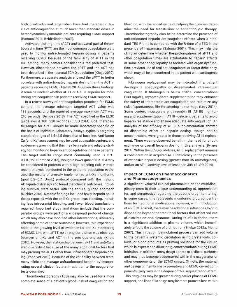
Anticoagulation During ECMO Support Unfractionated heparin is the most widely used anticoag-ulant for patients receiving ECMO support because it has a relatively short half-life, can be readily reversed, and can be titrated easily to achieve a patient-specific level of antico-agulation (ELSO 2014). At the time of cannulation, the ELSO guidelines recommend an initial bolus of 50–100 units/kg of unfractionated heparin, which may not be warranted when the patient is already actively anticoagulated (i.e., during percutaneous coronary intervention or on cardiopulmonary bypass) (ELSO 2014). Much debate remains regarding the most appropriate intensity of anticoagulation of patients receiving ECMO support, as well as the most effective method of monitoring this therapy (ELSO 2014). It is common practice to include several coagulation tests to monitor anticoagula-tion, which are combined with the patient’s clinical condition (e.g., the presence of bleeding or clinical thrombosis) to guide anticoagulation decision-making. For patients thought to have heparin-induced thrombocytopenia, the direct throm-bin inhibitors bivalirudin or argatroban should be considered. Dose selection of the direct thrombin inhibitors should fac-tor in existing coagulopathy and end-organ function because
CardSAP 2019 BOOK 1 • Heart Failure 13 Advanced Heart Failure
both bivalirudin and argatroban have had therapeutic lev-els of anticoagulation at much lower than standard doses in hemodynamically unstable patients requiring ECMO support (Ranucci 2011; Beiderlinden 2007).
Activated clotting time (ACT) and activated partial throm-boplastin time (PTT) are the most common coagulation tests used to monitor unfractionated heparin dosing in patients receiving ECMO. Because of the familiarity of aPTT in the ICU setting, many centers consider this the preferred test. However, discordance between the aPTT and the ACT has been described in the neonatal ECMO population (Khaja 2010). Furthermore, a separate analysis showed the aPTT to better correlate with unfractionated heparin dosing than the ACT in patientsreceivingECMO(Atallah2014).Giventhesefindings,it remains unclear whether aPTT or ACT is superior for mon-itoring anticoagulation in patients receiving ECMO support.
In a recent survey of anticoagulation practices for ECMO centers, the average minimum targeted ACT value was 183 seconds, and the reported average maximum ACT was 210seconds(Bembea2013).TheACTspecifiedintheELSOguidelines is 180–220 seconds (ELSO 2014). Goal therapeu-tic ranges for aPTT should bemade laboratory-specific onthe basis of individual laboratory assays, typically targeting standard ranges of 1.5–2.5 times that of baseline. Anti-factor Xa (anti-Xa) assessment may be used at capable centers, and evidence is growing that this may be a safe and reliable strat-egy for monitoring heparin anticoagulation in these patients. The target anti-Xa range most commonly used is 0.3– 0.7 IU/mL (Bembea 2013), though a lower goal of 0.2–0.4 may be considered in patients with a high bleeding risk. A more recent analysis conducted in the pediatric population evalu-ated the results of a newly implemented anti-Xa monitoring (goal 0.5–0.7 IU/mL) protocol compared with the historic ACT-guided strategy and found that clinical outcomes, includ-ing survival, were better with the anti-Xa–guided approach (Niebler2018).Notablefindingsincludedfewerheparinbolusdoses reported with the anti-Xa group; less bleeding, includ-ing less intracranial bleeding; and fewer blood transfusions required. Important study limitations include that the com-parator groups were part of a widespread protocol change, whichmayalsohavemodifiedotherinterventions,ultimatelyaffecting some of these outcomes; however, this experience adds to the growing level of evidence for anti-Xa monitoring of ECMO. Like with aPTT, no strong correlation was observed between anti-Xa and ACT in the previous analysis (Khaja 2010). However, the relationship between aPTT and anti-Xa is also discordant because of the many additional factors that may prolong the aPTT other than unfractionated heparin dos-ing (Vandiver 2012). Because of the variability between tests, many clinicians manage unfractionated heparin by incorpo-rating several clinical factors in addition to the coagulation tests described.
Thromboelastography (TEG) may also be used for a more complete sense of a patient’s global risk of coagulation and
bleeding, with the added value of helping the clinician deter-mine the need for transfusion or antifibrinolytic therapy.Thromboelastography also helps determine the presence of unfractionated heparin anticoagulant effects when a stan-dard TEG R-time is compared with the R-time of a TEG in the presence of heparinase (Salooja 2001). This may help the clinician determine whether the prolongations of aPTT and other coagulation times are attributable to heparin effects or some other coagulopathy associated with organ dysfunc-tion,previoususeoforalanticoagulants,orfactordeficiency,which may all be encountered in the patient with cardiogenic shock.
Fibrinogen replacement may be indicated if a patient develops a coagulopathy or disseminated intravascular coagulation. If fibrinogen is below critical concentrations(100 mg/dL), cryoprecipitate supplementation may enhance the safety of therapeutic anticoagulation and minimize any risk of spontaneous life-threatening hemorrhage (Levy 2014). Some centers incorporate antithrombin III (AT III) monitor-ingandsupplementationinATIII–deficientpatientstoavoidheparin resistance and ensure adequate anticoagulation. An analysis of the efficacy of AT III supplementation showedno discernible effect on heparin dosing, though anti-Xa concentrations were greater in those receiving AT III replace-ment.Therewasnoobservedbenefit intheneedforcircuitexchange or overall heparin dosing in this analysis (Byrnes 2014). Within the ELSO guidelines, AT III replacement remains aconsiderationinacquiredATIIIdeficiencyinthepresenceof excessive heparin dosing (greater than 35 units/kg/hour) and/or an AT III activity level of less than 30% (ELSO 2014).
Impact of ECMO on Pharmacokinetics and Pharmacodynamics Asignificantvalueofclinicalpharmacistsonthemultidisci-plinary team is their unique understanding of, appreciation for, and perspective regarding therapeutic drug monitoring. In some cases, this represents monitoring drug concentra-tions for traditional medications; however, with introduction ofanECMOcircuit,theremaybeadditionalinfluencestodrugdisposition beyond the traditional factors that affect volume of distribution and clearance. During ECMO initiation, there is a significant addition to plasma volume, which immedi-ately affects the volume of distribution (Shekar 2012a; Mehta 2007). This initiation (cannulation) process can add volume to the patient’s systemic circulation using crystalloids, col-loids, or blood products as priming solutions for the circuit, which is expected to dilute drug concentrations during ECMO initiation.Inaddition,manydrugsadheretoartificialsurfacesand may thus become sequestered within the oxygenator or other components of the ECMO circuit. Of note, the material and design of membrane oxygenators and ECMO circuit com-ponents likely vary in the degree of this sequestration effect. This drug loss may be greater during earlier phases of ECMO support, and lipophilic drugs may be more prone to loss within

CardSAP 2019 BOOK 1 • Heart Failure 14 Advanced Heart Failure
the circuit. Although hydrophilic drugs are less affected by drug loss, they are still likely to have a greater volume of dis-tribution because of the added plasma volume within the circuit.DrugsthathaveshownsignificantlosswithinECMOcircuits compared with control include fentanyl, midazolam, propofol, heparin, and voriconazole, potentially warranting different management strategies for each to ensure that ther-apeutic doses are maintained (Shekar 2012a; Shekar 2012b; Mehta 2007). Monitoring of available serum drug concentra-tions may be warranted, when possible, for medications in whichclinicalresponseisdifficulttoassessotherwise.
Sedation and Analgesia Optimizing sedation and analgesia can be one of the greater challenges in patients receiving ECMO support. Often, patients with refractory respiratory failure may require exces-sive doses of analgesic and sedative agents. Because oxygen consumption can be increased in agitated patients, poor sedation and persistent agitation may not only cause tis-sue hypoxia, but may also increase the danger that patients pose to themselves by dislodging ECMO cannulas, endotra-cheal tubes, nasogastric tubes, and intravascular catheters. Severalreportshaveshownsignificantsedativeoranalgesicdrug loss within ECMO circuits, including midazolam, propo-fol, dexmedetomidine, and fentanyl. This phenomenon may warrant greater doses, especially early in ECMO support, because the presence of the ECMO circuit may mimic the drug kinetics found in a multicompartment model (Wagner 2012; Mehta 2007; Mulla 2000). Morphine or hydromorphone may be a useful analgesic alternative to fentanyl in patients with uncontrolled pain receiving ECMO because both these agents are more hydrophilic than fentanyl (Shekar 2012b). Lorazepam may be a less lipophilic benzodiazepine option than midazolam or diazepam but still had some degradation compared with control concentrations in an in vitro model (Mulla 2000). In addition, as might be expected with any mul-ticompartment pharmacokinetic model, a period of drug redistribution may persist after discontinuation, possibly pro-longing the sedative effect. Many centers can keep patients awake and, in some cases, non-mechanically ventilated, to encourage mobility, minimizing the risk of mechanical venti-lation complications.
Antibiotic Therapy Because of the critical nature and hemodynamic instability of patients receiving ECMO support, suspicion for infection is quite common. Development of new infections while receiv-ing ECMO is a constant concern that should be met with rapid collection of blood, urine, and respiratory tract cultures as well as initiation of empiric broad-spectrum antimicro-bial agents on the basis of patient risk factors and local antimicrobial resistance patterns. With any antimicrobial agent selected, loading doses should be given with consid-eration for the greater volume of distribution present with
the ECMO circuit. It is especially important to achieve effec-tive therapeutic concentrations initially in a patient who may be experiencing septic shock. Like with the sedative medications, a component of drug sequestration of antimi-crobial agents may be within the circuit that poses the risk of treatment failure because of ineffective drug concentra-tions (Shekar 2012b). Antimicrobial agents that are quite lipophilic should be avoided, if possible. When therapeutic drug monitoring is feasible (e.g., vancomycin, aminoglyco-sides), drug concentrations should be monitored often. Most β-lactamsarehydrophilic,andcefotaxime,meropenem,andpiperacillin/tazobactam are minimally affected by the pres-ence of ECMO; therefore, clinicians can follow conventional dosing for these medications according to the patient’s CrCl (Donadello 2015; Ahsman 2010). For fungal infections, amphotericin B and hydrophilic azole antifungals may be the agents of choice to sustain effective antifungal concen-trations, depending on resistance patterns (Watt 2012; Ruiz 2009). Echinocandins have shown inconsistent pharma-cokinetics with ECMO, and voriconazole, a lipophilic azole antifungal, has consistently been shown to undergo signif-icant drug loss within the ECMO circuit (Ruiz 2009; Spriet 2009).Finally,forinfluenzainfection,higherdosesoftheanti-viral neuraminidase inhibitor oseltamivir (150 mg twice daily) have been described in patients receiving ECMO, for whom lower drug concentrations were reported than in patients not receiving ECMO (Eyler 2012). This report differed somewhat from a later study describing no difference in oseltamivir con-centrations between patients receiving ECMO and patients not receiving ECMO (Mulla 2013). Nonetheless, oseltamivir concentrations at standard doses were considered by both groupsofinvestigatorstoachievesufficientlyeffectivecon-centrations to treat severe influenza infections; therefore,a higher dosing strategy may not be necessary to provide therapeutic concentrations (Mulla 2013; Eyler 2012).
PERCUTANEOUS VENTRICULAR ASSIST DEVICES Indications for and Types of pVADsThe main indications for pVAD therapy are either for hemo-dynamic support during high-risk percutaneous coronary intervention or for use in the setting of refractory cardiogenic shock caused by acute myocardial infarction or severe HF. Currently, two FDA-approved devices may be used in these clinical settings. The first is the Impella series (Abiomed,Danvers, MA), which includes the Impella 2.5, the 5.0, the CP (Cardiac Power), and the RP. The Impella 2.5 is a cath-eter-mounted microaxial pump mounted on a 9-French catheter shaft, which houses the motor driveline and the purge line system. Insertion is usually done through a fem-oral approach, and the pump is positioned across the aortic valveintotheLVwithfluoroscopy(Allender2017)(Figure2).Expelling aspirated blood from the LV into the ascending

CardSAP 2019 BOOK 1 • Heart Failure 15 Advanced Heart Failure
aorta, the Impella 2.5 at its maximal rotation speed of 51,000 rpmprovidesflowofupto2.5L/minute.TheImpellaCPusesthe same platform as the 2.5 device but provides additional cardiac support and operates with a mean flow of 3–4 L/minute. The Impella 5.0 device carries a larger motor capa-ble of providing up to 5 L/minute of support and, as such, is inserted into the LV through femoral cutdown or through the axillary artery. Finally, the Impella RP is approved to provide circulatory support to those who develop acute right HF; this pump delivers blood from an inlet area in the inferior vena cava through the cannula to the outlet opening near the tip of the catheter in the pulmonary artery.
The TandemHeart pVAD (CardiacAssist, Pittsburgh, PA) is a low-speed centrifugal continuous-flow pump that canbe introduced percutaneously in the cardiac catheterization laboratory (Thiele 2001). Inserted by a venous transseptal puncture through the femoral vein, a 21-French left atrial can-nula channels blood into the pump, and a 15- to 17-French femoral artery cannula carries the blood to the systemic arte-rial circulation. The TandemHeart is capable of up to 4.5 L/minute of assisted cardiac output and can be used for both left- and right-sided mechanical support.
Hemodynamic Consequences of pVADs The Impella devices propel blood from the LV into the ascend-ing aorta, thereby unloading the LV and increasing cardiac output. They reduce myocardial oxygen consumption, improve
MAP, and reduce pulmonary capillary wedge pressure (PCWP) (Rihal 2015). The Impella 2.5 provides a greater increase in cardiac output than an IABP but less than the TandemHeart device (Table 2). The more powerful Impella CP and 5.0 devices are similar to the TandemHeart device with respect to MCS. Similar to the TandemHeart, adequate RV function is necessary to maintain LV preload and hemodynamic support during biventricular failure.
During MCS with the TandemHeart, both the LV and the device contribute flow to the aorta simultaneously (therebyworking in parallel, or tandem, rather than in series). Redirection of blood from the LA reduces LV preload, LV workload, fill-ing pressures, wall stress, and myocardial oxygen demand (see Table 2) (Rihal 2015). The increase in arterial blood pres-sure and cardiac output supports systemic perfusion. The aorta is thus perfused and pressured by both the LV and the TandemHeart, with the relative contribution of each varying, depending on LV response to the device. Not infrequently, LV contraction virtually ceases, and perfusion is pump- dependentwithaflatMAPcurve.LikewiththeImpelladevices,ventriculartachycardiaorfibrillationusually(butnotalways)renders the TandemHeart ineffective because of RV failure.
Anticoagulation During pVAD Support Successful use of the Impella devices is predicated on effective heparin-based anticoagulation. The manufacturer recommends administering unfractionated heparin through
Outlet area
To aortic valve/left ventricle
Blood flow
Pressure barrier createdby countercurrent flow
Purge flow
To aortic arch
Figure 2.SchematicofthepurgeflowsystemintheImpelladevices.AftersuccessfulinsertionoftheImpelladeviceacross the aortic valve, the outlet area expels blood withdrawn from the left ventricle (not shown) into the ascending aortathroughaxillaryflow.Adextrose-basedpurgesolutionisreleasedfromthecathetercountercurrenttobloodflowthat creates a pressure barrier and prevents entry of blood into the motor housing. After initial insertion of the device, the dextrose-only solution is commonly replaced with a purge solution that also contains heparin, which may further reduce the risk of thrombosis, should blood enter the motor housing.
Reprinted with permission from: Allender JE, Reed BN, Foster J et al. Pharmacologic considerations in the management of patients receiving left ventricular percutaneous mechanical circulatory support. Pharmacotherapy 2017;37:1272-83.

CardSAP 2019 BOOK 1 • Heart Failure 16 Advanced Heart Failure
a purge solution, which is used to lubricate the motor and maintain a pressure within the device of 300–1100 mm Hg. Historically, the default purge solution is 25,000 units of unfractionated heparin in 500 mL of 20% dextrose solution (50 units/mL), though lower concentrations of unfractionated heparin (e.g., 12.5 or 25 units/mL) can be used if the patient develops supratherapeutic aPTT values.
Recently, the device manufacturer changed the recom-mended default dextrose concentration to 5%, which is relevant because this reduction in the viscosity of the purge solutionwilllikelyincreasetheflowrate(andthustheunfrac-tionated heparin exposure) by as much as 30%–40%. The device console automatically adjusts the flow rate of thepurge to 2–30 mL/hour to maintain purge pressure, which is problematicbecausesuchfluctuationscansignificantlyalterthe patient’s exposure to unfractionated heparin. Adding to this already complicated scenario is the need to maintain therapeutic anticoagulation (ACT of 160–180 seconds or aPTT of 60–80 seconds), which often necessitates supple-mental intravenous unfractionated heparin (Seyfarth 2008). Simultaneous administration of unfractionated heparin in the purge solution (which is controlled by the console) together with intravenousunfractionatedheparinposesasignificanthazard for medication error and heparin over- or underdos-age. The Patient Case Scenario highlights this problem and offers potential solutions to avoid harm and optimize antico-agulation strategies in these patients.
Similar to the Impella devices, anticoagulation with the TandemHeart is complicated by the need for a heparinized infusate of 1000 mL of normal saline with 90,000 units of unfractionatedheparin.Thisinfusaterunsatafixedrateof10mL/hour,which,unlikewiththeImpelladevices,willnotfluc-tuate with unfractionated heparin exposure. Of importance, the infusate must be saline because dextrose-containing products can damage the motor and lead to catastrophic fail-ure of the device. Additional unfractionated heparin can be administered intravenously, as needed, to achieve therapeu-tic anticoagulation (Table 3).
Complications of pVAD Therapy The most commonly reported complications of Impella placement are limb ischemia, vascular injury, and bleeding requiring blood transfusion. Vascular complications com-mon to all transfemoral procedures such as hematoma, pseudoaneurysm, and arterial-venous fistula and retroperi-toneal hemorrhage can occur with any mechanical support device. Hemolysis as the result of mechanical erythrocyte shearinghasbeen reportedwithin thefirst24hoursofusein 5%–10% of patients, who may respond to repositioning of the device (Lauten 2013). Persistent hemolysis is an indica-tion for device removal. Because the Impella device traverses
Table 2. Comparison of Available Temporary Support Devices
IABP Impella TandemHeart VA ECMO
Maximum support 0.5–1 L/min 2.5, 3.5, or 5.0 L/min Up to 4.1 L/min ≥5L/min
LV unloading + ++,+++,++++ +++ ++
Coronary perfusion + + – –
Bleeding risk + ++ +++ ++++
Management complexity + ++ ++++ +++
Maximum implant timea Weeks 7 days 14 days Days to weeks
aAccording to manufacturer recommendations.VA ECMO = venoarterial extracorporeal membranous oxygenation.
Table 3. Example Anticoagulation Protocol for the TandemHeart Device
Initiating Anticoagulation
aPTT (seconds) Instructions
< 55 Continue current infusate (heparin 45,000 units/500 mL saline) at 10 mL/hr, initiate intravenous heparin 2 units/kg/hr
55–75 Therapeutic – No changes
76–90 Switch infusate to heparin 25,000 units/500 mL saline at 10 mL/hr, and initiate intravenous heparin at 2 units/kg/hr
91–110 Switch infusate to heparin 25,000 units/500 mL saline at 10 mL/hr, do not start intravenous heparin
>110 Switch infusate to saline (no heparin) at 10 mL/hr
Information from: Lee Y, Weeks PA. Effectiveness of protocol guided heparin anticoagulation in patients with the TandemHeart percutaneous ventricular assist device. ASAIO J 2015;61:207-8.

CardSAP 2019 BOOK 1 • Heart Failure 17 Advanced Heart Failure
the aortic valve and sits in the LV, it is contraindicated in patients with a mechanical aortic valve or in those with an LV thrombus. Aortic stenosis and regurgitation are relative con-traindications, though reports exist of hemodynamic rescue in critical aortic stenosis or to facilitate valvuloplasty. These devices should not be placed in patients with severe periph-eral arterial disease or in those who cannot tolerate systemic anticoagulation. Finally, Impella use may theoretically worsen right-to-left shunting and hypoxemia in patients with a preex-isting ventricular septal defect.
Because the TandemHeart does not pass through the aorta, it is safe in patients with aortic valvular pathology. However, the transseptal puncture needed to implant the device exposes patients to unique complications like cardiac tamponade. Other possible complications include throm-bosis, air embolism, and hemolysis. Care must be taken to prevent dislodgement of the left atrial cannula, particularly during patient transport or if patients move their legs, because dislodgement into the right atrium results in massive right-to-left shunting and severe systemic oxygen desaturation. The cannula may also migrate into a pulmonary vein, which leads to device malfunction.
DURABLE LVAD THERAPYIndications for and Types of Durable LVAD PumpsCurrent guidelines for managing HF suggest that dura-ble LVAD therapy can be considered to prolong survival in carefully selected patients (see Table 1) with stage D dis-ease (Yancy 2013). These devices can be used as either a bridge-to-transplantation or destination therapy in those who are not candidates for HT. All commercially available LVADs in theUnitedStatesoperateundercontinuousflow(CF-LVADs);the HeartMate II (Thoratec Corp., Pleasanton, CA) operates through a mechanical-bearing internal impeller (similar to an Archimedes screw) and delivers up to 10 L/minute of cardiac outputbyaxiallaminarflow.TheHeartWareHVAD(HeartWareInternational, Framingham, MA) is a smaller pump that is inserted directly into the pericardium; this device uses a water wheel–like impeller to generate full cardiac support by centrif-ugalflow.TheHeartMateIII(Thoratec)isafullymagneticallylevitated centrifugal pump that is also inserted directly into the pericardium. All devices cannulate the apex of the LV to provide direct mechanical cardiac unloading, and each pro-pels blood forward through an outflow graft anastomosisto the ascending aorta. The HeartMate II and HeartMate III devicesprovideasnapshotoffourdeviceparameters(flow,speed, pulsatility index, and power) on the device counsel, whereas the HeartWare HVAD monitor provides continuous waveformanalysisofflowandpower(Table4). Speed is set by the providing clinician with the goal of obtaining optimal flow(whichiscalculatedbythedeviceonthebasisofpowerconsumption).
Patient Care ScenarioA woman (weight 75 kg) is admitted to the ICU with an Impella CP device in place. Her purge solution (25,000 units/500 mL of dextrose 5% in water) is running at 10 mL/hour, or 500 units/hour of heparin. According to the heparin protocol for this hospital, her total hourly hep-arin dose should be 900 units (75 kg × 12 units/kg) to achieve an aPTT of 60–80 seconds.
Part 1According to this protocol, how much intravenous hepa-rin should the patient receive?
ANSWERThe patient should be initiated on an intravenous hepa-rin drip at a rate of 400 units/hour to equal a total hourly dose of 900 units/hour (500 units from the purge plus 400 units intravenously).
Part 2Two hours after starting intravenous heparin, the nurse notices that the Impella controller has reduced the flow rate of the purge solution to 8 mL/hour, or 400 units/hour of heparin. She notifies the physician, who asks the ICU pharmacist for assistance. What is the most appropriate action to take at this time?
ANSWERThe pharmacist should recommend increasing the rate of intravenous heparin by 100–500 units/hour so that the patient continues to receive a total hourly dose of 900 units/hour (400 units purge plus 500 units intravenously).
Part 3After 6 hours of support, the first aPTT value is 47 sec-onds (goal 60–80 seconds). The ICU team asks the pharmacist for assistance with anticoagulation man-agement. What is the most appropriate action at this time?
ANSWERBecause the aPTT is subtherapeutic, the patient will require more heparin. This can be accomplished by increasing the intravenous heparin. Remember that the purge flow rate is controlled by the device and cannot be titrated to achieve a therapeutic aPTT. A reasonable solution is to increase the infusion rate for the intrave-nous heparin by 100 units/hour, and the new total hourly dose will be 1000 units/hour (400 units purge plus 600 units intravenous).
1. Jennings DL, Nemerovski CW, Kalus JS. Effective anti-coagulation for a percutaneous ventricular assist device using a heparin-based purge solution. Ann Pharmacother 2013;47:1364-7.
2. Allender JE, Reed BN, Foster J, et al. Pharmacologic con-siderations in the management of patients receiving left ventricular percutaneous mechanical circulatory support. Pharmacotherapy 2017;37:1272-83.

CardSAP 2019 BOOK 1 • Heart Failure 18 Advanced Heart Failure
Hemodynamic Consequences of Durable LVADs Continuous pumping of blood directly from the LV inde-pendent of the cardiac cycle results in loss of the normal isovolumic periods. Unlike the other forms of support like the Impella devices, removal of blood from the LV in dura-ble LVADs does not depend on ejection through the aortic valve.Aspumpflowrateincreases,theLVbecomesincreas-ingly unloaded, peak LV pressure generation decreases, and myocardial oxygen consumption markedly decreases. At the same time, arterial blood pressure increases such that peak LV pressure and arterial pressure are increasingly dis-sociated. This direct unloading also results in decreased left atrial and PCWP. Over time, these improvements in blood oxy-genation, systemic pressures, and perfusion may reverse the metabolicmilieuofend-stageHFandinvokebeneficialsec-ondary changes in LV contractility and peripheral resistance.
Management of Hypertension All CF-LVADs are sensitive to increases in afterload, such that elevations in systemic arterial pressure can impede device functionand reduce forwardflow (seeTable4). The
presence of hypertension also presages a heightened risk of stroke in CF-LVAD recipients (Nassif 2015). Depending on the device speed and the residual native left heart function, these patients will lack pulsatility; thus, blood pressure targets are based on the MAP. Noninvasive blood pressure monitoring is done with a Doppler probe because of the aforementioned loss in pulsatility.
Clinicians facing systemic arterial hypertension (usually definedasaMAPgreaterthan90mmHg)intheearlypost-operativesettingshouldfirstassesssystemicperfusionandthen begin to withdraw inotropic support in patients with adequate mixed venous oxygen saturation (Svo2) and stable end-organ function. If hypertension persists or these medica-tions cannot be weaned because of low Svo2 (e.g., less than 65%), a systemic vasodilator should be initiated. If intrave-noustherapyisneeded,nicardipineisoftenthefirstdrugofchoice because of its relatively neutral effects on cardiac ino-tropy and chronotropy, though sodium nitroprusside can be considered as an alternative. Patients should be transitioned to oral therapy as soon as possible; ACE inhibitors are con-sideredfirst-line therapy in thosewithstable renal function
Table 4. Device Parameters for CF-LVAD
Parameter Normal Values Can Be Elevated by: Can Be Decreased by: Pharmacotherapy Considerations
Flow 4–6 L/mina SepsisDevice thrombosisAorticinsufficiency
RV failureDehydrationHemorrhageHypertensionArrhythmias
Monitorfordecreasesinflowwhentitratingβ-blockersordiuretics
Titrate afterload-reducing agents to avoid hypertensionandoptimizeflow
Monitor for evidence of blood loss or device thrombosis
Speed 8800–9800 rpmb
2800–3400 rpmc
5000–6000 rpmd
None – adjusted by health care team
Dehydration Sudden drops in speed (i.e., suction event) may indicate dehydration and should prompt assessment of diuretic regimen, fluidstatus,andpotentialhemorrhage
Pulsatility index
4–7 for HeartMate II and 3–5 for HeartMate IIIe
Hypertension HypotensionDehydrationRV failure
Sudden drops in pulsatility index (i.e., suction event) in HeartMate II/III may indicate dehydration and should prompt assessmentofdiureticregimenandfluidstatus
Power 5–7 W Device thrombosisHypertension
HypotensionSepsis
Sustained power elevation should prompt evaluation for device thrombosis
aNormal value depends on the patient’s BMI.bTypical range for HeartMate II device.cTypical range for HeartWare HVAD.dTypical range for HeartMate III device.eCalculated only by the HeartMate II and HeartMate III devices; when the LV contracts, the increase in ventricular pressure causes anincreaseinpumpflowduringcardiacsystole.Themagnitudeoftheseflowpulsesismeasuredandaveragedover15-sintervalstoproducea“pulsatilityindex”(PI).ThemagnitudeofthePIvalueisrelatedtotheamountofassistanceprovidedbythepump.Highervaluesindicatemoreventricularfillingandhigherpulsatility(i.e.,thepumpisprovidinglesssupporttotheLV).Lowervaluesindicatelessventricularfillingandlowerpulsatility(i.e.,thepumpisprovidinggreatersupportandfurtherunloadingtheventricle).Informationfrom:JenningsDL,SchilligJ,ChambersR.ThepharmacotherapyoftheHeartMateII,acontinuousflowleft-ventricularassist device, in patients with advanced heart failure: integration of disease, device and drug. Ann Pharmacother 2010;44:1647-50.
CardSAP 2019 BOOK 1 • Heart Failure 19 Advanced Heart Failure
and acceptable potassium concentrations (Lampert 2014). Dihydropyridine calcium channel blockers (e.g., amlodipine) are also acceptable, as areβ-receptor antagonists (assum-ing that RV function is adequate). Most CF-LVAD recipients require one or two antihypertensive medications to maintain an optimal MAP (less than 80 mm Hg) (Lampert 2014).
Management of Ventricular Arrhythmias Sustained ventricular arrhythmias can occur in up to 40% of CF-LVAD recipients and are most common in patients with a history of this condition (Raasch 2012). As the LV is com-pletely unloaded, the pathophysiologic sequela of these arrhythmias may be negligible. Reports exist of patients sur-viving hours and, in extreme cases, months of ventricular fibrillation.However, the lossoforganizedcontraction fromthe unsupported RV can lead to hemodynamic destabilization byreducingpumpflowsecondarytounsatisfactoryleft-sidedvolume for ventricular filling (see Table 4). Therefore, treat-ment decisions regarding ventricular arrhythmias should be made on a case-by-case basis according to the patient’s over-all condition. Although asymptomatic arrhythmias may not require intervention, those associated with hypotension, RV failure, or clinical symptoms (e.g., dizziness or palpitations) warrant treatment.
Amiodarone remains the preferred antiarrhythmic agent for CF-LVAD recipients. In addition to the customary moni-toring for this agent, clinical pharmacists should be mindful that initiating amiodarone therapy may alter the pharmaco-dynamic response to warfarin, which is germane because virtually all recipients of durable CF-LVADs require oral anticoagulation (Edwin 2010). For patients who develop intol-erance to amiodarone or whose amiodarone therapy fails, lidocaine or mexiletine is an appropriate secondary treatment option.β-Blockersareeffectiveantiarrhythmicmedicationsthat should also be initiated, when possible, in CF-LVAD recip-ients; however, the potential for negative inotropy and RV dysfunctionwithβ-blockersmaylimittheiruse(Refaat2008).Therefore, clinicians should monitor these patients closely for signs of RV failure (see below) whenever initiating or titrat-ingβ-blockers.
Management of RV Failure The anatomy and physiology of the RV are quite distinct from those of the LV. The LV has three muscle layers (oblique, circular, and longitudinal), whereas the RV only has two (cir-cumferential and longitudinal) (Sheehan 2008). Furthermore, although the LV exerts powerful torsional and rotational forces, the RV operates using peristaltic contractions (similar to the GI smooth muscles). The RV largely depends on the low hydraulic impedance characteristics of the pulmonary vascu-lar bed and, as such, can achieve comparable output with a myocardialenergycostofaboutone-fifththatoftheLV.
Unanticipated RV failure may occur in up to 40% of recipi-entsofdurableCF-LVADsandisassociatedwithsignificantly
worsenedsurvival (Tsiouris2015).Nouniformdefinition forsevere RV failure exists; however, this pathology is commonly described as the need for placement of an RV assist device or use of intravenous inotropes for more than 14 days post-operatively. Many preoperative risk factors for developing RV failurehavebeen identified, includingelevations incen-tral venous pressure (CVP), diminished RV stroke work index and RV contractility on echocardiography, and the presence of signs and symptoms suggestive of right HF (Morgan 2013).
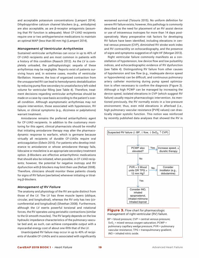
Right ventricular failure commonly manifests as a con-stellationofhypotension,lowdeviceflowandlowpulsatilityindices, and echocardiographic evidence of RV dysfunction (see Table 4). Distinguishing RV failure from other causes ofhypotensionandlowflow(e.g.,inadequatedevicespeedorhypovolemia)canbedifficult,andcontinuouspulmonaryartery catheter monitoring during pump speed optimiza-tionisoftennecessarytoconfirmthediagnosis(Figure3).Although a high PCWP can be managed by increasing the device speed, isolated elevations in CVP (which suggest RV failure) usually require pharmacologic intervention. As men-tioned previously, the RV normally exists in a low-pressure environment; thus, even mild elevations in afterload (i.e., high pulmonary vascular resistance [PVR] values) can dras-tically impair systolic function. This notion was reinforced by recently published data analyses that showed the RV is
No
No
Yes
Yes
Considerinodilator (e.g.,dobutamine)
Consider iNO,inhaled
epoprostenol,inhaled milrinoneInhaled iloprost
PVR > 3 Woodunits OR TPG >
12 mm Hg?
Increase speed, ±diuretic therapy
PCWP alsoelevated?
Suspected RV failure (↓ BP, ↓ flow, ↓ SvO2, ↑ CVP)
Figure 3. Flow chart for pharmacologic management of right-ventricular (RV) failure.
BP = blood pressure; CVP = central venous pressure; SvO2 = mixed venous oxygen saturation; PCWP = pulmonary capillary wedge pressure; PVR = pulmonary vascular resistance; TPG = transpulmonary gradient; iNO = inhaled nitric oxide.

CardSAP 2019 BOOK 1 • Heart Failure 20 Advanced Heart Failure
more sensitive to even small increases in afterload pressure early after CF-LVAD implantation (Houston 2016). Therefore, the choice of agent for pharmacologic support of the fail-ing RV hinges on the patient’s PVR value (Figure 4). For RV failure with normal PVR values, traditional inotropic ther-apy (i.e., dobutamine ormilrinone) should be sufficient toimprove contractility and RV output. Conversely, if the PVR is elevated (greater than 250 dynes/second/cm5 or 3 Wood units) or the patient has other evidence of a high RV after-load (e.g., a transpulmonary gradient greater than 12 mm Hg [mPAP-PCWP]), a selective pulmonary artery vasodila-tor is the preferred initial pharmacologic agent. A complete review of these agents is beyond the scope of this chap-ter; however, Table 5 includes a summary description of the medications commonly used in the ICU for acute RV failure. Inhaled nitric oxide is historically the most common treat-ment;however,thistherapyissignificantlylimitedbecauseof its high cost. Inhaled epoprostenol is significantly lessexpensive than inhaled nitric oxide but is cumbersome to administer and can increase bleeding risk through inhibi-tion of platelet aggregation (Groves 2014). Recent pilot data analyses suggest that inhaled milrinone can also be used for acute RV failure after CF-LVAD implantation; however, absorption from the pulmonary circulation produces ther-apeutic plasma milrinone concentrations; hence, patients receiving this modality may be at risk of hypotension and cardiac arrhythmias (Haglund 2015). For severe, refractory postoperative pulmonary arterial hypertension, combining inhaled pulmonary vasodilators with complimentary phar-macology (e.g., epoprostenol plus milrinone or inhaled nitric oxide plus iloprost) can be considered as salvage therapy (Antoniou 2013; Haraldsson 2001). If pharmacology does not
reverse RV failure, mechanical right heart support should be initiated. However, the need for an RV assist device after CF-LVAD implantation is associated with a high postoper-ative mortality (Morgan 2013). If patients with an elevated PVR respond to inhaled pulmonary vasodilators and recover from RV failure, transition to an oral pulmonary vasodilator (e.g.,sildenafil)canbeconsidered.
Thromboprophylaxis During LVAD Support All commercially available durable CF-LVADs carry a risk of thrombosis, which can include clotting within the device itself (i.e., pump thrombus) as well as ischemic stroke caused by device-related emboli. Lifelong combination therapy with warfarin-based anticoagulation and antiplatelet agents is required to mitigate these complications. No randomized data exist comparing anticoagulation regimens in CF-LVAD recipients; as such, significant heterogeneity exists withinthefield,andpracticeisoftenguidedbylocalcenterexperi-ence (Jennings 2016). For both the HeartMate II device and the HeartWare HVAD, intravenous unfractionated heparin should be initiated as soon as surgical hemostasis has been achieved. Both device manufacturers recommend targeting aloweraPTTforthefirst24–48hours(e.g.,45–50seconds),with the eventual goal of titrating it toward 55–65 seconds (Maltais 2017). Aspirin should also be initiated by postopera-tive day 2 at a dose of 81 mg daily for the HeartMate II/III and a dose of 162–325 mg daily for the HeartWare HVAD. For the HeartMate II device, some centers still use dual antiplatelet therapy with aspirin and dipyridamole, whereas other cen-ters (particularly in Europe) omit antiplatelet agents entirely (Jennings 2016).
Figure 4. Central venous pressure (CVP) is plotted on the x-axis against pulmonary capillary wedge pressure (PCWP) on the y-axis. Patients with elevations in both of these values likely have biventricular failure, whereas isolated elevations in either CVP or PCWP may represent either right or left heart failure, respectively. Point 1 represents a low speed that is not adequate to unload the LV. At point 2, the speed has been optimized, and both ventricles are adequately decompressed. At point 3, the speed has been increased too much, resulting in a leftward shift of the intraventricular septum and subsequent RV failure.

CardSAP 2019 BOOK 1 • Heart Failure 21 Advanced Heart Failure
When transitioning from unfractionated heparin to warfarin, the clinical pharmacist should assist with dosing, particularly when major drug-drug interactions are present. If the patient was taking warfarin before CF-LVAD implantation, historical requirements can be used as a basis for postoperative dosing (Jennings 2012). Warfarin genotype data, if available, can also help in selecting an appropriate initial dosing regimen (Jennings 2016). Of note, the pharmacist should ensure that unfraction-atedheparinisoverlappeduntilatleastfivedosesofwarfarinhave been administered and until the INR is therapeutic for at least two readings taken 24 hours apart (Colombo 2016).
The HeartMate II, HeartMate III, and HeartWare HVAD man-ufacturers advocate an INR target of 2–3; however, many centers use narrower ranges (e.g., 2–2.5 or 2.5–3) (Jennings 2016). Although no randomized data analyses have compared one INR target range with another, use of narrower targets is usually associated with lower time within the therapeutic range and poorer anticoagulation quality (Kuyumjian 2016). In light of this, a standard initial INR of 2–3 is recommended for all recipients of durable CF-LVADs. This recommenda-tion is further supported by a recent analysis of over 10,000 INR values in 249 HeartMate II recipients, which found that the optimal INR was 2.6 on the basis of weighted mortality of thrombotic and bleeding events, with low rates of adverse events at INR values of 2.0–3.2 (Nassif 2016).
TheHeartMate IIIdevicewasdesignedwithspecific fea-tures to enhance hemocompatibility and reduce the risk of thrombosis; these include a fully magnetically levitated rotor, widebloodflowpassages,andanintrinsicpulsedesignedtoavert stasis within the pump. In the 2-year results from the pivotal MOMENTUM 3 randomized trial, suspected events of pump thrombosis occurred in 2 patients (1.1%) in the centrifu-gal-flowpumpgroupcomparedwith27patients(15.7%)whohad33sucheventsintheaxial-flowpumpgroup(HR0.06;95%CI, 0.01–0.26; p<0.001) (Mehra 2018). Given this more forgiv-ingthromboticprofile,pilotdatahavealreadybeenpublishedexploring lower-intensity anticoagulation. MAGENTUM 1 is a prospective, single-arm pilot study of 15 HeartMate III recip-ients who received standard warfarin anticoagulation (INR 2.0–3.0) and aspirin for 6 weeks postimplantation, followed by a lower INR target of 1.5–1.9 for 6 months (Netuka 2018). The primary end point of survival free from pump thrombo-sis, disabling stroke, or major bleeding during follow-up was met in 14 of 15 patients (one patient developed GI bleeding). Although these data are preliminary, they support the feasibil-ity of reduced-intensity anticoagulation for the HeartMate III and pave the way for additional investigation of this promis-ing strategy.
Data analyses exploring the use of non–warfarin-based anti-coagulation are limited to one small report from a single-center,
Table 5. Comparison of Commonly Used Selective Pulmonary Artery Vasodilators in the ICU
Agent Mechanism of Action Common Doses Notes
Inhaled nitric oxide
Activates intracellular guanylyl cyclase, which increases concentrations of cyclic guanosine 3'5’-monophosphate
1–20 ppm through continuous inhalation
• Very short half-life• Can cause methemoglobinemia• Very expensive• Limited systemic exposure
Inhaled epoprostenol
Activates intracellular adenylate cyclase, which increases concentrations of cyclic adenosine monophosphate
25–50 ng/kg/min • Complicated administration• Less expensive• Some systemic exposure
○ Potential for platelet inhibition and bleeding
○ Potential hypotension
Iloprost Activates intracellular adenylate cyclase, which increases concentrations of cyclic adenosine monophosphate
2.5–5 mcg inhaled 6–9 times daily
• Ease of administration• Patient can be extubated• Expensive• Some systemic exposure
○ Potential for platelet inhibition and bleeding
○ Potential hypotension
Inhaled milrinone
Inhibits phosphodiesterase type 3, which increases concentrations of cyclic adenosine monophosphate
6 mg/hr continuous inhalation
• Complicated administration• Patient must be intubated• Less expensive• Systemic exposure
○ Potential hypotension ○ Arrhythmia

CardSAP 2019 BOOK 1 • Heart Failure 22 Advanced Heart Failure
randomized, open-label study of 16 HeartWare HVAD recipients in Vienna (Andreas 2017). Patients with normal or impaired renal function (GFR greater than 80 mL/minute/1.73 m2 or between 80 and 30 mL/minute) received phenprocoumon or dabigatran at a dose of 110 or 75 mg twice daily. The study was terminated prematurely when four of the eight HeartWare HVAD recipients had a thromboembolic event. Although dis-appointing, these results must be viewed in the context of the study limitations, most notably the small sample size and the nonstandardized dosing of dabigatran, which likely produced markedly insufficient drug concentrations to protect againstdevice thrombosis. Nonetheless, pending the publication of additional research, warfarin should remain the only antico-agulant used for thromboprophylaxis in all the commercially available CF-LVADs.
Infectious Complications of Durable LVAD Therapy Infection is a major complication associated with LVAD ther-apy, with reported rates of 25%–80% (Nienaber 2013). The clinical spectrum of infection in CF-LVAD recipients includes infections related to the device (e.g., the percutaneous drive-line) as well as non–LVAD-related infections (e.g., pneumonia, bacteremia). Chronic infection of the driveline is the most common infection site (Nienaber 2013). Some CF-LVAD recip-ients may develop severe infections, including bacteremia and sepsis. When patients present with severe infection and critical illness, clinical pharmacists must be familiar with the epidemiology of CF-LVAD infections as well as the potential for altered pharmacokinetics of antimicrobial therapy in dura-ble device recipients.
Several studies have shown that the continuum of patho-gens associated with LVAD-related infection encompasses gram-positive and gram-negative bacteria as well as fun-gal species. Methicillin-sensitive Staphylococcus aureus, coagulase-negative staphylococci, methicillin-resistant S. aureus (MRSA), Enterococcus sp., Pseudomonas aeruginosa, Klebsiella sp., Escherichia coli, Stenotrophomonas sp., Serratia sp., Candida sp., Propionibacterium sp., diphtheroids, and Corynebacterium sp.haveallbeenidentifiedinthesepatients(Nienaber 2013). As such, pharmacists caring for CF-LVAD recipients with severe infections should initiate broad- spectrum antimicrobial therapy, including agents that cover resistant bacteria (e.g., methicillin-resistant gram-positive cocci and Pseudomonas sp.) and Candida sp. (Kusne 2017). For patients with more benign infections (e.g., local drive-linesite),delayingtherapyuntilthepathogenisidentifiedisanacceptableapproach(Kusne2017).Specificagents(bothantimicrobial and antifungal) should be chosen together with infectious disease physicians and should incorporate local antibiogram data. However, the optimal treatment duration for CF-LVAD–related infections remains undefined. Recentguidelines advocate short courses for patients with uncom-plicated infections (e.g., 2 weeks), whereas patients with
more severe infections such as bacteremia or pump pocket infection may require treatment for 6–8 weeks or longer, depending on the infection (Kusne 2017).
Limited data analyses have suggested that CF-LVAD recipients have a larger volume of distribution and a lower- than-anticipated drug clearance compared with non-LVAD recipients, even in the face of apparent euvolemia and nor-mal hemodynamics (Jennings 2014). Clinical pharmacists must therefore be vigilant with monitoring for both clinical efficacy and toxicity associatedwith antimicrobial therapyand must implement therapeutic drug monitoring whenever possible.
Recently published guidelines have provided recommen-dationsforantimicrobialprophylaxisforthefirsttime.Theserecommendations are summarized in Box 2.
Bleeding Complications of Durable LVAD Therapy Gastrointestinal bleeding, often originating from arteriove-nous malformations within the small intestine and colon, affects over 15% of patients receiving durable LVAD support (Goldstein 2015). Recent evidence suggests that the sustained elevations in serum thrombin concentrations during mechan-ical support generate an excess of angiopoietin-2, which appears to drive the growth of these arteriovenous malforma-tions (Tabit 2016). Compounding these anatomic lesions is the depletion of high-molecular-weight von Willebrand mul-timers from LVAD-induced sheer stress, which produces a physiologic state of hypocoagulability (Bartoli 2015). Although these anatomic and physiologic derangements can lead to persistent and recurrent mucosal bleeding, they are sometimes types of life-threatening hemorrhage. Initial management of GI bleeding is predominantly nonpharmaco-logic (i.e., endoscopic intervention) (Goldstein 2015). Several drugtherapieshavehadprovenbenefitinsmallcaseseries
Box 2. Summary of Recommendations for Antimicrobial Prophylaxis During LVAD Implant• Regimen should target Staphylococcus sp.• Regimen should cover MRSA in colonized patients• Routine broad-spectrum gram-negative prophylaxis is not
recommended unless guided by local epidemiologic data• Rifampin prophylaxis is not recommended because of
drug-drug interactions• Routine antifungal prophylaxis is not recommended• Duration of antimicrobial prophylaxis should not exceed
48 hr• Duration of prophylaxis should not be based on the pres-
ence of chest tubes or drains
Information from: Kusne S, Mooney M, Danziger-Isakov L, et al. An ISHLT consensus document for prevention and manage-ment strategies for mechanical circulatory support infection. J Heart Lung Transplant 2017;36:1137-53.

CardSAP 2019 BOOK 1 • Heart Failure 23 Advanced Heart Failure
and case reports, including octreotide, ACE inhibitors, and thalidomide. A comprehensive review of the evidence for each of these agents was recently published (Sieg 2017). Although the supporting evidence for each of these modali-ties is limited, the authors of this review proposed a treatment algorithm that was based on their expert opinion (Figure 5). Finally, although the HeartMate III was clearly superior in reducing pump thrombosis, rates of GI hemorrhage were sim-ilar between this device and the HeartMate II (Mehra 2018). These findings highlight the need for continued advancesin pump technology to combat additional adverse events beyond device thrombosis.
Because of the innate thrombotic nature of CF-LVADs, bleeding necessitating anticoagulation reversal is a par-ticularly precarious clinical scenario with the potential for catastrophic complications. As such, anticoagulation reversal should be reserved for patients with a potentially life-threatening hemorrhage. Limited data analyses suggest that common modalities, including 3- and 4-factor PCCs, vitamin K, and fresh frozen plasma, are safe to reverse the effects of warfarin (Chen 2015; Jennings 2014). In accor-dance with published guidelines for anticoagulation in non-LVAD recipients, a regimen of a 4-factor PCCs and intra-venous vitamin K is the preferred warfarin reversal strategy
in CF-LVAD recipients (Guyatt 2012) with a life-threatening bleed. Because of the high risk of thromboembolism associ-ated with recombinant factor VIIa in CF-LVAD recipients, this agent should be avoided, if possible, for reversal of warfarin in the setting of acute hemorrhage (Jennings 2014). For patients with non–life-threatening bleeding or for those who require a nonemergency surgical procedure, anticoagulation reversal is not recommended, given the potential risk of thrombosis.
Should the clinical pharmacist be forced to reverse anti-coagulation, device settings (see Table 4), hemodynamic parameters (e.g., blood pressure and cardiac output), and laboratory measurements of hemolysis (e.g., serum lactate dehydrogenase and plasma free hemoglobin) should all be diligently monitored for signs of pump thrombosis (see dis-cussion in the text that follows in thrombotic complications). Recent literature suggests that acute hemorrhage and the ensuing interruption in anticoagulation is a risk factor for sub-sequent thrombotic complications (Stulak 2014). Therefore, once hemostasis has been achieved and the patient is clin-ically stable, anticoagulation should usually be reinitiated carefully with unfractionated heparin. Warfarin should only be reinitiated after the patient has remained free from recur-rent bleeding on therapeutic unfractionated heparin for 24–48 hours.
Initiate ACEi or ARB
Recurrent GIB? Recurrent GIB?
Continue therapy Continue thalidomide
Initiate octreotide
No
No No
Yes
Yes
Yes
Patient already taking ACEi orARB therapy?
Resume full intensityantithrombotic therapy after
hemostasis achieved
Figure 5. Treatment algorithm for GI bleeding in CF-LVAD recipients.
ACEi = ACE inhibitor; ARB = angiotensin receptor blocker; GIB = gastrointestinal bleed.Reprinted with permission from: Sieg AC, Moretz JD, Horn E, Jennings DL. Pharmacotherapeutic management of gastrointestinal bleedinginpatientswithcontinuous-flowleftventricularassistdevices.Pharmacotherapy2017;37:1432-48.

CardSAP 2019 BOOK 1 • Heart Failure 24 Advanced Heart Failure
Thrombotic Complications of Durable LVAD Support A comprehensive overview of the pathophysiology of device-related thrombosis is beyond the scope of this chap-ter and has recently been described elsewhere (de Biasi 2015). In brief, because of the inherent lack of hemocom-patibility of the blood-contacting surfaces within the pump, thrombus formation begins when activated platelets and the titanium alloy interface through adhesion proteins (e.g., von Willebrand factor). As activated platelets continue to aggre-gate, local concentrations of tissue factor spike and form complexes with factor VIIa, hence stimulating the extrinsic pathway. Concurrently, contact proteins (e.g., high-molecular- weight kininogen) also adhere to the metal surface of the device and promote further generation of thrombin through the intrinsic pathway. The net result of these converging coagulation cascades is the formation of a stabilized clot within the device, which exponentially increases sheer stress on erythrocytes. The ensuing hemolysis perpetuates this vicious cycle through carbon monoxide release, which is itself a procoagulant molecule.
As the clot expands, either within the pump motor itself or in the cannula, device function eventually becomes com-promised. Patients usually then begin to manifest clinical signs of device thrombosis, such as overt HF symptoms, cardiogenic shock and organ malperfusion, hemoly-sis (e.g., elevations in serum lactate dehydrogenase and hematuria), and derangements in device parameters (see Table 4). All the commercially available devices are radio-opaque, and as such, the diagnosis of device thrombosis is based on clinical suspicion and through the exclusion of alternative pathologies (Goldstein 2013). Pump-speed change testing, or a ramp test, can be a useful ancillary diagnostic modality in the HeartMate II patient population (Estep 2014). Pharmacotherapy for acute device thrombo-sis can include unfractionated heparin, direct thrombin inhibitors (e.g., argatroban), glycoprotein IIb/IIIa inhibi-tors, and thrombolytic agents. Evidence to support these therapies is limited to case reports and case series; the aggregate experience of these small series suggests that the failure rate for medical therapy is unacceptably high (Jennings 2015). One notable exception seems to be the use of thrombolysis with the HeartWare HVAD, but only when this therapy is guided by log file analysis (Jorde 2015). Log files of power readings stored in the device may identify signals of thrombosis and allow for early interven-tion before thrombi become too extensive to be treated pharmacologically. Outside this specific scenario, surgical therapy with device exchange should be pursued first line for suspected device thrombosis. Medical therapy should be reserved as salvage treatment for those who are not candidates for surgery, keeping in mind that outcomes in this scenario are poor and patients are at risk of hemor-rhagic complications.
HEART TRANSPLANTATION Heart transplantation remains the gold standard surgi-cal treatment for patients with advanced HF. One-year survival after HT is greater than 90%, and median survival was recently reported as greater than 10 years, making this a moredefinitivesolutiontotreatingadvancedHFintheappro-priately selected population (Lund 2017). Because of the limited donor availability relative to prospective recipients, candidates are screened to ensure they aremost qualified(see Table 1). As mentioned previously, the care team’s goals include optimizing the patient’s overall condition through positive inotropic medications, nutrition support, and MCS, if needed. Transplantation in hemodynamically unstable patients or patients with multiorgan dysfunction or severe nutritional deficiencies would cause poor outcomes andhence is avoided at most centers. According to the available statistics published by the International Society for Heart and Lung Transplantation (ISHLT), there is a growing trend of pro-viding MCS to patients with advanced HF before HT, which was reported in less than 20% of heart recipients in 2000 but increased to about 50% in 2013 (Lund 2017). There remains an evolving relationship and respective place in therapy for MCS and HT in advanced HF.
Immunosuppression and Rejection Immunosuppression is given to HT recipients beginning intraoperatively to prevent acute cellular- and antibody-medi-ated (humoral) rejection at all phases after HT. High doses of intravenous corticosteroids (usually 500–1000 mg of methyl-prednisolone) are given intraoperatively before the vascular clamps are removed and the new graft is perfused. Many cen-ters (about 50% worldwide) give an additional induction agent in combination with intravenous steroids. According to most recent registry data, the most common choice of agent is an interleukin-2 receptor antagonist (basiliximab), which is given to 30% of HT recipients. Around 20% of HT recipients receive induction with antithymocyte globulin at the time of trans-plantation (Lund 2017). Use of these induction agents remains controversialbecausethereisnoclearbenefitonlong-termsurvival reported in the registry data associated with their use. However, their use may be considered in patients determined to be at a higher risk of rejection, particularly antithymocyte globulin in patients who have elevated panel-reactive anti-bodies (Lund 2017; Costanzo 2010). Although no randomized trials have compared their use, an analysis of the ISHLT reg-istry showed potentially improved survival of antithymocyte globulin induction over basiliximab induction (Ansari 2015). After surgery, corticosteroids are tapered slowly as calci-neurin inhibitors and antiproliferative immunosuppression agents (mycophenolate mofetil, sirolimus or azathioprine) areinitiatedwithinthefirstfewpostoperativedaystoweeks.Tacrolimus is generally regarded as the preferred calcineurin inhibitor in HT, and target troughs early posttransplantation are 10–15 ng/mL (Lund 2017; Costanzo 2010). Adjustments

CardSAP 2019 BOOK 1 • Heart Failure 25 Advanced Heart Failure
to immunosuppression are based on the toxicities of medi-cations, graft function, and pathologic evidence of rejection from surveillance biopsy assessment.
Rejection diagnosis and treatment is quite complex, and management strategies remain largely controversial. Hence, thischapterwillbrieflyintroducerejectionafterHT.Rejectionis generally diagnosed when patients have evidence of graft dysfunction, or it may be diagnosed by routine surveillance biopsies early after HT. Rejection is classified as cellularmediated or antibody mediated, with cellular rejection more common. The diagnosis of rejection largely depends on a histopathologic assessment of an endomyocardial tissue specimen obtained from a biopsy, where several techniques and staining are used to identify the presence of lympho-cytes, macrophages, complement deposition, and evidence of myocyte damage. The type of rejection determines the appropriate drug therapies. If rejection is strongly suspected in a patient with hemodynamic compromise, high-dose intravenous corticosteroids (500–1000 mg of methylpred-nisolone) are given before analysis of biopsy specimens (Costanzo 2010). In severe hemodynamic instability in the presence of rejection, antithymocyte globulin should be con-sidered because it has cytolytic effects for all lymphocytes, including T cells and B cells. According to surveillance biopsy dataanalyses,thepresenceofdonor-specificantibodiesandthe patient’s known history of antibody-mediated rejection, plasmapheresis, and other drug therapies including ritux-imab, intravenous immunoglobulin, proteasome inhibitors, eculizumab, or alemtuzumab may be considered for anti-body-mediated rejection (Table 6).
Hemodynamic Support and Arrhythmia Management After HTAfter HT, patients are expected to receive vasoactive infu-sions to support cardiac function and increase systemic vascular resistance, if necessary, as they are transitioned
from cardiopulmonary bypass and the transplanted heart begins to assume its role in providing total cardiac output. Patient hemodynamics can be quite labile during the early postoperative period and may require frequent assessment and adjustment of vasoactive infusions. In many cases, patients in the immediate postoperative period have signs of systemicinflammatoryresponsesyndromeandmayrequirevasopressors to maintain a MAP greater than 60 mm Hg. Cardiac function in HT recipients can vary in the early post-operative stage, with some patients immediately achieving optimal cardiac output and others requiring time to recover. Causes of decreased cardiac output during this time can include rejection or primary graft dysfunction. Rejection at thistimepointisclassifiedashyperacutebecauseitoccurswithin minutes to hours of graft reperfusion (Costanzo 2010). Primary graft dysfunction is characterized by cardiac dys-function in the absence of immunologic-mediated injury. Inotropic support should be slowly tapered after HT, with careful attention given to changes in the patient’s heart rate and cardiac output. Early graft dysfunction may be so severe that it necessitates temporary MCS until recovery or treat-ment of rejection, if present, can be implemented.
Inhaled vasodilators including inhaled nitric oxide or pros-tacyclins may be used to reduce RV afterload or manage RV dysfunction after HT, which can occur in recipients with ele-vated PVR (Costanzo 2010). Typically, these agents are given with inotropes, which are potentially transitioned to oral phos-phodiesterasetype5inhibitors(e.g.,sildenafil),ifneeded.
Heart transplant recipients may be prone to bradyarrhyth-mias and sinus node dysfunction because the newly implanted heart has been denervated at the time of transplantation. This denervation results in autonomic deregulation of intrin-sicheartrate,andinstead,thenaturalheartrateisinfluencedby intrinsic pacemaker potential of cardiac myocytes and cir-culating catecholamines. Sympathetic denervation impairs the ability to generate an increased heart rate response to
Table 6. Summary of Drug Therapies for Antibody-Mediated Rejection
Drug Therapy Proposed Mechanism of Action Relative Drug Expense
Corticosteroids Up-regulationofanti-inflammatorygeneexpression $
Intravenous immunoglobulin BlocksFc-γreceptor;inhibitscomplement;down-regulatesB-cellreceptor; neutralizes circulating antibodies and cytokines
$$$$
Antithymocyte globulin Depletes T lymphocytes and some B lymphocytes $$$
Rituximab Monoclonal antibodies against CD20, depletes B cells $$
Bortezomib/carfilzomib 26S proteasome inhibitor on plasma cells, depletes B cells and plasma cells
$$
Eculizumab Terminal complement inhibitor $$$$$
Information from: Colvin MM, Cook JL, Chang P, et al. Antibody-mediated rejection in cardiac transplantation: emerging knowledge in diagnosis and management. Circulation 2015;131:1608-39.

CardSAP 2019 BOOK 1 • Heart Failure 26 Advanced Heart Failure
exercise or other stressors affecting exercise tolerance, and parasympathetic denervation may eventually lead to a higher resting heart rate. During early posttransplantation, brady-arrhythmias and relative chronotropic incompetence pose a greater concern to the ICU treatment of HT recipients and usually resolve within days to weeks after transplantation. The target heart rate immediately after transplantation is usually 90–110 beats/minute; however, in select patients, a slower heart rate may be safe as long as the rhythm is regu-larandcardiacoutputissufficient(Costanzo2010).Inotropicagents provide chronotropic support; then, in select patients, backup pacing may be necessary to maintain a heart rate sufficienttosupportcardiacoutput.Isoproterenolisusuallyregarded as the intravenous drug of choice to manage chro-notropic incompetence. Nevertheless, recent increases in isoproterenol acquisition costs have led clinicians to consider alternatives, including dobutamine, dopamine, and epineph-rine,dependingonotherpatient-specificfactors(e.g.,bloodpressure, cardiac output). All chronotropic agents can cause tachyarrhythmias, including ventricular tachycardia, making ongoing telemetry monitoring critical to ensuring the safety of these agents. Oral drug therapies including terbutaline and theophylline have been used with anecdotal success in transitioning patients from intravenous chronotropic support (Costanzo 2010; Coons 2004; Bertolet 1996; Redmond 1993; Ellenbogen 1988). Tachyarrhythmias may often be managed simply by reducing the use of chronotropic infusions, when present; however, occasionally, nonpharmacologic cardiover-sion of malignant arrhythmias may be necessary. Amiodarone has been used in patients after HT for both atrial and ventricu-lar arrhythmias; nonetheless, amiodarone use should always be considered temporary because its long-term toxic effects on the transplanted heart and other organs are undesirable (Costanzo 2010). Digoxin is generally considered ineffec-tive as a rate-controlling agent after HT, and agents with negative inotropic properties should be avoided in the early postoperative period after HT (Costanzo 2010; Stecker 2005). Attention should be paid to relevant drug-drug interactions if amiodarone is used in the presence of calcineurin inhibitors because amiodarone increases drug exposure (Page 2005).
Antimicrobial Prophylaxis and Infectious Complications Because of the immunosuppression required to prevent rejection in patients after HT, clinical pharmacists should be particularly aware of any infectious complications, especially in the postoperative ICU phase. Patients are routinely given standard antibacterial prophylaxis around the time of sur-gery,whichshouldincludecoverageofskinflora(e.g.,MRSA).Because many patients with ventricular assist devices may receive transplants with chronic device infections, an appro-priate treatment course tailored to the microbiologic culture data posttransplantation after device removal should be
considered (Anesi 2018; Costanza 2010). Opportunistic infec-tionsareuncommoninthefirstmonthaftertransplantation;however, the guidelines indicate initiation of Pneumocystis jiroveci pneumonia (PJP), cytomegalovirus (CMV) and mucocutaneous Candida prophylaxis within the first fewdays postoperatively (Costanzo 2010; Fishman 2007). Sulfamethoxazole/trimethoprim is considered the gold stan-dard for PJP prophylaxis in patients after HT. Alternatives can be considered for patients with sulfa allergies, hyperkalemia, or other potential adverse effects related to sulfamethoxazole/ trimethoprim. Antiviral prophylaxis should be based on donor and recipient CMV immunoglobulin G (IgG) matching to deter-mine the risk level and need for dual CMV plus herpes simplex virus prophylaxis compared with herpes simplex virus–only prophylaxis. Donor-positive CMV IgG and recipient-nega-tive CMV IgG is considered high risk, any recipient-positive CMV IgG is considered intermediate risk, and both donor- and recipient-negative CMV IgG is considered low risk. Prophylaxis with valganciclovir is generally recommended for both intermediate- and high-risk patients, whereas acy-clovir or valacyclovir prophylaxis is appropriate for low-risk patients (Costanzo 2010). Some centers follow preemptive therapy with weekly monitoring of CMV antigen titers instead of routine prophylaxis in intermediate-risk patients, in which case patients should receive acyclovir-based prophylaxis to prevent other types of herpes viral infections. Systemic anti-fungal prophylaxis is not currently a standard practice for all HT recipients; nevertheless, an analysis showed that those who, before transplantation, are receiving ECMO, receiving renal replacement therapy, or having Aspergillus airway colo-nization may be at a greater risk of invasive fungal infections after HT (Tissot 2014). Current standard prophylaxis for oro-pharyngeal candidiasis is nystatin 400,000–600,000 units four times daily or oral clotrimazole lozenges beginning after the patient is extubated postoperatively (Costanzo 2010).
CONCLUSIONS AND FUTURE DIRECTIONS Mechanical circulatory support devices and HT have greatly improved the survival of select patients with advanced HF. The clinical pharmacist plays an integral role in optimizing pharmacotherapy associated with these treatments. Future advances in device technology are focused on removing the necessary driveline through alternative internalized power sources, improving biocompatibility with the blood coagula-tion system, and addressing the common problem of RV failure through more effective biventricular support or improved total artificialhearttechnology.FutureadvancesinHTarefocusedon widening organ availability, improving the heart alloca-tion system, enhancing crossmatching techniques to allow for lower-intensity immunosuppression, and developing drug therapies that selectively prevent rejection without increasing

CardSAP 2019 BOOK 1 • Heart Failure 27 Advanced Heart Failure
the undesirable risks of infection and malignancy that remain commoninHTrecipientstoday.Asthefieldmovesforward,clinical pharmacists’ continued contributions to this patient population will ensure the best outcomes.
REFERENCES Ahsman MJ, Wildschut ED, Tibboel D, et al. Pharmacokinetics
of cefotaxime and desacetyl cefotaxime in infants under-going extracorporeal membrane oxygenation. Antimicrob Agents Chemother 2010;54:1734-41.
Allender JE, Reed BN, Foster J, et al. Pharmacologic con-siderations in the management of patients receiving left ventricular percutaneous mechanical circulatory support. Pharmacotherapy 2017;37:1272-83.
Andreas M, Moayedifar R, Wieselthaler G, et al. Increased thromboembolic events with dabigatran compared with vitamin K antagonism in left ventricular assist device patients: a randomized controlled pilot trial. Circ Heart Fail 2017;10(10).
Anesi JA, Blumberg EA, Abbo LM. Perioperative antibiotic prophylaxis to prevent surgical site infections in solid organ transplantation. Transplantation 2018;102:21-34.
Angiolillo DJ, Firstenberg MS, Price MJ, et al. Bridging anti-platelet therapy with cangrelor in patients undergoing cardiac surgery: a randomized controlled trial. JAMA 2012;307:265-74.
Ansari D, Lund LH, Stehlik J, et al. Induction with anti- thymocyte globulin in heart transplantation is associated with better long-term survival compared with basiliximab. J Heart Lung Transplant 2015;34:1283-91.
Antoniou T, Koletsis EN, Prokakis C, et al. Hemodynamic effects of combination therapy with inhaled nitric oxide and iloprost in patients with pulmonary hypertension and right ventricular dysfunction after high-risk cardiac sur-gery. J Cardiothorac Vasc Anesth 2013;27:459-66.
Atallah S, Liebl M, Fitousis K, et al. Evaluation of the acti-vated clotting time and activated partial thromboplastin time for the monitoring of heparin in adult extracorporeal membrane oxygenation patients. Perfusion 2014; 29:456-61.
Bartoli CR, Restle DJ, Zhang DM, et al. Pathologic von Willebrand factor degradation with a left ventricular assist device occurs via two distinct mechanisms: mechanical demolition and enzymatic cleavage. J Thorac Cardiovasc Surg 2015;149:281-9.
Beiderlinden M, Treschan T, Gorlinger K, et al. Argatroban in extracorporeal membrane oxygenation. Artif Organs 2007;31:461-5.
Bembea MM, Annich G, Rycus P, et al. Variability in anti-coagulation management of patients on extracorporeal membrane oxygenation: an international survey. Pediatr Crit Care Med 2013;14:e77-84.
Bertolet BD, Eagle DA, Conti JB, et al. Bradycardia after heart transplantation: reversal with theophylline. J Am Coll Cardiol 1996;28:396-9.
Byrnes JW, Swearingen CJ, Prodham P, et al. Antithrombin III supplementation on extracorporeal membrane oxygen-ation: impact on heparin dose and circuit life. ASAIO J 2014;60:57-62.
Chen P, Falvey J, Lowenstein L, et al. Anticoagulation rever-sal with 4-factor prothrombin complex concentrate in left ventricular assist device patients with acute hemorrhagic stroke. J Heart Lung Transplant 2015;34:S128.
Colombo PC, Yuzefpolskaya M, Jennings DL, et al. Usefulness of a multipronged strategy to prevent device thrombosis in HeartMate II left ventricular assist devices. J Heart Lung Transplant 2016;35:S248-S249.
Coons JC, Shullo M, Schonder K, et al. Terbutaline for chronotropic support in heart transplantation. Ann Pharmacother 2004;38:586-9.
Costanzo MR, Taylor D, Hunt S, et al. The International Society of Heart and Lung Transplantation guidelines for the care of heart transplant recipients. J Heart Lung Transplant 2010;29:914-56.
Practice Points for Treating the ECMO RecipientIn determining the optimal treatment for patients receiv-ing ECMO, the clinical pharmacist should monitor and optimize the following aspects of therapy:
• The cause of hemodynamic and/or respiratory decompen-sation leading to the need for ECMO should be determined to ensure the effective treatment of underlying causes (e.g., arrhythmia, pulmonary embolism, progressive systolic HF, acute coronary syndrome, pneumonia).
• Anticoagulation should be evaluated on the basis of labo-ratory and clinical parameters and adjusted to minimize the risk of thromboembolic complications on ECMO support. Bleeding should be carefully monitored because these patients remain at a high risk of coagulopathy and major bleeding throughout the support.
• Vasoactive drips may be adjusted to target a MAP of 60–90 mm Hg. Inotropes may be used to enhance pulsa-tility in patients receiving VA ECMO to minimize the risk of intracardiac thrombus formation and facilitate the weaning from MCS.
• Patients receiving ECMO should be monitored carefully for infection because of several risk factors. Prophylactic antimicrobial strategies may be considered, and in patients with signs/symptoms of infection, broad-spectrum anti-bacterial and antifungal therapy should be initiated and adjusted on the basis of available sensitivity results.
• Analgesia and sedation therapies should be tailored to patient-specific needs with the goals of achieving ventilation/perfusion goals, maintaining the patient’s safety, and minimizing the use of long-acting sedating agents or agents that could negatively affect the patient’s hemodynamics.

CardSAP 2019 BOOK 1 • Heart Failure 28 Advanced Heart Failure
De Backer D, Biston P, Devriendt J, et al. Comparison of dopamine and norepinephrine in the treatment of shock. N Engl J Med 2010;362:779-89.
de Biasi AR, Manning KB, Salemi A. Science for surgeons: understandingpumpthrombogenesisincontinuous-flowleft ventricular assist devices. J Thorac Cardiovasc Surg 2015;149:667-73.
Donadello K, Antonucci E, Cristallini S, et al. B-lactam pharmacokinetics during extracorporeal membrane oxy-genation therapy: a case-control study. Int J Antimicrob Agents 2015;45:278-82.
Edwin SB, Jennings DL, Kalus JS. An Evaluation of the early pharmacodynamic response after simultaneous initiation of warfarin and amiodarone. J Clin Pharmacol 2010;50:693-8.
Ellenbogen KA, Szentpetery S, Katz MR. Reversibility of prolonged chronotropic dysfunction with theophylline following orthotopic cardiac transplantation. Am Heart J 1988;116:202-6.
Estep JD, Vivo RP, Cordero-Reyes AM, et al. Asimplifiedechocardiographic technique for detecting contin-uous-flowleftventricularassistdevicemalfunctiondue to pump thrombosis. J Heart Lung Transplant 2014;33:575-86.
Extracorporeal Life Support Organization (ELSO). General Guidelines for All ECLS Cases. 2013. Version 1.3.
Extracorporeal Life Support Organization (ELSO). Anticoagulation Guidelines. 2014.
Eyler RF, Heung M, Pleva M, et al. Pharmacokinetics of osel-tamivir and oseltamivir carboxylate in critically ill patients receiving continuous venovenous hemodialysis and/or extracorporeal membrane oxygenation. Pharmacotherapy 2012;32:1061-9.
Fishman JA. Infection in solid-organ transplant recipients. N Engl J Med 2007;357:2601-14.
Goldstein DJ, Aaronson KD, Tatooles AJ, et al. Gastrointestinal bleeding in recipients of the HeartWare ventricular assist system. JACC Heart Fail 2015;3:303-13.
Goldstein DJ, John R, Salerno C, et al. Algorithm for the diag-nosis and management of suspected pump thrombus. J Heart Lung Transplant 2013;32:667-70.
Groves DS, Blum FE, Huffmyer JL, et al. Effects of early inhaled epoprostenol therapy on pulmonary artery pressure and blood loss during LVAD placement. J Cardiothorac Vasc Anesth 2014;28:652-60.
Guyatt GH, Akl EA, Crowther M, et al. Executive summary: Antithrombotic Therapy and Prevention of Thrombosis, 9th ed: American College of Chest Physicians Evidence-Based Clinical Practice Guidelines. Chest 2012;141(2 suppl):7S-47S.
Haglund NA, Burdorf A, Jones T, et al. Inhaled milrinone after left ventricular assist device implantation. J Card Fail 2015;21:792-7.
Haneke F, Schildhauer TA, Schlebes AD, et al. Infections and extracorporeal membrane oxygenation: incidence, therapy and outcome. ASAIO J 2016;62:80-6.
Haraldsson A, Kieler-Jensen N, Ricksten SE. The additive pulmonary vasodilatory effects of inhaled prostacyclin and inhaled milrinone in postcardiac surgical patients with pulmonary hypertension. Anesth Analg 2001;93:1439-45.
Herman CR, Buth KJ, Kent BA, et al. Clopidogrel increases blood transfusion and hemorrhagic complications in patients undergoing cardiac surgery. Ann Thorac Surg 2010;89:397-402.
Houston BA, Kalathiya RJ, Hsu S, et al. Right ventricu-lar afterload sensitivity dramatically increases after left ventricular assist device implantation: a multi-cen-ter hemodynamic analysis. J Heart Lung Transplant 2016;35:868-76.
Jennings DL, Brewer R, Williams CT. Impact of continuous flowleft-ventricularassistdeviceonthepharmacody-namic response to warfarin early after implantation. Ann Pharmacother 2012;46:1266-7.
Jennings DL, Garan AR, Yuzefpolskaya M, et al. Genotype-guidedwarfarindosingincontinuous-flowleft-ventricularassist device (CF-LVAD) patients. J Heart Lung Transplant 2016a;35:S75.
Jennings DL, Horn ET, Lyster H, et al. Assessing anticoagu-lation practice patterns in patients on durable mechanical circulatory support devices: an international survey. ASAIO J 2016b;62:28-32.
Jennings DL, Jacob M, Chopra A, et al. Safety of anticoagu-lationreversalinpatientssupportedwithcontinuous-flowleft-ventricular assist devices. ASAIO J 2014a;60:381-4.
Jennings DL, Makowski CT, Chambers RM, et al. Dosing of vancomycininpatientswithcontinuous-flowleftventricu-lar assist devices: a clinical pharmacokinetic analysis. Int J Artif Organs 2014b;37:270-4.
Jennings DL, Weeks PA. Thrombosisincontinuous-flowleft ventricular assist devices: pathophysiology, preven-tion, and pharmacologic management. Pharmacotherapy 2015;35:79-98.
Jorde UP, Aaronson KD, Najjar SS, et al. Identificationandmanagement of pump thrombus in the HeartWare left ven-tricular assist device system: a novel approach using log fileanalysis. JACC Heart Fail 2015;3:849-56.
Khaja WA, Bilen O, Lukner RB, et al. Evaluation of heparin assay for coagulation management in newborns undergo-ing ECMO. Am J Clin Pathol 2010;134:950-4.
Kirklin JK, Naftel DC, Pagani FD, et al. Seventh INTERMACS annual report: 15,000 patients and counting. J Heart Lung Transplant 2015;34:1495-504.
Kusne S, Mooney M, Danziger-Isakov L, et al. An ISHLT consensus document for prevention and management strategies for mechanical circulatory support infection. J Heart Lung Transplant 2017;36:1137-53.

CardSAP 2019 BOOK 1 • Heart Failure 29 Advanced Heart Failure
Kuyumjian YM, Leverock J, Miyares MA, et al. A multidis-ciplinary team-based process improves anticoagulation qualityinpatientswithcontinuous-flowleft-ventricularassist devices. J Heart Lung Transplant 2016;35:S123.
Lampert BC, Eckert C, Weaver S, et al. Blood pressure con-trolincontinuousflowleftventricularassistdevices:efficacyandimpactonadverseevents. Ann Thorac Surg 2014;97:139-46.
Lauten A, Engstrom AE, Jung C, et al. Percutaneous left-ven-tricular support with the Impella-2.5-assist device in acute cardiogenic shock: results of the Impella-EUROSHOCK-registry. Circ Heart Fail 2013;6:23-30.
Levy B, Perez P, Perny J, et al. Comparison of norepineph-rine-dobutamine to epinephrine for hemodynamics, lactate metabolism, and organ function variables in car-diogenic shock. A prospective, randomized pilot study. Crit Care Med 2011;39:450-5.
Levy JH, Welsby I, Goodnough LT. Fibrinogen as a thera-peutic target for bleeding: a review of critical levels and replacement therapy. Transfusion 2014;54:1389-405.
Lund LH, Khush KK, Cherikh WS, et al. The Registry of the International Society for Heart and Lung Transplantation: Thirty-Fourth Adult Heart Transplantation Report-2017; Focus Theme: allograft ischemic time. J Heart Lung Transplant 2017;36:1037-46.
MacGregor DA, Smith TE, Prielipp RC, et al. Pharmacokinetics of dopamine in healthy man subjects. Anesthesiology 2000;92:338-46.
Maltais S, Kilic A, Nathan S, et al. Prevention of HeartMate II Pump Thrombosis Through Clinical Management: the PREVENT multi-center study. J Heart Lung Transplant 2017;36:1-12.
Mehra MR, Canter CE, Hannan MM, et al. The 2016 International Society for Heart Lung Transplantation list-ing criteria for heart transplantation: a 10-year update. J Heart Lung Transplant 2016;35:1-23.
Mehra MR, Domanski MJ. Should left ventricular assist device should be standard of care for patients with refrac-tory heart failure who are not transplantation candidates?: left ventricular assist devices should be considered standard of care for patients with refractory heart fail-ure who are not transplantation candidates. Circulation 2012;126:3081-7.
Mehra MR, Goldstein DJ, Uriel N, et al. Two-year outcomes with a magnetically levitated cardiac pump in heart failure. N Engl J Med 2018;378:1386-95.
Meissner A, Herrmann G, Gerdesmeyer L, et al. Additive effects of milrinone and dobutamine in severe heart failure. Z Kardiol 1992;81:266-71.
Miceli A, Capoun R, Fino C, et al. Effects of angiotensin- converting enzyme inhibitor therapy on clinical outcome in patients undergoing coronary artery bypass grafting. J Am Coll Cardiol 2009;54:1778-84.
Morgan JA, Paone G, Nemeh HW, et al. Impact of con-tinuous-flowleftventricularassistdevicesupportonright ventricular function. J Heart Lung Transplant 2013;32:398-403.
Mulla H, Lawson G, Burke MD, et al. In vitro evaluation of sedative drug losses during extracorporeal membrane oxygenation. Perfusion 2000;15:21-6.
Mulla H, Peek GJ, Harvey C, et al. Oseltamivir pharmaco-kinetics in critically ill adults receiving extracorporeal membrane oxygenation support. Anaesth Intensive Care 2013;41:66-73.
Nassif ME, LaRue SJ, Raymer DS, et al. Relationship between anticoagulation intensity and thrombotic or bleeding outcomes among outpatients with continu-ous-flowleftventricularassistdevices. Circ Heart Fail 2016 May 9.
Nassif ME, Tibrewala A, Raymer DS, et al. Systolic blood pressure on discharge after left ventricular assist device insertion is associated with subsequent stroke. J Heart Lung Transplant 2015;34:503-8.
NetukaI,IvákP,TučanováZ,etal.Evaluation of low-intensity anti-coagulation with a fully magnetically levitated centrif-ugal-flowcirculatorypump-theMAGENTUM1study. J Heart Lung Transplant 2018;37:579-86.
Niebler RA, Parker H, Hoffman GM. Impact of anticoagu-lation and circuit technology on complications during extracorporeal membrane oxygenation. ASAIO J 2018. [Epub ahead of print]
Nienaber JJ, Kusne S, Riaz T, et al. Clinical manifestations and management of left ventricular assist device-associ-ated infections. Clin Infect Dis 2013;57:1438-48.
O’Horo JC, Cawcutt KA, Gallo de Moraes A, et al. The evi-dence base for prophylactic antibiotics in patients receiving extracorporeal membrane oxygenation. ASAIO J 2016;62:6-10.
PageRL,MillerGG,LindenfieldJ.Drug therapy in the heart transplant recipient: part IV: drug-drug interactions. Circulation 2005;111:230-9.
Raasch H, Jensen BC, Chang PP, et al. Epidemiology, management, and outcomes of sustained ventricular arrhythmiasaftercontinuous-flowleftventricularassistdevice implantation. Am Heart J 2012;164:373-8.
Ranucci M, Ballotta A, Kandil H, et al. Bivalirudin-based versus conventional heparin anticoagulation for postcar-diotomy extracorporeal membrane oxygenation. Crit Care 2011;15:R275.
Redmond JM, Zehr KJ, Gillinov MA, et al. Use of theophyl-line for treatment of prolonged sinus node dysfunction in human orthotopic heart transplantation. J Heart Lung Transplant 1993;12:133-9.
Refaat M, Chemaly E, Lebeche D, et al. Ventricular arrhyth-mias after left ventricular assist device implantation. Pacing Clin Electrophysiol 2008;31:1246-52.

CardSAP 2019 BOOK 1 • Heart Failure 30 Advanced Heart Failure
Rihal CS, Naidu SS, Givertz MM, et al. 2015 SCAI/ACC/HFSA/STS clinical expert consensus statement on the use of percutaneous mechanical circulatory support devices in cardiovascular care. J Card Fail 2015;21:499-518.
Ruiz S, Papy E, Da Silva D, et al. Potential voriconazole and caspofungin sequestration during extracorporeal mem-brane oxygenation. Intensive Care Med 2009;35:183-4.
Seyfarth M, Sibbing D, Bauer I, et al. A randomized clinical trialtoevaluatethesafetyandefficacyofapercutaneousleft ventricular assist device versus intra-aortic balloon pumping for treatment of cardiogenic shock caused by myocardial infarction. J Am Coll Cardiol 2008;52:1584-8.
Sheehan F, Redington A. The right ventricle: anatomy, physiology and clinical imaging. Heart 2008;94:1510-5.
Shekar K, Fraser JF, Smith MT, et al. Pharmacokinetic changes in patients receiving extracorporeal membrane oxygenation. J Crit Care 2012a;27:741.e9-18.
Shekar K, Roberts JA, Mcdonald CI, et al. Sequestration of drugs in the circuit may lead to therapeutic failure during extracorporeal membrane oxygenation. Critical Care 2012b;16:R194:1-7.
Sieg AC, Moretz JD, Horn E, et al. Pharmacotherapeutic management of gastrointestinal bleeding in patients withcontinuous-flowleftventricularassistdevices. Pharmacotherapy 2017;37:1432-48.
Spriet I, Annaert P, Meersseman P, et al. Pharmacokinetics of caspofungin and voriconazole in critically ill patients during extracorporeal membrane oxygenation. J Antimicrob Chemother 2009;63:767-70.
Stecker EC, Strelich KR, Chugh SS, et al. Arrhythmias after orthotopic heart transplantation. J Card Fail 2005;11:464-72.
Stulak JM, Lee D, Haft JW, et al. Gastrointestinal bleeding and subsequent risk of thromboembolic events during support with a left ventricular assist device. J Heart Lung Transplant 2014;33:60-4.
Tabit CE, Kim GH, Fedson SE, et al. Increased angiogenesis and non-surgical bleeding in patients with continu-ous-flowleftventricularassistdevicesaremediatedbythrombin-induced angiopoietin-2. J Heart Lung Transplant 2016;35:S36.
Thiele H, Lauer B, Hambrecht R, et al. Reversal of cardio-genic shock by percutaneous left atrial-to femoral arterial bypass assistance. Circulation 2001;104:2917-22.
Tissot F, Pascual M, Hullin R, et al. Impact of targeted anti-fungal prophylaxis in heart transplant recipients at high risk for early invasive fungal infection. Transplantation 2014;97:1192-7.
Tsiouris A, Paone G, Brewer RJ, et al. Outcomes of patients with right ventricular failure on milrinone after left ventric-ular assist device implantation. ASAIO J 2015;61:133-8.
Vandiver JW, Vondracek TG. Antifactor Xa levels versus activated partial thromboplastin time for monitoring unfractionated heparin. Pharmacotherapy 2012;32:546-58.
Wagner D, Pasko D, Phillips K, et al. In vitro clearance of dex-medetomidine in extracorporeal membrane oxygenation. Perfusion 2012;28:40-6.
Warshauer J, Patel VG, Christopoulos G, et al. Outcomes of preoperative bridging therapy for patients undergo-ing surgery after coronary stent implantation: a weighted meta-analysis of 280 patients from eight studies. Catheter Cardiovasc Interv 2015;85:25-31.
Watt KM, Benjamin Jr DK, Cheifetz IM, et al. Pharmacokinetics andsafetyoffluconazoleinyounginfantssupportedwithextracorporeal membrane oxygenation. Pediatr Infect Dis J 2012;31:1042-7.
Wiviott SD, Braunwald E, McCabe CH, et al.; for the TRITON TIMI 38 Investigators. Prasugrel versus clopidogrel in patients with acute coronary syndromes. N Engl J Med 2007;357:2001-15.
Yancy CW, Jessup M, Bozkurt B, et al. 2013 ACCF/AHA guide-line for the management of heart failure: a report of the American College of Cardiology Foundation/American Heart Association Task Force on Practice Guidelines. Circulation 2013;128:e240-e327.

CardSAP 2019 BOOK 1 • Heart Failure 31 Advanced Heart Failure
Self-Assessment Questions1. A 64-year-old man is readmitted for acute decom-
pensation of his longstanding heart failure (HF) with reducedejectionfraction.Healsohasatrialfibrillation,hypertension, hyperlipidemia, and ischemic coronary disease. Because of his persistently severe symptoms and frequent readmissions, he is worked up for heart transplantation (HT) and successfully listed. Given his disease severity, his listing status will ensure that his surgery will occur in the next 4–6 weeks. His active med-icationprofileincludesaspirin81mgdaily,atorvastatin80 mg daily, lisinopril 10 mg daily, furosemide 80 mg twice daily, and spironolactone 25 mg daily. You are the clinical pharmacist caring for this patient on the cardi-ology stepdown ward. Which one of the following would be most appropriate to discontinue in anticipation of this patient’s impending transplant surgery?
A. AspirinB. AtorvastatinC. LisinoprilD. Furosemide
2. A 46-year-old man presents with an acute myocardial infarction and cardiogenic shock. He receives percu-taneous coronary intervention with drug-eluting stent placement to the left anterior descending and right coronary arteries. An Impella 2.5 device is used for hemodynamic support. However, the patient remains dependent on percutaneous ventricular assist device (pVAD) and inotrope support. He is subsequently listed for HT as status 1a and will remain in the ICU until he receives an organ. He has a relatively common blood type, so he is expected to receive a transplant within 1– 2 weeks at status 1a. His medication regimen includes ticagrelor and aspirin. Which one of the following, in addition to discontinuing ticagrelor, would be best to rec-ommend for this patient?
A. Initiateeptifibatidetherapy.B. Initiate clopidogrel therapy.C. Continue aspirin monotherapy.D. Initiate cangrelor therapy.
3. A 67-year-old woman (weight 63 kg) is admitted with an acute anterior ST-segment elevation myocardial infarction (STEMI) with severe refractory cardiogenic shock requir-ing Impella CP support. She arrives in the ICU with a purge solution containing 25,000 units of heparin in a 500-mL solutionof5%dextrose.Thepurge isflowingat16mL/hour. J.T.’s current aPTT value is 128 seconds (goal 60– 80 seconds). Which one of the following is best to
recommend regarding this patient’s anticoagulation therapy?
A. Discontinue heparin from the purge solution and initiate intravenous heparin at 800 units/hour.
B. Decrease the heparin purge concentration to 12,500 units in 500 mL of 5% dextrose.
C. Continue the current regimen and recheck an aPTT value in 6 hours.
D. DecreasetheImpellapurgeflowrateto10mL/hour.
4. A 45-year-old man (weight 72 kg) has an acute anterior STEMI with severe refractory cardiogenic shock requir-ing TandemHeart support. He is currently receiving an infusate of 45,000 units of heparin in 500 mL of saline at 10 mL/hour. His aPTT 6 hours after arriving in the ICU is 65 seconds. Which one of the following recommenda-tions would be most appropriate for this patient?
A. Discontinue heparin from the infusate and initiate intravenous heparin 900 units/hour.
B. Add intravenous heparin at 300 units/hour to the patient’s current regimen.
C. Continue the current regimen and recheck an aPTT value in 6 hours.
D. Decrease the saline infusate rate to 6 mL/hour.
5. A 65-year-old woman with a history of stage D ischemic cardiomyopathy is postoperative day 2 from a Heart-Mate III implantation. The patient is clinically stable and has been tolerating intravenous heparin for several days with no signs of hemorrhage. The medical team wants to initiate oral antithrombotic therapy. Which one of the fol-lowing is best to recommend for this patient according to the current manufacturer’s guidelines?
A. Aspirin 325 mg daily, warfarin INR target 2.5–3.5B. Aspirin 325 mg daily, warfarin INR target 1.5–1.9C. Aspirin 81 mg daily, warfarin INR target 2.0–3.0D. Aspirin 325 mg daily, warfarin INR target 1.5–1.9
6. Whichoneofthefollowingcontinuous-flowleftventricu-lar assist device (CF-LVAD) recipients with suspected RV failurewouldmostbenefitfrominhalediloprosttherapy?
A. PCWPelevated,PVRelevated,deviceflowlow,bloodpressure low
B. PCWPlow,PVRelevated,deviceflowlow,bloodpressure low
C. PCWPlow,PVRnormal,deviceflowlow,bloodpressure low
D. PCWPelevated,PVRnormal,deviceflowlow,bloodpressure elevated

CardSAP 2019 BOOK 1 • Heart Failure 32 Advanced Heart Failure
7. A 76-year-old man with a history of ischemic cardio-myopathy has a HeartMate II device implantation. His postoperative course is uncomplicated, and he remains in good health for 8 months until he presents with fatigue and melenic stools. He undergoes upper and lower GI scoping, during which arteriovenous malformations are diagnosed and treated with argon laser coagulation. Which one of the following medications would most accuratelybeconsideredfirstlineforsecondarypreven-tion of GI bleeding?
A. LisinoprilB. OctreotideC. ThalidomideD. Danazol
8. A 68-year-old woman with a HeartWare HVAD presents with sudden-onset epistaxis. This is her third episode in the past month, and at this admission, the team contacts the otolaryngology team for possible nasal packing. In the meantime, the patient has ongoing epistaxis. Her INR is 2.9, and the team is considering anticoagulation rever-sal. Which one of the following is best to recommend for this patient?
A. Administer 4-factor prothrombin complex concentrates (PCCs) with intravenous vitamin K.
B. Administer 3-factor PCCs with intravenous vitamin K.
C. Administer fresh frozen plasma with intravenous vitamin K.
D. Continue with supportive care with no anticoagulation reversal.
9. A 32-year-old man presents with a history of non-isch-emic dilated cardiomyopathy after HeartMate II implantation. His operative course is uncomplicated, and the team asks for your input as the clinical phar-macist in devising the patient’s postoperative antibiotic regimen. Of note, his MRSA nasal swab is negative, indi-cating that he is not colonized with this organism. Which one of the following regimens would be most appropriate for this patient?
A. CefazolinpluslevofloxacinplusfluconazoleB. Cefazolin plus vancomycinC. CefazolinD. Cefazolinplusrifampinplusfluconazole
10. A 65-year-old man with a history of ischemic dilated cardiomyopathy is now postoperative day 2 from Heart-Ware HVAD implantation. Surgical hemostasis has been achieved, and the team wants to initiate a thrombopro-phylactic regimen. The patient’s CrCl is 62 mL/minute. Thepatientwastakingwarfarin5mgdailyforatrialfibril-lation before his surgery. No genotype data are available regarding his warfarin sensitivity. The patient has not
been initiated on any new medications that interact with warfarin. Which one of the following is best to recom-mend for this patient?
A. Aspirin 325 mg daily, dabigatran 75 mg twice dailyB. Aspirin 325 mg daily, warfarin 5 mg dailyC. Aspirin 81 mg daily, warfarin 5 mg dailyD. Aspirin 325 mg daily, warfarin 2.5 mg daily
Questions 11–13 pertain to the following case.
M.J. is a 54-year-old man (weight 85 kg) with a medical his-tory of hypertension and osteoarthritis. While recovering from knee arthroplasty (postoperative day 4), M.J. has a car-diac arrest (pulseless electrical activity). Before this arrest, he took enoxaparin 40 mg daily for deep venous thrombo-sis prophylaxis. The medical team begins administering advanced cardiac life support and contacts the ECMO team to initiate VA ECMO (ECPR). After several rounds of CPR, he has received two doses of epinephrine 1-mg intravenous push, and on rhythm checks, the patient has sinus tachycar-dia, still without return of spontaneous circulation. The team placing the cannulas asks you to initiate anticoagulation ther-apy for the ECMO.
11. Which one of the following is best to recommend for M.J.?
A. Bivalirudin 0.75-mg/kg intravenous bolusB. Enoxaparin 1 mg/kg subcutaneously onceC. Unfractionated heparin 8500-unit intravenous bolusD. Unfractionated heparin 5000 units subcutaneously
once
12. After initiation of ECMO support, M.J. is initiated on dopamine 5 mcg/kg/minute to enhance pulsatility and maintain MAP greater than 60 mm Hg. Since initiation, he has become more hypertensive with a MAP greater than100mmHg.Theteamishavingdifficultytaperingdopamine while maintaining pulsatility. Which one of the following is best to recommend for M.J. to eliminate the vasoconstrictive effects of dopamine?
A. Start epinephrine and simultaneously taper dopamine.
B. Start milrinone and begin tapering dopamine in 2 hours.
C. Start norepinephrine and simultaneously taper dopamine.
D. Discontinue dopamine; there is no need to maintain pulsatility in a patient receiving VA ECMO.
13. After 3 days of ECMO support, the heparin infusion for M.J. has been titrated to 38 units/kg/hour. The team is concerned that M.J. is becoming volume overloaded, and his heparin drip is infusing at 65 mL/hour (50 unit/mL concentration). An antithrombin functional assay shows 15% normal function. Which one of the following

CardSAP 2019 BOOK 1 • Heart Failure 33 Advanced Heart Failure
is best to recommend regarding antithrombin replace-ment for M.J.?
A. Administer cryoprecipitate.B. Administer fresh frozen plasma.C. Administer recombinant antithrombin.D. No replacement is indicated.
14. A 65-year-old white man with ischemic cardiomyopathy is accepted for transplantation. His complement- dependent cytotoxicity and calculated panel-reactive antibodies are both zero, and his crossmatch is nega-tive at the time of transplantation. The patient receives the heart of a 45-year-old man. One week later, tapering of isoproterenol and epinephrine results in decreases in his heart rate from 100–110 beats/minute to 75– 80 beats/minute with an accompanying decrease in his cardiac index from 2.5 L/minute/m2 to 1.9 L/minute/m2. Which one of the following is best to recommend for this patient?
A. Goal rate less than 60 beats/minute; current heart ratewillimprovediastolicfillingandcardiacoutput.
B. Goal rate 70–80 beats/minute; the patient is likely becoming vasodilated, explaining the decrease in cardiac index.
C. Goal rate 80–90 beats/minute; the team should initiate RV pacing to optimize the goal heart rate and improve cardiac synchrony.
D. Goal rate greater than 90 beats/minute; the team should titrate isoproterenol and/or epinephrine and wean more slowly during the next attempt.
15. A 28-year-old woman underwent HT 8 months ago. She was recently given a diagnosis of severe depres-sion and reported that she quit taking her medications about a week earlier, including her immunosuppression. She is now in the ED with symptoms of HF, volume over-load, and renal impairment. An echocardiogram reveals biventricular dysfunction, with left ventricular ejection fraction estimated at 25%–30%. The team admitting her to the ICU plans to further evaluate her cardiac function by performing a right heart catheterization and endo-myocardial biopsy (EMB). Which one of the following is best to recommend for this patient?
A. No treatment at this time; await results of EMB to determine.
B. Methylprednisolone 1 g intravenously every 24 hours for three doses.
C. Prednisone 2 mg/kg by mouth daily for 3 days.D. Plasmapheresis followed by 1 g/kg of intravenous
immunoglobulin and rituximab 375 mg/m2.