advanced glycation end products accelerate ischemia/reperfusion injury through receptor of
TRANSCRIPT
Thomas Jefferson UniversityJefferson Digital Commons
Department of Emergency Medicine Faculty Papers Department of Emergency Medicine
10-1-2011
Advanced glycation end products accelerateischemia/reperfusion injury through receptor ofadvanced end product/nitrative thioredoxininactivation in cardiac microvascular endothelialcells.Yi LiuDepartment of Cardiology, Xijing Hospital, Fourth Military Medical University, 15 Changle West Road, Xi'an 710032, China
Yanzhuo MaDepartment of Cardiology, Xijing Hospital, Fourth Military Medical University, 15 Changle West Road, Xi'an 710032, China
Rutao WangDepartment of Cardiology, Xijing Hospital, Fourth Military Medical University, 15 Changle West Road, Xi'an 710032, China
Chenhai XiaDepartment of Cardiology, Xijing Hospital, Fourth Military Medical University, 15 Changle West Road, Xi'an 710032, China
Rongqing ZhangDepartment of Cardiology, Xijing Hospital, Fourth Military Medical University, 15 Changle West Road, Xi'an 710032, China
This Article is brought to you for free and open access by the Jefferson Digital Commons. The Jefferson Digital Commons is a service of ThomasJefferson University's Center for Teaching and Learning (CTL). The Commons is a showcase for Jefferson books and journals, peer-reviewed scholarlypublications, unique historical collections from the University archives, and teaching tools. The Jefferson Digital Commons allows researchers andinterested readers anywhere in the world to learn about and keep up to date with Jefferson scholarship. This article has been accepted for inclusion in
Recommended CitationLiu, Yi; Ma, Yanzhuo; Wang, Rutao; Xia, Chenhai; Zhang, Rongqing; Lian, Kun; Luan, Ronghua;Sun, Lu; Yang, Lu; Lau, Wayne B; Wang, Haichang; and Tao, Ling, "Advanced glycation endproducts accelerate ischemia/reperfusion injury through receptor of advanced end product/nitrativethioredoxin inactivation in cardiac microvascular endothelial cells." (2011). Department of EmergencyMedicine Faculty Papers. Paper 10.https://jdc.jefferson.edu/emfp/10

See next page for additional authors
Let us know how access to this document benefits youFollow this and additional works at: https://jdc.jefferson.edu/emfp
Part of the Alternative and Complementary Medicine Commons, Cardiology Commons, and theEmergency Medicine Commons

AuthorsYi Liu, Yanzhuo Ma, Rutao Wang, Chenhai Xia, Rongqing Zhang, Kun Lian, Ronghua Luan, Lu Sun, Lu Yang,Wayne B Lau, Haichang Wang, and Ling Tao
This article is available at Jefferson Digital Commons: https://jdc.jefferson.edu/emfp/10
FORUM ORIGINAL RESEARCH COMMUNICATION
Advanced Glycation End ProductsAccelerate Ischemia/Reperfusion Injury Through Receptorof Advanced End Product/Nitrative Thioredoxin Inactivation
in Cardiac Microvascular Endothelial Cells
Yi Liu,1 Yanzhuo Ma,1 Rutao Wang,1 Chenhai Xia,1 Rongqing Zhang,1 Kun Lian,1 Ronghua Luan,1
Lu Sun,1 Lu Yang,1 Wayne B. Lau,2 Haichang Wang,1 and Ling Tao1
Abstract
The advanced glycation end products (AGEs) are associated with increased cardiac endothelial injury. How-ever, no causative link has been established between increased AGEs and enhanced endothelial injury afterischemia/reperfusion. More importantly, the molecular mechanisms by which AGEs may increase endothelialinjury remain unknown. Adult rat cardiac microvascular endothelial cells (CMECs) were isolated and incubatedwith AGE-modified bovine serum albumin (BSA) or BSA. After AGE-BSA or BSA preculture, CMECs weresubjected to simulated ischemia (SI)/reperfusion (R). AGE-BSA increased SI/R injury as evidenced byenhanced lactate dehydrogenase release and caspase-3 activity. Moreover, AGE-BSA significantly increasedSI/R-induced oxidative/nitrative stress in CMECs (as measured by increased inducible nitric oxide synthaseexpression, total nitric oxide production, superoxide generation, and peroxynitrite formation) and increasedSI/R-induced nitrative inactivation of thioredoxin-1 (Trx-1), an essential cytoprotective molecule. Supple-mentation of EUK134 (peroxynitrite decomposition catalyst), human Trx-1, or soluble receptor of advanced endproduct (sRAGE) (a RAGE decoy) in AGE-BSA precultured cells attenuated SI/R-induced oxidative/nitrativestress, reduced SI/R-induced Trx-1 nitration, preserved Trx-1 activity, and reduced SI/R injury. Our resultsdemonstrated that AGEs may increase SI/R-induced endothelial injury by increasing oxidative/nitrative injuryand subsequent nitrative inactivation of Trx-1. Interventions blocking RAGE signaling or restoring Trx activitymay be novel therapies to mitigate endothelial ischemia/reperfusion injury in the diabetic population. Antioxid.Redox Signal. 15, 1769–1778.
Introduction
Diabetes mellitus is a major risk factor for cardiovas-cular disease, with vascular complications as the leading
etiology of morbidity and mortality in the diabetic population(13). Despite interventional technique advances, the diabeticcondition portends an adverse outcome after revasculari-zation (21). Further, diabetic rats subjected to ischemia/reperfusion (I/R) injury manifest increased apoptosis of car-diac microvascular endothelial cells (CMECs) (33). However,the molecular mechanisms by which the diabetic state sensi-tizes CMECs to I/R injury are unclear.
Many hyperglycemia-induced metabolic derangementsand abnormalities have been identified as being responsible
for endothelial cell dysfunction. Among them, the advancedglycation end products (AGEs), and their receptor (RAGE),have been strongly implicated in the pathogenesis of diabeticvascular complications (24). It is well known that the inter-action of AGEs with RAGE increases the intracellular reactiveoxygen species (ROS) generation, subsequently inducing ap-optotic cell death and injury in endothelial cells (3, 7, 19).Recent evidence demonstrates that nitric oxide (NO) reactivenitrogen species such as peroxynitrite (ONOO - ), a criticalcontributor of protein nitrative modification and cell injury,play a crucial role in I/R-induced cardiomyocyte injury(26). However, whether AGEs could cause cardiac cell in-jury by nitrative stress and induce subsequent protein ni-trative modification remains incompletely understood. More
1Department of Cardiology, Xijing Hospital, The Fourth Military Medical University, Xi’an, China.2Department of Emergency Medicine, Thomas Jefferson University, Philadelphia, Pennsylvania.
ANTIOXIDANTS & REDOX SIGNALINGVolume 15, Number 7, 2011ª Mary Ann Liebert, Inc.DOI: 10.1089/ars.2010.3764
1769
importantly, specific intracellular molecules nitratively mod-ified and thereby contributive to increased endothelial dam-age in the diabetic patient is completely unknown.
Ubiquitously expressed in living cells, thioredoxin-1 (Trx-1)is a small protein with many protective biological functions.Trx-1 not only exerts cytoprotective functions against oxida-tive stress but also regulates cell survival signaling pathways(15, 25, 38). In addition to its upregulated or downregulatedexpression at the gene level, Trx activity is regulated byposttranslational modification (25). Previously, we demon-strated for the first time that Trx-1 can be modified at thetyrosine residue by nitration, resulting in loss of its cardio-protective action (28). In a recent study (37), we demonstratedthat nitrative inactivation of Trx-1 increases vulnerability ofdiabetic hearts to I/R injury. However, the upstream mole-cules and mechanisms causing increased nitrative Trx inacti-vation in diabetic endothelial cells remain unidentified.
Therefore, the aims of the present study were (i) to deter-mine whether AGEs could exacerbate CMECs I/R injury; (ii)to examine whether AGEs increase nitrative stress and sub-sequent nitrative Trx-1 inactivation; and (iii) to determine anycause-effect relationship between AGE-RAGE-induced ni-trative Trx inactivation and increased I/R injury in CMECs.
Materials and Methods
Preparation of AGE proteins
AGE-bovine serum albumin (BSA) was prepared as pre-viously described (35). Briefly, BSA (50 mg/ml) was incu-bated under sterile conditions with 0.5 M D-glucose in100 mM sodium phosphate buffer (phosphate-buffered saline[PBS], pH 7.4) at 37�C for 9 weeks. Unincorporated sugarswere removed by dialysis against PBS. Control BSA was in-cubated under the same conditions, in the absence of reducingsugars. AGE content was determined spectrofluorometrically(360 nm excitation, and 450 nm emission) and expressed as the
percentage of relative fluorescence compared with controlBSA. Preparations were tested for endotoxin using EndospecyES-20S system (Seikagaku Co.); no endotoxin was detectable.
CMECs culture and identification
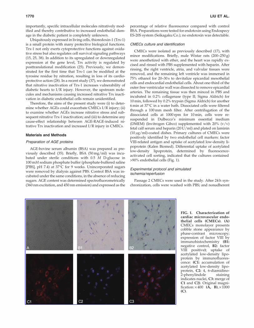
CMECs were isolated as previously described (17), withminor modifications. Briefly, male Wistar rats (200–250 g)were anesthetized with ether, and the heart was rapidly ex-cised and rinsed with PBS supplemented with heparin. Afterrinsing, the right ventricle, atria, and valvular tissues wereremoved, and the remaining left ventricle was immersed in75% ethanol for 20–30 s to devitalize epicardial mesothelialcells and endocardial endothelial cells. About one-third of theouter free ventricular wall was dissected to remove epicardialarteries. The remaining tissue was then minced in PBS andincubated in 0.2% collagenase (type II; Sigma Aldrich) for10 min, followed by 0.2% trypsin (Sigma Aldrich) for another6 min at 37�C in a water bath. Dissociated cells were filteredthrough a 100 mm mesh filter. After centrifugation of thedissociated cells at 1000 rpm for 10 min, cells were re-suspended in Dulbecco’s minimum essential medium(DMEM) (Invitrogen Gibco) supplemented with 20% (v/v)fetal calf serum and heparin (20 U/ml) and plated on laminin(10 lg/ml)-coated dishes. Primary cultures of CMECs werepositively identified by two endothelial cell markers: factorVIII-related antigen and uptake of acetylated low-density li-poprotein (Kalen Biomed). Differential uptake of acetylatedlow-density lipoprotein, determined by fluorescence-activated cell sorting, indicated that the cultures contained>90% endothelial cells (Fig. 1).
Experimental protocol and simulatedischemia/reperfusion
Passage 2 CMECs were used in the study. After 24 h syn-chronization, cells were washed with PBS; and nonadherent
FIG. 1. Characterization ofcardiac microvascular endo-thelial cells (CMECs). (A)CMECs monolayer presentscobble stone appearance byphase-contrast microscopy;expression of factor VIII byimmunohistochemistry (B1:negative control, B2: factorVIII positive); uptake ofacetylated low-density lipo-protein by immunofluores-cence: (C1: accumulation ofacetylated low-density lipo-protein, C2: 4, 6-diamidino-2-phenylindole stainingindicates nuclei, C3: merge ofC1 and C2). Original magni-fication: · 400 (A, B), · 1000(C).
1770 LIU ET AL.

cells were removed from the culturing system and were ran-domly assigned to one of the following treatments: BSA(100 lg/ml as control), AGE-BSA (100 lg/ml), AGE-BSA +EUK134 (7 lM, a peroxynitrite decomposition catalyst; Cay-man Chemical), AGE-BSA + human Trx-1 (hTrx-1) (1 lg/ml;Sigma), or AGE-BSA + sRAGE (4 lg/ml, a RAGE decoy;Adipobioscience). After 48 h incubation, cells were subjectedto either sham simulated ischemia/reperfusion (SI/R, 10 h ofnormoxia/normal-glucose environment) or SI/R (4 h hypoxia-hypoglycemic environment plus 6 h normoxia/normal glu-cose environment) as previously described (36). Briefly, theoxygen-glucose deprivation injury occurred by placing cellsin a hypoxic environment (1% O2/5% CO2/94% N2) main-tained by an incubator in the presence of glucose-free DMEMfor 4 h, at which time the medium was exchanged withoxygenated and normal glucose DMEM in an incubator at37�C to simulate the reperfusion condition for 6 h.
Assessment of SI/R-induced CMECs injury
To determine CMECs death, lactate dehydrogenase (LDH)release was determined by an enzyme activity assay kit(Nanjing Institute of Jiancheng Bioengineering). Caspase-3activity was determined by caspase-3 activity assay kit(Chemicon). Caspase-3 activity was expressed as nmol pNA/h/mg protein.
Quantification of superoxide production,cellular nitrotyrosine content
Superoxide production, an index of oxidative stress, in vi-able CMECs was measured by lucigenin-enhanced chemilu-minescence as previously described (17) and expressed asrelative light units per second per milligram protein. CMECsnitrotyrosine content, an index of protein nitration and ni-trative stress, was determined as described in our previousstudy (27).
Total NO assay
The supernatant fluid of CMECs was harvested, and NOconcentrations were measured with Griess reagent using anassay kit (Beyotime Company). The amount of total cellularprotein in the respective wells was determined by Lowry’smethod after lysis with a buffer containing 0.1% of sodiumdodecyl sulfate in 10 mM Tris, pH 7.4. Total nitrite accumu-lated in each well was defined as lM/mg of protein in thecorresponding well.
Western blot analysis for Trx-1, induciblenitric oxide synthase, RAGE, and gp91phox
CMECs were lysed in lysis buffer and centrifuged; the su-pernatant was utilized to determine Trx expression. Equalprotein amounts were electrophoresed on a 14% sodium do-decyl sulfate–polyacrylamide gel and then electrophoreticallytransferred to a polyvinylidene difluoride membrane (Milli-pore). After blocking with 5% skim milk in Tris-buffered sa-line containing 0.05% Tween 20 at room temperature for 1 h,the membrane was incubated with a monoclonal anti-murineTrx antibody (Redox Bioscience), an anti-murine RAGE anti-body (Santa Cruz), an anti-murine gp91phox (Santa Cruz)antibody, or an anti-murine inducible nitric oxide synthase
(iNOS) antibody (Cell Signaling) and then with the HRPlinked lgG (Cell Signaling). The blot was developed with anECL-Plus chemiluminescence reagent kit (Amersham) andvisualized with UVP Bio-Imaging Systems. Blot densitieswere analyzed with Vision Works LS Acquisition and Ana-lysis Software.
Trx activity assay
Trx activity was determined via the insulin disulfide re-duction assay (11). Briefly, 40 lg of cellular protein extractswere preincubated at 37�C for 15 min with 2 ml activationbuffer (100 mM HEPES, 2 mM ethylenediaminetetraaceticacid, 1 mg/ml BSA, and 2 mM DL-Dithiothreitol) to reduceTrx. After addition of 20 lL reaction buffer (100 mM HEPES,2.0 mM ethylenediaminetetraacetic acid, 0.2 mM NADPH,and 140 mM insulin), the reaction was initiated by addition ofmammalian Trx reductase (1 ml, 15 mU; Sigma) or water tocontrols. After incubation for 30 min at 37�C, the reaction wasterminated by 125 lL stopping solution (0.2M Tris–CL, 10 Mguanidine–HCl, and 1.7 mM 3-carboxy-4-nitrophenyl dis-ulfide, DTNB), followed by absorption measurement(412 nm). Trx-1 activity was expressed as oxidized NADPHlmol/min/mg of protein.
Detection of Trx-1 nitration
CMECs were homogenized with lysis buffer. EndogenousTrx-1 was immunoprecipitated with a monoclonal anti-murine Trx-1 antibody (Redox Bioscience). After sampleseparation, Trx-1 nitration was detected with a monoclonalantibody (Upstate) against nitrotyrosine. The blot was de-veloped with an ECL-Plus chemiluminescence reagent kit(Amersham) and visualized with UVP Bio-Imaging Systems.Blot densities were analyzed with Vision Works LS Acquisi-tion and Analysis Software.
Statistical analysis
All values in the text and figures are presented asmeans – standard error. All data (except Western blot density)were subjected to analysis of variance followed by Bonferronicorrection for post hoc t test. Western blot densities were an-alyzed with the Kruskal–Wallis test followed by Dunn’spost hoc test. Probabilities of 0.05 or less were considered to bestatistically significant.
Results
AGE-BSA increases the SI/R injury in CMECs
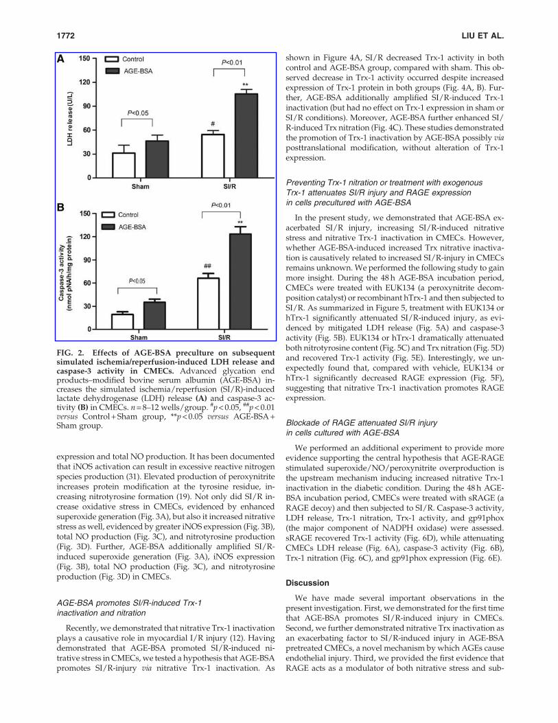
To investigate the role of AGE-BSA on SI/R-induced injuryin CMECs, we examined the effects of AGE-BSA on the SI/R-induced caspase-3 activity and LDH release in CMECs. SI/R induced a significant LDH release (Fig. 2A) and caspase-3activation (Fig. 2B). Compared with cells precultured in BSA,cells precultured in AGE-BSA had increased SI/R-inducedLDH release (Fig. 2A) and caspase-3 activity (Fig. 2B).
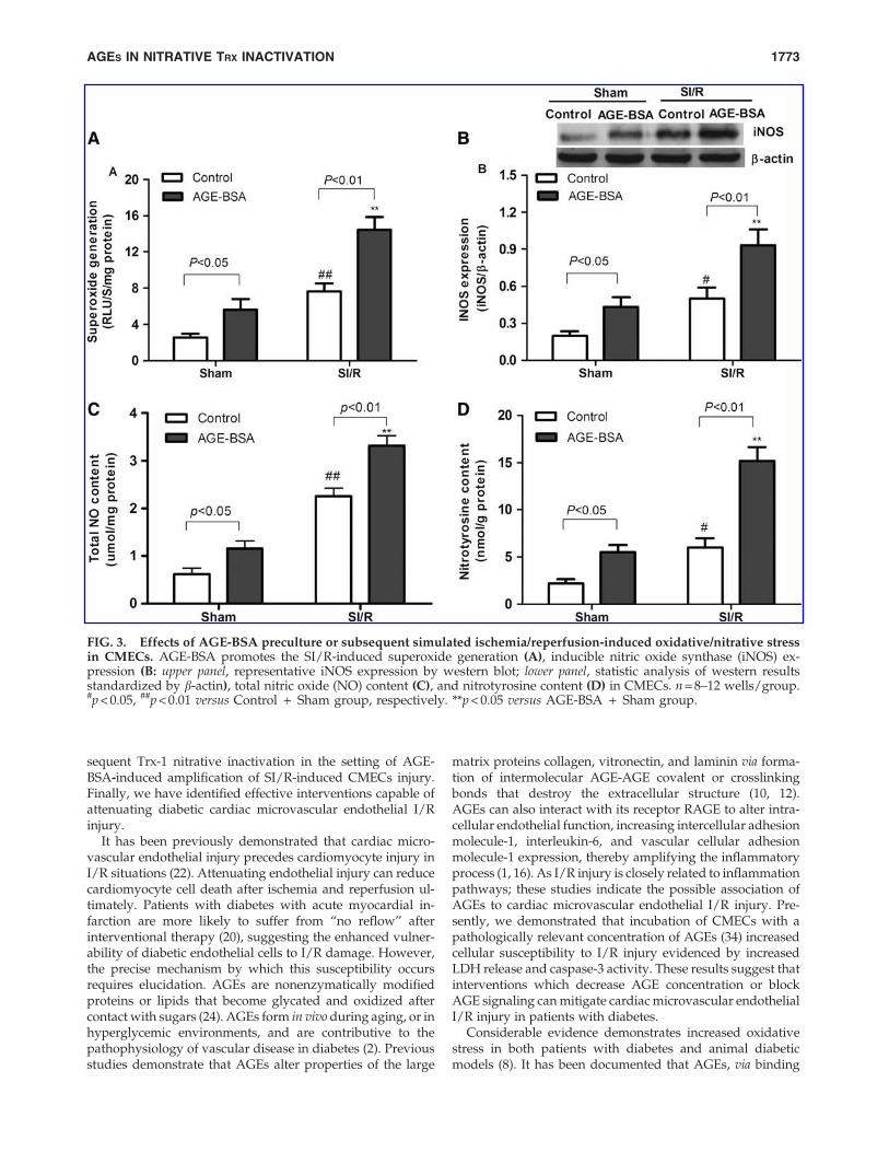
AGE-BSA promotes the SI/R-inducedoxidative/nitrative stress in CMECs
To determine whether AGE-BSA exacerbates SI/R-inducedoxidative/nitrative stress, we examined iNOS protein
AGES IN NITRATIVE TRX INACTIVATION 1771
expression and total NO production. It has been documentedthat iNOS activation can result in excessive reactive nitrogenspecies production (31). Elevated production of peroxynitriteincreases protein modification at the tyrosine residue, in-creasing nitrotyrosine formation (19). Not only did SI/R in-crease oxidative stress in CMECs, evidenced by enhancedsuperoxide generation (Fig. 3A), but also it increased nitrativestress as well, evidenced by greater iNOS expression (Fig. 3B),total NO production (Fig. 3C), and nitrotyrosine production(Fig. 3D). Further, AGE-BSA additionally amplified SI/R-induced superoxide generation (Fig. 3A), iNOS expression(Fig. 3B), total NO production (Fig. 3C), and nitrotyrosineproduction (Fig. 3D) in CMECs.
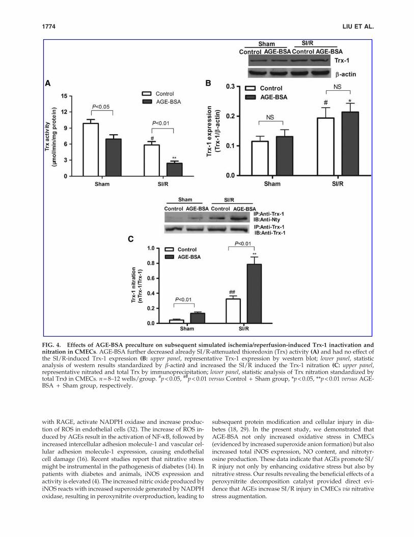
AGE-BSA promotes SI/R-induced Trx-1inactivation and nitration
Recently, we demonstrated that nitrative Trx-1 inactivationplays a causative role in myocardial I/R injury (12). Havingdemonstrated that AGE-BSA promoted SI/R-induced ni-trative stress in CMECs, we tested a hypothesis that AGE-BSApromotes SI/R-injury via nitrative Trx-1 inactivation. As
shown in Figure 4A, SI/R decreased Trx-1 activity in bothcontrol and AGE-BSA group, compared with sham. This ob-served decrease in Trx-1 activity occurred despite increasedexpression of Trx-1 protein in both groups (Fig. 4A, B). Fur-ther, AGE-BSA additionally amplified SI/R-induced Trx-1inactivation (but had no effect on Trx-1 expression in sham orSI/R conditions). Moreover, AGE-BSA further enhanced SI/R-induced Trx nitration (Fig. 4C). These studies demonstratedthe promotion of Trx-1 inactivation by AGE-BSA possibly viaposttranslational modification, without alteration of Trx-1expression.
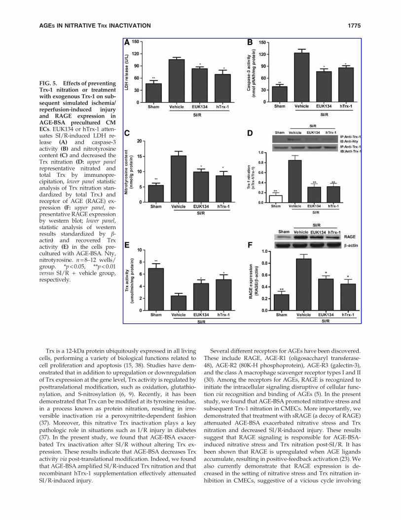
Preventing Trx-1 nitration or treatment with exogenousTrx-1 attenuates SI/R injury and RAGE expressionin cells precultured with AGE-BSA
In the present study, we demonstrated that AGE-BSA ex-acerbated SI/R injury, increasing SI/R-induced nitrativestress and nitrative Trx-1 inactivation in CMECs. However,whether AGE-BSA-induced increased Trx nitrative inactiva-tion is causatively related to increased SI/R-injury in CMECsremains unknown. We performed the following study to gainmore insight. During the 48 h AGE-BSA incubation period,CMECs were treated with EUK134 (a peroxynitrite decom-position catalyst) or recombinant hTrx-1 and then subjected toSI/R. As summarized in Figure 5, treatment with EUK134 orhTrx-1 significantly attenuated SI/R-induced injury, as evi-denced by mitigated LDH release (Fig. 5A) and caspase-3activity (Fig. 5B). EUK134 or hTrx-1 dramatically attenuatedboth nitrotyrosine content (Fig. 5C) and Trx nitration (Fig. 5D)and recovered Trx-1 activity (Fig. 5E). Interestingly, we un-expectedly found that, compared with vehicle, EUK134 orhTrx-1 significantly decreased RAGE expression (Fig. 5F),suggesting that nitrative Trx-1 inactivation promotes RAGEexpression.
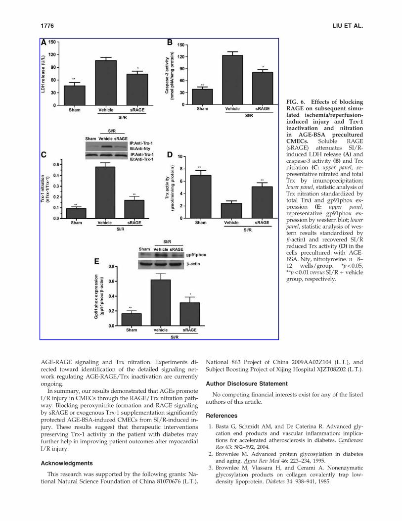
Blockade of RAGE attenuated SI/R injuryin cells cultured with AGE-BSA
We performed an additional experiment to provide moreevidence supporting the central hypothesis that AGE-RAGEstimulated superoxide/NO/peroxynitrite overproduction isthe upstream mechanism inducing increased nitrative Trx-1inactivation in the diabetic condition. During the 48 h AGE-BSA incubation period, CMECs were treated with sRAGE (aRAGE decoy) and then subjected to SI/R. Caspase-3 activity,LDH release, Trx-1 nitration, Trx-1 activity, and gp91phox(the major component of NADPH oxidase) were assessed.sRAGE recovered Trx-1 activity (Fig. 6D), while attenuatingCMECs LDH release (Fig. 6A), caspase-3 activity (Fig. 6B),Trx-1 nitration (Fig. 6C), and gp91phox expression (Fig. 6E).
Discussion
We have made several important observations in thepresent investigation. First, we demonstrated for the first timethat AGE-BSA promotes SI/R-induced injury in CMECs.Second, we further demonstrated nitrative Trx inactivation asan exacerbating factor to SI/R-induced injury in AGE-BSApretreated CMECs, a novel mechanism by which AGEs causeendothelial injury. Third, we provided the first evidence thatRAGE acts as a modulator of both nitrative stress and sub-
FIG. 2. Effects of AGE-BSA preculture on subsequentsimulated ischemia/reperfusion-induced LDH release andcaspase-3 activity in CMECs. Advanced glycation endproducts–modified bovine serum albumin (AGE-BSA) in-creases the simulated ischemia/reperfusion (SI/R)-inducedlactate dehydrogenase (LDH) release (A) and caspase-3 ac-tivity (B) in CMECs. n = 8–12 wells/group. #p < 0.05, ##p < 0.01versus Control + Sham group, **p < 0.05 versus AGE-BSA +Sham group.
1772 LIU ET AL.
sequent Trx-1 nitrative inactivation in the setting of AGE-BSA-induced amplification of SI/R-induced CMECs injury.Finally, we have identified effective interventions capable ofattenuating diabetic cardiac microvascular endothelial I/Rinjury.
It has been previously demonstrated that cardiac micro-vascular endothelial injury precedes cardiomyocyte injury inI/R situations (22). Attenuating endothelial injury can reducecardiomyocyte cell death after ischemia and reperfusion ul-timately. Patients with diabetes with acute myocardial in-farction are more likely to suffer from ‘‘no reflow’’ afterinterventional therapy (20), suggesting the enhanced vulner-ability of diabetic endothelial cells to I/R damage. However,the precise mechanism by which this susceptibility occursrequires elucidation. AGEs are nonenzymatically modifiedproteins or lipids that become glycated and oxidized aftercontact with sugars (24). AGEs form in vivo during aging, or inhyperglycemic environments, and are contributive to thepathophysiology of vascular disease in diabetes (2). Previousstudies demonstrate that AGEs alter properties of the large
matrix proteins collagen, vitronectin, and laminin via forma-tion of intermolecular AGE-AGE covalent or crosslinkingbonds that destroy the extracellular structure (10, 12).AGEs can also interact with its receptor RAGE to alter intra-cellular endothelial function, increasing intercellular adhesionmolecule-1, interleukin-6, and vascular cellular adhesionmolecule-1 expression, thereby amplifying the inflammatoryprocess (1, 16). As I/R injury is closely related to inflammationpathways; these studies indicate the possible association ofAGEs to cardiac microvascular endothelial I/R injury. Pre-sently, we demonstrated that incubation of CMECs with apathologically relevant concentration of AGEs (34) increasedcellular susceptibility to I/R injury evidenced by increasedLDH release and caspase-3 activity. These results suggest thatinterventions which decrease AGE concentration or blockAGE signaling can mitigate cardiac microvascular endothelialI/R injury in patients with diabetes.
Considerable evidence demonstrates increased oxidativestress in both patients with diabetes and animal diabeticmodels (8). It has been documented that AGEs, via binding
FIG. 3. Effects of AGE-BSA preculture or subsequent simulated ischemia/reperfusion-induced oxidative/nitrative stressin CMECs. AGE-BSA promotes the SI/R-induced superoxide generation (A), inducible nitric oxide synthase (iNOS) ex-pression (B: upper panel, representative iNOS expression by western blot; lower panel, statistic analysis of western resultsstandardized by b-actin), total nitric oxide (NO) content (C), and nitrotyrosine content (D) in CMECs. n = 8–12 wells/group.#p < 0.05, ##p < 0.01 versus Control + Sham group, respectively. **p < 0.05 versus AGE-BSA + Sham group.
AGES IN NITRATIVE TRX INACTIVATION 1773
with RAGE, activate NADPH oxidase and increase produc-tion of ROS in endothelial cells (32). The increase of ROS in-duced by AGEs result in the activation of NF-kB, followed byincreased intercellular adhesion molecule-1 and vascular cel-lular adhesion molecule-1 expression, causing endothelialcell damage (16). Recent studies report that nitrative stressmight be instrumental in the pathogenesis of diabetes (14). Inpatients with diabetes and animals, iNOS expression andactivity is elevated (4). The increased nitric oxide produced byiNOS reacts with increased superoxide generated by NADPHoxidase, resulting in peroxynitrite overproduction, leading to
subsequent protein modification and cellular injury in dia-betes (18, 29). In the present study, we demonstrated thatAGE-BSA not only increased oxidative stress in CMECs(evidenced by increased superoxide anion formation) but alsoincreased total iNOS expression, NO content, and nitrotyr-osine production. These data indicate that AGEs promote SI/R injury not only by enhancing oxidative stress but also bynitrative stress. Our results revealing the beneficial effects of aperoxynitrite decomposition catalyst provided direct evi-dence that AGEs increase SI/R injury in CMECs via nitrativestress augmentation.
FIG. 4. Effects of AGE-BSA preculture on subsequent simulated ischemia/reperfusion-induced Trx-1 inactivation andnitration in CMECs. AGE-BSA further decreased already SI/R-attenuated thioredoxin (Trx) activity (A) and had no effect ofthe SI/R-induced Trx-1 expression (B: upper panel, representative Trx-1 expression by western blot; lower panel, statisticanalysis of western results standardized by b-actin) and increased the SI/R induced the Trx-1 nitration (C: upper panel,representative nitrated and total Trx by immunoprecipitation; lower panel, statistic analysis of Trx nitration standardized bytotal Trx) in CMECs. n = 8–12 wells/group. #p < 0.05, ##p < 0.01 versus Control + Sham group, *p < 0.05, **p < 0.01 versus AGE-BSA + Sham group, respectively.
1774 LIU ET AL.
Trx is a 12-kDa protein ubiquitously expressed in all livingcells, performing a variety of biological functions related tocell proliferation and apoptosis (15, 38). Studies have dem-onstrated that in addition to upregulation or downregulationof Trx expression at the gene level, Trx activity is regulated byposttranslational modification, such as oxidation, glutathio-nylation, and S-nitrosylation (6, 9). Recently, it has beendemonstrated that Trx can be modified at its tyrosine residue,in a process known as protein nitration, resulting in irre-versible inactivation via a peroxynitrite-dependent fashion(37). Moreover, this nitrative Trx inactivation plays a keypathologic role in situations such as I/R injury in diabetes(37). In the present study, we found that AGE-BSA exacer-bated Trx inactivation after SI/R without altering Trx ex-pression. These results indicate that AGE-BSA decreases Trxactivity via post-translational modification. Indeed, we foundthat AGE-BSA amplified SI/R-induced Trx nitration and thatrecombinant hTrx-1 supplementation effectively attenuatedSI/R-induced injury.
Several different receptors for AGEs have been discovered.These include RAGE, AGE-R1 (oligosaccharyl transferase-48), AGE-R2 (80K-H phosphoprotein), AGE-R3 (galectin-3),and the class A macrophage scavenger receptor types I and II(30). Among the receptors for AGEs, RAGE is recognized toinitiate the intracellular signaling disruptive of cellular func-tion via recognition and binding of AGEs (5). In the presentstudy, we found that AGE-BSA promoted nitrative stress andsubsequent Trx-1 nitration in CMECs. More importantly, wedemonstrated that treatment with sRAGE (a decoy of RAGE)attenuated AGE-BSA exacerbated nitrative stress and Trxnitration and decreased SI/R-induced injury. These resultssuggest that RAGE signaling is responsible for AGE-BSA-induced nitrative stress and Trx nitration post-SI/R. It hasbeen shown that RAGE is upregulated when AGE ligandsaccumulate, resulting in positive-feedback activation (23). Wealso currently demonstrate that RAGE expression is de-creased in the setting of nitrative stress and Trx nitration in-hibition in CMECs, suggestive of a vicious cycle involving
FIG. 5. Effects of preventingTrx-1 nitration or treatmentwith exogenous Trx-1 on sub-sequent simulated ischemia/reperfusion-induced injuryand RAGE expression inAGE-BSA precultured CMECs. EUK134 or hTrx-1 atten-uates SI/R-induced LDH re-lease (A) and caspase-3activity (B) and nitrotyrosinecontent (C) and decreased theTrx nitration (D: upper panelrepresentative nitrated andtotal Trx by immunopre-cipitation, lower panel statisticanalysis of Trx nitration stan-dardized by total Trx.) andreceptor of AGE (RAGE) ex-pression (F: upper panel, re-presentative RAGE expressionby western blot; lower panel,statistic analysis of westernresults standardized by b-actin) and recovered Trxactivity (E) in the cells pre-cultured with AGE-BSA. Nty,nitrotyrosine. n = 8–12 wells/group. *p < 0.05, **p < 0.01versus SI/R + vehicle group,respectively.
AGES IN NITRATIVE TRX INACTIVATION 1775
AGE-RAGE signaling and Trx nitration. Experiments di-rected toward identification of the detailed signaling net-work regulating AGE-RAGE/Trx inactivation are currentlyongoing.
In summary, our results demonstrated that AGEs promoteI/R injury in CMECs through the RAGE/Trx nitration path-way. Blocking peroxynitrite formation and RAGE signalingby sRAGE or exogenous Trx-1 supplementation significantlyprotected AGE-BSA-induced CMECs from SI/R-induced in-jury. These results suggest that therapeutic interventionspreserving Trx-1 activity in the patient with diabetes mayfurther help in improving patient outcomes after myocardialI/R injury.
Acknowledgments
This research was supported by the following grants: Na-tional Natural Science Foundation of China 81070676 (L.T.),
National 863 Project of China 2009AA02Z104 (L.T.), andSubject Boosting Project of Xijing Hospital XJZT08Z02 (L.T.).
Author Disclosure Statement
No competing financial interests exist for any of the listedauthors of this article.
References
1. Basta G, Schmidt AM, and De Caterina R. Advanced gly-cation end products and vascular inflammation: implica-tions for accelerated atherosclerosis in diabetes. CardiovascRes 63: 582–592, 2004.
2. Brownlee M. Advanced protein glycosylation in diabetesand aging. Annu Rev Med 46: 223–234, 1995.
3. Brownlee M, Vlassara H, and Cerami A. Nonenzymaticglycosylation products on collagen covalently trap low-density lipoprotein. Diabetes 34: 938–941, 1985.
FIG. 6. Effects of blockingRAGE on subsequent simu-lated ischemia/reperfusion-induced injury and Trx-1inactivation and nitrationin AGE-BSA preculturedCMECs. Soluble RAGE(sRAGE) attenuates SI/R-induced LDH release (A) andcaspase-3 activity (B) and Trxnitration (C: upper panel, re-presentative nitrated and totalTrx by imunoprecipitation;lower panel, statistic analysis ofTrx nitration standardized bytotal Trx) and gp91phox ex-pression (E: upper panel,representative gp91phox ex-pression by western blot; lowerpanel, statistic analysis of wes-tern results standardized byb-actin) and recovered SI/Rreduced Trx activity (D) in thecells precultured with AGE-BSA. Nty, nitrotyrosine. n = 8–12 wells/group. *p < 0.05,**p < 0.01 versus SI/R + vehiclegroup, respectively.
1776 LIU ET AL.
4. Bucciarelli LG, Ananthakrishnan R, Hwang YC, Kaneko M,Song F, Sell DR, Strauch C, Monnier VM, Yan SF, Schmidt AM,and Ramasamy R. RAGE and modulation of ischemic injury inthe diabetic myocardium. Diabetes 57: 1941–1951, 2008.
5. Bucciarelli LG, Wendt T, Rong L, Lalla E, Hofmann MA,Goova MT, Taguchi A, Yan SF, Yan SD, Stern DM, andSchmidt AM. RAGE is a multiligand receptor of the im-munoglobulin superfamily: implications for homeostasisand chronic disease. Cell Mol Life Sci 59: 1117–1128, 2002.
6. Casagrande S, Bonetto V, Fratelli M, Gianazza E, Eberini I,Massignan T, Salmona M, Chang G, Holmgren A, andGhezzi P. Glutathionylation of human thioredoxin: a possi-ble crosstalk between the glutathione and thioredoxin sys-tems. Proc Natl Acad Sci U S A 99: 9745–9749, 2002.
7. Coughlan MT, Thorburn DR, Penfold SA, Laskowski A,Harcourt BE, Sourris KC, Tan AL, Fukami K, Thallas-BonkeV, Nawroth PP, Brownlee M, Bierhaus A, Cooper ME, andForbes JM. RAGE-induced cytosolic ROS promote mito-chondrial superoxide generation in diabetes. J Am Soc Ne-phrol 20: 742–752, 2009.
8. Forbes JM, Coughlan MT, and Cooper ME. Oxidative stressas a major culprit in kidney disease in diabetes. Diabetes 57:1446–1454, 2008.
9. Haendeler J, Hoffmann J, Tischler V, Berk BC, Zeiher AM,and Dimmeler S. Redox regulatory and anti-apoptoticfunctions of thioredoxin depend on S-nitrosylation at cys-teine 69. Nat Cell Biol 4: 743–749, 2002.
10. Hammes HP, Weiss A, Hess S, Araki N, Horiuchi S,Brownlee M, and Preissner KT. Modification of vitronectinby advanced glycation alters functional properties in vitroand in the diabetic retina. Lab Invest 75: 325–338, 1996.
11. Holmgren A and Bjornstedt M. Thioredoxin and thioredoxinreductase. Methods Enzymol 252: 199–208, 1995.
12. Howard EW, Benton R, Ahern-Moore J, and Tomasek JJ.Cellular contraction of collagen lattices is inhibited by non-enzymatic glycation. Exp Cell Res 228: 132–137, 1996.
13. King H, Aubert RE, and Herman WH. Global burden ofdiabetes, 1995–2025: prevalence, numerical estimates, andprojections. Diabetes Care 21: 1414–1431, 1998.
14. Kowluru RA. Effect of reinstitution of good glycemic controlon retinal oxidative stress and nitrative stress in diabeticrats. Diabetes 52: 818–823, 2003.
15. Lincoln DT, Ali Emadi EM, Tonissen KF, and Clarke FM.The thioredoxin-thioredoxin reductase system: over-expres-sion in human cancer. Anticancer Res 23: 2425–2433, 2003.
16. Neumann A, Schinzel R, Palm D, Riederer P, and Munch G.High molecular weight hyaluronic acid inhibits advancedglycation endproduct-induced NF-kappaB activation andcytokine expression. FEBS Lett 453: 283–287, 1999.
17. Nishida M, Carley WW, Gerritsen ME, Ellingsen O, KellyRA, and Smith TW. Isolation and characterization of humanand rat cardiac microvascular endothelial cells. Am J Physiol264: H639–H652, 1993.
18. Ren XY, Li YN, Qi JS, and Niu T. Peroxynitrite-inducedprotein nitration contributes to liver mitochondrial damagein diabetic rats. J Diabetes Complications 22: 357–364, 2008.
19. Rojas A, Mercadal E, Figueroa H, and Morales MA. Ad-vanced glycation and ROS: a link between diabetes andheart failure. Curr Vasc Pharmacol 6: 44–51, 2008.
20. Romano M, Buffoli F, Tomasi L, Aroldi M, Lettieri C, FerrariMR, and Zanini R. The no-reflow phenomenon in acutemyocardial infarction after primary angioplasty: incidence,predictive factors, and long-term outcomes. J Cardiovasc Med(Hagerstown) 9: 59–63, 2008.
21. Sangiorgi G, Romagnoli E, Biondi-Zoccai G, Margheri M,Tamburino C, Barbagallo R, Falchetti E, Vittori G, AgostoniP, Cosgrave J, and Colombo A. Percutaneous coronary im-plantation of sirolimus-eluting stents in unselected patientsand lesions: clinical results and multiple outcome predictors.Am Heart J 156: 871–878, 2008.
22. Scarabelli T, Stephanou A, Rayment N, Pasini E, CominiL, Curello S, Ferrari R, Knight R, and Latchman D.Apoptosis of endothelial cells precedes myocyte cell ap-optosis in ischemia/reperfusion injury. Circulation 104:253–256, 2001.
23. Schmidt AM and Stern DM. RAGE: a new target for theprevention and treatment of the vascular and inflammatorycomplications of diabetes. Trends Endocrinol Metab 11: 368–375, 2000.
24. Singh R, Barden A, Mori T, and Beilin L. Advanced gly-cation end-products: a review. Diabetologia 44: 129–146,2001.
25. Tao L, Gao E, Bryan NS, Qu Y, Liu HR, Hu A, ChristopherTA, Lopez BL, Yodoi J, Koch WJ, Feelisch M, and Ma XL.Cardioprotective effects of thioredoxin in myocardial ische-mia and reperfusion: role of S-nitrosation [corrected]. ProcNatl Acad Sci U S A 101: 11471–11476, 2004.
26. Tao L, Gao E, Hu A, Coletti C, Wang Y, Christopher TA,Lopez BL, Koch W, and Ma XL. Thioredoxin reduces post-ischemic myocardial apoptosis by reducing oxidative/nitrative stress. Br J Pharmacol 149: 311–318, 2006.
27. Tao L, Gao E, Jiao X, Yuan Y, Li S, Christopher TA, LopezBL, Koch W, Chan L, Goldstein BJ, and Ma XL. Adiponectincardioprotection after myocardial ischemia/reperfusion in-volves the reduction of oxidative/nitrative stress. Circulation115: 1408–1416, 2007.
28. Tao L, Jiao X, Gao E, Lau WB, Yuan Y, Lopez B, ChristopherT, Ramachandra Rao SP, Williams W, Southan G, Sharma K,Koch W, and Ma XL. Nitrative inactivation of thioredoxin-1and its role in postischemic myocardial apoptosis. Circula-tion 114: 1395–1402, 2006.
29. Vareniuk I, Pavlov IA, and Obrosova IG. Inducible nitricoxide synthase gene deficiency counteracts multiple mani-festations of peripheral neuropathy in a streptozotocin-induced mouse model of diabetes. Diabetologia 51: 2126–2133,2008.
30. Vlassara H. The AGE-receptor in the pathogenesis of dia-betic complications. Diabetes Metab Res Rev 17: 436–443, 2001.
31. Wang XL, Liu HR, Tao L, Liang F, Yan L, Zhao RR,Lopez BL, Christopher TA, and Ma XL. Role of iNOS-derived reactive nitrogen species and resultant nitrativestress in leukocytes-induced cardiomyocyte apoptosis aftermyocardial ischemia/reperfusion. Apoptosis 12: 1209–1217,2007.
32. Wautier MP, Chappey O, Corda S, Stern DM, Schmidt AM,and Wautier JL. Activation of NADPH oxidase by AGE linksoxidant stress to altered gene expression via RAGE. Am JPhysiol Endocrinol Metab 280: E685–E694, 2001.
33. Wei L, Sun D, Yin Z, Yuan Y, Hwang A, Zhang Y, Si R,Zhang R, Guo W, Cao F, and Wang H. A PKC-beta inhibitorprotects against cardiac microvascular ischemia reperfusioninjury in diabetic rats. Apoptosis 15: 488–498, 2010.
34. Xu B, Chibber R, Ruggiero D, Kohner E, Ritter J, and FerroA. Impairment of vascular endothelial nitric oxide synthaseactivity by advanced glycation end products. FASEB J 17:1289–1291, 2003.
35. Yamagishi S, Inagaki Y, Okamoto T, Amano S, Koga K,Takeuchi M, and Makita Z. Advanced glycation end
AGES IN NITRATIVE TRX INACTIVATION 1777
product-induced apoptosis and overexpression of vascularendothelial growth factor and monocyte chemoattractantprotein-1 in human-cultured mesangial cells. J Biol Chem 277:20309–20315, 2002.
36. Yang D, Guo S, Zhang T, and Li H. Hypothermia attenuatesischemia/reperfusion-induced endothelial cell apoptosis viaalterations in apoptotic pathways and JNK signaling. FEBSLett 583: 2500–2506, 2009.
37. Yin T, Hou R, Liu S, Lau WB, Wang H, and Tao L. Nitrativeinactivation of thioredoxin-1 increases vulnerability of dia-betic hearts to ischemia/reperfusion injury. J Mol Cell Cardiol49: 354–361, 2010.
38. Yoshida T, Oka S, Masutani H, Nakamura H, and YodoiJ. The role of thioredoxin in the aging process: involve-ment of oxidative stress. Antioxid Redox Signal 5: 563–570,2003.
Address correspondence to:Dr. Ling Tao
Department of CardiologyXijing Hospital
The Fourth Military Medical University15 Changle West Road
Xi’an 710032China
E-mail: [email protected]
Dr. Haichang WangDepartment of Cardiology
Xijing HospitalThe Fourth Military Medical University
15 Changle West RoadXi’an 710032
China
E-mail: [email protected]
Date of first submission to ARS Central, November 15, 2010;date of acceptance, December 2, 2010.
Abbreviations Used
AGEs¼ advanced glycation end productsBSA¼ bovine serum albumin
CMECs¼ cardiac microvascular endothelial cellsDMEM¼Dulbecco’s minimum essential medium
I/R¼ ischemia/reperfusionLDH¼ lactate dehydrogenase
NO¼nitric oxidePBS¼phosphate-buffered saline
RAGE¼ receptor of advanced end productROS¼ reactive oxygen speciesSI/R¼ simulated ischemia/reperfusion
sRAGE¼ soluble receptor of advanced end productTrx¼ thioredoxin
1778 LIU ET AL.

This article has been cited by:
1. Nilanjana Maulik , Juan A. Sanchez . 2011. Risk Factors in Heart Disease: Therapeutic Interventions. Antioxidants & RedoxSignaling 15:7, 1765-1767. [Citation] [Full Text] [PDF] [PDF Plus]