advanced clinical corneal topography for refractive surgeons · advanced clinical corneal...
TRANSCRIPT
Advanced Clinical Corneal Topography for Refractive Surgeons
Course director: Ming Wang, MD, PhD. Director, Wang Vision Institute
Clinical associate professor of ophthalmology, University of Tennessee, [email protected], www.wangvisioninstitute.com 1801 West End Ave, Ste 1150, Nashville, TN, 37203, USA, (615)321-8881 (phone), (615)321-8874(fax) International President, Shanghai Aier Eye Hospital (www.aier021.com), 上海爱尔眼科医院国际院长
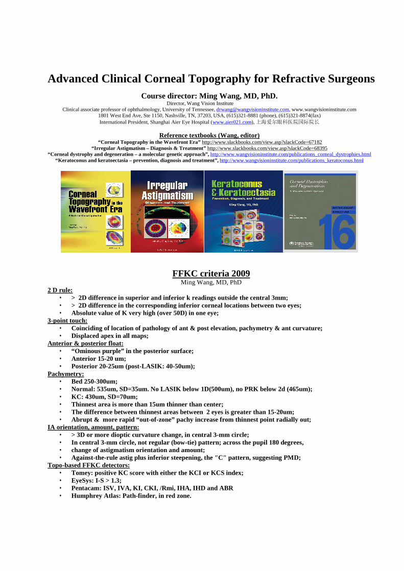
Reference textbooks (Wang, editor)
“Corneal Topography in the Wavefront Era” http://www.slackbooks.com/view.asp?slackCode=67182 “Irregular Astigmatism – Diagnosis & Treatment” http://www.slackbooks.com/view.asp?slackCode=68395
“Corneal dystrophy and degeneration – a molecular genetic approach”, http://www.wangvisioninstitute.com/publications_corneal_dystrophies.html “Keratoconus and keratoectasia – prevention, diagnosis and treatment”, http://www.wangvisioninstitute.com/publications_keratoconus.html
FFKC criteria 2009 Ming Wang, MD, PhD
2 D rule: • > 2D difference in superior and inferior k readings outside the central 3mm; • > 2D difference in the corresponding inferior corneal locations between two eyes; • Absolute value of K very high (over 50D) in one eye;
3-point touch: • Coinciding of location of pathology of ant & post elevation, pachymetry & ant curvature; • Displaced apex in all maps;
Anterior & posterior float: • “Ominous purple” in the posterior surface; • Anterior 15-20 um; • Posterior 20-25um (post-LASIK: 40-50um);
Pachymetry: • Bed 250-300um; • Normal: 535um, SD=35um. No LASIK below 1D(500um), no PRK below 2d (465um); • KC: 430um, SD=70um; • Thinnest area is more than 15um thinner than center; • The difference between thinnest areas between 2 eyes is greater than 15-20um; • Abrupt & more rapid “out-of-zone” pachy increase from thinnest point radially out;
IA orientation, amount, pattern: • > 3D or more dioptic curvature change, in central 3-mm circle; • In central 3-mm circle, not regular (bow-tie) pattern; across the pupil 180 degrees, • change of astigmatism orientation and amount; • Against-the-rule astig plus inferior steepening, the "C" pattern, suggesting PMD;
Topo-based FFKC detectors: • Tomey: positive KC score with either the KCI or KCS index; • EyeSys: I-S > 1.3; • Pentacam: ISV, IVA, KI, CKI, /Rmi, IHA, IHD and ABR • Humphrey Atlas: Path-finder, in red zone.

Ming Wang, MD, PhD -- Topographic Technologies
A. History of corneal topography a. Keratometry b. Keratoscopy c. Placido disc
B. Placido disc technology a. reflection of placido images b. two forms: large targets and small target machines
C. Tomography a. Scanning slit technology
i. Direct measurement of the anterior and posterior surfaces ii. No way to validate posterior curvature findings until recently
iii. Sensitive to eye movement due to longer testing time b. Scheimpflug imaging
i. Pentacam ii. Precisio
iii. Direct measurement of the anterior and posterior surfaces D. 3-D corneal technology
a. generates submicron precision b. fast data capture
E. Case examples
Stephen D. Klyce -- Corneal Topography: Screening and Identifying FFKC (NOTE: detailed outline will be available from www.wangvisioninstitute.com/for_physicians.html) 1. It is the standard of care in refractive surgery to routinely perform and evaluate corneal topography along with the slit lamp exam, careful pachymetry and refraction, and pupillometry. 2. There are a variety of corneal topography models to choose among, most of which offer similar features and presentation formats. It has been shown that through use of a fixed 1.5 D interval scale and an axial power map, clinical anomalies easily stand out when evaluating topography.3 3. A display of corneal topography that includes a color-coded contour map alongside familiar measures such as pupil diameter and simulated keratometry should suffice. 4. Use a routine benchmark to occasionally test your corneal topographer to ensure calibration is being maintained. 5. In reality, the measurement of corneal topography actually measures the shape of the tear film. Bring the patient to corneal topography as a first step in a screening and ask the patient to blink several times prior to capturing the image of the mires. 6. A normal cornea will have smooth contours, centrally uniform power, and flattening toward the periphery - particularly toward the nasal side. SimK readings should be ~42.75 +/- 1.6 D (standard deviation). 7. The most common abnormal corneal topography conditions found in screening and the known potential sequellae after LASIK are listed below:
Topographic Abnormality Cause Potential Consequence after LASIK
Irregular mires with mistracking dry eye, drops poor predictability
Irregular contours basement membrane dystrophy Kerectasia
Inferior or superior steepening contact lens warpage poor predictability

Inferior steepening keratoconus, pellucid corneal marginal degeneration
Kerectasia
“Negative” bow tie, “C” shaped steepening
pellucid corneal marginal degeneration
Kerectasia
Truncated bow tie Keratoconus Kerectasia
Asymmetric bow tie Keratoconus Kerectasia
Lazy 8 or skewed bow tie Keratoconus Kerectasia
Central or superior steepening Keratoconus Kerectasia
8. Note that contact lens warpage can masquerade as keratoconus by causing an inferior or superior steepening in corneas that have with-the-rule astigmatism. 9. Several classification schemes are available: Tomey Smolek/Klyce Keratoconus program, the Humphrey Pathfinder, and the NIDEK Magellan Navigator. 10. Pathology in one eye often forecasts the potential for pathology in the other eye (e.g. keratoconus). Y Ralph Chu, MD -- Scaling in Topography, challenging cases
A. Standard scaling B. Auto scaling C. Reading maps:
i. Check name of patient ii. date of exam iii. examined eye iv. Check the type of scale v. Type of measurement (height in microns, curvature in mm, power in diopters) vi. Step interval vii. Study the map (type of map, form of abnormality) viii. Evaluate statistical information ix. Compare with topography of the other eye x. Compare with previous maps (verify they are the same scale) xi. Apply statistical analysis or other needed software application xii. Explain the exams result to the patient xiii. Case examples
Amar Agarwal, MS; FRCS; FRCophth – Orbscan Analysis (NOTE: detailed outline will be available from www.wangvisioninstitute.com/for_physicians.html)
A. Slit scanning basics i. Paraxial optics (Keratometers, two-dimensional instruments) ii. Raytrace/geometric optics (Raytrace optics does not require surface curvature, but depends on elevation and especially surface slope) iii. Orbscan uses slit-beams and back-scattered light to triangulate surface shape. The derived mathematical surface is then raytraced using a basic keratometer model to produce simulated keratometer (SimK) values. iv. Color conventions:
a. Blue = low, level, flat, deep, thick, or aberrated. b. Red = high, steep, sharp, shallow, thin, or focused.

v. Quad maps: a. upper left: anterior elevation
b. upper right: posterior elevation c. lower left: keratometric d. lower right: pachymetry map
B. Elevation maps i. Elevation is measured directly ii. Standard measurement for shape of the cornea iii. Both slope and curvature can be mathematically derived from elevation data
C. Pachymetry maps i. Central value ii. Thinnest value (typically inferior temporal but varies) D. Screening prior to LASIK
i. Rule out posterior elevation, FFKC
Arun Gulani, MD -- Pentacam & Corneoplastique(TM)- The ART of Vision Surgery (NOTE: detailed outline will be available from www.wangvisioninstitute.com/for_physicians.html)
A. Pentacam overview – Pentacam is a rotating Scheimpflug Camera which generates images from the anterior surface of the cornea up to the posterior surface of the lens in less than 2 seconds thru 50 scans with 2760 true elevation points per image from the complete anterior eye segment as well as a special high resolution cornea scanning mode.
B. Provides a movable 3-D-presentation of the anterior chamber angle, volume, depth and lens thickness. C. The pachymetry of the complete cornea from the 3-D-model has an accuracy +/-5µm as a colored map
display along with a colored anterior chamber depth map and densitometry of the complete lens including the subcasular layer.
D. Applications: a. Patients seeking refractive surgery or lens implantation, b. S/P refractive surgery for corrective procedures (enhancements, ectasia) or IOL calculation c. Keratoconus screening
Giuseppe D'Ippolito, Dr Ing – Elevation topography using the Precisio elevation-based topography
A. Overview of Scheimpflug imaging B. Elevation maps C. Pachymetry maps D. Clinical cases
Kevin Miller, PhD -- A Simple Nomogram for LRIs
A. Implantation and LRIs between 1996 and 1999 a. Calculated the vector change in corneal astigmatism using topo-derived simK’s b. Compared the actual surgical results to nomogram and generated a predictive model c. Exclusion criteria: eyes with a history of previous refractive surgery d. Statistical analysis: determined the relationship between the operative variables (length, depth and
number of AK incisions; location of the phacoemulsification incision, and patient age.) and amount of correction achieved by multiple regression analysis
e. Corneal flattening (D) = 0.474 + -0.791 x Average LRI length in clock hours – 0.513 (if the phaco incision was outside the LRI.)
f. Amount of flattening increased by 0.12D for ever 10 years increase age g. Phaco incision reduces the effect of an LRI by 0.5D if it is made 90 degrees away.
Jack T. Holladay, MD -- Pentacam Tomography and direct corneal power measurements after LASIK and PRK (NOTE: Full handout is available on Dr. Holladay’s website at www.docholladay.com under HANDOUTS, # 8.)
A. Using data for IOL calculation a. History methods: PRE-OP K’s and MR) b. Current data: Current K’s, Post-OP central topo and change in MR c. Gas perm lens over-refraction d. True Power maps

Ming Wang, MD,PhD – Posterior corneal changes and its impact on refractive surgery patient management Posterior corneal surface – a traditionally ignored corneal surface •Relative small refractive contribution: 12.5%; •Assumed to be constant in traditional keratometry (hence the constant fudging factor 1.3375, I.e., posterior corneal shape mirrors that of the anterior); •This assumption was adequate in pre-LASIK era. The change (increase) in posterior cornea contribution can NO LONGER be ignored in LASIK era With the advent of anterior ablative corneal surgery, the shape of posterior cornea no longer mirrors that of anterior. Posterior contribution to total corneal refractive power is changed (increased): –“Unilateral “ K reduction due to anterior central tissue removal, resulting in a relative increase of posterior contribution; –Weakening of overall corneal strength due to tissue removal, the posterior surface bulges forward, resulting in an absolute increase of posterior contribution. Question: Does posterior corneal contribution indeed INCREASE after myopic LASIK? The study: 17 eyes of 17 consecutive myopic patients (-0.74 to –9.98D) s/p LASIK, Orbscan examination of all corneal surface powers. Does central posterior power increases MORE (i.e, more central bowing) than peripheral after myopic LASIK? Is K change a good predictor of refractive correction (at corneal plane), as in the case of pre-LASIK era? Why is K change less than refractive correction and hence not a good predictor of refractive correction? The answer: K change is less than refractive change is because the posterior contribution is increased after LASIK (K assumes a constant fudging factor 1.3357). If K change is not a good predictor of refractive correction, then what is? The answer: total corneal power change is the best predictor of refractive correction. Since in total corneal power change, the changing (increased) posterior contribution after LASIK is taken into account. Ignoring the change (increase) in posterior contribution is the cause of INACCURACY of IOL calculation in post-LASIK patients •IOL formulas use traditional K, which assumes that posterior cornea mirrors anterior and is a constant contribution (and hence the constant 1.3375 fudging factor); •Because of this fundamental error in the IOL formula which uses K, “just staying on a more myopic side” to avoid hyperopia is no longer good enough, since the width of scatter of the resultant refraction is also increased. Posterior contribution is not only increased after LASIK but also posterior surface is the “first surface to go” •Posterior cornea is the “first surface to go” when cornea begins ectasia:
–Theoretical consideration: The posterior surface faces the “direct assault” of intraocular fluid forces; –Clinical evidence: we often see normal anterior surfaces in the presence of abnormal posterior changes (increased float); however, we seldom see abnormal anterior surfaces without accompanying posterior pathology. Clinical importance in recognizing posterior change before and after LASIK
•Preop: identify early corneal ectasia (posterior ectasia occurs first) and thus exclude these patients;
•Postop: identify early ectasia, in which excessive forward movement of posterior cornea (“ominous purple”) is present, and thus avoid enhancement. Posterior changes affect postop visual QUALITY: 1st approximation of corneal refractive surgery
•Anterior surface: determines refraction;
•Posterior surface: determines visual quality - a new frontier in refractive surgery today. Clinical posterior float threshold for NOT to do LASIK: When there is inferior decentration of float, and the extent of float is: Primary LASIK: 20-30um; Enhancement: 30-50um. Clinical cases of impact of posterior corneal changes Case 1: Examination of posterior cornea helps identify poor LASIK candidate: A case of posterior KC “ominous purple”! Posterior change does occur EARLIER (anterior is still normal). Case 2: Examination of posterior cornea helps identify the true cause of “overcorrection” and thus avoid making things worse by doing enhancement: a case of excessive “overcorrection”, which is in fact NOT overcorrection. Rather than too much tissue being removed anteriorly, there is a gross forward MOVEMENT of posterior cornea – earliest sign of impending ectasia. Don’t enhance. It will result in PAN-CORNEAL thinning with hyperopic enhancement, further worsening ectasia! Case 3: Posterior changes occur EARLIER than anterior: a case of anterior change being typically accompanied by posterior changes Case 4: Examination of posterior cornea helps identify the true cause of resistance to enhancment: a case of s/p H-L, resistant to enh, why? Preop existing posterior decentered apex!!! Case 5: Examination of posterior cornea reveals earliest sign of ectasia: A case of posterior KC (“ominous purple”), with normal anterior. Don’t touch it! Case 6: Posterior cornea is an earlier and more sensitive predictor of impending ectasia: a case of posterior change being more pronounced than anterior. Summary of study of posterior changes after LASIK •Posterior corneal contribution to refraction is no longer constant after myopic LASIK, it is in fact INCREASED (12.5% to 25%!); •K is no longer the best predictor of refractive correction (0.8:1), total corneal power is (1:1); •Posterior surface is the “first surface to go” , and hence is the most sensitive and earliest indicator of impending ectasia. Summary of the effect of posterior contribution increase on IOL calculation •Not only there is a tendency towards hyperopia, but also the WIDTH of the scatter of the resultant refraction is increased, so it is no longer good enough to “just stay on a more myopic side” to avoid hyperopia; •Fundamental solution: Reformulate IOL formula to include the changing (increased) posterior corneal contribution, I.e.: So, discard the constant fudging factor 1.3375!