refractive and aberrometric outcomes of intracorneal … · diego cuevas, md,10 inmaculada pascual,...
TRANSCRIPT

Refractive and Aberrometric Outcomesof Intracorneal Ring Segments forKeratoconus: Mechanical versusFemtosecond-assisted Procedures
David P. Piñero, MSc,1,2 Jorge L. Alio, MD, PhD,1,3 Bassam El Kady, MD, PhD,4 Efekan Coskunseven, MD,5
Hector Morbelli, MD,6 Antonio Uceda-Montanes, MD,7,8 Miguel J. Maldonado, MD, PhD,9
Diego Cuevas, MD,10 Inmaculada Pascual, PhD2
Objective: To compare visual, refractive, and corneal aberrometric outcomes in keratoconic eyes implantedwith intracorneal ring segments (ICRS) implantation using either a mechanical or a femtosecond laser-assistedprocedure.
Design: Retrospective, consecutive case series.Participants: A total of 146 consecutive eyes of 106 patients with the diagnosis of keratoconus (68 unilateral
and 39 bilateral) were included. Two groups were created according to the surgical technique used for cornealtunnelization: Mechanical group (mechanical tunnelization, 63 eyes) and Femtosecond group (femtosecondlaser-assisted tunnelization, 83 eyes). Intracorneal ring segments implantation was indicated because of theexistence of reduced best spectacle-corrected visual acuity (BSCVA) or contact lens intolerance.
Methods: Intracorneal ring segments implantations were performed by 6 surgeons following the sameprotocol except for the incision location. A total of 55 eyes were implanted with Intacs (Addition Technology, Inc,Fremont, CA) and 8 eyes were implanted with KeraRings (Mediphacos, Belo Horizonte, Brazil) in the Mechanicalgroup, and 25 eyes were implanted with Intacs and 58 eyes were implanted with KeraRings in the Femtosecondgroup. Mean follow-up was 10.66�8.20 months, ranging from 1 month to 24 months.
Main Outcome Measures: Uncorrected visual acuity (UCVA), BSCVA, refraction, keratometry, and rootmean square (RMS) for different kinds of corneal aberrations.
Results: By reporting only for statistically significant changes, UCVA improved in both groups at 6 months(P�0.02) and BSCVA improved in the Femtosecond group (P�0.01). The refraction improved in both groups at6 months (P�0.02). The cornea on average was flatter in both groups at 6 months (P�0.01). Root mean squareastigmatism was reduced in the Femtosecond group (P � 0.03), but there was an increase in some higher-orderaberrations (P � 0.03). Significant differences were found between the 2 groups for eyes implanted with Intacsfor primary spherical aberration, coma, and other higher-order aberrations, favoring the Femtosecond group(P�0.01). A significant negative correlation was found between the preoperative corneal aberrations and thepostoperative BSCVA in the Mechanical group (r�0.63, P�0.04).
Conclusions: Intracorneal ring segments implantation using both mechanical and femtosecond laser-assisted procedures provide similar visual and refractive outcomes. A more limited aberrometric correction isobserved for eyes with mechanical implantation.
Financial Disclosure(s): The author(s) have no proprietary or commercial interest in any materials discussed
in this article. Ophthalmology 2009;116:1675–1687 © 2009 by the American Academy of Ophthalmology.Keratoconus is an ectatic corneal disorder characterized bya progressive corneal thinning that results in corneal pro-trusion, irregular astigmatism, and decreased vision.1 Themanagement of patients with keratoconus must include vi-sual rehabilitation because the visual function is devastatedas a result of the significant increase in all ocular aberra-tions.2–5 Spectacle correction is only adequate for earlycases, whereas in advanced or moderate cases, contactlenses or surgical solutions are necessary to achieve a sat-isfactory visual outcome. Rigid gas-permeable and hybrid
contact lenses provide good visual quality.6 However, some© 2009 by the American Academy of OphthalmologyPublished by Elsevier Inc.
patients can become intolerant to contact lenses7 or achievean unacceptable visual performance.8
Intrastromal corneal ring segments (ICRS) have beenproposed and investigated as an additive surgical procedurefor keratoconus correction,9–28 providing an interesting al-ternative aiming at delaying and preventing corneal graft inpatients with keratoconus.15,16 This type of surgical treat-ment has proved to be effective in improving visual acuity,reducing the refractive error and mean keratometry. Theaddition of extra material at the corneal mid-periphery in-
duces a displacement of the local anterior surface forward at1675ISSN 0161-6420/09/$–see front matterdoi:10.1016/j.ophtha.2009.05.016

Ophthalmology Volume 116, Number 9, September 2009
this area and a flattening of the central portion of theanterior cornea because of the morphologic structure ofcorneal lamellae (arc-shortening effect).29 The use of shortarc-length ring ICRS has proved to be effective for thecorrection of astigmatism,9,13,25 because this kind of proce-dure induces less corneal flattening and a significant changein corneal toricity as a result of the corneal architecture(predicted by finite element modeling).30
Mechanical dissection was the first method described forfacilitating the insertion of ring segments (mechanical pro-cedure).26–28 Good visual results and reduced complicationsrates have been reported using the mechanical procedure inearly to moderate keratoconus.10,11,14–17,19–28 However, theuse of femtosecond laser for corneal tunnelization becamewidely accepted after the approval of its use by the Foodand Drug Administration in the United States.31 This laserallows the surgeon to program the tunnelization at a prede-termined depth with a high degree of precision. Theoreti-cally, this femtosecond laser-assisted procedure would gen-erate a more accurate stromal dissection, reducing surgicalerror and leading to better visual and refractive results.However, in 2 studies32,33 comparing both femtosecond andmechanical tunnelization procedures in ectatic eyes, no dif-ferences in visual and refractive outcomes were detected.However, the follow-up period was no greater than 12months.
The aim of the present study was to compare visual,refractive, and corneal aberrometric outcomes in kerato-conic eyes in which ICRS implantation was facilitated usingeither the mechanical or the femtosecond-laser assisted pro-cedure with a follow-up period up to 24 months.
Patients and Methods
Patients
In the current study, a multicenter retrospective analysis of anonrandomized consecutive series of cases was performed. Data ofall patients who underwent ICRS implantation for keratoconustreatment from September 2000 to June 2007 in 6 different oph-thalmologic centers, 5 Spanish (Vissum Alicante, Vissum Sevilla,Vissum Albacete, Vissum Almería, University Clinic of Univer-sity of Navarra) and 1 Turkish (Refractive Surgery Department ofDunya Eye Hospital, Istanbul), were reviewed and analyzed com-prehensively. Table 1 summarizes the contribution of each partic-ipating center to the current study. A total of 146 consecutive eyesof 106 patients diagnosed with keratoconus (68 unilateral and 39bilateral cases) were included. Two different groups were createdaccording to the surgical technique used for creation of cornealchannels: eyes operated using mechanical tunnelization (Mechan-ical group: 63 eyes, 43.15%) and eyes operated using femtosecondlaser-assisted tunnelization (Femtosecond group: 83 eyes, 56.85%). Inall cases, ICRS implantation was indicated because of the exis-tence of reduced best spectacle-corrected visual acuity (BSCVA)or contact lens intolerance.
A comprehensive examination was performed in all cases be-fore ICRS implantation to ensure the viability of the surgery. Thisexamination included Snellen uncorrected visual acuity (UCVA)and BSCVA (decimal notation), manifest refraction, slit-lampbiomicroscopy, Goldman tonometry, fundus evaluation, ultrasonic
pachymetry, and corneal topography. Keratoconus diagnosis was1676
based on corneal topography and slit-lamp observation: asymmet-ric bowtie pattern with or without skewed axes and presence ofstromal thinning, corneal conical protrusion at the apex, Fleischerring, Vogt striae, or anterior stromal scar.1
Because topographic data were collected from different periodsand different centers, 3 different corneal topographic systems wereused for corneal examination: CMS 100 Topometer (G. Roden-stock Instrument GmbH, Ottobrunn, Germany), CSO (CSO,Firenze, Italy), and Orbscan IIz system (Bausch & Lomb, Roch-ester, NY). The first 2 devices are Placido-based systems, and theOrbscan IIz is a combined scanning-slit and Placido-disc topo-graphic system. Although agreement between these specific de-vices has not been reported, Orbscan and Placido-based deviceshave been proved to provide similar accuracy and precision oncalibrated spherical test surfaces.34 In the study, the followingtopographic data were evaluated and recorded with all cornealtopographic devices: the corneal dioptric power in the flattestmeridian for the 3 mm central zone (K1), corneal dioptric power inthe steepest meridian for the 3 mm central zone (K2), mean cornealpower in the 3 mm zone (KM), and inferosuperior asymmetryindex, calculated as the difference between the dioptric power at 3mm below and above the corneal geometric center.
Corneal aberrometry was also recorded and analyzed only inthose patients examined at all visits with the CSO topographysystem (85 eyes), because this device was the only one with thecapability to calculate directly this specific information. The CSOtopography system analyzes a total of 6144 corneal points of acorneal area enclosed in a circular annulus defined by an innerradius of 0.33 and an outer radius of 10 mm in respect to cornealvertex. The software of this system, the EyeTop2005 (CSO),automatically performs the conversion of corneal elevation profileinto corneal wavefront data using the Zernike polynomials with anexpansion up to the seventh order. In this study, aberration coef-ficients and root mean square (RMS) values were always calcu-lated for a 6-mm pupil. The following aberrometric parameterswere recorded and analyzed: higher-order RMS (computed forthird to seventh Zernike terms), primary coma RMS (computed forthe Zernike terms Z3
�1), coma-like RMS (computed for third,fifth, and seventh-order Zernike terms), spherical-like RMS (com-puted for fourth and sixth-order Zernike terms), and residual RMS(computed considering all Zernike terms except those correspond-ing with primary coma and spherical aberration). The correspond-ing Zernike coefficient for primary spherical aberration (Z 0) was
Table 1. Contribution of Each Participating OphthalmologicCenter to this Retrospective Study
Investigator Surgeon Ophthalmologic Center
EyesImplantedwith ICRS
1 Dr Alió Vissum Alicante (Spain) 1162 Dr Coskunseven Refractive Surgery
Department of DunyaEye Hospital, Istanbul(Turkey)
14
3 Dr Morbelli Vissum Albacete (Spain) 63 Dr Uceda Vissum Sevilla (Spain) 44 Dr Maldonado University Clinic,
University of Navarra3
5 Dr Cuevas Vissum Almería (Spain) 3
ICRS � intracorneal ring segments.
4
also reported with its sign.

Piñero et al � Intracorneal Ring Segment Implantation in Keratoconus
Ethical board committee approval of our institution (VissumInstituto Oftalmologico de Alicante) was obtained for this inves-tigation. In addition, during the process of consent for this surgery,consent was taken to later include clinical information in scientificstudies.
Surgery
Surgical procedures were performed by 6 surgeons, 1 from eachparticipating center in the study (JLA, Vissum Alicante; EC,Dunya Eye Hospital; HM, Vissum Albacete; AUM, VissumSevilla; MML, University of Navarra; and DC, Vissum Almería).In all cases an antibiotic prophylaxis was prescribed before sur-gery, consisting of topical ciprofloxacin (Oftacilox; Alcon Cusí,Barcelona, Spain) every 8 hours for 2 days. All procedures wereperformed under topical anesthesia.
The mechanical surgical procedure was initiated markinga reference point for centration (pupil center) and performing aradial incision of approximately 1.8 mm in length. After this, acalibrated diamond knife was set at approximately 70% of themean corneal thickness determined by ultrasonic pachymetry.From the base of the incision, pocketing hooks were used to createcorneal pockets on each side of the incision, taking care to main-tain a uniform depth. A device containing a semiautomated suctionring was placed around the limbus, guided by the previouslymarked reference point on the cornea. Two semicircular dissectorswere placed sequentially into the lamellar pocket to be steadilyadvanced by a rotational movement (counterclockwise and clock-wise dissectors).35 In the femtosecond laser-assisted surgical pro-cedure, the disposable glass lens of the laser system was firstapplanated to the cornea to fixate the eye and help maintain aprecise distance from the laser head to the focal point.13 Then, a
Table 2. Nomogram for Intacs (Addition Technology, Inc,Fremont, CA) Implantation Based on Corneal Topographic
Pattern and Defined by our Research Group36
Corneal Topography Pattern Indication
Steepening area not involving the180-degree meridian of thecornea (inferior cone)
1 segment of 0.45-mm thickness
Steepening extending at least 1 mmabove and beyond the 180-degreemeridian (central cone)
2 segments: 0.45-mm thicknesssegment inferiorly and0.25-mm thickness segmentsuperiorly
This nomogram was used in the current study.
Table 3. Nomogram for KeraRings (Mediphacos, Belo Horizo
SphericalEquivalent (D)
All Ectasia Is Limitedto One Half of the Cornea
75% of the Ectasia iof the Cornea an
Situated in the O
��10 D 25/35 25/35�8 to �10 D 20/30 20/30�6 to �8 D 15/25 15/25�2 to �6 D 0/20 0/20
D � diopters.This nomogram was created for 160-degree arc-length segments only, andequivalent and corneal topographic pattern (distribution of ectasia). For d
the steepest meridian as axis of separation. Nomogram notation: 25/35 � uppecontinuous circular stromal tunnel was created at approximately80% of corneal depth (if this depth was �400 �m; if not, a channelwas dissected exactly at 400 �m) within 15 seconds with nocorneal manipulation.13 The 30-kHz IntraLase femtosecond sys-tem was always used (IntraLase Corp, Irvine, CA), which couldnot dissect more than 400 �m. Incision location was dependent onthe surgeon criteria: on the steepest meridian in 122 eyes (83.56%)and on the flattest meridian in 24 eyes (16.44%).
In regard to the ICRS type, Intacs (Addition Technology, Inc,Fremont, CA) were implanted in 80 eyes (54.79%) and KeraRings(Mediphacos, Belo Horizonte, Brazil) were implanted in 66 eyes(45.21%). In the Mechanical group, 55 eyes were implanted withIntacs (37.67%) and only 8 eyes were implanted with KeraRings(5.48%). In the Femtosecond group, 25 eyes (17.12%) were im-planted with Intacs and 58 eyes were implanted with KeraRings(39.73%). A tunnel with an inner diameter of 6.6 mm and an outerdiameter of 7.8 mm was planned for Intacs implantation, and atunnel with an inner diameter of 4.8 mm and an outer diameter of5.7 mm was planned for KeraRings implantation.
The selection of the number (1 or 2) and thickness of Intacssegments was performed following the criteria defined and re-ported by our research group35 (Table 2). In regard to KeraRings,arc-length, thickness, and number of segments were selected con-sidering the nomogram defined by the manufacturer13 (Table 3).Only 1 ring segment was implanted in 26 eyes (17.81%), whereas2 segments were necessary in the other 120 eyes (82.19%).
Only 2 intraoperative complications were reported in our se-ries: a microperforation in 1 eye (0.68%) using the mechanicalspreader and decentered channels with segments over the pupillaryarea using the IntraLase technology in another eye (0.68%). Inaddition, in another eye operated using the mechanical tunneliza-tion, a superficial channel was created and finally had to beexplanted (extrusion at 1 month).
Topical tobramycin and dexamethasone eye drops (TobraDex;Alcon Laboratories, Inc., Fort Worth, TX) were used postopera-tively every 6 hours for 1 week and stopped. Topical lubricantswere also prescribed every 6 hours for 1 month (Systane, AlconLaboratories, Inc.).
Follow-up Evaluation
Postoperative visits were scheduled for the first postoperative dayand for months 1, 3, 6, 12, and 24 postoperatively. On the firstpostoperative day, UCVA measurement and slit-lamp examination(intracorneal rings position and corneal integrity) were performed.Snellen UCVA and BSCVA measurement, manifest refraction,slit-lamp examination, and corneal topography were performed inthe rest of postoperative examinations. The mean follow-up was
Brazil) Implantation Proposed by the Manufacturer (2007)13
e Half%alf
Two Thirds of the EctaticArea in One Half of the
Cornea and One Third inthe Other Half
Ectasia is Distributed Evenlyin Both Corneal Halves
30/35 35/3525/30 30/3020/25 25/2515/20 20/20
vided a selection of segment distribution and thickness based on sphericalg the distribution of the ectasia, the cornea is divided into 2 halves using
nte,
n Ond 25
ther H
it proefinin
r segment thickness/lower segment thickness (0.25 mm/0.35 mm).
1677

Ophthalmology Volume 116, Number 9, September 2009
10.66�8.20 months, ranging from 1 month to 24 months. A totalof 39 eyes completed the 24-month follow-up. In a total of 38 eyes,ring segments were explanted or repositioned, and the 24-monthfollow-up could not be completed. The postoperative visits afterring reposition or explantation were not included in the analysis toavoid bias. In addition, corneal crosslinking was performed in 6eyes during the follow-up; data were not included from visits aftercorneal crosslinking.
Main Outcome Measures
Uncorrected visual acuity, BSCVA, spherocylindrical refraction,keratometry, and corneal aberrometry were the main outcomemeasures.
Statistical Analysis
The Statistical Package for the Social Sciences version 15.0 forWindows (SPSS, Chicago, IL) was used for statistical analysis.Normality of all data samples was first checked by means of theKolmogorov–Smirnov test. When parametric analysis was possi-ble, the Student t test for paired data was performed for allparameter comparisons between preoperative and postoperativeexaminations or consecutive postoperative visits, whereas the Stu-dent t test for unpaired data was performed to compare outcomesobtained with mechanical and femtosecond-assisted techniques.
Table 4. Distribution of Keratoconus Cases According to theAmsler-Krumeich and Alió-Shabayek Classifications in the
Mechanical and Femtosecond Groups
Grade I Grade II Grade III Grade IV
MechanicalAmsler-
Krumeich30 (47.62%) 16 (25.40%) 5 (7.94%) 12 (19.05%)
Alió-Shabayek
6 (28.6%) 9 (14.3%) 1 (1.6%) 5 (7.9%)
FemtosecondAmsler-
Krumeich38 (45.78%) 21 (25.30%) 10 (12.05%) 14 (16.87%)
Alió-Shabayek
22 (36.1%) 18 (29.5%) 8 (13.1%) 13 (21.3%)
Parameter (Range) Preoperati
UCVA 0.18�0.18 (0.01–Sphere (D) �3.41�3.70 (�14.0Cylinder (D) �4.33�2.33 (�11.0SE (D) �5.58�3.85 (�15.0BSCVA 0.52�0.29 (0.05–K1 (D) 47.91�4.80 (40.60K2 (D) 53.16�6.00 (42.88KM (D) 50.08�5.20 (39.90ISAI (D) 9.38�6.27 (0.74–No. of eyes 63
BSCVA � best spectacle-corrected visual acuity; D � diopters; ISAI �3-mm zone; SE � spherical equivalent; UCVA � uncorrected visual acu
Ranges are shown in brackets below each mean value.1678
When parametric analysis was not possible, the Wilcoxonrank-sum test was applied to assess the significance of differencesbetween preoperative and postoperative data, and the Mann–Whitneytest was performed for the comparison of outcomes with bothtechniques, using the same level of significance (P�0.05) in allcases. Statistical analysis of differences in each complication ratebetween the Mechanical and Femtosecond groups was performedby the chi-square test.
Correlation coefficients (Pearson or Spearman depending ifnormality condition could be assumed) were used to assess thecorrelation between different variables. Finally, the efficacy indexwas calculated as the ratio of the postoperative UCVA to thepreoperative BSCVA, and the safety index was calculated as theratio of the postoperative BSCVA to the preoperative BSCVA.
Results
A total of 146 eyes of 106 patients with a mean age of 31.44�10.29years (range, 15–64 years) were included. Sixty-three patientswere male (59.43%), and 43 patients were female (40.57%). Therewas a balanced distribution of right and left eyes (74 vs. 72 eyes).Opacity of the cone area was observed in only 12 eyes (8.22%).According to the Amsler–Krumeich grading system,3 68 eyes hadcone grade I (46.58%), 37 eyes had cone grade II (25.34%), 15eyes had cone grade III (10.27%), and 26 eyes had cone grade IV(17.81%) (Table 4). By considering the corneal aberrations andaccording to the Alió-Shabayek grading system,3 28 eyes had conegrade I (32.94%), 27 eyes had cone grade II (31.76%), 10 eyes hadcone grade III (11.77%), and 20 eyes had cone grade IV (23.53%)(Table 4).
Mechanical Group
Table 5 summarizes the visual, refractive, and keratometric out-comes in eyes implanted with ICRS using the mechanical tunnel-ization (Mechanical group). At 6 months postoperatively, a statis-tically significant reduction was found in sphere, cylinder, andspherical equivalent (all P�0.02, Wilcoxon test). No statisticallysignificant changes were observed in these refractive parametersduring the rest of follow-up (P�0.34, Student t and Wilcoxontests), although a small but insignificant regression of the achievedspherical correction was observed at 12 months (P � 0.34, Wil-coxon test).
Table 5. Summary of Visual, Refractive, and
3 Mos
0.30�0.19 (0.05–0.70)2.50) �1.91�4.06 (�12.00 to �3.50)
.00) �2.48�1.74 (�6.00 to 0.00)0.50) �3.15�4.36 (�13.75 to �1.75)
0.55�0.25 (0.05–1.00)1) 45.63�3.82 (39.70–54.84)0) 50.01�4.49 (43.00–58.56)5) 46.92�4.26 (40.70–56.70)) 7.48�5.18 (�2.38 to 19.88)
33
osuperior asymmetry index; K1 � corneal dioptric power in the flattest
ve
0.60)0 to �0 to 00 to �
1.15)–59.1–67.9–60.624.00
inferity.

Piñero et al � Intracorneal Ring Segment Implantation in Keratoconus
A statistically significant improvement was found in UCVAat 6 months (P�0.01, Wilcoxon test), with no significantchanges during the rest of follow-up (P�0.66, Wilcoxon test).In contrast, BSCVA did not change significantly after surgery(P�0.33, Student t test); 41.18% of eyes at 6 months and42.11% of eyes at 24 months gained �2 lines of BSCVA (Fig 1), and35.29% of eyes at 6 months and a similar percentage of eyes(36.84%) at 24 months lost lines of BSCVA (Fig 1). Two eyeslosing lines of BSCVA had a significant cone opacity, and 2eyes presented a corneal melting at the incision area with aposterior ring segment extrusion (in both cases, segments wereexplanted). Mean efficacy and safety indices at 6 months were0.59�0.31 (range, 0.14 –1.40) and 1.14�0.63 (range, 0.17–2.67), respectively. These indices increased to 0.80�0.67 and1.35�0.80, respectively, at 24 months.
Mean keratometry decreased significantly from 50.08 diopterspreoperatively to 45.55 diopters at 6 months after surgery(P�0.01, Student t test). There was a regression of this flatteningeffect at 12 months, but it was not statistically significant (P �0.37, Student t test). In addition, no significant changes were foundin inferosuperior asymmetry index after surgery (P�0.71, Studentt test).
Table 6 summarizes corneal aberrometric outcomes in theMechanical group. At 6 months, no statistically significant changeswere found in any corneal aberrometric parameter, although aslight but insignificant reduction was observed in the RMS forhigher-order, astigmatism, primary coma, and coma-like aberra-tions (P�0.31, paired Student t and Wilcoxon tests). There was aninsignificant increase in higher-order, primary coma, spherical-like, and coma-like RMS between months 6 and 12 (P�0.35, pairedStudent t test). In addition, no significant changes were detectedbetween months 12 and 24, although there was a tendency toward areduction in higher-order, primary coma, spherical-like, and coma-like RMS (P�0.11, paired Student t and Wilcoxon tests).
Negative significant correlations were found between postop-erative BSCVA at 6 months and several preoperative cornealaberrometric parameters: higher-order (r � �0.67, P � 0.02),primary coma (r � �0.66, P � 0.02), spherical-like (r � �0.81,P�0.01), and coma-like (r � �0.63, P � 0.03) RMS. Significantcorrelations were also observed between postoperative BSCVA at12 months and the same aberrometric parameters: higher-order(r � �0.75, P�0.01), primary coma (r � �0.63, P � 0.04),spherical-like (r � �0.85, P�0.01), and coma-like (r � �0.72,P � 0.01) RMS.
Keratometric Outcomes in the Mechanical Group
6 Mos 1
0.36�0.23 (0.05–0.85) 0.33�0.25 (�1.26�2.71 (�11.00 to �3.00) �2.09�4.19 (�2.56�1.76 (�6.00 to 0.00) �2.86�1.75 (�2.48�2.74 (�11.00 to �1.00) �3.52�4.17 (
0.60�0.28 (0.05–1.00) 0.53�0.27 (43.97�2.82 (40.05–50.52) 46.37�5.28 (48.07�4.01 (41.50–56.49) 49.97�5.47 (45.55�3.31 (39.60–52.03) 47.90�5.22 (
8.77�1.47 (7.44–11.47) 8.84�4.48 (32
meridian for the 3-mm central zone; K2 � corneal dioptric power in the s
Femtosecond Group
Table 7 summarizes the visual, refractive, and keratometric outcomesin eyes implanted with ICRS using the femtosecond laser-assistedtunnelization (Femtosecond group). A statistically significant reduc-tion was found in sphere, cylinder, and spherical equivalent at 6months (P�0.01, Wilcoxon test). During the remainder of the follow-up, no significant changes in these parameters were found, although aslight but insignificant additional reduction was observed betweenmonths 12 and 24 (P�0.13, Wilcoxon test).
Statistically significant improvements in UCVA and BSCVAwere found at 6 months (P�0.02, Wilcoxon test), remaining stableduring the rest of the follow-up (P�0.33, Wilcoxon test); 46.55%of eyes gained �2 lines of BSCVA at 6 months, whereas 5 eyeslost lines of BSCVA (Fig 2). Ring segment extrusion occurred in2 of those eyes with BSCVA loss, and an irregular position of thering with no manifest extrusion was observed in 1 eye (tilted ring,not positioned in the correct plane). Mean efficacy and safetyindices at 6 months were 0.77�0.72 (range, 0.07–3.73) and1.61�1.66 (range, 0.50–12.00), respectively. These indices in-creased to 0.99�1.94 and 1.96�2.51, respectively, at 24 months.
A statistically significant central flattening was found 6 monthspostoperatively (P�0.01, Wilcoxon test). No significant changes
Figure 1. Changes in lines of BSCVA postoperatively in eyes operatedwith the mechanical procedure. Gains of �2 lines of BSCVA were foundin 41.18% of eyes at 6 months, 40.63% of eyes at 12 months, and 42.11%of eyes at 24 months. BSCVA � best spectacle-corrected visual acuity.
s 24 Mos
0.90) 0.35�0.25 (0.02–0.90)00 to �3.00) �2.15�4.35 (�12.00 to �2.50)0 to 0.00) �2.03�1.99 (�7.00 to 0.00)00 to �1.00) �3.16�4.52 (�14.25 to �0.75)1.00) 0.59�0.27 (0.20–1.00)–60.88) 46.75�4.71 (40.84–55.49)–62.07) 50.01�4.92 (43.05–59.09)–61.80) 47.89�4.66 (42.20–56.07)16.82) 5.22�4.13 (�1.00 to 12.18)
18
st meridian for the 3-mm central zone; KM � mean corneal power in the
2 Mo
0.02–�16.�6.0�16.0.05–36.8241.8939.362.15–30
teepe
1679

Ophthalmology Volume 116, Number 9, September 2009
were detected in keratometry during the rest of the follow-up(P�0.08, Student t and Wilcoxon tests). In addition, no significantchanges were found in inferosuperior asymmetry index after sur-gery (P � 0.82, Student t test). In regard to corneal aberrometry(Table 8), a statistically significant reduction in the RMS forastigmatism was observed (P � 0.03, Wilcoxon test) at 6 months.A significant increase in residual higher-order RMS was observed(P � 0.03, Wilcoxon test) at 6 months. A progressive reduction inhigher-order, primary coma, and coma-like aberrations RMS wasalso observed during the follow-up, but these changes did notreach statistical significance (P�0.34, Student t and Wilcoxontests). No significant correlations were found between postopera-tive BSCVA and preoperative corneal aberrometric parameters.
Mechanical versus Femtosecond
Comparison of outcomes obtained with the mechanical andfemtosecond-guided procedures for each segment type (Intacs andKeraRings) and for each keratoconus grade was planned initiallyto avoid the possible variability introduced by these 2 factors.However, only this comparison was feasible for cases of early tomoderate keratoconus (grade I and II) implanted with Intacs (me-chanical subgroup 24 eyes vs. femtosecond subgroup 19 eyes)because the samples in the remaining groups were not largeenough for plausible statistical testing. Only 8 eyes were implantedwith KeraRings using the mechanical procedure, 6 eyes withkeratoconus grade I and 2 eyes with keratoconus grade II (only 2of these 8 eyes with available aberrometric data). In addition, only2 cases of advanced keratoconus were implanted with Intacs using
Parameter Preop
Higher-order RMS (�m) 3.62�1.65RMS astigmatism (�m) 3.30�1.92Primary coma RMS (�m) 3.18�1.61Z4
0 (�m) �0.25�0.65Residual RMS (�m) 1.44�0.73Spherical-like RMS (�m) 1.06�0.47Coma-like RMS (�m) 3.45�1.62No. of eyes
RMS � root mean square.Ranges are given in brackets below each mean value. Aberrometric definitfifth, and seventh order.
Parameter (Range) Preopera
UCVA 0.26�0.25 (0.01Sphere (D) �3.60�4.88 (�20Cylinder (D) �3.67�2.50 (�9.SE (D) �5.42�5.17 (�22BSCVA 0.51�0.28 (0.03K1 (D) 46.53�4.61 (39.7K2 (D) 51.39�5.85 (42.1KM (D) 49.02�5.10 (41.3ISAI (D) 10.28�7.12 (�1.No. of eyes 83
BSCVA � best spectacle-corrected visual acuity; D � diopters; ISAI �3 mm zone; SE � spherical equivalent; UCVA � uncorrected visual acu
Ranges are shown in brackets below each mean value.1680
the IntraLase system. Therefore, only comparison of outcomesachieved with Intacs in early to moderate keratoconus (grades Iand II) using the mechanical and femtosecond-guided procedureswas performed because of these sample limitations.
Preoperatively, statistically significant differences between themechanical and the femtosecond Intacs subgroups for early tomoderate keratoconus were found only in sphere, K2, and KM(P�0.03, unpaired Student t and Mann–Whitney tests). Becausesignificant differences were present preoperatively, these parame-ters were not compared postoperatively. In regard to UCVA,cylinder, BSCVA, and K1, no statistically significant differenceswere found postoperatively between the mechanical and thefemtosecond Intacs subgroups (P�0.06, unpaired Student t andMann–Whitney tests). The postoperative spherical equivalent onlydiffered significantly at 12 and 24 months, with a mean highervalue in the Femtosecond group (P�0.04, unpaired Student t andMann–Whitney tests). Significant differences in corneal asymme-try were found at 6 months postoperatively (P � 0.02, unpairedStudent t test, mechanical 8.28�1.08 vs. femtosecond 4.41�4.76diopters), with a higher asymmetry in those eyes operated usingthe mechanical dissection. The trend of higher asymmetry in eyesfrom the mechanical subgroup was maintained at 12 and 24months postoperatively, but differences between the mechanicaland the femtosecond subgroups did not reach statistical signifi-cance (P�0.20, Mann–Whitney test).
In regard to the aberrometric analysis, no significant post-operative differences were found in RMS corneal astigmatism(P�0.13, unpaired Student t and Mann–Whitney tests), althoughmean postoperative values were higher in all visits for the me-
Table 6. Summary of the Corneal Aberrometric
e 3 Mos
5.99) 3.74�1.64 (1.19–7.32)7.35) 2.48�1.11 (0.79–4.61)5.65) 3.13�1.64 (1.05–7.08)0 to 0.78) �0.88�0.72 (�2.04 to 0.36)3.06) 1.60�0.69 (0.42–2.60)2.00) 1.41�0.63 (0.43–2.48)5.86) 3.43�1.61 (1.11–7.10)
13
primary coma, terms Z3�1; primary spherical aberration, term Z4
0; residual
Table 7. Summary of Visual, Refractive, and Keratometric
3 Mos
) 0.29�0.21 (0.01–0.75)3.00) �2.54�5.09 (�20.00–�4.00)
00) �2.72�1.60 (�7.00–0.00)1.25) �3.88�5.24 (�22.00–�3.13)
) 0.62�0.27 (0.10–1.20).20) 44.98�5.20 (37.00–59.18).53) 48.62�5.54 (38.70–63.25).25) 46.79�5.19 (37.85–60.71).67) 10.30�9.25 (�3.02–46.50)
46
superior asymmetry index; K1 � corneal dioptric power in the flattest
erativ
(0.91–(1.42–(0.57–(�1.9(0.38–(0.42–(0.62–22
ions:
tive
–0.90.00–�
00–0..00–�–1.008–604–680–6365–37
inferoity.

Piñero et al � Intracorneal Ring Segment Implantation in Keratoconus
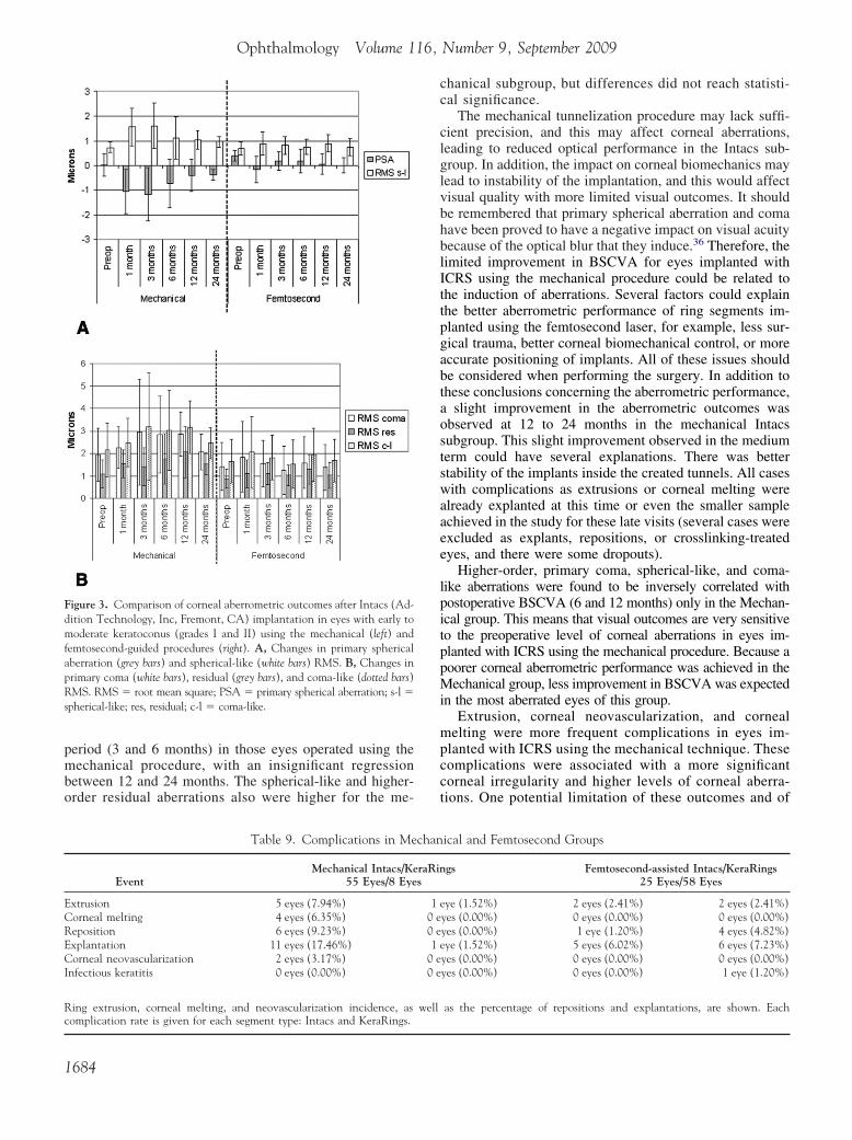
chanical subgroup. Higher-order RMS was significantly higher forthe mechanical subgroup at 6 months postoperatively (P � 0.03,Mann–Whitney test, mechanical 3.24�1.95 vs. femtosecond1.74�1.06 �m). Primary spherical aberration was significantlymore negative in the mechanical subgroup at 1, 3, 6, and 24months postoperatively (P�0.03, unpaired Student t and Mann–Whitney tests). Figure 3A shows the postoperative changes inprimary spherical aberration and spherical-like RMS in the me-chanical and femtosecond Intacs subgroups. As can be observed inthis graph, a significant negativization of primary spherical aber-ration occurred after Intacs implantation using the mechanicalprocedure (P � 0.03, Wilcoxon test). Spherical-like aberrationRMS also increased postoperatively, especially in the mechanicalsubgroup, but differences between the mechanical and femtosec-ond subgroups did not reach statistical significance (P�0.07, un-paired Student t test). Mean postoperative values of coma, residual,and coma-like RMS were higher at all postoperative visits for theeyes operated using the mechanical procedure (Fig 3B), and thedifferences reached statistical significance only for primary comaand coma-like aberration RMS at 3 and 6 months postoperatively(P�0.02, unpaired Student t and Mann–Whitney tests). In addi-tion, differences between the mechanical and femtosecond sub-groups were significant at 12 months for primary coma at 12months (P � 0.05, unpaired Student t test).
Complications
Complications in the Mechanical and Femtosecond groups aresummarized in Table 9. Segment ring explantation was performed
Outcomes in the Mechanical Group
6 Mos 12
2.99�0.86 (1.66–4.50) 4.10�2.10 (3.00�1.95 (1.65–7.09) 2.85�1.43 (2.62�0.77 (1.47–4.01) 3.20�1.51 (
�0.47�0.70 (�1.34 to 0.45) �1.09�1.11 (1.21�0.29 (0.68–1.55) 1.98�1.52 (0.95�0.41 (0.56–1.52) 1.83�1.41 (2.83�0.79 (1.56–4.24) 3.59�1.73 (
10
aberrations, all Zernike terms except Z3�1 and Z4
0; spherical-like aberratio
Outcomes in the Femtosecond Group
6 Mos 1
0.36�0.26 (0.03–1.00) 0.35�0.25�2.45�4.57 (�20.00–�3.50) �2.24�2.48�2.81�1.68 (�6.75–0.00) �3.08�1.86�3.88�4.71 (�22.00–�2.00) �3.79�3.56
0.65�0.28 (0.05–1.20) 0.66�0.2544.76�3.89 (36.88–54.20) 45.09�4.7048.39�4.75 (40.43–61.93) 48.34�5.0446.57�4.23 (39.89–57.34) 46.68�4.719.93�6.79 (�3.28–28.96) 8.02�6.93
58
meridian for the 3 mm central zone; K2 � corneal dioptric power in the s
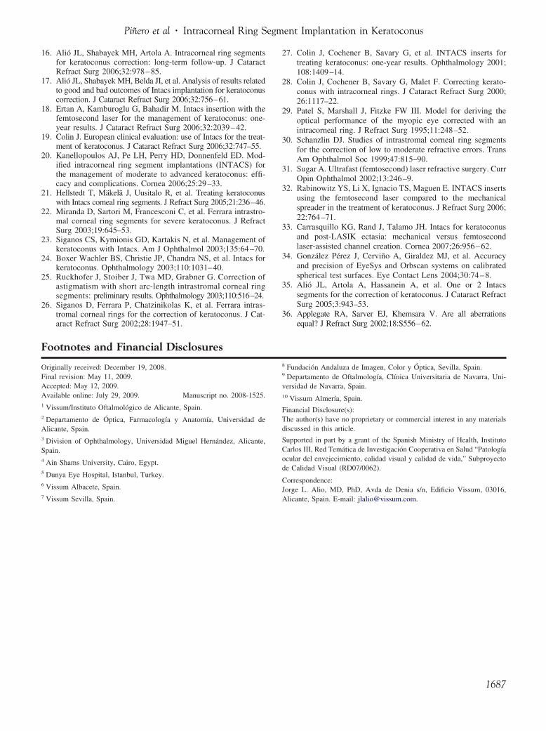
in 12 eyes (18.46%) of the Mechanical group and 11 eyes(13.25%) of the Femtosecond group (Table 9). The reasons forring segment explantation were extrusion (8 eyes), corneal melting(3 eyes), corneal neovascularization (2 eyes), and very poor visualoutcomes. All extrusion and melting cases (Table 9) showed asignificant increase in corneal irregularity with large amounts ofcorneal higher-order aberrations (Figs 4 and 5). Extrusion occurredat 6 months or later in 5 eyes that underwent mechanical tunnel-ization and in 3 eyes operated using the femtosecond laser. Ringreposition was performed in a total of 11 eyes with the aim ofimproving the ring effect and the visual and refractive outcomes.Seven of these repositioned segments were finally explanted be-cause of extrusion or poor visual outcomes. Infectious keratitisoccurred in 1 eye at 6 months postoperatively, which was appro-priately treated with an intensive fortified antibiotic and cortico-steroid combination. In this eye, an extrusion of superior ringsegment occurred and it was finally explanted.
When comparing the level of complications in the mechanicaland femtosecond procedures for eyes with early to moderatekeratoconus implanted with Intacs, we found similar rates forextrusion (mechanical 8.33% vs. femtosecond 10.52%), cornealmelting (mechanical 4.17% vs. femtosecond 0.00%), neovascular-ization (mechanical 4.17% vs. femtosecond 0.00%), infection(0.00% in both groups), and ring reposition (mechanical 4.17% vs.femtosecond 0.00%) (P�0.37, chi-square test). The explantationrate was higher in the mechanical subgroup (mechanical 20.83%vs. femtosecond 10.53%), but differences did not reach statisticalsignificance (P � 0.36, chi-square test).
24 Mos
9.49) 3.51�1.38 (1.83–5.17)6.32) 2.40�1.35 (0.83–3.93)6.53) 2.50�1.08 (1.26–3.89)3 to �0.10) �0.95�0.47 (�1.50 to �0.43)6.33) 2.10�1.23 (0.84–4.12)5.25) 1.46�0.75 (0.69–2.69)7.91) 3.18�1.21 (1.70–4.41)
10
rms from fourth and sixth order; coma-like aberrations, terms from third,
s 24 Mos
–1.00) 0.29�0.22 (0.05–0.90)50–�1.50) �1.17�2.12 (�6.00–�2.75)–�8.00) �2.67�1.40 (�7.00 to �1.00).00–�0.88) �2.51�2.11 (�7.00–�2.25)–1.00) 0.70�0.25 (0.30–1.00)5–59.17) 45.41�4.93 (39.33–58.54)5–62.32) 48.67�5.18 (42.29–61.41)9–60.59) 47.04�4.99 (41.37–59.97).98–26.71) 7.20�5.30 (�3.00–14.03)
19
st meridian for the 3 mm central zone; KM � mean corneal power in the
Mos
2.30–0.87–1.28–�3.11.03–0.57–1.92–18
ns, te
2 Mo
(0.02(�7.(0.00(�16(0.10(38.0(40.6(40.2(�1536
teepe
1681

Ophthalmology Volume 116, Number 9, September 2009
Discussion
The current study analyzed the refractive and corneal aber-rometric effect of ICRS in keratoconic eyes. We have alsostudied how this effect can be influenced by the surgicaltechnique used for corneal tunnelization: mechanical (Me-chanical group) or femtosecond laser-assisted (Femtosec-ond group). A significant reduction in manifest sphere,cylinder, and spherical equivalent was found after surgeryusing both surgical procedures for creation of corneal chan-nels. These refractive changes are in concordance with thosereported by previous authors.9–13,16–20,35 In the Mechanicalgroup, a slight but insignificant regression of spherical cor-rection occurred at 12 months. A similar variability insphere in a medium to long-term follow-up was also ob-served previously after Intacs implantation using the me-chanical procedure.15,16,19,23 Part of this variability in spher-ical correction could be due to the loss of effect of ringsegments leading to postoperative complications, such asextrusion or corneal melting (most complications occur at�6 months).
Uncorrected visual acuity significantly improved after sur-gery in both the Mechanical and Femtosecond groups, which isconsistent with the significant reduction achieved in refrac-tion. This visual improvement was also reported in other
Parameter Preo
Higher-order RMS (�m) 3.35�2.0RMS astigmatism (�m) 2.93�2.0Primary coma RMS (�m) 2.83�1.9Z4
0 (�m) �0.14�0.8Residual RMS (�m) 1.35�1.0Spherical-like RMS (�m) 1.10�0.8Coma-like RMS (�m) 3.11�1.9No. of eyes
RMS � root mean square.Aberrometric definitions: primary coma, terms Z3
�1; primary spherical ab
Figure 2. Changes in lines of BSCVA postoperatively in eyes operatedwith the femtosecond-assisted procedure. Gains of �2 lines of BSCVAwere found in 46.55% of eyes at 6 months. BSCVA � best spectacle-corrected visual acuity.
Ranges are shown in brackets below each mean value.
1682
studies on ICRS in corneal ectasia.9–14,18–20,23,24,26,27,33,35
BSCVA improved significantly only in the Femtosecondgroup, which supports the findings of other authors.9,11–13,18
However, there are published reports citing an improvementin BSCVA with ICRS using the mechanical procedure,which is contradictory to the findings of the presentstudy.10,14,16,17,19,20,23,24,26,33,35 Several factors could ex-plain the difference between our findings and the previousfindings. For example, we may have included more moder-ate and severe cases and observed more complications in theMechanical group compared with previous studies. Never-theless, the safety indices of both surgical techniques provedto be excellent with 41.18% (Mechanical group) and 46.55%(Femtosecond group) of eyes gaining �2 lines of BSCVAat 6 months.
In regard to corneal curvature, a significant central flat-tening was achieved in both surgical techniques, in keepingwith previous studies on ICRS for keratoconus manage-ment.9–20,23,35 This flattening is responsible for the reductionof refraction and increase in UCVA. However, a regression ofthe achieved corneal central flattening was observed at 12months in those eyes implanted with ICRS using the me-chanical surgical procedure. This change did not reachstatistical significance, but it was consistent with the regres-sion in myopic spherical correction also observed at 12months. This regression of the central flattening achievedwith ICRS implanted using the mechanical procedure in amedium to long-term follow-up has been reported.15,16,23
Inferosuperior asymmetry also decreased in both groups,but changes did not reach statistical significance because ofthe high variability of this parameter (not all cones weredecentered inferiorly).
In addition to visual and refractive outcomes, changes inanterior corneal aberrations were also evaluated in thisstudy. To the best of our knowledge, this is the first studycomparing the corneal aberrometric performance of ICRSimplanted using 2 different surgical procedures: mechanicaland femtosecond laser-assisted techniques. It should beremembered that anterior corneal aberrometric analysis isan important tool in clinical practice for evaluating theocular optical quality because the first refractive interface(air–cornea) is the most important contributor to the totalpower of the eye because of the large difference in refrac-tive index existing at this point. In highly aberrated corneas,
Table 8. Summary of the Corneal Aberrometric
ive 3 Mos
3–10.66) 3.19�1.87 (0.72–8.60)0–10.86) 2.65�1.72 (0.33–8.59)4–10.07) 2.62�1.88 (0.28–8.46).86–2.69) �0.17�0.71 (�1.75–0.98)7–8.10) 1.39�0.95 (0.40–5.72)8–6.38) 1.13�0.79 (0.36–4.41)7–10.46) 2.90�1.83 (0.53–8.50)
34
on, term Z40; residual aberrations, all Zernike terms except Z3
�1 and Z40;
perat
2 (0.39 (0.24 (0.04 (�11 (0.12 (0.23 (0.164
errati

Piñero et al � Intracorneal Ring Segment Implantation in Keratoconus
such as in keratoconus, the corneal aberrations of the ante-rior corneal surface are the most important source of opticalerrors in the eye. In the current study, we found the effect onthe corneal aberrations was dependent on the mechanismused to create the tunnel. After ICRS implantation using themechanical surgical procedure, no significant changes wereachieved in any corneal aberrometric parameter, althoughreduction was achieved in astigmatism, primary coma, andcoma-like aberrations. At 12 months, a significant increasewas found in higher-order, primary coma, spherical-like,and coma-like aberrations in the Mechanical group. Further-more, an increase in primary spherical aberration, higher-order residual, and spherical-like aberrations was inducedwith ICRS in this group, although changes did not reachstatistical significance. Therefore, there was a significantvariability in corneal aberrations after ICRS implantationusing the mechanical procedure, which implies that cornealirregularity is not well controlled with this kind of surgicalintervention. This supports the marginal improvement inBSCVA observed in the Mechanical group.
In corneas implanted with ICRS using the femtosecond-assisted surgical procedure, a significant reduction wasobserved in the RMS for astigmatism after surgery. Inaddition, a significant increase in higher-order residual ab-errations was found postoperatively. Total higher-order, pri-mary coma, and coma-like aberrations were also reducedpostoperatively, but changes did not reach statistical signif-icance. During all follow-up sessions, no significant regres-sions in the achieved aberrometric correction were ob-served, confirming the stability of ICRS effect on cornealirregularity in the Femtosecond group. These findings sup-port the conclusions reached by Shabayek and Alió,13 whofound a statistically significant reduction in higher-orderRMS for those eyes with a relatively high preoperativeRMS (�3.0 �m) and implanted with KeraRings using thefemtosecond laser for corneal tunnelization.
Part of these differences in the aberration profile betweenthe Mechanical and Femtosecond groups, as well as in therefractive and keratometric parameters, could be due to thedifferent ring segment profile implanted in each group (e.g.,Intacs were implanted in 87.30% of eyes in the Mechanicalgroup and in 31.25% of eyes in the Femtosecond group). Itshould be remembered that 2 different kinds of ring seg-ments were used, Intacs and KeraRings, each with a differ-
Outcomes in the Femtosecond Group
6 Mos 12
3.17�2.21 (0.75–10.39) 3.07�1.542.72�1.87 (0.81–8.83) 2.71�1.582.50�2.04 (0.31–9.08) 2.45�1.560.04�0.85 (�3.28–1.67) �0.09�0.711.58�1.16 (0.51–6.25) 1.49�0.831.28�0.99 (0.45–5.28) 1.15�0.552.83�2.08 (0.58–9.35) 2.79�1.56
41
spherical-like aberrations, terms from fourth and sixth order; coma-like a
ent cross-sectional profile and diameter of implantation.Another factor to consider is the severity of the ectaticdisease because higher rates of complications and pooreroutcomes have been reported for eyes with advanced kera-toconus implanted with ICRS.17,20 For all these reasons, acomparative analysis of the outcomes achieved with themechanical and femtosecond-based techniques for each seg-ment type and for early to moderate and advanced kerato-conus cases was initially planned to avoid the possiblevariability introduced by these 2 factors. With this detailedanalysis, the aberrometric and refractive variability inducedby the technique of corneal tunnelization could be evalu-ated. However, this comparative analysis was only feasiblefor eyes with early to moderate keratoconus (grades I and II)implanted with Intacs (mechanical subgroup 24 eyes vs.femtosecond subgroup 19 eyes) because the remaining sam-ples were not large enough to perform a plausible statisticalanalysis. Examples of the limitations are as follows: Only 8eyes were implanted with KeraRings using the mechanicalprocedure (only 2 of these 8 eyes with aberrometric data),and only 2 eyes with advanced keratoconus were implantedwith Intacs using the IntraLase system. This study is aretrospective analysis of a consecutive case series and assuch has its own limitations. For example, there were anunequal number of Intacs and KeraRings cases implantedwith the mechanical and femtosecond-guided procedures.
When comparing the mechanical and femtosecond sub-groups in early to moderate keratoconus eyes implantedwith Intacs, no significant differences were found in thevisual outcomes and magnitude of astigmatic correction.Significant differences were found at 6 months postopera-tively in the magnitude of corneal asymmetry. Furthermore,after surgery there was a tendency toward a greater extent ofasymmetry in those eyes operated with the mechanicaltechnique. This trend was maintained at 12 and 24 months,but the differences did not reach statistical significance. Thisgreater asymmetry in the mechanical subgroup was consis-tent with the significantly greater magnitude of primarycoma found in this subgroup at 3, 6, and 12 months. Inaddition, significant differences between the mechanicaland the femtosecond subgroups were found in other aber-rometric parameters, such as higher-order RMS, coma-likeRMS, and primary spherical aberration. Primary sphericalaberration changed significantly in the initial postoperative
24 Mos
1–6.78) 2.65�1.19 (0.63–5.44)4–6.95) 2.22�0.90 (0.82–4.05)6–6.38) 2.12�1.36 (0.22–4.87).30–1.61) �0.16�0.68 (�2.03–0.70)0–4.77) 1.18�0.53 (0.35–2.13)9–2.78) 1.02�0.49 (0.28–2.30)2–6.51) 2.39�1.20 (0.56–4.93)
18
ions, terms from third, fifth, and seventh order.
Mos
(0.8(0.3(0.1(�2(0.5(0.4(0.6
39
berrat
1683
Ophthalmology Volume 116, Number 9, September 2009
period (3 and 6 months) in those eyes operated using themechanical procedure, with an insignificant regressionbetween 12 and 24 months. The spherical-like and higher-order residual aberrations also were higher for the me-
Figure 3. Comparison of corneal aberrometric outcomes after Intacs (Ad-dition Technology, Inc, Fremont, CA) implantation in eyes with early tomoderate keratoconus (grades I and II) using the mechanical (left) andfemtosecond-guided procedures (right). A, Changes in primary sphericalaberration (grey bars) and spherical-like (white bars) RMS. B, Changes inprimary coma (white bars), residual (grey bars), and coma-like (dotted bars)RMS. RMS � root mean square; PSA � primary spherical aberration; s-l �spherical-like; res, residual; c-l � coma-like.
Table 9. Complications in Me
EventMechanical Intacs/Ke
55 Eyes/8 Eye
Extrusion 5 eyes (7.94%)Corneal melting 4 eyes (6.35%)Reposition 6 eyes (9.23%)Explantation 11 eyes (17.46%)Corneal neovascularization 2 eyes (3.17%)Infectious keratitis 0 eyes (0.00%)
Ring extrusion, corneal melting, and neovascularization incidence, as
complication rate is given for each segment type: Intacs and KeraRings.1684
chanical subgroup, but differences did not reach statisti-cal significance.
The mechanical tunnelization procedure may lack suffi-cient precision, and this may affect corneal aberrations,leading to reduced optical performance in the Intacs sub-group. In addition, the impact on corneal biomechanics maylead to instability of the implantation, and this would affectvisual quality with more limited visual outcomes. It shouldbe remembered that primary spherical aberration and comahave been proved to have a negative impact on visual acuitybecause of the optical blur that they induce.36 Therefore, thelimited improvement in BSCVA for eyes implanted withICRS using the mechanical procedure could be related tothe induction of aberrations. Several factors could explainthe better aberrometric performance of ring segments im-planted using the femtosecond laser, for example, less sur-gical trauma, better corneal biomechanical control, or moreaccurate positioning of implants. All of these issues shouldbe considered when performing the surgery. In addition tothese conclusions concerning the aberrometric performance,a slight improvement in the aberrometric outcomes wasobserved at 12 to 24 months in the mechanical Intacssubgroup. This slight improvement observed in the mediumterm could have several explanations. There was betterstability of the implants inside the created tunnels. All caseswith complications as extrusions or corneal melting werealready explanted at this time or even the smaller sampleachieved in the study for these late visits (several cases wereexcluded as explants, repositions, or crosslinking-treatedeyes, and there were some dropouts).
Higher-order, primary coma, spherical-like, and coma-like aberrations were found to be inversely correlated withpostoperative BSCVA (6 and 12 months) only in the Mechan-ical group. This means that visual outcomes are very sensitiveto the preoperative level of corneal aberrations in eyes im-planted with ICRS using the mechanical procedure. Because apoorer corneal aberrometric performance was achieved in theMechanical group, less improvement in BSCVA was expectedin the most aberrated eyes of this group.
Extrusion, corneal neovascularization, and cornealmelting were more frequent complications in eyes im-planted with ICRS using the mechanical technique. Thesecomplications were associated with a more significantcorneal irregularity and higher levels of corneal aberra-tions. One potential limitation of these outcomes and of
ical and Femtosecond Groups
gs Femtosecond-assisted Intacs/KeraRings25 Eyes/58 Eyes
eye (1.52%) 2 eyes (2.41%) 2 eyes (2.41%)yes (0.00%) 0 eyes (0.00%) 0 eyes (0.00%)yes (0.00%) 1 eye (1.20%) 4 eyes (4.82%)eye (1.52%) 5 eyes (6.02%) 6 eyes (7.23%)yes (0.00%) 0 eyes (0.00%) 0 eyes (0.00%)yes (0.00%) 0 eyes (0.00%) 1 eye (1.20%)
as the percentage of repositions and explantations, are shown. Each
chan
raRins
10 e0 e1
0 e0 e
well

n squ
Piñero et al � Intracorneal Ring Segment Implantation in Keratoconus
the current study is the learning curve of the surgeon. Wehave compared the outcomes from different surgeons,and their learning curve could be a potential source ofvariability, especially for corneal tunnelization with themechanical device, because it is highly dependent onsurgeon manual dexterity. A higher magnitude of vari-ability with 1 specific surgical procedure would implythat this surgical procedure is less reproducible andhighly dependent on surgeon skills, leading to less pre-dictable outcomes. Part of the variability in refractive andaberrometric outcomes observed during the follow-up in
Figure 4. Corneal topography and aberrometric analysis of a keratoconuextruded (superior and inferior) 12 months after surgery. Corneal topograp(up) and when extrusion was detected are shown (down). Each corneal aleft to right and from up to down, the following optical errors: all aberratithe residual aberrations without considering the astigmatism, primary comdescribed optical error is provided below each map. As can be observed, thincrease of 2.51 �m) and coma (increase of 1.22 �m). RMS � root mea
the Mechanical group could be attributed to this limiting
factor: the learning curve of the surgeon. In addition, wehave observed that no significant differences werepresent in the explantation, reposition, corneal neovascu-larization, corneal melting, extrusion, and infection ratesbetween the mechanical and the femtosecond Intacs sub-groups, although the explantation rate was slightly higherfor the mechanical subgroup. This could be associatedwith other patient-related factors.
In conclusion, intracorneal ring segments implantationwith Intacs or KeraRings is an effective option for thetreatment of spherocylindrical error and corneal irregularity
lanted with Intacs (mechanical procedure) and with both ring segmentsft) and aberrometric (right) maps 4 months before ring segments extrusionetry includes different maps simulating the wavefront considering, from
stigmatism, primary spherical aberration (Z40), primary coma (Z3
�1) andd primary spherical aberration. In addition, the RMS associated with thea large increase in primary spherical aberration (it becomes more negative,are.
s imphic (leberromons, aa, an
ere is
in keratoconus. As other authors have found,32,33 there were
1685

Ophthalmology Volume 116, Number 9, September 2009
no significant differences in refractive and visual outcomesbetween the mechanical and the femtosecond-assisted sur-gical techniques for ICRS implantation. Furthermore, it hasbeen demonstrated that final visual outcomes with ICRS(mostly Intacs) implanted using mechanical tunnelizationwere dependent on the preoperative magnitude of cornealirregularity, exerting a more limited visual improvement inhighly aberrated eyes. The use of mechanical tunnelizationspecifically for Intacs implantation in eyes with early tomoderate keratoconus has been demonstrated to limit thepotential aberrometric correction of these implants becausethe procedure itself generates new aberrations, especiallynegative primary spherical aberration and primary coma.This trend could not be specifically confirmed for KeraRingssegments because of the limitations of this retrospectivestudy in the sample size for this segment type. In addition,the impact of the surgical technique specifically in advancedkeratoconus should be addressed in future studies with amore complete series of advanced keratoconus cases(homogeneous samples of eyes implanted with Intacs andKeraRings). Longer follow-up is needed to corroborate thestability of visual, refractive, and aberrometric outcomesachieved by these implants using both surgical techniques,mechanical and femtosecond-assisted implantation.
References
1. Rabinowitz YS. Keratoconus. Surv Ophthalmol 1998;42:297–319.
2. Bühren J, Kühne C, Kohnen T. Defining subclinical kerato-conus using corneal first-surface higher-order aberrations.
Figure 5. Three-dimensional postoperative corneal profile of a cornea imexamination, extrusion of the inferior ring segment was present.
Am J Ophthalmol 2007;143:381–9.
1686
3. Alió JL, Shabayek MH. Corneal higher order aberrations: amethod to grade keratoconus. J Refract Surg 2006;22:539 –45.
4. Gobbe M, Guillon M. Corneal wavefront aberration measure-ments to detect keratoconus patients. Cont Lens Anterior Eye2005;28:57–66.
5. Barbero S, Marcos S, Merayo-Lloves J, Moreno-Barriuso E.Validation of the estimation of corneal aberrations from video-keratography in keratoconus. J Refract Surg 2002;18:263–70.
6. Garcia-Lledo M, Feinbaum C, Alió JL. Contact lens fitting inkeratoconus. Compr Ophthalmol Update 2006;7:47–52.
7. Smiddy WE, Hamburg TR, Kracher GP, Stark WJ. Keratoconus:contact lens or keratoplasty? Ophthalmology 1998;95:487–92.
8. Dana MR, Putz JL, Viana MA, et al. Contact lens failure inkeratoconus management. Ophthalmology 1992;99:1187–92.
9. Coskunseven E, Kymionis GD, Tsiklis NS, et al. One-yearresults of intrastromal corneal ring segment implantation(KeraRing) using femtosecond laser in patients with kerato-conus. Am J Ophthalmol 2008;145:775–9.
10. Shetty R, Kurian M, Anand D, et al. Intacs in advancedkeratoconus. Cornea 2008;27:1022–9.
11. Ertan A, Ozkilic E. Effect of age on outcomes in patients withkeratoconus treated by Intacs using a femtosecond laser. JRefract Surg 2008;24:690–5.
12. Ertan A, Kamburoglu G. Intacs implantation using femtosecondlaser for management of keratoconus: comparison of 306 cases indifferent stages. J Cataract Refract Surg 2008;34:1521–6.
13. Shabayek MH, Alió JL. Intrastromal corneal ring segmentimplantation by femtosecond laser for keratoconus correction.Ophthalmology 2007;114:1643–52.
14. Zare MA, Hashemi H, Salari MR. Intracorneal ring segmentimplantation for the management of keratoconus: safety andefficacy. J Cataract Refract Surg 2007;33:1886–91.
15. Kymionis GD, Siganos CS, Tsiklis NS, et al. Long-termfollow-up of Intacs in keratoconus. Am J Ophthalmol 2007;
ed with KeraRings (Mediphacos, Belo Horizonte, Brazil). In this corneal
plant143:236–44.
Piñero et al � Intracorneal Ring Segment Implantation in Keratoconus
16. Alió JL, Shabayek MH, Artola A. Intracorneal ring segmentsfor keratoconus correction: long-term follow-up. J CataractRefract Surg 2006;32:978–85.
17. Alió JL, Shabayek MH, Belda JI, et al. Analysis of results relatedto good and bad outcomes of Intacs implantation for keratoconuscorrection. J Cataract Refract Surg 2006;32:756–61.
18. Ertan A, Kamburoglu G, Bahadir M. Intacs insertion with thefemtosecond laser for the management of keratoconus: one-year results. J Cataract Refract Surg 2006;32:2039–42.
19. Colin J. European clinical evaluation: use of Intacs for the treat-ment of keratoconus. J Cataract Refract Surg 2006;32:747–55.
20. Kanellopoulos AJ, Pe LH, Perry HD, Donnenfeld ED. Mod-ified intracorneal ring segment implantations (INTACS) forthe management of moderate to advanced keratoconus: effi-cacy and complications. Cornea 2006;25:29–33.
21. Hellstedt T, Mäkelä J, Uusitalo R, et al. Treating keratoconuswith Intacs corneal ring segments. J Refract Surg 2005;21:236–46.
22. Miranda D, Sartori M, Francesconi C, et al. Ferrara intrastro-mal corneal ring segments for severe keratoconus. J RefractSurg 2003;19:645–53.
23. Siganos CS, Kymionis GD, Kartakis N, et al. Management ofkeratoconus with Intacs. Am J Ophthalmol 2003;135:64–70.
24. Boxer Wachler BS, Christie JP, Chandra NS, et al. Intacs forkeratoconus. Ophthalmology 2003;110:1031–40.
25. Ruckhofer J, Stoiber J, Twa MD, Grabner G. Correction ofastigmatism with short arc-length intrastromal corneal ringsegments: preliminary results. Ophthalmology 2003;110:516–24.
26. Siganos D, Ferrara P, Chatzinikolas K, et al. Ferrara intras-tromal corneal rings for the correction of keratoconus. J Cat-
aract Refract Surg 2002;28:1947–51.7 Vissum Sevilla, Spain.
27. Colin J, Cochener B, Savary G, et al. INTACS inserts fortreating keratoconus: one-year results. Ophthalmology 2001;108:1409–14.
28. Colin J, Cochener B, Savary G, Malet F. Correcting kerato-conus with intracorneal rings. J Cataract Refract Surg 2000;26:1117–22.
29. Patel S, Marshall J, Fitzke FW III. Model for deriving theoptical performance of the myopic eye corrected with anintracorneal ring. J Refract Surg 1995;11:248–52.
30. Schanzlin DJ. Studies of intrastromal corneal ring segmentsfor the correction of low to moderate refractive errors. TransAm Ophthalmol Soc 1999;47:815–90.
31. Sugar A. Ultrafast (femtosecond) laser refractive surgery. CurrOpin Ophthalmol 2002;13:246–9.
32. Rabinowitz YS, Li X, Ignacio TS, Maguen E. INTACS insertsusing the femtosecond laser compared to the mechanicalspreader in the treatment of keratoconus. J Refract Surg 2006;22:764–71.
33. Carrasquillo KG, Rand J, Talamo JH. Intacs for keratoconusand post-LASIK ectasia: mechanical versus femtosecondlaser-assisted channel creation. Cornea 2007;26:956–62.
34. González Pérez J, Cerviño A, Giraldez MJ, et al. Accuracyand precision of EyeSys and Orbscan systems on calibratedspherical test surfaces. Eye Contact Lens 2004;30:74–8.
35. Alió JL, Artola A, Hassanein A, et al. One or 2 Intacssegments for the correction of keratoconus. J Cataract RefractSurg 2005;3:943–53.
36. Applegate RA, Sarver EJ, Khemsara V. Are all aberrations
equal? J Refract Surg 2002;18:S556–62.Footnotes and Financial Disclosures
Originally received: December 19, 2008.Final revision: May 11, 2009.Accepted: May 12, 2009.Available online: July 29, 2009. Manuscript no. 2008-1525.1 Vissum/Instituto Oftalmológico de Alicante, Spain.2 Departamento de Óptica, Farmacología y Anatomía, Universidad deAlicante, Spain.3 Division of Ophthalmology, Universidad Miguel Hernández, Alicante,Spain.4 Ain Shams University, Cairo, Egypt.5 Dunya Eye Hospital, Istanbul, Turkey.6 Vissum Albacete, Spain.
8 Fundación Andaluza de Imagen, Color y Óptica, Sevilla, Spain.9 Departamento de Oftalmología, Clínica Universitaria de Navarra, Uni-versidad de Navarra, Spain.10 Vissum Almería, Spain.
Financial Disclosure(s):The author(s) have no proprietary or commercial interest in any materialsdiscussed in this article.
Supported in part by a grant of the Spanish Ministry of Health, InstitutoCarlos III, Red Temática de Investigación Cooperativa en Salud “Patologíaocular del envejecimiento, calidad visual y calidad de vida,” Subproyectode Calidad Visual (RD07/0062).
Correspondence:Jorge L. Alio, MD, PhD, Avda de Denia s/n, Edificio Vissum, 03016,
Alicante, Spain. E-mail: [email protected].1687