imaging and aberrometric analyses of diseases affecting
TRANSCRIPT

Imaging and aberrometric analyses of diseases affecting
biomechanical properties of the cornea
Modern disgnostic methods of keratoconus
PhD thesis
Dr Mihaacuteltz Kata
Semmelweis University
Clinical Medicine PhD School
Supervisor Dr Nagy Zoltaacuten Zsolt DSC
Reviewers Dr Kereacutenyi Aacutegnes PhD
Dr Farkas Aacutegnes PhD
Chairman of comprehensive exam Dr Matolcsy Andraacutes DSc
Members of comprehensive exam Dr Fuumlst Aacutegnes PhD
Dr Erdei Gaacutebor PhD
Budapest 2011
2
Contents
Table of contentshelliphelliphelliphelliphelliphelliphelliphelliphelliphelliphelliphelliphelliphelliphelliphelliphelliphelliphelliphelliphelliphelliphelliphelliphelliphelliphelliphelliphellip2
Abbreviationshelliphelliphelliphelliphelliphelliphelliphelliphelliphelliphelliphelliphelliphelliphelliphelliphelliphelliphelliphelliphelliphelliphelliphelliphelliphelliphelliphelliphelliphellip4
1 Introductionhelliphelliphelliphelliphelliphelliphelliphelliphelliphelliphelliphelliphelliphelliphelliphelliphelliphelliphelliphelliphelliphelliphelliphelliphelliphelliphelliphelliphelliphellip6
1 1 Epidemiology of keratoconushelliphelliphelliphelliphelliphelliphelliphelliphelliphelliphelliphelliphelliphelliphelliphelliphelliphelliphelliphelliphelliphellip7
1 2 Clinical Featureshelliphelliphelliphelliphelliphelliphelliphelliphelliphelliphelliphelliphelliphelliphelliphelliphelliphelliphelliphelliphelliphelliphelliphelliphelliphelliphellip7
1 3 Histopathologyhelliphelliphelliphelliphelliphelliphelliphelliphelliphelliphelliphelliphelliphelliphelliphelliphelliphelliphelliphelliphelliphelliphelliphelliphelliphelliphelliphellip10
1 4 Etiology and Pathogenesishelliphelliphelliphelliphelliphelliphelliphelliphelliphelliphelliphelliphelliphelliphelliphelliphelliphelliphelliphelliphelliphelliphellip11
14 1 Associated disordershelliphelliphelliphelliphelliphelliphelliphelliphelliphelliphelliphelliphelliphelliphelliphelliphelliphelliphelliphelliphelliphelliphelliphellip11
14 2 Biochemical studieshelliphelliphelliphelliphelliphelliphelliphelliphelliphelliphelliphelliphelliphelliphelliphelliphelliphelliphelliphelliphelliphelliphelliphellip12
14 3 Geneticshelliphelliphelliphelliphelliphelliphelliphelliphelliphelliphelliphelliphelliphelliphelliphelliphelliphelliphelliphelliphelliphelliphelliphelliphelliphelliphelliphelliphellip13
15 Diagnosis of keratoconushelliphelliphelliphelliphelliphelliphelliphelliphelliphelliphelliphelliphelliphelliphelliphelliphelliphelliphelliphelliphelliphelliphelliphellip14
151 Photokeratoscopy and keratometryhelliphelliphelliphelliphelliphelliphelliphelliphelliphelliphelliphelliphelliphelliphelliphelliphelliphelliphellip14
152 Computer-assisted videokeratoscopyhelliphelliphelliphelliphelliphelliphelliphelliphelliphelliphelliphelliphelliphelliphelliphelliphelliphellip15
153 Elevation based topographyhelliphelliphelliphelliphelliphelliphelliphelliphelliphelliphelliphelliphelliphelliphelliphelliphelliphelliphelliphelliphellip19
1531 Orbscanhelliphelliphelliphelliphelliphelliphelliphelliphelliphelliphelliphelliphelliphelliphelliphelliphelliphelliphelliphelliphelliphelliphelliphelliphelliphelliphelliphellip19
1532 Scheimpflug camerahelliphelliphelliphelliphelliphelliphelliphelliphelliphelliphelliphelliphelliphelliphelliphelliphelliphelliphelliphelliphelliphelliphellip20
154 Wavefront aberrometryhelliphelliphelliphelliphelliphelliphelliphelliphelliphelliphelliphelliphelliphelliphelliphelliphelliphelliphelliphelliphelliphelliphellip21
16 Treatment of Keratoconushelliphelliphelliphelliphelliphelliphelliphelliphelliphelliphelliphelliphelliphelliphelliphelliphelliphelliphelliphelliphelliphelliphelliphellip23
161 Contact lenseshelliphelliphelliphelliphelliphelliphelliphelliphelliphelliphelliphelliphelliphelliphelliphelliphelliphelliphelliphelliphelliphelliphelliphelliphelliphelliphellip23
162 Corneal crosslinkinghelliphelliphelliphelliphelliphelliphelliphelliphelliphelliphelliphelliphelliphelliphelliphelliphelliphelliphelliphelliphelliphelliphelliphellip23
1631 Surgical interventions ICRShelliphelliphelliphelliphelliphelliphelliphelliphelliphelliphelliphelliphelliphelliphelliphelliphelliphelliphelliphellip24
1632 Surgical interventions Keratoplastyhelliphelliphelliphelliphelliphelliphelliphelliphelliphelliphelliphelliphelliphelliphelliphelliphellip25
17 Other diseases affecting biomechanical properties of the corneahelliphelliphelliphelliphelliphelliphelliphelliphellip26
171 Pellucid marginal degenerationhelliphelliphelliphelliphelliphelliphelliphelliphelliphelliphelliphelliphelliphelliphelliphelliphelliphelliphelliphellip26
172 Keratoglobushelliphelliphelliphelliphelliphelliphelliphelliphelliphelliphelliphelliphelliphelliphelliphelliphelliphelliphelliphelliphelliphelliphelliphelliphelliphelliphellip28
173 Post-LASIK ectasiashellip helliphelliphelliphelliphelliphelliphelliphelliphelliphelliphelliphelliphelliphelliphelliphelliphelliphelliphelliphelliphelliphelliphellip28
2 Purposeshelliphelliphelliphelliphelliphelliphelliphelliphelliphelliphelliphelliphelliphelliphelliphelliphelliphelliphelliphelliphelliphelliphelliphelliphelliphelliphelliphelliphelliphelliphellip30
3 Methodshelliphelliphelliphelliphelliphelliphelliphelliphelliphelliphelliphelliphelliphelliphelliphelliphelliphelliphelliphelliphelliphelliphelliphelliphelliphelliphelliphelliphelliphelliphellip32
31 Demographic datahelliphelliphelliphelliphelliphelliphelliphelliphelliphelliphelliphelliphelliphelliphelliphelliphelliphelliphelliphelliphelliphelliphelliphelliphelliphellip32
32 Patient inclusion criteriahelliphelliphelliphelliphelliphelliphelliphelliphelliphelliphelliphelliphelliphelliphelliphelliphelliphelliphelliphelliphelliphelliphelliphellip33
33 Patient exclusion criteriahelliphelliphelliphelliphelliphelliphelliphelliphelliphelliphelliphelliphelliphelliphelliphelliphelliphelliphelliphelliphelliphelliphelliphellip33
34 Corneal topographic measurementshelliphelliphelliphelliphelliphelliphelliphelliphelliphelliphelliphelliphelliphelliphelliphelliphelliphelliphellip34
3
35 Pentacam measurementshelliphelliphelliphelliphelliphelliphelliphelliphelliphelliphelliphelliphelliphelliphelliphelliphelliphelliphelliphelliphelliphelliphellip34
36 Hartmann-Shack aberrometry measurementshelliphelliphelliphelliphelliphelliphelliphelliphelliphelliphelliphelliphelliphelliphellip35
37 Statistical analyseshelliphelliphelliphelliphelliphelliphelliphelliphelliphelliphelliphelliphelliphelliphelliphelliphelliphelliphelliphelliphelliphelliphelliphelliphelliphellip38
4 Resultshelliphelliphelliphelliphelliphelliphelliphelliphelliphelliphelliphelliphelliphelliphelliphelliphelliphelliphelliphelliphelliphelliphelliphelliphelliphelliphelliphelliphelliphelliphelliphellip40
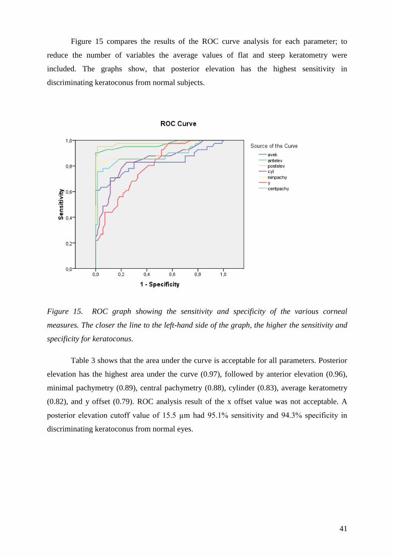
41 Evaluation of elevation parameters of keratoconus with Pentacamhelliphelliphelliphelliphelliphelliphellip40
42 Anterior chamber characteristics of keratoconus helliphelliphelliphelliphelliphelliphelliphelliphelliphelliphelliphelliphelliphellip45
43 Shifting of the line of sight in keratoconus measured by a Hartmann-Shack sensorhellip50
5 Discussionhelliphelliphelliphelliphelliphelliphelliphelliphelliphelliphelliphelliphelliphelliphelliphelliphelliphelliphelliphelliphelliphelliphelliphelliphelliphelliphelliphelliphelliphellip56
51 Keratoconus detection with Scheimpflug imaginghelliphelliphelliphelliphelliphelliphelliphelliphelliphelliphelliphelliphellip56
511 Characteristics of elevation based topographyhelliphelliphelliphelliphelliphelliphelliphelliphelliphelliphelliphelliphelliphellip56
512 The role of refernce body selection in the diagnosis of keratoconushelliphelliphelliphelliphellip58
513 The use of elevation data in the diagnosis of keratoconushelliphelliphelliphelliphelliphelliphelliphelliphellip61
514 Comparative analysis of anterior chamber characteristics of keratoconushelliphelliphellip63
52 Keratoconus detection with aberrometryhelliphelliphelliphelliphelliphelliphelliphelliphelliphelliphelliphellip65
521 Optical and visual axes of the eyehelliphelliphelliphelliphelliphelliphelliphelliphelliphelliphelliphelliphelliphelliphelliphelliphelliphelliphellip66
522 Spherical aberration in keratoconus helliphelliphelliphelliphelliphelliphelliphelliphelliphelliphelliphelliphelliphelliphelliphelliphelliphellip68
523 The pupil apodizing and Stiles-Crawford effecthelliphelliphelliphelliphelliphelliphelliphelliphelliphelliphelliphelliphellip70
6 Conclusionhelliphelliphelliphelliphelliphelliphelliphelliphelliphelliphelliphelliphelliphelliphelliphelliphelliphelliphelliphelliphelliphelliphelliphelliphelliphelliphelliphelliphelliphelliphellip72
7 Summaryhelliphelliphelliphelliphelliphelliphelliphelliphelliphelliphelliphelliphelliphelliphelliphelliphelliphelliphelliphelliphelliphelliphelliphelliphelliphelliphelliphelliphelliphelliphellip74
8 Referenceshelliphelliphelliphelliphelliphelliphelliphelliphelliphelliphelliphelliphelliphelliphelliphelliphelliphelliphelliphelliphelliphelliphelliphelliphelliphelliphelliphelliphelliphelliphellip75
9 List of publicationshelliphelliphelliphelliphelliphelliphelliphelliphelliphelliphelliphelliphelliphelliphelliphelliphelliphelliphelliphelliphelliphelliphelliphelliphelliphelliphellip84
Acknowledgementhelliphelliphelliphelliphelliphelliphelliphelliphelliphelliphelliphelliphelliphelliphelliphelliphelliphelliphelliphelliphelliphelliphelliphelliphelliphelliphelliphelliphellip89
4
Abbreviations
ACD anterior chamber depth
AIC Akaike information criterion
ANOVA analysis of variance
ASCRS American Society of Cataract and Refractive Surgery
AUROC area under the receiver operating characteristic curve
BCVA best corrected visual acuity
BFS best-fit sphere
BFTE best fit toric ellipsoid reference body
CCT central corneal thickness
CFA confirmatory factor analysis
CL contact lens
CRF corneal resistance factor
CH corneal hysteresis
CXL corneal crosslinking
D diopter
DLK diffuse lamellar keratitis
GEE generalized estimating equation
HORMS higher-order root mean square
ICRS intrastromal corneal ring segments
IOL intraocular lens
K keratometry
KISA central keratometry (K) inferior (I) minus superior (S) average (A) keratometry
LASIK laser-assisted in situ keratomileusis
LKP lamellar keratoplasty
LOS line of sight
OPD optical path difference
ORA ocular response analyzer
OSA optical society of America
pIOL phakic intraocular lens
PKP penetrating keratoplasty
PMD pellucid marginal degeneration
PRK photorefractive keratectomy
QICC quasi-likelihood under independence model criterion
5
RSB residual stromal bed
RMS root mean square
ROC receiver operating characteristic
ROS reactive oxygen species
SA spherical aberration
SCE Stiles-Crawford effect
SD standard deviation
TCT thinnest corneal thickness
TMS topographic modeling system
UCVA uncorrected visual acuity
US ultrasound
6
1 Introduction
The cornea a transparent tissue that covers the front of the eye performs
approximately 23 of the optical refraction and focuses light towards the lens and the retina
Thus even slight variations in the shape of the cornea can significantly diminish visual
performance It is reported though that localized loss of corneal thickness and likely also
degradation of corneal mechanical properties cause gradual tissue protrusion which results in
a more conical appearance of the cornea that imposes blurred vision Surgical interventions
and diseases of corneal tissue have been found to result in substantial changes in corneal
tissue structure which can then also alter the biomechanical properties of the cornea The
surgical procedures used to perform corneal refractive surgery result in changes in the corneal
tissue structure which affect the corneal thickness (CCT) and curvature of the cornea
Corneal refractive surgery and corneal diseases thus can alter corneal biomechanics
Keratoconus is the most frequently occurring disease of the cornea caused by a non-
inflammatory deterioration of the corneal structure (1)
Keratoconus which was first described in detail in 1854 derives from the Greek
words Kerato (cornea) and Konos (cone) (2) Keratoconus is the most common primary
ectasia It is a bilateral and asymmetric corneal degeneration characterized by localized
corneal thinning which leads to protrusion of the thinned cornea (34) Corneal thinning
normally occurs in the inferior-temporal as well as the central cornea although superior
localizations have also been described The disease induces myopia irregular astigmatism and
has well defined slit lamp findings (56) It has well-described clinical signs but early forms
of the disease may go undetected unless the anterior corneal topography is studied The
diagnosis of more advanced keratoconus is not complicated because of the typical
biomicroscopic and topographic findings but the detection of subclinical or forme fruste
cases may impose difficulty Early disease is now best detected with videokeratoscopy or
elevation based topography (78) It is particularly important to detect the disease among
refractive surgery candidates as keratorefractive procedures may worsen their condition (9)
Corneal protrusion causes high myopia and irregular astigmatism affecting visual quality
7
1 1 Epidemiology
The incidence and prevalence of keratoconus in the general population has been
estimated to be between 5 and 23 and 54 per 10000 respectively Differences on the rates
reported are attributed to different definitions and diagnostic criteria employed between
studies However it would not be surprising to expect an increase in the incidence and
prevalence rates of this disease over the next few years with the current wide spread use of
corneal topography leading to improved diagnosis(10) Keratoconus affects both genders
although it is unclear whether significant differences between males and females exist (11)
Keratoconus classically has its onset at puberty and is progressive until the third to
fourth decade of life when it usually arrests It is most commonly an isolated condition
despite multiple singular reports of coexistence with other disorders Commonly recognized
associations include Down syndrome Leberlsquos congenital amaurosis and connective tissue
disorders For example patients with advanced keratoconus have been reported to have a high
incidence of mitral valve prolapse Atopy eye rubbing and hard contact lenses have also
been reported to be highly associated with this disorder (3 12)
1 2 Clinical Features
The ocular symptoms and signs of keratoconus vary depending on disease severity At
incipient stages also referred to as subclinical or frustre forms keratoconus does not normally
produce any symptoms and thus can go unnoticed by the patient and practitioner unless
specific tests (ie corneal topography) are undertaken for diagnosis (23) Disease
progression is manifested by a significant loss of visual acuity which cannot be compensated
for with spectacles Therefore eye care practitioners should be suspicious about the presence
of keratoconus when a visual acuity of 10 is difficult to achieve with increasing against-the-
rule astigmatism Near visual acuity is generally found to be better than expected from the
refraction distance visual acuity and age of the patient (1314) The appearance of ―scissor
shadows while performing retinoscopy suggests the development of irregular astigmatism
Through retinoscopy it is possible to estimate the location of the cones apex and its diameter
and the adjustable spectacle corrected visual acuity achievable The Charleux oil drop that is
observed by backlighting the mydriatric pupil also poses a warning sign (23)
8
In moderate and advance cases of keratoconus a hemosiderin arc or circle line commonly
known as Fleischers ring (Figure 1) is frequently seen around the cone base This line has
been suggested to be an accumulation of iron deposits from the tear film onto the cornea as a
result of severe corneal curvature changes induced by the disease andor due to modification
of the normal epithelial slide process (1516)
Figure 1 Fleischers ring a hemosiderin arc around the cone base (from our own database )
Another characteristic sign is the presence of Vogts striae (Figure 2) which are fine vertical
lines produced by compression of Descemets membrane which tend to disappear when
physical pressure is exerted on the cornea
Figure 2 Vogts striae fine vertical lines produced by compression of Descemets membrane
(from our own database)
9
The increased visibility of corneal nerves and observation of superficial and deep corneal
opacities are also common signs which can be present at different severity stages of the
disease The majority of contact lens patients eventually develop corneal scarring Munsons
sign (Figure 3) a V-shape deformation of the lower eyelid when the eye is in downward
position and Rizzutis sign a bright reflection of the nasal area of the limbus when light is
directed to the temporal limbal area are signs frequently observed in advanced stages (23)
Figure 3 Munsons sign a V-shape deformation of the lower eyelid when the eye is in
downward position (from our own database)
Acute hydrops (Figure 4) is a well-known complication occurring in approximately 3 of
patients with keratoconus Hydrops occurs after rupture of the posterior cornea leads to an
influx of aqueous humor into the cornea resulting in edema Corneal edema typically resolves
in 6 to 10 weeks therefore hydrops is usually not an indication for emergency corneal
transplantation (17)
Figure 4 Acute hydrops rupture of the posterior cornea with a consequent
influx of aqueous humor into the cornea (17)
10
1 3 Histopathology
Thinning of the corneal stroma breaks in Bowmanlsquos layer and deposition of iron in
the basal layers of the corneal epithelium comprise a triad of the classical histopathologic
features found in keratoconus (Fig 5) Depending on the stage of the disease every layer and
tissue of the cornea can however become involved in the pathological process Fine details
of these processes are most clearly appreciated by electron microscopy (2)
Figure 5 Breaks in Bowmanrsquos layer (arrow) (3)
The epithelium may show degeneration of its basal cells breaks accompanied by downgrowth
of epithelium into Bowmanlsquos layer particles within a thickened subepithelial basement
membranelike layer and between basal epithelial cells and accumulation of ferritin particles
within and between epithelial cells most prominently in the basal layer of the epithelium
Histopathologic features detected in Bowmanlsquos layer may include breaks filled by eruptions
of underlying stromal collagen periodic acid Schiffndashpositive nodules and Z-shaped
interruptions possibly due to separation of collagen bundles and reticular scarring Features
noted in the stroma are compaction and loss of arrangement of fibrils in the anterior stroma
decrease in the number of collagen lamellae normal and degenerating fibroblasts in addition
to keratocytes and fine granular and microfibrillar material associated with the keratocytes
Descemetlsquos membrane is rarely affected except for breaks seen in acute hydrops The
endothelium is usually normal However some abnormalities have been reported including
intracellular ―dark structures pleomorphism and elongation of cells with their long axis
toward the cone Gross histopathologic analysis of corneal buttons undergoing penetrating
keratoplasty for keratoconus has revealed the presence of two types of cone morphology
―nipple-type cones located centrally and ―oval-(sagging) type cones located inferiorly or
inferotemporally These types of cones often can be distinguished on slit-lamp examination or
evaluation of the anterior corneal topography in keratoconus patients (23)
11
Histopathologic examination of corneal buttons in patients who have had acute
hydrops reveals stromal edema (Figure 6) Descemetlsquos membrane separates from the posterior
surface and retracts into scrolls ledges or ridges During the repair process corneal
endothelium extends over the anterior and posterior surfaces of the detached Descemetlsquos
membrane and denuded stroma endothelial integrity is usually reestablished 3ndash4 months after
the acute event (2-4)
Figure 6 Corneal hydrops and the break in Descemets membrane with scrolled edges (4)
1 4 Etiology and Pathogenesis
14 1 Associated disorders
Keratoconus commonly develops as an isolated condition although it has also been
described in association with many syndromes and diseases (2318) Studies have reported
that 05ndash15 of subjects with Downs syndrome suffer from keratoconus leading to an
association 10ndash300 times higher than that of the normal population This association has been
suggested to occur as a result of eye rubbing owing to the increased rate of blepharitis seen in
approximately 46 of Downs syndrome individuals (19) It has also been found that 30ndash41
of subjects with Lebers congenital amaurosis a rare genetic disorder also suffer from
keratoconus (20) Although keratoconus in Lebers congenital amaurosis has been
documented as an oculo-digital sign (ie patients rub their eyes with the fingers in a strongly
and compulsively manner) genetic rather than eye rubbing mechanisms for keratoconus have
also been identified (23) Certain studies suggest that keratoconus patients do rub their eyes
more often than normal (21) Contact lenses are also suggested as a source of mechanical
trauma related to keratoconus (23) Because early in the disease process patients have mild
myopic astigmatism with clinically normal-looking corneas and their vision is best corrected
12
with rigid contact lenses it is extremely difficult to determine which came first the
keratoconus or contact lens wear
Other associations between keratoconus and connective tissue disorders such as
Ehlers-Danlos syndrome subtype VI Osteogenesis imperfecta and joint hypermobility have
previously been reported (2) Additionally some studies have found an association between
advanced keratoconus and mitral valve prolapse (22)
Atopy is also reported to be highly associated with keratoconus (221)
14 2 Biochemical studies
Despite the intensive research activity over the last few decades into the etiology and
pathogenesis of keratoconus the causes and possible mechanisms for its development remain
poorly understood Corneal thinning appears to result from loss of structural components in
the cornea but why this occurs is not clear Theoretically the cornea can thin because it has
fewer collagen lamellae than normal fewer collagen fibrils per lamella closer packing of
collagen fibrils or various combinations of these factors These conditions may result from
defective formation of extracellular constituents of corneal tissue a destruction of previously
formed components an increased distensibility of corneal tissue with sliding collagen fibers
or collagen lamellae or a combination of these mechanisms (2) Several biochemical theories
for keratoconus development have been proposed to support the hypothesis that corneal
thinning occurs as a result of the loss of corneal structural components Maumlaumlttauml et al found
differences in collagen types XIII XV and XVIII between normal and keratoconic corneas
leading to the suggestion that these differences might play an active role in the wound healing
process observed between normal and keratoconic corneas (323 24)
The excessive degradation of the corneal stroma commonly observed in keratoconus
might be the result of proteolitic enzyme activity that can be initiated by an increased level of
proteases and other catabolic enzymes or decreased levels of proteinase inhibitors such as α2-
macroglobulin and α1-antiprotease (25)
It has also been found that keratocytes in keratoconus have four times greater numbers
of Interleukin-1 receptors compared to normal subjects As Interleukin-1 has been postulated
to be a modulator of keratocytes proliferation differentiation and death it has been suggested
that the loss of anterior stromal keratocytes might occur due to an excess of apoptotic cell
death and stromal mass loss Furthermore if epithelial microtrauma leads to an increased
release of Interleukin-1 the latter provides support towards the association of keratoconus
with eye rubbing contact lens wear and atopy (26)
13
The different distribution and lower number of stromal lamellae in keratoconic
compared with normal corneas and has been proposed as a precursor for corneal rigidity
reduction and thinning ultimately leading to keratoconus development Furthermore
oxidative damage has been described as a co-factor in keratoconus progression Keratoconic
corneas have decreased levels of aldehyde dehydrogenase Class 3 and superoxide dismutase
enzymes Both of these enzymes play important roles in the reactive oxygen processes of
different species The reactive oxygen accumulation causes cytotoxic deposition of
malondialdehyde and peroxynitrites which could potentially damage corneal tissues The
main factors related to increased oxidative damage are ultraviolet radiation atopy and
mechanical trauma the latter could occur as a result of chronic eye rubbing and contact lens
wear (3 27 28)
14 3 Genetics
Although formal genetic analyses using current methodology have not been reported
for keratoconus review of the published literature provides strong pointers to suggest genetic
influences in the pathogenesis of this disorder Several large series have reported a positive
family history in 6ndash10 of patients with keratoconus (3 21) The majority of reported studies
suggested an autosomal dominant mode of inheritance with variable expression and included
subtle forms of the disorder such as keratoconus fruste or mild irregular astigmatism in order
to resolve the mode of inheritance Although there are several reports in the literature that
suggest recessive inheritance none show clear evidence that three generations were examined
or that subtle forms of the disorder were sought for inclusion in the pedigree analysis (23 29
30) The degree of penetrance was approximately 20 The disease was characterized by
complete penetrance and variable expressivity (31)
Several loci have been associated to keratoconus disease in different studies Heacuteon et
al identified four mutations of the VSX1 gene (ie R166W L159M D144E and H244R) in
different keratoconic patients (32) Bisceglia et al also found four mutations of the VSX1
gene (ie D144R G160D P247R and L17P) in 7 out of 80 keratoconus subjects assessed
(33) Recently Eran et al identified the D144E mutation linkage in a Jewish family affected
by keratoconus (34) In contrast Aldave et al reported that just 2 out of 100 keratoconus
subjects showed any gene mutation (35) More recently Liskova et al have shown that
mutation of D144E is not the direct cause of keratoconus development (36) and Tang et al
have identified that mutations L159M R166W and H244R are not related to keratoconus
(237)
14
Although several previous reports have investigated the genetics of keratoconus
futher analyses are needed to accurately define hereditary patterns for various subtypes of
keratoconus and elucidate the role genetic influences may play in its pathogenesis Formal
genetic analyses of a disease or trait are used to test whether there is a significant genetic
influence in the etiology of the disease and to identify both the modes of inheritance of any
responsible genes and their locations in the human genome In a genetic analysis the first
question to be investigated is whether familial aggregation is the result of genetic factors
(23)
15 Diagnosis of keratoconus
151 Photokeratoscopy and keratometry
Several devices are currently available for detecting early keratoconus by measuring
anterior corneal topography The early diagnostic methods were simple inexpensive devices
such as handheld keratoscopes (placido disks) With the hand-held keratoscopes such as the
Klein keratoscope early keratoconus is characterized by a downward deviation of the
horizontal axis of the Placido disk reflection In 1938 Marc Amsler using a photographic
placido disk was the first to describe early corneal topographic changes in keratoconus before
clinical or biomicroscopic signs could be detected (Figure 7) His classical studies on the
natural history of keratoconus documented its progression from minor corneal surface
distortions to clinically detectable keratoconus He classified keratoconus into clinically
recognizable stages and an earlier latent stage recognizable only by placido disk examination
of corneal topography These early stages were subdivided into two categories keratoconus
fruste in which there is a 1ndash4 degree deviation of the horizontal axis of the placido disk and
early or mild keratoconus which has a 4ndash8 degree deviation Only slight degrees of
asymmetric oblique astigmatism could be detected in these early forms Similar findings were
absent in patients with regular astigmatism (38 39)
15
Figure 7 Photographic placido disk images used by Amsler Deflection of the horizontal
meridian labeled as keratoconus (3)
The photokeratoscope produces a topographic record of 55ndash80 of the total corneal contour
but it provides little or no information about the central 3 mm of the cornea The
ophthalmometer (keratometer) which provides information about only 2ndash3 points
approximately 3 mm apart can detect keratoconus by showing distortion of its mires or
central or inferior steepening While steep corneas might suggest keratoconus there are
patients with steep corneas and high degrees of regular astigmatism who do not have
keratoconus Conversely there are patients who have keratoconus with normal central corneal
curvatures but irregular astigmatism or inferior steepening only A documented increase in
corneal curvature over time as seen by keratometry is a sensitive indicator of keratoconus (3)
152 Computer-assisted videokeratoscopy
Computer-assisted videokeratoscopes which generate color-coded maps and
topographic indices are currently the most widespread devices for confirming the diagnosis
of keratoconus With such devices keratoconus appears as an area of increased surface power
surrounded by concentric zones of decreasing surface power Three features are common to
keratoconus videokeratographs that use sagittal topography a localized area of increased
surface power inferior-superior power asymmetry and skewed steep radial axes above and
below the horizontal meridian depicting irregular astigmatism the hallmark of keratoconus
(2-478)
16
Figure 8 Typical features of keratoconus seen on sagittal topography a localized area of
increased surface power inferior-superior power asymmetry and skewed steep radial axes
above and below the horizontal meridian (from our own database)
Placido-disk based computer videokeratoscopes such as the TMS (Topographic
Modeling System Computed Anatomy New York NY) have the combined features of both
a keratometer and photokeratoscope recording curvature changes in both the central and
paracentral cornea they are ideally suited for detecting subtle topographic changes present in
early keratoconus and for documenting their progression by serial topographic analysis (40)
Several studies have been performed to characterize the topographic phenotype of
clinically detectable keratoconus by videokeratography The majority of patients have
peripheral cones with steepening extending into the periphery (Figure 8) The steepening in
this group is usually confined to one or two quadrants A smaller group of patients have
central topographic alterations Many central cones have a bow tie configuration similar to
that found in naturally occurring astigmatism In the keratoconus patients however the bow
tie pattern is asymmetric with the inferior loop being larger in most instances In contrast to
eyes having with-the-rule astigmatism the steep radial axes above and below the horizontal
meridian in keratoconus appear skewed giving the bow tie a lazy-eight configuration
Another pattern found in central cones is more symmetric steepening without a bow tie
appearance (41 42) The pattern is usually the same in both eyes although it may be more
advanced in one eye than in the other (3) Figure 9 depicts the typical topographic patterns of
keratoconus
17
Fig 9 Classification scheme of normal videokeratographs in the absolute scale devised as a
baseline to monitor topographic progression to keratoconus (41)
Top round oval symmetric bow tie and symmetric bow tie (AB) with skewed radial axes
Middle asymmetric bow tie with superior steepening asymmetric bow tie with inferior
steepening (IS) asymmetric bow tie with skewed radial axes and superior steepening
Bottom inferior steepening irregular and bow tie with skewed radial axes (ABSRAX) Lower
right figure is a schematic illustration of how to determine whether a pattern is ABIS or
ABSRAX A line is drawn to bisect the upper and lower lobes of the asymmetric bow tie (see
solid lines) if there is no significant deviation from the vertical meridian (ie no skewing)
the pattern is designated as ABIS (as in bottom A) if the lines bisecting the two lobes appear
skewed by more than 30deg from the vertical meridian (ie 150deg from one another) it is labeled
as ABSRAX
Possible sources of confusion in the diagnosis of keratoconus are videokeratography
patterns simulating keratoconus (videokeratographic pseudokeratoconus) (3) The most
common culprit is contact lens wear (both hard and soft) which induces patterns of inferior
steepening that may be very difficult to distinguish from keratoconus (43) These patterns
18
however disappear with time (1-2 month) after contact lens wear is discontinued
Videokeratographic pseudokeratoconus may also result from technical errors during
videocapturing such as inferior eyeball compression misalignment of the eye with inferior or
superior rotation of the globe and incomplete digitization of mires causing formation of dry
spots which simulates inferior steepening (23)
Developing quantitative descriptors of videokeratography patterns in keratoconus
would allow for easier recognition of patterns and enable us to develop a quantitative
phenotype that could be universally used to formulate minimal topographic criteria for
diagnosing keratoconus The Smolek-Klyce method results in the keratoconus severity index
(KSI) which is obtained with a combination of neural network models and decision tree
analysis36 The KSI increases in a more or less linear fashion with the progression of
keratoconus Thus this variable can be used to track the natural history of the disease It is
also possible to detect asymptomatic keratoconus when KSI reaches 15 a KSI of 30 or
higher indicates clinical keratoconus (44) The network method accurately classified
keratoconus suspect cases and was found to be significandy sensitive and specific for these
cases The severity network established statistically validated numeric thresholds which can
be useful to clinically monitor and document cone development (45)
Rabinowitz et al (46) have developed three indices that distinguished eyes with
keratoconus from normals central K (descriptive of central steepening) I-S values (inferior-
superior dioptric asymmetry) and R versus L (difference between right and left central
corneal power) These abnormalities were similar to but less severe than those found in the
patients with keratoconus It is possible that these indices are descriptive of the earliest stages
of keratoconus in normal eyes before they progress to keratoconus and these abnormalities
might represent variable expression of a keratoconus gene in these families A new index has
also been developed that is more specific to keratoconus and that quantifies the irregular
astigmatism that typifies the keratoconus videokeratograph the SRAX index Corneal
topography is considered suspect of keratoconus when it meets the following criteria central
keratometry value gt472 diopters (D) or inferior minus superior (IndashS) average keratometry
gt14 D or KISA gt 60 (46-47)
Disease detection even at early stages has become increasingly important particularly
in an attempt to prevent iatrogenic ecstasia formation ndash the lost of corneal shape ndash which has
been widely documented in patients with subclinical forms of keratoconus who have
undergone refractive surgery procedures For this reason several above not mentioned
index-based classification methods build on corneal topography systems for grading the
severity of keratoconus have been developed
19
153 Elevation based topography
Measurement of Placido-disk based corneal topography and central corneal thickness
are widely used methods in the diagnosis of keratoconus however they are of limited use
Placido-disk based corneal topography only examines the anterior surface of the cornea and
alteration in the reference point or viewing angle may result in inaccuracy of curvature
measurement (48-51) Ultrasound pachymetry which is widely used for the measurement of
central corneal thickness is a contact device and precise measurement depends on correct
probe alignment and centration (52)
With the advent of Orbscan slit-scanning topography (Bausch amp Lomb Orbtek Inc
Salt Lake City UT) and the Pentacam Comprehensive Eye Scanner (Oculus Wetzlar
Germany) anterior and posterior corneal surface elevation data measurement and pachymetry
map detection have become possible Height data give a more accurate representation of the
true shape of the corneal surface because they are independent of axis orientation and
position (784849) Both the Orbscan and the Pentacam have the advantage of being non-
contact methods
1531 Orbscan
Another technique slit-scanning topographytomography scans the entire surface of
the cornea it measures elevation and pachymetric parameters the ACD and the anterior
chamber angle of the eye It is based on the principle of measuring the dimensions of a
slit‐scanning beam projected on the cornea Orbscan II and newer versions have a Placido-
disc attachment in order to obtain curvature measurements directly The latest hardware
upgrade Orbscan IIz can be integrated with a Shack Hartmann aberrometer in the Zyoptix
workstation This integrated system offers total wavefront analysis through the 5th order and
identifies the total aberrations of the eye The Orbscan IIz scans the entire surface of the
cornea and acquires over 9000 data points in 15 seconds The curvature of the anterior and
posterior surfaces of the cornea can be assessed along with the anterior surface of the lens and
the iris Mapping of the iris in conjunction with posterior surface corneal topography allows
an estimation of the iridocorneal angle of the eye Some studies demonstrated that Orbscan
seemed to underestimate corneal thickness readings in eyes with keratoconus and in eyes after
excimer laser keratorefractive surgery (52-57)
20
1532 Scheimpflug camera
Pentacam Scheimpflug uses the Scheimpflug principle in order to obtain images of
the anterior segment The Scheimpflug principle describes the optical properties involved in
the photography of objects when their plane is not parallel to the film of the camera It
requires that the plane containing the slit beam and the image plane intersect at one point
with the corresponding angles equal It has a rotating Scheimpflug camera that takes up to 50
slit images of the anterior segment in less than 2 seconds Software is then used to construct a
three dimensional image A second camera captures eye movements and makes appropriate
corrections It calculates data for corneal topography (anterior and posterior corneal surface)
and thickness anterior chamber depth (ACD) lens opacification and lens thickness It allows
the measurement of local elevation points by fitting the corneal shape to a best fit sphere
reference surface with variable diameters or to an ellipsoid surface Examination of the
posterior corneal surface is important in the early diagnosis of keratoconus as epithelial
compensation can mask the presence of an underlying cone on the anterior surface (Figure 10)
(56) It also provides data on corneal wavefront of the anterior and posterior corneal surface
using Zernike polynomials A newer version has recently become available the Pentacam
HR In addition to a higher resolution camera it has phakic intraocular lens (IOL) software
that simulates the position of the proposed lens The quality of the lens data depends on the
pupil size as only part of the lens can be examined through the pupillary aperture (535558-
60) Several studies have evaluated the reproducibility and repeatability of Scheimpflug
imaging devices which have been shown to give reliable corneal thickness measurements in
eyes with keratoconus (5257)
There are few studies of other devices useful for the detection of keratoconus The
most common method for measuring central corneal thickness (CCT) is ultrasound biometry
however this requires a contact device and precise measurement depends on correct probe
alignment and centration Although US biomicroscopy allows detailed measurement of the
cornea and anterior chamber it also requires contact with the eye (52) Several other methods
including optical coherence tomography specular microscopy and partial coherence
interferometry were recently developed to measure the cornea and anterior chamber (5457)
21
Figure 10 Typical features of keratoconus seen on an anterior elevation map of the
Pentacam an increased positive elevation (65microm) can be seen at the apex of the cone
Keratometric and pachymetric data are depicted at the left side of the image (from our own
database)
154 Wavefront aberrometry
Measurement of Placido-disk based corneal topography and central corneal thickness
are currently the most widely used methods in the diagnosis of keratoconus In more
advanced cases this is not complicated because of the typical biomicroscopic and topographic
findings but the detection of subclinical or forme fruste cases with corneal topography alone
may impose difficulty Understanding the characteristics of ocular higher-order aberrations
due to keratoconus might be very useful to differentiate early cases from simple myopia or
myopic astigmatism (6162) Corneal higher-order aberrations in keratoconus have been
investigated by several authors This latter is mathematically calculated from corneal
topographic elevation data and does not take into account the internal ocular aberrations The
advantage of measuring corneal aberrations is its ability to analyze most of the anterior
corneal surface it allows a better understanding of the optical behavior of the cornea (63-66)
Aberrometry uses wavefront sensing which is a technique of measuring the complete
refractive status including irregular astigmatism of an optical system Light is defined
differently in geometrical and physical optics In geometrical optics the rays from a point
source of light radiate out in all directions Light coming from infinity is considered to be
22
linear bundles of light rays In physical optics on the other hand light is expressed as a wave
and the light waves spread in all directions as a spherical wave The wavefront is the shape of
the light waves that are all in-phase Light coming from infinity is expressed as proceeding as
a plane wavefront A wavefront aberration is defined as the deviation of the wavefront that
originates from the measured optical system from reference wavefront that comes from an
ideal optic system The unit for wavefront aberrations is microns or fractions of wavelengths
and is expressed as the root mean square or RMS The purpose of wavefront analyses of the
eye is to evaluate the optical quality of the eye by measuring the shape of its wavefront as
wavefront aberrations For this an aberrometer or wavefront sensor is used and for
measuring the corneal wavefront aberrations a corneal topographer is used
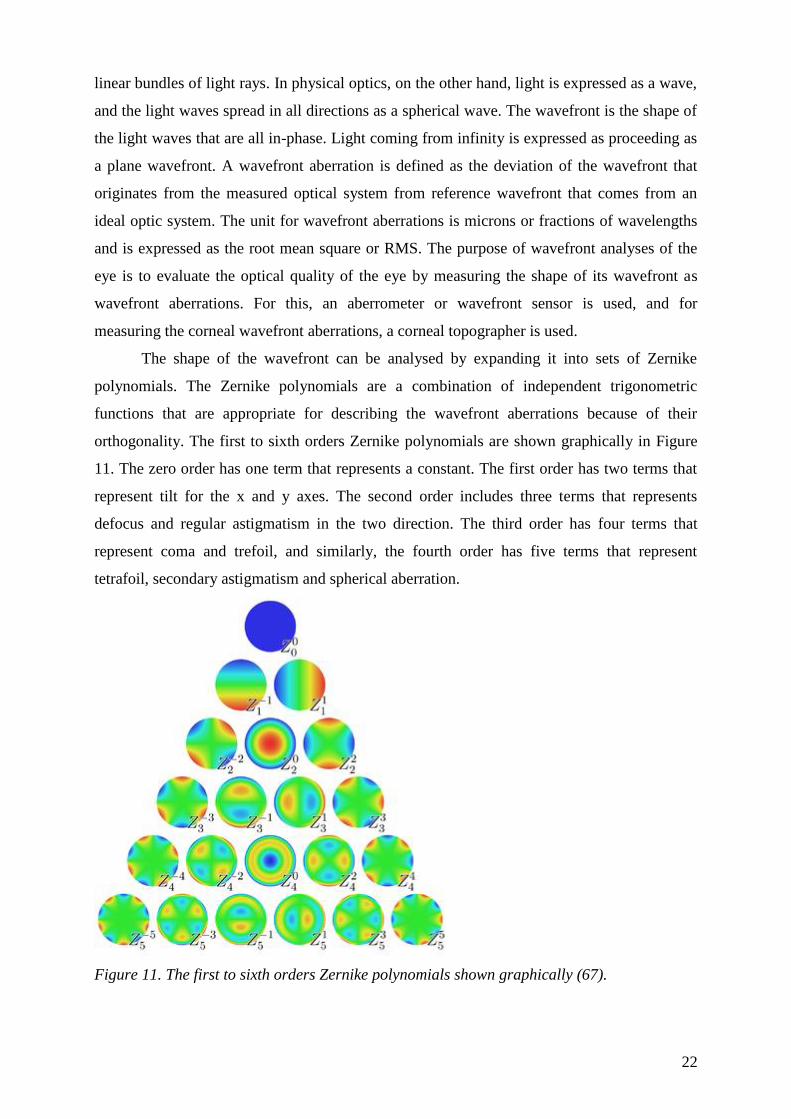
The shape of the wavefront can be analysed by expanding it into sets of Zernike
polynomials The Zernike polynomials are a combination of independent trigonometric
functions that are appropriate for describing the wavefront aberrations because of their
orthogonality The first to sixth orders Zernike polynomials are shown graphically in Figure
11 The zero order has one term that represents a constant The first order has two terms that
represent tilt for the x and y axes The second order includes three terms that represents
defocus and regular astigmatism in the two direction The third order has four terms that
represent coma and trefoil and similarly the fourth order has five terms that represent
tetrafoil secondary astigmatism and spherical aberration
Figure 11 The first to sixth orders Zernike polynomials shown graphically (67)
23
The polynomials can be expanded up to any arbitrary order if a sufficient number of
measurements are made for the calculations Spectacles can correct for only the second order
aberrations and not the third- and higher-orders that represent irregular astigmatism
Monochromatic aberrations can be evaluated quantitatively using the Zernike coefficients for
each term
Although the total HOAs can be used to estimate the severity of deterioration of
optical quality of the eye as the diagnostic purposes it will be essential for the surgical
treatments to quantify the details of wavefront of the eye using Zernike expansion or Fourier
expansionWavefront aberrations caused by the anterior andor posterior corneal surfaces can
be calculated using the height data of the corneal topographers such as videokeratoscopes or
slit-scanning corneal topographers (67)
16 Treatment of Keratoconus
161 Contact lenses
As irregular astigmatism can not be corrected with spectacles contact lens is the most
widly used optical correction method of keratoconus Although contact lenses for keratoconus
are manufactured with hydrogel silicone hydrogel gas permeable and hybrid (ie rigid
centre and soft skirt) materials gas permeable contact lenses remain the most commonly used
contact lens type as high levels of irregular astigmatism cannot normally be corrected with
other contact lens types Piggy back systems consisting on the fitting a gas permeable on top
of a soft contact lens have also been used for keratoconus management The soft contact lens
is used to improve wearing comfort and provide a more regular area for the gas permeable
contact lenses to sit whereas the gas permeable contact lens is primarily used for providing
adequate visual acuity The use of high oxygen permeability soft (ie silicone hydrogel) and
gas permeable contact lenses is highly recommended for keratoconus management as these
corneas are well known to be compromised (2 71-73)
24
162 Corneal crosslinking
Crosslinking is a widespread method in the polymer industry to harden materials and
also in bioengineering to stabilize tissue Using UVA at 370 nm and the photosensitizer
riboflavin the photosensitizer is excited into its triplet state generating so-called reactive
oxygen species (ROS) being mainly singlet oxygen and to a much lesser degree superoxide
anion radicals The ROS can react further with various molecules inducing chemical covalent
bonds bridging amino groups of collagen fibrils The wave length of 370 nm was chosen
because of an absorption peak of riboflavin at this wavelength (7475)
The first clinical study on the crosslinking treatment of keratoconus was performed by
Wollensak In this 3-year study 22 patients with progressive keratoconus were treated with
riboflavin and UVA In all treated eyes the progression of keratoconus was at least stopped
(freezinglsquo) In 16 there was also a slight reversal and flattening of the keratoconus by two
diopters Best corrected visual acuity improved slightly in 15 eyes (74)
Crosslinking treatment of keratoconus is a very promising new method of treating
keratoconus At the present stage of knowledge the treatment should only be performed in
patients with documented progression of keratoconus in the preoperative months With more
long-term experience prophylactic treatment of keratoconus at an early stage might become
possible Additional refractive corrections can also be considered if necessary In case a
recurrence of keratoconus progression should occur in the long run which has not been
observed so far a second crosslinking procedure might be a choice
However safe and promising technik CXL is the proper determination of inclusion
criteria may significantly reduce the complications and failures A preoperative maximum K
reading less than 5800 diopters may reduce the failure rate to less than 3 and restricting
patient age to younger than 35 years may reduce the complication rate to 1 (76) As
postopertiv complication stromal infiltrations and moderate anterior chamber inflammation
has been described and diffuse lamellar keratitis (DLK) after myopic laser in situ
keratomileusis Herpes simplex keratitis and iritis recurrence has been also associated with the
intervention (76)
1631 Surgical interventions ICRS
Intrastromal corneal ring segments (ICRS) were initially developed for the correction
of mild to moderate myopia and are now considered in the management of keratoconus and
25
other ectatic disorders such as ectasia after laser in situ keratomileusis and pellucid marginal
degeneration The ICRS acts as a passive element that flattens the central cornea by an arc-
shortening effect on the corneal lamellae structure (Figure 12) Although ICRS implantation
is an effective tool in managing corneal ectatic disease several complications have been
described These include incomplete tunnel creation anterior or posterior corneal perforation
epithelial defects segment extrusion and induced astigmatism by central migration of the
ICRS along the horizontal corneal diameter In most of these cases ICRS explantation is
mandatory and it is possible that a more appropriate ICRS can be implanted later A main
advantage of ICRS implantation is its potential reversibility because the technique does not
require tissue removal this has been shown in eyes with low to moderate myopia
The outcomes of corneal remodeling by ICRS are mainly dependent on the
biomechanical properties of the corneal tissue The corneal response depends on the
magnitude of the force and on the velocity of the application of the force A specific ICRS
inducing a specific force on the cornea will generate different changes in the ectatic corneal
profile depending on the underlying corneal biomechanical status Theoretically the cornea
should have the ability to return to its original state after removal of the application of force
In addition it should be able to be remodeled when new forces are applied (ie after
implantation of a new ICRS) (7778)
Figure 12 Clinical picture of an after ICRS implantation (79)
1632 Surgical interventions keratoplasty
The refractive error caused by the ectatic cornea is initially managed with either
spectacles or contact lenses When ectasia progresses to the point where contact lenses no
longer provide useful vision then surgical intervention may be considered Penetrating
keratoplasty is the most commonly performed surgical procedure for ectatic corneas but is
associated with complications including graft rejection induced astigmatism complications
26
of intraocular surgery such as glaucoma cataract formation retinal detachment cystoid
macular edema endophthalmitis and expulsive hemorrhage To avoid these complications
new methods such as lamellar keratoplasty (LKP) have evolved LKP has the advantages of
being extraocular and reversible if tissue complications occur Another advantage includes the
ability to replace only selected areas of diseased corneal tissue with healthy donor tissue LKP
results however may be limited by vision-reducing graft-host interface problems and the
technical nature of the surgical procedure (8081) In keratoconus conventional penetrating
keratoplasty has long been associated with good long-term outcomes with graft survival of
over 90 up to 138 years postoperatively Visual outcomes have also been good with 732
achieving BCVA over 2040 (8283)
Replacing weakened corneal tissue by corneal transplantation provides a permanent
means of substituting or augmenting of weakened corneal tissue and the various forms of
keratoplasty for ectatic corneal disease may be largely divided into penetrating or lamellar
keratoplasty procedures and central or peripheral keratoplasty procedures according to the
precise site of tectonic weakness of the cornea Recent innovations in lamellar keratoplasty
address issues in the management of corneal ectasia Nevertheless corneal endothelial
replacement is generally unnecessary in keratoconus which is primarily a corneal stromal
disorder and as the major causes of long-term graft failure and attrition are endothelial in
nature an anterior lamellar keratoplasty approach to keratoconus which avoids unnecessary
replacement of normal recipient endothelium is ideal Penetrating keratoplasty has in the past
been associated with better postoperative vision due in part to the problem of interface haze
and irregularities in lamellar keratoplasty (84)
New automated surgical technologies in anterior segment surgery which have largely
been developed for corneal refractive surgery are now being utilized in keratoplasty surgery
and are able to partially substitute for conventional manual surgical procedures providing
greater precision and reproducibility in lamellar corneal dissection These include an
automated microkeratome-assisted lamellar keratoplasty device and femtosecond laser-
assisted lamellar keratoplasty (85)
27
17 Other diseases affecting biomechanical properties of the
cornea
171 Pellucid marginal degeneration
Pellucid marginal degeneration (PMD) is characterized by a peripheral band of
thinning of the inferior cornea from the 4 to the 8 olsquoclock position There is 1ndash2-mm
uninvolved area between the thinning and the limbus The corneal protrusion is most marked
above the area of thinning and the thickness of the central cornea is usually normal Like
keratoconus pellucid marginal degeneration is a progressive disorder affecting both eyes
although eyes may be asymmetrically affected In moderate cases it can easily be
differentiated from keratoconus by slit-lamp evaluation because of the classical location of the
thinning In early cases the cornea may look relatively normal and in advanced cases it may
be difficult to distinguish from keratoconus because the thinning may involve most if not all
of the inferior cornea In both instances videokeratography is very useful to make the
distinction The videokeratograph has a classical ―butterfly appearance (Figure 13)
demonstrating large amounts of against-the-rule astigmatism (2-4)
Figure 13 Corneal topography of a patient with PMD The classical ldquobutterflyrdquo appearance
can be clearly seen on the axial map (from our own database)
Pellucid marginal degeneration can be differentiated from other peripheral corneal
thinning disorders such as Terrienlsquos marginal degeneration because the area of thinning is
always epithelialized clear avascular and without lipid deposition Terrienlsquos corneal
degeneration affects a similar age group and also causes high astigmatism however it may
affect both the superior and inferior cornea and is accompanied by lipid deposition and
28
vascular invasion Videokeratography can also be used to differentiate these two disorders
because they have distinctly different topographic patterns (86)
Because of the large amounts of against-the-rule astigmatism patients with pellucid
marginal degeneration are much more difficult to fit with contact lenses than patients with
keratoconus although spherical or aspheric contact lenses with large overall diameter should
initially be attempted in early-to-moderate cases Surgery may be considered for patients
whose vision is not adequately corrected by contact lenses or in patients who are contact lens-
intolerant Patients with pellucid marginal degeneration however are typically poor
candidates for penetrating keratoplasty for two reasons First thinning occurs so near the
limbus that the donor cornea must be placed very close to the corneal limbus thus increasing
the chances of graft rejection Second because of the extreme thinning and the location of the
thinning penetrating keratoplasty typically induces large amounts of postoperative
astigmatism which may be extremely difficult to correct because of disparity in graft-host
thickness
Crescentic lamellar keratoplasty is a useful initial surgical procedure in patients with
pellucid marginal degeneration This procedure involves removing a crescentic inferior layer
of ectatic tissue by lamellar dissection and replacing it with a thicker lamellar donor graft
This will in most cases eliminate large amounts of against-the-rule astigmatism In some
cases the patient may become contact lens-tolerant thus obviating a full-thickness procedure
(87)
172 Keratoglobus
Keratoglobus is a rare disorder in which the entire cornea is thinned most markedly
near the corneal limbus in contrast to the localized thinning centrally or paracentrally in
keratoconus The cornea may be thinned to as little as 20 of normal thickness and it
assumes a globular shape In advanced keratoconus the entire cornea can also be thinned and
globular-shaped making it difficult to distinguish these two entities However even in very
advanced keratoconus there may be a small area of uninvolved cornea superiorly that
approaches normal corneal thickness (2-4)
Keratoglobus is bilateral but it is usually present from birth and tends to be
nonprogressive It can be distinguished from megalocornea and congenital glaucoma because
the cornea is usually of normal diameter It is a recessive disorder and is often associated with
blue sclerae and other systemic features in contrast to keratoconus which is most commonly
an isolated disorder In contrast to keratoconus the corneas in keratoglobus are prone to
29
corneal rupture from even minimal trauma Thus hard contact lenses are contraindicated and
protective spectacles should be strongly encouraged If the cornea is extremely thin a tectonic
limbus-to-limbus lamellar keratoplasty should be considered to strengthen the cornea A
subsequent central penetrating keratoplasty may be considered if adequate visual
rehabilitation cannot be achieved with glasses (88)
173 Post-LASIK ectasias
Corneal ectasia after LASIK is a progressive corneal steepening usually inferiorly
with an increase in myopia and astigmatism loss of uncorrected visual acuity and often loss
of best-corrected visual acuity that can present days to years after LASIK The actual
incidence remains undetermined and no good data support firm predictions previous
estimates however have ranged from 004 to 06 Among members of the International
Society of Refractive Surgery (ISRS) of the American Academy of Ophthalmology (AAO)
responding to the practice patterns survey in 2004 more than 50 had at least one case of
ectasia develop in their practice Approximately 50 of cases present within the first 12
months but late onset ectasia can also occur (89)
Corneal refractive surgery alters the effective shape thickness curvature and tensile
strength of the cornea The specific mechanisms resulting in post-LASIK ectasia remain
undetermined although complex biomechanical modeling that takes into account factors such
as corneal plasticity and viscoelasticity and corneal parameters such as Youngs modulus
Poissons ratio and curvature radius among others may provide insight in the future (90)
LASIK inevitably reduces the tensile strength of the cornea Among the first four
reported cases all had greater than 10 diopters of myopia preoperatively and less than 250 microm
residual stromal bed thickness (RSB) postoperatively and two patients had forme fruste
keratoconus According to recommendation a RSB of at least 250ndash300 microm be maintained to
prevent corneal ectasia after myopic keratomileusis (91)
Most authors recognize preoperative topographic abnormalities as uniquely indicative
of increased risk for post-LASIK ectasia the definition of abnormallsquo however remains a
source of great debate Forme fruste keratoconus as defined by the Rabinowitz criteria is a
risk factor for post-LASIK ectasia Pellucid marginal corneal degeneration suspects are also at
increased risk Based on extensive review of the literature the members of the American
Society of Cataract and Refractive Surgery (ASCRS) joint committee now recommend
avoiding Lasik in patients with asymmetric inferior corneal steepening or asymmetric bowtie
patterns with skewed steep radial axes above and below the horizontal meridian (92)
30
2 Purposes
1 Currently the diagnosis of keratoconus is based on biomicroscopic findings corneal
topography and ultrasound pachymetry Placido-disk based corneal topography only
examines the anterior surface of the cornea and alteration in the reference point or viewing
angle may result in inaccuracy of curvature measurement Height data give a more accurate
representation of the true shape of the corneal surface because they are independent of axis
orientation and position The Pentacam Comprehensive Eye Scanner uses a rotating
Scheimpflug camera and measures both anterior and posterior corneal surfaces by an
elevation based system It allows the measurement of local elevation points by fitting the
corneal shape to a best fit sphere reference surface with variable diameters or to an ellipsoid
surface Examination of the posterior corneal surface is important in the early diagnosis of
keratoconus as epithelial compensation can mask the presence of an underlying cone on the
anterior surface The purpose of our first study (I) was to evaluate the discriminating ability of
pachymetric and elevation data obtained by Pentacam and to rank them according to their
usefulness in the differential diagnosis of keratoconus
2 The purpose of the second study (II) was to find the anterior chamber characteristics of
mild keratoconus with the aid of the rotating Scheimpflug imaging and to evaluate trends in
corneal protrusion progression We also aimed to demonstrate that morphological alterations
of the cornea are accompanied by localized anterior chamber changes
3 Understanding the characteristics of ocular higher order aberrations owing to
keratoconus might be very useful to differentiate early cases from simple myopia or myopic
astigmatism It is particularly important to detect the disease among refractive surgery
candidates because keratorefractive procedures may worsen their condition Corneal higher
order aberrations are calculated from corneal topographic elevation data and do not take into
account the internal ocular aberrations The advantage of measuring corneal aberrations
relates to their ability to analyze most of the anterior corneal surface which allows a better
understanding of the optical behavior of the cornea The limitation of this method is that it
neglects the pupil area and position and it is not aligned to the line of sight (LoS) which is the
most relevant reference axis for defining retinal image quality at the point of fixation The
purpose of our third study (III) was to prove our hypothesis that keratoconus causes a shift in
the LoS which acts as a compensating mechanism for increased corneal higher order
aberrations To test our hypothesis we examined location of the LoS its position relative to
the pupil center and their impact on ocular higher order aberrations in keratoconic and control
31
eyes Taking into account the changes of the LoS allows a better understanding of the
pathologic optics of keratoconus and may have a beneficial effect on increasing the precision
of correcting higher order aberrations According to our knowledge no such comparisons
were performed in previous articles
32
3 Methods
31 Demographic data
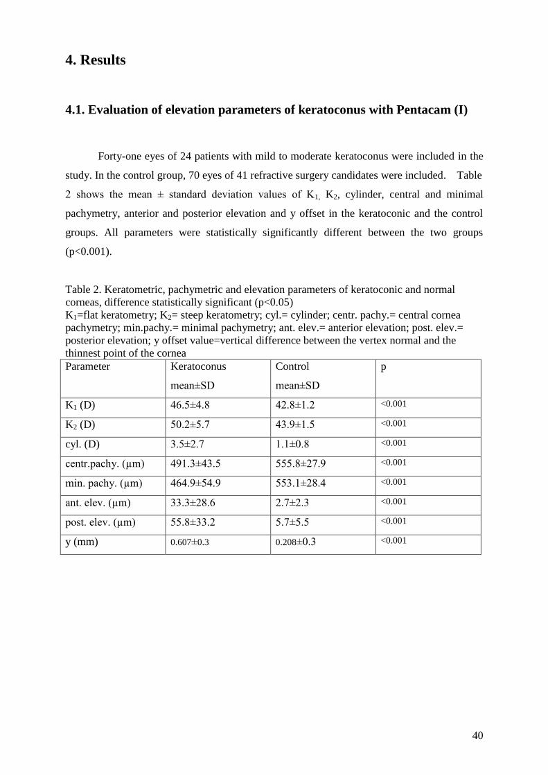
In the first and second studies (I II) forty-one eyes of 24 patients with mild to
moderate keratoconus were included In the control group 70 eyes of 41 refractive surgery
candidates were included Among the forty-one eyes with keratoconus 18 patients had
binocular keratoconus 2 patients had unilateral disease in 3 patients the fellow eye has
already undergone penetrating keratoplasty The control group consisted of 41 normal
individuals of 29 patients both eyes could be evaluated of 12 patients only one eye could be
included in the study because of previous refractive surgery or corneal disease of the fellow
eye Table 1A shows the patientslsquo demographic data There were no statistically significant
differences between the keratoconus and the control groups in age or sex distribution
(pgt005)
Table 1A Demographic features of the patients (I-II) difference statistically significant
(plt005)
Subjects Eyes (n) Age (y)
meanplusmnSD
Gender (MF)
Keratoconus 24 41 387plusmn141 159
Controls 41 68 402plusmn151 2417
p 061 021
Table 1B Demographic features of the patients (III) difference statistically significant
(plt005)
Subjects Eyes (n) Age (y)
meanplusmnSD
Gender (MF)
Keratoconus 30 50 315plusmn82 1317
Controls 50 100 303plusmn109 2228
p gt005 gt005
In the third study (III) (Table 1B) fifty-five eyes of 30 patients with mild to moderate
keratoconus and 100 eyes of 50 refractive surgery candidates with normal corneas were
included Among keratoconus patients 25 had binocular keratoconus 2 patients had unilateral
disease in 3 patients the fellow eye has already undergone penetrating keratoplasty The
control group consisted of 100 healthy eyes of 50 refractive surgery candidates There were
33
no statistically significant differences between the keratoconus (mean age 315plusmn82 years 13
men 17 women) and the control groups by MannndashWhitney nonparametric test (mean age
303plusmn109 years 22men 28 women) in age or sex distribution (p=01)
32 Patient inclusion criteria
Both eyes of every patient have undergone a complete ophthalmologic evaluation
including slit-lamp biomicroscopy keratometry retinoscopy ophthalmoscopy and Placido-
disk based videokeratography Keratoconic patients included in the study were either mild or
forme fruste cases The criteria for diagnosing keratoconus were defined as the existence of
central thinning of the cornea with Fleischer ring Vogtlsquos striae or both by slit-lamp
examination Forme fruste keratoconus was diagnosed when an abnormal localized
steepening was observed by corneal topography without any slit-lamp findings
Both eyes of every subject were used in the study except for those with previous
ocular surgery trauma or other pathology Control subjects were age matched and had a
refractive error of less than plusmn 5 spheric diopters and or astigmatism less than 3 diopters
Patients who wear rigid contact lenses were asked to stop using them for 4 weeks and soft
contact lenses were ceased for at least one week before assesment
From all patients informed consent was obtained after the characteristics of the study
was explained A review by the local ethics committee was not required For all study
procedures the tenets of the declaration of Helsinki were followed
33 Patient exclusion criteria
Subjects with corneal scarring previous ocular surgery trauma or any other ocular
diseases except for keratoconus and refractive errors were excluded from the study
34
Severe cases were excluded because of potential stromal haze or scar formation
which may alter the optical transparency of the cornea and image acquisition of the Pentacam
34 Corneal topographic measurements
Corneal topographic measurement were taken with the Tomey corneal topographer
(Tomey Topographic Modeling System software version-4 Tomey Corp Nagoya Japan)
Both topographic and wavefront aberration images were taken at 5 to 10 seconds after a
complete blink when the tear film layer is the most stable (93)
35 Pentacam measurements
All eyes were examined with the Pentacam HR (Oculus Inc) used by three trained
examiners The readings were taken as recommended in the instruction manual Briefly the
patients were instructed to keep both eyes open and fixate on the black target in the center of
the blue fixation beam After attaining perfect alignment the instrument automatically took
25 Scheimpflug images within 2 seconds For each eye one high quality image was recorded
In the first study (I) the following data were then exported to a Microsoft Excel spreadsheet
keratometry values in the flat (K1) and steep (K2) meridian corneal astigmatism (cylinder)
corneal thickness at the center (central pachymetry) and at the thinnest point of the cornea
(minimal pachymetry) the distance of the thinnest point of the cornea (apex of the cone in
keratoconic patients) from the center of the cornea in the vertical meridian (y) and local
elevation (anterior elevation posterior elevation) values For height data measurement the
best fit sphere (BFS) was used as a reference body using the float option over a 9 mm fit The
float map means that the reference body has no fixed center the distance between the cornea
and the sphere surface is optimized to be as small as possible Elevation maps show the
difference in height between cornea and reference body their value is positive when the
measured point of the cornea is above the reference body and negative when it lies below
Anterior and posterior elevation data were read as the maximum values above the BFS in the
central 5 mm of the cornea
In the second study (II) the following data were exported for further analysis the ACD
measured from the posterior corneal surface centrally the ACD at the thinnest point of the
cornea and the ACD 10 mm 20 mm and 30 mm inferiorndashparacentral For local posterior
35
elevation measurements a toric ellipsoid was used as a reference body The elevation maps
from the device show the difference in height between the cornea and reference body the
value is positive when the measured point of the cornea is above the reference body and
negative when it is below Posterior elevation data were read as the maximum values above
the toric ellipsoid surface in the central 50 mm of the cornea which in the keratoconus eyes
in this study was at the thinnest point of the cornea The mean keratometry values and the
smaller of the 2 chamber angles in the horizontal section calculated from the 3-dimensional
model were also determined and processed for analysis
36 Hartmann-Shack aberrometry measurements
All eyes were examined with the WASCA Hartmann-Shack wavefront sensor (Carl
Zeiss Meditec AG Jena Germany) used by three trained examiners (III) The readings were
taken as recommended in the instruction manual Briefly the patient positions his or her head
on a chin rest and fixates on the center of a circular grid which is optically fogged The
fixation target and the probe beam were coaxial located at infinity in order that the retinal
image of the narrow probe beam will remain centered on the retinal image of the fixation
target (94) The operator manually aligns a reference box on a video monitor with the pupil
This comprises a set of six symmetrical infrared diodes which are seen as reflections off the
corneal surface on the control monitor The anterior-posterior focus point of measurement is
determined by bringing into focus these corneal reflexions This is achieved by first moving
the instrument towards the patient and slowly drawing it back until the optimal focus is found
Measurements are taken until the criteria of measurement selection are met These are 1 the
CCD spot array is perfectly centered within the reference box both horizontally and vertically
2 to ensure that there is no or as little as possible missing data within the analysis zone (95)
Wavefront aberration measurements were standardized to a 45 mm pupil We used no
dilating drop as the aberration profile of a pharmacologically dilated pupil is less relevant to
the natural pupil view normally experienced by the patient and in order to avoid instrument
myopia (96) Wavefront measurements were performed in a dark room We used the right-
hand coordinate reference frame and the double-index convention for naming the Zernike
coefficients and polynomials recommended by the OSAVSIA Standards Taskforce (9798)
The signs of bilaterally asymmetrical Zernike coefficients of the left eyes affected by
enantiomorphism were reversed in order to allow comparison between right and left eyes
(99)
36
The following data were then exported to a Microsoft Excel spreadsheet average and
the steep keratometry values and the axis of the steepest meridian measured by corneal
topography Root mean square (RMS) higher-order RMS (HORMS) (μm) values of Zernike
polynomials up to the sixth-order (μm) and the distance of LoS relative to the pupillary axis
given by the aberrometer as x and y offset (mm) values
To calculate the axis of the LoS relative to the pupil center vector analysis following
the recommendations of the Astigmatism Project Group of the American National Standards
Institute and the method suggested by Alpins was applied (100101) Figure 14 shows the
design of vector analysis The WASCA aberrometer gives the parameters of x and y offsets in
mm as the distance of the center of the pupil from the point of reference the LoS The angle α
was calculated using trigonometrical analysis by equation tangent α=xy The angle α was
added or subtracted from the 90deg or 270deg meridian according to the sign of the x and y offset
values If both x and y had a negative sign (this is the case represented in Figure 15) the angle
α was subtracted from the 270deg meridian If both x and y had a positive sign the angle α was
subtracted from the 90deg meridian When x was negative and y positive the angle α was added
to the 90deg meridian when x was positive and y negative the angle α was added to the 270deg
meridian Than the vector was reversed by 180 degrees in order to get the distance of the LoS
relative to the pupil center In order to calculate the distance between the LoS and the pupil
center we used Pythagoralsquos rule
37
Figure 14 Design of vector analysis The angle α was added or subtracted from the 90deg or
270deg meridian according to the sign of the x and y offset values If both x and y had a negative
sign See further explanation in text
- +
+
-
X axis
Y axis
LOS
pupil center
α
y offset
x offset
180deg
270deg
0deg
90deg
site of LOS
refered to
pupil center
38
37 Statistical analyses
Statistical analyses were performed with Statistica 80 software (StatSoft Inc Tulsa
OK USA) Receiver operating characteristic (ROC) curves were obtained using SPSS 150
(SPSS Inc Chicago Il USA) (I-III) Normal distribution assumption could not be accepted
for anterior elevation according to the Shapiro-Wilkslsquo W test For group comparisons of
continuous variables the Mann-Whitney nonparametric test was used A p value less than 005
was considered statistically significant Area under the ROC curve (AUROC) was calculated
to describe the predictive accuracy of the different indices and to determine the cutoff point
for the most sensitive parameter Logistic regression analysis was performed to investigate
predictors of classical topographic and central pachymetric findings contrasted with
keratometric pachymetric and elevation data obtained by the Pentacam with calculation of r2
positive and negative predictive values Confirmatory factor analysis (CFA) was performed in
the keratoconic group for quantification of the validity of parameters characterizing the
disease CFA is a commonly used statistical method to detect responsible latent factors for the
measurable clinical variables which is becoming more and more popular in ophthalmologic
research23
Briefly during CFA the most representative variables can be determined using
correlation matrices without incorporating any gold standard and several model structures
can be examined During our analysis different models (with two- and three latent factors)
based on our previous assumptions on clinical manifestations of the disease were tested and
the best fitted model was chosen by values of fit-indices
Multivariable regression analysis using a generalized estimating equation (GEE) was
performed to determine predictors of keratoconus data from the eyes were statistically
analyzed as repeated measures (102) Thus the analysis took into account the correlated
nature of data from patients who had measurements in both eyes The construction of the
multivariable logistic regression model started with variables that showed the best fit to data
in univariable modeling assessed using the value of the corrected quasi-likelihood under
independence model criterion (QICC) Lower QICC values indicate a better fit to data Next
variables were added and the change in the QICC value was tested Variables were kept in the
model if they were associated with a P value less than 005 and the overall fit of the model
improved as indicated by a decrease in the QICC value compared with the value in a model
that did not include the variable (58)
Correlations between posterior elevation and corneal protrusion (ACD at minimum
pachymetry) were tested using linear and linear piecewise (segmented) regression analysis
Linear piecewise regression analysis models data to identify potentially meaningful trends
39
Breakpoints are independent-variable values at which the slope of the linear function changes
Breakpoint values are unknown and are estimated A nonlinear least-squares regression
technique was used to fit the model to the data and the significance of the breakpoint and the
regression coefficients of the slopes were determined The accuracy of fit of the piecewise
regression model was then compared with that of the linear regression model using the
explanation coefficients and Akaike Information Criterion (AIC) values of the 2 models
Higher explanation coefficients and lower AIC values indicate that the given regression
model is more likely
Bootstrap method was used to overcome the correlated nature between eyes of the
same subject by treating each subject as a cluster (103) Normal distribution assumption could
not be accepted for most of the Zernike polynomials according to the Shapiro-Wilkslsquo W test
For group comparisons of continuous variables the Mann-Whitney nonparametric test was
used To test correlations the Spearman rank correlation was used Factorial regression
analysis was used to test the interactive effects of higher order aberrations on the extent of
LoS shift A p value less than 005 was considered statistically significant
40
4 Results
41 Evaluation of elevation parameters of keratoconus with Pentacam (I)
Forty-one eyes of 24 patients with mild to moderate keratoconus were included in the
study In the control group 70 eyes of 41 refractive surgery candidates were included Table
2 shows the mean plusmn standard deviation values of K1 K2 cylinder central and minimal
pachymetry anterior and posterior elevation and y offset in the keratoconic and the control
groups All parameters were statistically significantly different between the two groups
(plt0001)
Table 2 Keratometric pachymetric and elevation parameters of keratoconic and normal
corneas difference statistically significant (plt005)
K1=flat keratometry K2= steep keratometry cyl= cylinder centr pachy= central cornea
pachymetry minpachy= minimal pachymetry ant elev= anterior elevation post elev=
posterior elevation y offset value=vertical difference between the vertex normal and the
thinnest point of the cornea
Parameter Keratoconus
meanplusmnSD
Control
meanplusmnSD
p
K1 (D) 465plusmn48 428plusmn12 lt0001
K2 (D) 502plusmn57 439plusmn15 lt0001
cyl (D) 35plusmn27 11plusmn08 lt0001
centrpachy (microm) 4913plusmn435 5558plusmn279 lt0001
min pachy (microm) 4649plusmn549 5531plusmn284 lt0001
ant elev (microm) 333plusmn286 27plusmn23 lt0001
post elev (microm) 558plusmn332 57plusmn55 lt0001
y (mm) 0607plusmn03 0208plusmn03 lt0001
41
Figure 15 compares the results of the ROC curve analysis for each parameter to
reduce the number of variables the average values of flat and steep keratometry were
included The graphs show that posterior elevation has the highest sensitivity in
discriminating keratoconus from normal subjects
Figure 15 ROC graph showing the sensitivity and specificity of the various corneal
measures The closer the line to the left-hand side of the graph the higher the sensitivity and
specificity for keratoconus
Table 3 shows that the area under the curve is acceptable for all parameters Posterior
elevation has the highest area under the curve (097) followed by anterior elevation (096)
minimal pachymetry (089) central pachymetry (088) cylinder (083) average keratometry
(082) and y offset (079) ROC analysis result of the x offset value was not acceptable A
posterior elevation cutoff value of 155 microm had 951 sensitivity and 943 specificity in
discriminating keratoconus from normal eyes
42
Table 3 Area under the curve standard error and p values of various parameters (keratoconic
vs normal eyes)
aveK= average keratometry cyl= cylinder centr pachy= central cornea pachymetry
minpachy= minimal pachymetry ant elev= anterior elevation post elev= posterior
elevation y offset value=vertical difference between the vertex normal and the thinnest point
of the cornea
Variables Area under
the curve
Std Error
p value
postelev (microm) 097 0017 0000
ant elev (microm) 096 0020 0000
minpachy (microm) 090 0037 0000
centrpachy (microm) 088 0040 0000
cylinder (D) 083 0042 0000
aveK (D) 082 0049 0000
y (mm) 079 0042 0000
Table 4 shows the sensitivity and specificity identified by cutoff points of posterior
corneal elevation
Table 4 Cutoff point sensitivity and specificity values of posterior elevation
posterior
elevation cutoff
point (microm)
Sensitivity
()
Specificity
()
-50 100 0
-05 100 114
15 100 271
45 976 500
75 976 671
105 976 800
145 951 900
155 951 943
175 927 986
190 878 986
215 878 100
375 68 100
cutoff point
43
Logistic regression analysis was performed to investigate predictors of two models
The first model was constructed of variables representing classical topographic (average
keratometry cylinder) and central pachymetric findings Into the second model new variables
obtained by the height data of Pentacam (minimal pachymetry anterior and posterior
elevation) were added The goodness-of-fit of the second model was superior to that of the
first (r2 067 vs 051) For the first model positive predictive value was 075 negative
predictive value was 095 for the second model positive predictive value was 092 negative
predictive value was 10
Figure 16 shows the structure of a three-factor model of CFA which showed the best
indices of fit (chi-Square p-level 060 RMSEA 0001) identifying corneal curvature (CC)
corneal thinning (CT) and cone apex elevation (CE) as latent factors responsible for clinical
alterations as detected by the measured variables Good correlation was found between these
factors (CC vs CT 075 CC vs CE 072 CT vs CE 075 respectively) The most
representative variable for the detection of this corneal shape disorder in the overall model
was minimal pachymetry (-099) followed by anterior elevation (098) keratometry results
(095) central pachymetry (-094) posterior elevation (092) and corneal cylinder (038)
Table 5 demonstrates the results of the confirmatory factor analysis
44
Figure 16 Structure of confirmatory factor analysis model
Table 5 Results of confirmatory factor analysis (chi-Square p-level 06 Steiger-Lind
RMSEA Point Estimate 0001)
parameter path coefficient Standard error
minimal pachymetry (microm) -099 0000
anterior elevation (microm) 098 0030
K1+K2 (D) 095 0153
central pachymetry(microm) -094 0018
posterior elevation (microm) 092 0036
cylinder (D) 039 0148
corneal
shape
disorder
corneal
curvature
corneal
thinning
cone apex
elevation
K1+K2
cylinder
central
pachymetry
minimal
pachymetry
anterior
elevation
posterior
elevation
45
42 Anterior chamber characteristics of keratoconus (II)
Table 6 shows the mean measurement values in the keratoconus and control groups
Table 7 shows the correlation (r values and P values) between posterior corneal elevation and
anterior chamber morphology parameters The anterior chamber was significantly deeper in
the keratoconus group than in the control group at all measurement points (ie center 10 mm
20 mm and 30 mm paracentral thinnest) There was no statistically significant difference
between the 2 groups in anterior chamber volume anterior chamber angle or spherical
equivalent The posterior elevation was significantly higher in the keratoconus group than in
the control group and had a strong correlation with minimum pachymetry and central
pachymetry In contrast no significant correlation was found between the posterior elevation
and pachymetry results in the control group In the keratoconus group posterior elevation was
statistically significantly correlated with ACD in the central cornea (r = 057) at the thinnest
point of the cornea (r = 041) and 10 mm paracentrally (r = 045) Posterior elevation did not
correlate with the ACD 20 and 30 mm paracentrally anterior chamber angle and anterior
chamber volume in the keratoconus group (Table 7)
46
Table 6 Anterior chamber and corneal morphology by group
Mean plusmn SD
T test
Parameter Keratoconus Group Control Group P Value
Anterior chamber
Volume (mm3) 17528 plusmn 2042 16003 plusmn 6082 015
Angle (degrees) 3783 plusmn 549 3498 plusmn 799 006
Anterior chamber depth
(mm)
Central 319 plusmn 028 283 plusmn 055 lt0001
At minimum pachymetry 329 plusmn 033 287 plusmn 055 lt0001
10 mm paracentral 315 plusmn 025 282 plusmn 057 lt001
20 mm paracentral 283 plusmn 027 248 plusmn 071 lt001
30 mm paracentral 244 plusmn 029 212 plusmn 062 lt001
Corneal thickness (μm)
Central 491 plusmn 43 555 plusmn 28 lt0001
At minimum pachymetry 469 plusmn 57 554 plusmn 26 lt0001
Posterior elevation (μm) 4077 plusmn 229 minus063 plusmn 688 lt0001
Spherical equivalent (D) minus37 plusmn 27 minus29 plusmn 37 014
Average keratometry (D) 4790 plusmn 509 4337 plusmn 133 lt0001
t test
47
Table 7Correlation between posterior corneal elevation and anterior chamber morphology
parameters
Linear Regression
Parameter r P Value
Anterior chamber
Volume (mm3) 025 gt005
Angle (degrees) minus010 gt005
Anterior chamber depth
(mm)
Central 057 lt0001
At minimum pachymetry 041 0006
10 mm paracentral 045 002
20 mm paracentral 027 gt005
30 mm paracentral 020 gt005
Corneal thickness (μm)
Central minus062 lt0001
At minimum pachymetry minus075 lt0001
Mean keratometry (D) 058 lt0001
On univariable GEE logistic regression analysis the mean keratometry posterior
elevation central ACD ACD at minimum pachymetry CCT and minimum corneal thickness
were significantly different between normal eyes and keratoconus eyes However on
multivariable logistic regression analysis only the ACD at minimum pachymetry remained
significant after posterior elevation was entered into the model (Table 8)
48
Table 8 Results of univariable and the multivariable GEEs to predict keratoconus
GEE ModelParameter OR (95 CI) QICC P Value
Univariable
Posterior elevation 144 (125-165) 294 lt0001
ACD
Central 519 (19-1418) 13816 0001
At minimum pachymetry 603 (218-1667) 13539 0001
Pachymetry
Minimum 095 (092-098) 7501 0001
Central 095 (092-098) 9069 0001
Average keratometry 179 (133-241) 10346 lt0001
Multivariable
Posterior elevation 146 (125-171) 2774 lt0001
ACD at minimum
pachymetry 754 (135-4205) mdash 002
ACD = anterior chamber depth CI = confidence interval GEE = generalized estimating
equation OR = odds ratio QICC = corrected quasi-likelihood under independence model
criterion
A piecewise linear regression model to fit linear segments to different ranges of
posterior elevation showed a statistically significant explanation coefficient and breakpoint
value (Table 9) The linear piecewise model yielded better explanation coefficients and lower
AIC values than the linear model showing the segmented nature of the correlation Figure 17
shows the regression curves of the 2 models and the estimated breakpoint location determined
by the piecewise regression analysis The curves show that beyond a threshold the corneal
protrusion rate was significantly different Above 40 μm of posterior elevation corneal
ectasia accelerated by 7 times (regression coefficient ratio) between the second segment and
the first segment of the curves (Table 9)
49
Table 9 Linear and piecewise linear regression results of correlation between posterior
elevation and ACD at the minimum pachymetry CI = confidence interval
Parameter Mean (95 CI) P Value
Linear
Model explanation coefficient (r2) 023 0001
Akaike information criterion 13788 mdash
Piecewise linear
Regression coefficient
Of first segment 0001 (minus001 to 001) 085
Of second segment 0007 (0003 to 0012) 0002
Estimated breakpoint (μm) 40 (365 to 433) 0002
Model explanation coefficient 041 lt0001
Akaike information criterion 10167 mdash
Figure 17 Piecewise linear regression model used to fit linear segments to different ranges of
posterior elevation determined a breakpoint of the curve at 40 μm (95 CI 365-433 μm
vertical dotted line) This segmented regression model (continuous line) showed better fit to
the data than the linear regression model (dotted line) (ACD = anterior chamber depth)
50
The univariable GEE logistic regression model to validate the threshold value and test
the effect of minimum pachymetry had a lowest QICC value of 4064 and a 450 μm cutoff
point for minimum pachymetry This confirmed that 40 μm was the best cutoff point for
posterior elevation
43 Shifting of the line of sight in keratoconus measured by a Hartmann-
Shack sensor (III)
Fifty-five eyes of 30 patients with keratoconus and 100 healthy eyes of 50 refractive
surgery candidates were included in this study There were no statistically significant
differences between the keratoconus (mean age 315plusmn82 years 13 men 17 women) and the
control groups (mean age 303plusmn109 years 22men 28 women) in age or sex distribution
(p=01) There was no statistically significant differences between the refractive state of the
eyes in the two group neither as most of the refractive surgery candidates had a myopic
refractive error mean spherical equivalent in the keratoconic group was -37plusmn27 diopters
(D) in the control group -286plusmn37D p=014
Internal validation of original data using bootstrapping method showed high
consistency with results of the original sample confirming our selection method statistically
applicable
Table 10 shows the mean and standard deviation values of RMS HORMS Zernike
polynomials and xy offset parameters of keratoconic and normal corneas There was no
statistically significant difference between the two groups in total RMS value which can be
explained by the similar refractive states of the two groups The HORMS values in the
keratoconic group were significantly higher than in the control group The following Zernike
terms were significantly higher among keratoconic patients oblique astigmatism (Z2-2
) trefoil
(Z3-3
and Z33) vertical coma (Z3
-1) secondary astigmatism (Z4
-2 and Z4
2) quadrafoil (Z4
4)
pentafoil (Z5-5
) secondary coma and trefoil (Z5-1
and Z53) Vertical coma showed the highest
values among higher-order Zernike coefficients There was no statistically significant
difference in the x offset values but y offset showed a marked increase (-019plusmn038) in
keratoconic patients the negative sign means that the LoS shifted downwards in average
51
Table10 Average and standard deviation values of RMS HORMS Zernike polynomials and
x y offset parameters of keratoconic and normal corneas statistically significant difference
(plt005) marked with
Control
meanplusmnSD (μm)
Keratoconus
meanplusmnSD (μm)
p
RMS 30plusmn17 306plusmn19 092
HORMS 018plusmn014 059plusmn026 0000
Z2-2
0052plusmn031 -0397plusmn079 0001
Z20 2074plusmn276 2594plusmn192 0466
Z22 -0284plusmn069 -0134plusmn097 0430
Z3-3
-0048plusmn01 0071plusmn025 0000
Z3-1
0026plusmn01 -0418plusmn029 0000
Z31 0013plusmn006 0016plusmn017 0534
Z33 -0005plusmn006 -0083plusmn019 0001
Z4-4
0002plusmn003 -0005plusmn007 0368
Z4-2
0005plusmn004 0045plusmn007 0000
Z40 0021plusmn005 0002plusmn013 0868
Z42 -0007plusmn004 -0051plusmn011 0000
Z44 0011plusmn004 0032plusmn005 0016
Z5-5
0002plusmn002 -0004plusmn003 0033
Z5-3
0009plusmn003 -0004plusmn005 0546
Z5-1
-0006plusmn002 0018plusmn006 0045
Z51 -0002plusmn002 -0005plusmn002 0284
Z53 -00002plusmn002 0004plusmn003 0014
Z55 -0005plusmn004 -0001plusmn002 0539
Z6-6
-0001plusmn002 -0001plusmn001 0776
Z6-4
0001plusmn001 00001plusmn001 0673
Z6-2
-0002plusmn001 -0002plusmn001 0641
Z60 00055plusmn002 0007plusmn003 0202
Z62 0001plusmn001 -0002plusmn003 0517
Z64 -0001plusmn001 -0002plusmn002 0126
Z66 00012plusmn003 -0002plusmn001 0845
x offset 029plusmn038 024plusmn048 022
y offset 002plusmn033 -019plusmn038 00007
52
Our hypothesis was that the LoS shifted in the direction of the cone To test this we
have calculated the axis of the shift of the line of sight as mentioned above The cone location
can be characterized by the steepest keratometric axis (Ks) on topography only the topograph
always locates this meridian between 0 and 180 degrees in case of inferiorly located cones
we have reversed the Ks to the inferior half of the cornea by adding 180deg to the original axis
Figure 18 shows the correlation between the axis of line of sight (LoS) and the axis of the
steepest meridian on topography The correlation was statistically significant (r=059
plt0001)
-50 0 50 100 150 200 250 300 350 400
LoS axis (degree)
0
50
100
150
200
250
300
350
400
Ks a
xis
(de
gre
e)
R= 059 plt0001
Figure 18 Correlation in keratoconic patients between the axis of the shift of line of sight
(LoS) and the axis of the steepest meridian on topography (Ks) which in case of inferior cones
is reversed to the inferior half of the cornea by adding 180deg to the original axis The
correlation is statistically significant R and p values were calculated by Spearman rank
correlation
We found a significant correlation between the extent of the shift (hypotenuse) and the
negative value of vertical coma (r=-039 p=0004) Figure 19 shows that as vertical coma
increases in a negative direction the line of sight moves farther from the pupil center
53
00 02 04 06 08 10 12 14
hypotenuse (mm)
-12
-10
-08
-06
-04
-02
00
02
04
Z
3-1(μm)
R= -039 p=0004
Figure 19 Correlation between the hypotenuse which is the distance of LoS from the pupil
center and vertical coma (Z3-1
) among keratoconic patients The correlation is statistically
significant R and p values were calculated by Spearman rank correlation
Figure 20 shows the marked correlation (r=-049 plt0001) we have found between the
average keratometry value (aveK) measured by topography and spherical aberration (Z40)
According to the diagram as progression in steepening occurs the spherical aberration (SA)
becomes more and more negative which means that there is an excess of power in the center
relative to the pupil margin
40 41 42 43 44 45 46 47 48 49 50 51
aveK (diopter)
-04
-03
-02
-01
00
01
02
03
04
Z
40(μm)
R= -049 plt0001
Figure 20 Correlation between the average keratometry value (aveK) measured by
topography and spherical aberration (Z40) among keratoconic patients The correlation is
statistically significant R and p values were calculated by Spearman rank correlation
54
We have also found a correlation (r=029 plt004 Fig 21) between the size of the
hypotenuse which is the distance of LoS from the pupil center and spherical aberration As
spherical aberration increases the distance of the LoS from the pupil center increases We
found no significant correlation between average keratometry and the hypotenuse
00 02 04 06 08 10 12 14
hypotenuse (mm)
-04
-03
-02
-01
00
01
02
03
04
Z
40(μm)
R= 029 p=004
Figure 21 Correlation between the hypotenuse which is the distance of LoS from the pupil
center and spherical aberration (Z40) among keratoconic patients The correlation is
statistically significant R and p values were calculated by Spearman rank correlation
Figure 22 represents the axial topography map of a patient with a nasal inferior cone
by whom the LoS is shifted in the same direction and a schematic illustration how the Wasca
aberrometer measures the aberrations in a reduced and infero-nasally shifted circular area In
the circle the higher-order aberration map of the same person can be seen It is evident that the
main higher-order aberration is coma and that the line of sight is shifted towards the coma
The aberrometer can measure the aberrations only in a regular circle and its reference axis is
always the LoS This is the explanation why in many cases we couldnlsquot get an aberration map
of a larger area than 4-5 mms even if the pupil dilated to 6-7 mms in the dark room
55
Figure 22A Axial topography map of a patient with a nasal inferior cone by whom the LoS is
shifted in the same direction
Figure 22B Schematic illustration how the Wasca aberrometer measures the aberrations in a
reduced and infero-nasally shifted circular area in the same patient y=125mm x=046mm
hypotenuse (distance of LoS from pupil center)= 133mm
Finally a factorial regression model testing the interactive effects of vertical coma and
spherical aberration on the shift of LoS had a good fit on our data (r2=069 plt0001) These
result suggests a close correlation between ideally balanced polynomials and the LoS shift
56
5 Discussion
51 Keratoconus detection with Scheimpflug imaging
511 Characteristics of elevation based topography
According to the classical Placido based topographic findings in keratoconus the
most specific changes in the curvature are steepening and protrusion of the cornea usually
inferior to the visual axis (2-4642) More recent studies based on the elevation data obtained
by Orbscan and Pentacam measurements indicate that deformation occurs not only in the
anterior but also in the posterior corneal surface of keratoconus eyes (48505359104) As
Orbscan derives the posterior elevation map mathematically from the reflection mires of a
Placido-disk and 20 slit scans of the anterior segment there had been caution raised regarding
its posterior elevation data measurements (105) Rotating Scheimpflug imaging technology is
used by instruments such as the Pentacam (Oculus Inc Dutenhofen Germany) to measure
the anterior and posterior corneal surfaces as well as other anterior segment structures
Pentacam uses Scheimpflug imagery and measures 25000 elevation points This method is
independent of axis orientation and position which is not true for Placido disc based
videokeratography This later method has however significant limitations when trying to
describe the cornea with a curvature map First there are the physical limitations of a Placido-
based reflecting type system The area of corneal coverage is limited to about 60 of the
corneal surface eliminating important data for many peripheral or para-central pathologies
(ie pellucid marginal degeneration keratoconus) Second there is no information about the
posterior surface of the cornea (5051) Additionally there are limitations in attempting to
reconstruct the corneal surface based on curvature measurements The standard topographic
curvature (axial or sagittal curvature) is a referenced based measurement It is not a unique
property of the cornea The same shape can have many different curvatureslsquo depending on
which axis is used to make the measurement The line of sight and the measurement axis of
the videokeratoscope are not the same True topographic imaging implies shape and requires
the generation of an X Y and Z coordinate system (7841424445)
Scheimpflug camera displays elevation data not in its raw form (actual elevation data)
but compared with some reference shape The maps typically display how the actual corneal
elevation data compares (deviates) with this known shape The reason for this is to magnify
57
the differences and allow the clinician a qualitative map that will highlight clinically
significant areas The reason for viewing elevation data in this format is that the actual raw
elevation data lacks qualitative patterns that would allow the clinician to easily separate
normal from abnormal corneas Although not an elevation map the pachymetric map
represents the spatial difference between the anterior and posterior corneal surface and in as
such is totally dependent on accurate elevation data In addition to identifying thin corneas
the overall pachymetric distribution may be another indicator of pathology Normal corneas
are typically thinnest in the central region and thicken in the periphery Displacement of the
thinnest region is often seen in keratoconus and may at times predate changes on either the
anterior or posterior surfaces (78)
Evaluating corneal shape using Scheimpflug imaging may allow validation of the
measurements previously obtained by the Orbscan Ciolino and Belin (9) studied changes in
posterior corneal elevation following excimer treatment in 121 myopic eyes using the
Pentacam Although trend lines suggested a relationship between a thinner corneal and more
negative posterior corneal surface in LASIK photorefractive keratectomy (PRK) trend lines
are essentially flat There was no significant difference between the posterior displacement
between LASIK and PRK eyes No LASIK eyes showed significant forward displacement
They concluded that subclinical incidence of post-LASIK ectasia previously based on
Orbscan measurement may be less than previously thought
Quisling et al (59) compared measurements from Orbscan IIz and the Pentacam in
eyes with keratoconus The Orbscan IIz best fit sphere fixed to the apex was compared with
the Pentacam posterior best fit sphere with the float option removed The average best fit
sphere radius and average thinnest points were not significantly different between the two
systems The posterior elevation was significantly different despite similar radii of curvature
This may be due to a difference in data analysis by the two machines The Orbscan estimates
the central 3 mm while the Pentacam images the cornea directly It was suggested that the
Orbscan IIz may be more accurate in the periphery but less so in the center overestimating
the posterior height Both machines found the location of the cones most commonly located in
the inferior temporal quadrant
Ambrosio et al (49) used the Pentacam to evaluate normal and keratoconic eyes to
determine characteristics which may help to detect keratoconus The Pentacam yields
information regarding the thickness volume and spatial profile from the three-dimensional
model Corneal thickness at the thinnest point was used to create spatial thickness profiles and
volume centered around the thinnest point Significant differences were found in all positions
58
of the spatial thickness profile and volume distributions with lower values for each in
keratoconic eyes Keratoconic eyes were thinner with less volume and a more abrupt increase
as one moved outward from the thinnest point of the cornea than normal eyes
512 The role of reference body selection in the diagnosis of keratoconus
The corneal surface is variably aspheric and toric this diversity is even more
pronounced in keratoconic patients Approximating posterior corneal surface to a conic is
useful as it permits the description of its shape by the apical radius and the rapidity of
steepening or flattening from the apex The ellipso-toric model besides corneal asphericity
incorporates the difference in curvature between the two principal meridians (48 106)
Therefore an aspherical and toric reference surface like a toric ellipsoid would lie closer to the
actual corneal surface and enhances local changes more sensitively than a reference sphere
There are several previous papers concerning the discriminating potential of posterior
elevation in keratoconus however there is no accordance in reference body selection which
makes the comparison of different study result difficult The selection of reference body
influences the magnitude of posterior elevation and thus the identified cutoff values for
discriminating keratoconic corneas from normal
There are several previous papers reporting posterior elevation cutoff values
characteristic of keratoconus however there is no accordance in reference body selection
which makes the comparison difficult The disparity between the different results obtained by
Pentacam and Orbscan can be explained by the different scanning methods but also by the
reference body selection A larger diameter best fit sphere results in larger posterior elevation
values (485359104)
Toric ellipsoid reference bodies approximate the aspheric corneal surface seen in
keratoconus better than spherical models This concept can be better understood when looking
at the schematic illustration of posterior elevation calculating method on Figure 23 An
increased prolateness results in higher elevation values in the center It is also apparent that
when a toric ellipsoid reference surface is used the central vaulting above the reference
surface has less variation and posterior elevation values around the cone apex are smaller than
with a BFS The selection of reference body also determines the sign of elevation data in
corneal astigmatism With toric ellipsoid as a reference body surface elevation data along the
steep meridian at the mid-periphery are positive while data along the flat meridian are
negative Using sphere as a reference surface corneal astigmatism shows negative elevation
59
data along the steep meridians and positive elevation data along the flat meridians It can be
also observed that the toric ellipsoid reference body crosses the cornea twice at the steepest
meridian while only once at the flat meridian This is probably due to the variable asphericity
values in different meridians of the cornea Even in healthy corneas the value of asphericity
changes between meridians the most oblate meridian was found to be horizontally
(100101105106)
Figure 23 Posterior elevation maps of a keratoconic patient with a best fit toric ellipsoid
(BFTE) reference body (A) and a fixed 8mm best fit sphere (BFS) reference body (B)
Schematic cross-sectional view of the cornea and ellipsoid reference body (C) and spherical
reference body (D) in the steepest meridian (99deg) Areas colored red correspond to corneal
elevation relative to reference surface elevation + value Areas colored blue correspond to
corneal depression relative to reference surface elevation - value Site of the thinnest point
marked (images are from our own database schematic illustrations are drawn by the
author)
AB
- ectatic
corneal surface-ellipsoid reference
body
C
- ectatic corneal
surface-spherical
reference body
D
60
By definition an astigmatic surface is one that has two meridians of different
curvature When these principal meridians are orthogonal (90deg apart) the surface is said to be
regular Regular astigmatism shows a classic pattern where the flat meridian is raised off the
BFS and the steep meridian is below (or depressed) the BFS (Fig 25) The larger the
astigmatism the greater the difference between corresponding points on the principal
meridians Additionally the further you go out from the centre the greater the deviation from
the BFS Irregular astigmatism is by definition where the principal meridians are non-
orthogonal This is readily apparent in the standard elevation subtraction map Mild changes
may still be associated with good best spectacle corrected vision (BSCVA) but larger
amounts of irregular astigmatism are typically associated with a reduction in BSCVA
Although the BFS is still best for screening a toric ellipsoid can mimic what is correctable by
spectacles (both sphere and cylinder) and differences from the toric ellipsoid should correlate
to the reduction in BSCVA (this is effectively what some have called an irregularity map)
Irregularly irregular corneas are so distorted that the principal meridians can often not be
identified These corneas are almost always pathologic associated with a significant reduction
in BSCVA and may be seen in conditions such as keratoconus anterior dystrophies and
corneal scarring An ectasia is a protrusion of the corneal surface These can occur on the
anterior corneal surface the posterior surface or both In keratoconus when a BFS is fit to a
cone the apex of the cone appears as a circular area of positive deviation off the BFS
(islandlsquo) This pattern (islandlsquo) is distinct from the positive elevations seen on the flat
meridian of an astigmatic eye The purpose of utilizing the reference surface is to allow for
qualitative separation of normal and abnormal corneas The magnitude (height) of the island
corresponds to the degree of elevation off the more normal cornea The size of the base of the
island corresponds to the extent of the cornea involved in the ecstatic process The location of
the islandlsquo more clearly demonstrates the location of the cone (485359104106)
61
513 The use of elevation data in the diagnosis of keratoconus (I)
In our first study ROC curve analysis showed that posterior elevation was the most
effective parameter in the diagnosis of keratoconus A cutoff value of 155microm had 951
sensitivity and 943 specificity for discriminating normal eyes from keratoconus De Sanctis
et al (104) also found posterior elevation a sensitive parameter for the detection of
keratoconus with Pentacam they identified a cutoff value of 35 microm for keratoconus and 32
microm for subclinical keratoconus Rao et al (106) has found a posterior elevation greater than 40
microm as a diagnostic index for keratoconus using Orbscan data Fam et al (48) identified a
cutoff value of 165 microm for the anterior elevation as the most sensitive index with ROC curve
analysis for discriminating keratoconus with Orbscan They have found that posterior
elevation of ge 40 microns had a sensitivity of 577 and specificity of 898 when
keratoconus and keratoconus suspects were classified from normals from the study The
sensitivity and specificity of posterior elevation increased to 99 and 928 respectively
when keratoconus was differentiated from normals They find that the posterior elevation was
limited in its clinical application compared to anterior elevation and anterior elevation ratio
They have concluded that anterior corneal elevation is a better indicator than posterior
elevation of keratoconus and suspected keratoconus The disparity between the different
results obtained by Pentacam and Orbscan can be explained by the different scanning
methods The Orbscan II is a hybrid system that acquires data through slit-scanning
(projective) and Placido ring (reflective) technology Slit-scan and corneal pachymetry data
are used to generate relative elevation maps of the anterior and posterior cornea Placido disk
reflections supplement slit-scan data to generate curvature-based maps such as axial and
meridional (tangential) keratometric maps An elevation map is calculated by fitting the
corneal shape to a best-fit sphere (BFS) Without a BFS minute variations in corneal
elevation may be masked by the overwhelmingly large corneal curvature Micro variations in
corneal elevation (μm) may be optically significant but can be lost in the overall sagittal depth
of the cornea (mm) Elevations are plotted against a sphere for which the algebraic sums of
elevations and depressions are equal
The cutoff point of 38microm identified by de Sanctis et al using our data has a
sensitivity of 68 and a specificity of 100 Both studies have used the same settings for
posterior elevation the maximum values above the BFS in the central 5 mm of the cornea
using the float option The explanation for the diverse results is most probably the obvious
heterogeneity of disease severity among previously diagnosed patients Moreover our study
included already known keratoconus patients from mild to moderate disease severity severe
62
cases were excluded De Sanctis et al have subdivided their patients in two groups subclinical
keratoconus and keratoconus groups Further investigations on larger population is required to
determine precise cutoff values for posterior elevation
In contrast to ROC curve analysis confirmatory factor analysis (CFA) is not used
for discriminating keratoconus from normal corneas but it tries to identify and rank the
underlying mechanisms leading to the corneal shape disorder observed in the disease
Therefore this analysis was performed only on the keratoconic patient group CFA is a
statistical tool which avoids the use of any pre-established gold standard diagnostic method
(103) During CFA the most representative variables can be determined using correlation
matrices and several model structures can be examined To our knowledge this is the first
application of CFA in the evaluation of keratoconus In the CFA model we examined three
topographic parameters [(flat and steep keratometry cylinder) characterizing corneal
curvature factor] two pachymetric parameters [(minimal and central pachymetry) the corneal
thinning factor] and two elevation parameters [(anterior and posterior elevation at the apex of
the cone) cone apex elevation] All these parameters have been shown to affect corneal
morphological alteration observed in keratoconus The size of the path coefficients (Table 5)
show the ability of the parameter to quantify the corneal damage A path coefficient of 1
corresponds to a measurement that perfectly quantifies the disease In our model best results
were obtained for the minimal pachymetry (-099) followed by anterior elevation (098)
keratometry results (095) central pachymetry (-094) posterior elevation (092) and corneal
cylinder (038) These results show that elevation measurements and localization of the apex
of the cone by corneal pachymetric map play an important role in the detection of disease
progression Before the advent of Orbscan and Pentacam these data could not be measured
The same hypothesis is supported by the results of the logistic regression analysis according
to which the model completed with the height data of the Pentacam shows a better ability for
disease prediction
Although it has been suggested that an increase in posterior elevation may be the
earliest sign of subclinical keratoconus (104) indices derived from Placido-disk based
videokeratography may be also sensitive in discriminating this condition which is in large
part defined on the basis of topographic patterns produced with this method However in eyes
with subclinical keratoconus the Pentacam rotating Scheimpflug camera may add useful
information to Placido disk-based videokeratography Axial curvature analysis provided by
the latter method suffers from some limitations when studying an abnormal shape Axial
curvature analysis is based on the assumption that the reference axis used to generate the
63
maps is the same as the visual axis and the corneal apex In many normal eyes the corneal
apex and the corneal sighting point do not correspond and Placido-disk based
videokeratography may generate incomplete or sometimes misleading pictures (425051)
Because the cornea is analyzed around a point other than its center a normal aspherical
surface may generate an asymmetrical bow-tie pattern or inferior steepening which are the
commonest patterns seen in subclinical keratoconus In such eyes the Pentacam rotating
Scheimpflug camera may help to distinguish normal from abnormal corneal shapes because
this system directly acquires elevation points from the corneal surfaces
514 Comparative analysis of anterior chamber characteristics of
keratoconus (II)
The result of the second studymdashthat the anterior chamber was significantly deeper in
eyes with keratoconus than in normal eyesmdashconfirms previous studies using Scheimpflug
imaging The Pentacam system can evaluate the cornea and anterior segment of the eye from
the anterior surface of the cornea to the posterior surface of the lens Anterior chamber depth
is a major parameter of the Pentacam In the study of Emre et al (53) the mean ACD in all
keratoconic eyes was higher than in the control group Even in mild cases the ACD was
significantly deeper than in the control group and the ACD became deeper as the disease
progressed In their study the difference in mean ACD between the severe and mild
keratoconic groups was 046 mm an almost 14 increase in depth with the progression of the
disease This increase in ACD was larger than the change in thinnest corneal thickness (TCT)
They have found that the anterior protrusion of the central corneal may be a source of the
increase Accurate measurement of the ACD is of paramount importance in the implantation
of phakic intraocular lenses (pIOL) may be implanted for the management of keratoconus
Thus the progressive increase in ACD may be an advantage in keratoconic patients in terms
of implantation of pIOLs
More recent studies based on the elevation data obtained by slit-scanning topography
and Scheimpflug measurements indicate that deformation occurs in both the anterior surface
and the posterior corneal surface in eyes with keratoconus (50 104107) Rotating
Scheimpflug imaging not only measures the thickness of the cornea over a wide area but also
finds the exact location of the minimum pachymetry and the maximum elevation points
allowing assessment of morphologic parameters across the anterior chamber In a study by
64
Meinhardt et al (107) ACD measurements by Scheimpflug imaging showed less
intraobserver variation than measurements by partial coherence interferometry whereas
Buehl et al (108) found that both are reliable easy-to-use methods for measuring pachymetry
and ACD
In our study we characterized the severity of keratoconus using the posterior corneal
elevation because it has been described as the most sensitive indicator of keratoconus
(50104) Results in our linear piecewise regression analysis suggest there is a specific
threshold level of disease progression at which level the rate of protrusion is significantly
elevated Linear piecewise model had better fit to data than linear regression analysis
confirming the different correlation across the data set The jump of the regression line at the
threshold level supports the separated nature of keratoconus cases below and above this
threshold level The higher regression coefficient of the second segment of piecewise
regression curve indicates a closer correlation between corneal protrusion and anterior
chamber deepening suggesting that decreased corneal stiffness is a more important
determinant of corneal ectasia in advanced forms
Ciolino et al (9) used Pentacam imaging after laser in situ keratomileusis in healthy
eyes over a 14-month follow-up They found no progressive changes in the posterior corneal
surface that would be typical of ectasia In addition the posterior corneal elevation was not
correlated with the residual stromal bed (RSB) thickness in these healthy eyes suggesting that
corneal thinning does not cause corneal ectasia when a minimum RSB of 327 μm is respected
In contrast our results in corneas with keratoconus proved that progressive forward
displacement of the posterior corneal surface correlates with corneal thinning and anterior
chamber deepening The segmented nature of the correlation between corneal parameters and
ACD suggests that beyond a certain threshold ectatic corneas have decreased resistance to
intraocular pressure After our keratoconus cases were dichotomized according to the 40 μm
cutoff value a univariable GEE model with minimum pachymetry as the predictor identified
450 μm as the cutoff point of minimum pachymetry for the best prediction results and a
validated posterior elevation cutoff value We identified the threshold value for central
pachymetry as well because it can be measured during later examinations using commercially
available instruments
Hysteresis a parameter that characterizes the biomechanical status of the cornea is
lower in eyes with keratoconus than in normal eyes (109) There is decreased stiffness and
increased extensibility of keratoconus tissue relative to normal tissue this could be a
consequence of abnormal collagen lamella in the areas of ectasia (90) Collagen crosslinking
(CXL) is a recently introduced therapeutic method that effectively stabilizes the cornea in
65
eyes with keratoconus by increasing corneal rigidity (110) Because CXL requires a minimum
stromal bed of 400 μm and according to our results disease progression is more rapid beyond
a threshold level we recommend treatment be performed before this breakpoint Finally
posterior elevation and minimum pachymetry results seem to be useful parameters in
following corneal protrusion in eyes with keratoconus Moreover the threshold levels we
identified will further increase the precision of assessing progression of the disease
52 Keratoconus detection with aberrometry (III)
Irregular corneal astigmatism is the hallmark of keratoconus Irregular astigmatism
and higher order aberrations cannot be assessed using clinical refraction Instead Placido
disk-based videokeratography to evaluate curvature and elevation is better able to define
irregular astigmatism Corneal topography using videokeratography is currently the most
commonly used diagnostic modality for diagnosing keratoconus when obvious clinical signs
of the disease are absent The diagnosis of keratoconus can be challenging Early to moderate
cases may not present with typical microscopic signs such as stress lines or obvious
protrusion of the cornea Missed diagnosis occasionally can lead to a disastrous outcome for
example when refractive surgery is performed on a cornea at risk Earlier diagnosis of
keratoconus would allow identification of the intervention modality that is best suited for a
patient In subtle cases this may allow clinicians to provide earlier intervention and a more
accurate clinical prognosis Earlier diagnosis could have a great impact on clinical decision-
making with respect to refractive surgery The development of better wavefront criteria for
keratoconus and suspected keratoconus has the potential for widespread clinical applications
allowing improved health care delivery through imaging that is fast relatively inexpensive
and noninvasive (62-65)
More recently wavefront analysis has been introduced in the field of refractive
surgery Wavefront analysis is able to measure higher order aberrations and irregular
astigmatism beyond simple sphere and cylinder The optical aberrations of the eye are
assessed using aberrometers such as the Hartmann-Schack aberrometer Higher order
aberrations are represented in mathematical calculations such as point spread function and
Zernike polynomials For practical reasons ophthalmology only deals with approximately the
first 10 levels of higher order aberrations The wavefront method is reported to discern
refraction of the eye to within 005 D with approximately 25 to 50 times greater accuracy
66
than phorometry autorefraction or topographic analysis Furthermore because it analyzes
different data points compared to topography wavefront has the potential to become another
sensitive and specific diagnostic tool in keratoconus (65-67)
Currently a variety of aberrometers providing measurements of lower order
aberrations as well as higher order aberrations have been widely used to measure
monochromatic aberrations of the human eye in pre- and postoperative examinations for
refractive surgery1-3 One of these instruments the Wavefront Supported Custom Ablation
(WASCA) aberrometer (Carl Zeiss Meditec AG Jena Germany) which works in accordance
with the Hartmann-Shack principle A Hartmann-Shack device uses a narrow laser beam that
is sent along the ocular line of sight into the eye where it reflects on the retina This reflection
serves as secondary source that illuminates the pupil area from behind The outgoing light is
then guided through a set of relay lenses that projects the pupil plane onto an array of tiny
lenses that splits up the wavefront into a number of individually focused spots on a charged
coupled device camera Because of focal shift the resulting spot pattern shows spot
displacements compared with the reference positions This way the wavefront slopes are
determined for the entire pupil at once (114116117)
In our study eyes with keratoconus had significantly more higher-order aberrations
than normal control individuals while there was no statistically significant difference in
whole eye RMS values which is probably due to the fact that myopia and astigmatism was a
common refractive error among refractive surgery candidates It has been described in several
papers previously that the dominant aberration of keratoconic patients is vertical coma (62 -
67111-115) According to our findings vertical coma was the dominant higher order
aberration in keratoconus and there were significant differences in higher order astigmatic
polynomials trefoil and secondary coma also These aberrations can be attributed to the
characteristic cone shape and the inferior shift of the conelsquos apex (64 66)
521 Optical and visual axes of the eye
The eye is a decentered optical system with non-rotationally symmetric components
The principle elements of the eyelsquos optical system are the cornea pupil and the crystalline
lens Each is decentered and tilted with respect to other components rendering an optical
system that is typically dominated by coma at the foveola The eye is also an imaging device
designed to form an in-focus inverted image on a screen In the case of the eye the imaging
screen is the retina However unlike film the ―grain of the retina is not uniform over its
67
extent Instead the grain is finest at the foveola and falls off quickly as the distance from the
foveola increases Consequently when viewing fine detail we rotate our eye such that the
object of regard falls on the foveola Thus with respect to an individuallsquos ability to see fine
detail aberration at the foveola has the greatest impact
There are two axes of interest which are centered on the foveolamdashthe visual axis and
the line of sight In object space the visual axis is typically defined as the line connecting the
fixation object point to the eyelsquos first nodal point In contrast the line of sight is defined as
the (broken) line passing through the center of the eyelsquos entrance and exit pupils connecting
the object of regard to the foveola (Figure 24) The line of sight is equivalent to the foveal
chief ray The visual axis and the line of sight are not the same and in some eyes the
difference can have a large impact on retinal image quality (98116117)
Figure 24 Image illustrates the position of the pupilary axis and the line of sight (122)
The optical industry has a tradition of calculating the optical aberration of systems
with respect to the center of the systemlsquos exit pupil In a centered optical system using the
center of the exit pupil as a reference for measurement of on axis aberration is the same as
measuring the optical aberration with respect to the chief ray from an axial object point
It is the committeelsquos recommendation that the ophthalmic community remain in optical
industry tradition and use the line of sight as the reference axis for the purposes of calculating
and measuring the ocular optical aberrations The rationale is that the line-of-sight is the chief
ray for the fixation point and therefore aberrations measured with respect to this axis will
have the pupil center as the origin of a Cartesian reference frame (117)
68
The angle between the pupillary axis and the line of sight is the angle lambda Our
primary observation was that keratoconic patients had a greater angle lambda than control
patients and the difference was more pronounced in the vertical direction than in the
horizontal To evaluate the nature of this shift and find possible explanations for the direction
and magnitude of the displacement we performed vector analysis of the LoS displacement and
astigmatic axes
Our hypothesis that the shift in the LoS is an internal compensation mechanism to
reduce corneal coma caused by keratoconus was confirmed by result of vector analysis as the
axis of the shift of the LoS showed a marked correlation with the steepest keratometric axis
on topography Since the steepest keratometric axis characterizes the location of the cone1 the
axis of shift of LoS corresponds to the location of maximal protrusion In addition a
correlation was found between the hypotenuse which is the distance of LoS from the pupil
center and the increase of vertical coma in the negative direction The negative sign of vertical
coma means that there is a relative phase retardation of the wavefront in the inferior cornea
due to the longer intraocular light path caused by inferior corneal protrusion (65) The
displacement of the LoS also occurred inferiorly in most of cases (Figure 18)
522 Spherical aberration in keratoconus
We have also found evident correlation between the distance of the LoS from the pupil
center and the spherical aberration (Figure 21) Spherical aberration is a rotationally
symmetric aberration in which the light rays that pass through the paraxial zone of the pupil
focus at a different distance than the rays that pass through the marginal pupil (118)
In a normal eye a certain quantity of positive spherical aberration is present which is
considered physiological and is compensated for at least in part by the internal optics
(121122)
Following myopic photoablative treatment the effect of reduction in the spherical
aberration due to flattening is generally not sufficient to compensate for the increase in
spherical aberration due to the substantial variation in shape obtained with the majority of the
current ablation profiles This effect is even greater in the incisional operations of radial
keratotomy where with equal dioptric correction the cornea becomes even more oblate The
opposite occurs with hyperopic treatments as the current ablation profiles produce a hyper-
prolate cornea This variation in shape produces a negative spherical aberration which usually
is not compensated by the increase in positive from the increased curvature (101117)
69
The spherical aberration of the anterior corneal surface is added to that of the posterior
surface and crystalline lens These will tend to counterbalance if they are of opposite signs If
all of the components of spherical aberrations do not mutually compensate the image of a
point-object will consist of a disk surrounded by a diffused halo If the overall spherical
aberration is not excessive a slight loss in contrast transfer will be present with an
improvement in the depth of the field The latter phenomenon is due to the multifocal effect
of the spherical aberration This is the reason why in the event of residual ametropia eyes
operated by corneal refractive surgery have a better unaided visual acuity than would be
expected on the basis of the residual refractive error A slight residue of spherical aberration
may also prove useful in the event of presbyopia This is the principle of some types of
multifocal contact lenses with simultaneous vision or multifocal intraocular lenses which
have been created in such a way as to produce a certain degree of spherical aberration With
these lenses if the spherical aberration is positive (as in a myopic treatment) the center of the
pupil is used for distance vision and the peripheral zones for near If the spherical aberration
is negative (as in a hyperopic photoablative treatment) the center is for near vision and the
periphery for distance (118)
By convention spherical aberration is positive when the marginal rays focus ahead of
the paraxial rays whereas it is negative when the opposite is true The magnitude of spherical
aberration depends on entrance pupil diameter radius of curvature refractive index and the
value of asphericity Positive spherical aberration is greater the more the cornea is oblate and
it becomes negative if the surface is more prolate As progression in keratoconus occurs the
cone bulges anteriorly the cornea thins and steepens the cornea becomes more prolate which
explains why the spherical aberration becomes more negative as the average keratometry
value increases We have also found a correlation between the size of the hypotenuse and SA
With an increasing spherical aberration the LoS became more decentered If the apex is not
centered on the visual axis a prismatic effect astigmatism from oblique incidence and coma
occurs (119) Kollbaum et al demonstrated that as spherical aberration is decentered coma is
induced (120) They also stated that depending on the sign of the eyelsquos inherent coma typical
decentration of a spherical aberration-correcting lens could eliminate or change the direction
andor magnitude of the coma These findings suggest that the decentration of the line of sight
is an internal compensation mechanism of the eye in order to find the ideal balance between
spherical aberration and coma This hypothesis was confirmed using factorial regression in
our study which proved the interactive nature of these polynomials on the shift of LoS
70
Previous investigations have revealed that there is a balance between corneal aberrations and
the aberrations of the internal optics of the eye that results in smaller ocular aberrations
(121122) Kelly et al have found a correlation between angle kappa and corneal lateral coma
They stated that shifting the location and alignment of the optical elements of the eye may be
a mechanism how corneal coma is compensated (121)
523 The pupil apodizing and Stiles-Crawford effect
Figure 24B shows a schematic illustration how the Wasca aberrometer measures the
aberrations in a reduced area in a keratoconic patient The pupil apodizing effect of the Stiles-
Crawford effect (SCE) has a similar impact on image formation This effect means that light
entering on or near the receptor axis will have an increased effect on photoreceptors and the
sensitivity for light entering the pupil 4 mm from the peak of the SCE is about 16 of the
peak sensitivity (123) It is due to the SCE that the marginal rays from the relatively
decentered pupil edge have a diminished visual impact on image formation of keratoconic
patients
Geometrical optics gives a serviceable description of the role of the eyes pupil in
admitting the rays that contribute light to the image formed on the retina (Figure 25) To a
first approximation the rays passing through the centre of the pupil strike the retina head-on
The rays coming in near the edge of a fully dilated pupil (approx 8 mm in diameter) will have
an obliquity of approximately 10deg In visual experiments although the object and its resultant
retinal image remain fixed the location and size in the pupillary plane of the rays can be
controlled either by placing an artificial aperture in front of the eye or by a special mode of
imagery called Maxwellian viewing in which auxiliary optical components in the light beam
limit the pupillary region through which it passes into the eye In either case the data to be
discussed are based on the effect of the pupil entry point on the way light affects the visual
process (124)
71
Figure 25 Schematic eye with a wide pupil showing two identical bundles of rays
from a distant object focused on the retina that enter through different pupillary regions A
the centre and B near the edge (124)
It is now unanimously accepted that the StilesndashCrawford effect has its origin
somewhere in the retinal receptor cells and the photon capture by the photopigment
molecules Because these cells diameter approaches that of the wavelength of light it is
important to know the spectral dependency of the StilesndashCrawford effect When we try to find
an explanation of the StilesndashCrawford effect attention centers on the retinal receptor cells
and particularly on rodndashcone differences A variety of morphological factors bears on the
approach to an explanation of directional sensitivity The overall shape of cones varies
depending on their retinal location the ones in the centre of the fovea having thin long outer
segments resembling rods Histological examinations have revealed that cones outer
segments are tapered though the taper in the foveal centre is very slight Cones differ from
rods at the levels of the cell nucleus the inner segment and the joining portion called the
ellipsoid however in all these dimensions cone measurements vary markedly between the
fovea and the retinal periphery where the cells are several times wider and prominently
tapered The phototransduction process takes place in the outer segments Cone
photopigments are located in folds of the cell membrane and the rod pigment rhodopsin in
stacks of intracellular discs These polar molecules have a forward-pointing directional
acceptance lobe which at the maximum would follow a cosine law cosine squared if the
calculation is in terms of the intensity of the radiation For light entering near the edge of a
very wide pupil (say 8 mm in diameter) when the obliquity angle is 10deg or less this would
reduce acceptance by only a few per cent and is quite insufficient to account for the actual
attenuation of obliquely incident light in cone vision (123124)
Our study however has a few limitations We could not include advanced keratoconus
patients in the keratoconus group due to the difficulty of digitalizing the image using the
72
Hartmann-Shack sensor so we donlsquot know how the site of the line of sight changes in these
patients The other limitation is that there is an ambiguity in locating the pupil center since its
location depends on the pupil diameter As mentioned by Kollbaum et al (120) the eye is a
dynamical biological system which accommodates the tear film the eye the pupil the lens
moves continuously This causes a variability in the detection of aberrations and the
localization of the LoS The visual aberrations in keratoconus also depend on the cone shape
dimension location and its distance from the visual axis (125) Our study did not take into
consideration these parameters Involving these factors could probably be helpful to better
understand the underlying mechanisms responsible for the shifting of line of sight observed in
keratoconus
6 Conclusion
According to the classical Placido based topographic findings in keratoconus the
most specific changes in the curvature are steepening and protrusion of the cornea usually
inferior to the visual axis Pentacam rotating Scheimpflug camera adds useful information to
Placido disk-based videokeratography According to the elevation data obtained by Pentacam
deformation occurs not only in the anterior but also in the posterior corneal surface of
keratoconus eyes An increase in posterior elevation may be the earliest sign of keratoconus
We have demonstrated in our first study (I) among the firsts in the international
literature the diagnostic capability of the different parameters obtained by the Pentacam ROC
curve analysis showed that posterior elevation was the most effective parameter in the
diagnosis of keratoconus A cutoff value of 155microm had 951 sensitivity and 943
specificity for discriminating normal eyes from keratoconus We have found an optimal cutoff
point for posterior elevation which was 155 μm for the discrimination of keratoconus
corneas from normals Logistic regression analysis showed best fit to the data for the model
completed with the height data of the Pentacam Confirmatory factor analysis (CFA)
explained a three factor model satisfactorily showing minimal pachymetry (-099) anterior
elevation (098) and keratometry (095) as the most representative clinical variables of the
disease Central and minimal pachymetric and keratometric parameters measured by the
Pentacam camera can also effectively discriminate keratoconus from normal corneas serving
as a useful diagnostic tool for disease staging We were the first authors in the international
literature who used a CFA to determine the differentiating ability of keratometric and
73
elevation parameters of keratoconus Our findings provide a useful tool for the detection of
the disease The posterior elevation cutoff value which we have identified can serve as a
diagnostic guide in the screening of refractive surgery candidates
In our second study (II) we have demonstrated first in the international literature that
there is a threshold level of posterior corneal elevation and corneal thickness values beyond
which the level of corneal protrusion in keratoconus is accelerated According to our results
anterior chamber depth (ACD) in keratoconus significantly correlated with posterior
elevation In the multivariable GEE model posterior elevation and ACD at minimum
pachymetry were significant predictors of keratoconus Segmented analysis of the correlation
between posterior elevation and central ACD showed a better fit to data than linear regression
and identified a 40 microm threshold level for posterior elevation The GEE determined 450 microm
as a similar threshold for central corneal thickness This finding may serve as a helpful
indication for the timing of surgical intervention or riboflavin treatment of the disease
Understanding the characteristics of ocular higher order aberrations owing to
keratoconus might be very useful to differentiate early cases from simple myopia or myopic
astigmatism In our third study (III) we evaluated ocular wavefront aberrations owing to
keratoconus compared with normal controls We have described first in the international
literature the changes in the axis of line of sight (LoS) among keratoconic patients Higher
order root mean square error primary and secondary vertical coma trefoil secondary
astigmatism and y offset values were significantly higher in the keratoconus group compared
to the control group In keratoconic patients there was a significant correlation between the
axis of the shift in the LoS and the steepest keratometric axis on topography the distance of
the LoS from pupil center and vertical coma and spherical aberration There was also a
significant correlation between the average keratometry value measured by topography and
sphaerical aberration Taking into account the changes of the LoS allows a better
understanding of the pathologic optics of keratoconus and may have a beneficial effect on
increasing the precision of correcting higher order aberrations The displacement observed in
the line of sight is an internal optical compensation mechanism in order to reduce corneal
aberrations
74
7 Summary
Keratoconus is a bilateral noninflammatory progressive disorder characterized by
corneal ectasia thinning and protrusion The disease induces myopia irregular astigmatism
and has well defined slit lamp findings The diagnosis of more advanced keratoconus is not
complicated because of the typical biomicroscopic and topographic findings but the
detection of subclinical or forme fruste cases may impose difficulty Elevation-based
topography offers important advances over Placido-disc based devices The ability to image
the posterior cornea and to produce an accurate pachymetric map is itself significant
Elevation subtraction maps are also more accurate in determining the cone morphology and in
separating the false-positive keratoconus suspect cases
The aim of our study was to compare the keratometric pachymetric and elevation
parameters of normal to keratoconic corneas measured by Pentacam and to evaluate their
importance in the detection of the disease ROC curve analyses showed best predictive
accuracy for posterior and anterior elevation (area under the curve 097 and 096) followed
by minimal and central pachymetry (089 and 088) in the detection of keratoconus Optimal
cutoff point for posterior elevation was found to be 155 μm for the discrimination of
keratoconus corneas from normals According to our results both posterior and anterior
elevation pachymetric and keratometric parameters measured by the Pentacam camera can
effectively discriminate keratoconus from normal corneas serving as a useful diagnostic tool
for disease staging
The second study was designed in order to compare anterior chamber parameters in
normal eyes and eyes with mild to moderate keratoconus corneas using rotating Scheimpflug
imaging and evaluate trends in corneal protrusion progression We could identify a threshold
level of posterior corneal elevation (40microm) and corneal thickness (450 microm) values beyond
which the level of corneal protrusion in keratoconus is accelerated
Understanding the characteristics of ocular higher order aberrations owing to
keratoconus might be very useful to differentiate early cases from simple myopia or myopic
astigmatism The purpose of our third study was to prove our hypothesis that keratoconus
causes a shift in the LoS which acts as a compensating mechanism for increased corneal
higher order aberrations To test our hypothesis we examined location of the LoS its position
relative to the pupil center and their impact on ocular higher order aberrations in keratoconic
and control eyes A significant shift of the line of sight was observed in keratoconic patients
by aberrometry The direction of the shift correlated with cone location and its measure
correlated with vertical coma and spherical aberration
75
8 References
1 Gefen A Shalom R Elad D Mandel Y Biomechanical analysis of the keratoconic cornea
J Mech Behav Biomed Mater 20092224-36
2 Romero-Jimeacutenez M Santodomingo-Rubido J Wolffsohn JS Keratoconus a review
Cont Lens Anterior Eye 2010 33157-66
3 Rabinowitz YS Keratoconus Surv Ophthalmol 1998 42297-319 Section 3Chapter 91
4 Krachmer JH Mannis MJ Holland EJ Palay DA Cornea text and color atlas CD-Rom
Mosby 1998
5 Barr JT Wilson BS Gordon MO et al Estimation of the incidence and factors predictive
of corneal scarring in the Collaborative Longitudinal Evaluation of Keratoconus (CLEK)
Study Cornea 2006 2516-25
6 McMahon TT Edrington TB Szczotka-Flynn L et al Longitudinal changes in corneal
curvature in keratoconus Cornea 2006 25 296-305
7 Swartz T Marten L Wang M Measuring the cornea the latest developments in corneal
topography Curr Opin Ophthalmol 2007 18325-33
8 Konstantopoulos A Hossain P Anderson DF Recent advances in ophthalmic anterior
segment imaging a new era for ophthalmic diagnosis Br J Ophthalmol 2007 91551-5
9 Ciolino JB Belin MW Changes in the posterior cornea after laser in situ keratomieleusis
and photorecractive keratectomy J Cataract Refract Surg 2006 321426-1431
10 Kennedy RH Bourne WM Dyer JA A 48-year clinical and epidemiologic study of
keratoconus Am J Ophthalmol 101 (1986) pp 267ndash273
11 Li X Rabinowitz YS Rasheed K Yang H Longitudinal study of the normal eyes in
unilateral keratoconus patients Ophthalmology 111 (2004) pp 440ndash446
12 Krachmer JH Feder RS Belin MW Keratoconus and related noninflammatory corneal
thinning disorders Surv Ophthalmol 28 (1984) pp 293ndash322
13 Stabuc-Silih M Strazisar M Ravnik-Glavac M Hawlina M Glavac DGenetics and
clinical characteristics of keratoconus Acta Dermatovenerol Alp Panonica Adriat 201019
3-10
14 Barr JT Wilson BS Gordon MO et al Estimation of the incidence and factors predictive
of corneal scarring in the Collaborative Longitudinal Evaluation of Keratoconus (CLEK)
Study Cornea 20062516-25
76
15 Barraquer-Somers E Chang CC Green WR Corneal epithelial iron deposition
Ophthalmology 1983 90729ndash734
16 Ozdamar A Aras C Ustundag C Bahcecioglu H Ozkan S Corneal iron ring associated
with iatrogenic keratectasia after myopic laser in situ keratomileusis J Cataract Refract Surg
2000 261684-6
17 Suzuki H Minarcik JR Images in clinical medicine Keratoconus complicated by acute
corneal hydrops N Engl J Med 2009 15361-62
18 Li X Rabinowitz YS Rasheed K Yang H Longitudinal study of the normal eyes in
unilateral keratoconus patients Ophthalmology 2004 111 440ndash446
19 Shrapiro MB France TD The ocular features of Downs syndrome Am J Ophthalmol
1985 99659ndash663
20 Damji KF Sohocki MM Khan R Gupta SK Rahim M Loyer M et al Lebers congenital
amaurosis with anterior keratoconus in Pakistani families is caused by the Trp278X mutation
in the AIPL1 gene on 17p Can J Ophthalmol 2001 36252ndash259
21 Tretter T Rabinowitz YS Yang H et al Aetiological factors in keratoconus
Ophthalmology 1995 102 (Suppl)156
22 Litcher H Loya N Sagie A Cohen N Muzmacher L Yassur Y et al Keratoconus and
mitral valve prolapse Am J Ophthalmol 2000 129667ndash668
23 Maumlaumlttauml M Vaumlisaumlnen T Vaumlisaumlnen MR Pihlajaniemi T Tervo T Altered expression of type
XIII collagen in keratoconus and scarred human corneas increased expression in scarred
cornea is associated with myofibroblast transformation Cornea 2006 25448ndash453
24 Maumlaumlttauml M Heljasvaara R Sormunen R Pihlajaniemi T Autio-Harmainen H Tervo T
Differential expression of collagen types XVIIIendostatin and XV in normal keratoconus
and scarred human corneas Cornea 2006 25 341ndash349
25 Fukuchi T Yue BY Sugar J Lam S Lysosomal enzyme activities in conjunctival tissues
of patients with keratoconus Arch Ophthalmol 1994112 1368ndash1374
26 Sawaguchi S Twining SS Yue BY Chang SH Zhou X Loushin G et al Alpha 2
macroglobulin levels in normal human and keratoconus corneas Invest Ophthalmol Vis Sci
1994 35 4008ndash4014
27 Behndig A Karlsson K Johansson BO Braumlnnstroumlm T Marklund SL Superoxide
dismutase isoenzymes in the human eye Invest Ophthalmol Vis Sci 1998 39 471ndash475
28 Kenney MC Brown DJ The cascade hypothesis of keratoconus Contact Lens Anterior
Eye 2003 26 139ndash146
77
29 Wang Y Rabinowitz YS Rotter JI Yang H Genetic epidemiological study of
keratoconus evidence for major gene determination Am J Med Genet 2000 28 403-9
30 Rabinowitz YS Dong L Wistow G Gene expression profile studies of human
keratoconus cornea for NEIBank a novel cornea-expressed gene and the absence of
transcripts for aquaporin 5 Invest Ophthalmol Vis Sci 2005 461239-46
31 Ihalainen A Clinical and epidemiological features of keratoconus genetic and external
factors in the pathogenesis of the disease Acta Ophthalmol 1986 178 (Suppl) 5ndash64
32 Heacuteon E Greenberg A Kopp KK Rootman D Vincent AL Billingsley G et al VSX1 a
gene for posterior polymorphous dystrophy and keratoconus Hum Mol Genet 2002 11
1029ndash1036
33 Bisceglia L Ciaschetti M De Bonis P Campo PA Pizzicoli C Scala C et al VSX1
mutational analysis in a series of Italian patients affected by keratoconus detection of a novel
mutation Invest Ophthalmol Vis Sci 2005 46 39ndash45
34 Eran P Almogit A David Z Wolf HR Hana G Yaniv B et al The D144E substitution in
the VSX1gene a non-pathogenic variant or disease causing mutation Ophthalmic Genet
2008 29 53ndash59
35 Aldave AJ Yellore VS Salem AK Yoo GL Rayner SA Yang H et al No VSX1 gene
mutations associated with keratoconus Invest Ophthalmol Vis Sci 2006 47 2820ndash2822
36 Liskova P Ebenezer ND Hysi PG Gwilliam R El-Ashry MF Moodaley LC et al
Molecular analysis of the VSX1 gene in familial keratoconus Mol Vis 2007 4 1887ndash1891
37 Tang YG Picornell Y Su X Li X Yang H Rabinowitz YS Three VSX1 gene mutations
L159M R166W and H244R are not associated with keratoconus Cornea 2008 27 189ndash
192
38 Amsler M Le keratocone fruste au javal Ophthalmologica 1938 96 77ndash83
39 Amsler M Keratocone classique et keratocone fruste arguments unitaires
Ophthalmologica 1946 111 96ndash101
40 McMahon TT Anderson RJ Roberts C Mahmoud AM Szczotka-Flynn LB Raasch TW
Friedman NE Davis LJ CLEK Study Group Repeatability of corneal topography
measurement in keratoconus with the TMS-1 Optom Vis Sci 2005 May82(5)405-15
41 Rabinowitz YS Yang H and Akkina J et al Videokeratography of normal human corneas
Br J Ophthalmol 1996 80 610ndash616
42 Neshburn AB Bahri S SalzJ Keratoconus detected by videokeratography in candidates
for photorefractive keratectomy J Refract Surg 1995 11 194-201
78
43 Cheng HC Lin KK Chen YF Hsiao CH Pseudokeratoconus in a patient with soft contact
lens-induced keratopathy assessment with Orbscan I J Cataract Refract Surg 2004 30 925-
8
44 Avitabile T Franco L Ortisi E Castiglione F Pulvirenti M Torrisi B Castiglione F
Reibaldi A Keratoconus staging a computer-assisted ultrabiomicroscopic method compared
with videokeratographic analysis Cornea 2004 Oct23(7)655-60
45 Smolek MK Klyce SD Current keratoconus detection methods compared with a neural
network approach Invest Ophthalmol Vis Sci 1997 382290-9
46 Rabinowitz YS Yang H Elashoff J Rotter J The KI-SA index a quantitative new
videokeratography algorithm embodying minimal topographic criteria for diagnosing
keratoconus and ―suspects (abstract) Invest Ophthalmol Vis Sci 1996 37 S910
47 Rabinowitz YS Videokeratographic indices to aid in screening for keratoconus J Refract
Surg 1995 11371-379
48 Fam HB Lim KL Corneal elevation indices in normal and keratoconic eyes J Cataract
Refract Surg 2006321281-1287
49 Ambroacutesio R Alonso RS Luz A Velarde LGC Corneal-thickness spatial profile and
corneal-volume distribution Tomographic indices to detect keratoconus J Cataract Refract
Surg 2006 321851-1859
50 Tomidokoro A Oshika T Amano et al Changes in anterior and posterior corneal
curvatures in keratoconus Ophthalmology 2000 1071328-1332
51 Wilson SE Cautions regarding measurement of the posterior corneal curvature
Ophthalmology 2000 1071223
52 De Sanctis U Missolunghi A Mutani B et al Reproducibility and repeatability of central
corneal thickness measurement in keratoconus using the rotating Scheimpflug camera and
pachymetry Am J Ophthalmol 2007 144 712-718
53 Emre S Doganay S Yologlu S Evaluation of anterior segment parameters in keratoconic
eyes measured with the Pentacam system J Cataract Refract Surg 2007 331708-1712
54 Kawana K Miyata K Tokunaga T et al Central corneal thickness measurements using
Orbscan II scanning slit topography noncontact specular microscopy and ultrasonic
pachymetry in eyes with keratoconus Cornea 200524967-71
55 Ho T Cheng ACK Rao SK et al Central corneal thickness measurements using Orbscan
II Visante ultrasound and Pentacam pachymetry after laser in situ keratomileusis for
myopia J Cataract Refract Surg 2007 331177-1182
79
56 Chen D Lam AKC Intrasession and intersession repeatability of the Pentacam system on
posterior corneal assessment in the normal human eye J Cataract Refract Surg 2007
33448-454
57 Ucakhan OO Ozkan M Kanpolat A Corneal thickness measurements in normal and
keratoconic eyes Pentacam comprehensive eye scanner versus noncontact specular
microscopy and ultrasound pachymetry J Cataract Refract Surg 2006 32970-977
58 Nilforoushan MR Marmor M Abramson J et al Comparative evaluation of refractive
surgery candidates with Placido topography Orbscan II Pentacam and wavefront analysis J
Cataract Refract Surg 2008 34 623-631
59 Quisling S Sjoberg S Zimmerman B Goins K Comparison of Pentacam and Orbscan II
on posterior curvature topography measurements in keratoconus eyes Ophthalmology 2006
1131629-1632
60 Ruumlfer S Schroumlder A Arvani MK et al Central and peripheral corneal pachymetry ndash
standard evaluation with the Pentacam system Klin Monatsbl Augenheilkd 2005 222117-
122
61 Kosaki R Maeda N Bessho K et al Magnitude and orientation of Zernike terms in
patients with keratoconus Invest Ophthalmol Vis Sci 2007483062-8
62 Oie Y Maeda N Kosaki R et al Characteristics of ocular higher-order aberrations in
patients with pellucid marginal corneal degeneration J Cataract Refract Surg
2008341928-34
63 Nakagawa T Maeda N Kosaki R et al Higher-order aberrations due to the posterior
corneal surface in patients with keratoconusInvest Ophthalmol Vis Sci 2008 Nov ―in press
64 Pantanelli S MacRae S Jeong TM Yoon G Characterizing the wave aberration in eyes
with keratoconus or penetrating keratoplasty using a high-dynamic range wavefront sensor
Ophthalmology 20071142013-21
65 Buumlhren J Kuumlhne C Kohnen T Defining subclinical keratoconus using corneal first-
surface higher-order aberrations Am J Ophthalmol 2007143381-9
66 Shah S Naroo S Hosking S et al Nidek OPD-scan analysis of normal keratoconic and
penetrating keratoplasty eyes J Refract Surg 200319S255-9
67 Maeda N Clinical applications of wavefront aberrometry - a review Clin Experiment
Ophthalmol 200937 118-29
68 Kirwan C OMalley D OKeefe M Corneal hysteresis and corneal resistance factor in
keratoectasia findings using the Reichert ocular response analyzer Ophthalmologica 2008
222 334ndash337
80
69 Kirwan C OKeefe M Corneal hysteresis using the Reichert ocular response analyser
findings pre- and post-LASIK and LASEK Acta Ophthalmol 2008 86 215ndash218
70 Kida T Liu JH Weinreb RN Effects of aging on corneal biomechanical properties and
their impact on 24-hour measurement of intraocular pressure Am J Ophthalmol 2008 146
567ndash572
71 Lim N Vogt U Characteristics and functional outcomes of 130 patients with keratoconus
attending a specialist contact lens clinic Eye 2002 16 54ndash59
72 Marsack JD Parker KE Applegate RA Performance of wavefront-guided soft lenses in
three keratoconus subjects Optom Vis Sci 2008 85 E1172ndash78
73 Zadnik K Barr JT Steger-May K Edrington TB McMahon TT Gordon M Comparison
of flat and steep rigid contact lens fitting methods in keratoconus Optom Vis Sci 200582
1014ndash1021
74 Wollensak G Crosslinking treatment of progressive keratoconus new hope Curr Opin
Ophthalmol 2006 17356-60
75 Koller T Seiler T Therapeutic cross-linking of the cornea using riboflavinUVA Klin
Monbl Augenheilkd 2007 224 700-6
76 Koller T Mrochen M Seiler TComplication and failure rates after corneal crosslinking J
Cataract Refract Surg 2009 351358-62
77 Alioacute JL Pintildeero DP Soumlğuumltluuml E Kubaloglu A Implantation of new intracorneal ring
segments after segment explantation for unsuccessful outcomes in eyes with keratoconus J
Cataract Refract Surg 2010 36 1303-10
78 Rabinowitz YSINTACS for KeratoconusInt Ophthalmol Clin 2010 5063-76
79 Alioacute JL Shabayek MH Artola AIntracorneal ring segments for keratoconus correction
long-term follow-up J Cataract Refract Surg 200632 978-85
80 Tan BU Purcell TL Torres LF Schanzlin DJ New surgical approaches to the
management of keratoconus and post-LASIK ectasia Trans Am Ophthalmol Soc 2006104
212-20
81 Tan DT Por YM Current treatment options for corneal ectasia Curr Opin Ophthalmol
2007 18284-9
82 Pramanik S Musch DC Sutphin JE et al Extended long-term outcomes of penetrating
keratoplasty for keratoconus Ophthalmology 2006 1131633ndash1638
83 Zadok D Schwarts S Marcovich A et al Penetrating keratoplasty for keratoconus Long
term results Cornea 2005 24959ndash961
84 Al-Torbak AA Al-Motowa S Al-Assiri A et al Deep anterior lamellar keratoplasty for
keratoconus Cornea 2006 25408ndash412
81
85 Tan DT Ang LPK Automated lamellar therapeutic keratoplasty for post-PRK corneal
scarring and thinning Am J Ophthalmol 2004 1381067ndash1069
86 Tzelikis PF Cohen EJ Rapuano CJ et al Management of pellucid marginal degeneration
Cornea 2005 24555ndash560
87 Cheng CL Theng JT Tan DT Compressive C-shaped lamellar keratoplasty a surgical
alternative for the management of severe astigmatism from peripheral corneal degeneration
Ophthalmology 2005 112425ndash430
88 Moacutedis L Sohajda Z Komaacuter T Hassan Z Berta A Clinicopathological characteristic of
keratoglobus Klin Monbl Augenheilkd 2005 222505-8
89 Randleman JB Post-laser in-situ keratomileusis ectasia current understanding and future
directions Curr Opin Ophthalmol 2006 17406-12
90 Dupps WJ Jr Biomechanical modeling of corneal ectasia J Refract Surg 2005 21186ndash
190
91 Binder PS Ectasia after laser in situ keratomileusis J Cataract Refract Surg 2003 29
2419-29
92 Binder PS Lindstrom RL Stulting RD et al Keratoconus and corneal ectasia after
LASIK J Cataract Refract Surg 2005 312035ndash2038
93 Neacutemeth J Erdeacutelyi B Csaacutekaacuteny BCorneal topography changes after a 15 second pause in
blinking J Cataract Refract Surg 2001 27589-92
94 Applegate RA Thibos LN Twa MD Sarver EJ Importance of fixation pupil center and
reference axis in ocular wavefront sensing videokeratography and retinal image quality J
Cataract Refract Surg 2009 35139-52
95 Reinstein DZ Archer TJ Couch D Accuracy of the WASCA aberrometer refraction
compared to manifest refraction in myopia J Refract Surg 200622268-74
96 Cervino A Hosking SL Rai GK et al Wavefront analyzers induce instrument myopia J
Refract Surg 200622795-803
97 Thibos LN Applegate RRA Schwiegerling JT Webb R Visison science and its
applications standards for reporting the optical aberrations of eyes J Refract Surg
200218S652-60
98 Applegate RA Thibos LN Bradely A et al Reference axis selection subcommittee
report of the OSA working group to establish standards for measurement and reporting of
optical aberrations of the eye J Refract Surg 200016S656-8
82
99 Smolek MK Klyce SD Sarver EJ Inattention to nonsuperimposable midline symmetry
causes wavefront analysis error Arch Ophthalmol 2002120 439-47
100 Alpins NA Astigmatism analysis by the Alpins method J Cataract Refract Surg
2001 2731-49
101 Eydelman MB Drum B Holladay J et al Standardized analysis of correction of
astigmatism by laser systems that reshape the cornea J Refract Surg 2006 22 81-95
102 Hanley JA Negassa A Edwardes MD Forrester JE Statistical analysis of correlated
data using generalized estimating equations an orientation Am J Epidemiol 2003 157 364ndash
375
103 Martus P Juumlnemann A Wisse M Multivariate approach for quantification of
morphologic and functional damage in glaucoma Invest Ophthalmol Vis Sci 2000 411099-
1110
104 De Sanctis U Loiacono C Richiardi L et al Sensitivity and Specificity of Posterior
Corneal Elevation Measured by Pentacam in Discriminating KeratoconusSubclinical
Keratoconus Ophthalmology 2008 1151534-1539
105 Cairns G McGhee CN Orbscan computerized topography attributes applications and
limitations J Cataract Refract Surg 2005 31205-220
106 Rao SN Raviv T Majmudar PA Epstein RJ Role of Orbscan II in screening
keratoconus suspects before refractive corneal surgery Ophthalmology 2002 1091642-1646
107 Meinhardt B Stachs O Stave J Beck R Guthoff R Evaluation of biometric methods
for measuring the anterior chamber depth in the non-contact mode Graefes Arch Clin Exp
Ophthalmol 2006244 559ndash564
108 Buehl W Stojanac D Sacu S Drexler W Findl O Comparison of three methods of
measuring corneal thickness and anterior chamber depth Am J Ophthalmol 2006 141 7ndash12
109 Mollan SP Wolffsohn JS Nessim M Laiquzzaman M Sivakumar S Hartley S Shah
S Accuracy of Goldmann ocular response analyser Pascal and TonoPen XL tonometry in
keratoconic and normal eyes Br J Ophthalmol 2008 921661ndash1665
110 Wollensak G Spoerl E Seiler T Riboflavinultraviolet-A-induced collagen
crosslinking for the treatment of keratoconus Am J Ophthalmol 2003 135 620ndash627
111 Alio JL Shabayek MH Corneal higher order aberrations a method to grade
keratoconus J Refract Surg 200622539-45
112 Jafri B Li X Yang H Rabinowitz YS Higher order wavefront aberrations and
topography in early and suspected keratoconus J Refract Surg 2007 23774-81
113 Lim L Wei RH Chan WK Tan DTH Evaluation of higher order ocular aberrations in
patients with keratoconus J Refract Surg 2007 23825-8
83
114 Maeda N Fujikado T Kuroda T et al Wavefront aberrations measured with Hartmann-
Shack sensor in patients with keratoconus Ophthalmology 2002109 1996-2003
115 Negishi K Kumanomido T Utsumi Y Tsubota K Effect of higher-order aberrations on
visual function in keratoconic eyes with a rigid gas permeable contact lens Am J Ophthalmol
2007144 924-9
116 Klein SA Garcia DD Line of sight and alternative representations of aberrations of the
eye J Refract Surg 200011S630-5
117 Bueeler M Iseli HP Jankov M Mrochen M Treatment-induced shifts of ocular
reference axes used for measurement centration J Cataract Refract Surg 2005 311986-94
118 Calossi A Corneal asphericity and spherical aberration J Refract Surg 200723505-14
119 Salmon TO Thibos LN Videokeratoscope-line-of sight misalignement and its effect on
measurements of corneal and internal ocular aberrations J Opt Soc Am A Opt Image Sci Vis
200219657-69
120 Kollbaum PS Bradley A Correcting aberrations with contact lenses Contact Lens
Spectrum 2007 November 24-31
121 Kelly J Mihashi T Howland Compensation of corneal horizontalvertical astigmatism
lateral coma and spherical aberration by internal optics of the eye Journal of Vision 2004
4262-71
122 Artal P Benito A Tabernero J The human eye is an axample of robust optical design
Journal of Vision 2006 61-7
123 Zhang X Ye M Bradley A Thibos L Apodization by the Stiles-Crawford effect
moderates the visual impact of retinal image defocus Opt Soc Am A Opt Image Sci Vis
1999 16812-20
124 Westheimer G Directional sensitivity of the retina 75 years of Stiles-Crawford effect
Proc Biol Sci 2008 2752777-86
125 Tan B Baker K Chen YL et al How keratoconus influences optical performance of the
eye Journal of Vision 2008 131-10
84
9List of publications
Publications attached to PhD thesis
I Mihaacuteltz K Kovaacutecs I Takaacutecs A Nagy ZZ Evaluation of keratometric pachymetric and
elevation parameters of keratoconic corneas with Pentacam Cornea 2009 28976-80
IF2106
II Mihaacuteltz K Kraacutenitz K Kovaacutecs I Takaacutecs Aacute Neacutemeth J Nagy ZZ Shifting of the line of
sight in keratoconus measured by a Hartmann-Shack sensor Ophthalmology201011741-48
IF5491
III Kovaacutecs I Mihaacuteltz K Neacutemeth J Nagy ZZ Anterior chamber characteristics of
keratoconus measured by rotating Scheimpflug camera J Cat Refr Surg 2010 36 1101-
1106 IF2745
IF 10342
Publications not attached to PhD thesis
Publications in Hungarian journals
1 Mihaacuteltz K Vaacutemos R Salacz Gy A cornea endotheliumaacutenak spekulaacuter mikroszkoacutepos
vizsgaacutelata kontaktlencseacutet viselőkoumln Szemeacuteszet 2002139 183-186
2 Mihaacuteltz K Salacz Gy A szaacuteraz szem eacutes kezeleacutese Hyppocrates 2003V41-44
3 Mihaacuteltz K Reacutecsaacuten Zs Radoacute G Salacz Gy Corneaperforaacutecioacutehoz vezető autoimmun
koacuterkeacutepek Szemeacuteszet 200414183-88
4 Nagy Z Z Mihaacuteltz K Filkorn T Takaacutecs Aacute Neacutemeth J A corneaacutelis crosslinking szerepe a
keratoconus kezeleacuteseacuteben ndash előzetes eredmeacutenyek Szemeacuteszet 2008 145 85-88
85
5 Kraacutenitz K Mihaacuteltz K Kovaacutevs I Takaacutecs A Nagy Z Z A hummaacutemfront-aberraacutecioacutek szerepe
a keratoconus diagnosztikaacutejaacuteban
2010 147 28-32
Publications in foreign journals
1 Somfai GM Mihaacuteltz K Tulassay E Rigoacute J Jr Diagnosis of serous neuroretinal
detachments of the macula in severe preeclamptic patients with optical coherence
tomography Hypertens Pregnancy 200625(1)11-20
2 Mihaacuteltz K Takaacutecs A Filkorn T Nagy ZZ Outcome of iris and lens perforation during
keratoplasty for keratoconus South-East European Journal of Ophthalmology 2008 2(3-4)
51-54
3 Takaacutecs A Mihaacuteltz K Nagy ZZ Evaluation of corneal density and haze with the rotating
Scheimpflug camera after photorefractive keratectomy in myopic and hyperopic patients J
Refract Surg 2010 Jul 131-9 doi 1039281081597X-20100618-02
Papers accepted for publication
1 Kovaacutecs I Mihaacuteltz K Ecsedy M Neacutemeth J Nagy ZZ The role of reference body selection
in calculating posterior corneal elevation and prediction of keratoconus using rotating
Scheimpflug camera Acta Ophthalmologica (accepted for publication) doi 101111j1755-
3768201002053x
2 Mihaacuteltz K Kovaacutecs I Kraacutenitz K Erdei G Neacutemeth J Nagy ZZ The mechanism of
aberration balance and its effect on retinal image quality in keratoconus J Cat Refr Surg
(accepted for publication)
Cumulative IF 17658
Presentations posters abstracts in the topic of the Phd thesis
86
International
1 Mihaacuteltz K Kraacutenitz K Kovaacutecs I Nagy ZZ Evaluation of Wavefront Aberrations in Patients
With Keratoconus Measured With a Hartmann-Shack Sensor Invest Ophthalmol Vis Sci
2009 ARVO E-Abstract 3541
2 Kovaacutecs I Mihaacuteltz K Takaacutecs A Kraacutenitz K Nagy ZZ Evaluation of the Effect of Corneal
Protrusion on Anterior Chamber Morphology in Keratoconic Patients Using Pentacam
Scheimpflug Camera Invest Ophthalmol Vis Sci 2009 ARVO E-Abstract 3531
3 Mihaacuteltz K Kovaacutecs I Takaacutecs A Nagy ZZ Assessment of elevation parameters in
keratoconus Congress of the European Society of Cataract and Refractive Surgery (ESCRS)
Barcelona Spain 2009 September poster
4 Mihaacuteltz K Kovaacutecs I Kraacutenitz K Neacutemeth J Nagy ZZ Wavefront aberrometry
characteristics and their effect on visual metrics measured by a Hartmann-Schack sensor in
keratoconus Congress of the European Society of Cataract and Refractive Surgery (ESCRS)
Budapest 2010 February free paper
5 Kovaacutecs I Mihaacuteltz K Ecsedy M Kraacutenitz K Neacutemeth J Nagy ZZ The role of reference body
selection in calculating posterior corneal elevation and prediction of keratoconus using
rotating Scheimpflug camera Congress of the European Society of Cataract and Refractive
Surgery (ESCRS) Budapest 2010 February free paper
6 Mihaacuteltz K Kovaacutecs I Kraacutenitz K Erdei G Nagy ZZ The mechanism of aberration balance
and its effect on retinal image quality in keratoconus Congress of the European Society of
Cataract and Refractive Surgery (ESCRS) Paris 2010 September poster
Hungarian
1 Mihaacuteltz K Nagy ZZ Topographic characteristics of keratoconus patients Congress of the
Hungarian Ophthalmologic Society Peacutecs 2008 May free paper
87
2 Mihaacuteltz K Nagy ZZ Topographic and aberrometric characteristics of keratoconus
Continuation course of the Department of Ophthalmology Semmelweis University 2008
December course
3 Mihaacuteltz K Nagy ZZ Aberrometric characteristics of keratoconus Congress of the
Hungarian Ophthalmologic Society Budapest 2009 June course
4 Mihaacuteltz K Nagy ZZ Aberrometric characteristics of keratoconus Continuation course of
the Department of Ophthalmology Semmelweis University 2010 January course
5 Mihaacuteltz K Nagy ZZ Wavefront analyses of keratoconic patients Congress of the
Hungarian Ophthalmologic Society Szeged 2010 June course
Presentations posters abstracts not in the topic of the Phd thesis
International
1 Mihaacuteltz K Radoacute G Salacz Gy Corneal amnion covering Congress of the Romanian
Society of Ophthalmology Timisoara 2002 April free paper
2 Radoacute G Mihaacuteltz K Salacz Gy Corneal amnion covering in patients suffering from Mooren
ulcer Congress of the German Society of Ophthalmology (DOG) 2001 September Berlin
free paper
3 Radoacute G Mihaacuteltz K Salacz Gy Carbon dioxide laser treatment in partial limbal deficiency
Congress of the German Society of Ophthalmology (DOG) Berlin 2002 September free
paper
4 Radoacute G Mihaacuteltz K Salacz Gy Fixation of the Iris-claw Lens of the German Cataract
Surgeons Ludwigshafen 2003 February free paper
88
5 Mihaacuteltz K Győry J Salacz Gy Angiographic evidence of choroidal thrombosis in
toxaemia of pregnancy ndash A case report 14th Congress of Societas Ophthalmologica Europaea
(SOE) Madrid 2003 June poster
6 Kovaacutecs I Rigo J Mihaacuteltz K Somfai GM Serous neuroretinal detachment of the macula
diagnosed by optical coherence tomography in patients with severe preeclampsia 9th Vision
Research Conference ndash Neuroimaging the Retina Fort Lauderdale 2005 Poster Nr 16
7 Nagy ZZ Takaacutecs A Kraacutenitz K Mihaacuteltz K Comparative analysis of femtolaser-assisted
and manual capsulorhexis during phacoemulsification Congress of the European Society of
Cataract and Refractive Surgery (ESCRS) Paris 2010 September free paper
Hungarian
1 Mihaacuteltz K Radoacute G Amnion covering of eyes with ulcus Mooren Congress of the
Hungarian Ophthalmologic Society Miskolc 2002 August free paper
2 Mihaacuteltz K Autoimmun diseases leading to cornea perforation Congress of the Hungarian
Ophthalmologic Society Budapest 2003 August free paper
3 Mihaacuteltz K Takaacutecs Aacute Somfai G Nagy ZZ Topographic and refractive characteristics of
myopic patients Societas Hungarica ad Implantandam Oculi Lenticulam (SHIOL) Keszthely
2008 March free paper
4 Mihaacuteltz K Nagy ZZ Anterior segment characteristics of different refractive states of the
eye Societas Hungarica ad Implantandam Oculi Lenticulam (SHIOL) Keszthely 2009
March free paper
89
Acknowledgement
First and foremost I would like to thank to my supervisor Dr Zoltaacuten Nagy for the
valuable guidance and advice He inspired me to work in this scientific project I also would
like to thank him for providing instrumental facilities to complete my researches
Besides I would like to thank the head of our Department Dr Jaacutenos Neacutemeth for
supporting my research and for providing a good environment to this work Also I would like
to take this opportunity to thank Dr Kinga Kraacutenitz Dr Aacutegnes Takaacutecs and Judit Zadravecz for
the great help in data acquisition In addition I would also like to thank to Dr Illeacutes Kovaacutecs
who provided me valuable help in statistics and for his guidance in writing my articles
Finally an honorable mention goes to my family and friends for their understandings
and supports in completing this work

2
Contents
Table of contentshelliphelliphelliphelliphelliphelliphelliphelliphelliphelliphelliphelliphelliphelliphelliphelliphelliphelliphelliphelliphelliphelliphelliphelliphelliphelliphelliphelliphellip2
Abbreviationshelliphelliphelliphelliphelliphelliphelliphelliphelliphelliphelliphelliphelliphelliphelliphelliphelliphelliphelliphelliphelliphelliphelliphelliphelliphelliphelliphelliphelliphellip4
1 Introductionhelliphelliphelliphelliphelliphelliphelliphelliphelliphelliphelliphelliphelliphelliphelliphelliphelliphelliphelliphelliphelliphelliphelliphelliphelliphelliphelliphelliphelliphellip6
1 1 Epidemiology of keratoconushelliphelliphelliphelliphelliphelliphelliphelliphelliphelliphelliphelliphelliphelliphelliphelliphelliphelliphelliphelliphelliphellip7
1 2 Clinical Featureshelliphelliphelliphelliphelliphelliphelliphelliphelliphelliphelliphelliphelliphelliphelliphelliphelliphelliphelliphelliphelliphelliphelliphelliphelliphelliphellip7
1 3 Histopathologyhelliphelliphelliphelliphelliphelliphelliphelliphelliphelliphelliphelliphelliphelliphelliphelliphelliphelliphelliphelliphelliphelliphelliphelliphelliphelliphelliphellip10
1 4 Etiology and Pathogenesishelliphelliphelliphelliphelliphelliphelliphelliphelliphelliphelliphelliphelliphelliphelliphelliphelliphelliphelliphelliphelliphelliphellip11
14 1 Associated disordershelliphelliphelliphelliphelliphelliphelliphelliphelliphelliphelliphelliphelliphelliphelliphelliphelliphelliphelliphelliphelliphelliphelliphellip11
14 2 Biochemical studieshelliphelliphelliphelliphelliphelliphelliphelliphelliphelliphelliphelliphelliphelliphelliphelliphelliphelliphelliphelliphelliphelliphelliphellip12
14 3 Geneticshelliphelliphelliphelliphelliphelliphelliphelliphelliphelliphelliphelliphelliphelliphelliphelliphelliphelliphelliphelliphelliphelliphelliphelliphelliphelliphelliphelliphellip13
15 Diagnosis of keratoconushelliphelliphelliphelliphelliphelliphelliphelliphelliphelliphelliphelliphelliphelliphelliphelliphelliphelliphelliphelliphelliphelliphelliphellip14
151 Photokeratoscopy and keratometryhelliphelliphelliphelliphelliphelliphelliphelliphelliphelliphelliphelliphelliphelliphelliphelliphelliphelliphellip14
152 Computer-assisted videokeratoscopyhelliphelliphelliphelliphelliphelliphelliphelliphelliphelliphelliphelliphelliphelliphelliphelliphelliphellip15
153 Elevation based topographyhelliphelliphelliphelliphelliphelliphelliphelliphelliphelliphelliphelliphelliphelliphelliphelliphelliphelliphelliphelliphellip19
1531 Orbscanhelliphelliphelliphelliphelliphelliphelliphelliphelliphelliphelliphelliphelliphelliphelliphelliphelliphelliphelliphelliphelliphelliphelliphelliphelliphelliphelliphellip19
1532 Scheimpflug camerahelliphelliphelliphelliphelliphelliphelliphelliphelliphelliphelliphelliphelliphelliphelliphelliphelliphelliphelliphelliphelliphelliphellip20
154 Wavefront aberrometryhelliphelliphelliphelliphelliphelliphelliphelliphelliphelliphelliphelliphelliphelliphelliphelliphelliphelliphelliphelliphelliphelliphellip21
16 Treatment of Keratoconushelliphelliphelliphelliphelliphelliphelliphelliphelliphelliphelliphelliphelliphelliphelliphelliphelliphelliphelliphelliphelliphelliphelliphellip23
161 Contact lenseshelliphelliphelliphelliphelliphelliphelliphelliphelliphelliphelliphelliphelliphelliphelliphelliphelliphelliphelliphelliphelliphelliphelliphelliphelliphelliphellip23
162 Corneal crosslinkinghelliphelliphelliphelliphelliphelliphelliphelliphelliphelliphelliphelliphelliphelliphelliphelliphelliphelliphelliphelliphelliphelliphelliphellip23
1631 Surgical interventions ICRShelliphelliphelliphelliphelliphelliphelliphelliphelliphelliphelliphelliphelliphelliphelliphelliphelliphelliphelliphellip24
1632 Surgical interventions Keratoplastyhelliphelliphelliphelliphelliphelliphelliphelliphelliphelliphelliphelliphelliphelliphelliphelliphellip25
17 Other diseases affecting biomechanical properties of the corneahelliphelliphelliphelliphelliphelliphelliphelliphellip26
171 Pellucid marginal degenerationhelliphelliphelliphelliphelliphelliphelliphelliphelliphelliphelliphelliphelliphelliphelliphelliphelliphelliphelliphellip26
172 Keratoglobushelliphelliphelliphelliphelliphelliphelliphelliphelliphelliphelliphelliphelliphelliphelliphelliphelliphelliphelliphelliphelliphelliphelliphelliphelliphelliphellip28
173 Post-LASIK ectasiashellip helliphelliphelliphelliphelliphelliphelliphelliphelliphelliphelliphelliphelliphelliphelliphelliphelliphelliphelliphelliphelliphelliphellip28
2 Purposeshelliphelliphelliphelliphelliphelliphelliphelliphelliphelliphelliphelliphelliphelliphelliphelliphelliphelliphelliphelliphelliphelliphelliphelliphelliphelliphelliphelliphelliphelliphellip30
3 Methodshelliphelliphelliphelliphelliphelliphelliphelliphelliphelliphelliphelliphelliphelliphelliphelliphelliphelliphelliphelliphelliphelliphelliphelliphelliphelliphelliphelliphelliphelliphellip32
31 Demographic datahelliphelliphelliphelliphelliphelliphelliphelliphelliphelliphelliphelliphelliphelliphelliphelliphelliphelliphelliphelliphelliphelliphelliphelliphelliphellip32
32 Patient inclusion criteriahelliphelliphelliphelliphelliphelliphelliphelliphelliphelliphelliphelliphelliphelliphelliphelliphelliphelliphelliphelliphelliphelliphelliphellip33
33 Patient exclusion criteriahelliphelliphelliphelliphelliphelliphelliphelliphelliphelliphelliphelliphelliphelliphelliphelliphelliphelliphelliphelliphelliphelliphelliphellip33
34 Corneal topographic measurementshelliphelliphelliphelliphelliphelliphelliphelliphelliphelliphelliphelliphelliphelliphelliphelliphelliphelliphellip34
3
35 Pentacam measurementshelliphelliphelliphelliphelliphelliphelliphelliphelliphelliphelliphelliphelliphelliphelliphelliphelliphelliphelliphelliphelliphelliphellip34
36 Hartmann-Shack aberrometry measurementshelliphelliphelliphelliphelliphelliphelliphelliphelliphelliphelliphelliphelliphelliphellip35
37 Statistical analyseshelliphelliphelliphelliphelliphelliphelliphelliphelliphelliphelliphelliphelliphelliphelliphelliphelliphelliphelliphelliphelliphelliphelliphelliphelliphellip38
4 Resultshelliphelliphelliphelliphelliphelliphelliphelliphelliphelliphelliphelliphelliphelliphelliphelliphelliphelliphelliphelliphelliphelliphelliphelliphelliphelliphelliphelliphelliphelliphelliphellip40
41 Evaluation of elevation parameters of keratoconus with Pentacamhelliphelliphelliphelliphelliphelliphellip40
42 Anterior chamber characteristics of keratoconus helliphelliphelliphelliphelliphelliphelliphelliphelliphelliphelliphelliphelliphellip45
43 Shifting of the line of sight in keratoconus measured by a Hartmann-Shack sensorhellip50
5 Discussionhelliphelliphelliphelliphelliphelliphelliphelliphelliphelliphelliphelliphelliphelliphelliphelliphelliphelliphelliphelliphelliphelliphelliphelliphelliphelliphelliphelliphelliphellip56
51 Keratoconus detection with Scheimpflug imaginghelliphelliphelliphelliphelliphelliphelliphelliphelliphelliphelliphelliphellip56
511 Characteristics of elevation based topographyhelliphelliphelliphelliphelliphelliphelliphelliphelliphelliphelliphelliphelliphellip56
512 The role of refernce body selection in the diagnosis of keratoconushelliphelliphelliphelliphellip58
513 The use of elevation data in the diagnosis of keratoconushelliphelliphelliphelliphelliphelliphelliphelliphellip61
514 Comparative analysis of anterior chamber characteristics of keratoconushelliphelliphellip63
52 Keratoconus detection with aberrometryhelliphelliphelliphelliphelliphelliphelliphelliphelliphelliphelliphellip65
521 Optical and visual axes of the eyehelliphelliphelliphelliphelliphelliphelliphelliphelliphelliphelliphelliphelliphelliphelliphelliphelliphelliphellip66
522 Spherical aberration in keratoconus helliphelliphelliphelliphelliphelliphelliphelliphelliphelliphelliphelliphelliphelliphelliphelliphelliphellip68
523 The pupil apodizing and Stiles-Crawford effecthelliphelliphelliphelliphelliphelliphelliphelliphelliphelliphelliphelliphellip70
6 Conclusionhelliphelliphelliphelliphelliphelliphelliphelliphelliphelliphelliphelliphelliphelliphelliphelliphelliphelliphelliphelliphelliphelliphelliphelliphelliphelliphelliphelliphelliphelliphellip72
7 Summaryhelliphelliphelliphelliphelliphelliphelliphelliphelliphelliphelliphelliphelliphelliphelliphelliphelliphelliphelliphelliphelliphelliphelliphelliphelliphelliphelliphelliphelliphelliphellip74
8 Referenceshelliphelliphelliphelliphelliphelliphelliphelliphelliphelliphelliphelliphelliphelliphelliphelliphelliphelliphelliphelliphelliphelliphelliphelliphelliphelliphelliphelliphelliphelliphellip75
9 List of publicationshelliphelliphelliphelliphelliphelliphelliphelliphelliphelliphelliphelliphelliphelliphelliphelliphelliphelliphelliphelliphelliphelliphelliphelliphelliphelliphellip84
Acknowledgementhelliphelliphelliphelliphelliphelliphelliphelliphelliphelliphelliphelliphelliphelliphelliphelliphelliphelliphelliphelliphelliphelliphelliphelliphelliphelliphelliphelliphellip89
4
Abbreviations
ACD anterior chamber depth
AIC Akaike information criterion
ANOVA analysis of variance
ASCRS American Society of Cataract and Refractive Surgery
AUROC area under the receiver operating characteristic curve
BCVA best corrected visual acuity
BFS best-fit sphere
BFTE best fit toric ellipsoid reference body
CCT central corneal thickness
CFA confirmatory factor analysis
CL contact lens
CRF corneal resistance factor
CH corneal hysteresis
CXL corneal crosslinking
D diopter
DLK diffuse lamellar keratitis
GEE generalized estimating equation
HORMS higher-order root mean square
ICRS intrastromal corneal ring segments
IOL intraocular lens
K keratometry
KISA central keratometry (K) inferior (I) minus superior (S) average (A) keratometry
LASIK laser-assisted in situ keratomileusis
LKP lamellar keratoplasty
LOS line of sight
OPD optical path difference
ORA ocular response analyzer
OSA optical society of America
pIOL phakic intraocular lens
PKP penetrating keratoplasty
PMD pellucid marginal degeneration
PRK photorefractive keratectomy
QICC quasi-likelihood under independence model criterion
5
RSB residual stromal bed
RMS root mean square
ROC receiver operating characteristic
ROS reactive oxygen species
SA spherical aberration
SCE Stiles-Crawford effect
SD standard deviation
TCT thinnest corneal thickness
TMS topographic modeling system
UCVA uncorrected visual acuity
US ultrasound
6
1 Introduction
The cornea a transparent tissue that covers the front of the eye performs
approximately 23 of the optical refraction and focuses light towards the lens and the retina
Thus even slight variations in the shape of the cornea can significantly diminish visual
performance It is reported though that localized loss of corneal thickness and likely also
degradation of corneal mechanical properties cause gradual tissue protrusion which results in
a more conical appearance of the cornea that imposes blurred vision Surgical interventions
and diseases of corneal tissue have been found to result in substantial changes in corneal
tissue structure which can then also alter the biomechanical properties of the cornea The
surgical procedures used to perform corneal refractive surgery result in changes in the corneal
tissue structure which affect the corneal thickness (CCT) and curvature of the cornea
Corneal refractive surgery and corneal diseases thus can alter corneal biomechanics
Keratoconus is the most frequently occurring disease of the cornea caused by a non-
inflammatory deterioration of the corneal structure (1)
Keratoconus which was first described in detail in 1854 derives from the Greek
words Kerato (cornea) and Konos (cone) (2) Keratoconus is the most common primary
ectasia It is a bilateral and asymmetric corneal degeneration characterized by localized
corneal thinning which leads to protrusion of the thinned cornea (34) Corneal thinning
normally occurs in the inferior-temporal as well as the central cornea although superior
localizations have also been described The disease induces myopia irregular astigmatism and
has well defined slit lamp findings (56) It has well-described clinical signs but early forms
of the disease may go undetected unless the anterior corneal topography is studied The
diagnosis of more advanced keratoconus is not complicated because of the typical
biomicroscopic and topographic findings but the detection of subclinical or forme fruste
cases may impose difficulty Early disease is now best detected with videokeratoscopy or
elevation based topography (78) It is particularly important to detect the disease among
refractive surgery candidates as keratorefractive procedures may worsen their condition (9)
Corneal protrusion causes high myopia and irregular astigmatism affecting visual quality
7
1 1 Epidemiology
The incidence and prevalence of keratoconus in the general population has been
estimated to be between 5 and 23 and 54 per 10000 respectively Differences on the rates
reported are attributed to different definitions and diagnostic criteria employed between
studies However it would not be surprising to expect an increase in the incidence and
prevalence rates of this disease over the next few years with the current wide spread use of
corneal topography leading to improved diagnosis(10) Keratoconus affects both genders
although it is unclear whether significant differences between males and females exist (11)
Keratoconus classically has its onset at puberty and is progressive until the third to
fourth decade of life when it usually arrests It is most commonly an isolated condition
despite multiple singular reports of coexistence with other disorders Commonly recognized
associations include Down syndrome Leberlsquos congenital amaurosis and connective tissue
disorders For example patients with advanced keratoconus have been reported to have a high
incidence of mitral valve prolapse Atopy eye rubbing and hard contact lenses have also
been reported to be highly associated with this disorder (3 12)
1 2 Clinical Features
The ocular symptoms and signs of keratoconus vary depending on disease severity At
incipient stages also referred to as subclinical or frustre forms keratoconus does not normally
produce any symptoms and thus can go unnoticed by the patient and practitioner unless
specific tests (ie corneal topography) are undertaken for diagnosis (23) Disease
progression is manifested by a significant loss of visual acuity which cannot be compensated
for with spectacles Therefore eye care practitioners should be suspicious about the presence
of keratoconus when a visual acuity of 10 is difficult to achieve with increasing against-the-
rule astigmatism Near visual acuity is generally found to be better than expected from the
refraction distance visual acuity and age of the patient (1314) The appearance of ―scissor
shadows while performing retinoscopy suggests the development of irregular astigmatism
Through retinoscopy it is possible to estimate the location of the cones apex and its diameter
and the adjustable spectacle corrected visual acuity achievable The Charleux oil drop that is
observed by backlighting the mydriatric pupil also poses a warning sign (23)
8
In moderate and advance cases of keratoconus a hemosiderin arc or circle line commonly
known as Fleischers ring (Figure 1) is frequently seen around the cone base This line has
been suggested to be an accumulation of iron deposits from the tear film onto the cornea as a
result of severe corneal curvature changes induced by the disease andor due to modification
of the normal epithelial slide process (1516)
Figure 1 Fleischers ring a hemosiderin arc around the cone base (from our own database )
Another characteristic sign is the presence of Vogts striae (Figure 2) which are fine vertical
lines produced by compression of Descemets membrane which tend to disappear when
physical pressure is exerted on the cornea
Figure 2 Vogts striae fine vertical lines produced by compression of Descemets membrane
(from our own database)
9
The increased visibility of corneal nerves and observation of superficial and deep corneal
opacities are also common signs which can be present at different severity stages of the
disease The majority of contact lens patients eventually develop corneal scarring Munsons
sign (Figure 3) a V-shape deformation of the lower eyelid when the eye is in downward
position and Rizzutis sign a bright reflection of the nasal area of the limbus when light is
directed to the temporal limbal area are signs frequently observed in advanced stages (23)
Figure 3 Munsons sign a V-shape deformation of the lower eyelid when the eye is in
downward position (from our own database)
Acute hydrops (Figure 4) is a well-known complication occurring in approximately 3 of
patients with keratoconus Hydrops occurs after rupture of the posterior cornea leads to an
influx of aqueous humor into the cornea resulting in edema Corneal edema typically resolves
in 6 to 10 weeks therefore hydrops is usually not an indication for emergency corneal
transplantation (17)
Figure 4 Acute hydrops rupture of the posterior cornea with a consequent
influx of aqueous humor into the cornea (17)
10
1 3 Histopathology
Thinning of the corneal stroma breaks in Bowmanlsquos layer and deposition of iron in
the basal layers of the corneal epithelium comprise a triad of the classical histopathologic
features found in keratoconus (Fig 5) Depending on the stage of the disease every layer and
tissue of the cornea can however become involved in the pathological process Fine details
of these processes are most clearly appreciated by electron microscopy (2)
Figure 5 Breaks in Bowmanrsquos layer (arrow) (3)
The epithelium may show degeneration of its basal cells breaks accompanied by downgrowth
of epithelium into Bowmanlsquos layer particles within a thickened subepithelial basement
membranelike layer and between basal epithelial cells and accumulation of ferritin particles
within and between epithelial cells most prominently in the basal layer of the epithelium
Histopathologic features detected in Bowmanlsquos layer may include breaks filled by eruptions
of underlying stromal collagen periodic acid Schiffndashpositive nodules and Z-shaped
interruptions possibly due to separation of collagen bundles and reticular scarring Features
noted in the stroma are compaction and loss of arrangement of fibrils in the anterior stroma
decrease in the number of collagen lamellae normal and degenerating fibroblasts in addition
to keratocytes and fine granular and microfibrillar material associated with the keratocytes
Descemetlsquos membrane is rarely affected except for breaks seen in acute hydrops The
endothelium is usually normal However some abnormalities have been reported including
intracellular ―dark structures pleomorphism and elongation of cells with their long axis
toward the cone Gross histopathologic analysis of corneal buttons undergoing penetrating
keratoplasty for keratoconus has revealed the presence of two types of cone morphology
―nipple-type cones located centrally and ―oval-(sagging) type cones located inferiorly or
inferotemporally These types of cones often can be distinguished on slit-lamp examination or
evaluation of the anterior corneal topography in keratoconus patients (23)
11
Histopathologic examination of corneal buttons in patients who have had acute
hydrops reveals stromal edema (Figure 6) Descemetlsquos membrane separates from the posterior
surface and retracts into scrolls ledges or ridges During the repair process corneal
endothelium extends over the anterior and posterior surfaces of the detached Descemetlsquos
membrane and denuded stroma endothelial integrity is usually reestablished 3ndash4 months after
the acute event (2-4)
Figure 6 Corneal hydrops and the break in Descemets membrane with scrolled edges (4)
1 4 Etiology and Pathogenesis
14 1 Associated disorders
Keratoconus commonly develops as an isolated condition although it has also been
described in association with many syndromes and diseases (2318) Studies have reported
that 05ndash15 of subjects with Downs syndrome suffer from keratoconus leading to an
association 10ndash300 times higher than that of the normal population This association has been
suggested to occur as a result of eye rubbing owing to the increased rate of blepharitis seen in
approximately 46 of Downs syndrome individuals (19) It has also been found that 30ndash41
of subjects with Lebers congenital amaurosis a rare genetic disorder also suffer from
keratoconus (20) Although keratoconus in Lebers congenital amaurosis has been
documented as an oculo-digital sign (ie patients rub their eyes with the fingers in a strongly
and compulsively manner) genetic rather than eye rubbing mechanisms for keratoconus have
also been identified (23) Certain studies suggest that keratoconus patients do rub their eyes
more often than normal (21) Contact lenses are also suggested as a source of mechanical
trauma related to keratoconus (23) Because early in the disease process patients have mild
myopic astigmatism with clinically normal-looking corneas and their vision is best corrected
12
with rigid contact lenses it is extremely difficult to determine which came first the
keratoconus or contact lens wear
Other associations between keratoconus and connective tissue disorders such as
Ehlers-Danlos syndrome subtype VI Osteogenesis imperfecta and joint hypermobility have
previously been reported (2) Additionally some studies have found an association between
advanced keratoconus and mitral valve prolapse (22)
Atopy is also reported to be highly associated with keratoconus (221)
14 2 Biochemical studies
Despite the intensive research activity over the last few decades into the etiology and
pathogenesis of keratoconus the causes and possible mechanisms for its development remain
poorly understood Corneal thinning appears to result from loss of structural components in
the cornea but why this occurs is not clear Theoretically the cornea can thin because it has
fewer collagen lamellae than normal fewer collagen fibrils per lamella closer packing of
collagen fibrils or various combinations of these factors These conditions may result from
defective formation of extracellular constituents of corneal tissue a destruction of previously
formed components an increased distensibility of corneal tissue with sliding collagen fibers
or collagen lamellae or a combination of these mechanisms (2) Several biochemical theories
for keratoconus development have been proposed to support the hypothesis that corneal
thinning occurs as a result of the loss of corneal structural components Maumlaumlttauml et al found
differences in collagen types XIII XV and XVIII between normal and keratoconic corneas
leading to the suggestion that these differences might play an active role in the wound healing
process observed between normal and keratoconic corneas (323 24)
The excessive degradation of the corneal stroma commonly observed in keratoconus
might be the result of proteolitic enzyme activity that can be initiated by an increased level of
proteases and other catabolic enzymes or decreased levels of proteinase inhibitors such as α2-
macroglobulin and α1-antiprotease (25)
It has also been found that keratocytes in keratoconus have four times greater numbers
of Interleukin-1 receptors compared to normal subjects As Interleukin-1 has been postulated
to be a modulator of keratocytes proliferation differentiation and death it has been suggested
that the loss of anterior stromal keratocytes might occur due to an excess of apoptotic cell
death and stromal mass loss Furthermore if epithelial microtrauma leads to an increased
release of Interleukin-1 the latter provides support towards the association of keratoconus
with eye rubbing contact lens wear and atopy (26)
13
The different distribution and lower number of stromal lamellae in keratoconic
compared with normal corneas and has been proposed as a precursor for corneal rigidity
reduction and thinning ultimately leading to keratoconus development Furthermore
oxidative damage has been described as a co-factor in keratoconus progression Keratoconic
corneas have decreased levels of aldehyde dehydrogenase Class 3 and superoxide dismutase
enzymes Both of these enzymes play important roles in the reactive oxygen processes of
different species The reactive oxygen accumulation causes cytotoxic deposition of
malondialdehyde and peroxynitrites which could potentially damage corneal tissues The
main factors related to increased oxidative damage are ultraviolet radiation atopy and
mechanical trauma the latter could occur as a result of chronic eye rubbing and contact lens
wear (3 27 28)
14 3 Genetics
Although formal genetic analyses using current methodology have not been reported
for keratoconus review of the published literature provides strong pointers to suggest genetic
influences in the pathogenesis of this disorder Several large series have reported a positive
family history in 6ndash10 of patients with keratoconus (3 21) The majority of reported studies
suggested an autosomal dominant mode of inheritance with variable expression and included
subtle forms of the disorder such as keratoconus fruste or mild irregular astigmatism in order
to resolve the mode of inheritance Although there are several reports in the literature that
suggest recessive inheritance none show clear evidence that three generations were examined
or that subtle forms of the disorder were sought for inclusion in the pedigree analysis (23 29
30) The degree of penetrance was approximately 20 The disease was characterized by
complete penetrance and variable expressivity (31)
Several loci have been associated to keratoconus disease in different studies Heacuteon et
al identified four mutations of the VSX1 gene (ie R166W L159M D144E and H244R) in
different keratoconic patients (32) Bisceglia et al also found four mutations of the VSX1
gene (ie D144R G160D P247R and L17P) in 7 out of 80 keratoconus subjects assessed
(33) Recently Eran et al identified the D144E mutation linkage in a Jewish family affected
by keratoconus (34) In contrast Aldave et al reported that just 2 out of 100 keratoconus
subjects showed any gene mutation (35) More recently Liskova et al have shown that
mutation of D144E is not the direct cause of keratoconus development (36) and Tang et al
have identified that mutations L159M R166W and H244R are not related to keratoconus
(237)
14
Although several previous reports have investigated the genetics of keratoconus
futher analyses are needed to accurately define hereditary patterns for various subtypes of
keratoconus and elucidate the role genetic influences may play in its pathogenesis Formal
genetic analyses of a disease or trait are used to test whether there is a significant genetic
influence in the etiology of the disease and to identify both the modes of inheritance of any
responsible genes and their locations in the human genome In a genetic analysis the first
question to be investigated is whether familial aggregation is the result of genetic factors
(23)
15 Diagnosis of keratoconus
151 Photokeratoscopy and keratometry
Several devices are currently available for detecting early keratoconus by measuring
anterior corneal topography The early diagnostic methods were simple inexpensive devices
such as handheld keratoscopes (placido disks) With the hand-held keratoscopes such as the
Klein keratoscope early keratoconus is characterized by a downward deviation of the
horizontal axis of the Placido disk reflection In 1938 Marc Amsler using a photographic
placido disk was the first to describe early corneal topographic changes in keratoconus before
clinical or biomicroscopic signs could be detected (Figure 7) His classical studies on the
natural history of keratoconus documented its progression from minor corneal surface
distortions to clinically detectable keratoconus He classified keratoconus into clinically
recognizable stages and an earlier latent stage recognizable only by placido disk examination
of corneal topography These early stages were subdivided into two categories keratoconus
fruste in which there is a 1ndash4 degree deviation of the horizontal axis of the placido disk and
early or mild keratoconus which has a 4ndash8 degree deviation Only slight degrees of
asymmetric oblique astigmatism could be detected in these early forms Similar findings were
absent in patients with regular astigmatism (38 39)
15
Figure 7 Photographic placido disk images used by Amsler Deflection of the horizontal
meridian labeled as keratoconus (3)
The photokeratoscope produces a topographic record of 55ndash80 of the total corneal contour
but it provides little or no information about the central 3 mm of the cornea The
ophthalmometer (keratometer) which provides information about only 2ndash3 points
approximately 3 mm apart can detect keratoconus by showing distortion of its mires or
central or inferior steepening While steep corneas might suggest keratoconus there are
patients with steep corneas and high degrees of regular astigmatism who do not have
keratoconus Conversely there are patients who have keratoconus with normal central corneal
curvatures but irregular astigmatism or inferior steepening only A documented increase in
corneal curvature over time as seen by keratometry is a sensitive indicator of keratoconus (3)
152 Computer-assisted videokeratoscopy
Computer-assisted videokeratoscopes which generate color-coded maps and
topographic indices are currently the most widespread devices for confirming the diagnosis
of keratoconus With such devices keratoconus appears as an area of increased surface power
surrounded by concentric zones of decreasing surface power Three features are common to
keratoconus videokeratographs that use sagittal topography a localized area of increased
surface power inferior-superior power asymmetry and skewed steep radial axes above and
below the horizontal meridian depicting irregular astigmatism the hallmark of keratoconus
(2-478)
16
Figure 8 Typical features of keratoconus seen on sagittal topography a localized area of
increased surface power inferior-superior power asymmetry and skewed steep radial axes
above and below the horizontal meridian (from our own database)
Placido-disk based computer videokeratoscopes such as the TMS (Topographic
Modeling System Computed Anatomy New York NY) have the combined features of both
a keratometer and photokeratoscope recording curvature changes in both the central and
paracentral cornea they are ideally suited for detecting subtle topographic changes present in
early keratoconus and for documenting their progression by serial topographic analysis (40)
Several studies have been performed to characterize the topographic phenotype of
clinically detectable keratoconus by videokeratography The majority of patients have
peripheral cones with steepening extending into the periphery (Figure 8) The steepening in
this group is usually confined to one or two quadrants A smaller group of patients have
central topographic alterations Many central cones have a bow tie configuration similar to
that found in naturally occurring astigmatism In the keratoconus patients however the bow
tie pattern is asymmetric with the inferior loop being larger in most instances In contrast to
eyes having with-the-rule astigmatism the steep radial axes above and below the horizontal
meridian in keratoconus appear skewed giving the bow tie a lazy-eight configuration
Another pattern found in central cones is more symmetric steepening without a bow tie
appearance (41 42) The pattern is usually the same in both eyes although it may be more
advanced in one eye than in the other (3) Figure 9 depicts the typical topographic patterns of
keratoconus
17
Fig 9 Classification scheme of normal videokeratographs in the absolute scale devised as a
baseline to monitor topographic progression to keratoconus (41)
Top round oval symmetric bow tie and symmetric bow tie (AB) with skewed radial axes
Middle asymmetric bow tie with superior steepening asymmetric bow tie with inferior
steepening (IS) asymmetric bow tie with skewed radial axes and superior steepening
Bottom inferior steepening irregular and bow tie with skewed radial axes (ABSRAX) Lower
right figure is a schematic illustration of how to determine whether a pattern is ABIS or
ABSRAX A line is drawn to bisect the upper and lower lobes of the asymmetric bow tie (see
solid lines) if there is no significant deviation from the vertical meridian (ie no skewing)
the pattern is designated as ABIS (as in bottom A) if the lines bisecting the two lobes appear
skewed by more than 30deg from the vertical meridian (ie 150deg from one another) it is labeled
as ABSRAX
Possible sources of confusion in the diagnosis of keratoconus are videokeratography
patterns simulating keratoconus (videokeratographic pseudokeratoconus) (3) The most
common culprit is contact lens wear (both hard and soft) which induces patterns of inferior
steepening that may be very difficult to distinguish from keratoconus (43) These patterns
18
however disappear with time (1-2 month) after contact lens wear is discontinued
Videokeratographic pseudokeratoconus may also result from technical errors during
videocapturing such as inferior eyeball compression misalignment of the eye with inferior or
superior rotation of the globe and incomplete digitization of mires causing formation of dry
spots which simulates inferior steepening (23)
Developing quantitative descriptors of videokeratography patterns in keratoconus
would allow for easier recognition of patterns and enable us to develop a quantitative
phenotype that could be universally used to formulate minimal topographic criteria for
diagnosing keratoconus The Smolek-Klyce method results in the keratoconus severity index
(KSI) which is obtained with a combination of neural network models and decision tree
analysis36 The KSI increases in a more or less linear fashion with the progression of
keratoconus Thus this variable can be used to track the natural history of the disease It is
also possible to detect asymptomatic keratoconus when KSI reaches 15 a KSI of 30 or
higher indicates clinical keratoconus (44) The network method accurately classified
keratoconus suspect cases and was found to be significandy sensitive and specific for these
cases The severity network established statistically validated numeric thresholds which can
be useful to clinically monitor and document cone development (45)
Rabinowitz et al (46) have developed three indices that distinguished eyes with
keratoconus from normals central K (descriptive of central steepening) I-S values (inferior-
superior dioptric asymmetry) and R versus L (difference between right and left central
corneal power) These abnormalities were similar to but less severe than those found in the
patients with keratoconus It is possible that these indices are descriptive of the earliest stages
of keratoconus in normal eyes before they progress to keratoconus and these abnormalities
might represent variable expression of a keratoconus gene in these families A new index has
also been developed that is more specific to keratoconus and that quantifies the irregular
astigmatism that typifies the keratoconus videokeratograph the SRAX index Corneal
topography is considered suspect of keratoconus when it meets the following criteria central
keratometry value gt472 diopters (D) or inferior minus superior (IndashS) average keratometry
gt14 D or KISA gt 60 (46-47)
Disease detection even at early stages has become increasingly important particularly
in an attempt to prevent iatrogenic ecstasia formation ndash the lost of corneal shape ndash which has
been widely documented in patients with subclinical forms of keratoconus who have
undergone refractive surgery procedures For this reason several above not mentioned
index-based classification methods build on corneal topography systems for grading the
severity of keratoconus have been developed
19
153 Elevation based topography
Measurement of Placido-disk based corneal topography and central corneal thickness
are widely used methods in the diagnosis of keratoconus however they are of limited use
Placido-disk based corneal topography only examines the anterior surface of the cornea and
alteration in the reference point or viewing angle may result in inaccuracy of curvature
measurement (48-51) Ultrasound pachymetry which is widely used for the measurement of
central corneal thickness is a contact device and precise measurement depends on correct
probe alignment and centration (52)
With the advent of Orbscan slit-scanning topography (Bausch amp Lomb Orbtek Inc
Salt Lake City UT) and the Pentacam Comprehensive Eye Scanner (Oculus Wetzlar
Germany) anterior and posterior corneal surface elevation data measurement and pachymetry
map detection have become possible Height data give a more accurate representation of the
true shape of the corneal surface because they are independent of axis orientation and
position (784849) Both the Orbscan and the Pentacam have the advantage of being non-
contact methods
1531 Orbscan
Another technique slit-scanning topographytomography scans the entire surface of
the cornea it measures elevation and pachymetric parameters the ACD and the anterior
chamber angle of the eye It is based on the principle of measuring the dimensions of a
slit‐scanning beam projected on the cornea Orbscan II and newer versions have a Placido-
disc attachment in order to obtain curvature measurements directly The latest hardware
upgrade Orbscan IIz can be integrated with a Shack Hartmann aberrometer in the Zyoptix
workstation This integrated system offers total wavefront analysis through the 5th order and
identifies the total aberrations of the eye The Orbscan IIz scans the entire surface of the
cornea and acquires over 9000 data points in 15 seconds The curvature of the anterior and
posterior surfaces of the cornea can be assessed along with the anterior surface of the lens and
the iris Mapping of the iris in conjunction with posterior surface corneal topography allows
an estimation of the iridocorneal angle of the eye Some studies demonstrated that Orbscan
seemed to underestimate corneal thickness readings in eyes with keratoconus and in eyes after
excimer laser keratorefractive surgery (52-57)
20
1532 Scheimpflug camera
Pentacam Scheimpflug uses the Scheimpflug principle in order to obtain images of
the anterior segment The Scheimpflug principle describes the optical properties involved in
the photography of objects when their plane is not parallel to the film of the camera It
requires that the plane containing the slit beam and the image plane intersect at one point
with the corresponding angles equal It has a rotating Scheimpflug camera that takes up to 50
slit images of the anterior segment in less than 2 seconds Software is then used to construct a
three dimensional image A second camera captures eye movements and makes appropriate
corrections It calculates data for corneal topography (anterior and posterior corneal surface)
and thickness anterior chamber depth (ACD) lens opacification and lens thickness It allows
the measurement of local elevation points by fitting the corneal shape to a best fit sphere
reference surface with variable diameters or to an ellipsoid surface Examination of the
posterior corneal surface is important in the early diagnosis of keratoconus as epithelial
compensation can mask the presence of an underlying cone on the anterior surface (Figure 10)
(56) It also provides data on corneal wavefront of the anterior and posterior corneal surface
using Zernike polynomials A newer version has recently become available the Pentacam
HR In addition to a higher resolution camera it has phakic intraocular lens (IOL) software
that simulates the position of the proposed lens The quality of the lens data depends on the
pupil size as only part of the lens can be examined through the pupillary aperture (535558-
60) Several studies have evaluated the reproducibility and repeatability of Scheimpflug
imaging devices which have been shown to give reliable corneal thickness measurements in
eyes with keratoconus (5257)
There are few studies of other devices useful for the detection of keratoconus The
most common method for measuring central corneal thickness (CCT) is ultrasound biometry
however this requires a contact device and precise measurement depends on correct probe
alignment and centration Although US biomicroscopy allows detailed measurement of the
cornea and anterior chamber it also requires contact with the eye (52) Several other methods
including optical coherence tomography specular microscopy and partial coherence
interferometry were recently developed to measure the cornea and anterior chamber (5457)
21
Figure 10 Typical features of keratoconus seen on an anterior elevation map of the
Pentacam an increased positive elevation (65microm) can be seen at the apex of the cone
Keratometric and pachymetric data are depicted at the left side of the image (from our own
database)
154 Wavefront aberrometry
Measurement of Placido-disk based corneal topography and central corneal thickness
are currently the most widely used methods in the diagnosis of keratoconus In more
advanced cases this is not complicated because of the typical biomicroscopic and topographic
findings but the detection of subclinical or forme fruste cases with corneal topography alone
may impose difficulty Understanding the characteristics of ocular higher-order aberrations
due to keratoconus might be very useful to differentiate early cases from simple myopia or
myopic astigmatism (6162) Corneal higher-order aberrations in keratoconus have been
investigated by several authors This latter is mathematically calculated from corneal
topographic elevation data and does not take into account the internal ocular aberrations The
advantage of measuring corneal aberrations is its ability to analyze most of the anterior
corneal surface it allows a better understanding of the optical behavior of the cornea (63-66)
Aberrometry uses wavefront sensing which is a technique of measuring the complete
refractive status including irregular astigmatism of an optical system Light is defined
differently in geometrical and physical optics In geometrical optics the rays from a point
source of light radiate out in all directions Light coming from infinity is considered to be
22
linear bundles of light rays In physical optics on the other hand light is expressed as a wave
and the light waves spread in all directions as a spherical wave The wavefront is the shape of
the light waves that are all in-phase Light coming from infinity is expressed as proceeding as
a plane wavefront A wavefront aberration is defined as the deviation of the wavefront that
originates from the measured optical system from reference wavefront that comes from an
ideal optic system The unit for wavefront aberrations is microns or fractions of wavelengths
and is expressed as the root mean square or RMS The purpose of wavefront analyses of the
eye is to evaluate the optical quality of the eye by measuring the shape of its wavefront as
wavefront aberrations For this an aberrometer or wavefront sensor is used and for
measuring the corneal wavefront aberrations a corneal topographer is used
The shape of the wavefront can be analysed by expanding it into sets of Zernike
polynomials The Zernike polynomials are a combination of independent trigonometric
functions that are appropriate for describing the wavefront aberrations because of their
orthogonality The first to sixth orders Zernike polynomials are shown graphically in Figure
11 The zero order has one term that represents a constant The first order has two terms that
represent tilt for the x and y axes The second order includes three terms that represents
defocus and regular astigmatism in the two direction The third order has four terms that
represent coma and trefoil and similarly the fourth order has five terms that represent
tetrafoil secondary astigmatism and spherical aberration
Figure 11 The first to sixth orders Zernike polynomials shown graphically (67)
23
The polynomials can be expanded up to any arbitrary order if a sufficient number of
measurements are made for the calculations Spectacles can correct for only the second order
aberrations and not the third- and higher-orders that represent irregular astigmatism
Monochromatic aberrations can be evaluated quantitatively using the Zernike coefficients for
each term
Although the total HOAs can be used to estimate the severity of deterioration of
optical quality of the eye as the diagnostic purposes it will be essential for the surgical
treatments to quantify the details of wavefront of the eye using Zernike expansion or Fourier
expansionWavefront aberrations caused by the anterior andor posterior corneal surfaces can
be calculated using the height data of the corneal topographers such as videokeratoscopes or
slit-scanning corneal topographers (67)
16 Treatment of Keratoconus
161 Contact lenses
As irregular astigmatism can not be corrected with spectacles contact lens is the most
widly used optical correction method of keratoconus Although contact lenses for keratoconus
are manufactured with hydrogel silicone hydrogel gas permeable and hybrid (ie rigid
centre and soft skirt) materials gas permeable contact lenses remain the most commonly used
contact lens type as high levels of irregular astigmatism cannot normally be corrected with
other contact lens types Piggy back systems consisting on the fitting a gas permeable on top
of a soft contact lens have also been used for keratoconus management The soft contact lens
is used to improve wearing comfort and provide a more regular area for the gas permeable
contact lenses to sit whereas the gas permeable contact lens is primarily used for providing
adequate visual acuity The use of high oxygen permeability soft (ie silicone hydrogel) and
gas permeable contact lenses is highly recommended for keratoconus management as these
corneas are well known to be compromised (2 71-73)
24
162 Corneal crosslinking
Crosslinking is a widespread method in the polymer industry to harden materials and
also in bioengineering to stabilize tissue Using UVA at 370 nm and the photosensitizer
riboflavin the photosensitizer is excited into its triplet state generating so-called reactive
oxygen species (ROS) being mainly singlet oxygen and to a much lesser degree superoxide
anion radicals The ROS can react further with various molecules inducing chemical covalent
bonds bridging amino groups of collagen fibrils The wave length of 370 nm was chosen
because of an absorption peak of riboflavin at this wavelength (7475)
The first clinical study on the crosslinking treatment of keratoconus was performed by
Wollensak In this 3-year study 22 patients with progressive keratoconus were treated with
riboflavin and UVA In all treated eyes the progression of keratoconus was at least stopped
(freezinglsquo) In 16 there was also a slight reversal and flattening of the keratoconus by two
diopters Best corrected visual acuity improved slightly in 15 eyes (74)
Crosslinking treatment of keratoconus is a very promising new method of treating
keratoconus At the present stage of knowledge the treatment should only be performed in
patients with documented progression of keratoconus in the preoperative months With more
long-term experience prophylactic treatment of keratoconus at an early stage might become
possible Additional refractive corrections can also be considered if necessary In case a
recurrence of keratoconus progression should occur in the long run which has not been
observed so far a second crosslinking procedure might be a choice
However safe and promising technik CXL is the proper determination of inclusion
criteria may significantly reduce the complications and failures A preoperative maximum K
reading less than 5800 diopters may reduce the failure rate to less than 3 and restricting
patient age to younger than 35 years may reduce the complication rate to 1 (76) As
postopertiv complication stromal infiltrations and moderate anterior chamber inflammation
has been described and diffuse lamellar keratitis (DLK) after myopic laser in situ
keratomileusis Herpes simplex keratitis and iritis recurrence has been also associated with the
intervention (76)
1631 Surgical interventions ICRS
Intrastromal corneal ring segments (ICRS) were initially developed for the correction
of mild to moderate myopia and are now considered in the management of keratoconus and
25
other ectatic disorders such as ectasia after laser in situ keratomileusis and pellucid marginal
degeneration The ICRS acts as a passive element that flattens the central cornea by an arc-
shortening effect on the corneal lamellae structure (Figure 12) Although ICRS implantation
is an effective tool in managing corneal ectatic disease several complications have been
described These include incomplete tunnel creation anterior or posterior corneal perforation
epithelial defects segment extrusion and induced astigmatism by central migration of the
ICRS along the horizontal corneal diameter In most of these cases ICRS explantation is
mandatory and it is possible that a more appropriate ICRS can be implanted later A main
advantage of ICRS implantation is its potential reversibility because the technique does not
require tissue removal this has been shown in eyes with low to moderate myopia
The outcomes of corneal remodeling by ICRS are mainly dependent on the
biomechanical properties of the corneal tissue The corneal response depends on the
magnitude of the force and on the velocity of the application of the force A specific ICRS
inducing a specific force on the cornea will generate different changes in the ectatic corneal
profile depending on the underlying corneal biomechanical status Theoretically the cornea
should have the ability to return to its original state after removal of the application of force
In addition it should be able to be remodeled when new forces are applied (ie after
implantation of a new ICRS) (7778)
Figure 12 Clinical picture of an after ICRS implantation (79)
1632 Surgical interventions keratoplasty
The refractive error caused by the ectatic cornea is initially managed with either
spectacles or contact lenses When ectasia progresses to the point where contact lenses no
longer provide useful vision then surgical intervention may be considered Penetrating
keratoplasty is the most commonly performed surgical procedure for ectatic corneas but is
associated with complications including graft rejection induced astigmatism complications
26
of intraocular surgery such as glaucoma cataract formation retinal detachment cystoid
macular edema endophthalmitis and expulsive hemorrhage To avoid these complications
new methods such as lamellar keratoplasty (LKP) have evolved LKP has the advantages of
being extraocular and reversible if tissue complications occur Another advantage includes the
ability to replace only selected areas of diseased corneal tissue with healthy donor tissue LKP
results however may be limited by vision-reducing graft-host interface problems and the
technical nature of the surgical procedure (8081) In keratoconus conventional penetrating
keratoplasty has long been associated with good long-term outcomes with graft survival of
over 90 up to 138 years postoperatively Visual outcomes have also been good with 732
achieving BCVA over 2040 (8283)
Replacing weakened corneal tissue by corneal transplantation provides a permanent
means of substituting or augmenting of weakened corneal tissue and the various forms of
keratoplasty for ectatic corneal disease may be largely divided into penetrating or lamellar
keratoplasty procedures and central or peripheral keratoplasty procedures according to the
precise site of tectonic weakness of the cornea Recent innovations in lamellar keratoplasty
address issues in the management of corneal ectasia Nevertheless corneal endothelial
replacement is generally unnecessary in keratoconus which is primarily a corneal stromal
disorder and as the major causes of long-term graft failure and attrition are endothelial in
nature an anterior lamellar keratoplasty approach to keratoconus which avoids unnecessary
replacement of normal recipient endothelium is ideal Penetrating keratoplasty has in the past
been associated with better postoperative vision due in part to the problem of interface haze
and irregularities in lamellar keratoplasty (84)
New automated surgical technologies in anterior segment surgery which have largely
been developed for corneal refractive surgery are now being utilized in keratoplasty surgery
and are able to partially substitute for conventional manual surgical procedures providing
greater precision and reproducibility in lamellar corneal dissection These include an
automated microkeratome-assisted lamellar keratoplasty device and femtosecond laser-
assisted lamellar keratoplasty (85)
27
17 Other diseases affecting biomechanical properties of the
cornea
171 Pellucid marginal degeneration
Pellucid marginal degeneration (PMD) is characterized by a peripheral band of
thinning of the inferior cornea from the 4 to the 8 olsquoclock position There is 1ndash2-mm
uninvolved area between the thinning and the limbus The corneal protrusion is most marked
above the area of thinning and the thickness of the central cornea is usually normal Like
keratoconus pellucid marginal degeneration is a progressive disorder affecting both eyes
although eyes may be asymmetrically affected In moderate cases it can easily be
differentiated from keratoconus by slit-lamp evaluation because of the classical location of the
thinning In early cases the cornea may look relatively normal and in advanced cases it may
be difficult to distinguish from keratoconus because the thinning may involve most if not all
of the inferior cornea In both instances videokeratography is very useful to make the
distinction The videokeratograph has a classical ―butterfly appearance (Figure 13)
demonstrating large amounts of against-the-rule astigmatism (2-4)
Figure 13 Corneal topography of a patient with PMD The classical ldquobutterflyrdquo appearance
can be clearly seen on the axial map (from our own database)
Pellucid marginal degeneration can be differentiated from other peripheral corneal
thinning disorders such as Terrienlsquos marginal degeneration because the area of thinning is
always epithelialized clear avascular and without lipid deposition Terrienlsquos corneal
degeneration affects a similar age group and also causes high astigmatism however it may
affect both the superior and inferior cornea and is accompanied by lipid deposition and
28
vascular invasion Videokeratography can also be used to differentiate these two disorders
because they have distinctly different topographic patterns (86)
Because of the large amounts of against-the-rule astigmatism patients with pellucid
marginal degeneration are much more difficult to fit with contact lenses than patients with
keratoconus although spherical or aspheric contact lenses with large overall diameter should
initially be attempted in early-to-moderate cases Surgery may be considered for patients
whose vision is not adequately corrected by contact lenses or in patients who are contact lens-
intolerant Patients with pellucid marginal degeneration however are typically poor
candidates for penetrating keratoplasty for two reasons First thinning occurs so near the
limbus that the donor cornea must be placed very close to the corneal limbus thus increasing
the chances of graft rejection Second because of the extreme thinning and the location of the
thinning penetrating keratoplasty typically induces large amounts of postoperative
astigmatism which may be extremely difficult to correct because of disparity in graft-host
thickness
Crescentic lamellar keratoplasty is a useful initial surgical procedure in patients with
pellucid marginal degeneration This procedure involves removing a crescentic inferior layer
of ectatic tissue by lamellar dissection and replacing it with a thicker lamellar donor graft
This will in most cases eliminate large amounts of against-the-rule astigmatism In some
cases the patient may become contact lens-tolerant thus obviating a full-thickness procedure
(87)
172 Keratoglobus
Keratoglobus is a rare disorder in which the entire cornea is thinned most markedly
near the corneal limbus in contrast to the localized thinning centrally or paracentrally in
keratoconus The cornea may be thinned to as little as 20 of normal thickness and it
assumes a globular shape In advanced keratoconus the entire cornea can also be thinned and
globular-shaped making it difficult to distinguish these two entities However even in very
advanced keratoconus there may be a small area of uninvolved cornea superiorly that
approaches normal corneal thickness (2-4)
Keratoglobus is bilateral but it is usually present from birth and tends to be
nonprogressive It can be distinguished from megalocornea and congenital glaucoma because
the cornea is usually of normal diameter It is a recessive disorder and is often associated with
blue sclerae and other systemic features in contrast to keratoconus which is most commonly
an isolated disorder In contrast to keratoconus the corneas in keratoglobus are prone to
29
corneal rupture from even minimal trauma Thus hard contact lenses are contraindicated and
protective spectacles should be strongly encouraged If the cornea is extremely thin a tectonic
limbus-to-limbus lamellar keratoplasty should be considered to strengthen the cornea A
subsequent central penetrating keratoplasty may be considered if adequate visual
rehabilitation cannot be achieved with glasses (88)
173 Post-LASIK ectasias
Corneal ectasia after LASIK is a progressive corneal steepening usually inferiorly
with an increase in myopia and astigmatism loss of uncorrected visual acuity and often loss
of best-corrected visual acuity that can present days to years after LASIK The actual
incidence remains undetermined and no good data support firm predictions previous
estimates however have ranged from 004 to 06 Among members of the International
Society of Refractive Surgery (ISRS) of the American Academy of Ophthalmology (AAO)
responding to the practice patterns survey in 2004 more than 50 had at least one case of
ectasia develop in their practice Approximately 50 of cases present within the first 12
months but late onset ectasia can also occur (89)
Corneal refractive surgery alters the effective shape thickness curvature and tensile
strength of the cornea The specific mechanisms resulting in post-LASIK ectasia remain
undetermined although complex biomechanical modeling that takes into account factors such
as corneal plasticity and viscoelasticity and corneal parameters such as Youngs modulus
Poissons ratio and curvature radius among others may provide insight in the future (90)
LASIK inevitably reduces the tensile strength of the cornea Among the first four
reported cases all had greater than 10 diopters of myopia preoperatively and less than 250 microm
residual stromal bed thickness (RSB) postoperatively and two patients had forme fruste
keratoconus According to recommendation a RSB of at least 250ndash300 microm be maintained to
prevent corneal ectasia after myopic keratomileusis (91)
Most authors recognize preoperative topographic abnormalities as uniquely indicative
of increased risk for post-LASIK ectasia the definition of abnormallsquo however remains a
source of great debate Forme fruste keratoconus as defined by the Rabinowitz criteria is a
risk factor for post-LASIK ectasia Pellucid marginal corneal degeneration suspects are also at
increased risk Based on extensive review of the literature the members of the American
Society of Cataract and Refractive Surgery (ASCRS) joint committee now recommend
avoiding Lasik in patients with asymmetric inferior corneal steepening or asymmetric bowtie
patterns with skewed steep radial axes above and below the horizontal meridian (92)
30
2 Purposes
1 Currently the diagnosis of keratoconus is based on biomicroscopic findings corneal
topography and ultrasound pachymetry Placido-disk based corneal topography only
examines the anterior surface of the cornea and alteration in the reference point or viewing
angle may result in inaccuracy of curvature measurement Height data give a more accurate
representation of the true shape of the corneal surface because they are independent of axis
orientation and position The Pentacam Comprehensive Eye Scanner uses a rotating
Scheimpflug camera and measures both anterior and posterior corneal surfaces by an
elevation based system It allows the measurement of local elevation points by fitting the
corneal shape to a best fit sphere reference surface with variable diameters or to an ellipsoid
surface Examination of the posterior corneal surface is important in the early diagnosis of
keratoconus as epithelial compensation can mask the presence of an underlying cone on the
anterior surface The purpose of our first study (I) was to evaluate the discriminating ability of
pachymetric and elevation data obtained by Pentacam and to rank them according to their
usefulness in the differential diagnosis of keratoconus
2 The purpose of the second study (II) was to find the anterior chamber characteristics of
mild keratoconus with the aid of the rotating Scheimpflug imaging and to evaluate trends in
corneal protrusion progression We also aimed to demonstrate that morphological alterations
of the cornea are accompanied by localized anterior chamber changes
3 Understanding the characteristics of ocular higher order aberrations owing to
keratoconus might be very useful to differentiate early cases from simple myopia or myopic
astigmatism It is particularly important to detect the disease among refractive surgery
candidates because keratorefractive procedures may worsen their condition Corneal higher
order aberrations are calculated from corneal topographic elevation data and do not take into
account the internal ocular aberrations The advantage of measuring corneal aberrations
relates to their ability to analyze most of the anterior corneal surface which allows a better
understanding of the optical behavior of the cornea The limitation of this method is that it
neglects the pupil area and position and it is not aligned to the line of sight (LoS) which is the
most relevant reference axis for defining retinal image quality at the point of fixation The
purpose of our third study (III) was to prove our hypothesis that keratoconus causes a shift in
the LoS which acts as a compensating mechanism for increased corneal higher order
aberrations To test our hypothesis we examined location of the LoS its position relative to
the pupil center and their impact on ocular higher order aberrations in keratoconic and control
31
eyes Taking into account the changes of the LoS allows a better understanding of the
pathologic optics of keratoconus and may have a beneficial effect on increasing the precision
of correcting higher order aberrations According to our knowledge no such comparisons
were performed in previous articles
32
3 Methods
31 Demographic data
In the first and second studies (I II) forty-one eyes of 24 patients with mild to
moderate keratoconus were included In the control group 70 eyes of 41 refractive surgery
candidates were included Among the forty-one eyes with keratoconus 18 patients had
binocular keratoconus 2 patients had unilateral disease in 3 patients the fellow eye has
already undergone penetrating keratoplasty The control group consisted of 41 normal
individuals of 29 patients both eyes could be evaluated of 12 patients only one eye could be
included in the study because of previous refractive surgery or corneal disease of the fellow
eye Table 1A shows the patientslsquo demographic data There were no statistically significant
differences between the keratoconus and the control groups in age or sex distribution
(pgt005)
Table 1A Demographic features of the patients (I-II) difference statistically significant
(plt005)
Subjects Eyes (n) Age (y)
meanplusmnSD
Gender (MF)
Keratoconus 24 41 387plusmn141 159
Controls 41 68 402plusmn151 2417
p 061 021
Table 1B Demographic features of the patients (III) difference statistically significant
(plt005)
Subjects Eyes (n) Age (y)
meanplusmnSD
Gender (MF)
Keratoconus 30 50 315plusmn82 1317
Controls 50 100 303plusmn109 2228
p gt005 gt005
In the third study (III) (Table 1B) fifty-five eyes of 30 patients with mild to moderate
keratoconus and 100 eyes of 50 refractive surgery candidates with normal corneas were
included Among keratoconus patients 25 had binocular keratoconus 2 patients had unilateral
disease in 3 patients the fellow eye has already undergone penetrating keratoplasty The
control group consisted of 100 healthy eyes of 50 refractive surgery candidates There were
33
no statistically significant differences between the keratoconus (mean age 315plusmn82 years 13
men 17 women) and the control groups by MannndashWhitney nonparametric test (mean age
303plusmn109 years 22men 28 women) in age or sex distribution (p=01)
32 Patient inclusion criteria
Both eyes of every patient have undergone a complete ophthalmologic evaluation
including slit-lamp biomicroscopy keratometry retinoscopy ophthalmoscopy and Placido-
disk based videokeratography Keratoconic patients included in the study were either mild or
forme fruste cases The criteria for diagnosing keratoconus were defined as the existence of
central thinning of the cornea with Fleischer ring Vogtlsquos striae or both by slit-lamp
examination Forme fruste keratoconus was diagnosed when an abnormal localized
steepening was observed by corneal topography without any slit-lamp findings
Both eyes of every subject were used in the study except for those with previous
ocular surgery trauma or other pathology Control subjects were age matched and had a
refractive error of less than plusmn 5 spheric diopters and or astigmatism less than 3 diopters
Patients who wear rigid contact lenses were asked to stop using them for 4 weeks and soft
contact lenses were ceased for at least one week before assesment
From all patients informed consent was obtained after the characteristics of the study
was explained A review by the local ethics committee was not required For all study
procedures the tenets of the declaration of Helsinki were followed
33 Patient exclusion criteria
Subjects with corneal scarring previous ocular surgery trauma or any other ocular
diseases except for keratoconus and refractive errors were excluded from the study
34
Severe cases were excluded because of potential stromal haze or scar formation
which may alter the optical transparency of the cornea and image acquisition of the Pentacam
34 Corneal topographic measurements
Corneal topographic measurement were taken with the Tomey corneal topographer
(Tomey Topographic Modeling System software version-4 Tomey Corp Nagoya Japan)
Both topographic and wavefront aberration images were taken at 5 to 10 seconds after a
complete blink when the tear film layer is the most stable (93)
35 Pentacam measurements
All eyes were examined with the Pentacam HR (Oculus Inc) used by three trained
examiners The readings were taken as recommended in the instruction manual Briefly the
patients were instructed to keep both eyes open and fixate on the black target in the center of
the blue fixation beam After attaining perfect alignment the instrument automatically took
25 Scheimpflug images within 2 seconds For each eye one high quality image was recorded
In the first study (I) the following data were then exported to a Microsoft Excel spreadsheet
keratometry values in the flat (K1) and steep (K2) meridian corneal astigmatism (cylinder)
corneal thickness at the center (central pachymetry) and at the thinnest point of the cornea
(minimal pachymetry) the distance of the thinnest point of the cornea (apex of the cone in
keratoconic patients) from the center of the cornea in the vertical meridian (y) and local
elevation (anterior elevation posterior elevation) values For height data measurement the
best fit sphere (BFS) was used as a reference body using the float option over a 9 mm fit The
float map means that the reference body has no fixed center the distance between the cornea
and the sphere surface is optimized to be as small as possible Elevation maps show the
difference in height between cornea and reference body their value is positive when the
measured point of the cornea is above the reference body and negative when it lies below
Anterior and posterior elevation data were read as the maximum values above the BFS in the
central 5 mm of the cornea
In the second study (II) the following data were exported for further analysis the ACD
measured from the posterior corneal surface centrally the ACD at the thinnest point of the
cornea and the ACD 10 mm 20 mm and 30 mm inferiorndashparacentral For local posterior
35
elevation measurements a toric ellipsoid was used as a reference body The elevation maps
from the device show the difference in height between the cornea and reference body the
value is positive when the measured point of the cornea is above the reference body and
negative when it is below Posterior elevation data were read as the maximum values above
the toric ellipsoid surface in the central 50 mm of the cornea which in the keratoconus eyes
in this study was at the thinnest point of the cornea The mean keratometry values and the
smaller of the 2 chamber angles in the horizontal section calculated from the 3-dimensional
model were also determined and processed for analysis
36 Hartmann-Shack aberrometry measurements
All eyes were examined with the WASCA Hartmann-Shack wavefront sensor (Carl
Zeiss Meditec AG Jena Germany) used by three trained examiners (III) The readings were
taken as recommended in the instruction manual Briefly the patient positions his or her head
on a chin rest and fixates on the center of a circular grid which is optically fogged The
fixation target and the probe beam were coaxial located at infinity in order that the retinal
image of the narrow probe beam will remain centered on the retinal image of the fixation
target (94) The operator manually aligns a reference box on a video monitor with the pupil
This comprises a set of six symmetrical infrared diodes which are seen as reflections off the
corneal surface on the control monitor The anterior-posterior focus point of measurement is
determined by bringing into focus these corneal reflexions This is achieved by first moving
the instrument towards the patient and slowly drawing it back until the optimal focus is found
Measurements are taken until the criteria of measurement selection are met These are 1 the
CCD spot array is perfectly centered within the reference box both horizontally and vertically
2 to ensure that there is no or as little as possible missing data within the analysis zone (95)
Wavefront aberration measurements were standardized to a 45 mm pupil We used no
dilating drop as the aberration profile of a pharmacologically dilated pupil is less relevant to
the natural pupil view normally experienced by the patient and in order to avoid instrument
myopia (96) Wavefront measurements were performed in a dark room We used the right-
hand coordinate reference frame and the double-index convention for naming the Zernike
coefficients and polynomials recommended by the OSAVSIA Standards Taskforce (9798)
The signs of bilaterally asymmetrical Zernike coefficients of the left eyes affected by
enantiomorphism were reversed in order to allow comparison between right and left eyes
(99)
36
The following data were then exported to a Microsoft Excel spreadsheet average and
the steep keratometry values and the axis of the steepest meridian measured by corneal
topography Root mean square (RMS) higher-order RMS (HORMS) (μm) values of Zernike
polynomials up to the sixth-order (μm) and the distance of LoS relative to the pupillary axis
given by the aberrometer as x and y offset (mm) values
To calculate the axis of the LoS relative to the pupil center vector analysis following
the recommendations of the Astigmatism Project Group of the American National Standards
Institute and the method suggested by Alpins was applied (100101) Figure 14 shows the
design of vector analysis The WASCA aberrometer gives the parameters of x and y offsets in
mm as the distance of the center of the pupil from the point of reference the LoS The angle α
was calculated using trigonometrical analysis by equation tangent α=xy The angle α was
added or subtracted from the 90deg or 270deg meridian according to the sign of the x and y offset
values If both x and y had a negative sign (this is the case represented in Figure 15) the angle
α was subtracted from the 270deg meridian If both x and y had a positive sign the angle α was
subtracted from the 90deg meridian When x was negative and y positive the angle α was added
to the 90deg meridian when x was positive and y negative the angle α was added to the 270deg
meridian Than the vector was reversed by 180 degrees in order to get the distance of the LoS
relative to the pupil center In order to calculate the distance between the LoS and the pupil
center we used Pythagoralsquos rule
37
Figure 14 Design of vector analysis The angle α was added or subtracted from the 90deg or
270deg meridian according to the sign of the x and y offset values If both x and y had a negative
sign See further explanation in text
- +
+
-
X axis
Y axis
LOS
pupil center
α
y offset
x offset
180deg
270deg
0deg
90deg
site of LOS
refered to
pupil center
38
37 Statistical analyses
Statistical analyses were performed with Statistica 80 software (StatSoft Inc Tulsa
OK USA) Receiver operating characteristic (ROC) curves were obtained using SPSS 150
(SPSS Inc Chicago Il USA) (I-III) Normal distribution assumption could not be accepted
for anterior elevation according to the Shapiro-Wilkslsquo W test For group comparisons of
continuous variables the Mann-Whitney nonparametric test was used A p value less than 005
was considered statistically significant Area under the ROC curve (AUROC) was calculated
to describe the predictive accuracy of the different indices and to determine the cutoff point
for the most sensitive parameter Logistic regression analysis was performed to investigate
predictors of classical topographic and central pachymetric findings contrasted with
keratometric pachymetric and elevation data obtained by the Pentacam with calculation of r2
positive and negative predictive values Confirmatory factor analysis (CFA) was performed in
the keratoconic group for quantification of the validity of parameters characterizing the
disease CFA is a commonly used statistical method to detect responsible latent factors for the
measurable clinical variables which is becoming more and more popular in ophthalmologic
research23
Briefly during CFA the most representative variables can be determined using
correlation matrices without incorporating any gold standard and several model structures
can be examined During our analysis different models (with two- and three latent factors)
based on our previous assumptions on clinical manifestations of the disease were tested and
the best fitted model was chosen by values of fit-indices
Multivariable regression analysis using a generalized estimating equation (GEE) was
performed to determine predictors of keratoconus data from the eyes were statistically
analyzed as repeated measures (102) Thus the analysis took into account the correlated
nature of data from patients who had measurements in both eyes The construction of the
multivariable logistic regression model started with variables that showed the best fit to data
in univariable modeling assessed using the value of the corrected quasi-likelihood under
independence model criterion (QICC) Lower QICC values indicate a better fit to data Next
variables were added and the change in the QICC value was tested Variables were kept in the
model if they were associated with a P value less than 005 and the overall fit of the model
improved as indicated by a decrease in the QICC value compared with the value in a model
that did not include the variable (58)
Correlations between posterior elevation and corneal protrusion (ACD at minimum
pachymetry) were tested using linear and linear piecewise (segmented) regression analysis
Linear piecewise regression analysis models data to identify potentially meaningful trends
39
Breakpoints are independent-variable values at which the slope of the linear function changes
Breakpoint values are unknown and are estimated A nonlinear least-squares regression
technique was used to fit the model to the data and the significance of the breakpoint and the
regression coefficients of the slopes were determined The accuracy of fit of the piecewise
regression model was then compared with that of the linear regression model using the
explanation coefficients and Akaike Information Criterion (AIC) values of the 2 models
Higher explanation coefficients and lower AIC values indicate that the given regression
model is more likely
Bootstrap method was used to overcome the correlated nature between eyes of the
same subject by treating each subject as a cluster (103) Normal distribution assumption could
not be accepted for most of the Zernike polynomials according to the Shapiro-Wilkslsquo W test
For group comparisons of continuous variables the Mann-Whitney nonparametric test was
used To test correlations the Spearman rank correlation was used Factorial regression
analysis was used to test the interactive effects of higher order aberrations on the extent of
LoS shift A p value less than 005 was considered statistically significant
40
4 Results
41 Evaluation of elevation parameters of keratoconus with Pentacam (I)
Forty-one eyes of 24 patients with mild to moderate keratoconus were included in the
study In the control group 70 eyes of 41 refractive surgery candidates were included Table
2 shows the mean plusmn standard deviation values of K1 K2 cylinder central and minimal
pachymetry anterior and posterior elevation and y offset in the keratoconic and the control
groups All parameters were statistically significantly different between the two groups
(plt0001)
Table 2 Keratometric pachymetric and elevation parameters of keratoconic and normal
corneas difference statistically significant (plt005)
K1=flat keratometry K2= steep keratometry cyl= cylinder centr pachy= central cornea
pachymetry minpachy= minimal pachymetry ant elev= anterior elevation post elev=
posterior elevation y offset value=vertical difference between the vertex normal and the
thinnest point of the cornea
Parameter Keratoconus
meanplusmnSD
Control
meanplusmnSD
p
K1 (D) 465plusmn48 428plusmn12 lt0001
K2 (D) 502plusmn57 439plusmn15 lt0001
cyl (D) 35plusmn27 11plusmn08 lt0001
centrpachy (microm) 4913plusmn435 5558plusmn279 lt0001
min pachy (microm) 4649plusmn549 5531plusmn284 lt0001
ant elev (microm) 333plusmn286 27plusmn23 lt0001
post elev (microm) 558plusmn332 57plusmn55 lt0001
y (mm) 0607plusmn03 0208plusmn03 lt0001
41
Figure 15 compares the results of the ROC curve analysis for each parameter to
reduce the number of variables the average values of flat and steep keratometry were
included The graphs show that posterior elevation has the highest sensitivity in
discriminating keratoconus from normal subjects
Figure 15 ROC graph showing the sensitivity and specificity of the various corneal
measures The closer the line to the left-hand side of the graph the higher the sensitivity and
specificity for keratoconus
Table 3 shows that the area under the curve is acceptable for all parameters Posterior
elevation has the highest area under the curve (097) followed by anterior elevation (096)
minimal pachymetry (089) central pachymetry (088) cylinder (083) average keratometry
(082) and y offset (079) ROC analysis result of the x offset value was not acceptable A
posterior elevation cutoff value of 155 microm had 951 sensitivity and 943 specificity in
discriminating keratoconus from normal eyes
42
Table 3 Area under the curve standard error and p values of various parameters (keratoconic
vs normal eyes)
aveK= average keratometry cyl= cylinder centr pachy= central cornea pachymetry
minpachy= minimal pachymetry ant elev= anterior elevation post elev= posterior
elevation y offset value=vertical difference between the vertex normal and the thinnest point
of the cornea
Variables Area under
the curve
Std Error
p value
postelev (microm) 097 0017 0000
ant elev (microm) 096 0020 0000
minpachy (microm) 090 0037 0000
centrpachy (microm) 088 0040 0000
cylinder (D) 083 0042 0000
aveK (D) 082 0049 0000
y (mm) 079 0042 0000
Table 4 shows the sensitivity and specificity identified by cutoff points of posterior
corneal elevation
Table 4 Cutoff point sensitivity and specificity values of posterior elevation
posterior
elevation cutoff
point (microm)
Sensitivity
()
Specificity
()
-50 100 0
-05 100 114
15 100 271
45 976 500
75 976 671
105 976 800
145 951 900
155 951 943
175 927 986
190 878 986
215 878 100
375 68 100
cutoff point
43
Logistic regression analysis was performed to investigate predictors of two models
The first model was constructed of variables representing classical topographic (average
keratometry cylinder) and central pachymetric findings Into the second model new variables
obtained by the height data of Pentacam (minimal pachymetry anterior and posterior
elevation) were added The goodness-of-fit of the second model was superior to that of the
first (r2 067 vs 051) For the first model positive predictive value was 075 negative
predictive value was 095 for the second model positive predictive value was 092 negative
predictive value was 10
Figure 16 shows the structure of a three-factor model of CFA which showed the best
indices of fit (chi-Square p-level 060 RMSEA 0001) identifying corneal curvature (CC)
corneal thinning (CT) and cone apex elevation (CE) as latent factors responsible for clinical
alterations as detected by the measured variables Good correlation was found between these
factors (CC vs CT 075 CC vs CE 072 CT vs CE 075 respectively) The most
representative variable for the detection of this corneal shape disorder in the overall model
was minimal pachymetry (-099) followed by anterior elevation (098) keratometry results
(095) central pachymetry (-094) posterior elevation (092) and corneal cylinder (038)
Table 5 demonstrates the results of the confirmatory factor analysis
44
Figure 16 Structure of confirmatory factor analysis model
Table 5 Results of confirmatory factor analysis (chi-Square p-level 06 Steiger-Lind
RMSEA Point Estimate 0001)
parameter path coefficient Standard error
minimal pachymetry (microm) -099 0000
anterior elevation (microm) 098 0030
K1+K2 (D) 095 0153
central pachymetry(microm) -094 0018
posterior elevation (microm) 092 0036
cylinder (D) 039 0148
corneal
shape
disorder
corneal
curvature
corneal
thinning
cone apex
elevation
K1+K2
cylinder
central
pachymetry
minimal
pachymetry
anterior
elevation
posterior
elevation
45
42 Anterior chamber characteristics of keratoconus (II)
Table 6 shows the mean measurement values in the keratoconus and control groups
Table 7 shows the correlation (r values and P values) between posterior corneal elevation and
anterior chamber morphology parameters The anterior chamber was significantly deeper in
the keratoconus group than in the control group at all measurement points (ie center 10 mm
20 mm and 30 mm paracentral thinnest) There was no statistically significant difference
between the 2 groups in anterior chamber volume anterior chamber angle or spherical
equivalent The posterior elevation was significantly higher in the keratoconus group than in
the control group and had a strong correlation with minimum pachymetry and central
pachymetry In contrast no significant correlation was found between the posterior elevation
and pachymetry results in the control group In the keratoconus group posterior elevation was
statistically significantly correlated with ACD in the central cornea (r = 057) at the thinnest
point of the cornea (r = 041) and 10 mm paracentrally (r = 045) Posterior elevation did not
correlate with the ACD 20 and 30 mm paracentrally anterior chamber angle and anterior
chamber volume in the keratoconus group (Table 7)
46
Table 6 Anterior chamber and corneal morphology by group
Mean plusmn SD
T test
Parameter Keratoconus Group Control Group P Value
Anterior chamber
Volume (mm3) 17528 plusmn 2042 16003 plusmn 6082 015
Angle (degrees) 3783 plusmn 549 3498 plusmn 799 006
Anterior chamber depth
(mm)
Central 319 plusmn 028 283 plusmn 055 lt0001
At minimum pachymetry 329 plusmn 033 287 plusmn 055 lt0001
10 mm paracentral 315 plusmn 025 282 plusmn 057 lt001
20 mm paracentral 283 plusmn 027 248 plusmn 071 lt001
30 mm paracentral 244 plusmn 029 212 plusmn 062 lt001
Corneal thickness (μm)
Central 491 plusmn 43 555 plusmn 28 lt0001
At minimum pachymetry 469 plusmn 57 554 plusmn 26 lt0001
Posterior elevation (μm) 4077 plusmn 229 minus063 plusmn 688 lt0001
Spherical equivalent (D) minus37 plusmn 27 minus29 plusmn 37 014
Average keratometry (D) 4790 plusmn 509 4337 plusmn 133 lt0001
t test
47
Table 7Correlation between posterior corneal elevation and anterior chamber morphology
parameters
Linear Regression
Parameter r P Value
Anterior chamber
Volume (mm3) 025 gt005
Angle (degrees) minus010 gt005
Anterior chamber depth
(mm)
Central 057 lt0001
At minimum pachymetry 041 0006
10 mm paracentral 045 002
20 mm paracentral 027 gt005
30 mm paracentral 020 gt005
Corneal thickness (μm)
Central minus062 lt0001
At minimum pachymetry minus075 lt0001
Mean keratometry (D) 058 lt0001
On univariable GEE logistic regression analysis the mean keratometry posterior
elevation central ACD ACD at minimum pachymetry CCT and minimum corneal thickness
were significantly different between normal eyes and keratoconus eyes However on
multivariable logistic regression analysis only the ACD at minimum pachymetry remained
significant after posterior elevation was entered into the model (Table 8)
48
Table 8 Results of univariable and the multivariable GEEs to predict keratoconus
GEE ModelParameter OR (95 CI) QICC P Value
Univariable
Posterior elevation 144 (125-165) 294 lt0001
ACD
Central 519 (19-1418) 13816 0001
At minimum pachymetry 603 (218-1667) 13539 0001
Pachymetry
Minimum 095 (092-098) 7501 0001
Central 095 (092-098) 9069 0001
Average keratometry 179 (133-241) 10346 lt0001
Multivariable
Posterior elevation 146 (125-171) 2774 lt0001
ACD at minimum
pachymetry 754 (135-4205) mdash 002
ACD = anterior chamber depth CI = confidence interval GEE = generalized estimating
equation OR = odds ratio QICC = corrected quasi-likelihood under independence model
criterion
A piecewise linear regression model to fit linear segments to different ranges of
posterior elevation showed a statistically significant explanation coefficient and breakpoint
value (Table 9) The linear piecewise model yielded better explanation coefficients and lower
AIC values than the linear model showing the segmented nature of the correlation Figure 17
shows the regression curves of the 2 models and the estimated breakpoint location determined
by the piecewise regression analysis The curves show that beyond a threshold the corneal
protrusion rate was significantly different Above 40 μm of posterior elevation corneal
ectasia accelerated by 7 times (regression coefficient ratio) between the second segment and
the first segment of the curves (Table 9)
49
Table 9 Linear and piecewise linear regression results of correlation between posterior
elevation and ACD at the minimum pachymetry CI = confidence interval
Parameter Mean (95 CI) P Value
Linear
Model explanation coefficient (r2) 023 0001
Akaike information criterion 13788 mdash
Piecewise linear
Regression coefficient
Of first segment 0001 (minus001 to 001) 085
Of second segment 0007 (0003 to 0012) 0002
Estimated breakpoint (μm) 40 (365 to 433) 0002
Model explanation coefficient 041 lt0001
Akaike information criterion 10167 mdash
Figure 17 Piecewise linear regression model used to fit linear segments to different ranges of
posterior elevation determined a breakpoint of the curve at 40 μm (95 CI 365-433 μm
vertical dotted line) This segmented regression model (continuous line) showed better fit to
the data than the linear regression model (dotted line) (ACD = anterior chamber depth)
50
The univariable GEE logistic regression model to validate the threshold value and test
the effect of minimum pachymetry had a lowest QICC value of 4064 and a 450 μm cutoff
point for minimum pachymetry This confirmed that 40 μm was the best cutoff point for
posterior elevation
43 Shifting of the line of sight in keratoconus measured by a Hartmann-
Shack sensor (III)
Fifty-five eyes of 30 patients with keratoconus and 100 healthy eyes of 50 refractive
surgery candidates were included in this study There were no statistically significant
differences between the keratoconus (mean age 315plusmn82 years 13 men 17 women) and the
control groups (mean age 303plusmn109 years 22men 28 women) in age or sex distribution
(p=01) There was no statistically significant differences between the refractive state of the
eyes in the two group neither as most of the refractive surgery candidates had a myopic
refractive error mean spherical equivalent in the keratoconic group was -37plusmn27 diopters
(D) in the control group -286plusmn37D p=014
Internal validation of original data using bootstrapping method showed high
consistency with results of the original sample confirming our selection method statistically
applicable
Table 10 shows the mean and standard deviation values of RMS HORMS Zernike
polynomials and xy offset parameters of keratoconic and normal corneas There was no
statistically significant difference between the two groups in total RMS value which can be
explained by the similar refractive states of the two groups The HORMS values in the
keratoconic group were significantly higher than in the control group The following Zernike
terms were significantly higher among keratoconic patients oblique astigmatism (Z2-2
) trefoil
(Z3-3
and Z33) vertical coma (Z3
-1) secondary astigmatism (Z4
-2 and Z4
2) quadrafoil (Z4
4)
pentafoil (Z5-5
) secondary coma and trefoil (Z5-1
and Z53) Vertical coma showed the highest
values among higher-order Zernike coefficients There was no statistically significant
difference in the x offset values but y offset showed a marked increase (-019plusmn038) in
keratoconic patients the negative sign means that the LoS shifted downwards in average
51
Table10 Average and standard deviation values of RMS HORMS Zernike polynomials and
x y offset parameters of keratoconic and normal corneas statistically significant difference
(plt005) marked with
Control
meanplusmnSD (μm)
Keratoconus
meanplusmnSD (μm)
p
RMS 30plusmn17 306plusmn19 092
HORMS 018plusmn014 059plusmn026 0000
Z2-2
0052plusmn031 -0397plusmn079 0001
Z20 2074plusmn276 2594plusmn192 0466
Z22 -0284plusmn069 -0134plusmn097 0430
Z3-3
-0048plusmn01 0071plusmn025 0000
Z3-1
0026plusmn01 -0418plusmn029 0000
Z31 0013plusmn006 0016plusmn017 0534
Z33 -0005plusmn006 -0083plusmn019 0001
Z4-4
0002plusmn003 -0005plusmn007 0368
Z4-2
0005plusmn004 0045plusmn007 0000
Z40 0021plusmn005 0002plusmn013 0868
Z42 -0007plusmn004 -0051plusmn011 0000
Z44 0011plusmn004 0032plusmn005 0016
Z5-5
0002plusmn002 -0004plusmn003 0033
Z5-3
0009plusmn003 -0004plusmn005 0546
Z5-1
-0006plusmn002 0018plusmn006 0045
Z51 -0002plusmn002 -0005plusmn002 0284
Z53 -00002plusmn002 0004plusmn003 0014
Z55 -0005plusmn004 -0001plusmn002 0539
Z6-6
-0001plusmn002 -0001plusmn001 0776
Z6-4
0001plusmn001 00001plusmn001 0673
Z6-2
-0002plusmn001 -0002plusmn001 0641
Z60 00055plusmn002 0007plusmn003 0202
Z62 0001plusmn001 -0002plusmn003 0517
Z64 -0001plusmn001 -0002plusmn002 0126
Z66 00012plusmn003 -0002plusmn001 0845
x offset 029plusmn038 024plusmn048 022
y offset 002plusmn033 -019plusmn038 00007
52
Our hypothesis was that the LoS shifted in the direction of the cone To test this we
have calculated the axis of the shift of the line of sight as mentioned above The cone location
can be characterized by the steepest keratometric axis (Ks) on topography only the topograph
always locates this meridian between 0 and 180 degrees in case of inferiorly located cones
we have reversed the Ks to the inferior half of the cornea by adding 180deg to the original axis
Figure 18 shows the correlation between the axis of line of sight (LoS) and the axis of the
steepest meridian on topography The correlation was statistically significant (r=059
plt0001)
-50 0 50 100 150 200 250 300 350 400
LoS axis (degree)
0
50
100
150
200
250
300
350
400
Ks a
xis
(de
gre
e)
R= 059 plt0001
Figure 18 Correlation in keratoconic patients between the axis of the shift of line of sight
(LoS) and the axis of the steepest meridian on topography (Ks) which in case of inferior cones
is reversed to the inferior half of the cornea by adding 180deg to the original axis The
correlation is statistically significant R and p values were calculated by Spearman rank
correlation
We found a significant correlation between the extent of the shift (hypotenuse) and the
negative value of vertical coma (r=-039 p=0004) Figure 19 shows that as vertical coma
increases in a negative direction the line of sight moves farther from the pupil center
53
00 02 04 06 08 10 12 14
hypotenuse (mm)
-12
-10
-08
-06
-04
-02
00
02
04
Z
3-1(μm)
R= -039 p=0004
Figure 19 Correlation between the hypotenuse which is the distance of LoS from the pupil
center and vertical coma (Z3-1
) among keratoconic patients The correlation is statistically
significant R and p values were calculated by Spearman rank correlation
Figure 20 shows the marked correlation (r=-049 plt0001) we have found between the
average keratometry value (aveK) measured by topography and spherical aberration (Z40)
According to the diagram as progression in steepening occurs the spherical aberration (SA)
becomes more and more negative which means that there is an excess of power in the center
relative to the pupil margin
40 41 42 43 44 45 46 47 48 49 50 51
aveK (diopter)
-04
-03
-02
-01
00
01
02
03
04
Z
40(μm)
R= -049 plt0001
Figure 20 Correlation between the average keratometry value (aveK) measured by
topography and spherical aberration (Z40) among keratoconic patients The correlation is
statistically significant R and p values were calculated by Spearman rank correlation
54
We have also found a correlation (r=029 plt004 Fig 21) between the size of the
hypotenuse which is the distance of LoS from the pupil center and spherical aberration As
spherical aberration increases the distance of the LoS from the pupil center increases We
found no significant correlation between average keratometry and the hypotenuse
00 02 04 06 08 10 12 14
hypotenuse (mm)
-04
-03
-02
-01
00
01
02
03
04
Z
40(μm)
R= 029 p=004
Figure 21 Correlation between the hypotenuse which is the distance of LoS from the pupil
center and spherical aberration (Z40) among keratoconic patients The correlation is
statistically significant R and p values were calculated by Spearman rank correlation
Figure 22 represents the axial topography map of a patient with a nasal inferior cone
by whom the LoS is shifted in the same direction and a schematic illustration how the Wasca
aberrometer measures the aberrations in a reduced and infero-nasally shifted circular area In
the circle the higher-order aberration map of the same person can be seen It is evident that the
main higher-order aberration is coma and that the line of sight is shifted towards the coma
The aberrometer can measure the aberrations only in a regular circle and its reference axis is
always the LoS This is the explanation why in many cases we couldnlsquot get an aberration map
of a larger area than 4-5 mms even if the pupil dilated to 6-7 mms in the dark room
55
Figure 22A Axial topography map of a patient with a nasal inferior cone by whom the LoS is
shifted in the same direction
Figure 22B Schematic illustration how the Wasca aberrometer measures the aberrations in a
reduced and infero-nasally shifted circular area in the same patient y=125mm x=046mm
hypotenuse (distance of LoS from pupil center)= 133mm
Finally a factorial regression model testing the interactive effects of vertical coma and
spherical aberration on the shift of LoS had a good fit on our data (r2=069 plt0001) These
result suggests a close correlation between ideally balanced polynomials and the LoS shift
56
5 Discussion
51 Keratoconus detection with Scheimpflug imaging
511 Characteristics of elevation based topography
According to the classical Placido based topographic findings in keratoconus the
most specific changes in the curvature are steepening and protrusion of the cornea usually
inferior to the visual axis (2-4642) More recent studies based on the elevation data obtained
by Orbscan and Pentacam measurements indicate that deformation occurs not only in the
anterior but also in the posterior corneal surface of keratoconus eyes (48505359104) As
Orbscan derives the posterior elevation map mathematically from the reflection mires of a
Placido-disk and 20 slit scans of the anterior segment there had been caution raised regarding
its posterior elevation data measurements (105) Rotating Scheimpflug imaging technology is
used by instruments such as the Pentacam (Oculus Inc Dutenhofen Germany) to measure
the anterior and posterior corneal surfaces as well as other anterior segment structures
Pentacam uses Scheimpflug imagery and measures 25000 elevation points This method is
independent of axis orientation and position which is not true for Placido disc based
videokeratography This later method has however significant limitations when trying to
describe the cornea with a curvature map First there are the physical limitations of a Placido-
based reflecting type system The area of corneal coverage is limited to about 60 of the
corneal surface eliminating important data for many peripheral or para-central pathologies
(ie pellucid marginal degeneration keratoconus) Second there is no information about the
posterior surface of the cornea (5051) Additionally there are limitations in attempting to
reconstruct the corneal surface based on curvature measurements The standard topographic
curvature (axial or sagittal curvature) is a referenced based measurement It is not a unique
property of the cornea The same shape can have many different curvatureslsquo depending on
which axis is used to make the measurement The line of sight and the measurement axis of
the videokeratoscope are not the same True topographic imaging implies shape and requires
the generation of an X Y and Z coordinate system (7841424445)
Scheimpflug camera displays elevation data not in its raw form (actual elevation data)
but compared with some reference shape The maps typically display how the actual corneal
elevation data compares (deviates) with this known shape The reason for this is to magnify
57
the differences and allow the clinician a qualitative map that will highlight clinically
significant areas The reason for viewing elevation data in this format is that the actual raw
elevation data lacks qualitative patterns that would allow the clinician to easily separate
normal from abnormal corneas Although not an elevation map the pachymetric map
represents the spatial difference between the anterior and posterior corneal surface and in as
such is totally dependent on accurate elevation data In addition to identifying thin corneas
the overall pachymetric distribution may be another indicator of pathology Normal corneas
are typically thinnest in the central region and thicken in the periphery Displacement of the
thinnest region is often seen in keratoconus and may at times predate changes on either the
anterior or posterior surfaces (78)
Evaluating corneal shape using Scheimpflug imaging may allow validation of the
measurements previously obtained by the Orbscan Ciolino and Belin (9) studied changes in
posterior corneal elevation following excimer treatment in 121 myopic eyes using the
Pentacam Although trend lines suggested a relationship between a thinner corneal and more
negative posterior corneal surface in LASIK photorefractive keratectomy (PRK) trend lines
are essentially flat There was no significant difference between the posterior displacement
between LASIK and PRK eyes No LASIK eyes showed significant forward displacement
They concluded that subclinical incidence of post-LASIK ectasia previously based on
Orbscan measurement may be less than previously thought
Quisling et al (59) compared measurements from Orbscan IIz and the Pentacam in
eyes with keratoconus The Orbscan IIz best fit sphere fixed to the apex was compared with
the Pentacam posterior best fit sphere with the float option removed The average best fit
sphere radius and average thinnest points were not significantly different between the two
systems The posterior elevation was significantly different despite similar radii of curvature
This may be due to a difference in data analysis by the two machines The Orbscan estimates
the central 3 mm while the Pentacam images the cornea directly It was suggested that the
Orbscan IIz may be more accurate in the periphery but less so in the center overestimating
the posterior height Both machines found the location of the cones most commonly located in
the inferior temporal quadrant
Ambrosio et al (49) used the Pentacam to evaluate normal and keratoconic eyes to
determine characteristics which may help to detect keratoconus The Pentacam yields
information regarding the thickness volume and spatial profile from the three-dimensional
model Corneal thickness at the thinnest point was used to create spatial thickness profiles and
volume centered around the thinnest point Significant differences were found in all positions
58
of the spatial thickness profile and volume distributions with lower values for each in
keratoconic eyes Keratoconic eyes were thinner with less volume and a more abrupt increase
as one moved outward from the thinnest point of the cornea than normal eyes
512 The role of reference body selection in the diagnosis of keratoconus
The corneal surface is variably aspheric and toric this diversity is even more
pronounced in keratoconic patients Approximating posterior corneal surface to a conic is
useful as it permits the description of its shape by the apical radius and the rapidity of
steepening or flattening from the apex The ellipso-toric model besides corneal asphericity
incorporates the difference in curvature between the two principal meridians (48 106)
Therefore an aspherical and toric reference surface like a toric ellipsoid would lie closer to the
actual corneal surface and enhances local changes more sensitively than a reference sphere
There are several previous papers concerning the discriminating potential of posterior
elevation in keratoconus however there is no accordance in reference body selection which
makes the comparison of different study result difficult The selection of reference body
influences the magnitude of posterior elevation and thus the identified cutoff values for
discriminating keratoconic corneas from normal
There are several previous papers reporting posterior elevation cutoff values
characteristic of keratoconus however there is no accordance in reference body selection
which makes the comparison difficult The disparity between the different results obtained by
Pentacam and Orbscan can be explained by the different scanning methods but also by the
reference body selection A larger diameter best fit sphere results in larger posterior elevation
values (485359104)
Toric ellipsoid reference bodies approximate the aspheric corneal surface seen in
keratoconus better than spherical models This concept can be better understood when looking
at the schematic illustration of posterior elevation calculating method on Figure 23 An
increased prolateness results in higher elevation values in the center It is also apparent that
when a toric ellipsoid reference surface is used the central vaulting above the reference
surface has less variation and posterior elevation values around the cone apex are smaller than
with a BFS The selection of reference body also determines the sign of elevation data in
corneal astigmatism With toric ellipsoid as a reference body surface elevation data along the
steep meridian at the mid-periphery are positive while data along the flat meridian are
negative Using sphere as a reference surface corneal astigmatism shows negative elevation
59
data along the steep meridians and positive elevation data along the flat meridians It can be
also observed that the toric ellipsoid reference body crosses the cornea twice at the steepest
meridian while only once at the flat meridian This is probably due to the variable asphericity
values in different meridians of the cornea Even in healthy corneas the value of asphericity
changes between meridians the most oblate meridian was found to be horizontally
(100101105106)
Figure 23 Posterior elevation maps of a keratoconic patient with a best fit toric ellipsoid
(BFTE) reference body (A) and a fixed 8mm best fit sphere (BFS) reference body (B)
Schematic cross-sectional view of the cornea and ellipsoid reference body (C) and spherical
reference body (D) in the steepest meridian (99deg) Areas colored red correspond to corneal
elevation relative to reference surface elevation + value Areas colored blue correspond to
corneal depression relative to reference surface elevation - value Site of the thinnest point
marked (images are from our own database schematic illustrations are drawn by the
author)
AB
- ectatic
corneal surface-ellipsoid reference
body
C
- ectatic corneal
surface-spherical
reference body
D
60
By definition an astigmatic surface is one that has two meridians of different
curvature When these principal meridians are orthogonal (90deg apart) the surface is said to be
regular Regular astigmatism shows a classic pattern where the flat meridian is raised off the
BFS and the steep meridian is below (or depressed) the BFS (Fig 25) The larger the
astigmatism the greater the difference between corresponding points on the principal
meridians Additionally the further you go out from the centre the greater the deviation from
the BFS Irregular astigmatism is by definition where the principal meridians are non-
orthogonal This is readily apparent in the standard elevation subtraction map Mild changes
may still be associated with good best spectacle corrected vision (BSCVA) but larger
amounts of irregular astigmatism are typically associated with a reduction in BSCVA
Although the BFS is still best for screening a toric ellipsoid can mimic what is correctable by
spectacles (both sphere and cylinder) and differences from the toric ellipsoid should correlate
to the reduction in BSCVA (this is effectively what some have called an irregularity map)
Irregularly irregular corneas are so distorted that the principal meridians can often not be
identified These corneas are almost always pathologic associated with a significant reduction
in BSCVA and may be seen in conditions such as keratoconus anterior dystrophies and
corneal scarring An ectasia is a protrusion of the corneal surface These can occur on the
anterior corneal surface the posterior surface or both In keratoconus when a BFS is fit to a
cone the apex of the cone appears as a circular area of positive deviation off the BFS
(islandlsquo) This pattern (islandlsquo) is distinct from the positive elevations seen on the flat
meridian of an astigmatic eye The purpose of utilizing the reference surface is to allow for
qualitative separation of normal and abnormal corneas The magnitude (height) of the island
corresponds to the degree of elevation off the more normal cornea The size of the base of the
island corresponds to the extent of the cornea involved in the ecstatic process The location of
the islandlsquo more clearly demonstrates the location of the cone (485359104106)
61
513 The use of elevation data in the diagnosis of keratoconus (I)
In our first study ROC curve analysis showed that posterior elevation was the most
effective parameter in the diagnosis of keratoconus A cutoff value of 155microm had 951
sensitivity and 943 specificity for discriminating normal eyes from keratoconus De Sanctis
et al (104) also found posterior elevation a sensitive parameter for the detection of
keratoconus with Pentacam they identified a cutoff value of 35 microm for keratoconus and 32
microm for subclinical keratoconus Rao et al (106) has found a posterior elevation greater than 40
microm as a diagnostic index for keratoconus using Orbscan data Fam et al (48) identified a
cutoff value of 165 microm for the anterior elevation as the most sensitive index with ROC curve
analysis for discriminating keratoconus with Orbscan They have found that posterior
elevation of ge 40 microns had a sensitivity of 577 and specificity of 898 when
keratoconus and keratoconus suspects were classified from normals from the study The
sensitivity and specificity of posterior elevation increased to 99 and 928 respectively
when keratoconus was differentiated from normals They find that the posterior elevation was
limited in its clinical application compared to anterior elevation and anterior elevation ratio
They have concluded that anterior corneal elevation is a better indicator than posterior
elevation of keratoconus and suspected keratoconus The disparity between the different
results obtained by Pentacam and Orbscan can be explained by the different scanning
methods The Orbscan II is a hybrid system that acquires data through slit-scanning
(projective) and Placido ring (reflective) technology Slit-scan and corneal pachymetry data
are used to generate relative elevation maps of the anterior and posterior cornea Placido disk
reflections supplement slit-scan data to generate curvature-based maps such as axial and
meridional (tangential) keratometric maps An elevation map is calculated by fitting the
corneal shape to a best-fit sphere (BFS) Without a BFS minute variations in corneal
elevation may be masked by the overwhelmingly large corneal curvature Micro variations in
corneal elevation (μm) may be optically significant but can be lost in the overall sagittal depth
of the cornea (mm) Elevations are plotted against a sphere for which the algebraic sums of
elevations and depressions are equal
The cutoff point of 38microm identified by de Sanctis et al using our data has a
sensitivity of 68 and a specificity of 100 Both studies have used the same settings for
posterior elevation the maximum values above the BFS in the central 5 mm of the cornea
using the float option The explanation for the diverse results is most probably the obvious
heterogeneity of disease severity among previously diagnosed patients Moreover our study
included already known keratoconus patients from mild to moderate disease severity severe
62
cases were excluded De Sanctis et al have subdivided their patients in two groups subclinical
keratoconus and keratoconus groups Further investigations on larger population is required to
determine precise cutoff values for posterior elevation
In contrast to ROC curve analysis confirmatory factor analysis (CFA) is not used
for discriminating keratoconus from normal corneas but it tries to identify and rank the
underlying mechanisms leading to the corneal shape disorder observed in the disease
Therefore this analysis was performed only on the keratoconic patient group CFA is a
statistical tool which avoids the use of any pre-established gold standard diagnostic method
(103) During CFA the most representative variables can be determined using correlation
matrices and several model structures can be examined To our knowledge this is the first
application of CFA in the evaluation of keratoconus In the CFA model we examined three
topographic parameters [(flat and steep keratometry cylinder) characterizing corneal
curvature factor] two pachymetric parameters [(minimal and central pachymetry) the corneal
thinning factor] and two elevation parameters [(anterior and posterior elevation at the apex of
the cone) cone apex elevation] All these parameters have been shown to affect corneal
morphological alteration observed in keratoconus The size of the path coefficients (Table 5)
show the ability of the parameter to quantify the corneal damage A path coefficient of 1
corresponds to a measurement that perfectly quantifies the disease In our model best results
were obtained for the minimal pachymetry (-099) followed by anterior elevation (098)
keratometry results (095) central pachymetry (-094) posterior elevation (092) and corneal
cylinder (038) These results show that elevation measurements and localization of the apex
of the cone by corneal pachymetric map play an important role in the detection of disease
progression Before the advent of Orbscan and Pentacam these data could not be measured
The same hypothesis is supported by the results of the logistic regression analysis according
to which the model completed with the height data of the Pentacam shows a better ability for
disease prediction
Although it has been suggested that an increase in posterior elevation may be the
earliest sign of subclinical keratoconus (104) indices derived from Placido-disk based
videokeratography may be also sensitive in discriminating this condition which is in large
part defined on the basis of topographic patterns produced with this method However in eyes
with subclinical keratoconus the Pentacam rotating Scheimpflug camera may add useful
information to Placido disk-based videokeratography Axial curvature analysis provided by
the latter method suffers from some limitations when studying an abnormal shape Axial
curvature analysis is based on the assumption that the reference axis used to generate the
63
maps is the same as the visual axis and the corneal apex In many normal eyes the corneal
apex and the corneal sighting point do not correspond and Placido-disk based
videokeratography may generate incomplete or sometimes misleading pictures (425051)
Because the cornea is analyzed around a point other than its center a normal aspherical
surface may generate an asymmetrical bow-tie pattern or inferior steepening which are the
commonest patterns seen in subclinical keratoconus In such eyes the Pentacam rotating
Scheimpflug camera may help to distinguish normal from abnormal corneal shapes because
this system directly acquires elevation points from the corneal surfaces
514 Comparative analysis of anterior chamber characteristics of
keratoconus (II)
The result of the second studymdashthat the anterior chamber was significantly deeper in
eyes with keratoconus than in normal eyesmdashconfirms previous studies using Scheimpflug
imaging The Pentacam system can evaluate the cornea and anterior segment of the eye from
the anterior surface of the cornea to the posterior surface of the lens Anterior chamber depth
is a major parameter of the Pentacam In the study of Emre et al (53) the mean ACD in all
keratoconic eyes was higher than in the control group Even in mild cases the ACD was
significantly deeper than in the control group and the ACD became deeper as the disease
progressed In their study the difference in mean ACD between the severe and mild
keratoconic groups was 046 mm an almost 14 increase in depth with the progression of the
disease This increase in ACD was larger than the change in thinnest corneal thickness (TCT)
They have found that the anterior protrusion of the central corneal may be a source of the
increase Accurate measurement of the ACD is of paramount importance in the implantation
of phakic intraocular lenses (pIOL) may be implanted for the management of keratoconus
Thus the progressive increase in ACD may be an advantage in keratoconic patients in terms
of implantation of pIOLs
More recent studies based on the elevation data obtained by slit-scanning topography
and Scheimpflug measurements indicate that deformation occurs in both the anterior surface
and the posterior corneal surface in eyes with keratoconus (50 104107) Rotating
Scheimpflug imaging not only measures the thickness of the cornea over a wide area but also
finds the exact location of the minimum pachymetry and the maximum elevation points
allowing assessment of morphologic parameters across the anterior chamber In a study by
64
Meinhardt et al (107) ACD measurements by Scheimpflug imaging showed less
intraobserver variation than measurements by partial coherence interferometry whereas
Buehl et al (108) found that both are reliable easy-to-use methods for measuring pachymetry
and ACD
In our study we characterized the severity of keratoconus using the posterior corneal
elevation because it has been described as the most sensitive indicator of keratoconus
(50104) Results in our linear piecewise regression analysis suggest there is a specific
threshold level of disease progression at which level the rate of protrusion is significantly
elevated Linear piecewise model had better fit to data than linear regression analysis
confirming the different correlation across the data set The jump of the regression line at the
threshold level supports the separated nature of keratoconus cases below and above this
threshold level The higher regression coefficient of the second segment of piecewise
regression curve indicates a closer correlation between corneal protrusion and anterior
chamber deepening suggesting that decreased corneal stiffness is a more important
determinant of corneal ectasia in advanced forms
Ciolino et al (9) used Pentacam imaging after laser in situ keratomileusis in healthy
eyes over a 14-month follow-up They found no progressive changes in the posterior corneal
surface that would be typical of ectasia In addition the posterior corneal elevation was not
correlated with the residual stromal bed (RSB) thickness in these healthy eyes suggesting that
corneal thinning does not cause corneal ectasia when a minimum RSB of 327 μm is respected
In contrast our results in corneas with keratoconus proved that progressive forward
displacement of the posterior corneal surface correlates with corneal thinning and anterior
chamber deepening The segmented nature of the correlation between corneal parameters and
ACD suggests that beyond a certain threshold ectatic corneas have decreased resistance to
intraocular pressure After our keratoconus cases were dichotomized according to the 40 μm
cutoff value a univariable GEE model with minimum pachymetry as the predictor identified
450 μm as the cutoff point of minimum pachymetry for the best prediction results and a
validated posterior elevation cutoff value We identified the threshold value for central
pachymetry as well because it can be measured during later examinations using commercially
available instruments
Hysteresis a parameter that characterizes the biomechanical status of the cornea is
lower in eyes with keratoconus than in normal eyes (109) There is decreased stiffness and
increased extensibility of keratoconus tissue relative to normal tissue this could be a
consequence of abnormal collagen lamella in the areas of ectasia (90) Collagen crosslinking
(CXL) is a recently introduced therapeutic method that effectively stabilizes the cornea in
65
eyes with keratoconus by increasing corneal rigidity (110) Because CXL requires a minimum
stromal bed of 400 μm and according to our results disease progression is more rapid beyond
a threshold level we recommend treatment be performed before this breakpoint Finally
posterior elevation and minimum pachymetry results seem to be useful parameters in
following corneal protrusion in eyes with keratoconus Moreover the threshold levels we
identified will further increase the precision of assessing progression of the disease
52 Keratoconus detection with aberrometry (III)
Irregular corneal astigmatism is the hallmark of keratoconus Irregular astigmatism
and higher order aberrations cannot be assessed using clinical refraction Instead Placido
disk-based videokeratography to evaluate curvature and elevation is better able to define
irregular astigmatism Corneal topography using videokeratography is currently the most
commonly used diagnostic modality for diagnosing keratoconus when obvious clinical signs
of the disease are absent The diagnosis of keratoconus can be challenging Early to moderate
cases may not present with typical microscopic signs such as stress lines or obvious
protrusion of the cornea Missed diagnosis occasionally can lead to a disastrous outcome for
example when refractive surgery is performed on a cornea at risk Earlier diagnosis of
keratoconus would allow identification of the intervention modality that is best suited for a
patient In subtle cases this may allow clinicians to provide earlier intervention and a more
accurate clinical prognosis Earlier diagnosis could have a great impact on clinical decision-
making with respect to refractive surgery The development of better wavefront criteria for
keratoconus and suspected keratoconus has the potential for widespread clinical applications
allowing improved health care delivery through imaging that is fast relatively inexpensive
and noninvasive (62-65)
More recently wavefront analysis has been introduced in the field of refractive
surgery Wavefront analysis is able to measure higher order aberrations and irregular
astigmatism beyond simple sphere and cylinder The optical aberrations of the eye are
assessed using aberrometers such as the Hartmann-Schack aberrometer Higher order
aberrations are represented in mathematical calculations such as point spread function and
Zernike polynomials For practical reasons ophthalmology only deals with approximately the
first 10 levels of higher order aberrations The wavefront method is reported to discern
refraction of the eye to within 005 D with approximately 25 to 50 times greater accuracy
66
than phorometry autorefraction or topographic analysis Furthermore because it analyzes
different data points compared to topography wavefront has the potential to become another
sensitive and specific diagnostic tool in keratoconus (65-67)
Currently a variety of aberrometers providing measurements of lower order
aberrations as well as higher order aberrations have been widely used to measure
monochromatic aberrations of the human eye in pre- and postoperative examinations for
refractive surgery1-3 One of these instruments the Wavefront Supported Custom Ablation
(WASCA) aberrometer (Carl Zeiss Meditec AG Jena Germany) which works in accordance
with the Hartmann-Shack principle A Hartmann-Shack device uses a narrow laser beam that
is sent along the ocular line of sight into the eye where it reflects on the retina This reflection
serves as secondary source that illuminates the pupil area from behind The outgoing light is
then guided through a set of relay lenses that projects the pupil plane onto an array of tiny
lenses that splits up the wavefront into a number of individually focused spots on a charged
coupled device camera Because of focal shift the resulting spot pattern shows spot
displacements compared with the reference positions This way the wavefront slopes are
determined for the entire pupil at once (114116117)
In our study eyes with keratoconus had significantly more higher-order aberrations
than normal control individuals while there was no statistically significant difference in
whole eye RMS values which is probably due to the fact that myopia and astigmatism was a
common refractive error among refractive surgery candidates It has been described in several
papers previously that the dominant aberration of keratoconic patients is vertical coma (62 -
67111-115) According to our findings vertical coma was the dominant higher order
aberration in keratoconus and there were significant differences in higher order astigmatic
polynomials trefoil and secondary coma also These aberrations can be attributed to the
characteristic cone shape and the inferior shift of the conelsquos apex (64 66)
521 Optical and visual axes of the eye
The eye is a decentered optical system with non-rotationally symmetric components
The principle elements of the eyelsquos optical system are the cornea pupil and the crystalline
lens Each is decentered and tilted with respect to other components rendering an optical
system that is typically dominated by coma at the foveola The eye is also an imaging device
designed to form an in-focus inverted image on a screen In the case of the eye the imaging
screen is the retina However unlike film the ―grain of the retina is not uniform over its
67
extent Instead the grain is finest at the foveola and falls off quickly as the distance from the
foveola increases Consequently when viewing fine detail we rotate our eye such that the
object of regard falls on the foveola Thus with respect to an individuallsquos ability to see fine
detail aberration at the foveola has the greatest impact
There are two axes of interest which are centered on the foveolamdashthe visual axis and
the line of sight In object space the visual axis is typically defined as the line connecting the
fixation object point to the eyelsquos first nodal point In contrast the line of sight is defined as
the (broken) line passing through the center of the eyelsquos entrance and exit pupils connecting
the object of regard to the foveola (Figure 24) The line of sight is equivalent to the foveal
chief ray The visual axis and the line of sight are not the same and in some eyes the
difference can have a large impact on retinal image quality (98116117)
Figure 24 Image illustrates the position of the pupilary axis and the line of sight (122)
The optical industry has a tradition of calculating the optical aberration of systems
with respect to the center of the systemlsquos exit pupil In a centered optical system using the
center of the exit pupil as a reference for measurement of on axis aberration is the same as
measuring the optical aberration with respect to the chief ray from an axial object point
It is the committeelsquos recommendation that the ophthalmic community remain in optical
industry tradition and use the line of sight as the reference axis for the purposes of calculating
and measuring the ocular optical aberrations The rationale is that the line-of-sight is the chief
ray for the fixation point and therefore aberrations measured with respect to this axis will
have the pupil center as the origin of a Cartesian reference frame (117)
68
The angle between the pupillary axis and the line of sight is the angle lambda Our
primary observation was that keratoconic patients had a greater angle lambda than control
patients and the difference was more pronounced in the vertical direction than in the
horizontal To evaluate the nature of this shift and find possible explanations for the direction
and magnitude of the displacement we performed vector analysis of the LoS displacement and
astigmatic axes
Our hypothesis that the shift in the LoS is an internal compensation mechanism to
reduce corneal coma caused by keratoconus was confirmed by result of vector analysis as the
axis of the shift of the LoS showed a marked correlation with the steepest keratometric axis
on topography Since the steepest keratometric axis characterizes the location of the cone1 the
axis of shift of LoS corresponds to the location of maximal protrusion In addition a
correlation was found between the hypotenuse which is the distance of LoS from the pupil
center and the increase of vertical coma in the negative direction The negative sign of vertical
coma means that there is a relative phase retardation of the wavefront in the inferior cornea
due to the longer intraocular light path caused by inferior corneal protrusion (65) The
displacement of the LoS also occurred inferiorly in most of cases (Figure 18)
522 Spherical aberration in keratoconus
We have also found evident correlation between the distance of the LoS from the pupil
center and the spherical aberration (Figure 21) Spherical aberration is a rotationally
symmetric aberration in which the light rays that pass through the paraxial zone of the pupil
focus at a different distance than the rays that pass through the marginal pupil (118)
In a normal eye a certain quantity of positive spherical aberration is present which is
considered physiological and is compensated for at least in part by the internal optics
(121122)
Following myopic photoablative treatment the effect of reduction in the spherical
aberration due to flattening is generally not sufficient to compensate for the increase in
spherical aberration due to the substantial variation in shape obtained with the majority of the
current ablation profiles This effect is even greater in the incisional operations of radial
keratotomy where with equal dioptric correction the cornea becomes even more oblate The
opposite occurs with hyperopic treatments as the current ablation profiles produce a hyper-
prolate cornea This variation in shape produces a negative spherical aberration which usually
is not compensated by the increase in positive from the increased curvature (101117)
69
The spherical aberration of the anterior corneal surface is added to that of the posterior
surface and crystalline lens These will tend to counterbalance if they are of opposite signs If
all of the components of spherical aberrations do not mutually compensate the image of a
point-object will consist of a disk surrounded by a diffused halo If the overall spherical
aberration is not excessive a slight loss in contrast transfer will be present with an
improvement in the depth of the field The latter phenomenon is due to the multifocal effect
of the spherical aberration This is the reason why in the event of residual ametropia eyes
operated by corneal refractive surgery have a better unaided visual acuity than would be
expected on the basis of the residual refractive error A slight residue of spherical aberration
may also prove useful in the event of presbyopia This is the principle of some types of
multifocal contact lenses with simultaneous vision or multifocal intraocular lenses which
have been created in such a way as to produce a certain degree of spherical aberration With
these lenses if the spherical aberration is positive (as in a myopic treatment) the center of the
pupil is used for distance vision and the peripheral zones for near If the spherical aberration
is negative (as in a hyperopic photoablative treatment) the center is for near vision and the
periphery for distance (118)
By convention spherical aberration is positive when the marginal rays focus ahead of
the paraxial rays whereas it is negative when the opposite is true The magnitude of spherical
aberration depends on entrance pupil diameter radius of curvature refractive index and the
value of asphericity Positive spherical aberration is greater the more the cornea is oblate and
it becomes negative if the surface is more prolate As progression in keratoconus occurs the
cone bulges anteriorly the cornea thins and steepens the cornea becomes more prolate which
explains why the spherical aberration becomes more negative as the average keratometry
value increases We have also found a correlation between the size of the hypotenuse and SA
With an increasing spherical aberration the LoS became more decentered If the apex is not
centered on the visual axis a prismatic effect astigmatism from oblique incidence and coma
occurs (119) Kollbaum et al demonstrated that as spherical aberration is decentered coma is
induced (120) They also stated that depending on the sign of the eyelsquos inherent coma typical
decentration of a spherical aberration-correcting lens could eliminate or change the direction
andor magnitude of the coma These findings suggest that the decentration of the line of sight
is an internal compensation mechanism of the eye in order to find the ideal balance between
spherical aberration and coma This hypothesis was confirmed using factorial regression in
our study which proved the interactive nature of these polynomials on the shift of LoS
70
Previous investigations have revealed that there is a balance between corneal aberrations and
the aberrations of the internal optics of the eye that results in smaller ocular aberrations
(121122) Kelly et al have found a correlation between angle kappa and corneal lateral coma
They stated that shifting the location and alignment of the optical elements of the eye may be
a mechanism how corneal coma is compensated (121)
523 The pupil apodizing and Stiles-Crawford effect
Figure 24B shows a schematic illustration how the Wasca aberrometer measures the
aberrations in a reduced area in a keratoconic patient The pupil apodizing effect of the Stiles-
Crawford effect (SCE) has a similar impact on image formation This effect means that light
entering on or near the receptor axis will have an increased effect on photoreceptors and the
sensitivity for light entering the pupil 4 mm from the peak of the SCE is about 16 of the
peak sensitivity (123) It is due to the SCE that the marginal rays from the relatively
decentered pupil edge have a diminished visual impact on image formation of keratoconic
patients
Geometrical optics gives a serviceable description of the role of the eyes pupil in
admitting the rays that contribute light to the image formed on the retina (Figure 25) To a
first approximation the rays passing through the centre of the pupil strike the retina head-on
The rays coming in near the edge of a fully dilated pupil (approx 8 mm in diameter) will have
an obliquity of approximately 10deg In visual experiments although the object and its resultant
retinal image remain fixed the location and size in the pupillary plane of the rays can be
controlled either by placing an artificial aperture in front of the eye or by a special mode of
imagery called Maxwellian viewing in which auxiliary optical components in the light beam
limit the pupillary region through which it passes into the eye In either case the data to be
discussed are based on the effect of the pupil entry point on the way light affects the visual
process (124)
71
Figure 25 Schematic eye with a wide pupil showing two identical bundles of rays
from a distant object focused on the retina that enter through different pupillary regions A
the centre and B near the edge (124)
It is now unanimously accepted that the StilesndashCrawford effect has its origin
somewhere in the retinal receptor cells and the photon capture by the photopigment
molecules Because these cells diameter approaches that of the wavelength of light it is
important to know the spectral dependency of the StilesndashCrawford effect When we try to find
an explanation of the StilesndashCrawford effect attention centers on the retinal receptor cells
and particularly on rodndashcone differences A variety of morphological factors bears on the
approach to an explanation of directional sensitivity The overall shape of cones varies
depending on their retinal location the ones in the centre of the fovea having thin long outer
segments resembling rods Histological examinations have revealed that cones outer
segments are tapered though the taper in the foveal centre is very slight Cones differ from
rods at the levels of the cell nucleus the inner segment and the joining portion called the
ellipsoid however in all these dimensions cone measurements vary markedly between the
fovea and the retinal periphery where the cells are several times wider and prominently
tapered The phototransduction process takes place in the outer segments Cone
photopigments are located in folds of the cell membrane and the rod pigment rhodopsin in
stacks of intracellular discs These polar molecules have a forward-pointing directional
acceptance lobe which at the maximum would follow a cosine law cosine squared if the
calculation is in terms of the intensity of the radiation For light entering near the edge of a
very wide pupil (say 8 mm in diameter) when the obliquity angle is 10deg or less this would
reduce acceptance by only a few per cent and is quite insufficient to account for the actual
attenuation of obliquely incident light in cone vision (123124)
Our study however has a few limitations We could not include advanced keratoconus
patients in the keratoconus group due to the difficulty of digitalizing the image using the
72
Hartmann-Shack sensor so we donlsquot know how the site of the line of sight changes in these
patients The other limitation is that there is an ambiguity in locating the pupil center since its
location depends on the pupil diameter As mentioned by Kollbaum et al (120) the eye is a
dynamical biological system which accommodates the tear film the eye the pupil the lens
moves continuously This causes a variability in the detection of aberrations and the
localization of the LoS The visual aberrations in keratoconus also depend on the cone shape
dimension location and its distance from the visual axis (125) Our study did not take into
consideration these parameters Involving these factors could probably be helpful to better
understand the underlying mechanisms responsible for the shifting of line of sight observed in
keratoconus
6 Conclusion
According to the classical Placido based topographic findings in keratoconus the
most specific changes in the curvature are steepening and protrusion of the cornea usually
inferior to the visual axis Pentacam rotating Scheimpflug camera adds useful information to
Placido disk-based videokeratography According to the elevation data obtained by Pentacam
deformation occurs not only in the anterior but also in the posterior corneal surface of
keratoconus eyes An increase in posterior elevation may be the earliest sign of keratoconus
We have demonstrated in our first study (I) among the firsts in the international
literature the diagnostic capability of the different parameters obtained by the Pentacam ROC
curve analysis showed that posterior elevation was the most effective parameter in the
diagnosis of keratoconus A cutoff value of 155microm had 951 sensitivity and 943
specificity for discriminating normal eyes from keratoconus We have found an optimal cutoff
point for posterior elevation which was 155 μm for the discrimination of keratoconus
corneas from normals Logistic regression analysis showed best fit to the data for the model
completed with the height data of the Pentacam Confirmatory factor analysis (CFA)
explained a three factor model satisfactorily showing minimal pachymetry (-099) anterior
elevation (098) and keratometry (095) as the most representative clinical variables of the
disease Central and minimal pachymetric and keratometric parameters measured by the
Pentacam camera can also effectively discriminate keratoconus from normal corneas serving
as a useful diagnostic tool for disease staging We were the first authors in the international
literature who used a CFA to determine the differentiating ability of keratometric and
73
elevation parameters of keratoconus Our findings provide a useful tool for the detection of
the disease The posterior elevation cutoff value which we have identified can serve as a
diagnostic guide in the screening of refractive surgery candidates
In our second study (II) we have demonstrated first in the international literature that
there is a threshold level of posterior corneal elevation and corneal thickness values beyond
which the level of corneal protrusion in keratoconus is accelerated According to our results
anterior chamber depth (ACD) in keratoconus significantly correlated with posterior
elevation In the multivariable GEE model posterior elevation and ACD at minimum
pachymetry were significant predictors of keratoconus Segmented analysis of the correlation
between posterior elevation and central ACD showed a better fit to data than linear regression
and identified a 40 microm threshold level for posterior elevation The GEE determined 450 microm
as a similar threshold for central corneal thickness This finding may serve as a helpful
indication for the timing of surgical intervention or riboflavin treatment of the disease
Understanding the characteristics of ocular higher order aberrations owing to
keratoconus might be very useful to differentiate early cases from simple myopia or myopic
astigmatism In our third study (III) we evaluated ocular wavefront aberrations owing to
keratoconus compared with normal controls We have described first in the international
literature the changes in the axis of line of sight (LoS) among keratoconic patients Higher
order root mean square error primary and secondary vertical coma trefoil secondary
astigmatism and y offset values were significantly higher in the keratoconus group compared
to the control group In keratoconic patients there was a significant correlation between the
axis of the shift in the LoS and the steepest keratometric axis on topography the distance of
the LoS from pupil center and vertical coma and spherical aberration There was also a
significant correlation between the average keratometry value measured by topography and
sphaerical aberration Taking into account the changes of the LoS allows a better
understanding of the pathologic optics of keratoconus and may have a beneficial effect on
increasing the precision of correcting higher order aberrations The displacement observed in
the line of sight is an internal optical compensation mechanism in order to reduce corneal
aberrations
74
7 Summary
Keratoconus is a bilateral noninflammatory progressive disorder characterized by
corneal ectasia thinning and protrusion The disease induces myopia irregular astigmatism
and has well defined slit lamp findings The diagnosis of more advanced keratoconus is not
complicated because of the typical biomicroscopic and topographic findings but the
detection of subclinical or forme fruste cases may impose difficulty Elevation-based
topography offers important advances over Placido-disc based devices The ability to image
the posterior cornea and to produce an accurate pachymetric map is itself significant
Elevation subtraction maps are also more accurate in determining the cone morphology and in
separating the false-positive keratoconus suspect cases
The aim of our study was to compare the keratometric pachymetric and elevation
parameters of normal to keratoconic corneas measured by Pentacam and to evaluate their
importance in the detection of the disease ROC curve analyses showed best predictive
accuracy for posterior and anterior elevation (area under the curve 097 and 096) followed
by minimal and central pachymetry (089 and 088) in the detection of keratoconus Optimal
cutoff point for posterior elevation was found to be 155 μm for the discrimination of
keratoconus corneas from normals According to our results both posterior and anterior
elevation pachymetric and keratometric parameters measured by the Pentacam camera can
effectively discriminate keratoconus from normal corneas serving as a useful diagnostic tool
for disease staging
The second study was designed in order to compare anterior chamber parameters in
normal eyes and eyes with mild to moderate keratoconus corneas using rotating Scheimpflug
imaging and evaluate trends in corneal protrusion progression We could identify a threshold
level of posterior corneal elevation (40microm) and corneal thickness (450 microm) values beyond
which the level of corneal protrusion in keratoconus is accelerated
Understanding the characteristics of ocular higher order aberrations owing to
keratoconus might be very useful to differentiate early cases from simple myopia or myopic
astigmatism The purpose of our third study was to prove our hypothesis that keratoconus
causes a shift in the LoS which acts as a compensating mechanism for increased corneal
higher order aberrations To test our hypothesis we examined location of the LoS its position
relative to the pupil center and their impact on ocular higher order aberrations in keratoconic
and control eyes A significant shift of the line of sight was observed in keratoconic patients
by aberrometry The direction of the shift correlated with cone location and its measure
correlated with vertical coma and spherical aberration
75
8 References
1 Gefen A Shalom R Elad D Mandel Y Biomechanical analysis of the keratoconic cornea
J Mech Behav Biomed Mater 20092224-36
2 Romero-Jimeacutenez M Santodomingo-Rubido J Wolffsohn JS Keratoconus a review
Cont Lens Anterior Eye 2010 33157-66
3 Rabinowitz YS Keratoconus Surv Ophthalmol 1998 42297-319 Section 3Chapter 91
4 Krachmer JH Mannis MJ Holland EJ Palay DA Cornea text and color atlas CD-Rom
Mosby 1998
5 Barr JT Wilson BS Gordon MO et al Estimation of the incidence and factors predictive
of corneal scarring in the Collaborative Longitudinal Evaluation of Keratoconus (CLEK)
Study Cornea 2006 2516-25
6 McMahon TT Edrington TB Szczotka-Flynn L et al Longitudinal changes in corneal
curvature in keratoconus Cornea 2006 25 296-305
7 Swartz T Marten L Wang M Measuring the cornea the latest developments in corneal
topography Curr Opin Ophthalmol 2007 18325-33
8 Konstantopoulos A Hossain P Anderson DF Recent advances in ophthalmic anterior
segment imaging a new era for ophthalmic diagnosis Br J Ophthalmol 2007 91551-5
9 Ciolino JB Belin MW Changes in the posterior cornea after laser in situ keratomieleusis
and photorecractive keratectomy J Cataract Refract Surg 2006 321426-1431
10 Kennedy RH Bourne WM Dyer JA A 48-year clinical and epidemiologic study of
keratoconus Am J Ophthalmol 101 (1986) pp 267ndash273
11 Li X Rabinowitz YS Rasheed K Yang H Longitudinal study of the normal eyes in
unilateral keratoconus patients Ophthalmology 111 (2004) pp 440ndash446
12 Krachmer JH Feder RS Belin MW Keratoconus and related noninflammatory corneal
thinning disorders Surv Ophthalmol 28 (1984) pp 293ndash322
13 Stabuc-Silih M Strazisar M Ravnik-Glavac M Hawlina M Glavac DGenetics and
clinical characteristics of keratoconus Acta Dermatovenerol Alp Panonica Adriat 201019
3-10
14 Barr JT Wilson BS Gordon MO et al Estimation of the incidence and factors predictive
of corneal scarring in the Collaborative Longitudinal Evaluation of Keratoconus (CLEK)
Study Cornea 20062516-25
76
15 Barraquer-Somers E Chang CC Green WR Corneal epithelial iron deposition
Ophthalmology 1983 90729ndash734
16 Ozdamar A Aras C Ustundag C Bahcecioglu H Ozkan S Corneal iron ring associated
with iatrogenic keratectasia after myopic laser in situ keratomileusis J Cataract Refract Surg
2000 261684-6
17 Suzuki H Minarcik JR Images in clinical medicine Keratoconus complicated by acute
corneal hydrops N Engl J Med 2009 15361-62
18 Li X Rabinowitz YS Rasheed K Yang H Longitudinal study of the normal eyes in
unilateral keratoconus patients Ophthalmology 2004 111 440ndash446
19 Shrapiro MB France TD The ocular features of Downs syndrome Am J Ophthalmol
1985 99659ndash663
20 Damji KF Sohocki MM Khan R Gupta SK Rahim M Loyer M et al Lebers congenital
amaurosis with anterior keratoconus in Pakistani families is caused by the Trp278X mutation
in the AIPL1 gene on 17p Can J Ophthalmol 2001 36252ndash259
21 Tretter T Rabinowitz YS Yang H et al Aetiological factors in keratoconus
Ophthalmology 1995 102 (Suppl)156
22 Litcher H Loya N Sagie A Cohen N Muzmacher L Yassur Y et al Keratoconus and
mitral valve prolapse Am J Ophthalmol 2000 129667ndash668
23 Maumlaumlttauml M Vaumlisaumlnen T Vaumlisaumlnen MR Pihlajaniemi T Tervo T Altered expression of type
XIII collagen in keratoconus and scarred human corneas increased expression in scarred
cornea is associated with myofibroblast transformation Cornea 2006 25448ndash453
24 Maumlaumlttauml M Heljasvaara R Sormunen R Pihlajaniemi T Autio-Harmainen H Tervo T
Differential expression of collagen types XVIIIendostatin and XV in normal keratoconus
and scarred human corneas Cornea 2006 25 341ndash349
25 Fukuchi T Yue BY Sugar J Lam S Lysosomal enzyme activities in conjunctival tissues
of patients with keratoconus Arch Ophthalmol 1994112 1368ndash1374
26 Sawaguchi S Twining SS Yue BY Chang SH Zhou X Loushin G et al Alpha 2
macroglobulin levels in normal human and keratoconus corneas Invest Ophthalmol Vis Sci
1994 35 4008ndash4014
27 Behndig A Karlsson K Johansson BO Braumlnnstroumlm T Marklund SL Superoxide
dismutase isoenzymes in the human eye Invest Ophthalmol Vis Sci 1998 39 471ndash475
28 Kenney MC Brown DJ The cascade hypothesis of keratoconus Contact Lens Anterior
Eye 2003 26 139ndash146
77
29 Wang Y Rabinowitz YS Rotter JI Yang H Genetic epidemiological study of
keratoconus evidence for major gene determination Am J Med Genet 2000 28 403-9
30 Rabinowitz YS Dong L Wistow G Gene expression profile studies of human
keratoconus cornea for NEIBank a novel cornea-expressed gene and the absence of
transcripts for aquaporin 5 Invest Ophthalmol Vis Sci 2005 461239-46
31 Ihalainen A Clinical and epidemiological features of keratoconus genetic and external
factors in the pathogenesis of the disease Acta Ophthalmol 1986 178 (Suppl) 5ndash64
32 Heacuteon E Greenberg A Kopp KK Rootman D Vincent AL Billingsley G et al VSX1 a
gene for posterior polymorphous dystrophy and keratoconus Hum Mol Genet 2002 11
1029ndash1036
33 Bisceglia L Ciaschetti M De Bonis P Campo PA Pizzicoli C Scala C et al VSX1
mutational analysis in a series of Italian patients affected by keratoconus detection of a novel
mutation Invest Ophthalmol Vis Sci 2005 46 39ndash45
34 Eran P Almogit A David Z Wolf HR Hana G Yaniv B et al The D144E substitution in
the VSX1gene a non-pathogenic variant or disease causing mutation Ophthalmic Genet
2008 29 53ndash59
35 Aldave AJ Yellore VS Salem AK Yoo GL Rayner SA Yang H et al No VSX1 gene
mutations associated with keratoconus Invest Ophthalmol Vis Sci 2006 47 2820ndash2822
36 Liskova P Ebenezer ND Hysi PG Gwilliam R El-Ashry MF Moodaley LC et al
Molecular analysis of the VSX1 gene in familial keratoconus Mol Vis 2007 4 1887ndash1891
37 Tang YG Picornell Y Su X Li X Yang H Rabinowitz YS Three VSX1 gene mutations
L159M R166W and H244R are not associated with keratoconus Cornea 2008 27 189ndash
192
38 Amsler M Le keratocone fruste au javal Ophthalmologica 1938 96 77ndash83
39 Amsler M Keratocone classique et keratocone fruste arguments unitaires
Ophthalmologica 1946 111 96ndash101
40 McMahon TT Anderson RJ Roberts C Mahmoud AM Szczotka-Flynn LB Raasch TW
Friedman NE Davis LJ CLEK Study Group Repeatability of corneal topography
measurement in keratoconus with the TMS-1 Optom Vis Sci 2005 May82(5)405-15
41 Rabinowitz YS Yang H and Akkina J et al Videokeratography of normal human corneas
Br J Ophthalmol 1996 80 610ndash616
42 Neshburn AB Bahri S SalzJ Keratoconus detected by videokeratography in candidates
for photorefractive keratectomy J Refract Surg 1995 11 194-201
78
43 Cheng HC Lin KK Chen YF Hsiao CH Pseudokeratoconus in a patient with soft contact
lens-induced keratopathy assessment with Orbscan I J Cataract Refract Surg 2004 30 925-
8
44 Avitabile T Franco L Ortisi E Castiglione F Pulvirenti M Torrisi B Castiglione F
Reibaldi A Keratoconus staging a computer-assisted ultrabiomicroscopic method compared
with videokeratographic analysis Cornea 2004 Oct23(7)655-60
45 Smolek MK Klyce SD Current keratoconus detection methods compared with a neural
network approach Invest Ophthalmol Vis Sci 1997 382290-9
46 Rabinowitz YS Yang H Elashoff J Rotter J The KI-SA index a quantitative new
videokeratography algorithm embodying minimal topographic criteria for diagnosing
keratoconus and ―suspects (abstract) Invest Ophthalmol Vis Sci 1996 37 S910
47 Rabinowitz YS Videokeratographic indices to aid in screening for keratoconus J Refract
Surg 1995 11371-379
48 Fam HB Lim KL Corneal elevation indices in normal and keratoconic eyes J Cataract
Refract Surg 2006321281-1287
49 Ambroacutesio R Alonso RS Luz A Velarde LGC Corneal-thickness spatial profile and
corneal-volume distribution Tomographic indices to detect keratoconus J Cataract Refract
Surg 2006 321851-1859
50 Tomidokoro A Oshika T Amano et al Changes in anterior and posterior corneal
curvatures in keratoconus Ophthalmology 2000 1071328-1332
51 Wilson SE Cautions regarding measurement of the posterior corneal curvature
Ophthalmology 2000 1071223
52 De Sanctis U Missolunghi A Mutani B et al Reproducibility and repeatability of central
corneal thickness measurement in keratoconus using the rotating Scheimpflug camera and
pachymetry Am J Ophthalmol 2007 144 712-718
53 Emre S Doganay S Yologlu S Evaluation of anterior segment parameters in keratoconic
eyes measured with the Pentacam system J Cataract Refract Surg 2007 331708-1712
54 Kawana K Miyata K Tokunaga T et al Central corneal thickness measurements using
Orbscan II scanning slit topography noncontact specular microscopy and ultrasonic
pachymetry in eyes with keratoconus Cornea 200524967-71
55 Ho T Cheng ACK Rao SK et al Central corneal thickness measurements using Orbscan
II Visante ultrasound and Pentacam pachymetry after laser in situ keratomileusis for
myopia J Cataract Refract Surg 2007 331177-1182
79
56 Chen D Lam AKC Intrasession and intersession repeatability of the Pentacam system on
posterior corneal assessment in the normal human eye J Cataract Refract Surg 2007
33448-454
57 Ucakhan OO Ozkan M Kanpolat A Corneal thickness measurements in normal and
keratoconic eyes Pentacam comprehensive eye scanner versus noncontact specular
microscopy and ultrasound pachymetry J Cataract Refract Surg 2006 32970-977
58 Nilforoushan MR Marmor M Abramson J et al Comparative evaluation of refractive
surgery candidates with Placido topography Orbscan II Pentacam and wavefront analysis J
Cataract Refract Surg 2008 34 623-631
59 Quisling S Sjoberg S Zimmerman B Goins K Comparison of Pentacam and Orbscan II
on posterior curvature topography measurements in keratoconus eyes Ophthalmology 2006
1131629-1632
60 Ruumlfer S Schroumlder A Arvani MK et al Central and peripheral corneal pachymetry ndash
standard evaluation with the Pentacam system Klin Monatsbl Augenheilkd 2005 222117-
122
61 Kosaki R Maeda N Bessho K et al Magnitude and orientation of Zernike terms in
patients with keratoconus Invest Ophthalmol Vis Sci 2007483062-8
62 Oie Y Maeda N Kosaki R et al Characteristics of ocular higher-order aberrations in
patients with pellucid marginal corneal degeneration J Cataract Refract Surg
2008341928-34
63 Nakagawa T Maeda N Kosaki R et al Higher-order aberrations due to the posterior
corneal surface in patients with keratoconusInvest Ophthalmol Vis Sci 2008 Nov ―in press
64 Pantanelli S MacRae S Jeong TM Yoon G Characterizing the wave aberration in eyes
with keratoconus or penetrating keratoplasty using a high-dynamic range wavefront sensor
Ophthalmology 20071142013-21
65 Buumlhren J Kuumlhne C Kohnen T Defining subclinical keratoconus using corneal first-
surface higher-order aberrations Am J Ophthalmol 2007143381-9
66 Shah S Naroo S Hosking S et al Nidek OPD-scan analysis of normal keratoconic and
penetrating keratoplasty eyes J Refract Surg 200319S255-9
67 Maeda N Clinical applications of wavefront aberrometry - a review Clin Experiment
Ophthalmol 200937 118-29
68 Kirwan C OMalley D OKeefe M Corneal hysteresis and corneal resistance factor in
keratoectasia findings using the Reichert ocular response analyzer Ophthalmologica 2008
222 334ndash337
80
69 Kirwan C OKeefe M Corneal hysteresis using the Reichert ocular response analyser
findings pre- and post-LASIK and LASEK Acta Ophthalmol 2008 86 215ndash218
70 Kida T Liu JH Weinreb RN Effects of aging on corneal biomechanical properties and
their impact on 24-hour measurement of intraocular pressure Am J Ophthalmol 2008 146
567ndash572
71 Lim N Vogt U Characteristics and functional outcomes of 130 patients with keratoconus
attending a specialist contact lens clinic Eye 2002 16 54ndash59
72 Marsack JD Parker KE Applegate RA Performance of wavefront-guided soft lenses in
three keratoconus subjects Optom Vis Sci 2008 85 E1172ndash78
73 Zadnik K Barr JT Steger-May K Edrington TB McMahon TT Gordon M Comparison
of flat and steep rigid contact lens fitting methods in keratoconus Optom Vis Sci 200582
1014ndash1021
74 Wollensak G Crosslinking treatment of progressive keratoconus new hope Curr Opin
Ophthalmol 2006 17356-60
75 Koller T Seiler T Therapeutic cross-linking of the cornea using riboflavinUVA Klin
Monbl Augenheilkd 2007 224 700-6
76 Koller T Mrochen M Seiler TComplication and failure rates after corneal crosslinking J
Cataract Refract Surg 2009 351358-62
77 Alioacute JL Pintildeero DP Soumlğuumltluuml E Kubaloglu A Implantation of new intracorneal ring
segments after segment explantation for unsuccessful outcomes in eyes with keratoconus J
Cataract Refract Surg 2010 36 1303-10
78 Rabinowitz YSINTACS for KeratoconusInt Ophthalmol Clin 2010 5063-76
79 Alioacute JL Shabayek MH Artola AIntracorneal ring segments for keratoconus correction
long-term follow-up J Cataract Refract Surg 200632 978-85
80 Tan BU Purcell TL Torres LF Schanzlin DJ New surgical approaches to the
management of keratoconus and post-LASIK ectasia Trans Am Ophthalmol Soc 2006104
212-20
81 Tan DT Por YM Current treatment options for corneal ectasia Curr Opin Ophthalmol
2007 18284-9
82 Pramanik S Musch DC Sutphin JE et al Extended long-term outcomes of penetrating
keratoplasty for keratoconus Ophthalmology 2006 1131633ndash1638
83 Zadok D Schwarts S Marcovich A et al Penetrating keratoplasty for keratoconus Long
term results Cornea 2005 24959ndash961
84 Al-Torbak AA Al-Motowa S Al-Assiri A et al Deep anterior lamellar keratoplasty for
keratoconus Cornea 2006 25408ndash412
81
85 Tan DT Ang LPK Automated lamellar therapeutic keratoplasty for post-PRK corneal
scarring and thinning Am J Ophthalmol 2004 1381067ndash1069
86 Tzelikis PF Cohen EJ Rapuano CJ et al Management of pellucid marginal degeneration
Cornea 2005 24555ndash560
87 Cheng CL Theng JT Tan DT Compressive C-shaped lamellar keratoplasty a surgical
alternative for the management of severe astigmatism from peripheral corneal degeneration
Ophthalmology 2005 112425ndash430
88 Moacutedis L Sohajda Z Komaacuter T Hassan Z Berta A Clinicopathological characteristic of
keratoglobus Klin Monbl Augenheilkd 2005 222505-8
89 Randleman JB Post-laser in-situ keratomileusis ectasia current understanding and future
directions Curr Opin Ophthalmol 2006 17406-12
90 Dupps WJ Jr Biomechanical modeling of corneal ectasia J Refract Surg 2005 21186ndash
190
91 Binder PS Ectasia after laser in situ keratomileusis J Cataract Refract Surg 2003 29
2419-29
92 Binder PS Lindstrom RL Stulting RD et al Keratoconus and corneal ectasia after
LASIK J Cataract Refract Surg 2005 312035ndash2038
93 Neacutemeth J Erdeacutelyi B Csaacutekaacuteny BCorneal topography changes after a 15 second pause in
blinking J Cataract Refract Surg 2001 27589-92
94 Applegate RA Thibos LN Twa MD Sarver EJ Importance of fixation pupil center and
reference axis in ocular wavefront sensing videokeratography and retinal image quality J
Cataract Refract Surg 2009 35139-52
95 Reinstein DZ Archer TJ Couch D Accuracy of the WASCA aberrometer refraction
compared to manifest refraction in myopia J Refract Surg 200622268-74
96 Cervino A Hosking SL Rai GK et al Wavefront analyzers induce instrument myopia J
Refract Surg 200622795-803
97 Thibos LN Applegate RRA Schwiegerling JT Webb R Visison science and its
applications standards for reporting the optical aberrations of eyes J Refract Surg
200218S652-60
98 Applegate RA Thibos LN Bradely A et al Reference axis selection subcommittee
report of the OSA working group to establish standards for measurement and reporting of
optical aberrations of the eye J Refract Surg 200016S656-8
82
99 Smolek MK Klyce SD Sarver EJ Inattention to nonsuperimposable midline symmetry
causes wavefront analysis error Arch Ophthalmol 2002120 439-47
100 Alpins NA Astigmatism analysis by the Alpins method J Cataract Refract Surg
2001 2731-49
101 Eydelman MB Drum B Holladay J et al Standardized analysis of correction of
astigmatism by laser systems that reshape the cornea J Refract Surg 2006 22 81-95
102 Hanley JA Negassa A Edwardes MD Forrester JE Statistical analysis of correlated
data using generalized estimating equations an orientation Am J Epidemiol 2003 157 364ndash
375
103 Martus P Juumlnemann A Wisse M Multivariate approach for quantification of
morphologic and functional damage in glaucoma Invest Ophthalmol Vis Sci 2000 411099-
1110
104 De Sanctis U Loiacono C Richiardi L et al Sensitivity and Specificity of Posterior
Corneal Elevation Measured by Pentacam in Discriminating KeratoconusSubclinical
Keratoconus Ophthalmology 2008 1151534-1539
105 Cairns G McGhee CN Orbscan computerized topography attributes applications and
limitations J Cataract Refract Surg 2005 31205-220
106 Rao SN Raviv T Majmudar PA Epstein RJ Role of Orbscan II in screening
keratoconus suspects before refractive corneal surgery Ophthalmology 2002 1091642-1646
107 Meinhardt B Stachs O Stave J Beck R Guthoff R Evaluation of biometric methods
for measuring the anterior chamber depth in the non-contact mode Graefes Arch Clin Exp
Ophthalmol 2006244 559ndash564
108 Buehl W Stojanac D Sacu S Drexler W Findl O Comparison of three methods of
measuring corneal thickness and anterior chamber depth Am J Ophthalmol 2006 141 7ndash12
109 Mollan SP Wolffsohn JS Nessim M Laiquzzaman M Sivakumar S Hartley S Shah
S Accuracy of Goldmann ocular response analyser Pascal and TonoPen XL tonometry in
keratoconic and normal eyes Br J Ophthalmol 2008 921661ndash1665
110 Wollensak G Spoerl E Seiler T Riboflavinultraviolet-A-induced collagen
crosslinking for the treatment of keratoconus Am J Ophthalmol 2003 135 620ndash627
111 Alio JL Shabayek MH Corneal higher order aberrations a method to grade
keratoconus J Refract Surg 200622539-45
112 Jafri B Li X Yang H Rabinowitz YS Higher order wavefront aberrations and
topography in early and suspected keratoconus J Refract Surg 2007 23774-81
113 Lim L Wei RH Chan WK Tan DTH Evaluation of higher order ocular aberrations in
patients with keratoconus J Refract Surg 2007 23825-8
83
114 Maeda N Fujikado T Kuroda T et al Wavefront aberrations measured with Hartmann-
Shack sensor in patients with keratoconus Ophthalmology 2002109 1996-2003
115 Negishi K Kumanomido T Utsumi Y Tsubota K Effect of higher-order aberrations on
visual function in keratoconic eyes with a rigid gas permeable contact lens Am J Ophthalmol
2007144 924-9
116 Klein SA Garcia DD Line of sight and alternative representations of aberrations of the
eye J Refract Surg 200011S630-5
117 Bueeler M Iseli HP Jankov M Mrochen M Treatment-induced shifts of ocular
reference axes used for measurement centration J Cataract Refract Surg 2005 311986-94
118 Calossi A Corneal asphericity and spherical aberration J Refract Surg 200723505-14
119 Salmon TO Thibos LN Videokeratoscope-line-of sight misalignement and its effect on
measurements of corneal and internal ocular aberrations J Opt Soc Am A Opt Image Sci Vis
200219657-69
120 Kollbaum PS Bradley A Correcting aberrations with contact lenses Contact Lens
Spectrum 2007 November 24-31
121 Kelly J Mihashi T Howland Compensation of corneal horizontalvertical astigmatism
lateral coma and spherical aberration by internal optics of the eye Journal of Vision 2004
4262-71
122 Artal P Benito A Tabernero J The human eye is an axample of robust optical design
Journal of Vision 2006 61-7
123 Zhang X Ye M Bradley A Thibos L Apodization by the Stiles-Crawford effect
moderates the visual impact of retinal image defocus Opt Soc Am A Opt Image Sci Vis
1999 16812-20
124 Westheimer G Directional sensitivity of the retina 75 years of Stiles-Crawford effect
Proc Biol Sci 2008 2752777-86
125 Tan B Baker K Chen YL et al How keratoconus influences optical performance of the
eye Journal of Vision 2008 131-10
84
9List of publications
Publications attached to PhD thesis
I Mihaacuteltz K Kovaacutecs I Takaacutecs A Nagy ZZ Evaluation of keratometric pachymetric and
elevation parameters of keratoconic corneas with Pentacam Cornea 2009 28976-80
IF2106
II Mihaacuteltz K Kraacutenitz K Kovaacutecs I Takaacutecs Aacute Neacutemeth J Nagy ZZ Shifting of the line of
sight in keratoconus measured by a Hartmann-Shack sensor Ophthalmology201011741-48
IF5491
III Kovaacutecs I Mihaacuteltz K Neacutemeth J Nagy ZZ Anterior chamber characteristics of
keratoconus measured by rotating Scheimpflug camera J Cat Refr Surg 2010 36 1101-
1106 IF2745
IF 10342
Publications not attached to PhD thesis
Publications in Hungarian journals
1 Mihaacuteltz K Vaacutemos R Salacz Gy A cornea endotheliumaacutenak spekulaacuter mikroszkoacutepos
vizsgaacutelata kontaktlencseacutet viselőkoumln Szemeacuteszet 2002139 183-186
2 Mihaacuteltz K Salacz Gy A szaacuteraz szem eacutes kezeleacutese Hyppocrates 2003V41-44
3 Mihaacuteltz K Reacutecsaacuten Zs Radoacute G Salacz Gy Corneaperforaacutecioacutehoz vezető autoimmun
koacuterkeacutepek Szemeacuteszet 200414183-88
4 Nagy Z Z Mihaacuteltz K Filkorn T Takaacutecs Aacute Neacutemeth J A corneaacutelis crosslinking szerepe a
keratoconus kezeleacuteseacuteben ndash előzetes eredmeacutenyek Szemeacuteszet 2008 145 85-88
85
5 Kraacutenitz K Mihaacuteltz K Kovaacutevs I Takaacutecs A Nagy Z Z A hummaacutemfront-aberraacutecioacutek szerepe
a keratoconus diagnosztikaacutejaacuteban
2010 147 28-32
Publications in foreign journals
1 Somfai GM Mihaacuteltz K Tulassay E Rigoacute J Jr Diagnosis of serous neuroretinal
detachments of the macula in severe preeclamptic patients with optical coherence
tomography Hypertens Pregnancy 200625(1)11-20
2 Mihaacuteltz K Takaacutecs A Filkorn T Nagy ZZ Outcome of iris and lens perforation during
keratoplasty for keratoconus South-East European Journal of Ophthalmology 2008 2(3-4)
51-54
3 Takaacutecs A Mihaacuteltz K Nagy ZZ Evaluation of corneal density and haze with the rotating
Scheimpflug camera after photorefractive keratectomy in myopic and hyperopic patients J
Refract Surg 2010 Jul 131-9 doi 1039281081597X-20100618-02
Papers accepted for publication
1 Kovaacutecs I Mihaacuteltz K Ecsedy M Neacutemeth J Nagy ZZ The role of reference body selection
in calculating posterior corneal elevation and prediction of keratoconus using rotating
Scheimpflug camera Acta Ophthalmologica (accepted for publication) doi 101111j1755-
3768201002053x
2 Mihaacuteltz K Kovaacutecs I Kraacutenitz K Erdei G Neacutemeth J Nagy ZZ The mechanism of
aberration balance and its effect on retinal image quality in keratoconus J Cat Refr Surg
(accepted for publication)
Cumulative IF 17658
Presentations posters abstracts in the topic of the Phd thesis
86
International
1 Mihaacuteltz K Kraacutenitz K Kovaacutecs I Nagy ZZ Evaluation of Wavefront Aberrations in Patients
With Keratoconus Measured With a Hartmann-Shack Sensor Invest Ophthalmol Vis Sci
2009 ARVO E-Abstract 3541
2 Kovaacutecs I Mihaacuteltz K Takaacutecs A Kraacutenitz K Nagy ZZ Evaluation of the Effect of Corneal
Protrusion on Anterior Chamber Morphology in Keratoconic Patients Using Pentacam
Scheimpflug Camera Invest Ophthalmol Vis Sci 2009 ARVO E-Abstract 3531
3 Mihaacuteltz K Kovaacutecs I Takaacutecs A Nagy ZZ Assessment of elevation parameters in
keratoconus Congress of the European Society of Cataract and Refractive Surgery (ESCRS)
Barcelona Spain 2009 September poster
4 Mihaacuteltz K Kovaacutecs I Kraacutenitz K Neacutemeth J Nagy ZZ Wavefront aberrometry
characteristics and their effect on visual metrics measured by a Hartmann-Schack sensor in
keratoconus Congress of the European Society of Cataract and Refractive Surgery (ESCRS)
Budapest 2010 February free paper
5 Kovaacutecs I Mihaacuteltz K Ecsedy M Kraacutenitz K Neacutemeth J Nagy ZZ The role of reference body
selection in calculating posterior corneal elevation and prediction of keratoconus using
rotating Scheimpflug camera Congress of the European Society of Cataract and Refractive
Surgery (ESCRS) Budapest 2010 February free paper
6 Mihaacuteltz K Kovaacutecs I Kraacutenitz K Erdei G Nagy ZZ The mechanism of aberration balance
and its effect on retinal image quality in keratoconus Congress of the European Society of
Cataract and Refractive Surgery (ESCRS) Paris 2010 September poster
Hungarian
1 Mihaacuteltz K Nagy ZZ Topographic characteristics of keratoconus patients Congress of the
Hungarian Ophthalmologic Society Peacutecs 2008 May free paper
87
2 Mihaacuteltz K Nagy ZZ Topographic and aberrometric characteristics of keratoconus
Continuation course of the Department of Ophthalmology Semmelweis University 2008
December course
3 Mihaacuteltz K Nagy ZZ Aberrometric characteristics of keratoconus Congress of the
Hungarian Ophthalmologic Society Budapest 2009 June course
4 Mihaacuteltz K Nagy ZZ Aberrometric characteristics of keratoconus Continuation course of
the Department of Ophthalmology Semmelweis University 2010 January course
5 Mihaacuteltz K Nagy ZZ Wavefront analyses of keratoconic patients Congress of the
Hungarian Ophthalmologic Society Szeged 2010 June course
Presentations posters abstracts not in the topic of the Phd thesis
International
1 Mihaacuteltz K Radoacute G Salacz Gy Corneal amnion covering Congress of the Romanian
Society of Ophthalmology Timisoara 2002 April free paper
2 Radoacute G Mihaacuteltz K Salacz Gy Corneal amnion covering in patients suffering from Mooren
ulcer Congress of the German Society of Ophthalmology (DOG) 2001 September Berlin
free paper
3 Radoacute G Mihaacuteltz K Salacz Gy Carbon dioxide laser treatment in partial limbal deficiency
Congress of the German Society of Ophthalmology (DOG) Berlin 2002 September free
paper
4 Radoacute G Mihaacuteltz K Salacz Gy Fixation of the Iris-claw Lens of the German Cataract
Surgeons Ludwigshafen 2003 February free paper
88
5 Mihaacuteltz K Győry J Salacz Gy Angiographic evidence of choroidal thrombosis in
toxaemia of pregnancy ndash A case report 14th Congress of Societas Ophthalmologica Europaea
(SOE) Madrid 2003 June poster
6 Kovaacutecs I Rigo J Mihaacuteltz K Somfai GM Serous neuroretinal detachment of the macula
diagnosed by optical coherence tomography in patients with severe preeclampsia 9th Vision
Research Conference ndash Neuroimaging the Retina Fort Lauderdale 2005 Poster Nr 16
7 Nagy ZZ Takaacutecs A Kraacutenitz K Mihaacuteltz K Comparative analysis of femtolaser-assisted
and manual capsulorhexis during phacoemulsification Congress of the European Society of
Cataract and Refractive Surgery (ESCRS) Paris 2010 September free paper
Hungarian
1 Mihaacuteltz K Radoacute G Amnion covering of eyes with ulcus Mooren Congress of the
Hungarian Ophthalmologic Society Miskolc 2002 August free paper
2 Mihaacuteltz K Autoimmun diseases leading to cornea perforation Congress of the Hungarian
Ophthalmologic Society Budapest 2003 August free paper
3 Mihaacuteltz K Takaacutecs Aacute Somfai G Nagy ZZ Topographic and refractive characteristics of
myopic patients Societas Hungarica ad Implantandam Oculi Lenticulam (SHIOL) Keszthely
2008 March free paper
4 Mihaacuteltz K Nagy ZZ Anterior segment characteristics of different refractive states of the
eye Societas Hungarica ad Implantandam Oculi Lenticulam (SHIOL) Keszthely 2009
March free paper
89
Acknowledgement
First and foremost I would like to thank to my supervisor Dr Zoltaacuten Nagy for the
valuable guidance and advice He inspired me to work in this scientific project I also would
like to thank him for providing instrumental facilities to complete my researches
Besides I would like to thank the head of our Department Dr Jaacutenos Neacutemeth for
supporting my research and for providing a good environment to this work Also I would like
to take this opportunity to thank Dr Kinga Kraacutenitz Dr Aacutegnes Takaacutecs and Judit Zadravecz for
the great help in data acquisition In addition I would also like to thank to Dr Illeacutes Kovaacutecs
who provided me valuable help in statistics and for his guidance in writing my articles
Finally an honorable mention goes to my family and friends for their understandings
and supports in completing this work

8
In moderate and advance cases of keratoconus a hemosiderin arc or circle line commonly
known as Fleischers ring (Figure 1) is frequently seen around the cone base This line has
been suggested to be an accumulation of iron deposits from the tear film onto the cornea as a
result of severe corneal curvature changes induced by the disease andor due to modification
of the normal epithelial slide process (1516)
Figure 1 Fleischers ring a hemosiderin arc around the cone base (from our own database )
Another characteristic sign is the presence of Vogts striae (Figure 2) which are fine vertical
lines produced by compression of Descemets membrane which tend to disappear when
physical pressure is exerted on the cornea
Figure 2 Vogts striae fine vertical lines produced by compression of Descemets membrane
(from our own database)
9
The increased visibility of corneal nerves and observation of superficial and deep corneal
opacities are also common signs which can be present at different severity stages of the
disease The majority of contact lens patients eventually develop corneal scarring Munsons
sign (Figure 3) a V-shape deformation of the lower eyelid when the eye is in downward
position and Rizzutis sign a bright reflection of the nasal area of the limbus when light is
directed to the temporal limbal area are signs frequently observed in advanced stages (23)
Figure 3 Munsons sign a V-shape deformation of the lower eyelid when the eye is in
downward position (from our own database)
Acute hydrops (Figure 4) is a well-known complication occurring in approximately 3 of
patients with keratoconus Hydrops occurs after rupture of the posterior cornea leads to an
influx of aqueous humor into the cornea resulting in edema Corneal edema typically resolves
in 6 to 10 weeks therefore hydrops is usually not an indication for emergency corneal
transplantation (17)
Figure 4 Acute hydrops rupture of the posterior cornea with a consequent
influx of aqueous humor into the cornea (17)
10
1 3 Histopathology
Thinning of the corneal stroma breaks in Bowmanlsquos layer and deposition of iron in
the basal layers of the corneal epithelium comprise a triad of the classical histopathologic
features found in keratoconus (Fig 5) Depending on the stage of the disease every layer and
tissue of the cornea can however become involved in the pathological process Fine details
of these processes are most clearly appreciated by electron microscopy (2)
Figure 5 Breaks in Bowmanrsquos layer (arrow) (3)
The epithelium may show degeneration of its basal cells breaks accompanied by downgrowth
of epithelium into Bowmanlsquos layer particles within a thickened subepithelial basement
membranelike layer and between basal epithelial cells and accumulation of ferritin particles
within and between epithelial cells most prominently in the basal layer of the epithelium
Histopathologic features detected in Bowmanlsquos layer may include breaks filled by eruptions
of underlying stromal collagen periodic acid Schiffndashpositive nodules and Z-shaped
interruptions possibly due to separation of collagen bundles and reticular scarring Features
noted in the stroma are compaction and loss of arrangement of fibrils in the anterior stroma
decrease in the number of collagen lamellae normal and degenerating fibroblasts in addition
to keratocytes and fine granular and microfibrillar material associated with the keratocytes
Descemetlsquos membrane is rarely affected except for breaks seen in acute hydrops The
endothelium is usually normal However some abnormalities have been reported including
intracellular ―dark structures pleomorphism and elongation of cells with their long axis
toward the cone Gross histopathologic analysis of corneal buttons undergoing penetrating
keratoplasty for keratoconus has revealed the presence of two types of cone morphology
―nipple-type cones located centrally and ―oval-(sagging) type cones located inferiorly or
inferotemporally These types of cones often can be distinguished on slit-lamp examination or
evaluation of the anterior corneal topography in keratoconus patients (23)
11
Histopathologic examination of corneal buttons in patients who have had acute
hydrops reveals stromal edema (Figure 6) Descemetlsquos membrane separates from the posterior
surface and retracts into scrolls ledges or ridges During the repair process corneal
endothelium extends over the anterior and posterior surfaces of the detached Descemetlsquos
membrane and denuded stroma endothelial integrity is usually reestablished 3ndash4 months after
the acute event (2-4)
Figure 6 Corneal hydrops and the break in Descemets membrane with scrolled edges (4)
1 4 Etiology and Pathogenesis
14 1 Associated disorders
Keratoconus commonly develops as an isolated condition although it has also been
described in association with many syndromes and diseases (2318) Studies have reported
that 05ndash15 of subjects with Downs syndrome suffer from keratoconus leading to an
association 10ndash300 times higher than that of the normal population This association has been
suggested to occur as a result of eye rubbing owing to the increased rate of blepharitis seen in
approximately 46 of Downs syndrome individuals (19) It has also been found that 30ndash41
of subjects with Lebers congenital amaurosis a rare genetic disorder also suffer from
keratoconus (20) Although keratoconus in Lebers congenital amaurosis has been
documented as an oculo-digital sign (ie patients rub their eyes with the fingers in a strongly
and compulsively manner) genetic rather than eye rubbing mechanisms for keratoconus have
also been identified (23) Certain studies suggest that keratoconus patients do rub their eyes
more often than normal (21) Contact lenses are also suggested as a source of mechanical
trauma related to keratoconus (23) Because early in the disease process patients have mild
myopic astigmatism with clinically normal-looking corneas and their vision is best corrected
12
with rigid contact lenses it is extremely difficult to determine which came first the
keratoconus or contact lens wear
Other associations between keratoconus and connective tissue disorders such as
Ehlers-Danlos syndrome subtype VI Osteogenesis imperfecta and joint hypermobility have
previously been reported (2) Additionally some studies have found an association between
advanced keratoconus and mitral valve prolapse (22)
Atopy is also reported to be highly associated with keratoconus (221)
14 2 Biochemical studies
Despite the intensive research activity over the last few decades into the etiology and
pathogenesis of keratoconus the causes and possible mechanisms for its development remain
poorly understood Corneal thinning appears to result from loss of structural components in
the cornea but why this occurs is not clear Theoretically the cornea can thin because it has
fewer collagen lamellae than normal fewer collagen fibrils per lamella closer packing of
collagen fibrils or various combinations of these factors These conditions may result from
defective formation of extracellular constituents of corneal tissue a destruction of previously
formed components an increased distensibility of corneal tissue with sliding collagen fibers
or collagen lamellae or a combination of these mechanisms (2) Several biochemical theories
for keratoconus development have been proposed to support the hypothesis that corneal
thinning occurs as a result of the loss of corneal structural components Maumlaumlttauml et al found
differences in collagen types XIII XV and XVIII between normal and keratoconic corneas
leading to the suggestion that these differences might play an active role in the wound healing
process observed between normal and keratoconic corneas (323 24)
The excessive degradation of the corneal stroma commonly observed in keratoconus
might be the result of proteolitic enzyme activity that can be initiated by an increased level of
proteases and other catabolic enzymes or decreased levels of proteinase inhibitors such as α2-
macroglobulin and α1-antiprotease (25)
It has also been found that keratocytes in keratoconus have four times greater numbers
of Interleukin-1 receptors compared to normal subjects As Interleukin-1 has been postulated
to be a modulator of keratocytes proliferation differentiation and death it has been suggested
that the loss of anterior stromal keratocytes might occur due to an excess of apoptotic cell
death and stromal mass loss Furthermore if epithelial microtrauma leads to an increased
release of Interleukin-1 the latter provides support towards the association of keratoconus
with eye rubbing contact lens wear and atopy (26)
13
The different distribution and lower number of stromal lamellae in keratoconic
compared with normal corneas and has been proposed as a precursor for corneal rigidity
reduction and thinning ultimately leading to keratoconus development Furthermore
oxidative damage has been described as a co-factor in keratoconus progression Keratoconic
corneas have decreased levels of aldehyde dehydrogenase Class 3 and superoxide dismutase
enzymes Both of these enzymes play important roles in the reactive oxygen processes of
different species The reactive oxygen accumulation causes cytotoxic deposition of
malondialdehyde and peroxynitrites which could potentially damage corneal tissues The
main factors related to increased oxidative damage are ultraviolet radiation atopy and
mechanical trauma the latter could occur as a result of chronic eye rubbing and contact lens
wear (3 27 28)
14 3 Genetics
Although formal genetic analyses using current methodology have not been reported
for keratoconus review of the published literature provides strong pointers to suggest genetic
influences in the pathogenesis of this disorder Several large series have reported a positive
family history in 6ndash10 of patients with keratoconus (3 21) The majority of reported studies
suggested an autosomal dominant mode of inheritance with variable expression and included
subtle forms of the disorder such as keratoconus fruste or mild irregular astigmatism in order
to resolve the mode of inheritance Although there are several reports in the literature that
suggest recessive inheritance none show clear evidence that three generations were examined
or that subtle forms of the disorder were sought for inclusion in the pedigree analysis (23 29
30) The degree of penetrance was approximately 20 The disease was characterized by
complete penetrance and variable expressivity (31)
Several loci have been associated to keratoconus disease in different studies Heacuteon et
al identified four mutations of the VSX1 gene (ie R166W L159M D144E and H244R) in
different keratoconic patients (32) Bisceglia et al also found four mutations of the VSX1
gene (ie D144R G160D P247R and L17P) in 7 out of 80 keratoconus subjects assessed
(33) Recently Eran et al identified the D144E mutation linkage in a Jewish family affected
by keratoconus (34) In contrast Aldave et al reported that just 2 out of 100 keratoconus
subjects showed any gene mutation (35) More recently Liskova et al have shown that
mutation of D144E is not the direct cause of keratoconus development (36) and Tang et al
have identified that mutations L159M R166W and H244R are not related to keratoconus
(237)
14
Although several previous reports have investigated the genetics of keratoconus
futher analyses are needed to accurately define hereditary patterns for various subtypes of
keratoconus and elucidate the role genetic influences may play in its pathogenesis Formal
genetic analyses of a disease or trait are used to test whether there is a significant genetic
influence in the etiology of the disease and to identify both the modes of inheritance of any
responsible genes and their locations in the human genome In a genetic analysis the first
question to be investigated is whether familial aggregation is the result of genetic factors
(23)
15 Diagnosis of keratoconus
151 Photokeratoscopy and keratometry
Several devices are currently available for detecting early keratoconus by measuring
anterior corneal topography The early diagnostic methods were simple inexpensive devices
such as handheld keratoscopes (placido disks) With the hand-held keratoscopes such as the
Klein keratoscope early keratoconus is characterized by a downward deviation of the
horizontal axis of the Placido disk reflection In 1938 Marc Amsler using a photographic
placido disk was the first to describe early corneal topographic changes in keratoconus before
clinical or biomicroscopic signs could be detected (Figure 7) His classical studies on the
natural history of keratoconus documented its progression from minor corneal surface
distortions to clinically detectable keratoconus He classified keratoconus into clinically
recognizable stages and an earlier latent stage recognizable only by placido disk examination
of corneal topography These early stages were subdivided into two categories keratoconus
fruste in which there is a 1ndash4 degree deviation of the horizontal axis of the placido disk and
early or mild keratoconus which has a 4ndash8 degree deviation Only slight degrees of
asymmetric oblique astigmatism could be detected in these early forms Similar findings were
absent in patients with regular astigmatism (38 39)
15
Figure 7 Photographic placido disk images used by Amsler Deflection of the horizontal
meridian labeled as keratoconus (3)
The photokeratoscope produces a topographic record of 55ndash80 of the total corneal contour
but it provides little or no information about the central 3 mm of the cornea The
ophthalmometer (keratometer) which provides information about only 2ndash3 points
approximately 3 mm apart can detect keratoconus by showing distortion of its mires or
central or inferior steepening While steep corneas might suggest keratoconus there are
patients with steep corneas and high degrees of regular astigmatism who do not have
keratoconus Conversely there are patients who have keratoconus with normal central corneal
curvatures but irregular astigmatism or inferior steepening only A documented increase in
corneal curvature over time as seen by keratometry is a sensitive indicator of keratoconus (3)
152 Computer-assisted videokeratoscopy
Computer-assisted videokeratoscopes which generate color-coded maps and
topographic indices are currently the most widespread devices for confirming the diagnosis
of keratoconus With such devices keratoconus appears as an area of increased surface power
surrounded by concentric zones of decreasing surface power Three features are common to
keratoconus videokeratographs that use sagittal topography a localized area of increased
surface power inferior-superior power asymmetry and skewed steep radial axes above and
below the horizontal meridian depicting irregular astigmatism the hallmark of keratoconus
(2-478)
16
Figure 8 Typical features of keratoconus seen on sagittal topography a localized area of
increased surface power inferior-superior power asymmetry and skewed steep radial axes
above and below the horizontal meridian (from our own database)
Placido-disk based computer videokeratoscopes such as the TMS (Topographic
Modeling System Computed Anatomy New York NY) have the combined features of both
a keratometer and photokeratoscope recording curvature changes in both the central and
paracentral cornea they are ideally suited for detecting subtle topographic changes present in
early keratoconus and for documenting their progression by serial topographic analysis (40)
Several studies have been performed to characterize the topographic phenotype of
clinically detectable keratoconus by videokeratography The majority of patients have
peripheral cones with steepening extending into the periphery (Figure 8) The steepening in
this group is usually confined to one or two quadrants A smaller group of patients have
central topographic alterations Many central cones have a bow tie configuration similar to
that found in naturally occurring astigmatism In the keratoconus patients however the bow
tie pattern is asymmetric with the inferior loop being larger in most instances In contrast to
eyes having with-the-rule astigmatism the steep radial axes above and below the horizontal
meridian in keratoconus appear skewed giving the bow tie a lazy-eight configuration
Another pattern found in central cones is more symmetric steepening without a bow tie
appearance (41 42) The pattern is usually the same in both eyes although it may be more
advanced in one eye than in the other (3) Figure 9 depicts the typical topographic patterns of
keratoconus
17
Fig 9 Classification scheme of normal videokeratographs in the absolute scale devised as a
baseline to monitor topographic progression to keratoconus (41)
Top round oval symmetric bow tie and symmetric bow tie (AB) with skewed radial axes
Middle asymmetric bow tie with superior steepening asymmetric bow tie with inferior
steepening (IS) asymmetric bow tie with skewed radial axes and superior steepening
Bottom inferior steepening irregular and bow tie with skewed radial axes (ABSRAX) Lower
right figure is a schematic illustration of how to determine whether a pattern is ABIS or
ABSRAX A line is drawn to bisect the upper and lower lobes of the asymmetric bow tie (see
solid lines) if there is no significant deviation from the vertical meridian (ie no skewing)
the pattern is designated as ABIS (as in bottom A) if the lines bisecting the two lobes appear
skewed by more than 30deg from the vertical meridian (ie 150deg from one another) it is labeled
as ABSRAX
Possible sources of confusion in the diagnosis of keratoconus are videokeratography
patterns simulating keratoconus (videokeratographic pseudokeratoconus) (3) The most
common culprit is contact lens wear (both hard and soft) which induces patterns of inferior
steepening that may be very difficult to distinguish from keratoconus (43) These patterns
18
however disappear with time (1-2 month) after contact lens wear is discontinued
Videokeratographic pseudokeratoconus may also result from technical errors during
videocapturing such as inferior eyeball compression misalignment of the eye with inferior or
superior rotation of the globe and incomplete digitization of mires causing formation of dry
spots which simulates inferior steepening (23)
Developing quantitative descriptors of videokeratography patterns in keratoconus
would allow for easier recognition of patterns and enable us to develop a quantitative
phenotype that could be universally used to formulate minimal topographic criteria for
diagnosing keratoconus The Smolek-Klyce method results in the keratoconus severity index
(KSI) which is obtained with a combination of neural network models and decision tree
analysis36 The KSI increases in a more or less linear fashion with the progression of
keratoconus Thus this variable can be used to track the natural history of the disease It is
also possible to detect asymptomatic keratoconus when KSI reaches 15 a KSI of 30 or
higher indicates clinical keratoconus (44) The network method accurately classified
keratoconus suspect cases and was found to be significandy sensitive and specific for these
cases The severity network established statistically validated numeric thresholds which can
be useful to clinically monitor and document cone development (45)
Rabinowitz et al (46) have developed three indices that distinguished eyes with
keratoconus from normals central K (descriptive of central steepening) I-S values (inferior-
superior dioptric asymmetry) and R versus L (difference between right and left central
corneal power) These abnormalities were similar to but less severe than those found in the
patients with keratoconus It is possible that these indices are descriptive of the earliest stages
of keratoconus in normal eyes before they progress to keratoconus and these abnormalities
might represent variable expression of a keratoconus gene in these families A new index has
also been developed that is more specific to keratoconus and that quantifies the irregular
astigmatism that typifies the keratoconus videokeratograph the SRAX index Corneal
topography is considered suspect of keratoconus when it meets the following criteria central
keratometry value gt472 diopters (D) or inferior minus superior (IndashS) average keratometry
gt14 D or KISA gt 60 (46-47)
Disease detection even at early stages has become increasingly important particularly
in an attempt to prevent iatrogenic ecstasia formation ndash the lost of corneal shape ndash which has
been widely documented in patients with subclinical forms of keratoconus who have
undergone refractive surgery procedures For this reason several above not mentioned
index-based classification methods build on corneal topography systems for grading the
severity of keratoconus have been developed
19
153 Elevation based topography
Measurement of Placido-disk based corneal topography and central corneal thickness
are widely used methods in the diagnosis of keratoconus however they are of limited use
Placido-disk based corneal topography only examines the anterior surface of the cornea and
alteration in the reference point or viewing angle may result in inaccuracy of curvature
measurement (48-51) Ultrasound pachymetry which is widely used for the measurement of
central corneal thickness is a contact device and precise measurement depends on correct
probe alignment and centration (52)
With the advent of Orbscan slit-scanning topography (Bausch amp Lomb Orbtek Inc
Salt Lake City UT) and the Pentacam Comprehensive Eye Scanner (Oculus Wetzlar
Germany) anterior and posterior corneal surface elevation data measurement and pachymetry
map detection have become possible Height data give a more accurate representation of the
true shape of the corneal surface because they are independent of axis orientation and
position (784849) Both the Orbscan and the Pentacam have the advantage of being non-
contact methods
1531 Orbscan
Another technique slit-scanning topographytomography scans the entire surface of
the cornea it measures elevation and pachymetric parameters the ACD and the anterior
chamber angle of the eye It is based on the principle of measuring the dimensions of a
slit‐scanning beam projected on the cornea Orbscan II and newer versions have a Placido-
disc attachment in order to obtain curvature measurements directly The latest hardware
upgrade Orbscan IIz can be integrated with a Shack Hartmann aberrometer in the Zyoptix
workstation This integrated system offers total wavefront analysis through the 5th order and
identifies the total aberrations of the eye The Orbscan IIz scans the entire surface of the
cornea and acquires over 9000 data points in 15 seconds The curvature of the anterior and
posterior surfaces of the cornea can be assessed along with the anterior surface of the lens and
the iris Mapping of the iris in conjunction with posterior surface corneal topography allows
an estimation of the iridocorneal angle of the eye Some studies demonstrated that Orbscan
seemed to underestimate corneal thickness readings in eyes with keratoconus and in eyes after
excimer laser keratorefractive surgery (52-57)
20
1532 Scheimpflug camera
Pentacam Scheimpflug uses the Scheimpflug principle in order to obtain images of
the anterior segment The Scheimpflug principle describes the optical properties involved in
the photography of objects when their plane is not parallel to the film of the camera It
requires that the plane containing the slit beam and the image plane intersect at one point
with the corresponding angles equal It has a rotating Scheimpflug camera that takes up to 50
slit images of the anterior segment in less than 2 seconds Software is then used to construct a
three dimensional image A second camera captures eye movements and makes appropriate
corrections It calculates data for corneal topography (anterior and posterior corneal surface)
and thickness anterior chamber depth (ACD) lens opacification and lens thickness It allows
the measurement of local elevation points by fitting the corneal shape to a best fit sphere
reference surface with variable diameters or to an ellipsoid surface Examination of the
posterior corneal surface is important in the early diagnosis of keratoconus as epithelial
compensation can mask the presence of an underlying cone on the anterior surface (Figure 10)
(56) It also provides data on corneal wavefront of the anterior and posterior corneal surface
using Zernike polynomials A newer version has recently become available the Pentacam
HR In addition to a higher resolution camera it has phakic intraocular lens (IOL) software
that simulates the position of the proposed lens The quality of the lens data depends on the
pupil size as only part of the lens can be examined through the pupillary aperture (535558-
60) Several studies have evaluated the reproducibility and repeatability of Scheimpflug
imaging devices which have been shown to give reliable corneal thickness measurements in
eyes with keratoconus (5257)
There are few studies of other devices useful for the detection of keratoconus The
most common method for measuring central corneal thickness (CCT) is ultrasound biometry
however this requires a contact device and precise measurement depends on correct probe
alignment and centration Although US biomicroscopy allows detailed measurement of the
cornea and anterior chamber it also requires contact with the eye (52) Several other methods
including optical coherence tomography specular microscopy and partial coherence
interferometry were recently developed to measure the cornea and anterior chamber (5457)
21
Figure 10 Typical features of keratoconus seen on an anterior elevation map of the
Pentacam an increased positive elevation (65microm) can be seen at the apex of the cone
Keratometric and pachymetric data are depicted at the left side of the image (from our own
database)
154 Wavefront aberrometry
Measurement of Placido-disk based corneal topography and central corneal thickness
are currently the most widely used methods in the diagnosis of keratoconus In more
advanced cases this is not complicated because of the typical biomicroscopic and topographic
findings but the detection of subclinical or forme fruste cases with corneal topography alone
may impose difficulty Understanding the characteristics of ocular higher-order aberrations
due to keratoconus might be very useful to differentiate early cases from simple myopia or
myopic astigmatism (6162) Corneal higher-order aberrations in keratoconus have been
investigated by several authors This latter is mathematically calculated from corneal
topographic elevation data and does not take into account the internal ocular aberrations The
advantage of measuring corneal aberrations is its ability to analyze most of the anterior
corneal surface it allows a better understanding of the optical behavior of the cornea (63-66)
Aberrometry uses wavefront sensing which is a technique of measuring the complete
refractive status including irregular astigmatism of an optical system Light is defined
differently in geometrical and physical optics In geometrical optics the rays from a point
source of light radiate out in all directions Light coming from infinity is considered to be
22
linear bundles of light rays In physical optics on the other hand light is expressed as a wave
and the light waves spread in all directions as a spherical wave The wavefront is the shape of
the light waves that are all in-phase Light coming from infinity is expressed as proceeding as
a plane wavefront A wavefront aberration is defined as the deviation of the wavefront that
originates from the measured optical system from reference wavefront that comes from an
ideal optic system The unit for wavefront aberrations is microns or fractions of wavelengths
and is expressed as the root mean square or RMS The purpose of wavefront analyses of the
eye is to evaluate the optical quality of the eye by measuring the shape of its wavefront as
wavefront aberrations For this an aberrometer or wavefront sensor is used and for
measuring the corneal wavefront aberrations a corneal topographer is used
The shape of the wavefront can be analysed by expanding it into sets of Zernike
polynomials The Zernike polynomials are a combination of independent trigonometric
functions that are appropriate for describing the wavefront aberrations because of their
orthogonality The first to sixth orders Zernike polynomials are shown graphically in Figure
11 The zero order has one term that represents a constant The first order has two terms that
represent tilt for the x and y axes The second order includes three terms that represents
defocus and regular astigmatism in the two direction The third order has four terms that
represent coma and trefoil and similarly the fourth order has five terms that represent
tetrafoil secondary astigmatism and spherical aberration
Figure 11 The first to sixth orders Zernike polynomials shown graphically (67)
23
The polynomials can be expanded up to any arbitrary order if a sufficient number of
measurements are made for the calculations Spectacles can correct for only the second order
aberrations and not the third- and higher-orders that represent irregular astigmatism
Monochromatic aberrations can be evaluated quantitatively using the Zernike coefficients for
each term
Although the total HOAs can be used to estimate the severity of deterioration of
optical quality of the eye as the diagnostic purposes it will be essential for the surgical
treatments to quantify the details of wavefront of the eye using Zernike expansion or Fourier
expansionWavefront aberrations caused by the anterior andor posterior corneal surfaces can
be calculated using the height data of the corneal topographers such as videokeratoscopes or
slit-scanning corneal topographers (67)
16 Treatment of Keratoconus
161 Contact lenses
As irregular astigmatism can not be corrected with spectacles contact lens is the most
widly used optical correction method of keratoconus Although contact lenses for keratoconus
are manufactured with hydrogel silicone hydrogel gas permeable and hybrid (ie rigid
centre and soft skirt) materials gas permeable contact lenses remain the most commonly used
contact lens type as high levels of irregular astigmatism cannot normally be corrected with
other contact lens types Piggy back systems consisting on the fitting a gas permeable on top
of a soft contact lens have also been used for keratoconus management The soft contact lens
is used to improve wearing comfort and provide a more regular area for the gas permeable
contact lenses to sit whereas the gas permeable contact lens is primarily used for providing
adequate visual acuity The use of high oxygen permeability soft (ie silicone hydrogel) and
gas permeable contact lenses is highly recommended for keratoconus management as these
corneas are well known to be compromised (2 71-73)
24
162 Corneal crosslinking
Crosslinking is a widespread method in the polymer industry to harden materials and
also in bioengineering to stabilize tissue Using UVA at 370 nm and the photosensitizer
riboflavin the photosensitizer is excited into its triplet state generating so-called reactive
oxygen species (ROS) being mainly singlet oxygen and to a much lesser degree superoxide
anion radicals The ROS can react further with various molecules inducing chemical covalent
bonds bridging amino groups of collagen fibrils The wave length of 370 nm was chosen
because of an absorption peak of riboflavin at this wavelength (7475)
The first clinical study on the crosslinking treatment of keratoconus was performed by
Wollensak In this 3-year study 22 patients with progressive keratoconus were treated with
riboflavin and UVA In all treated eyes the progression of keratoconus was at least stopped
(freezinglsquo) In 16 there was also a slight reversal and flattening of the keratoconus by two
diopters Best corrected visual acuity improved slightly in 15 eyes (74)
Crosslinking treatment of keratoconus is a very promising new method of treating
keratoconus At the present stage of knowledge the treatment should only be performed in
patients with documented progression of keratoconus in the preoperative months With more
long-term experience prophylactic treatment of keratoconus at an early stage might become
possible Additional refractive corrections can also be considered if necessary In case a
recurrence of keratoconus progression should occur in the long run which has not been
observed so far a second crosslinking procedure might be a choice
However safe and promising technik CXL is the proper determination of inclusion
criteria may significantly reduce the complications and failures A preoperative maximum K
reading less than 5800 diopters may reduce the failure rate to less than 3 and restricting
patient age to younger than 35 years may reduce the complication rate to 1 (76) As
postopertiv complication stromal infiltrations and moderate anterior chamber inflammation
has been described and diffuse lamellar keratitis (DLK) after myopic laser in situ
keratomileusis Herpes simplex keratitis and iritis recurrence has been also associated with the
intervention (76)
1631 Surgical interventions ICRS
Intrastromal corneal ring segments (ICRS) were initially developed for the correction
of mild to moderate myopia and are now considered in the management of keratoconus and
25
other ectatic disorders such as ectasia after laser in situ keratomileusis and pellucid marginal
degeneration The ICRS acts as a passive element that flattens the central cornea by an arc-
shortening effect on the corneal lamellae structure (Figure 12) Although ICRS implantation
is an effective tool in managing corneal ectatic disease several complications have been
described These include incomplete tunnel creation anterior or posterior corneal perforation
epithelial defects segment extrusion and induced astigmatism by central migration of the
ICRS along the horizontal corneal diameter In most of these cases ICRS explantation is
mandatory and it is possible that a more appropriate ICRS can be implanted later A main
advantage of ICRS implantation is its potential reversibility because the technique does not
require tissue removal this has been shown in eyes with low to moderate myopia
The outcomes of corneal remodeling by ICRS are mainly dependent on the
biomechanical properties of the corneal tissue The corneal response depends on the
magnitude of the force and on the velocity of the application of the force A specific ICRS
inducing a specific force on the cornea will generate different changes in the ectatic corneal
profile depending on the underlying corneal biomechanical status Theoretically the cornea
should have the ability to return to its original state after removal of the application of force
In addition it should be able to be remodeled when new forces are applied (ie after
implantation of a new ICRS) (7778)
Figure 12 Clinical picture of an after ICRS implantation (79)
1632 Surgical interventions keratoplasty
The refractive error caused by the ectatic cornea is initially managed with either
spectacles or contact lenses When ectasia progresses to the point where contact lenses no
longer provide useful vision then surgical intervention may be considered Penetrating
keratoplasty is the most commonly performed surgical procedure for ectatic corneas but is
associated with complications including graft rejection induced astigmatism complications
26
of intraocular surgery such as glaucoma cataract formation retinal detachment cystoid
macular edema endophthalmitis and expulsive hemorrhage To avoid these complications
new methods such as lamellar keratoplasty (LKP) have evolved LKP has the advantages of
being extraocular and reversible if tissue complications occur Another advantage includes the
ability to replace only selected areas of diseased corneal tissue with healthy donor tissue LKP
results however may be limited by vision-reducing graft-host interface problems and the
technical nature of the surgical procedure (8081) In keratoconus conventional penetrating
keratoplasty has long been associated with good long-term outcomes with graft survival of
over 90 up to 138 years postoperatively Visual outcomes have also been good with 732
achieving BCVA over 2040 (8283)
Replacing weakened corneal tissue by corneal transplantation provides a permanent
means of substituting or augmenting of weakened corneal tissue and the various forms of
keratoplasty for ectatic corneal disease may be largely divided into penetrating or lamellar
keratoplasty procedures and central or peripheral keratoplasty procedures according to the
precise site of tectonic weakness of the cornea Recent innovations in lamellar keratoplasty
address issues in the management of corneal ectasia Nevertheless corneal endothelial
replacement is generally unnecessary in keratoconus which is primarily a corneal stromal
disorder and as the major causes of long-term graft failure and attrition are endothelial in
nature an anterior lamellar keratoplasty approach to keratoconus which avoids unnecessary
replacement of normal recipient endothelium is ideal Penetrating keratoplasty has in the past
been associated with better postoperative vision due in part to the problem of interface haze
and irregularities in lamellar keratoplasty (84)
New automated surgical technologies in anterior segment surgery which have largely
been developed for corneal refractive surgery are now being utilized in keratoplasty surgery
and are able to partially substitute for conventional manual surgical procedures providing
greater precision and reproducibility in lamellar corneal dissection These include an
automated microkeratome-assisted lamellar keratoplasty device and femtosecond laser-
assisted lamellar keratoplasty (85)
27
17 Other diseases affecting biomechanical properties of the
cornea
171 Pellucid marginal degeneration
Pellucid marginal degeneration (PMD) is characterized by a peripheral band of
thinning of the inferior cornea from the 4 to the 8 olsquoclock position There is 1ndash2-mm
uninvolved area between the thinning and the limbus The corneal protrusion is most marked
above the area of thinning and the thickness of the central cornea is usually normal Like
keratoconus pellucid marginal degeneration is a progressive disorder affecting both eyes
although eyes may be asymmetrically affected In moderate cases it can easily be
differentiated from keratoconus by slit-lamp evaluation because of the classical location of the
thinning In early cases the cornea may look relatively normal and in advanced cases it may
be difficult to distinguish from keratoconus because the thinning may involve most if not all
of the inferior cornea In both instances videokeratography is very useful to make the
distinction The videokeratograph has a classical ―butterfly appearance (Figure 13)
demonstrating large amounts of against-the-rule astigmatism (2-4)
Figure 13 Corneal topography of a patient with PMD The classical ldquobutterflyrdquo appearance
can be clearly seen on the axial map (from our own database)
Pellucid marginal degeneration can be differentiated from other peripheral corneal
thinning disorders such as Terrienlsquos marginal degeneration because the area of thinning is
always epithelialized clear avascular and without lipid deposition Terrienlsquos corneal
degeneration affects a similar age group and also causes high astigmatism however it may
affect both the superior and inferior cornea and is accompanied by lipid deposition and
28
vascular invasion Videokeratography can also be used to differentiate these two disorders
because they have distinctly different topographic patterns (86)
Because of the large amounts of against-the-rule astigmatism patients with pellucid
marginal degeneration are much more difficult to fit with contact lenses than patients with
keratoconus although spherical or aspheric contact lenses with large overall diameter should
initially be attempted in early-to-moderate cases Surgery may be considered for patients
whose vision is not adequately corrected by contact lenses or in patients who are contact lens-
intolerant Patients with pellucid marginal degeneration however are typically poor
candidates for penetrating keratoplasty for two reasons First thinning occurs so near the
limbus that the donor cornea must be placed very close to the corneal limbus thus increasing
the chances of graft rejection Second because of the extreme thinning and the location of the
thinning penetrating keratoplasty typically induces large amounts of postoperative
astigmatism which may be extremely difficult to correct because of disparity in graft-host
thickness
Crescentic lamellar keratoplasty is a useful initial surgical procedure in patients with
pellucid marginal degeneration This procedure involves removing a crescentic inferior layer
of ectatic tissue by lamellar dissection and replacing it with a thicker lamellar donor graft
This will in most cases eliminate large amounts of against-the-rule astigmatism In some
cases the patient may become contact lens-tolerant thus obviating a full-thickness procedure
(87)
172 Keratoglobus
Keratoglobus is a rare disorder in which the entire cornea is thinned most markedly
near the corneal limbus in contrast to the localized thinning centrally or paracentrally in
keratoconus The cornea may be thinned to as little as 20 of normal thickness and it
assumes a globular shape In advanced keratoconus the entire cornea can also be thinned and
globular-shaped making it difficult to distinguish these two entities However even in very
advanced keratoconus there may be a small area of uninvolved cornea superiorly that
approaches normal corneal thickness (2-4)
Keratoglobus is bilateral but it is usually present from birth and tends to be
nonprogressive It can be distinguished from megalocornea and congenital glaucoma because
the cornea is usually of normal diameter It is a recessive disorder and is often associated with
blue sclerae and other systemic features in contrast to keratoconus which is most commonly
an isolated disorder In contrast to keratoconus the corneas in keratoglobus are prone to
29
corneal rupture from even minimal trauma Thus hard contact lenses are contraindicated and
protective spectacles should be strongly encouraged If the cornea is extremely thin a tectonic
limbus-to-limbus lamellar keratoplasty should be considered to strengthen the cornea A
subsequent central penetrating keratoplasty may be considered if adequate visual
rehabilitation cannot be achieved with glasses (88)
173 Post-LASIK ectasias
Corneal ectasia after LASIK is a progressive corneal steepening usually inferiorly
with an increase in myopia and astigmatism loss of uncorrected visual acuity and often loss
of best-corrected visual acuity that can present days to years after LASIK The actual
incidence remains undetermined and no good data support firm predictions previous
estimates however have ranged from 004 to 06 Among members of the International
Society of Refractive Surgery (ISRS) of the American Academy of Ophthalmology (AAO)
responding to the practice patterns survey in 2004 more than 50 had at least one case of
ectasia develop in their practice Approximately 50 of cases present within the first 12
months but late onset ectasia can also occur (89)
Corneal refractive surgery alters the effective shape thickness curvature and tensile
strength of the cornea The specific mechanisms resulting in post-LASIK ectasia remain
undetermined although complex biomechanical modeling that takes into account factors such
as corneal plasticity and viscoelasticity and corneal parameters such as Youngs modulus
Poissons ratio and curvature radius among others may provide insight in the future (90)
LASIK inevitably reduces the tensile strength of the cornea Among the first four
reported cases all had greater than 10 diopters of myopia preoperatively and less than 250 microm
residual stromal bed thickness (RSB) postoperatively and two patients had forme fruste
keratoconus According to recommendation a RSB of at least 250ndash300 microm be maintained to
prevent corneal ectasia after myopic keratomileusis (91)
Most authors recognize preoperative topographic abnormalities as uniquely indicative
of increased risk for post-LASIK ectasia the definition of abnormallsquo however remains a
source of great debate Forme fruste keratoconus as defined by the Rabinowitz criteria is a
risk factor for post-LASIK ectasia Pellucid marginal corneal degeneration suspects are also at
increased risk Based on extensive review of the literature the members of the American
Society of Cataract and Refractive Surgery (ASCRS) joint committee now recommend
avoiding Lasik in patients with asymmetric inferior corneal steepening or asymmetric bowtie
patterns with skewed steep radial axes above and below the horizontal meridian (92)
30
2 Purposes
1 Currently the diagnosis of keratoconus is based on biomicroscopic findings corneal
topography and ultrasound pachymetry Placido-disk based corneal topography only
examines the anterior surface of the cornea and alteration in the reference point or viewing
angle may result in inaccuracy of curvature measurement Height data give a more accurate
representation of the true shape of the corneal surface because they are independent of axis
orientation and position The Pentacam Comprehensive Eye Scanner uses a rotating
Scheimpflug camera and measures both anterior and posterior corneal surfaces by an
elevation based system It allows the measurement of local elevation points by fitting the
corneal shape to a best fit sphere reference surface with variable diameters or to an ellipsoid
surface Examination of the posterior corneal surface is important in the early diagnosis of
keratoconus as epithelial compensation can mask the presence of an underlying cone on the
anterior surface The purpose of our first study (I) was to evaluate the discriminating ability of
pachymetric and elevation data obtained by Pentacam and to rank them according to their
usefulness in the differential diagnosis of keratoconus
2 The purpose of the second study (II) was to find the anterior chamber characteristics of
mild keratoconus with the aid of the rotating Scheimpflug imaging and to evaluate trends in
corneal protrusion progression We also aimed to demonstrate that morphological alterations
of the cornea are accompanied by localized anterior chamber changes
3 Understanding the characteristics of ocular higher order aberrations owing to
keratoconus might be very useful to differentiate early cases from simple myopia or myopic
astigmatism It is particularly important to detect the disease among refractive surgery
candidates because keratorefractive procedures may worsen their condition Corneal higher
order aberrations are calculated from corneal topographic elevation data and do not take into
account the internal ocular aberrations The advantage of measuring corneal aberrations
relates to their ability to analyze most of the anterior corneal surface which allows a better
understanding of the optical behavior of the cornea The limitation of this method is that it
neglects the pupil area and position and it is not aligned to the line of sight (LoS) which is the
most relevant reference axis for defining retinal image quality at the point of fixation The
purpose of our third study (III) was to prove our hypothesis that keratoconus causes a shift in
the LoS which acts as a compensating mechanism for increased corneal higher order
aberrations To test our hypothesis we examined location of the LoS its position relative to
the pupil center and their impact on ocular higher order aberrations in keratoconic and control
31
eyes Taking into account the changes of the LoS allows a better understanding of the
pathologic optics of keratoconus and may have a beneficial effect on increasing the precision
of correcting higher order aberrations According to our knowledge no such comparisons
were performed in previous articles
32
3 Methods
31 Demographic data
In the first and second studies (I II) forty-one eyes of 24 patients with mild to
moderate keratoconus were included In the control group 70 eyes of 41 refractive surgery
candidates were included Among the forty-one eyes with keratoconus 18 patients had
binocular keratoconus 2 patients had unilateral disease in 3 patients the fellow eye has
already undergone penetrating keratoplasty The control group consisted of 41 normal
individuals of 29 patients both eyes could be evaluated of 12 patients only one eye could be
included in the study because of previous refractive surgery or corneal disease of the fellow
eye Table 1A shows the patientslsquo demographic data There were no statistically significant
differences between the keratoconus and the control groups in age or sex distribution
(pgt005)
Table 1A Demographic features of the patients (I-II) difference statistically significant
(plt005)
Subjects Eyes (n) Age (y)
meanplusmnSD
Gender (MF)
Keratoconus 24 41 387plusmn141 159
Controls 41 68 402plusmn151 2417
p 061 021
Table 1B Demographic features of the patients (III) difference statistically significant
(plt005)
Subjects Eyes (n) Age (y)
meanplusmnSD
Gender (MF)
Keratoconus 30 50 315plusmn82 1317
Controls 50 100 303plusmn109 2228
p gt005 gt005
In the third study (III) (Table 1B) fifty-five eyes of 30 patients with mild to moderate
keratoconus and 100 eyes of 50 refractive surgery candidates with normal corneas were
included Among keratoconus patients 25 had binocular keratoconus 2 patients had unilateral
disease in 3 patients the fellow eye has already undergone penetrating keratoplasty The
control group consisted of 100 healthy eyes of 50 refractive surgery candidates There were
33
no statistically significant differences between the keratoconus (mean age 315plusmn82 years 13
men 17 women) and the control groups by MannndashWhitney nonparametric test (mean age
303plusmn109 years 22men 28 women) in age or sex distribution (p=01)
32 Patient inclusion criteria
Both eyes of every patient have undergone a complete ophthalmologic evaluation
including slit-lamp biomicroscopy keratometry retinoscopy ophthalmoscopy and Placido-
disk based videokeratography Keratoconic patients included in the study were either mild or
forme fruste cases The criteria for diagnosing keratoconus were defined as the existence of
central thinning of the cornea with Fleischer ring Vogtlsquos striae or both by slit-lamp
examination Forme fruste keratoconus was diagnosed when an abnormal localized
steepening was observed by corneal topography without any slit-lamp findings
Both eyes of every subject were used in the study except for those with previous
ocular surgery trauma or other pathology Control subjects were age matched and had a
refractive error of less than plusmn 5 spheric diopters and or astigmatism less than 3 diopters
Patients who wear rigid contact lenses were asked to stop using them for 4 weeks and soft
contact lenses were ceased for at least one week before assesment
From all patients informed consent was obtained after the characteristics of the study
was explained A review by the local ethics committee was not required For all study
procedures the tenets of the declaration of Helsinki were followed
33 Patient exclusion criteria
Subjects with corneal scarring previous ocular surgery trauma or any other ocular
diseases except for keratoconus and refractive errors were excluded from the study
34
Severe cases were excluded because of potential stromal haze or scar formation
which may alter the optical transparency of the cornea and image acquisition of the Pentacam
34 Corneal topographic measurements
Corneal topographic measurement were taken with the Tomey corneal topographer
(Tomey Topographic Modeling System software version-4 Tomey Corp Nagoya Japan)
Both topographic and wavefront aberration images were taken at 5 to 10 seconds after a
complete blink when the tear film layer is the most stable (93)
35 Pentacam measurements
All eyes were examined with the Pentacam HR (Oculus Inc) used by three trained
examiners The readings were taken as recommended in the instruction manual Briefly the
patients were instructed to keep both eyes open and fixate on the black target in the center of
the blue fixation beam After attaining perfect alignment the instrument automatically took
25 Scheimpflug images within 2 seconds For each eye one high quality image was recorded
In the first study (I) the following data were then exported to a Microsoft Excel spreadsheet
keratometry values in the flat (K1) and steep (K2) meridian corneal astigmatism (cylinder)
corneal thickness at the center (central pachymetry) and at the thinnest point of the cornea
(minimal pachymetry) the distance of the thinnest point of the cornea (apex of the cone in
keratoconic patients) from the center of the cornea in the vertical meridian (y) and local
elevation (anterior elevation posterior elevation) values For height data measurement the
best fit sphere (BFS) was used as a reference body using the float option over a 9 mm fit The
float map means that the reference body has no fixed center the distance between the cornea
and the sphere surface is optimized to be as small as possible Elevation maps show the
difference in height between cornea and reference body their value is positive when the
measured point of the cornea is above the reference body and negative when it lies below
Anterior and posterior elevation data were read as the maximum values above the BFS in the
central 5 mm of the cornea
In the second study (II) the following data were exported for further analysis the ACD
measured from the posterior corneal surface centrally the ACD at the thinnest point of the
cornea and the ACD 10 mm 20 mm and 30 mm inferiorndashparacentral For local posterior
35
elevation measurements a toric ellipsoid was used as a reference body The elevation maps
from the device show the difference in height between the cornea and reference body the
value is positive when the measured point of the cornea is above the reference body and
negative when it is below Posterior elevation data were read as the maximum values above
the toric ellipsoid surface in the central 50 mm of the cornea which in the keratoconus eyes
in this study was at the thinnest point of the cornea The mean keratometry values and the
smaller of the 2 chamber angles in the horizontal section calculated from the 3-dimensional
model were also determined and processed for analysis
36 Hartmann-Shack aberrometry measurements
All eyes were examined with the WASCA Hartmann-Shack wavefront sensor (Carl
Zeiss Meditec AG Jena Germany) used by three trained examiners (III) The readings were
taken as recommended in the instruction manual Briefly the patient positions his or her head
on a chin rest and fixates on the center of a circular grid which is optically fogged The
fixation target and the probe beam were coaxial located at infinity in order that the retinal
image of the narrow probe beam will remain centered on the retinal image of the fixation
target (94) The operator manually aligns a reference box on a video monitor with the pupil
This comprises a set of six symmetrical infrared diodes which are seen as reflections off the
corneal surface on the control monitor The anterior-posterior focus point of measurement is
determined by bringing into focus these corneal reflexions This is achieved by first moving
the instrument towards the patient and slowly drawing it back until the optimal focus is found
Measurements are taken until the criteria of measurement selection are met These are 1 the
CCD spot array is perfectly centered within the reference box both horizontally and vertically
2 to ensure that there is no or as little as possible missing data within the analysis zone (95)
Wavefront aberration measurements were standardized to a 45 mm pupil We used no
dilating drop as the aberration profile of a pharmacologically dilated pupil is less relevant to
the natural pupil view normally experienced by the patient and in order to avoid instrument
myopia (96) Wavefront measurements were performed in a dark room We used the right-
hand coordinate reference frame and the double-index convention for naming the Zernike
coefficients and polynomials recommended by the OSAVSIA Standards Taskforce (9798)
The signs of bilaterally asymmetrical Zernike coefficients of the left eyes affected by
enantiomorphism were reversed in order to allow comparison between right and left eyes
(99)
36
The following data were then exported to a Microsoft Excel spreadsheet average and
the steep keratometry values and the axis of the steepest meridian measured by corneal
topography Root mean square (RMS) higher-order RMS (HORMS) (μm) values of Zernike
polynomials up to the sixth-order (μm) and the distance of LoS relative to the pupillary axis
given by the aberrometer as x and y offset (mm) values
To calculate the axis of the LoS relative to the pupil center vector analysis following
the recommendations of the Astigmatism Project Group of the American National Standards
Institute and the method suggested by Alpins was applied (100101) Figure 14 shows the
design of vector analysis The WASCA aberrometer gives the parameters of x and y offsets in
mm as the distance of the center of the pupil from the point of reference the LoS The angle α
was calculated using trigonometrical analysis by equation tangent α=xy The angle α was
added or subtracted from the 90deg or 270deg meridian according to the sign of the x and y offset
values If both x and y had a negative sign (this is the case represented in Figure 15) the angle
α was subtracted from the 270deg meridian If both x and y had a positive sign the angle α was
subtracted from the 90deg meridian When x was negative and y positive the angle α was added
to the 90deg meridian when x was positive and y negative the angle α was added to the 270deg
meridian Than the vector was reversed by 180 degrees in order to get the distance of the LoS
relative to the pupil center In order to calculate the distance between the LoS and the pupil
center we used Pythagoralsquos rule
37
Figure 14 Design of vector analysis The angle α was added or subtracted from the 90deg or
270deg meridian according to the sign of the x and y offset values If both x and y had a negative
sign See further explanation in text
- +
+
-
X axis
Y axis
LOS
pupil center
α
y offset
x offset
180deg
270deg
0deg
90deg
site of LOS
refered to
pupil center
38
37 Statistical analyses
Statistical analyses were performed with Statistica 80 software (StatSoft Inc Tulsa
OK USA) Receiver operating characteristic (ROC) curves were obtained using SPSS 150
(SPSS Inc Chicago Il USA) (I-III) Normal distribution assumption could not be accepted
for anterior elevation according to the Shapiro-Wilkslsquo W test For group comparisons of
continuous variables the Mann-Whitney nonparametric test was used A p value less than 005
was considered statistically significant Area under the ROC curve (AUROC) was calculated
to describe the predictive accuracy of the different indices and to determine the cutoff point
for the most sensitive parameter Logistic regression analysis was performed to investigate
predictors of classical topographic and central pachymetric findings contrasted with
keratometric pachymetric and elevation data obtained by the Pentacam with calculation of r2
positive and negative predictive values Confirmatory factor analysis (CFA) was performed in
the keratoconic group for quantification of the validity of parameters characterizing the
disease CFA is a commonly used statistical method to detect responsible latent factors for the
measurable clinical variables which is becoming more and more popular in ophthalmologic
research23
Briefly during CFA the most representative variables can be determined using
correlation matrices without incorporating any gold standard and several model structures
can be examined During our analysis different models (with two- and three latent factors)
based on our previous assumptions on clinical manifestations of the disease were tested and
the best fitted model was chosen by values of fit-indices
Multivariable regression analysis using a generalized estimating equation (GEE) was
performed to determine predictors of keratoconus data from the eyes were statistically
analyzed as repeated measures (102) Thus the analysis took into account the correlated
nature of data from patients who had measurements in both eyes The construction of the
multivariable logistic regression model started with variables that showed the best fit to data
in univariable modeling assessed using the value of the corrected quasi-likelihood under
independence model criterion (QICC) Lower QICC values indicate a better fit to data Next
variables were added and the change in the QICC value was tested Variables were kept in the
model if they were associated with a P value less than 005 and the overall fit of the model
improved as indicated by a decrease in the QICC value compared with the value in a model
that did not include the variable (58)
Correlations between posterior elevation and corneal protrusion (ACD at minimum
pachymetry) were tested using linear and linear piecewise (segmented) regression analysis
Linear piecewise regression analysis models data to identify potentially meaningful trends
39
Breakpoints are independent-variable values at which the slope of the linear function changes
Breakpoint values are unknown and are estimated A nonlinear least-squares regression
technique was used to fit the model to the data and the significance of the breakpoint and the
regression coefficients of the slopes were determined The accuracy of fit of the piecewise
regression model was then compared with that of the linear regression model using the
explanation coefficients and Akaike Information Criterion (AIC) values of the 2 models
Higher explanation coefficients and lower AIC values indicate that the given regression
model is more likely
Bootstrap method was used to overcome the correlated nature between eyes of the
same subject by treating each subject as a cluster (103) Normal distribution assumption could
not be accepted for most of the Zernike polynomials according to the Shapiro-Wilkslsquo W test
For group comparisons of continuous variables the Mann-Whitney nonparametric test was
used To test correlations the Spearman rank correlation was used Factorial regression
analysis was used to test the interactive effects of higher order aberrations on the extent of
LoS shift A p value less than 005 was considered statistically significant
40
4 Results
41 Evaluation of elevation parameters of keratoconus with Pentacam (I)
Forty-one eyes of 24 patients with mild to moderate keratoconus were included in the
study In the control group 70 eyes of 41 refractive surgery candidates were included Table
2 shows the mean plusmn standard deviation values of K1 K2 cylinder central and minimal
pachymetry anterior and posterior elevation and y offset in the keratoconic and the control
groups All parameters were statistically significantly different between the two groups
(plt0001)
Table 2 Keratometric pachymetric and elevation parameters of keratoconic and normal
corneas difference statistically significant (plt005)
K1=flat keratometry K2= steep keratometry cyl= cylinder centr pachy= central cornea
pachymetry minpachy= minimal pachymetry ant elev= anterior elevation post elev=
posterior elevation y offset value=vertical difference between the vertex normal and the
thinnest point of the cornea
Parameter Keratoconus
meanplusmnSD
Control
meanplusmnSD
p
K1 (D) 465plusmn48 428plusmn12 lt0001
K2 (D) 502plusmn57 439plusmn15 lt0001
cyl (D) 35plusmn27 11plusmn08 lt0001
centrpachy (microm) 4913plusmn435 5558plusmn279 lt0001
min pachy (microm) 4649plusmn549 5531plusmn284 lt0001
ant elev (microm) 333plusmn286 27plusmn23 lt0001
post elev (microm) 558plusmn332 57plusmn55 lt0001
y (mm) 0607plusmn03 0208plusmn03 lt0001
41
Figure 15 compares the results of the ROC curve analysis for each parameter to
reduce the number of variables the average values of flat and steep keratometry were
included The graphs show that posterior elevation has the highest sensitivity in
discriminating keratoconus from normal subjects
Figure 15 ROC graph showing the sensitivity and specificity of the various corneal
measures The closer the line to the left-hand side of the graph the higher the sensitivity and
specificity for keratoconus
Table 3 shows that the area under the curve is acceptable for all parameters Posterior
elevation has the highest area under the curve (097) followed by anterior elevation (096)
minimal pachymetry (089) central pachymetry (088) cylinder (083) average keratometry
(082) and y offset (079) ROC analysis result of the x offset value was not acceptable A
posterior elevation cutoff value of 155 microm had 951 sensitivity and 943 specificity in
discriminating keratoconus from normal eyes
42
Table 3 Area under the curve standard error and p values of various parameters (keratoconic
vs normal eyes)
aveK= average keratometry cyl= cylinder centr pachy= central cornea pachymetry
minpachy= minimal pachymetry ant elev= anterior elevation post elev= posterior
elevation y offset value=vertical difference between the vertex normal and the thinnest point
of the cornea
Variables Area under
the curve
Std Error
p value
postelev (microm) 097 0017 0000
ant elev (microm) 096 0020 0000
minpachy (microm) 090 0037 0000
centrpachy (microm) 088 0040 0000
cylinder (D) 083 0042 0000
aveK (D) 082 0049 0000
y (mm) 079 0042 0000
Table 4 shows the sensitivity and specificity identified by cutoff points of posterior
corneal elevation
Table 4 Cutoff point sensitivity and specificity values of posterior elevation
posterior
elevation cutoff
point (microm)
Sensitivity
()
Specificity
()
-50 100 0
-05 100 114
15 100 271
45 976 500
75 976 671
105 976 800
145 951 900
155 951 943
175 927 986
190 878 986
215 878 100
375 68 100
cutoff point
43
Logistic regression analysis was performed to investigate predictors of two models
The first model was constructed of variables representing classical topographic (average
keratometry cylinder) and central pachymetric findings Into the second model new variables
obtained by the height data of Pentacam (minimal pachymetry anterior and posterior
elevation) were added The goodness-of-fit of the second model was superior to that of the
first (r2 067 vs 051) For the first model positive predictive value was 075 negative
predictive value was 095 for the second model positive predictive value was 092 negative
predictive value was 10
Figure 16 shows the structure of a three-factor model of CFA which showed the best
indices of fit (chi-Square p-level 060 RMSEA 0001) identifying corneal curvature (CC)
corneal thinning (CT) and cone apex elevation (CE) as latent factors responsible for clinical
alterations as detected by the measured variables Good correlation was found between these
factors (CC vs CT 075 CC vs CE 072 CT vs CE 075 respectively) The most
representative variable for the detection of this corneal shape disorder in the overall model
was minimal pachymetry (-099) followed by anterior elevation (098) keratometry results
(095) central pachymetry (-094) posterior elevation (092) and corneal cylinder (038)
Table 5 demonstrates the results of the confirmatory factor analysis
44
Figure 16 Structure of confirmatory factor analysis model
Table 5 Results of confirmatory factor analysis (chi-Square p-level 06 Steiger-Lind
RMSEA Point Estimate 0001)
parameter path coefficient Standard error
minimal pachymetry (microm) -099 0000
anterior elevation (microm) 098 0030
K1+K2 (D) 095 0153
central pachymetry(microm) -094 0018
posterior elevation (microm) 092 0036
cylinder (D) 039 0148
corneal
shape
disorder
corneal
curvature
corneal
thinning
cone apex
elevation
K1+K2
cylinder
central
pachymetry
minimal
pachymetry
anterior
elevation
posterior
elevation
45
42 Anterior chamber characteristics of keratoconus (II)
Table 6 shows the mean measurement values in the keratoconus and control groups
Table 7 shows the correlation (r values and P values) between posterior corneal elevation and
anterior chamber morphology parameters The anterior chamber was significantly deeper in
the keratoconus group than in the control group at all measurement points (ie center 10 mm
20 mm and 30 mm paracentral thinnest) There was no statistically significant difference
between the 2 groups in anterior chamber volume anterior chamber angle or spherical
equivalent The posterior elevation was significantly higher in the keratoconus group than in
the control group and had a strong correlation with minimum pachymetry and central
pachymetry In contrast no significant correlation was found between the posterior elevation
and pachymetry results in the control group In the keratoconus group posterior elevation was
statistically significantly correlated with ACD in the central cornea (r = 057) at the thinnest
point of the cornea (r = 041) and 10 mm paracentrally (r = 045) Posterior elevation did not
correlate with the ACD 20 and 30 mm paracentrally anterior chamber angle and anterior
chamber volume in the keratoconus group (Table 7)
46
Table 6 Anterior chamber and corneal morphology by group
Mean plusmn SD
T test
Parameter Keratoconus Group Control Group P Value
Anterior chamber
Volume (mm3) 17528 plusmn 2042 16003 plusmn 6082 015
Angle (degrees) 3783 plusmn 549 3498 plusmn 799 006
Anterior chamber depth
(mm)
Central 319 plusmn 028 283 plusmn 055 lt0001
At minimum pachymetry 329 plusmn 033 287 plusmn 055 lt0001
10 mm paracentral 315 plusmn 025 282 plusmn 057 lt001
20 mm paracentral 283 plusmn 027 248 plusmn 071 lt001
30 mm paracentral 244 plusmn 029 212 plusmn 062 lt001
Corneal thickness (μm)
Central 491 plusmn 43 555 plusmn 28 lt0001
At minimum pachymetry 469 plusmn 57 554 plusmn 26 lt0001
Posterior elevation (μm) 4077 plusmn 229 minus063 plusmn 688 lt0001
Spherical equivalent (D) minus37 plusmn 27 minus29 plusmn 37 014
Average keratometry (D) 4790 plusmn 509 4337 plusmn 133 lt0001
t test
47
Table 7Correlation between posterior corneal elevation and anterior chamber morphology
parameters
Linear Regression
Parameter r P Value
Anterior chamber
Volume (mm3) 025 gt005
Angle (degrees) minus010 gt005
Anterior chamber depth
(mm)
Central 057 lt0001
At minimum pachymetry 041 0006
10 mm paracentral 045 002
20 mm paracentral 027 gt005
30 mm paracentral 020 gt005
Corneal thickness (μm)
Central minus062 lt0001
At minimum pachymetry minus075 lt0001
Mean keratometry (D) 058 lt0001
On univariable GEE logistic regression analysis the mean keratometry posterior
elevation central ACD ACD at minimum pachymetry CCT and minimum corneal thickness
were significantly different between normal eyes and keratoconus eyes However on
multivariable logistic regression analysis only the ACD at minimum pachymetry remained
significant after posterior elevation was entered into the model (Table 8)
48
Table 8 Results of univariable and the multivariable GEEs to predict keratoconus
GEE ModelParameter OR (95 CI) QICC P Value
Univariable
Posterior elevation 144 (125-165) 294 lt0001
ACD
Central 519 (19-1418) 13816 0001
At minimum pachymetry 603 (218-1667) 13539 0001
Pachymetry
Minimum 095 (092-098) 7501 0001
Central 095 (092-098) 9069 0001
Average keratometry 179 (133-241) 10346 lt0001
Multivariable
Posterior elevation 146 (125-171) 2774 lt0001
ACD at minimum
pachymetry 754 (135-4205) mdash 002
ACD = anterior chamber depth CI = confidence interval GEE = generalized estimating
equation OR = odds ratio QICC = corrected quasi-likelihood under independence model
criterion
A piecewise linear regression model to fit linear segments to different ranges of
posterior elevation showed a statistically significant explanation coefficient and breakpoint
value (Table 9) The linear piecewise model yielded better explanation coefficients and lower
AIC values than the linear model showing the segmented nature of the correlation Figure 17
shows the regression curves of the 2 models and the estimated breakpoint location determined
by the piecewise regression analysis The curves show that beyond a threshold the corneal
protrusion rate was significantly different Above 40 μm of posterior elevation corneal
ectasia accelerated by 7 times (regression coefficient ratio) between the second segment and
the first segment of the curves (Table 9)
49
Table 9 Linear and piecewise linear regression results of correlation between posterior
elevation and ACD at the minimum pachymetry CI = confidence interval
Parameter Mean (95 CI) P Value
Linear
Model explanation coefficient (r2) 023 0001
Akaike information criterion 13788 mdash
Piecewise linear
Regression coefficient
Of first segment 0001 (minus001 to 001) 085
Of second segment 0007 (0003 to 0012) 0002
Estimated breakpoint (μm) 40 (365 to 433) 0002
Model explanation coefficient 041 lt0001
Akaike information criterion 10167 mdash
Figure 17 Piecewise linear regression model used to fit linear segments to different ranges of
posterior elevation determined a breakpoint of the curve at 40 μm (95 CI 365-433 μm
vertical dotted line) This segmented regression model (continuous line) showed better fit to
the data than the linear regression model (dotted line) (ACD = anterior chamber depth)
50
The univariable GEE logistic regression model to validate the threshold value and test
the effect of minimum pachymetry had a lowest QICC value of 4064 and a 450 μm cutoff
point for minimum pachymetry This confirmed that 40 μm was the best cutoff point for
posterior elevation
43 Shifting of the line of sight in keratoconus measured by a Hartmann-
Shack sensor (III)
Fifty-five eyes of 30 patients with keratoconus and 100 healthy eyes of 50 refractive
surgery candidates were included in this study There were no statistically significant
differences between the keratoconus (mean age 315plusmn82 years 13 men 17 women) and the
control groups (mean age 303plusmn109 years 22men 28 women) in age or sex distribution
(p=01) There was no statistically significant differences between the refractive state of the
eyes in the two group neither as most of the refractive surgery candidates had a myopic
refractive error mean spherical equivalent in the keratoconic group was -37plusmn27 diopters
(D) in the control group -286plusmn37D p=014
Internal validation of original data using bootstrapping method showed high
consistency with results of the original sample confirming our selection method statistically
applicable
Table 10 shows the mean and standard deviation values of RMS HORMS Zernike
polynomials and xy offset parameters of keratoconic and normal corneas There was no
statistically significant difference between the two groups in total RMS value which can be
explained by the similar refractive states of the two groups The HORMS values in the
keratoconic group were significantly higher than in the control group The following Zernike
terms were significantly higher among keratoconic patients oblique astigmatism (Z2-2
) trefoil
(Z3-3
and Z33) vertical coma (Z3
-1) secondary astigmatism (Z4
-2 and Z4
2) quadrafoil (Z4
4)
pentafoil (Z5-5
) secondary coma and trefoil (Z5-1
and Z53) Vertical coma showed the highest
values among higher-order Zernike coefficients There was no statistically significant
difference in the x offset values but y offset showed a marked increase (-019plusmn038) in
keratoconic patients the negative sign means that the LoS shifted downwards in average
51
Table10 Average and standard deviation values of RMS HORMS Zernike polynomials and
x y offset parameters of keratoconic and normal corneas statistically significant difference
(plt005) marked with
Control
meanplusmnSD (μm)
Keratoconus
meanplusmnSD (μm)
p
RMS 30plusmn17 306plusmn19 092
HORMS 018plusmn014 059plusmn026 0000
Z2-2
0052plusmn031 -0397plusmn079 0001
Z20 2074plusmn276 2594plusmn192 0466
Z22 -0284plusmn069 -0134plusmn097 0430
Z3-3
-0048plusmn01 0071plusmn025 0000
Z3-1
0026plusmn01 -0418plusmn029 0000
Z31 0013plusmn006 0016plusmn017 0534
Z33 -0005plusmn006 -0083plusmn019 0001
Z4-4
0002plusmn003 -0005plusmn007 0368
Z4-2
0005plusmn004 0045plusmn007 0000
Z40 0021plusmn005 0002plusmn013 0868
Z42 -0007plusmn004 -0051plusmn011 0000
Z44 0011plusmn004 0032plusmn005 0016
Z5-5
0002plusmn002 -0004plusmn003 0033
Z5-3
0009plusmn003 -0004plusmn005 0546
Z5-1
-0006plusmn002 0018plusmn006 0045
Z51 -0002plusmn002 -0005plusmn002 0284
Z53 -00002plusmn002 0004plusmn003 0014
Z55 -0005plusmn004 -0001plusmn002 0539
Z6-6
-0001plusmn002 -0001plusmn001 0776
Z6-4
0001plusmn001 00001plusmn001 0673
Z6-2
-0002plusmn001 -0002plusmn001 0641
Z60 00055plusmn002 0007plusmn003 0202
Z62 0001plusmn001 -0002plusmn003 0517
Z64 -0001plusmn001 -0002plusmn002 0126
Z66 00012plusmn003 -0002plusmn001 0845
x offset 029plusmn038 024plusmn048 022
y offset 002plusmn033 -019plusmn038 00007
52
Our hypothesis was that the LoS shifted in the direction of the cone To test this we
have calculated the axis of the shift of the line of sight as mentioned above The cone location
can be characterized by the steepest keratometric axis (Ks) on topography only the topograph
always locates this meridian between 0 and 180 degrees in case of inferiorly located cones
we have reversed the Ks to the inferior half of the cornea by adding 180deg to the original axis
Figure 18 shows the correlation between the axis of line of sight (LoS) and the axis of the
steepest meridian on topography The correlation was statistically significant (r=059
plt0001)
-50 0 50 100 150 200 250 300 350 400
LoS axis (degree)
0
50
100
150
200
250
300
350
400
Ks a
xis
(de
gre
e)
R= 059 plt0001
Figure 18 Correlation in keratoconic patients between the axis of the shift of line of sight
(LoS) and the axis of the steepest meridian on topography (Ks) which in case of inferior cones
is reversed to the inferior half of the cornea by adding 180deg to the original axis The
correlation is statistically significant R and p values were calculated by Spearman rank
correlation
We found a significant correlation between the extent of the shift (hypotenuse) and the
negative value of vertical coma (r=-039 p=0004) Figure 19 shows that as vertical coma
increases in a negative direction the line of sight moves farther from the pupil center
53
00 02 04 06 08 10 12 14
hypotenuse (mm)
-12
-10
-08
-06
-04
-02
00
02
04
Z
3-1(μm)
R= -039 p=0004
Figure 19 Correlation between the hypotenuse which is the distance of LoS from the pupil
center and vertical coma (Z3-1
) among keratoconic patients The correlation is statistically
significant R and p values were calculated by Spearman rank correlation
Figure 20 shows the marked correlation (r=-049 plt0001) we have found between the
average keratometry value (aveK) measured by topography and spherical aberration (Z40)
According to the diagram as progression in steepening occurs the spherical aberration (SA)
becomes more and more negative which means that there is an excess of power in the center
relative to the pupil margin
40 41 42 43 44 45 46 47 48 49 50 51
aveK (diopter)
-04
-03
-02
-01
00
01
02
03
04
Z
40(μm)
R= -049 plt0001
Figure 20 Correlation between the average keratometry value (aveK) measured by
topography and spherical aberration (Z40) among keratoconic patients The correlation is
statistically significant R and p values were calculated by Spearman rank correlation
54
We have also found a correlation (r=029 plt004 Fig 21) between the size of the
hypotenuse which is the distance of LoS from the pupil center and spherical aberration As
spherical aberration increases the distance of the LoS from the pupil center increases We
found no significant correlation between average keratometry and the hypotenuse
00 02 04 06 08 10 12 14
hypotenuse (mm)
-04
-03
-02
-01
00
01
02
03
04
Z
40(μm)
R= 029 p=004
Figure 21 Correlation between the hypotenuse which is the distance of LoS from the pupil
center and spherical aberration (Z40) among keratoconic patients The correlation is
statistically significant R and p values were calculated by Spearman rank correlation
Figure 22 represents the axial topography map of a patient with a nasal inferior cone
by whom the LoS is shifted in the same direction and a schematic illustration how the Wasca
aberrometer measures the aberrations in a reduced and infero-nasally shifted circular area In
the circle the higher-order aberration map of the same person can be seen It is evident that the
main higher-order aberration is coma and that the line of sight is shifted towards the coma
The aberrometer can measure the aberrations only in a regular circle and its reference axis is
always the LoS This is the explanation why in many cases we couldnlsquot get an aberration map
of a larger area than 4-5 mms even if the pupil dilated to 6-7 mms in the dark room
55
Figure 22A Axial topography map of a patient with a nasal inferior cone by whom the LoS is
shifted in the same direction
Figure 22B Schematic illustration how the Wasca aberrometer measures the aberrations in a
reduced and infero-nasally shifted circular area in the same patient y=125mm x=046mm
hypotenuse (distance of LoS from pupil center)= 133mm
Finally a factorial regression model testing the interactive effects of vertical coma and
spherical aberration on the shift of LoS had a good fit on our data (r2=069 plt0001) These
result suggests a close correlation between ideally balanced polynomials and the LoS shift
56
5 Discussion
51 Keratoconus detection with Scheimpflug imaging
511 Characteristics of elevation based topography
According to the classical Placido based topographic findings in keratoconus the
most specific changes in the curvature are steepening and protrusion of the cornea usually
inferior to the visual axis (2-4642) More recent studies based on the elevation data obtained
by Orbscan and Pentacam measurements indicate that deformation occurs not only in the
anterior but also in the posterior corneal surface of keratoconus eyes (48505359104) As
Orbscan derives the posterior elevation map mathematically from the reflection mires of a
Placido-disk and 20 slit scans of the anterior segment there had been caution raised regarding
its posterior elevation data measurements (105) Rotating Scheimpflug imaging technology is
used by instruments such as the Pentacam (Oculus Inc Dutenhofen Germany) to measure
the anterior and posterior corneal surfaces as well as other anterior segment structures
Pentacam uses Scheimpflug imagery and measures 25000 elevation points This method is
independent of axis orientation and position which is not true for Placido disc based
videokeratography This later method has however significant limitations when trying to
describe the cornea with a curvature map First there are the physical limitations of a Placido-
based reflecting type system The area of corneal coverage is limited to about 60 of the
corneal surface eliminating important data for many peripheral or para-central pathologies
(ie pellucid marginal degeneration keratoconus) Second there is no information about the
posterior surface of the cornea (5051) Additionally there are limitations in attempting to
reconstruct the corneal surface based on curvature measurements The standard topographic
curvature (axial or sagittal curvature) is a referenced based measurement It is not a unique
property of the cornea The same shape can have many different curvatureslsquo depending on
which axis is used to make the measurement The line of sight and the measurement axis of
the videokeratoscope are not the same True topographic imaging implies shape and requires
the generation of an X Y and Z coordinate system (7841424445)
Scheimpflug camera displays elevation data not in its raw form (actual elevation data)
but compared with some reference shape The maps typically display how the actual corneal
elevation data compares (deviates) with this known shape The reason for this is to magnify
57
the differences and allow the clinician a qualitative map that will highlight clinically
significant areas The reason for viewing elevation data in this format is that the actual raw
elevation data lacks qualitative patterns that would allow the clinician to easily separate
normal from abnormal corneas Although not an elevation map the pachymetric map
represents the spatial difference between the anterior and posterior corneal surface and in as
such is totally dependent on accurate elevation data In addition to identifying thin corneas
the overall pachymetric distribution may be another indicator of pathology Normal corneas
are typically thinnest in the central region and thicken in the periphery Displacement of the
thinnest region is often seen in keratoconus and may at times predate changes on either the
anterior or posterior surfaces (78)
Evaluating corneal shape using Scheimpflug imaging may allow validation of the
measurements previously obtained by the Orbscan Ciolino and Belin (9) studied changes in
posterior corneal elevation following excimer treatment in 121 myopic eyes using the
Pentacam Although trend lines suggested a relationship between a thinner corneal and more
negative posterior corneal surface in LASIK photorefractive keratectomy (PRK) trend lines
are essentially flat There was no significant difference between the posterior displacement
between LASIK and PRK eyes No LASIK eyes showed significant forward displacement
They concluded that subclinical incidence of post-LASIK ectasia previously based on
Orbscan measurement may be less than previously thought
Quisling et al (59) compared measurements from Orbscan IIz and the Pentacam in
eyes with keratoconus The Orbscan IIz best fit sphere fixed to the apex was compared with
the Pentacam posterior best fit sphere with the float option removed The average best fit
sphere radius and average thinnest points were not significantly different between the two
systems The posterior elevation was significantly different despite similar radii of curvature
This may be due to a difference in data analysis by the two machines The Orbscan estimates
the central 3 mm while the Pentacam images the cornea directly It was suggested that the
Orbscan IIz may be more accurate in the periphery but less so in the center overestimating
the posterior height Both machines found the location of the cones most commonly located in
the inferior temporal quadrant
Ambrosio et al (49) used the Pentacam to evaluate normal and keratoconic eyes to
determine characteristics which may help to detect keratoconus The Pentacam yields
information regarding the thickness volume and spatial profile from the three-dimensional
model Corneal thickness at the thinnest point was used to create spatial thickness profiles and
volume centered around the thinnest point Significant differences were found in all positions
58
of the spatial thickness profile and volume distributions with lower values for each in
keratoconic eyes Keratoconic eyes were thinner with less volume and a more abrupt increase
as one moved outward from the thinnest point of the cornea than normal eyes
512 The role of reference body selection in the diagnosis of keratoconus
The corneal surface is variably aspheric and toric this diversity is even more
pronounced in keratoconic patients Approximating posterior corneal surface to a conic is
useful as it permits the description of its shape by the apical radius and the rapidity of
steepening or flattening from the apex The ellipso-toric model besides corneal asphericity
incorporates the difference in curvature between the two principal meridians (48 106)
Therefore an aspherical and toric reference surface like a toric ellipsoid would lie closer to the
actual corneal surface and enhances local changes more sensitively than a reference sphere
There are several previous papers concerning the discriminating potential of posterior
elevation in keratoconus however there is no accordance in reference body selection which
makes the comparison of different study result difficult The selection of reference body
influences the magnitude of posterior elevation and thus the identified cutoff values for
discriminating keratoconic corneas from normal
There are several previous papers reporting posterior elevation cutoff values
characteristic of keratoconus however there is no accordance in reference body selection
which makes the comparison difficult The disparity between the different results obtained by
Pentacam and Orbscan can be explained by the different scanning methods but also by the
reference body selection A larger diameter best fit sphere results in larger posterior elevation
values (485359104)
Toric ellipsoid reference bodies approximate the aspheric corneal surface seen in
keratoconus better than spherical models This concept can be better understood when looking
at the schematic illustration of posterior elevation calculating method on Figure 23 An
increased prolateness results in higher elevation values in the center It is also apparent that
when a toric ellipsoid reference surface is used the central vaulting above the reference
surface has less variation and posterior elevation values around the cone apex are smaller than
with a BFS The selection of reference body also determines the sign of elevation data in
corneal astigmatism With toric ellipsoid as a reference body surface elevation data along the
steep meridian at the mid-periphery are positive while data along the flat meridian are
negative Using sphere as a reference surface corneal astigmatism shows negative elevation
59
data along the steep meridians and positive elevation data along the flat meridians It can be
also observed that the toric ellipsoid reference body crosses the cornea twice at the steepest
meridian while only once at the flat meridian This is probably due to the variable asphericity
values in different meridians of the cornea Even in healthy corneas the value of asphericity
changes between meridians the most oblate meridian was found to be horizontally
(100101105106)
Figure 23 Posterior elevation maps of a keratoconic patient with a best fit toric ellipsoid
(BFTE) reference body (A) and a fixed 8mm best fit sphere (BFS) reference body (B)
Schematic cross-sectional view of the cornea and ellipsoid reference body (C) and spherical
reference body (D) in the steepest meridian (99deg) Areas colored red correspond to corneal
elevation relative to reference surface elevation + value Areas colored blue correspond to
corneal depression relative to reference surface elevation - value Site of the thinnest point
marked (images are from our own database schematic illustrations are drawn by the
author)
AB
- ectatic
corneal surface-ellipsoid reference
body
C
- ectatic corneal
surface-spherical
reference body
D
60
By definition an astigmatic surface is one that has two meridians of different
curvature When these principal meridians are orthogonal (90deg apart) the surface is said to be
regular Regular astigmatism shows a classic pattern where the flat meridian is raised off the
BFS and the steep meridian is below (or depressed) the BFS (Fig 25) The larger the
astigmatism the greater the difference between corresponding points on the principal
meridians Additionally the further you go out from the centre the greater the deviation from
the BFS Irregular astigmatism is by definition where the principal meridians are non-
orthogonal This is readily apparent in the standard elevation subtraction map Mild changes
may still be associated with good best spectacle corrected vision (BSCVA) but larger
amounts of irregular astigmatism are typically associated with a reduction in BSCVA
Although the BFS is still best for screening a toric ellipsoid can mimic what is correctable by
spectacles (both sphere and cylinder) and differences from the toric ellipsoid should correlate
to the reduction in BSCVA (this is effectively what some have called an irregularity map)
Irregularly irregular corneas are so distorted that the principal meridians can often not be
identified These corneas are almost always pathologic associated with a significant reduction
in BSCVA and may be seen in conditions such as keratoconus anterior dystrophies and
corneal scarring An ectasia is a protrusion of the corneal surface These can occur on the
anterior corneal surface the posterior surface or both In keratoconus when a BFS is fit to a
cone the apex of the cone appears as a circular area of positive deviation off the BFS
(islandlsquo) This pattern (islandlsquo) is distinct from the positive elevations seen on the flat
meridian of an astigmatic eye The purpose of utilizing the reference surface is to allow for
qualitative separation of normal and abnormal corneas The magnitude (height) of the island
corresponds to the degree of elevation off the more normal cornea The size of the base of the
island corresponds to the extent of the cornea involved in the ecstatic process The location of
the islandlsquo more clearly demonstrates the location of the cone (485359104106)
61
513 The use of elevation data in the diagnosis of keratoconus (I)
In our first study ROC curve analysis showed that posterior elevation was the most
effective parameter in the diagnosis of keratoconus A cutoff value of 155microm had 951
sensitivity and 943 specificity for discriminating normal eyes from keratoconus De Sanctis
et al (104) also found posterior elevation a sensitive parameter for the detection of
keratoconus with Pentacam they identified a cutoff value of 35 microm for keratoconus and 32
microm for subclinical keratoconus Rao et al (106) has found a posterior elevation greater than 40
microm as a diagnostic index for keratoconus using Orbscan data Fam et al (48) identified a
cutoff value of 165 microm for the anterior elevation as the most sensitive index with ROC curve
analysis for discriminating keratoconus with Orbscan They have found that posterior
elevation of ge 40 microns had a sensitivity of 577 and specificity of 898 when
keratoconus and keratoconus suspects were classified from normals from the study The
sensitivity and specificity of posterior elevation increased to 99 and 928 respectively
when keratoconus was differentiated from normals They find that the posterior elevation was
limited in its clinical application compared to anterior elevation and anterior elevation ratio
They have concluded that anterior corneal elevation is a better indicator than posterior
elevation of keratoconus and suspected keratoconus The disparity between the different
results obtained by Pentacam and Orbscan can be explained by the different scanning
methods The Orbscan II is a hybrid system that acquires data through slit-scanning
(projective) and Placido ring (reflective) technology Slit-scan and corneal pachymetry data
are used to generate relative elevation maps of the anterior and posterior cornea Placido disk
reflections supplement slit-scan data to generate curvature-based maps such as axial and
meridional (tangential) keratometric maps An elevation map is calculated by fitting the
corneal shape to a best-fit sphere (BFS) Without a BFS minute variations in corneal
elevation may be masked by the overwhelmingly large corneal curvature Micro variations in
corneal elevation (μm) may be optically significant but can be lost in the overall sagittal depth
of the cornea (mm) Elevations are plotted against a sphere for which the algebraic sums of
elevations and depressions are equal
The cutoff point of 38microm identified by de Sanctis et al using our data has a
sensitivity of 68 and a specificity of 100 Both studies have used the same settings for
posterior elevation the maximum values above the BFS in the central 5 mm of the cornea
using the float option The explanation for the diverse results is most probably the obvious
heterogeneity of disease severity among previously diagnosed patients Moreover our study
included already known keratoconus patients from mild to moderate disease severity severe
62
cases were excluded De Sanctis et al have subdivided their patients in two groups subclinical
keratoconus and keratoconus groups Further investigations on larger population is required to
determine precise cutoff values for posterior elevation
In contrast to ROC curve analysis confirmatory factor analysis (CFA) is not used
for discriminating keratoconus from normal corneas but it tries to identify and rank the
underlying mechanisms leading to the corneal shape disorder observed in the disease
Therefore this analysis was performed only on the keratoconic patient group CFA is a
statistical tool which avoids the use of any pre-established gold standard diagnostic method
(103) During CFA the most representative variables can be determined using correlation
matrices and several model structures can be examined To our knowledge this is the first
application of CFA in the evaluation of keratoconus In the CFA model we examined three
topographic parameters [(flat and steep keratometry cylinder) characterizing corneal
curvature factor] two pachymetric parameters [(minimal and central pachymetry) the corneal
thinning factor] and two elevation parameters [(anterior and posterior elevation at the apex of
the cone) cone apex elevation] All these parameters have been shown to affect corneal
morphological alteration observed in keratoconus The size of the path coefficients (Table 5)
show the ability of the parameter to quantify the corneal damage A path coefficient of 1
corresponds to a measurement that perfectly quantifies the disease In our model best results
were obtained for the minimal pachymetry (-099) followed by anterior elevation (098)
keratometry results (095) central pachymetry (-094) posterior elevation (092) and corneal
cylinder (038) These results show that elevation measurements and localization of the apex
of the cone by corneal pachymetric map play an important role in the detection of disease
progression Before the advent of Orbscan and Pentacam these data could not be measured
The same hypothesis is supported by the results of the logistic regression analysis according
to which the model completed with the height data of the Pentacam shows a better ability for
disease prediction
Although it has been suggested that an increase in posterior elevation may be the
earliest sign of subclinical keratoconus (104) indices derived from Placido-disk based
videokeratography may be also sensitive in discriminating this condition which is in large
part defined on the basis of topographic patterns produced with this method However in eyes
with subclinical keratoconus the Pentacam rotating Scheimpflug camera may add useful
information to Placido disk-based videokeratography Axial curvature analysis provided by
the latter method suffers from some limitations when studying an abnormal shape Axial
curvature analysis is based on the assumption that the reference axis used to generate the
63
maps is the same as the visual axis and the corneal apex In many normal eyes the corneal
apex and the corneal sighting point do not correspond and Placido-disk based
videokeratography may generate incomplete or sometimes misleading pictures (425051)
Because the cornea is analyzed around a point other than its center a normal aspherical
surface may generate an asymmetrical bow-tie pattern or inferior steepening which are the
commonest patterns seen in subclinical keratoconus In such eyes the Pentacam rotating
Scheimpflug camera may help to distinguish normal from abnormal corneal shapes because
this system directly acquires elevation points from the corneal surfaces
514 Comparative analysis of anterior chamber characteristics of
keratoconus (II)
The result of the second studymdashthat the anterior chamber was significantly deeper in
eyes with keratoconus than in normal eyesmdashconfirms previous studies using Scheimpflug
imaging The Pentacam system can evaluate the cornea and anterior segment of the eye from
the anterior surface of the cornea to the posterior surface of the lens Anterior chamber depth
is a major parameter of the Pentacam In the study of Emre et al (53) the mean ACD in all
keratoconic eyes was higher than in the control group Even in mild cases the ACD was
significantly deeper than in the control group and the ACD became deeper as the disease
progressed In their study the difference in mean ACD between the severe and mild
keratoconic groups was 046 mm an almost 14 increase in depth with the progression of the
disease This increase in ACD was larger than the change in thinnest corneal thickness (TCT)
They have found that the anterior protrusion of the central corneal may be a source of the
increase Accurate measurement of the ACD is of paramount importance in the implantation
of phakic intraocular lenses (pIOL) may be implanted for the management of keratoconus
Thus the progressive increase in ACD may be an advantage in keratoconic patients in terms
of implantation of pIOLs
More recent studies based on the elevation data obtained by slit-scanning topography
and Scheimpflug measurements indicate that deformation occurs in both the anterior surface
and the posterior corneal surface in eyes with keratoconus (50 104107) Rotating
Scheimpflug imaging not only measures the thickness of the cornea over a wide area but also
finds the exact location of the minimum pachymetry and the maximum elevation points
allowing assessment of morphologic parameters across the anterior chamber In a study by
64
Meinhardt et al (107) ACD measurements by Scheimpflug imaging showed less
intraobserver variation than measurements by partial coherence interferometry whereas
Buehl et al (108) found that both are reliable easy-to-use methods for measuring pachymetry
and ACD
In our study we characterized the severity of keratoconus using the posterior corneal
elevation because it has been described as the most sensitive indicator of keratoconus
(50104) Results in our linear piecewise regression analysis suggest there is a specific
threshold level of disease progression at which level the rate of protrusion is significantly
elevated Linear piecewise model had better fit to data than linear regression analysis
confirming the different correlation across the data set The jump of the regression line at the
threshold level supports the separated nature of keratoconus cases below and above this
threshold level The higher regression coefficient of the second segment of piecewise
regression curve indicates a closer correlation between corneal protrusion and anterior
chamber deepening suggesting that decreased corneal stiffness is a more important
determinant of corneal ectasia in advanced forms
Ciolino et al (9) used Pentacam imaging after laser in situ keratomileusis in healthy
eyes over a 14-month follow-up They found no progressive changes in the posterior corneal
surface that would be typical of ectasia In addition the posterior corneal elevation was not
correlated with the residual stromal bed (RSB) thickness in these healthy eyes suggesting that
corneal thinning does not cause corneal ectasia when a minimum RSB of 327 μm is respected
In contrast our results in corneas with keratoconus proved that progressive forward
displacement of the posterior corneal surface correlates with corneal thinning and anterior
chamber deepening The segmented nature of the correlation between corneal parameters and
ACD suggests that beyond a certain threshold ectatic corneas have decreased resistance to
intraocular pressure After our keratoconus cases were dichotomized according to the 40 μm
cutoff value a univariable GEE model with minimum pachymetry as the predictor identified
450 μm as the cutoff point of minimum pachymetry for the best prediction results and a
validated posterior elevation cutoff value We identified the threshold value for central
pachymetry as well because it can be measured during later examinations using commercially
available instruments
Hysteresis a parameter that characterizes the biomechanical status of the cornea is
lower in eyes with keratoconus than in normal eyes (109) There is decreased stiffness and
increased extensibility of keratoconus tissue relative to normal tissue this could be a
consequence of abnormal collagen lamella in the areas of ectasia (90) Collagen crosslinking
(CXL) is a recently introduced therapeutic method that effectively stabilizes the cornea in
65
eyes with keratoconus by increasing corneal rigidity (110) Because CXL requires a minimum
stromal bed of 400 μm and according to our results disease progression is more rapid beyond
a threshold level we recommend treatment be performed before this breakpoint Finally
posterior elevation and minimum pachymetry results seem to be useful parameters in
following corneal protrusion in eyes with keratoconus Moreover the threshold levels we
identified will further increase the precision of assessing progression of the disease
52 Keratoconus detection with aberrometry (III)
Irregular corneal astigmatism is the hallmark of keratoconus Irregular astigmatism
and higher order aberrations cannot be assessed using clinical refraction Instead Placido
disk-based videokeratography to evaluate curvature and elevation is better able to define
irregular astigmatism Corneal topography using videokeratography is currently the most
commonly used diagnostic modality for diagnosing keratoconus when obvious clinical signs
of the disease are absent The diagnosis of keratoconus can be challenging Early to moderate
cases may not present with typical microscopic signs such as stress lines or obvious
protrusion of the cornea Missed diagnosis occasionally can lead to a disastrous outcome for
example when refractive surgery is performed on a cornea at risk Earlier diagnosis of
keratoconus would allow identification of the intervention modality that is best suited for a
patient In subtle cases this may allow clinicians to provide earlier intervention and a more
accurate clinical prognosis Earlier diagnosis could have a great impact on clinical decision-
making with respect to refractive surgery The development of better wavefront criteria for
keratoconus and suspected keratoconus has the potential for widespread clinical applications
allowing improved health care delivery through imaging that is fast relatively inexpensive
and noninvasive (62-65)
More recently wavefront analysis has been introduced in the field of refractive
surgery Wavefront analysis is able to measure higher order aberrations and irregular
astigmatism beyond simple sphere and cylinder The optical aberrations of the eye are
assessed using aberrometers such as the Hartmann-Schack aberrometer Higher order
aberrations are represented in mathematical calculations such as point spread function and
Zernike polynomials For practical reasons ophthalmology only deals with approximately the
first 10 levels of higher order aberrations The wavefront method is reported to discern
refraction of the eye to within 005 D with approximately 25 to 50 times greater accuracy
66
than phorometry autorefraction or topographic analysis Furthermore because it analyzes
different data points compared to topography wavefront has the potential to become another
sensitive and specific diagnostic tool in keratoconus (65-67)
Currently a variety of aberrometers providing measurements of lower order
aberrations as well as higher order aberrations have been widely used to measure
monochromatic aberrations of the human eye in pre- and postoperative examinations for
refractive surgery1-3 One of these instruments the Wavefront Supported Custom Ablation
(WASCA) aberrometer (Carl Zeiss Meditec AG Jena Germany) which works in accordance
with the Hartmann-Shack principle A Hartmann-Shack device uses a narrow laser beam that
is sent along the ocular line of sight into the eye where it reflects on the retina This reflection
serves as secondary source that illuminates the pupil area from behind The outgoing light is
then guided through a set of relay lenses that projects the pupil plane onto an array of tiny
lenses that splits up the wavefront into a number of individually focused spots on a charged
coupled device camera Because of focal shift the resulting spot pattern shows spot
displacements compared with the reference positions This way the wavefront slopes are
determined for the entire pupil at once (114116117)
In our study eyes with keratoconus had significantly more higher-order aberrations
than normal control individuals while there was no statistically significant difference in
whole eye RMS values which is probably due to the fact that myopia and astigmatism was a
common refractive error among refractive surgery candidates It has been described in several
papers previously that the dominant aberration of keratoconic patients is vertical coma (62 -
67111-115) According to our findings vertical coma was the dominant higher order
aberration in keratoconus and there were significant differences in higher order astigmatic
polynomials trefoil and secondary coma also These aberrations can be attributed to the
characteristic cone shape and the inferior shift of the conelsquos apex (64 66)
521 Optical and visual axes of the eye
The eye is a decentered optical system with non-rotationally symmetric components
The principle elements of the eyelsquos optical system are the cornea pupil and the crystalline
lens Each is decentered and tilted with respect to other components rendering an optical
system that is typically dominated by coma at the foveola The eye is also an imaging device
designed to form an in-focus inverted image on a screen In the case of the eye the imaging
screen is the retina However unlike film the ―grain of the retina is not uniform over its
67
extent Instead the grain is finest at the foveola and falls off quickly as the distance from the
foveola increases Consequently when viewing fine detail we rotate our eye such that the
object of regard falls on the foveola Thus with respect to an individuallsquos ability to see fine
detail aberration at the foveola has the greatest impact
There are two axes of interest which are centered on the foveolamdashthe visual axis and
the line of sight In object space the visual axis is typically defined as the line connecting the
fixation object point to the eyelsquos first nodal point In contrast the line of sight is defined as
the (broken) line passing through the center of the eyelsquos entrance and exit pupils connecting
the object of regard to the foveola (Figure 24) The line of sight is equivalent to the foveal
chief ray The visual axis and the line of sight are not the same and in some eyes the
difference can have a large impact on retinal image quality (98116117)
Figure 24 Image illustrates the position of the pupilary axis and the line of sight (122)
The optical industry has a tradition of calculating the optical aberration of systems
with respect to the center of the systemlsquos exit pupil In a centered optical system using the
center of the exit pupil as a reference for measurement of on axis aberration is the same as
measuring the optical aberration with respect to the chief ray from an axial object point
It is the committeelsquos recommendation that the ophthalmic community remain in optical
industry tradition and use the line of sight as the reference axis for the purposes of calculating
and measuring the ocular optical aberrations The rationale is that the line-of-sight is the chief
ray for the fixation point and therefore aberrations measured with respect to this axis will
have the pupil center as the origin of a Cartesian reference frame (117)
68
The angle between the pupillary axis and the line of sight is the angle lambda Our
primary observation was that keratoconic patients had a greater angle lambda than control
patients and the difference was more pronounced in the vertical direction than in the
horizontal To evaluate the nature of this shift and find possible explanations for the direction
and magnitude of the displacement we performed vector analysis of the LoS displacement and
astigmatic axes
Our hypothesis that the shift in the LoS is an internal compensation mechanism to
reduce corneal coma caused by keratoconus was confirmed by result of vector analysis as the
axis of the shift of the LoS showed a marked correlation with the steepest keratometric axis
on topography Since the steepest keratometric axis characterizes the location of the cone1 the
axis of shift of LoS corresponds to the location of maximal protrusion In addition a
correlation was found between the hypotenuse which is the distance of LoS from the pupil
center and the increase of vertical coma in the negative direction The negative sign of vertical
coma means that there is a relative phase retardation of the wavefront in the inferior cornea
due to the longer intraocular light path caused by inferior corneal protrusion (65) The
displacement of the LoS also occurred inferiorly in most of cases (Figure 18)
522 Spherical aberration in keratoconus
We have also found evident correlation between the distance of the LoS from the pupil
center and the spherical aberration (Figure 21) Spherical aberration is a rotationally
symmetric aberration in which the light rays that pass through the paraxial zone of the pupil
focus at a different distance than the rays that pass through the marginal pupil (118)
In a normal eye a certain quantity of positive spherical aberration is present which is
considered physiological and is compensated for at least in part by the internal optics
(121122)
Following myopic photoablative treatment the effect of reduction in the spherical
aberration due to flattening is generally not sufficient to compensate for the increase in
spherical aberration due to the substantial variation in shape obtained with the majority of the
current ablation profiles This effect is even greater in the incisional operations of radial
keratotomy where with equal dioptric correction the cornea becomes even more oblate The
opposite occurs with hyperopic treatments as the current ablation profiles produce a hyper-
prolate cornea This variation in shape produces a negative spherical aberration which usually
is not compensated by the increase in positive from the increased curvature (101117)
69
The spherical aberration of the anterior corneal surface is added to that of the posterior
surface and crystalline lens These will tend to counterbalance if they are of opposite signs If
all of the components of spherical aberrations do not mutually compensate the image of a
point-object will consist of a disk surrounded by a diffused halo If the overall spherical
aberration is not excessive a slight loss in contrast transfer will be present with an
improvement in the depth of the field The latter phenomenon is due to the multifocal effect
of the spherical aberration This is the reason why in the event of residual ametropia eyes
operated by corneal refractive surgery have a better unaided visual acuity than would be
expected on the basis of the residual refractive error A slight residue of spherical aberration
may also prove useful in the event of presbyopia This is the principle of some types of
multifocal contact lenses with simultaneous vision or multifocal intraocular lenses which
have been created in such a way as to produce a certain degree of spherical aberration With
these lenses if the spherical aberration is positive (as in a myopic treatment) the center of the
pupil is used for distance vision and the peripheral zones for near If the spherical aberration
is negative (as in a hyperopic photoablative treatment) the center is for near vision and the
periphery for distance (118)
By convention spherical aberration is positive when the marginal rays focus ahead of
the paraxial rays whereas it is negative when the opposite is true The magnitude of spherical
aberration depends on entrance pupil diameter radius of curvature refractive index and the
value of asphericity Positive spherical aberration is greater the more the cornea is oblate and
it becomes negative if the surface is more prolate As progression in keratoconus occurs the
cone bulges anteriorly the cornea thins and steepens the cornea becomes more prolate which
explains why the spherical aberration becomes more negative as the average keratometry
value increases We have also found a correlation between the size of the hypotenuse and SA
With an increasing spherical aberration the LoS became more decentered If the apex is not
centered on the visual axis a prismatic effect astigmatism from oblique incidence and coma
occurs (119) Kollbaum et al demonstrated that as spherical aberration is decentered coma is
induced (120) They also stated that depending on the sign of the eyelsquos inherent coma typical
decentration of a spherical aberration-correcting lens could eliminate or change the direction
andor magnitude of the coma These findings suggest that the decentration of the line of sight
is an internal compensation mechanism of the eye in order to find the ideal balance between
spherical aberration and coma This hypothesis was confirmed using factorial regression in
our study which proved the interactive nature of these polynomials on the shift of LoS
70
Previous investigations have revealed that there is a balance between corneal aberrations and
the aberrations of the internal optics of the eye that results in smaller ocular aberrations
(121122) Kelly et al have found a correlation between angle kappa and corneal lateral coma
They stated that shifting the location and alignment of the optical elements of the eye may be
a mechanism how corneal coma is compensated (121)
523 The pupil apodizing and Stiles-Crawford effect
Figure 24B shows a schematic illustration how the Wasca aberrometer measures the
aberrations in a reduced area in a keratoconic patient The pupil apodizing effect of the Stiles-
Crawford effect (SCE) has a similar impact on image formation This effect means that light
entering on or near the receptor axis will have an increased effect on photoreceptors and the
sensitivity for light entering the pupil 4 mm from the peak of the SCE is about 16 of the
peak sensitivity (123) It is due to the SCE that the marginal rays from the relatively
decentered pupil edge have a diminished visual impact on image formation of keratoconic
patients
Geometrical optics gives a serviceable description of the role of the eyes pupil in
admitting the rays that contribute light to the image formed on the retina (Figure 25) To a
first approximation the rays passing through the centre of the pupil strike the retina head-on
The rays coming in near the edge of a fully dilated pupil (approx 8 mm in diameter) will have
an obliquity of approximately 10deg In visual experiments although the object and its resultant
retinal image remain fixed the location and size in the pupillary plane of the rays can be
controlled either by placing an artificial aperture in front of the eye or by a special mode of
imagery called Maxwellian viewing in which auxiliary optical components in the light beam
limit the pupillary region through which it passes into the eye In either case the data to be
discussed are based on the effect of the pupil entry point on the way light affects the visual
process (124)
71
Figure 25 Schematic eye with a wide pupil showing two identical bundles of rays
from a distant object focused on the retina that enter through different pupillary regions A
the centre and B near the edge (124)
It is now unanimously accepted that the StilesndashCrawford effect has its origin
somewhere in the retinal receptor cells and the photon capture by the photopigment
molecules Because these cells diameter approaches that of the wavelength of light it is
important to know the spectral dependency of the StilesndashCrawford effect When we try to find
an explanation of the StilesndashCrawford effect attention centers on the retinal receptor cells
and particularly on rodndashcone differences A variety of morphological factors bears on the
approach to an explanation of directional sensitivity The overall shape of cones varies
depending on their retinal location the ones in the centre of the fovea having thin long outer
segments resembling rods Histological examinations have revealed that cones outer
segments are tapered though the taper in the foveal centre is very slight Cones differ from
rods at the levels of the cell nucleus the inner segment and the joining portion called the
ellipsoid however in all these dimensions cone measurements vary markedly between the
fovea and the retinal periphery where the cells are several times wider and prominently
tapered The phototransduction process takes place in the outer segments Cone
photopigments are located in folds of the cell membrane and the rod pigment rhodopsin in
stacks of intracellular discs These polar molecules have a forward-pointing directional
acceptance lobe which at the maximum would follow a cosine law cosine squared if the
calculation is in terms of the intensity of the radiation For light entering near the edge of a
very wide pupil (say 8 mm in diameter) when the obliquity angle is 10deg or less this would
reduce acceptance by only a few per cent and is quite insufficient to account for the actual
attenuation of obliquely incident light in cone vision (123124)
Our study however has a few limitations We could not include advanced keratoconus
patients in the keratoconus group due to the difficulty of digitalizing the image using the
72
Hartmann-Shack sensor so we donlsquot know how the site of the line of sight changes in these
patients The other limitation is that there is an ambiguity in locating the pupil center since its
location depends on the pupil diameter As mentioned by Kollbaum et al (120) the eye is a
dynamical biological system which accommodates the tear film the eye the pupil the lens
moves continuously This causes a variability in the detection of aberrations and the
localization of the LoS The visual aberrations in keratoconus also depend on the cone shape
dimension location and its distance from the visual axis (125) Our study did not take into
consideration these parameters Involving these factors could probably be helpful to better
understand the underlying mechanisms responsible for the shifting of line of sight observed in
keratoconus
6 Conclusion
According to the classical Placido based topographic findings in keratoconus the
most specific changes in the curvature are steepening and protrusion of the cornea usually
inferior to the visual axis Pentacam rotating Scheimpflug camera adds useful information to
Placido disk-based videokeratography According to the elevation data obtained by Pentacam
deformation occurs not only in the anterior but also in the posterior corneal surface of
keratoconus eyes An increase in posterior elevation may be the earliest sign of keratoconus
We have demonstrated in our first study (I) among the firsts in the international
literature the diagnostic capability of the different parameters obtained by the Pentacam ROC
curve analysis showed that posterior elevation was the most effective parameter in the
diagnosis of keratoconus A cutoff value of 155microm had 951 sensitivity and 943
specificity for discriminating normal eyes from keratoconus We have found an optimal cutoff
point for posterior elevation which was 155 μm for the discrimination of keratoconus
corneas from normals Logistic regression analysis showed best fit to the data for the model
completed with the height data of the Pentacam Confirmatory factor analysis (CFA)
explained a three factor model satisfactorily showing minimal pachymetry (-099) anterior
elevation (098) and keratometry (095) as the most representative clinical variables of the
disease Central and minimal pachymetric and keratometric parameters measured by the
Pentacam camera can also effectively discriminate keratoconus from normal corneas serving
as a useful diagnostic tool for disease staging We were the first authors in the international
literature who used a CFA to determine the differentiating ability of keratometric and
73
elevation parameters of keratoconus Our findings provide a useful tool for the detection of
the disease The posterior elevation cutoff value which we have identified can serve as a
diagnostic guide in the screening of refractive surgery candidates
In our second study (II) we have demonstrated first in the international literature that
there is a threshold level of posterior corneal elevation and corneal thickness values beyond
which the level of corneal protrusion in keratoconus is accelerated According to our results
anterior chamber depth (ACD) in keratoconus significantly correlated with posterior
elevation In the multivariable GEE model posterior elevation and ACD at minimum
pachymetry were significant predictors of keratoconus Segmented analysis of the correlation
between posterior elevation and central ACD showed a better fit to data than linear regression
and identified a 40 microm threshold level for posterior elevation The GEE determined 450 microm
as a similar threshold for central corneal thickness This finding may serve as a helpful
indication for the timing of surgical intervention or riboflavin treatment of the disease
Understanding the characteristics of ocular higher order aberrations owing to
keratoconus might be very useful to differentiate early cases from simple myopia or myopic
astigmatism In our third study (III) we evaluated ocular wavefront aberrations owing to
keratoconus compared with normal controls We have described first in the international
literature the changes in the axis of line of sight (LoS) among keratoconic patients Higher
order root mean square error primary and secondary vertical coma trefoil secondary
astigmatism and y offset values were significantly higher in the keratoconus group compared
to the control group In keratoconic patients there was a significant correlation between the
axis of the shift in the LoS and the steepest keratometric axis on topography the distance of
the LoS from pupil center and vertical coma and spherical aberration There was also a
significant correlation between the average keratometry value measured by topography and
sphaerical aberration Taking into account the changes of the LoS allows a better
understanding of the pathologic optics of keratoconus and may have a beneficial effect on
increasing the precision of correcting higher order aberrations The displacement observed in
the line of sight is an internal optical compensation mechanism in order to reduce corneal
aberrations
74
7 Summary
Keratoconus is a bilateral noninflammatory progressive disorder characterized by
corneal ectasia thinning and protrusion The disease induces myopia irregular astigmatism
and has well defined slit lamp findings The diagnosis of more advanced keratoconus is not
complicated because of the typical biomicroscopic and topographic findings but the
detection of subclinical or forme fruste cases may impose difficulty Elevation-based
topography offers important advances over Placido-disc based devices The ability to image
the posterior cornea and to produce an accurate pachymetric map is itself significant
Elevation subtraction maps are also more accurate in determining the cone morphology and in
separating the false-positive keratoconus suspect cases
The aim of our study was to compare the keratometric pachymetric and elevation
parameters of normal to keratoconic corneas measured by Pentacam and to evaluate their
importance in the detection of the disease ROC curve analyses showed best predictive
accuracy for posterior and anterior elevation (area under the curve 097 and 096) followed
by minimal and central pachymetry (089 and 088) in the detection of keratoconus Optimal
cutoff point for posterior elevation was found to be 155 μm for the discrimination of
keratoconus corneas from normals According to our results both posterior and anterior
elevation pachymetric and keratometric parameters measured by the Pentacam camera can
effectively discriminate keratoconus from normal corneas serving as a useful diagnostic tool
for disease staging
The second study was designed in order to compare anterior chamber parameters in
normal eyes and eyes with mild to moderate keratoconus corneas using rotating Scheimpflug
imaging and evaluate trends in corneal protrusion progression We could identify a threshold
level of posterior corneal elevation (40microm) and corneal thickness (450 microm) values beyond
which the level of corneal protrusion in keratoconus is accelerated
Understanding the characteristics of ocular higher order aberrations owing to
keratoconus might be very useful to differentiate early cases from simple myopia or myopic
astigmatism The purpose of our third study was to prove our hypothesis that keratoconus
causes a shift in the LoS which acts as a compensating mechanism for increased corneal
higher order aberrations To test our hypothesis we examined location of the LoS its position
relative to the pupil center and their impact on ocular higher order aberrations in keratoconic
and control eyes A significant shift of the line of sight was observed in keratoconic patients
by aberrometry The direction of the shift correlated with cone location and its measure
correlated with vertical coma and spherical aberration
75
8 References
1 Gefen A Shalom R Elad D Mandel Y Biomechanical analysis of the keratoconic cornea
J Mech Behav Biomed Mater 20092224-36
2 Romero-Jimeacutenez M Santodomingo-Rubido J Wolffsohn JS Keratoconus a review
Cont Lens Anterior Eye 2010 33157-66
3 Rabinowitz YS Keratoconus Surv Ophthalmol 1998 42297-319 Section 3Chapter 91
4 Krachmer JH Mannis MJ Holland EJ Palay DA Cornea text and color atlas CD-Rom
Mosby 1998
5 Barr JT Wilson BS Gordon MO et al Estimation of the incidence and factors predictive
of corneal scarring in the Collaborative Longitudinal Evaluation of Keratoconus (CLEK)
Study Cornea 2006 2516-25
6 McMahon TT Edrington TB Szczotka-Flynn L et al Longitudinal changes in corneal
curvature in keratoconus Cornea 2006 25 296-305
7 Swartz T Marten L Wang M Measuring the cornea the latest developments in corneal
topography Curr Opin Ophthalmol 2007 18325-33
8 Konstantopoulos A Hossain P Anderson DF Recent advances in ophthalmic anterior
segment imaging a new era for ophthalmic diagnosis Br J Ophthalmol 2007 91551-5
9 Ciolino JB Belin MW Changes in the posterior cornea after laser in situ keratomieleusis
and photorecractive keratectomy J Cataract Refract Surg 2006 321426-1431
10 Kennedy RH Bourne WM Dyer JA A 48-year clinical and epidemiologic study of
keratoconus Am J Ophthalmol 101 (1986) pp 267ndash273
11 Li X Rabinowitz YS Rasheed K Yang H Longitudinal study of the normal eyes in
unilateral keratoconus patients Ophthalmology 111 (2004) pp 440ndash446
12 Krachmer JH Feder RS Belin MW Keratoconus and related noninflammatory corneal
thinning disorders Surv Ophthalmol 28 (1984) pp 293ndash322
13 Stabuc-Silih M Strazisar M Ravnik-Glavac M Hawlina M Glavac DGenetics and
clinical characteristics of keratoconus Acta Dermatovenerol Alp Panonica Adriat 201019
3-10
14 Barr JT Wilson BS Gordon MO et al Estimation of the incidence and factors predictive
of corneal scarring in the Collaborative Longitudinal Evaluation of Keratoconus (CLEK)
Study Cornea 20062516-25
76
15 Barraquer-Somers E Chang CC Green WR Corneal epithelial iron deposition
Ophthalmology 1983 90729ndash734
16 Ozdamar A Aras C Ustundag C Bahcecioglu H Ozkan S Corneal iron ring associated
with iatrogenic keratectasia after myopic laser in situ keratomileusis J Cataract Refract Surg
2000 261684-6
17 Suzuki H Minarcik JR Images in clinical medicine Keratoconus complicated by acute
corneal hydrops N Engl J Med 2009 15361-62
18 Li X Rabinowitz YS Rasheed K Yang H Longitudinal study of the normal eyes in
unilateral keratoconus patients Ophthalmology 2004 111 440ndash446
19 Shrapiro MB France TD The ocular features of Downs syndrome Am J Ophthalmol
1985 99659ndash663
20 Damji KF Sohocki MM Khan R Gupta SK Rahim M Loyer M et al Lebers congenital
amaurosis with anterior keratoconus in Pakistani families is caused by the Trp278X mutation
in the AIPL1 gene on 17p Can J Ophthalmol 2001 36252ndash259
21 Tretter T Rabinowitz YS Yang H et al Aetiological factors in keratoconus
Ophthalmology 1995 102 (Suppl)156
22 Litcher H Loya N Sagie A Cohen N Muzmacher L Yassur Y et al Keratoconus and
mitral valve prolapse Am J Ophthalmol 2000 129667ndash668
23 Maumlaumlttauml M Vaumlisaumlnen T Vaumlisaumlnen MR Pihlajaniemi T Tervo T Altered expression of type
XIII collagen in keratoconus and scarred human corneas increased expression in scarred
cornea is associated with myofibroblast transformation Cornea 2006 25448ndash453
24 Maumlaumlttauml M Heljasvaara R Sormunen R Pihlajaniemi T Autio-Harmainen H Tervo T
Differential expression of collagen types XVIIIendostatin and XV in normal keratoconus
and scarred human corneas Cornea 2006 25 341ndash349
25 Fukuchi T Yue BY Sugar J Lam S Lysosomal enzyme activities in conjunctival tissues
of patients with keratoconus Arch Ophthalmol 1994112 1368ndash1374
26 Sawaguchi S Twining SS Yue BY Chang SH Zhou X Loushin G et al Alpha 2
macroglobulin levels in normal human and keratoconus corneas Invest Ophthalmol Vis Sci
1994 35 4008ndash4014
27 Behndig A Karlsson K Johansson BO Braumlnnstroumlm T Marklund SL Superoxide
dismutase isoenzymes in the human eye Invest Ophthalmol Vis Sci 1998 39 471ndash475
28 Kenney MC Brown DJ The cascade hypothesis of keratoconus Contact Lens Anterior
Eye 2003 26 139ndash146
77
29 Wang Y Rabinowitz YS Rotter JI Yang H Genetic epidemiological study of
keratoconus evidence for major gene determination Am J Med Genet 2000 28 403-9
30 Rabinowitz YS Dong L Wistow G Gene expression profile studies of human
keratoconus cornea for NEIBank a novel cornea-expressed gene and the absence of
transcripts for aquaporin 5 Invest Ophthalmol Vis Sci 2005 461239-46
31 Ihalainen A Clinical and epidemiological features of keratoconus genetic and external
factors in the pathogenesis of the disease Acta Ophthalmol 1986 178 (Suppl) 5ndash64
32 Heacuteon E Greenberg A Kopp KK Rootman D Vincent AL Billingsley G et al VSX1 a
gene for posterior polymorphous dystrophy and keratoconus Hum Mol Genet 2002 11
1029ndash1036
33 Bisceglia L Ciaschetti M De Bonis P Campo PA Pizzicoli C Scala C et al VSX1
mutational analysis in a series of Italian patients affected by keratoconus detection of a novel
mutation Invest Ophthalmol Vis Sci 2005 46 39ndash45
34 Eran P Almogit A David Z Wolf HR Hana G Yaniv B et al The D144E substitution in
the VSX1gene a non-pathogenic variant or disease causing mutation Ophthalmic Genet
2008 29 53ndash59
35 Aldave AJ Yellore VS Salem AK Yoo GL Rayner SA Yang H et al No VSX1 gene
mutations associated with keratoconus Invest Ophthalmol Vis Sci 2006 47 2820ndash2822
36 Liskova P Ebenezer ND Hysi PG Gwilliam R El-Ashry MF Moodaley LC et al
Molecular analysis of the VSX1 gene in familial keratoconus Mol Vis 2007 4 1887ndash1891
37 Tang YG Picornell Y Su X Li X Yang H Rabinowitz YS Three VSX1 gene mutations
L159M R166W and H244R are not associated with keratoconus Cornea 2008 27 189ndash
192
38 Amsler M Le keratocone fruste au javal Ophthalmologica 1938 96 77ndash83
39 Amsler M Keratocone classique et keratocone fruste arguments unitaires
Ophthalmologica 1946 111 96ndash101
40 McMahon TT Anderson RJ Roberts C Mahmoud AM Szczotka-Flynn LB Raasch TW
Friedman NE Davis LJ CLEK Study Group Repeatability of corneal topography
measurement in keratoconus with the TMS-1 Optom Vis Sci 2005 May82(5)405-15
41 Rabinowitz YS Yang H and Akkina J et al Videokeratography of normal human corneas
Br J Ophthalmol 1996 80 610ndash616
42 Neshburn AB Bahri S SalzJ Keratoconus detected by videokeratography in candidates
for photorefractive keratectomy J Refract Surg 1995 11 194-201
78
43 Cheng HC Lin KK Chen YF Hsiao CH Pseudokeratoconus in a patient with soft contact
lens-induced keratopathy assessment with Orbscan I J Cataract Refract Surg 2004 30 925-
8
44 Avitabile T Franco L Ortisi E Castiglione F Pulvirenti M Torrisi B Castiglione F
Reibaldi A Keratoconus staging a computer-assisted ultrabiomicroscopic method compared
with videokeratographic analysis Cornea 2004 Oct23(7)655-60
45 Smolek MK Klyce SD Current keratoconus detection methods compared with a neural
network approach Invest Ophthalmol Vis Sci 1997 382290-9
46 Rabinowitz YS Yang H Elashoff J Rotter J The KI-SA index a quantitative new
videokeratography algorithm embodying minimal topographic criteria for diagnosing
keratoconus and ―suspects (abstract) Invest Ophthalmol Vis Sci 1996 37 S910
47 Rabinowitz YS Videokeratographic indices to aid in screening for keratoconus J Refract
Surg 1995 11371-379
48 Fam HB Lim KL Corneal elevation indices in normal and keratoconic eyes J Cataract
Refract Surg 2006321281-1287
49 Ambroacutesio R Alonso RS Luz A Velarde LGC Corneal-thickness spatial profile and
corneal-volume distribution Tomographic indices to detect keratoconus J Cataract Refract
Surg 2006 321851-1859
50 Tomidokoro A Oshika T Amano et al Changes in anterior and posterior corneal
curvatures in keratoconus Ophthalmology 2000 1071328-1332
51 Wilson SE Cautions regarding measurement of the posterior corneal curvature
Ophthalmology 2000 1071223
52 De Sanctis U Missolunghi A Mutani B et al Reproducibility and repeatability of central
corneal thickness measurement in keratoconus using the rotating Scheimpflug camera and
pachymetry Am J Ophthalmol 2007 144 712-718
53 Emre S Doganay S Yologlu S Evaluation of anterior segment parameters in keratoconic
eyes measured with the Pentacam system J Cataract Refract Surg 2007 331708-1712
54 Kawana K Miyata K Tokunaga T et al Central corneal thickness measurements using
Orbscan II scanning slit topography noncontact specular microscopy and ultrasonic
pachymetry in eyes with keratoconus Cornea 200524967-71
55 Ho T Cheng ACK Rao SK et al Central corneal thickness measurements using Orbscan
II Visante ultrasound and Pentacam pachymetry after laser in situ keratomileusis for
myopia J Cataract Refract Surg 2007 331177-1182
79
56 Chen D Lam AKC Intrasession and intersession repeatability of the Pentacam system on
posterior corneal assessment in the normal human eye J Cataract Refract Surg 2007
33448-454
57 Ucakhan OO Ozkan M Kanpolat A Corneal thickness measurements in normal and
keratoconic eyes Pentacam comprehensive eye scanner versus noncontact specular
microscopy and ultrasound pachymetry J Cataract Refract Surg 2006 32970-977
58 Nilforoushan MR Marmor M Abramson J et al Comparative evaluation of refractive
surgery candidates with Placido topography Orbscan II Pentacam and wavefront analysis J
Cataract Refract Surg 2008 34 623-631
59 Quisling S Sjoberg S Zimmerman B Goins K Comparison of Pentacam and Orbscan II
on posterior curvature topography measurements in keratoconus eyes Ophthalmology 2006
1131629-1632
60 Ruumlfer S Schroumlder A Arvani MK et al Central and peripheral corneal pachymetry ndash
standard evaluation with the Pentacam system Klin Monatsbl Augenheilkd 2005 222117-
122
61 Kosaki R Maeda N Bessho K et al Magnitude and orientation of Zernike terms in
patients with keratoconus Invest Ophthalmol Vis Sci 2007483062-8
62 Oie Y Maeda N Kosaki R et al Characteristics of ocular higher-order aberrations in
patients with pellucid marginal corneal degeneration J Cataract Refract Surg
2008341928-34
63 Nakagawa T Maeda N Kosaki R et al Higher-order aberrations due to the posterior
corneal surface in patients with keratoconusInvest Ophthalmol Vis Sci 2008 Nov ―in press
64 Pantanelli S MacRae S Jeong TM Yoon G Characterizing the wave aberration in eyes
with keratoconus or penetrating keratoplasty using a high-dynamic range wavefront sensor
Ophthalmology 20071142013-21
65 Buumlhren J Kuumlhne C Kohnen T Defining subclinical keratoconus using corneal first-
surface higher-order aberrations Am J Ophthalmol 2007143381-9
66 Shah S Naroo S Hosking S et al Nidek OPD-scan analysis of normal keratoconic and
penetrating keratoplasty eyes J Refract Surg 200319S255-9
67 Maeda N Clinical applications of wavefront aberrometry - a review Clin Experiment
Ophthalmol 200937 118-29
68 Kirwan C OMalley D OKeefe M Corneal hysteresis and corneal resistance factor in
keratoectasia findings using the Reichert ocular response analyzer Ophthalmologica 2008
222 334ndash337
80
69 Kirwan C OKeefe M Corneal hysteresis using the Reichert ocular response analyser
findings pre- and post-LASIK and LASEK Acta Ophthalmol 2008 86 215ndash218
70 Kida T Liu JH Weinreb RN Effects of aging on corneal biomechanical properties and
their impact on 24-hour measurement of intraocular pressure Am J Ophthalmol 2008 146
567ndash572
71 Lim N Vogt U Characteristics and functional outcomes of 130 patients with keratoconus
attending a specialist contact lens clinic Eye 2002 16 54ndash59
72 Marsack JD Parker KE Applegate RA Performance of wavefront-guided soft lenses in
three keratoconus subjects Optom Vis Sci 2008 85 E1172ndash78
73 Zadnik K Barr JT Steger-May K Edrington TB McMahon TT Gordon M Comparison
of flat and steep rigid contact lens fitting methods in keratoconus Optom Vis Sci 200582
1014ndash1021
74 Wollensak G Crosslinking treatment of progressive keratoconus new hope Curr Opin
Ophthalmol 2006 17356-60
75 Koller T Seiler T Therapeutic cross-linking of the cornea using riboflavinUVA Klin
Monbl Augenheilkd 2007 224 700-6
76 Koller T Mrochen M Seiler TComplication and failure rates after corneal crosslinking J
Cataract Refract Surg 2009 351358-62
77 Alioacute JL Pintildeero DP Soumlğuumltluuml E Kubaloglu A Implantation of new intracorneal ring
segments after segment explantation for unsuccessful outcomes in eyes with keratoconus J
Cataract Refract Surg 2010 36 1303-10
78 Rabinowitz YSINTACS for KeratoconusInt Ophthalmol Clin 2010 5063-76
79 Alioacute JL Shabayek MH Artola AIntracorneal ring segments for keratoconus correction
long-term follow-up J Cataract Refract Surg 200632 978-85
80 Tan BU Purcell TL Torres LF Schanzlin DJ New surgical approaches to the
management of keratoconus and post-LASIK ectasia Trans Am Ophthalmol Soc 2006104
212-20
81 Tan DT Por YM Current treatment options for corneal ectasia Curr Opin Ophthalmol
2007 18284-9
82 Pramanik S Musch DC Sutphin JE et al Extended long-term outcomes of penetrating
keratoplasty for keratoconus Ophthalmology 2006 1131633ndash1638
83 Zadok D Schwarts S Marcovich A et al Penetrating keratoplasty for keratoconus Long
term results Cornea 2005 24959ndash961
84 Al-Torbak AA Al-Motowa S Al-Assiri A et al Deep anterior lamellar keratoplasty for
keratoconus Cornea 2006 25408ndash412
81
85 Tan DT Ang LPK Automated lamellar therapeutic keratoplasty for post-PRK corneal
scarring and thinning Am J Ophthalmol 2004 1381067ndash1069
86 Tzelikis PF Cohen EJ Rapuano CJ et al Management of pellucid marginal degeneration
Cornea 2005 24555ndash560
87 Cheng CL Theng JT Tan DT Compressive C-shaped lamellar keratoplasty a surgical
alternative for the management of severe astigmatism from peripheral corneal degeneration
Ophthalmology 2005 112425ndash430
88 Moacutedis L Sohajda Z Komaacuter T Hassan Z Berta A Clinicopathological characteristic of
keratoglobus Klin Monbl Augenheilkd 2005 222505-8
89 Randleman JB Post-laser in-situ keratomileusis ectasia current understanding and future
directions Curr Opin Ophthalmol 2006 17406-12
90 Dupps WJ Jr Biomechanical modeling of corneal ectasia J Refract Surg 2005 21186ndash
190
91 Binder PS Ectasia after laser in situ keratomileusis J Cataract Refract Surg 2003 29
2419-29
92 Binder PS Lindstrom RL Stulting RD et al Keratoconus and corneal ectasia after
LASIK J Cataract Refract Surg 2005 312035ndash2038
93 Neacutemeth J Erdeacutelyi B Csaacutekaacuteny BCorneal topography changes after a 15 second pause in
blinking J Cataract Refract Surg 2001 27589-92
94 Applegate RA Thibos LN Twa MD Sarver EJ Importance of fixation pupil center and
reference axis in ocular wavefront sensing videokeratography and retinal image quality J
Cataract Refract Surg 2009 35139-52
95 Reinstein DZ Archer TJ Couch D Accuracy of the WASCA aberrometer refraction
compared to manifest refraction in myopia J Refract Surg 200622268-74
96 Cervino A Hosking SL Rai GK et al Wavefront analyzers induce instrument myopia J
Refract Surg 200622795-803
97 Thibos LN Applegate RRA Schwiegerling JT Webb R Visison science and its
applications standards for reporting the optical aberrations of eyes J Refract Surg
200218S652-60
98 Applegate RA Thibos LN Bradely A et al Reference axis selection subcommittee
report of the OSA working group to establish standards for measurement and reporting of
optical aberrations of the eye J Refract Surg 200016S656-8
82
99 Smolek MK Klyce SD Sarver EJ Inattention to nonsuperimposable midline symmetry
causes wavefront analysis error Arch Ophthalmol 2002120 439-47
100 Alpins NA Astigmatism analysis by the Alpins method J Cataract Refract Surg
2001 2731-49
101 Eydelman MB Drum B Holladay J et al Standardized analysis of correction of
astigmatism by laser systems that reshape the cornea J Refract Surg 2006 22 81-95
102 Hanley JA Negassa A Edwardes MD Forrester JE Statistical analysis of correlated
data using generalized estimating equations an orientation Am J Epidemiol 2003 157 364ndash
375
103 Martus P Juumlnemann A Wisse M Multivariate approach for quantification of
morphologic and functional damage in glaucoma Invest Ophthalmol Vis Sci 2000 411099-
1110
104 De Sanctis U Loiacono C Richiardi L et al Sensitivity and Specificity of Posterior
Corneal Elevation Measured by Pentacam in Discriminating KeratoconusSubclinical
Keratoconus Ophthalmology 2008 1151534-1539
105 Cairns G McGhee CN Orbscan computerized topography attributes applications and
limitations J Cataract Refract Surg 2005 31205-220
106 Rao SN Raviv T Majmudar PA Epstein RJ Role of Orbscan II in screening
keratoconus suspects before refractive corneal surgery Ophthalmology 2002 1091642-1646
107 Meinhardt B Stachs O Stave J Beck R Guthoff R Evaluation of biometric methods
for measuring the anterior chamber depth in the non-contact mode Graefes Arch Clin Exp
Ophthalmol 2006244 559ndash564
108 Buehl W Stojanac D Sacu S Drexler W Findl O Comparison of three methods of
measuring corneal thickness and anterior chamber depth Am J Ophthalmol 2006 141 7ndash12
109 Mollan SP Wolffsohn JS Nessim M Laiquzzaman M Sivakumar S Hartley S Shah
S Accuracy of Goldmann ocular response analyser Pascal and TonoPen XL tonometry in
keratoconic and normal eyes Br J Ophthalmol 2008 921661ndash1665
110 Wollensak G Spoerl E Seiler T Riboflavinultraviolet-A-induced collagen
crosslinking for the treatment of keratoconus Am J Ophthalmol 2003 135 620ndash627
111 Alio JL Shabayek MH Corneal higher order aberrations a method to grade
keratoconus J Refract Surg 200622539-45
112 Jafri B Li X Yang H Rabinowitz YS Higher order wavefront aberrations and
topography in early and suspected keratoconus J Refract Surg 2007 23774-81
113 Lim L Wei RH Chan WK Tan DTH Evaluation of higher order ocular aberrations in
patients with keratoconus J Refract Surg 2007 23825-8
83
114 Maeda N Fujikado T Kuroda T et al Wavefront aberrations measured with Hartmann-
Shack sensor in patients with keratoconus Ophthalmology 2002109 1996-2003
115 Negishi K Kumanomido T Utsumi Y Tsubota K Effect of higher-order aberrations on
visual function in keratoconic eyes with a rigid gas permeable contact lens Am J Ophthalmol
2007144 924-9
116 Klein SA Garcia DD Line of sight and alternative representations of aberrations of the
eye J Refract Surg 200011S630-5
117 Bueeler M Iseli HP Jankov M Mrochen M Treatment-induced shifts of ocular
reference axes used for measurement centration J Cataract Refract Surg 2005 311986-94
118 Calossi A Corneal asphericity and spherical aberration J Refract Surg 200723505-14
119 Salmon TO Thibos LN Videokeratoscope-line-of sight misalignement and its effect on
measurements of corneal and internal ocular aberrations J Opt Soc Am A Opt Image Sci Vis
200219657-69
120 Kollbaum PS Bradley A Correcting aberrations with contact lenses Contact Lens
Spectrum 2007 November 24-31
121 Kelly J Mihashi T Howland Compensation of corneal horizontalvertical astigmatism
lateral coma and spherical aberration by internal optics of the eye Journal of Vision 2004
4262-71
122 Artal P Benito A Tabernero J The human eye is an axample of robust optical design
Journal of Vision 2006 61-7
123 Zhang X Ye M Bradley A Thibos L Apodization by the Stiles-Crawford effect
moderates the visual impact of retinal image defocus Opt Soc Am A Opt Image Sci Vis
1999 16812-20
124 Westheimer G Directional sensitivity of the retina 75 years of Stiles-Crawford effect
Proc Biol Sci 2008 2752777-86
125 Tan B Baker K Chen YL et al How keratoconus influences optical performance of the
eye Journal of Vision 2008 131-10
84
9List of publications
Publications attached to PhD thesis
I Mihaacuteltz K Kovaacutecs I Takaacutecs A Nagy ZZ Evaluation of keratometric pachymetric and
elevation parameters of keratoconic corneas with Pentacam Cornea 2009 28976-80
IF2106
II Mihaacuteltz K Kraacutenitz K Kovaacutecs I Takaacutecs Aacute Neacutemeth J Nagy ZZ Shifting of the line of
sight in keratoconus measured by a Hartmann-Shack sensor Ophthalmology201011741-48
IF5491
III Kovaacutecs I Mihaacuteltz K Neacutemeth J Nagy ZZ Anterior chamber characteristics of
keratoconus measured by rotating Scheimpflug camera J Cat Refr Surg 2010 36 1101-
1106 IF2745
IF 10342
Publications not attached to PhD thesis
Publications in Hungarian journals
1 Mihaacuteltz K Vaacutemos R Salacz Gy A cornea endotheliumaacutenak spekulaacuter mikroszkoacutepos
vizsgaacutelata kontaktlencseacutet viselőkoumln Szemeacuteszet 2002139 183-186
2 Mihaacuteltz K Salacz Gy A szaacuteraz szem eacutes kezeleacutese Hyppocrates 2003V41-44
3 Mihaacuteltz K Reacutecsaacuten Zs Radoacute G Salacz Gy Corneaperforaacutecioacutehoz vezető autoimmun
koacuterkeacutepek Szemeacuteszet 200414183-88
4 Nagy Z Z Mihaacuteltz K Filkorn T Takaacutecs Aacute Neacutemeth J A corneaacutelis crosslinking szerepe a
keratoconus kezeleacuteseacuteben ndash előzetes eredmeacutenyek Szemeacuteszet 2008 145 85-88
85
5 Kraacutenitz K Mihaacuteltz K Kovaacutevs I Takaacutecs A Nagy Z Z A hummaacutemfront-aberraacutecioacutek szerepe
a keratoconus diagnosztikaacutejaacuteban
2010 147 28-32
Publications in foreign journals
1 Somfai GM Mihaacuteltz K Tulassay E Rigoacute J Jr Diagnosis of serous neuroretinal
detachments of the macula in severe preeclamptic patients with optical coherence
tomography Hypertens Pregnancy 200625(1)11-20
2 Mihaacuteltz K Takaacutecs A Filkorn T Nagy ZZ Outcome of iris and lens perforation during
keratoplasty for keratoconus South-East European Journal of Ophthalmology 2008 2(3-4)
51-54
3 Takaacutecs A Mihaacuteltz K Nagy ZZ Evaluation of corneal density and haze with the rotating
Scheimpflug camera after photorefractive keratectomy in myopic and hyperopic patients J
Refract Surg 2010 Jul 131-9 doi 1039281081597X-20100618-02
Papers accepted for publication
1 Kovaacutecs I Mihaacuteltz K Ecsedy M Neacutemeth J Nagy ZZ The role of reference body selection
in calculating posterior corneal elevation and prediction of keratoconus using rotating
Scheimpflug camera Acta Ophthalmologica (accepted for publication) doi 101111j1755-
3768201002053x
2 Mihaacuteltz K Kovaacutecs I Kraacutenitz K Erdei G Neacutemeth J Nagy ZZ The mechanism of
aberration balance and its effect on retinal image quality in keratoconus J Cat Refr Surg
(accepted for publication)
Cumulative IF 17658
Presentations posters abstracts in the topic of the Phd thesis
86
International
1 Mihaacuteltz K Kraacutenitz K Kovaacutecs I Nagy ZZ Evaluation of Wavefront Aberrations in Patients
With Keratoconus Measured With a Hartmann-Shack Sensor Invest Ophthalmol Vis Sci
2009 ARVO E-Abstract 3541
2 Kovaacutecs I Mihaacuteltz K Takaacutecs A Kraacutenitz K Nagy ZZ Evaluation of the Effect of Corneal
Protrusion on Anterior Chamber Morphology in Keratoconic Patients Using Pentacam
Scheimpflug Camera Invest Ophthalmol Vis Sci 2009 ARVO E-Abstract 3531
3 Mihaacuteltz K Kovaacutecs I Takaacutecs A Nagy ZZ Assessment of elevation parameters in
keratoconus Congress of the European Society of Cataract and Refractive Surgery (ESCRS)
Barcelona Spain 2009 September poster
4 Mihaacuteltz K Kovaacutecs I Kraacutenitz K Neacutemeth J Nagy ZZ Wavefront aberrometry
characteristics and their effect on visual metrics measured by a Hartmann-Schack sensor in
keratoconus Congress of the European Society of Cataract and Refractive Surgery (ESCRS)
Budapest 2010 February free paper
5 Kovaacutecs I Mihaacuteltz K Ecsedy M Kraacutenitz K Neacutemeth J Nagy ZZ The role of reference body
selection in calculating posterior corneal elevation and prediction of keratoconus using
rotating Scheimpflug camera Congress of the European Society of Cataract and Refractive
Surgery (ESCRS) Budapest 2010 February free paper
6 Mihaacuteltz K Kovaacutecs I Kraacutenitz K Erdei G Nagy ZZ The mechanism of aberration balance
and its effect on retinal image quality in keratoconus Congress of the European Society of
Cataract and Refractive Surgery (ESCRS) Paris 2010 September poster
Hungarian
1 Mihaacuteltz K Nagy ZZ Topographic characteristics of keratoconus patients Congress of the
Hungarian Ophthalmologic Society Peacutecs 2008 May free paper
87
2 Mihaacuteltz K Nagy ZZ Topographic and aberrometric characteristics of keratoconus
Continuation course of the Department of Ophthalmology Semmelweis University 2008
December course
3 Mihaacuteltz K Nagy ZZ Aberrometric characteristics of keratoconus Congress of the
Hungarian Ophthalmologic Society Budapest 2009 June course
4 Mihaacuteltz K Nagy ZZ Aberrometric characteristics of keratoconus Continuation course of
the Department of Ophthalmology Semmelweis University 2010 January course
5 Mihaacuteltz K Nagy ZZ Wavefront analyses of keratoconic patients Congress of the
Hungarian Ophthalmologic Society Szeged 2010 June course
Presentations posters abstracts not in the topic of the Phd thesis
International
1 Mihaacuteltz K Radoacute G Salacz Gy Corneal amnion covering Congress of the Romanian
Society of Ophthalmology Timisoara 2002 April free paper
2 Radoacute G Mihaacuteltz K Salacz Gy Corneal amnion covering in patients suffering from Mooren
ulcer Congress of the German Society of Ophthalmology (DOG) 2001 September Berlin
free paper
3 Radoacute G Mihaacuteltz K Salacz Gy Carbon dioxide laser treatment in partial limbal deficiency
Congress of the German Society of Ophthalmology (DOG) Berlin 2002 September free
paper
4 Radoacute G Mihaacuteltz K Salacz Gy Fixation of the Iris-claw Lens of the German Cataract
Surgeons Ludwigshafen 2003 February free paper
88
5 Mihaacuteltz K Győry J Salacz Gy Angiographic evidence of choroidal thrombosis in
toxaemia of pregnancy ndash A case report 14th Congress of Societas Ophthalmologica Europaea
(SOE) Madrid 2003 June poster
6 Kovaacutecs I Rigo J Mihaacuteltz K Somfai GM Serous neuroretinal detachment of the macula
diagnosed by optical coherence tomography in patients with severe preeclampsia 9th Vision
Research Conference ndash Neuroimaging the Retina Fort Lauderdale 2005 Poster Nr 16
7 Nagy ZZ Takaacutecs A Kraacutenitz K Mihaacuteltz K Comparative analysis of femtolaser-assisted
and manual capsulorhexis during phacoemulsification Congress of the European Society of
Cataract and Refractive Surgery (ESCRS) Paris 2010 September free paper
Hungarian
1 Mihaacuteltz K Radoacute G Amnion covering of eyes with ulcus Mooren Congress of the
Hungarian Ophthalmologic Society Miskolc 2002 August free paper
2 Mihaacuteltz K Autoimmun diseases leading to cornea perforation Congress of the Hungarian
Ophthalmologic Society Budapest 2003 August free paper
3 Mihaacuteltz K Takaacutecs Aacute Somfai G Nagy ZZ Topographic and refractive characteristics of
myopic patients Societas Hungarica ad Implantandam Oculi Lenticulam (SHIOL) Keszthely
2008 March free paper
4 Mihaacuteltz K Nagy ZZ Anterior segment characteristics of different refractive states of the
eye Societas Hungarica ad Implantandam Oculi Lenticulam (SHIOL) Keszthely 2009
March free paper
89
Acknowledgement
First and foremost I would like to thank to my supervisor Dr Zoltaacuten Nagy for the
valuable guidance and advice He inspired me to work in this scientific project I also would
like to thank him for providing instrumental facilities to complete my researches
Besides I would like to thank the head of our Department Dr Jaacutenos Neacutemeth for
supporting my research and for providing a good environment to this work Also I would like
to take this opportunity to thank Dr Kinga Kraacutenitz Dr Aacutegnes Takaacutecs and Judit Zadravecz for
the great help in data acquisition In addition I would also like to thank to Dr Illeacutes Kovaacutecs
who provided me valuable help in statistics and for his guidance in writing my articles
Finally an honorable mention goes to my family and friends for their understandings
and supports in completing this work