activity report present tua grant - fou.nu · acting on the ameloblast lineage during early stages....
TRANSCRIPT

Amel Gritli-Linde – page 1
ATTACHMENT 2
ACTIVITY REPORT – PRESENT TUA GRANT The project involves two PhD students, one of which is financed by the present TUA grant. In addition, our present TUA grant covers the salary for 50% forskningslaborant. Since several years, we have focused our interest on the role and mechanisms of action of signaling pathways during development of organs such as the tooth, palate, tongue as well as the brain, limb bud, skin/hair follicles [see list of applicant’s publications]. Development of these organs reiteratively uses similar molecular networks, yet the outcomes are different. Insights from such studies aiming at deciphering the native biological processes that govern embryogenesis and organogenesis are instrumental not only for understanding the etiopathogenesis of congenital malformations but also of cancer, since many developmental pathways are deregulated in tumors. In addition, deciphering the cellular and molecular processes during development is of paramount importance for future stem cell- and drug-based therapies as well as tissue engineering. Knowing the expression patterns of genes and their protein products during organogenesis is a necessary first step towards understanding their function during normal and abnormal development. We have studied the expression patterns of genes encoding a novel family of transmembrane proteins (TMEM16) during development of cranio-oro-facial structures, including the tooth, palate, bone, cartilage, tongue as well as the central and peripheral nervous systems. One of these proteins (TMEM16A/anoctamin 1) has recently been shown to function as a calcium-activated chloride channel. We showed that the different family members exhibited unique spatio-temporal expression patterns during the development of cranio-oro-facial structures [1]. In addition, we studied (at the protein and mRNA levels) the expression patterns of cellular and extracellular structural molecules during the different stages of palate development and unveiled LewisX/SSEA1 as a specific marker of palatal peridermal cells (these cells are necessary for palatal fusion, the failure of which generates cleft palate) [2]. We have recently uncovered the functions of the Sonic Hedgehog (SHH) and Fibroblast growth factor (FGF) signaling pathways during palate and limb development. We further revealed a novel function of SHH signaling during limb patterning [3], whereas in the developing palate, we showed that SHH and FGF signalings function as a pair through a Turing-type reaction-diffusion mechanism [4]. We have also been using genetic and experimental approaches to decipher the role of the Bone morphogenetic protein (BMP) and the SHH signaling pathways during tooth and tongue development. These investigations led to the discovery of novel and interesting phenotypes in our mutants, which in turn led us to generate additional, and different mutants. These, together with our novel protocols, are necessary to provide a mechanistic basis for the biological processes we are investigating. Thus, despite accumulating an impressive amount of data, we decided to delay publication of our completed studies until we have finished the analysis of our newly generated mutants.

Amel Gritli-Linde – page 2
In general, these studies are highly time-consuming since they require extensive serial mouse crosses (to generate the many different mutants and controls - but also an adequate number of specimens for statistical analyses), genotyping, several weeks of demineralization of the jaws, as well as developing new techniques to address specific biological questions. THE ROLE AND MECHANISMS OF ACTION OF BMP SIGNALING DURING ENAMEL FORMATION Previous studies using deletion or overexpression of genes encoding BMP inhibitors suggested that BMP signaling is necessary for secretory ameloblast differentiation (cells that secrete the pre-enamel matrix). However, genes encoding several BMPs are expressed in both the ameloblast lineage and the nearby dental mesenchyme. Thus, earlier studies were unable to discern the intrinsic (autocrine) role of BMP signaling in the ameloblast lineage from that emanating from the dental mesenchyme and acting on the ameloblast lineage during early stages. To provide a better understanding of the role of intrinsic BMP signaling in ameloblasts, we have generated mutant mice with deletions of both the Bmp2 and Bmp4 genes specifically in the ameloblast lineage. This was achieved by crossing transgenic mice carrying the ShhGFPCre fusion transgene with mice expressing conditional alleles for the Bmp2 and the Bmp4 genes. We showed that the ShhGFPCre, Bmp2c/c; Bmp4c/c (“c” indicates the conditional/floxed allele) mutants display enamel defects phenocopying those in Hypomaturation Amelogenesis Imperfecta in humans. The poor quality of enamel in our mutants led to severe dental abrasion. We have performed a multitude of cell and molecular analyses to determine the pathogenesis of the enamel abnormalities. We found that the defects resulted from abnormal differentiation and function of maturation-stage ameloblasts. In contrast to secretory ameloblasts, which secrete the pre-enamel organic matrix, maturation-stage ameloblasts are normally involved in absorbing the bulk of pre-enamel organic matrix necessary for production of fully mineralized enamel. We found, that maturation-stage ameloblasts in our double mutants not only failed to form a ruffle border (ruffle-ended ameloblasts), necessary for their function, but also displayed cytoplasmic extensions that remained trapped in the immature enamel matrix. Furthermore, instead of re-absorbing the organic matrix, the Bmp2- and Bmp4-deleted cells abnormally secreted a glycoprotein-rich layer on top of the immature enamel. Our biochemical analyses showed that this abnormal extracellular matrix layer was devoid of amelogenein protein (amelogenin was found to accumulate abnormally in the rest of the enamel in the mutants) but contained other proteins. To mechanistically probe the function of BMP signaling in maturation-stage ameloblasts, in addition to our in vivo analyses, we had to design a protocol (by testing numerous culture media and conditions) that enabled us to keep these highly differentiated cells alive and functional in an in vitro organ culture system. While culturing developing teeth in organ culture during early stages (before ameloblast differentiation) is straightforward, it has been a challenge to the scientific community to culture developing teeth with already differentiated cells and maintaining them in vitro. Our new in vitro organ culture system combined with in vivo analyses allowed us to reveal a BMP-dependent non-linear function for the Smad, c-Jun N-terminal kinase (JNK) and p38MAPK signalings in differentiation of maturation-stage ameloblasts. This part of our studies has been completed since some time. To be able to provide a conclusive story and to publish in a top journal, we decided to delay publication and incorporate data from our recently generated triple mutants carrying deletions in the Bmp2, Bmp4 and Bmp7 genes, not only in

Amel Gritli-Linde – page 3
the Shh expressing ameloblasts and their descendants (triple mutants: ShhGFPCre; Bmp2c/c; Bmp4c/c, Bmp7c/c) but also in the Keratin14-Cre (K14-Cre) expressing ameloblasts and their progeny (triple mutants: K14-Cre; Bmp2c/c; Bmp4c/c; Bmp7c/c). Using two different Cre-drivers enabled us to provide strong evidence for our data and conclusions. It is noteworthy that at the early secretory stage, ameloblasts express Bmp4, Bmp5 and Bmp7 (Bmp5 expression levels dwindle at the late secretory stage), whereas at the maturation stage ameloblasts express Bmp2, Bmp4 and Bmp7 genes. We found that, in contrast to the double mutants lacking BMP2 and BMP4 function, the triple mutants displayed enamel defects phenocopying Hypoplastic Amelogenesis Imperfecta in humans. In these, the immature enamel layer is reduced in thickness, leading to severe tooth abrasion. Our cellular and molecular analyses indicated that the defects in the triple mutants start during the late secretory stage, during which late secretory ameloblasts undergo squamous cell metaplasia following loss of important direct targets of BMP signalling necessary for induction and maintenance of ameloblast differentiation. During the studies referred above, we had to develop a protocol for the detection of phosphorylayed Smad1, 5 and 8 (which provide a readout of Smad-dependent BMP signaling) in demineralized specimens. This enabled us to provide further evidence (in the same tooth) for removal of Smad-dependent BMP signaling in the mutant ameloblasts and its preservation in cells of the dental mesenchyme, including odontoblasts as well as the the pulp and the dental follicle mesenchyme. The above study is essentially finished. We only need to generate additional (3-4) triple mutants and controls from different litters for statistical analyses. THE ROLE OF CARBONIC ANHYDRASES AND THEIR REGULATION DURING TOOTH DEVELOPMENT At present there is an active interest in the biology of carbonic anhydrases (CAs) due to their involvement in several human ailments such as glaucoma, brain and vascular calcifications, neurovascular edema (retinal and cerebral), osteoporosis, osteopetrosis, obesity, as well as cancer. The expression patterns, role and regulation of CAs during tooth development are to date unclear. In parallel with the above studies, we have made a systematic analysis of the expression patterns (at the protein and mRNA levels) as well as the activity of several α-carbonic anhydrase isoenzymes during development of the tooth and other craniofacial structures, including e.g. the developing and postnatal brain, tongue, nasal cavity, salivary and lacrimal glands. We thus uncovered novel and intriguing domains of expression (both at the tissue and subcellular levels) of a set of CAs during craniofacial development. We also studied the regulation of a set of those isoenzymes by using tooth organ cultures in the presence of specific inhibitors, bioactive molecules and small molecule agonists and antagonists of a set of signaling pathways. This study is completed and is presently being written. The two studies described above will be presented by one of our PhD students (Claes Göran Reibring) as half-time control during his PhD training (a Forskarskolan-supported position).

Amel Gritli-Linde – page 4
THE ROLE AND MECHANISMS OF ACTION OF THE SHH SIGNALING PATHWAY AND ITS INTERACTION WITH OTHER SIGNALING MECHANISMS DURING PATTERNING OF LINGUAL TASTE PAPILLAE During tongue development Sonic Hedgehog (Shh) is expressed first in the lingual epithelium, thereafter expression becomes confined to the epithelium of developing taste papillae. Postnatally, Shh is expressed only in basal cells of the taste buds, which are believed to be taste bud stem cells. The molecular machinery that enable cells to respond to SHH signaling is expressed in both the lingual epithelium (LE) and mesenchyme (LM). However, to date the role of SHH signaling during tongue development and taste papilla/taste cell specification and renewal is unknown. Shh null mutant mice generated by conventional gene targeting (gene inactivation in all cells of the body) are not suitable to address the role of SHH signaling during tongue development, as these totally lack several branchial arch derivatives, including the tongue. To overcome this hurdle, we generated a series of tissue-specific (conditional) mutant mice either lacking SHH signaling in both the LE and LM (genotype: K14-Cre; Shh c/c) or lacking SHH signaling only in the LE (K14-Cre; Smoc/c and ShhGFPCre; Smoc/c). Smo encodes Smoothened, a factor that is necessary for SHH signal transduction. Without SMO function, cells are unable to respond to SHH signaling even in the presence of the SHH ligand. Thus, in the K14-Cre; Smoc/c and ShhGFPCre; Smoc/c mutants, SHH is still produced by the LE. However, while the LM remains SHH-responsive, the LE can no longer respond to SHH inputs. On the other hand, as soon as Keratin14 is expressed, SHH signaling is abrogated in both the LE and LM in the K14-Cre; Shh c/c mutants. We found that while the K14-Cre; Smoc/c mutants were devoid of tongue defects, the K14-Cre; Shhc/c displayed only lack of non-taste filiform papillae. By contrast and interestingly, we found that during early tongue development [up to embryonic day 15 (E15)], the ShhGFPCre; Smoc/c mutants displayed patterning defects represented by the development of supernumerary and ectopic fungiform taste papillae bearing taste bud cell progenitors (Fig. 1B). However at later developmental stages all taste papillae virtually disappeared, and the Figure 1: Whole-mounts of E14.5 tongues from a control (A), a ShhGFPCre; Smoc/c mutant (B) and a ShhGFPCre; Bmp2c/c; Bmp4c/c double mutant (C) showing green fluorescent protein (GFP) expression in taste papillae. GFP indicates the exact sites of Shh expression in taste papillae. The Smo mutant (B) displays ectopic and supernumerary fungiform papillae. In the Bmp double mutant (C) fungiform papillae are decreased in number, but they are increased in size and develop at ectopic sites.
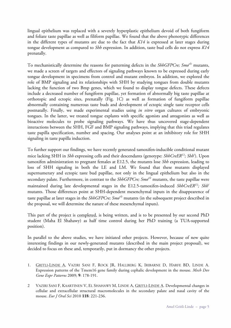
Amel Gritli-Linde – page 5
lingual epithelium was replaced with a severely hyperplastic epithelium devoid of both fungiform and foliate taste papillae as well as filiform papillae. We found that the above phenotypic differences in the different types of mutants are due to the fact that K14 is expressed at later stages during tongue development as compared to Shh expression. In addition, taste bud cells do not express K14 prenatally. To mechanistically determine the reasons for patterning defects in the ShhGFPCre; Smoc/c mutants, we made a screen of targets and effectors of signaling pathways known to be expressed during early tongue development in specimens from control and mutant embryos. In addition, we explored the role of BMP signaling and its relationships with SHH by studying tongues from double mutants lacking the function of two Bmp genes, which we found to display tongue defects. These defects include a decreased number of fungiform papillae, yet formation of abnormally big taste papillae at orthotopic and ectopic sites, prenatally (Fig. 1C) as well as formation of fungiform papillae abnormally containing numerous taste buds and development of ectopic single taste receptor cells postnatally. Finally, we made experimental studies using in vitro organ cultures of embryonic tongues. In the latter, we treated tongue explants with specific agonists and antagonists as well as bioactive molecules to probe signaling pathways. We have thus uncovered stage-dependent interactions between the SHH, FGF and BMP signaling pathways, implying that this triad regulates taste papilla specification, number and spacing. Our analyses point at an inhibitory role for SHH signaling in taste papilla induction. To further support our findings, we have recently generated tamoxifen-inducible conditional mutant mice lacking SHH in Shh expressing cells and their descendants (genotype: ShhCreERT2; Shhc). Upon tamoxifen administration to pregnant females at E12.5, the mutants lose Shh expression, leading to loss of SHH signaling in both the LE and LM. We found that these mutants displayed supernumerary and ectopic taste bud papillae, not only in the lingual epithelium but also in the secondary palate. Furthermore, in contrast to the ShhGFPCre; Smoc/c mutants, the taste papillae were maintained during late developmental stages in the E12.5-tamoxifen-induced ShhCreERT2; Shhc mutants. Those differences point at SHH-dependent mesenchymal inputs in the disappearence of taste papillae at later stages in the ShhGFPCre; Smoc/c mutants (in the subsequent project described in the proposal, we will determine the nature of these mesenchymal inputs). This part of the project is completed, is being written, and is to be presented by our second PhD student (Maha El Shahawy) as half time control during her PhD training (a TUA-supported position). In parallel to the above studies, we have initiated other projects. However, because of new quite interesting findings in our newly-generated mutants (described in the main project proposal), we decided to focus on these and, temporarily, put in dormancy the other projects. 1. GRITLI-LINDE A, VAZIRI SANI F, ROCK JR, HALLBERG K, IRIBARNE D, HARFE BD, LINDE A.
Expression patterns of the Tmem16 gene family during cephalic development in the mouse. Mech Dev Gene Expr Patterns 2009; 9: 178-191.
2 VAZIRI SANI F, KAARTINEN V, EL SHAHAWY M, LINDE A, GRITLI-LINDE A. Developmental changes in
cellular and extracellular structural macromolecules in the secondary palate and nasal cavity of the mouse. Eur J Oral Sci 2010 118: 221-236.

Amel Gritli-Linde – page 6
3. BOULDIN C, GRITLI-LINDE A, AHN S, HARFE B. The Shh signaling pathway is present and required within the vertebrate limb bud apical ectodermal ridge for normal autopod patterning. Proc Natl Acad Sci USA 2010; 107: 5489-5494.
4. ECONOMOU AD, PORNTAVEETUS T, OHAZAMA A, SHARPE PT, KONDO S, BASSON MA, GRITLI-
LINDE A, COBOURNE MT, GREEN JBA. Periodic stripe formation by a Turing-mechanism operating at growth zones in the mammalian palate. Nat Genet 2012; 44: 348-351.