acls study guide - enrollwarebasics!!!!! pvatrialdepolarization!! qrsvventriculardepolarization!!...
TRANSCRIPT

ACLS Study Guide
2012

ACLS Assessment
During the ACLS assessment, we will categorize our patient into the BLS Survey or the ACLS Survey. BLS Survey: *** Unconscious*** 1. Check for response 2. Activate the emergency response system and get an AED 3. Check the carotid pulse
a. If no pulse, Start CPR b. If pulse is present, start rescue breathing
4. Defibrillation ACLS Survey: *** Conscious*** Airway: Ensure a patent airway Use an OPA/NPA if needed Consider an Advanced Airway Breathing: Give oxygen Confirm placement of advanced airway Monitor waveform capnography Avoid hyperventilation Circulation: Establish IV/IO access Identify and treat the rhythm Monitor CPR quality Defibrillation or Cardioversion Differential Diagnosis: H’s and T’s Consider all reversible causes

ECG Basics
P -‐ Atrial Depolarization QRS-‐ Ventricular Depolarization T- Ventricular Repolarization By the Numbers PR Interval -‐ .12-‐.20 QRS Complex-‐ < .12
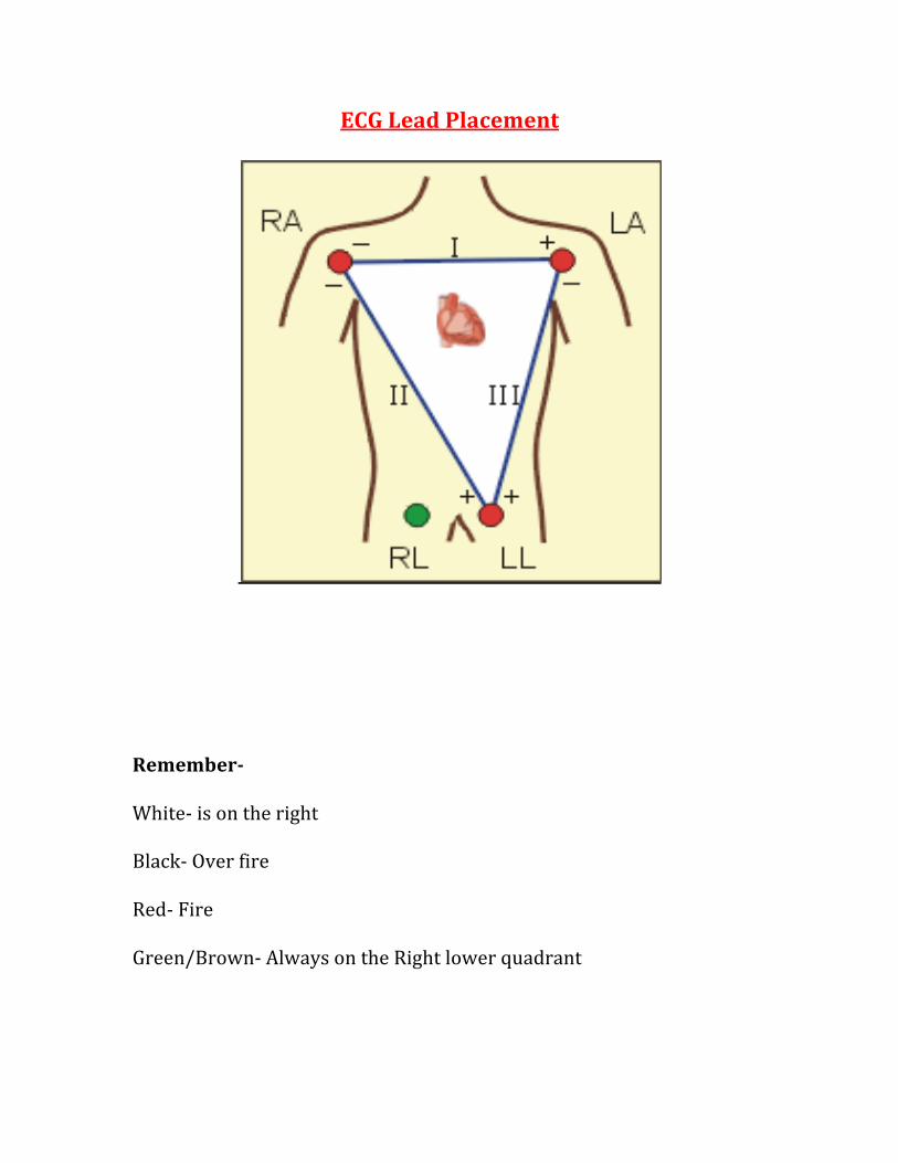
ECG Lead Placement
Remember- White-‐ is on the right Black-‐ Over fire Red-‐ Fire Green/Brown-‐ Always on the Right lower quadrant

12 Lead EKG
Indications for 12 Lead EKG: -‐ Chest Pain -‐ Shortness of Breath -‐ Syncope -‐ Diaphoresis -‐ Nausea/Vomiting -‐ Unexplained general weakness -‐ Diabetic patients -‐ Heart Palpitations
Placement of Precordial Leads
V1 – 4th intercostal space, just to the right of the sternum V2 – 4th intercostal space, just to the left of the sternum V3 – Halfway between V2 and V4 V4 – 5th intercostal space in the mid-clavicular line V5 – Halfway between V4 and V6 V6 – 5th intercostal space in the mid-axillary line

12 Lead EKG
Each lead records the electrical activity of the heart from its own vantage point. 12 Lead EKG’s will provide a detailed view of the heart V1-‐ V2 Septal View V3-‐V4 Anterior View V5-‐V6 Lateral View

ECG Review
Sinus Rhythms: 1. Sinus Bradycardia 2. Normal Sinus Rhythm 3. Sinus Tachycardia
Sinus Bradycardia Description: The Sinus Node is firing at a rate below 60. * This is normal for athletes to have resting heart rates below 60
Treatment: Are they symptomatic?
Chest Pain Fatigue Dizziness Shortness of Breath Altered Mental Status Hypotension Symptomatic Bradycardia: Atropine 0.5 mg every 3-‐5 mins Max : 3 mg

Normal Sinus Rhythm
Description: Normal firing rate is 60 – 100 Sinus Tachycardia Description: Firing rate 100-‐150 Common causes of Tachycardia: Fever Anxiety Sepsis Drugs Pain Asthma Hypovolemia Hypotension

Supraventricular Tachycardia
Description: SVT is characterized as a narrow complex rhythm with no P waves. The heart rate must be above 150 . Treatment:
Are they symptomatic? Chest Pain Fatigue Dizziness Shortness of Breath Altered Mental Status Hypotension Stable Treatment: ( Medicine) Vagal Maneuvers Adenosine 6 mg Rapid IVP ( 1st Dose) Adenosine 12 mg Rapid IVP (2nd Dose) Unstable Treatment: ( Edison) Sedate if possible Immediate Synchronized Cardioversion ( 100 Joules)
Pulseless Rhythms: 1. Ventricular Fibrillation 2. Ventricular Tachycardia 3. Pulseless Electrical Activity 4. Asystole

Ventricular Fibrillation
Description: Ventricular Fibrillation is a chaotic and unorganized rhythm that is unable to pump any blood. V-‐Fib or VF is the most common rhythm to occur during sudden cardiac arrest. Treatment:
V-Fib gets Defib
SHOCK 200 J (Debrillation) ↓
CPR ( 30 X 2) 2 mins ↓
Meds ( Epi 1 mg and Amiodarone 300 mg 1st/ 150 mg 2nd )

Ventricular Tachycardia
Description: Ventricular Tachycardia occurs when the ventricle takes over and generates a wide QRS complex. CHECK FOR A PULSE Treatment: Pulseless V- Tach
SHOCK 200 J (Debrillation) ↓
CPR ( 30 X 2) 2 mins ↓
Meds ( Epi 1 mg and Amiodarone 300 mg 1st / 150 mg 2nd )

Pulseless Electrical Activity
Description: Pulseless Electrical Activity ( PEA ) occurs when the heart is conducting an electrical impulse however lacks a pulse. The following rhythms can be PEA:
• Sinus Bradycardia • Normal Sinus Rhythm • Sinus Tachycardia
*** Remember a Rhythm without a pulse is PEA ***
Treatment: PEA
CPR ( 30 X 2) 2 mins ↓
Meds ( Epi 1 mg )
Consider H’s and T’s ( Differential Diagnosis )

Asystole
Description: Asystole occurs when we have no electrical activity and non-‐functioning pump. *** Ensure Leads and Defib pads are still on the patient ***
Treatment: Asystole
CPR ( 30 X 2) 2 mins ↓
Meds ( Epi 1 mg )
Consider H’s and T’s ( Differential Diagnosis )

ACLS Cardiac Medications
Atropine: 0.5 mg max 3 mg
Symptomatic Bradycardia
Speed up slow rate ↑
Adenosine 6 mg 12 mg RIVP
Stable SVT Symptomatic / HR ↑ 150 Slow down a fast rate ↓ Epinephrine 1 mg * Give to any pulseless PT * VF VT Asystole PEA
Amiodarone 300 mg/ 150 mg VF VT ( Pulseless )
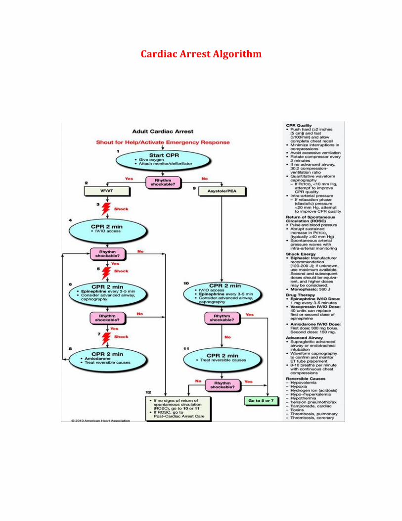
Cardiac Arrest Algorithm

Bradycardia Algorithm

Tachycardia Algorithm

Post Cardiac Arrest Algorithm

Heart Blocks
1st Degree Heart Block
Normal PR Interval .12-.20 seconds
The PR interval is delayed. Greater than .20 seconds.
The DELAY IS CONSTANT !
Treatment
Symptomatic ?
Atropine 0.5 mg every 3-‐ 5 mins
If Atropine is ineffective , consider TCP
If TCP ineffective, consider Dopamine 2-‐10 mcg/kg/min

Heart Blocks
2nd Degree Type 1
The PR interval is progressively getting longer with an eventual drop of the QRS complex
Remember:
Going , going, GONE !!!
Treatment
Symptomatic ?
Atropine 0.5 mg every 3-‐ 5 mins
If Atropine is ineffective , consider TCP
If TCP ineffective, consider Dopamine 2-‐10 mcg/kg/min

Heart Blocks
2nd Degree Type 2
The PR interval is constant however the QRS will drop
Remember:
More P waves than complexes
Treatment
Symptomatic ?
Consider TCP
Increase milliamp until you gain full capture
If TCP ineffective, consider Dopamine 2-‐10 mcg/kg/min

Heart Blocks
3rd Degree ( Complete AV Block)
There is total disassociation with the P wave and QRS complex
The QRS complex will contract on a regular interval
Treatment
Symptomatic ?
Consider TCP
Increase milliamp until you gain full capture
If TCP ineffective, consider Dopamine 2-‐10 mcg/kg/min

Capnography
Applications on intubated patients: Verification of ET tube placement Monitoring and detection ET tube dislodgment Loss of circulatory function Determination of adequate CPR compressions Confirmation of return of spontaneous circulation
American Heart Association recommends an ETCo2 reading of:
35-40 mm hg
10-12 Breaths per min
SPo2 > 94%

Capnography
Chest compression quality can be measured by the use of waveform capnography
< ETCo2 10 mm hg
CPR Quality is poor
Increase Depth and Increase Rate
ROSC
Return of Spontaneous Circulation
Sustained increase in ETCo2 >40 mm hg
