acetylcholinesterase inhibition and micronucleus frequency ... · acetylcholinesterase inhibition...
TRANSCRIPT
ISJ 11: 247-256, 2014 ISSN 1824-307X
RESEARCH REPORT Acetylcholinesterase inhibition and micronucleus frequency in oysters (Crassostrea corteziensis) exposed to chlorpyrifos AB Benitez-Trinidad1, YY Bernal-Hernández2, CL Moreno-Hernández2, IM Medina-Díaz2, ML Robledo-Marenco2, BS Barrón-Vivanco2, D Domínguez-Ojeda3, CA Romero-Bañuelos2, MI Girón-Pérez2, AE Rojas-García2 1Posgrado en Ciencias Biológico Agropecuarias y Pesqueras, Unidad Académica de Agricultura 2Laboratorio de Contaminación y Toxicología Ambiental, Universidad Autónoma de Nayarit (UAN), Av. de la Cultura s/n, Col. Los Fresnos, CP 63190 Tepic, Nayarit, México 3Escuela Nacional de Ingeniería Pesquera, UAN, Bahía de Matanchén Km. 12, San Blas, 63740 Nayarit, México
Accepted September 15, 2014
Abstract
Chlorpyrifos (CPF) is an Organophosphorous pesticide (OP) that has been widely used for both agricultural and domestic pest control. To date, there is little information regarding the effects of this pesticide on aquatic organisms, particularly oysters. The aim of this study was to evaluate Acetylcholinesterase (AChE) activity and Micronucleus (MN) frequency in the oyster Crassostrea corteziensis in laboratory exposure with CPF (20, 40, 60, 80, and 160 µg/L) and in a field study. The results showed that AChE was reduced 60 - 82 % in oysters exposed to CPF, relative to the negative control. Similar AChE results were observed in oysters collected from the Boca de Camichín Estuary in Nayarit, Mexico; with respect to genetic damage, evaluated through MN, treatment with CPF did not induce the MN frequency, nor did the oyster from the field study exhibit an increase in this biomarker. These results suggest that C. corteziensis is a sensitive model for evaluating the acute toxicity of OP in laboratory studies as well in the field. In addition, it generates prospects on studying mechanisms through which the oyster could possess resistance to genotoxic agents, as well as its being a reliable model for evaluating the genotoxic effects of xenobiotics through the MN technique. Key Words: acetylcholinesterase; micronucleus; Crassostrea corteziensis; chlorpyrifos
Introduction
Marine pollutants have consequences at
multiples biological levels (Mix, 1986; Malins et al., 1988; Bolognesi, 1990; Gopal and Pathak, 1993) and have been traditionally documented in terms of chemical concentrations of contaminants; however, these measurements do not provide estimations of harmful effects on living organisms and are now complemented with the measurement of additional biomarkers that are indicative of such effects. Biomarkers represent integral and measurable biochemical and physiological changes in organisms exposed to contaminants, thus indicating initial responses to environmental perturbations and contamination (McCarthy and Shugart, 1990; Bengtson and Henshel, 1996; Roy et al., 1996). For ___________________________________________________________________________
Corresponding author: Aurora Elizabeth Rojas-García Laboratorio de Contaminación y Toxicología Ambiental Secretaría de Investigación y Posgrado Universidad Autónoma de Nayarit CP 63155 Tepic, Nayarit, México E-mail: [email protected]
example, the measurement of Acetylcholinesterase (AChE) activity is widely used to evaluate the exposure and effects of anti-AChE compounds (Galgani and Bocquené, 2000). AChE is the enzyme responsible for the hydrolysis of the neurotransmitter acetylcholine into choline and acetic acid in the nerve synapse. AChE inhibition is directly linked with the action mechanism of the Organophosphorus (OP) and carbamate pesticides that block the action of this enzyme (Reigart and Roberts, 1999). Measurement of AChE activity is widely employed in several species, including aquatic species (Cajaraville et al., 2000; Singh and Sharma, 2005; Bernal-Hernández, et al., 2010).
The rapid increase in the production and use of OP and carbamate pesticides has raised concerns regarding their potential negative effects on humans and non-target wildlife populations. Pesticides enter waterways from agricultural and urban run-off, groundwater discharge, and after direct application (Schulz and Leiss, 1999), and may be transported to estuaries and coastal waters (Magni et al., 2006; Ismail et al., 2014).
247

Fig. 1 Map of the Boca de Camichín Estuary showing the three sampling stations. S1: Station 1; S2: Station 2, and S3: Station 3.
Chlorpyrifos (CPF) is an OP utilized for domestic and agricultural pest control. Although CPF is a pesticide used worldwide, there is little information concerning its acute and chronic effects on aquatic organisms, particularly oysters. Some reports have suggested that CPF has the potential to cause neuro- and genotoxic effects in aquatic organisms, such as inhibition of AChE and the formation of micronuclei (MN), respectively (Shugart, 1995). In some cases, the formulated pesticides are found to be more toxic than the active ingredient, particularly to aquatic organisms (Ali et al., 2009).
MN frequency is one of the most widely used genotoxicity biomarkers in aquatic organisms. MN are small intracytoplasmic masses of chromatin resulting from chromosomal breakages during cell division. An increase in the frequency of micronucleated cells is thought to result from chromosomal and genomic damage caused by clastogens or spindle poisons (Bolognesi and Fenech, 2012). At present, the MN assay is applied in laboratory and field studies using hemocytes and gill cells from bivalves, or simultaneously in both cell types (Barsiene et al., 2006; Bolognesi and Fenech, 2012). However, few studies have reported MN frequency in oysters.
The oyster Crassostrea corteziensis is a species native to the eastern Tropical Pacific where it is naturally distributed in mangrove zones from Mexico to Peru (Stuardo and Martínez, 1975). This species
supports artisanal and commercial fisheries in northwestern Mexico. The Boca de Camichín Estuary situated in Nayarit state is one of the largest oyster producers in Mexico. This ecosystem is important not only for its oyster production, but also for its biologic diversity. High agricultural activity around the estuary is reported, with elevated use of pesticides. One of the most utilized insecticides in the ecosystem is CPF (González-Arias et al., 2010; Rojas-García et al., 2011). Therefore, the aim of this study was to determine AChE activity and MN frequency in order to evaluate the degree of exposure to CPF and the integrity of genetic material, respectively, in vivo and in field study. Materials and Methods Sampling collection
For in vivo and in-field study, Crassostrea corteziensis oysters approximately 4 - 8 cm in shell length were collected at the Boca de Camichín Estuary in Nayarit state, Mexico. This area is characterized by its biological diversity and in part to the high-priority National Wetlands (Robledo-Marenco et al., 2006). Three sampling sites at the estuary were chosen (Fig. 1), taking into account the hydrographic and biological characteristics of the system. One site was localized toward the head of the estuary (S1: 21º44′06.8″N, 105º29′19.06″), another station was localized downstream and near
248

the mouth of the estuary (S2: 21º44′53.0″N, 105º29′39.0″W), and one was situated in the middle of the oyster-culture zone (S3: 21º45′41.1″N, 105º29′43.5″). The oysters were collected at each of the three sites at two levels: from the upper and the lower part of the oyster string. Gills were dissected immediately and frozen until use to screen for total protein, AChE activity, and MN frequency.
Exposure to CPF under in vivo conditions
The oysters were carried to the laboratory, placed in 40-L tanks filled with filtered seawater, and acclimated for 15 days, during which time they were fed 2 L/day of microalgae species: Chaetoceros spp. (6×10⁵ cells/mL). For exposure to CPF, oysters were placed in aquariums with 10 L of aerated water and were acclimated to estuary conditions (26 ‰; 32 ± 1 ºC). Ten organisms were utilized for each treatment. The organisms were divided into seven lots as follows: one for negative control (maintained under the same condition, but without treatment), one for positive control (treatment with mitomycin C as MN control) and five for CPF treatments (20, 40, 60, 80, and 160 µg/L). The organisms were incubated in 12-h:12-h dark-light cycles for > 96 h. Each experiment was run in triplicate. At the end of exposure, the organisms were washed, opened, and the gills were excised to screen for total protein, AChE activity, and MN frequency. For the present study, CPF (44.5 % Emulsifiable concentrate [EC]) with the Magnum L-480 brand was employed.
Total protein content and AChE activity
Gills were homogenized (1:2 W/V) in an extraction buffer (20 mM Tris-Base, 1 mM EDTA, 1 mM DTT, 500 mM sucrose, 150 mM KCl, 0.1 mM PMSF, pH 7.6) (Monserrat et al., 2002). The homogenate was centrifuged at 9,000 rpm for 30 min at 4 °C. The pellet was discarded and the supernatant (S9 fraction) was used to determine protein and AChE activity. Total protein was determined in fraction S9 according to the Lowry method (Lowry et al., 1951) using Bovine serum albumin (BSA) as the standard. AChE activity was
performed according to Ellman et al. (1961), modified by Monserrat et al. (2002). Enzyme activity was determined in triplicate using 300 µL of supernatant, 2700 µL of phosphate buffer (pH 8, 0.1 M), 100 µL of 0.5 mM Ellman’s reagent (DTNB), and 20 µL of 0.5 mM acetylthiocholine iodide as substrate. The rate of absorbance change at 412 nm was recorded over 120 sec at 25 °C using a Spectronic Genesys 10 Bio spectrophotometer (Genesys, WI, USA).
Micronucleus (MN) determination Cell preparation
This technique was based on the methodology described by Bolognesi et al. (1999) with some modifications. To prepare the cell suspension, gills were excised and cut into fragments with dissection scissors in 5 mL of saline solution. The cell suspension was centrifuged at 1,500 rpm at 25 °C for 5 min and then fixed in 5 mL of Carnoy’s fixative solution (CFS) (3:1 methanol:acetic acid) for 20 min. Afterward, the cell suspension was centrifuged and washed three times with CFS. The pellet was resuspended in 1 mL of CFS and treated with 1 mL of trypsin (0.00015 %). Trypsin activity was blocked by the addition of 5 mL of CFS. The cell suspension was centrifuged and washed twice with CFS. Finally the samples were resuspended in 1 mL of CFS and were maintained at 4 ºC until slide preparation.
Exposure to mitomycin C as positive control
Mitomycin C (MMC) is a DNA cross-linking agent that has been extensively used as a reference mutagen in numerous MN studies (Das and Nanda, 1986; Majone et al., 1987; Scarpato et al., 1990, Williams and Metcalfe, 1992). Oysters were placed in aquariums with 10 L of water (26 ‰). Prior to the exposure, they were acclimatized for 24 h. Subsequently, a group of 10 organisms with one replicate were exposed to MMC (2.5 mg/L) for 48 h. At the end of treatment, the oysters were sacrificed and their gills were separated to determine the presence of MN. The negative control group was run with one replicate.
Fig. 2 Representative gill cells from Crassostrea corteziensis (a) and a micronucleated gill cell (b). The micronucleus (MN) is indicated by an arrow beside the main nucleus. Staining with the Merck Hemacolor kit (100x).
249

Fig. 3 Total protein content (a) and remaining acetylcholinesterase activity in oyster (Crassostrea corteziensis) gills (b) exposed to different chlorpyrifos concentrations for 96 h. Data are expressed as means ± Standard deviations (SD) of three independent determinations. NC, Negative control. The asterisk (*) indicates a significant difference from control animals (NC) at p < 0.05. Criteria for scoring MN
MN were identified according to following criteria described by Fenech et al. (2003): (1) round and ovoid-shaped nonrefractory particles in the cytoplasm; (2) chromatin-like color and structure; (3) diameter of 1/3 - 1/20 of the main nucleus, and (4) particles completely separated from the main nucleus. Figure 2 shows the cell morphology and micronucleated cell observed in the MN test. Slides were analyzed by light microscope (Carl Zeiss, Axiostar plus, Göttingen, Germany) at a magnification of 100x for nuclear abnormalities (MN) and 1,000 gill cells were scored. Only intact cells were scored. All MN were checked by a second operator to ensure that unambiguous micronucleated cells were exclusively scored. Statistical analysis
The results were expressed as the average of three independent experiments. The results were analyzed by Mann-Whitney U test and Dunn multiple comparison tests. p values < 0.05 were considered statistically significant. Statistical analyses were conducted using the Stata ver. 8.0 program (Stata statistical software; Stata
Corporation, College Station, TX, USA) and GraphPad Prism 5.01 for Windows (GraphPad Software, San Diego, CA, USA).
Results and Discussion
The treatments with CFP did not cause mortality
in the oysters, but we observed that the organisms closed their valves for 12 h. There are reports that suggest that these oysters are able to isolate themselves from a contaminated environment by closing their valves (Kramer et al., 1989; Mersch et al., 1993). In addition, in bivalve molluscs, contact with toxic compounds causes a reduction in filtration activity; thus, this might reduce exposure time (Van der Gaag et al., 1990; Wrisberg et al., 1992). Total protein content and AChE activity
The results showed that C. corteziensis oysters exposed to 80 and 160 µg/L had the highest protein concentrations (8.50 and 12.77 mg/mL, respectively) compared with those of the negative control; the opposite was observed at 20 and 40 µg/L (2.32 and 2.41 mg/mL) (p < 0.05) (Fig. 3a). In addition, significant variations in remaining AChE activity of gills exposed to different concentrations of CFP
250

Fig. 4 Total protein content (a) and remaining acetylcholinesterase activity (b) in Crassostrea corteziensis oysters collected from the Boca de Camichín Estuary. Data are expressed as means ± Standard deviations (SD) of three independent determinations. NC, Negative control; S1, Sampling site 1; S2, Sampling site 2; S3, Sampling site 3. U: Upper part of string; L: Lower part of string. The asterisk (*) indicates a significant difference from control animals (NC) at p < 0.05. during 96 h were recorded (Fig. 3b). At the highest CFP treatments (80 and 160 µg/L), the remaining AChE activity was 40 and 18 % with respect the control group, which confirms the inhibitory effects of CFP on oyster exposure.
Proteins and other oyster components vary considerably among species and among individuals of the same species, also depend on age, gender, size, environment, and season of the year, and are closely related with food organisms (Maeda-Martínez et al., 2001). In this regard, average total protein content in the gills of oysters in this study was 5.93 mg/mL in CPF-treated organisms and 4.59 mg/mL in the negative control group. It has been reported that protein concentrations in the tissues of gills can be affected both positively and negatively by stress-associated physiological changes, as well as by the presence of chemicals (Rank et al., 2007).
On the other hand, in the field study no changes in protein content in C. corteziensis oysters was observed (Fig. 4a), but all of the organisms collected at the estuary exhibited lower AChE activities with respect the negative control, as illustrated in Figure 4b; this indicates the presence
of anticholinesterase compounds in the estuary and, possibly, OP pesticides.
Oysters have been proposed as bioindicators of aquatic pollution, in addition to mussels (Bebianno et al., 1993, 1994; Viarengo et al., 1998; Blasco and Puppo, 1999; Rodríguez-Ortega et al., 2001), and data regarding the measurement of AChE activity in oyster tissues are available in the literature (Mora et al., 1999; Dellali et al., 2001; Bernal-Hernández et al., 2010). Previous studies suggest that AChE activity is found in the gills, mantle, and adductor muscles of bivalves (Bocquene et al. 1997; Escartín and Porte 1997; Radenac et al., 1998; Mora et al., 1999; Monserrat et al., 2002; Damiens et al., 2004). Gills were chosen for measurement of esterase activities because gill tissue exhibited high activity levels and higher sensitivity to contaminants with respect to those of other tissues (Mora et al., 1999). Also, oyster gills are easy to dissect and have a relatively important and constant mass (apparently independent of field conditions) (Mora et al., 1999).
251
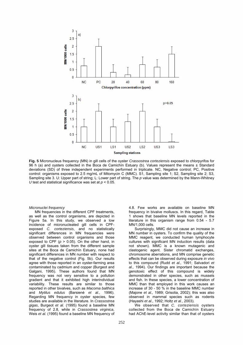
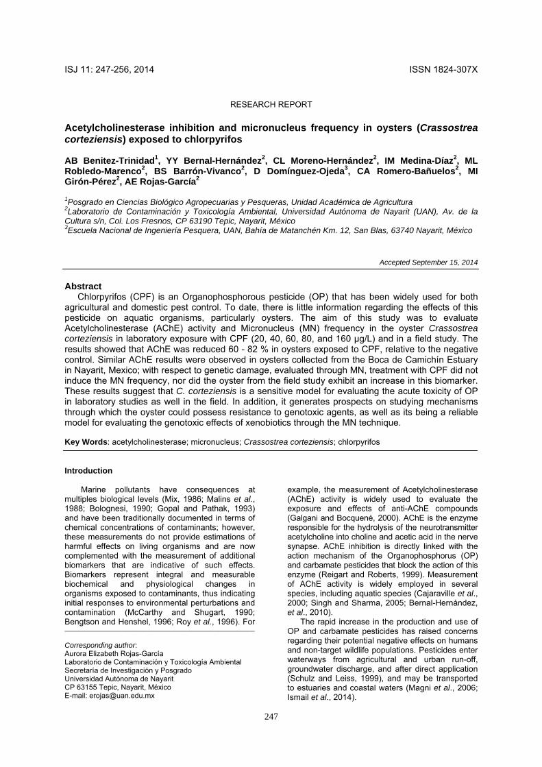
Fig. 5 Micronucleus frequency (MN) in gill cells of the oyster Crassostrea corteziensis exposed to chlorpyrifos for 96 h (a) and oysters collected in the Boca de Camichín Estuary (b). Values represent the means ± Standard deviations (SD) of three independent experiments performed in triplicate. NC, Negative control; PC, Positive control: organisms exposed to 2.5 mg/mL of Mitomycin C (MMC). S1, Sampling site 1; S2, Sampling site 2; S3, Sampling site 3. U: Upper part of string; L: Lower part of string. The p value was determined by the Mann-Whitney U test and statistical significance was set at p < 0.05. Micronuclei frequency
MN frequencies in the different CPF treatments, as well as the control organisms, are depicted in Figure 5a. In this study, we observed a low incidence of micronucleated gill cells in CPF-exposed C. corteziensis, and no statistically significant differences in MN frequencies were observed between control organisms and those exposed to CPF (p > 0.05). On the other hand, in oyster gill tissues taken from the different sample sites at the Boca de Camichín Estuary, none had significant differences in MN number with respect to that of the negative control (Fig. 5b). Our results agree with those reported in an oyster-farming area contaminated by cadmium and copper (Burgeot and Galgani, 1995). These authors found that MN frequency was not very sensitive to a pollution gradient and that it exhibited high interindividual variability. These results are similar to those reported in other bivalves, such as Macoma balthica and Mytilus edulus (Barsiené et al., 1996). Regarding MN frequency in oyster species, few studies are available in the literature. In Crassostrea gigas, Burgeot et al. (1995) found a baseline MN frequency of 2.8, while in Crassostrea virginica, Weis et al. (1995) found a baseline MN frequency of
4.8. Few works are available on baseline MN frequency in bivalve molluscs. In this regard, Table 1 shows that baseline MN levels reported in the literature in this organism range from 0.54 - 5.7 MN/1,000 cells.
Surprisingly, MMC did not cause an increase in MN number in oysters. To confirm the quality of the MMC reagent, we conducted human lymphocyte cultures with significant MN induction results (data not shown). MMC is a known mutagenic and clastogenic agent. Sister chromatid exchanges, chromosome aberrations, and MN comprise genetic effects that can be observed during exposure in vivo to this compound (Rudd et al., 1991; Salvadori et al., 1994). Our findings are important because the genotoxic effect of this compound is widely demonstrated in other species, such as mussels and fish. In these species, a lower concentration of MMC than that employed in this work causes an increase of 30 - 50 % in the baseline MMC number (Majone et al., 1989; Grisolia, 2002); this was also observed in mammal species such as rodents (Hayashi et al., 1992; Holtz et al., 2003).
We observed that C. corteziensis oysters collected from the Boca de Camichín Estuary had AChE-level activity similar than that of oysters
252
Table 1 Micronucleus (MN) frequency in bivalve organisms
ND: Not determined; SD: Standard deviation; PCB: Polychlorinated biphenyl; DDT: Dichlorodiphenyltrichloroethane; HCB: Hexachlorobenzene; HCH: Hexachlorocyclohexane; Cr: Chromium; Ni: Nickel. exposed under in vivo conditions to CPF, which caused marked inhibition of AChE activity but did not induce the presence of MN. These results confirm that AChE activity in C. corteziensis oysters can serve as a sensitive biomarker to assess exposure to OP, such as CPF (Bernal-Hernández et
al., 2010). On the other hand, these results generate perspectives regarding the mechanisms through which oyster could possess resistance to genotoxic agents, as well as whether this is a reliable model for evaluating the genotoxic effects of xenobiotics.
Organism Tissue Compound MN/
1,000 cells
SD Reference
Mytilus galloprovincialis Gill Zinc chloride 2.2 ND Majone et al., 1987
Crassostrea gigas Heart cells Benzo[a]-pyrene 2.8 ±2.3 Burgeot et al., 1995
Dreissena polymorpha
Hemolymph
Gill
Clastogens: Mitomycin C (MMC)
Bleomycin
Di-methylar-sinic acid Potassium chromate
Mitomycin C (MMC)
Bleomycin Dimethyl-arsinic acid Potassium chromate
1.2 4.4
6.9 3.2
6.1 6.7 5.6 5.4
±1.0 ± 1.8
± 2.4 ± 1.1
± 1.4 ± 3.1 ± 1.9 ± 2.6
Mersch et al., 1996
Mytilus trossulus Gill PCB, DDT, HCB, HCH,
and Polybrominated diethyl ethers (PBDE)
6 ND Kopecka et al., 2006
Mytilus edulis Gill Crude oil 2.4 ND Baršienė and
Andreikė-Naitė, 2007
Macoma balthica Gill Contaminated zone 1.2-3.63 ND Baršienė et al., 2008
Mytilus edulis Gill Contaminated zone 1.7-3.3 ND Baršienė et al., 2008
Mytilus galloprovincialis Hemolymph and gills
Urban and industrial pollution 5.5-5.1 ND Taleb et al., 2009
Crassostrea corteziensis Gill Chlorpyrifos 1.2 ±1.1 This study
Cerastoderma edule Hemolymph Chemical contaminant 0.4 ±0.6 Ruiz et al., 2013
Mya arenaria Hemolymph
Organotin compounds, arsenic, Polyaromatic hydro-carbons (PAH)
and PCB
15.4 ND Debenest et al., 2013
Mytilus galloprovincialis Digestive glands Hg2+ 2.3 ND Pytharopoulou et
al., 2013
Ruditapes philippinarum Gill PAH and trace metals such as Cr and Ni 4–16 ND Sacchi et al., 2013
253

Acknowledgments The authors thank Ing. ML Loza and ML Llamas
García for their technical assistance during oyster collection and experimental work. This research was made possible through grants NAYARIT-2006-C01-66170 and PROMEP/103.5/07/2746. References Ali D, Nagpure NS, Kumar S, Kumar R, Kushwaha
B, Lakra WS. Assesment of genotoxic and mutagenic effects of chlorpyrifos in freshwater fish Channa punctatus (Bloch) using micronucleus assay and alkaline single-cell gel electrophoresis. Food Chem. Toxicol. 47: 650-656, 2009.
Baršienė J, Dedonytėa V, Rybakovasa A, Andreikėnaitėa, L, Andersenc OK. Investigation of micronuclei and other nuclear abnormalities in peripheral blood and kidney of marine fish treated with crude oil. Aquat. Toxicol. 78: S99-S104, 2006.
Baršiené, J, Tapia G, Barsyte D. Chromosome of molluscs inhabiting some mountain spring of eastern Spain. J. Mollus. Stud. 62: 539-543, 1996.
Baršienė J. Andreikėnaitė L, Induction of micronuclei and other nuclear abnormalities in blue mussels exposed to crude oil from the North Sea. Ekologija 53: 9-15, 2007.
Baršienė J, Andreikėnaitė L, Garnaga G, Rybakovas A. Genotoxic and cytotoxic e�ects in the bivalve mollusks Macoma balthica and Mytilus edulis from the Baltic Sea. Ekologija 54: 44-50, 2008.
Bebianno MJ, Nott JA, Langston WJ. Cadmium metabolism in the clam Ruditapes decusata: the role of metallothioneins. Aquat. Toxicol. 27: 315-334, 1993.
Bebianno MJ, Serafim MAP, Rita MF. Involvement of metallothionein in cadmium accumulation and elimination in the clam Ruditapes decussata. Bull. Environ. Contam. Toxicol. 53: 726-732, 1994.
Bengtson DA, Henshel DS. Environmental toxicology and risk assessment: Biomarkers and risk assessment. Vol. V. ASTM, STP 1306, West Conshohocken, PA, 1996.
Bernal-Hernández YY, Medina-Díaz IM, Robledo-Marenco ML, Velázquez-Fernández JB, Girón-Pérez MI, Ortega-Cervantes L, et al. Acetylcholinesterase and metallothionein in oysters (Crassostrea corteziensis) from a subtropical Mexican Pacific estuary. Ecotoxicology 19: 819-25, 2010.
Blasco J, Puppo J. Effect of heavy metals (Cu, Cd and Pb) on aspartate and alanine aminotransferase in Ruditapes philippinarum (Mollusca: Bivalvia). Comp. Biochem. Physiol. 122C: 253-63, 1999.
Bocquene G, Roig A, Fournier D. Cholinesterases from common oyster (Crassostrea gigas). Evidence for the presence of a soluble acetylcholinesterase insensitive to organophosphate and carbamate inhibitors. FEBS lett. 407: 261-266, 1997.
Bolognesi C. Carcinogenic and mutagenic effects of pollutants in marine organisms: a review. In:
Grandjean E. (ed), Carcinogenic, mutagenic, and teratogenic marine pollutants: impact on human healt and the environment, Portfolio Publishing Company, The Woodland, TX, pp 67-83, 1990.
Bolognesi L, Landini E, Roggieri P, Fabbri R, Viarengo A. Genotoxicity biomarkers in the assessment of heavy metal effects in mussels: Experimental studies. Environ. Mol. Mutagen. 33: 287-292, 1999.
Bolognesi C, Fenech M. Mussel micronucleus cytome assay. Nature Protocols 7: 1125-1137, 2012.
Burgeot T, His E, Galgani F. The micronucleus assay in Crassostrea gigas for the detection of seawater genotoxicity. Mut. Res. 343: 125-140, 1995.
Cajaraville MP, Bebianno MJ, Blasco J, Porte C, Sarasquete C, Viarengo A. The use of biomarkers to assess the impact of pollution in coastal environments of the Iberian Peninsula: a practical approach. Sci. Total. Environ. 247: 295-311, 2000.
Damiens G, His E, Gnassia BM, Quiniou F, Roméo M. Evaluation of biomarkers in oyster larvae in natural and polluted conditions. Comp. Biochem. Physiol. 138C: 121-128. 2004.
Das RK, Nanda NK. Induction of micronuclei in pe-ripheral erythrocytes of fish Heteropneustes fossilis by mitomycin C and paper mill effluent. Mut. Res. 175: 67-71, 1986.
Debenest T, Gagné F, Burgeot T, Blaise C, Pellerin J. DNA integrity assessment in hemocytes of soft-shell clams (Mya arenaria) in the Saguenay Fjord (Québec, Canada). Environ. Sci. Pollut. Res. 20: 621-629, 2013.
Dellali M, Gnassia BM, Romeo M, Aaissa P. The use of acetylcholinesterase activity in Ruditapes decussatus and Mytilus galloprovincialis in the biomonitoring of Bizerta lagoon. Comp. Biochem. Physiol. 130C: 227-235, 2001.
Ellman G, Courtney KD, Andres V, Feather-Stone RM. A new and rapid colorimetric determination of acetylcholinesterase activity. Biochem. Pharmacol. 7: 88-95, 1961.
Escartín E, Porte C. The use of cholinesterase and carboxylesterase activities from Mytilus galloprovincialis in pollution monitoring. Environ. Toxicol. Chem. 16: 2090-2095, 1997.
Fenech M, Chang WP, Kirsch-Volders M, Holland N, Bonassi S, Zeiger E. Human Micronnucleus Project. "HUMN project: detailed description of the scoring criteria for the cytokinesis-block micronucleus assay using isolated human lymphocyte cultures". Mut. Res. 534: 65-75, 2003.
Galgani F, Bocquene G. Molecular biomarkers of exposure of marine organisms to organophosphorus pesticides and carbamates. In: Lagadic L, Caquet T, Amiard JC, Ramade F (eds), Use of Biomarkers for Environmental Quality Assessment, Enfield USA, Plymouth UK, pp 113-137, 2000.
González-Arias CA, Robledo-Marenco ML, Medina-Díaz IM, Velázquez-Fernández JB, Girón-Pérez MI, Quintanilla-Vega MB, et al. Patrón de uso y venta de plaguicidas en Nayarit, México. Rev. Int. Contam. Ambient 3: 221-228,2010.
254
Gopal K, Pathak SP. Possible causes of outbreaks of fish diseases and mortality in polluted waters. J. Adv. Zool. 14: 53-60, 1993.
Grisolia CK. A comparation between mouse and fish micronucleus test using cyclophosphamide, mitomicyn C and variouse pesticides. Mut. Res. 512: 145-150, 2002.
Hayashi M, Kodama Y, Awogi T, Suzuki T, Asita AO, Sofuni T. The micronucleus assay using peripheral blood reticulocytes from mitomycin C- and cyclophosphamide treated rats. Mut. Res. 278: 209-213, 1992.
Holtz KM, Rockwell S, Tomasz M, Sartorelli AC. Nuclear overexpression of NADH: cytochrome b5 reductase activity increases the cytotoxicity of mitomycin C (MC) and the total number of MC-DNA adducts in chinese hamster ovary cells. J. Biol. Chem. 278: 5029-5034, 2003.
Ismail N, Mahmood-Khan Q, Ali R, Ali T, Mobeen A. Evaluation of the genotoxicity of chlorpyrifos in common indus valley toad, Bufo stomaticus using alkaline single-cell gel electrophoresis (Comet) assay. Agric. Sci. 5: 376-382, 2014.
Kopecka J, Lehtonen KK, Baršienė J, Broeg K, Vuorinen PJ, Gercken, J, et al. Measurements of biomarker levels in flounder (Platichthys flesus) and blue mussel (Mytilus trossulus) from the gulf of Gdansk (southern Baltic). Mar. Pollut. Bull. 53: 406-421, 2006.
Kramer KJM, Jenner HA, De Zwart D. The valve movement response of mussels: a tool in biological monitoring. Hydrobiologia 188/189: 433-443, 1989.
Lowry OH, Rosebrough NJ, Farr AI, Randall RJ. Protein measurement with the Folin phenol reagent. J. Biol. Chem.193: 265-275, 1951.
Maeda-Martínez AN. Los moluscos pectínidos de Iberoamérica: Ciencia y acuicultura, editorial Limusa, México, 2001.
Magni P, De Falco G, Falugi C, Franzoni M, Monteverde M, Perrone E, et al. Genotoxicity biomarkers and acetylcholinesterase activity in natural populations of Mytilus galloprovincialis along a pollution gradient in the gulf of Oristano (Sardinia, western Mediterranean). Environ. Pollut., 142: 65-72, 2006.
Majone F, Brunetti R, Gola I, Levis AG. Persistence of micronuclei in the marine mussel, Mytilus galloprovincialis, after treatment with mitomycin C. Mut. Res. 191:157-161, 1987.
Majone F, Brunetti R, Fumagalli O, Gabriele M, Levis AG. Induction of micronuclei by mitomycin C and colchicine in the marine mussel Mytilus galloprovincialis. Mut. Res. 244: 147-151, 1989.
Malins DC, McCain BB, Landahl JT, Myers MS, Krahn MM, Brown DW, et al. Neoplastic and other diseases in fish in relation to toxic chemicals: an overview. Aquat. Toxicol. 11: 43-67, 1988.
McCarthy JF, Shugart LR. Biomarkers of environmental contamination, Lewis Publ, Boca Raton, pp 31, 1990.
Mersch J, Pihan JC. Simultaneous assessment of environmental impact on condition and trace metal availability in zebra mussels Dreissena polymorpha transplanted into the Wiltz River, Luxembourg. Comparison with the aquatic moss
Fontinalis antipyretica. Archiv. Environ. Contam. Toxicol. 25: 353-364, 1993.
Mersch J, Beauvais MN, Nagel P. Induction of micronuclei in haemocytes and gill cells of zebra mussels, Dreissena polymorpha, exposed to clastogens. Mut. Res. 371: 47-55, 1996.
Mix MC. Cancerous disease in aquatic animals and their association with environmental pollutants: a critical literature review. Mar. Environ. Res. 20: 1-141, 1986.
Monserrat JM, Bianchini A, Bainy AC. Kinetic and toxicological characteristics of acetylcholinesterase from the gills of oysters (Crassostrea rhizophorae) and other aquatic species. Mar. Environ. Res. 54: 781-785, 2002.
Mora P, Fournier D, Narbonne JF. Cholinesterases from the marine mussels Mytilus galloprovincialis Lmk. and M. edulis L. and from the freshwater bivalve Corbicula fluminea Muller. Comp. Biochem. Physiol. 122C: 353-361. 1999.
Pytharopoulou S, Kournoutoua GG, Leotsinidisb M, Georgiouc CD, Kalpaxis DL. Dysfunctions of the translational machinery in digestive glands of mussels exposed to mercury ions. Aquat. Toxicol. 134-135: 23-33, 2013.
Radenac G, Bocquené G, Fichet D, Miramand P. Contamination of a dredged-material disposal site (La Rochelle Bay, France). The use of the acetylcholinesterase activity of Mytilus edulis as a biomarker of pesticides: the need for a critical approach. Biomarkers 3: 305-315, 1998.
Rank J, Lehtonen KK, Strand J, Laursen M. DNA damage, acetylcholinesterase activity and lysosomal stability in native and transplanted mussels (Mytilus edulis) in areas close to coastal chemical dumping sites in Denmark. Aquat. Toxicol. 84: 50-61, 2007.
Robledo-Marenco ML, Botello AV, Romero-Bañuelos CA, Díaz-González G. Presence of persistent organochlorine pesticides in estuaries of the subtropical Mexican Pacific. Int. J. Environ. Pollut. 26: 284-294, 2006.
Rodriguez-Ortega MJ, Alhama J, Funes V, Romero-Ruiz A, Rodriguez-Ariza A, Lopez-Barea J. Biochemical biomarkers of pollution in the clam Chamelea gallina from South-Spanish littoral. Environ. Toxicol. Chem. 21: 542-549, 2001.
Rojas-García AE, Medina-Díaz IM, Robledo-Marenco ML, Barrón-Vivanco BS, Girón-Pérez MI, et al. Hematological, Biochemical Effects, and Self-reported Symptoms in Pesticide Retailers. JOEM 53: 517, 2011
Roy S, Lindström-Seppä P, Hänninen O., Integrative approach to aquatic environment biomonitoring. In: Richardson M (ed), Environmental Xenobiotics. Taylor and Francis, London, pp 123-142, 1996.
Rudd NL, Williams SE, Evans, M, Hennig UG, Hoar D. Kinetochore analysis of micronuclei allows insights into the actions of colcemid and mitomycin C. Mut. Res. 261, 57-68. 1991.
Ruiz P, Diaz S, Orbea A, Carballal MJ, Villalba A, Cajaraville MP. Biomarkers and transcription levels of cancer-related genes in cockles Cerastoderma edule from Galicia (NW Spain) with disseminated Neoplasia. Aquat. Toxicol. 101-111, 2013.
255

Salvadori S, Cau A, Coluccia E, Milia A, Deiana AM. Karyotype, C-banding and G-banding, and nucleolar organizer regions of Conger conger (Osteichthyes, Anguilliformes). Boll. Zool. 1: 59-63, 1994.
Sacchi A, Mouneyrac C, Bolognesi C, Sciuttoc A, Roggieri P, Fusi M, et al. E. Biomonitoring study of an estuarine coastal ecosystem, the Sacca di Goro lagoon, using Ruditapes philippinarum (Mollusca: Bivalvia). Environ. Pollut. 177:82-89, 2013
Scarpato R, Migliore L, Barale L. The micronucleus assay in Anodonta cygnea for the detection of drinking water mutagenicity. Mut. Res. 245: 231-237,1990.
Schulz R, Leiss M. A field study of the effects of agriculturally derived insecticide input on stream invertebrate dynamics. Aquat. Toxicol. 46: 155-76, 1999.
Shugart LR. Environmental genotoxicology. In: Rand GM (ed), Fundamentals of aquatic toxicology: effects, environmental fate, and risk assessment, Taylor & Francis, Bristol, PA, pp 405-420, 1995.
Stuardo J, Martínez, A. Relación entre algunos factores ecológicos y la biología de poblaciones de Crassostrea corteziensis Hertlein, 1951, de San Blas, Nayarit, México. An Centro Cienc. del Mar y Limnol. Univ. Nal. Autón. México. 2: 89-130, 1975.
Taleb ZM, Benali I, Gherras H, Ykhlef-Allal A, Bachir-Bouiadjra B, Amiard JC, et al. Biomonitoring of environmental pollution on the Algerian west coast using caged mussels Mytilus galloprovincialis. Oceanología 51: 63-84 2009.
Van der Gaag MA, Gauthier L, Noordsij A, Lévi, Y, Wrisberg MN. Methods to measure genotoxins in wastewater: evaluation with in vivo and in vitro tests. In: Waters MD, et al. (eds), Genetic toxicology of complex mixtures, Plenum Press, New York, 6: 215-232, 1990.
Viarengo A, Abele-Oeschger D, Burlando B. Effects of low temperature on prooxidants and antioxidant defence systems in marine organisms. In: Pfrtner HO, Playle R. (eds), Cold Ocean Physiology, Cambridge Univ., Press Cambridge, pp 213-235,1998.
Weis P, Weiqb JS, Couch J, Daniels CC, Chend T. Pathological and genotoxicological observations in oysters (Crassostrea virginica) living on chromated copper arsenate (CCA)-treated wood. Mar. Environ. Res. 39: 275-278, 1995.
Williams RC, Metcalfe CD. Development of an in vivo hepatic micronucleus assay with rainbow trout. Aquat. Toxicol. 23: 193-202, 1992.
Wrisberg MN, Bilbo CM, Spliid H. Induction of micronuclei in hemocytes of Mytilus edulis and statistical analysis. Ecotoxicol. Environ. Saf. 23: 191-205, 1992.
256