absorption, distribution, metabolism and excretion of the...
TRANSCRIPT

DMD #75358
1
Title page
Absorption, distribution, metabolism and excretion of the oral prostaglandin D2
receptor 2 (DP2) antagonist fevipiprant (QAW039) in healthy volunteers and in
vitro
David Pearson, H. Markus Weiss, Yi Jin, Jan Jaap van Lier, Veit J. Erpenbeck, Ulrike Glaenzel,
Peter End, Ralph Woessner, Fabian Eggimann, Gian Camenisch.
Novartis Institutes for Biomedical Research, Novartis Pharma AG, Basel, Switzerland (DP,
HMW, YJ, VJE, UG, PE, RW, FE, GC), PRA Health Sciences (JJvL)
This article has not been copyedited and formatted. The final version may differ from this version.DMD Fast Forward. Published on April 25, 2017 as DOI: 10.1124/dmd.117.075358
at ASPE
T Journals on June 2, 2018
dmd.aspetjournals.org
Dow
nloaded from

DMD #75358
2
Running Title Page
Fevipiprant ADME in healthy volunteers and in vitro
Corresponding author: David Pearson
Pharmacokinetics Sciences, Novartis Institutes for Biomedical Research, Postfach, CH-4002
Basel, Switzerland
Tel: +41799112654
Email: [email protected]
Number of text pages: 23
Number of tables: 5
Number of figures: 5
Number of references: 29
Number of words in the Abstract: 220
Number of words in the Introduction: 406
Number of words in the Discussion: 1466
Abbreviations: ADME, absorption, distribution, metabolism, and excretion; AE, adverse event;
Ae0−240 h, amount of drug excreted into the urine from time zero to 240 hours post-dose; AG,
acyl-glucuronide; AUC, area under the concentration-time curve; AUC0−t, the area under the
concentration-time curve from time zero to t; AUCinf, the area under the concentration-time
curve from time zero to infinity; BMI, body mass index; CL/F, the systemic clearance CL of the
drug from the plasma divided by the bioavailability F; CLr, renal clearance; Cmax, the maximum
(peak) plasma or blood drug concentration after single administration; CRTh2, chemoattractant
receptor-homologous molecule expressed on T-helper type 2 cells; DDI, drug-drug interaction;
DMPK, Drug Metabolism and Pharmacokinetics; DP2, prostaglandin D2 receptor 2; EU,
This article has not been copyedited and formatted. The final version may differ from this version.DMD Fast Forward. Published on April 25, 2017 as DOI: 10.1124/dmd.117.075358
at ASPE
T Journals on June 2, 2018
dmd.aspetjournals.org
Dow
nloaded from

DMD #75358
3
European Union; HLM, human liver microsomes; HPLC, high-performance liquid
chromatography; IL, Isotope Laboratory; LC-MS/MS, liquid chromatography coupled to tandem
mass spectrometry; LSC, liquid scintillation counting; MATE, multidrug and toxin extrusion
protein; MDR, multi-drug resistance gene; NOAEL, no observed adverse effect level; OAT,
organic anion transporter; OATP, organic anion-transporting polypeptide; OCT, organic cation
transporter; PK, pharmacokinetic(s); SEC, size exclusion chromatography; TRD, Technical
Research and Development; Tmax, the time to reach peak or maximum concentration following
drug administration; T1/2, terminal half-life of elimination; UGT, uridine 5'-diphospho (UDP)-
glucuronosyltransferase; Vz/F, the apparent volume of distribution during the terminal phase
divided by the bioavailability F.
This article has not been copyedited and formatted. The final version may differ from this version.DMD Fast Forward. Published on April 25, 2017 as DOI: 10.1124/dmd.117.075358
at ASPE
T Journals on June 2, 2018
dmd.aspetjournals.org
Dow
nloaded from
DMD #75358
4
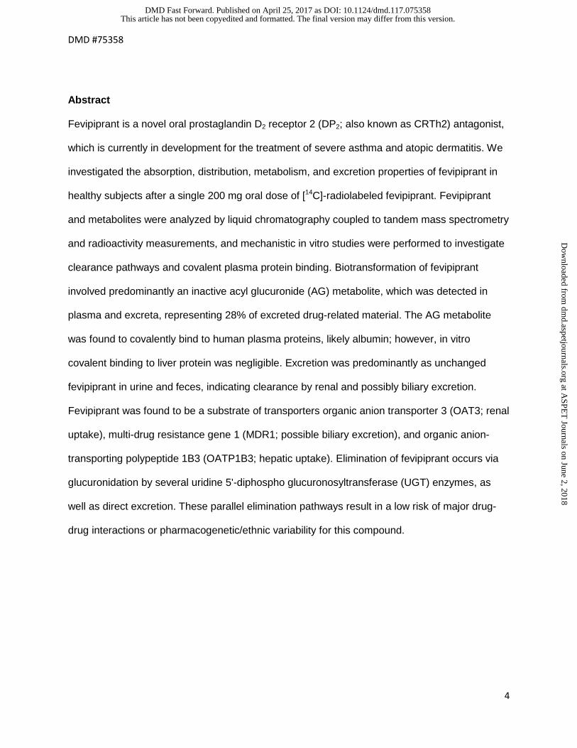
Abstract
Fevipiprant is a novel oral prostaglandin D2 receptor 2 (DP2; also known as CRTh2) antagonist,
which is currently in development for the treatment of severe asthma and atopic dermatitis. We
investigated the absorption, distribution, metabolism, and excretion properties of fevipiprant in
healthy subjects after a single 200 mg oral dose of [14C]-radiolabeled fevipiprant. Fevipiprant
and metabolites were analyzed by liquid chromatography coupled to tandem mass spectrometry
and radioactivity measurements, and mechanistic in vitro studies were performed to investigate
clearance pathways and covalent plasma protein binding. Biotransformation of fevipiprant
involved predominantly an inactive acyl glucuronide (AG) metabolite, which was detected in
plasma and excreta, representing 28% of excreted drug-related material. The AG metabolite
was found to covalently bind to human plasma proteins, likely albumin; however, in vitro
covalent binding to liver protein was negligible. Excretion was predominantly as unchanged
fevipiprant in urine and feces, indicating clearance by renal and possibly biliary excretion.
Fevipiprant was found to be a substrate of transporters organic anion transporter 3 (OAT3; renal
uptake), multi-drug resistance gene 1 (MDR1; possible biliary excretion), and organic anion-
transporting polypeptide 1B3 (OATP1B3; hepatic uptake). Elimination of fevipiprant occurs via
glucuronidation by several uridine 5'-diphospho glucuronosyltransferase (UGT) enzymes, as
well as direct excretion. These parallel elimination pathways result in a low risk of major drug-
drug interactions or pharmacogenetic/ethnic variability for this compound.
This article has not been copyedited and formatted. The final version may differ from this version.DMD Fast Forward. Published on April 25, 2017 as DOI: 10.1124/dmd.117.075358
at ASPE
T Journals on June 2, 2018
dmd.aspetjournals.org
Dow
nloaded from

DMD #75358
5
Introduction
The prevalence of allergic diseases is increasing worldwide, with the World Health Organization
estimating that 400 million people globally will be asthma sufferers by 2025 (Pawankar, 2014).
In the European Union (EU), 44−76 million individuals of the 217 million EU employees suffer
from allergic diseases of the airways or the skin and up to 90% of these individuals are
untreated or insufficiently treated (Zuberbier et al., 2014).
Fevipiprant (QAW039; [(2-[2-methyl-1-(4-[methylsulfonyl]-2-[trifluoromethyl]benzyl)-1H-
pyrrolo(2,3-b)pyridin-3-yl] acetic acid)]) is a potent and highly selective novel antagonist of the
human prostaglandin D2 receptor 2 (DP2, also known as CRTh2), which is a class A G protein–
coupled receptor involved in the modulation of inflammatory responses (Sykes et al., 2016). DP2
is also expressed on innate immune cells, such as eosinophils and ILC-2 cells, and plays a role
in the pathophysiology of respiratory disease (Townley and Agrawal, 2012; Xue et al., 2014).
Recently, human functional studies across diverse cellular systems have shown that fevipiprant
shows high potency for competitive inhibition of disease-relevant DP2-mediated responses in
human cells, such as Th2 cell cytokine production, and eosinophil activation (Sykes et al.,
2016). Fevipiprant is currently in development as a once-daily oral therapy for respiratory and
dermatologic disorders, such as severe asthma and atopic dermatitis (Sykes et al., 2016).
Two Phase 1 studies investigated the pharmacokinetics (PK), safety, and tolerability of
fevipiprant after single and multiple ascending doses in healthy subjects (Erpenbeck et al.,
2016). On administration of single and multiple oral doses, fevipiprant peak plasma
concentrations were observed 1–3 hours post-dose, and the apparent terminal half-life was
approximately 20 hours. Steady state was achieved within four days, with less than two-fold
accumulation. An acyl glucuronide (AG) metabolite without DP2 antagonist activity was detected
in plasma. Fevipiprant was well-tolerated at single and multiple oral doses up to 500 mg/day
(Erpenbeck et al., 2016).
This article has not been copyedited and formatted. The final version may differ from this version.DMD Fast Forward. Published on April 25, 2017 as DOI: 10.1124/dmd.117.075358
at ASPE
T Journals on June 2, 2018
dmd.aspetjournals.org
Dow
nloaded from
DMD #75358
6
With any new chemical entities, it is important to identify and quantify all relevant metabolites
and elimination pathways, for assessment of possible metabolite pharmacology, safety (FDA,
2008; ICH, 2009; Gao et al., 2013), clearance mechanisms and drug-drug interactions (DDIs)
(EMA, 2012; FDA, 2012), including planning of suitable clinical DDI and hepatic/renal
impairment studies.
We report the assessment of human absorption, distribution, metabolism, and excretion (ADME)
of a single oral 200 mg dose of [14C]-fevipiprant in healthy male subjects, as well as
identification of enzymes and transporters involved in the human PK of fevipiprant, and
characterization of covalent binding to plasma proteins in humans.
This article has not been copyedited and formatted. The final version may differ from this version.DMD Fast Forward. Published on April 25, 2017 as DOI: 10.1124/dmd.117.075358
at ASPE
T Journals on June 2, 2018
dmd.aspetjournals.org
Dow
nloaded from

DMD #75358
7
Materials and Methods
The primary objectives of the clinical ADME study were to identify and quantify fevipiprant and
metabolites in plasma and excreta for analysis of absorption, PK, and elimination pathways.
Study Drug
The parent batch of [14C]-radiolabeled fevipiprant was prepared by the Isotope Laboratory (IL),
Drug Metabolism and Pharmacokinetics (DMPK), Novartis, Switzerland. This batch was
adjusted to a final specific radioactivity of 15.7 kBq/mg by dilution with non-radiolabeled
fevipiprant, produced under Good Manufacturing Practice and released for human use by
Technical Research and Development (TRD), Novartis. The chemical structure of the
compound and the position of the radiolabel are shown in Figure 1.
Chemical and radiochemical purity 99.0% (with no single impurity ≥ 1%) was verified by high
performance liquid chromatography (HPLC) and stability was ascertained from the period from
manufacturing to dose administration. The radiolabeled study drug was provided as individually
manufactured doses of four 50 mg (monohydrate; 787.5 kBq) [14C]-fevipiprant capsules per
bottle providing a total radioactive dose of 3.15 MBq (IL and TRD departments, Novartis).
Chemicals and Standards
Radiolabeled and non-radiolabeled fevipiprant, radiolabeled and non-radiolabeled AG
metabolite and non-radiolabeled lactone metabolite were synthesized as described in (Bala et
al., 2005) and (Supplemental Data). All other reagents were of analytical grade and obtained
from commercial sources.
Subjects and Study Design
This was an open-label, single-center, single-arm study to investigate the ADME of a single oral
dose of 200 mg [14C]-fevipiprant in four healthy male subjects. Informed consent was obtained
from each subject in writing before any assessment was performed. The study (registered with
This article has not been copyedited and formatted. The final version may differ from this version.DMD Fast Forward. Published on April 25, 2017 as DOI: 10.1124/dmd.117.075358
at ASPE
T Journals on June 2, 2018
dmd.aspetjournals.org
Dow
nloaded from

DMD #75358
8
EudraCT as 2011-002842-10) was conducted at PRA International, Early Development
Services, Zuidlaren, The Netherlands, from February 17 through March 26, 2012, according to
the ethical principles of the Declaration of Helsinki. The study protocol was reviewed by the
Independent Ethics Committee of Stichting Beordeling Ethiek Biomedisch Onderzoek, Assen,
The Netherlands.
Subjects were male, aged 18 to 55 years, weighed ≥50 kg, with a body mass index (BMI) of 18
to 29 kg/m2, and could communicate well with the Investigator, and understand and comply with
the requirements of the study. The subjects were nonsmokers, had no history of alcoholism or
drug abuse, and did not use any prescription drugs or herbal medication within four weeks prior
to dosing and/or over-the-counter medication or dietary supplements (vitamins included) within
two weeks prior to dosing.
The study consisted of a screening visit (between Day -14 and Day -2), a baseline visit (on Day
-1), a single-dose treatment (on Day 1), 240 hours in-house observation period (Days 1−11),
optional collections and assessments (at Day 14 and Day 21), and a study Completion Visit
(Day 25). Each subject received a single oral [14C]-radiolabeled dose of 200 mg fevipiprant (3.15
MBq, 85 μCi), as four 50 mg capsules, in the morning, after an overnight fast of at least 10
hours. Subjects continued to fast for four hours post-dose (water was allowed after two hours).
The radiation exposure was not deemed to pose any relevant health risk to the subjects. Based
on human PK data (with non-radiolabeled fevipiprant) and animal mass balance data (with [14C]-
fevipiprant), the expected effective radiation dose was estimated to be up to 0.87 mSv, lower
than the recommended limit of 1 mSv/year defined by the International Commission on
Radiological Protection (ICRP, 2007). It was not expected that the clinical and laboratory staff
handling the radioactive materials and samples would be exposed to any radiation and
associated health risk.
This article has not been copyedited and formatted. The final version may differ from this version.DMD Fast Forward. Published on April 25, 2017 as DOI: 10.1124/dmd.117.075358
at ASPE
T Journals on June 2, 2018
dmd.aspetjournals.org
Dow
nloaded from

DMD #75358
9
Safety assessments included the monitoring and recording of all adverse events (AEs), regular
checks of routine blood chemistry, hematology and urine values, electrocardiogram recordings,
measurements of vital signs, and physical examinations.
Sample Collection and Aliquoting
Samples of whole blood, urine, and feces were collected over an 11-day period. Part of each
whole blood sample was processed to plasma. Each urine and plasma sample was divided into
two parts, and one part of each sample was acidified by addition of 1% volume of a 70% lactic
acid solution, in order to stabilize the known AG metabolite. Details of sample collection and
processing are given in the (Supplemental Data).
Quantification of total radioactivity, fevipiprant and metabolites
Total radioactivity in blood, non-acidified plasma, non-acidified urine, and feces was measured
by liquid scintillation counting (LSC). Concentrations of fevipiprant and AG metabolite in all
acidified plasma samples up to 240 hours post-dose and in acidified urine were assessed by a
validated specific liquid chromatography (LC)-mass spectrometry (MS)/MS assay. The AG
metabolite was quantified as the sum of all isomers formed by acyl glucuronide rearrangement.
Fevipiprant and metabolites were analyzed in acidified plasma, acidified urine and feces extract
pools of individual subjects by LC-MS/MS, with offline radioactivity detection. Selected plasma
samples were analyzed by size exclusion chromatography (SEC), with off-line radioactivity
detection. Details of these analytical methods are provided in the (Supplemental Data).
Structural Characterization of Metabolites
The structural characterization of metabolites in plasma and excreta was carried out by MS/MS
analysis after LC separation of analytes (described above).
Single stage and product ion spectra with exact mass measurements were obtained in positive
ion mode. The structures of the metabolites were derived from their product ion mass spectra,
This article has not been copyedited and formatted. The final version may differ from this version.DMD Fast Forward. Published on April 25, 2017 as DOI: 10.1124/dmd.117.075358
at ASPE
T Journals on June 2, 2018
dmd.aspetjournals.org
Dow
nloaded from

DMD #75358
10
the elemental composition determined by exact mass measurement and comparison with
synthetic standards.
Pharmacokinetic Evaluations
The present study evaluated the following PK parameters, which were determined from blood
(total radioactivity), plasma, and urine: Cmax (the maximum [peak] plasma or blood drug
concentration after single administration [amount × volume-1]), Tmax (the time to reach peak or
maximum concentration following drug administration [time]), T½ (the elimination half-life
associated with the terminal slope [lambda z], respectively, of a semi-logarithmic concentration-
time curve [time]), AUC0−t (the area under the concentration-time curve from time zero to t
[amount × time × volume-1]), AUCinf (the area under the concentration-time curve from time zero
to infinity [amount × time × volume-1]), CL/F (the systemic clearance CL of the drug from the
plasma [volume × time-1] divided by the bioavailability F), Vz/F (the apparent volume of
distribution during the terminal [lambda z] phase divided by the bioavailability F [volume]),
Ae0−240 h (amount of drug excreted into the urine from time zero to 240 hours post-dose [% of
dose]), CLr (renal clearance; calculated as Ae0−240 h/AUC0−240 h). PK parameters were calculated
using Phoenix WinNonlin 6.3 with noncompartmental analysis.
In vitro Investigations
Fevipiprant covalent binding assay in microsomes and hepatocytes
[14C]-fevipiprant (5 μM, 4.9 MBq/mg) was incubated with human liver microsomes (0.5 mg/mL)
at 37°C in sodium phosphate buffer (0.1 M, pH 7.4) in the presence of several sets of cofactors
(a) glutathione [GSH, 5 mM]; b) β-nicotinamide adenine dinucleotide phosphate [NADPH, 1
mM]; c) NADPH and uridine 5’- diphosphoglucuronic acid [UDPGA, 4 mM]; d) NADPH, UDPGA
and GSH). Aliquots were taken after 1 hour and precipitated with 3 volumes of acetonitrile.
This article has not been copyedited and formatted. The final version may differ from this version.DMD Fast Forward. Published on April 25, 2017 as DOI: 10.1124/dmd.117.075358
at ASPE
T Journals on June 2, 2018
dmd.aspetjournals.org
Dow
nloaded from

DMD #75358
11
[14C]-fevipiprant (10 μM, 4.9 MBq/mg) was incubated with cryopreserved human hepatocytes
(0.5x10⁶ cells/mL) 37°C, 95% humidity and 5% CO2, in Williams E medium. Aliquots were taken
after 0, 1.5 and 3 hours, and precipitated with 4 volumes of acetonitrile.
The precipitate obtained from each experiment was filtered and washed extensively with 90%
methanol, then the filters were removed and protein precipitate was solubilized in Solvable, then
analyzed by LSC. Covalent protein binding was calculated as pmol/mg protein and pmol/106
cells for microsomes and hepatocytes, respectively.
Incubations of radiolabeled fevipiprant and AG-metabolite with human plasma
[14C]-fevipiprant (4.9 MBq/mg) and [14C]-AG metabolite (3.3 MBq/mg) were each incubated with
1:1 human plasma/PBS at a final concentration of 20 µM at 37°C for 24 hours. Incubation of
[14C]-AG metabolite at 100 µM, and control incubations without plasma were also performed.
During the 20 µM incubations, aliquots were removed, precipitated by addition of 5 volumes of
acetonitrile and incubation at 4°C for 60 minutes, and then filtered. Filters were washed with
methanol/water (90:10) until eluate contained less than 5 pmol/mL radioactivity, as measured by
LSC. Retained radioactivity in the filters was then measured by dissolution of filters in Solvable
(Perkin Elmer) at room temperature for 16 hours, followed by addition of HCl (1M, 0.25 mL),
then scintillant (Irgasafe plus, Zinsser Analytic, 5 mL) was added and radioactivity was
measured by LSC. Retained radioactivity was assumed to be bound to proteins.
Aliquots from 21-hour incubations were further analyzed by sodium dodecyl sulfate
polyacrylamide gel electrophoresis (SDS-PAGE): 5, 10 and 20 μg aliquots were prepared in XT
sample buffer (BioRad) supplemented with 10 mM DTT and heated at 95°C for 5 minutes.
Subsequently, prepared samples were loaded to a CriterionTM XT precast gradient Midi gel 4-
12% Bis Tris (BioRad). The gel was run for 35 minutes at 200 Volt in Mes buffer (BioRad). The
gel was blotted to a PVDF membrane (Transblot Turbo, BioRad) and the resulting membrane
was exposed for 4 days to a radio imaging plate (Fujifilm, BAS-IP TR 2040) to detect the radio-
This article has not been copyedited and formatted. The final version may differ from this version.DMD Fast Forward. Published on April 25, 2017 as DOI: 10.1124/dmd.117.075358
at ASPE
T Journals on June 2, 2018
dmd.aspetjournals.org
Dow
nloaded from
DMD #75358
12
labeled proteins. Molecular weight and intensities of detected bands was measured by labeling
the size marker (all blue standard, BioRad) on the membrane with 3H supplemented ink and the
analysis tool of the AIDA software (v.4.25One®).
Additionally, aliquots from 21-hour incubations were depleted of albumin and analyzed by SDS-
PAGE: 10 μL aliquots of 20 μM incubations were purified using ProteoExtract Albumin Removal
Kit (Calbiochem #122640). Sample aliquots were diluted with albumin Binding Buffer (350 μL)
and purified according to the kit manual. The volumes of each eluate were reduced to 110 μL by
centrifuged at 14000 × g in 30 kDa Amicon spin filters (Millipore). Protein content of the samples
was determined using the Protein DC Assay (BioRad) and a BSA standard in PBS. Samples
were then analyzed by SDS-PAGE, as described above.
SDS-PAGE gels were additionally analyzed by gel excision and protein characterization, as
follows: the radiolabeled band was excised and digested with trypsin, as described in the In-gel
tryptic digestion kit (Thermo Scientific) procedure. The bands were first destained, reduced,
alkylated, and washed, as recommended in the procedure before digestion. Digestions with
trypsin and chymotrypsin were performed in ammonium bicarbonate (500 mM, pH 8) for 24
hours at 37°C. After incubation, incubates were acidified by adding 1% volume of lactic acid
(70% in water). Acidified incubates were analyzed by LC-MS/MS with an Acuity I class system
(Waters). Samples were injected onto an Acuity BEH130 C18 column (150 x 2.1 mm, 1.7 µm
particles, Waters) with a corresponding guard column (10 x 2.1 mm), heated to 30°C.
Separation was performed with a gradient of mobile phases formic acid (0.1%; phase A) and
acetonitrile (phase B) at a flow rate of 0.2 mL/minute. The gradient was as follows: 0 to 2
minutes: 5% B; 2 to 70 minutes: 5 to 50% B; 70 to 75 minutes: 50 to 75% B; 75 to 76 minutes:
75 to 5% B; 76 to 86 minutes: 5% B. The eluate was directed to an LTQ-Orbitrap XL mass
spectrometer (ThermoFisher) for MS/MS analysis. Protein fingerprint analysis was then
performed using ProteinProspector MS-Fit software version 2.
This article has not been copyedited and formatted. The final version may differ from this version.DMD Fast Forward. Published on April 25, 2017 as DOI: 10.1124/dmd.117.075358
at ASPE
T Journals on June 2, 2018
dmd.aspetjournals.org
Dow
nloaded from
DMD #75358
13
Aliquots from the 24-hour 100 µM incubation of the AG-metabolite were analyzed by SEC, as
described for plasma samples (Supplemental Data).
Other in vitro methods
Descriptions of all other in vitro methods are provided in the (Supplemental Data).
Demographics
A total of four male Caucasian subjects were enrolled and all completed the study. Subjects had
a mean age of 43.0 years (range: 27–54), weight of 85.6 kg (range: 77.9–93.5), height of 183.3
cm (range: 176–192), and BMI of 25.5 kg/m2 (range: 23.8–28.3). Only male subjects were
selected in order to perform a detailed analysis in a well-defined uniform group, and as the
study size is insufficient to make statistical comparisons of sub-populations such as genders. No
significant differences in PK or metabolism are known or anticipated between males and
females.
This article has not been copyedited and formatted. The final version may differ from this version.DMD Fast Forward. Published on April 25, 2017 as DOI: 10.1124/dmd.117.075358
at ASPE
T Journals on June 2, 2018
dmd.aspetjournals.org
Dow
nloaded from

DMD #75358
14
Results
Safety and Tolerability Data
Two (50%) of the subjects reported a total of four AEs during the study, which were considered
to be mild in severity. There were no serious AEs or discontinuations due to AEs. Two AEs
(headache and somnolence) were considered to be treatment-related. No clinically significant
abnormalities or changes were observed in clinical laboratory assessments, vital signs, or
electrocardiogram parameters.
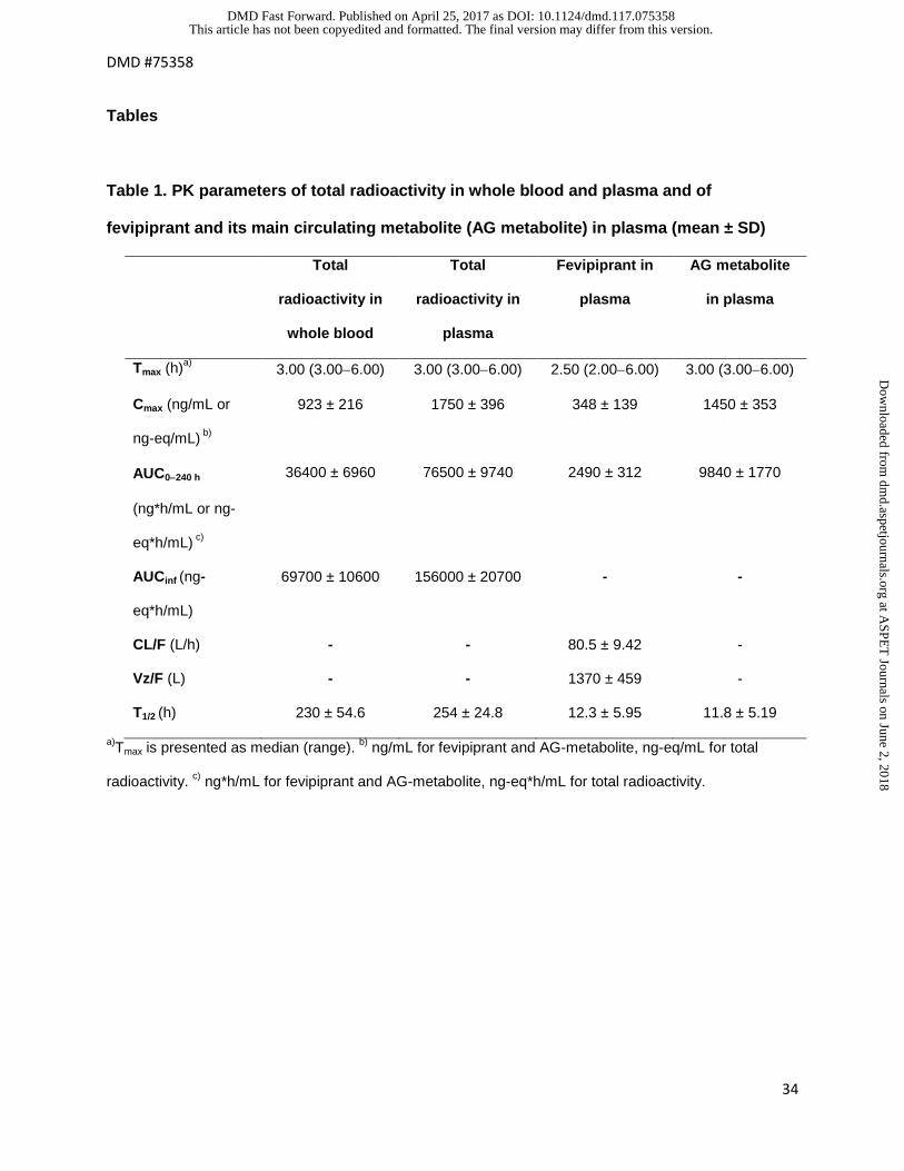
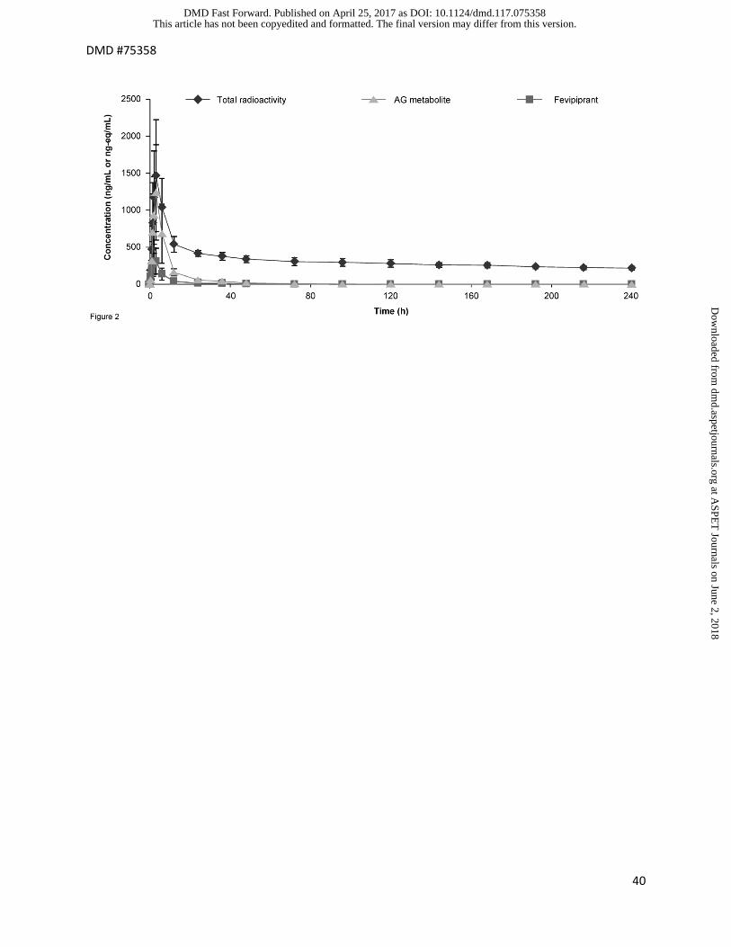
Pharmacokinetics of Total Radioactivity, Fevipiprant, and AG metabolite
Key PK variables calculated for total radioactivity, fevipiprant and AG metabolite are
summarized in Table 1 and Figure 2. Values for AG metabolite represent the sum of AG
metabolite and all isomers formed by acyl glucuronide rearrangement (assigned by comparison
with rearrangement products formed from synthetic AG metabolite).
Following oral administration of [14C]-fevipiprant, levels of radioactivity in blood and plasma, and
levels of fevipiprant and the AG metabolite in plasma, were initially aligned and reached a
maximum at approximately 3 hours post-dose. Radioactivity was detectable in blood and
plasma for up to 240 hours post-dose, with a mean terminal half-life in plasma of 254 hours (230
hours in blood). Contrastingly, fevipiprant and its major metabolite, the AG metabolite, were only
detected in plasma up to 96 and 120 hours post-dose, respectively, with average half-lives of
approximately 12 hours. The total exposure (AUC0−240 h) and Cmax of radioactivity in plasma were
approximately two-fold higher than observed in blood, indicating that blood radioactivity was
almost entirely located in plasma. Inter-subject variability was low to moderate.
Metabolite Identification and Profiles, and Metabolite Pharmacokinetic Analysis
Plasma, urine and feces extracts were analyzed by LC-MS/MS with radioactivity detection for
metabolite identification and profiles. The structures of all identified metabolites could be
This article has not been copyedited and formatted. The final version may differ from this version.DMD Fast Forward. Published on April 25, 2017 as DOI: 10.1124/dmd.117.075358
at ASPE
T Journals on June 2, 2018
dmd.aspetjournals.org
Dow
nloaded from

DMD #75358
15
confirmed by comparison of retention time and mass spectral data with synthesized standards,
and are given in Figure 1. Mass spectral data and representative product ion mass spectra of
fevipiprant and metabolites are shown in Table S1 and Figure S1 (Supplemental Data). The
only abundant metabolite detected was the AG metabolite. A minor lactone metabolite was also
identified, likely resulting from oxidative ring-closure of fevipiprant (see discussion section for
more detail). The structures of minor components P5.3 and P8.5 could not be determined.
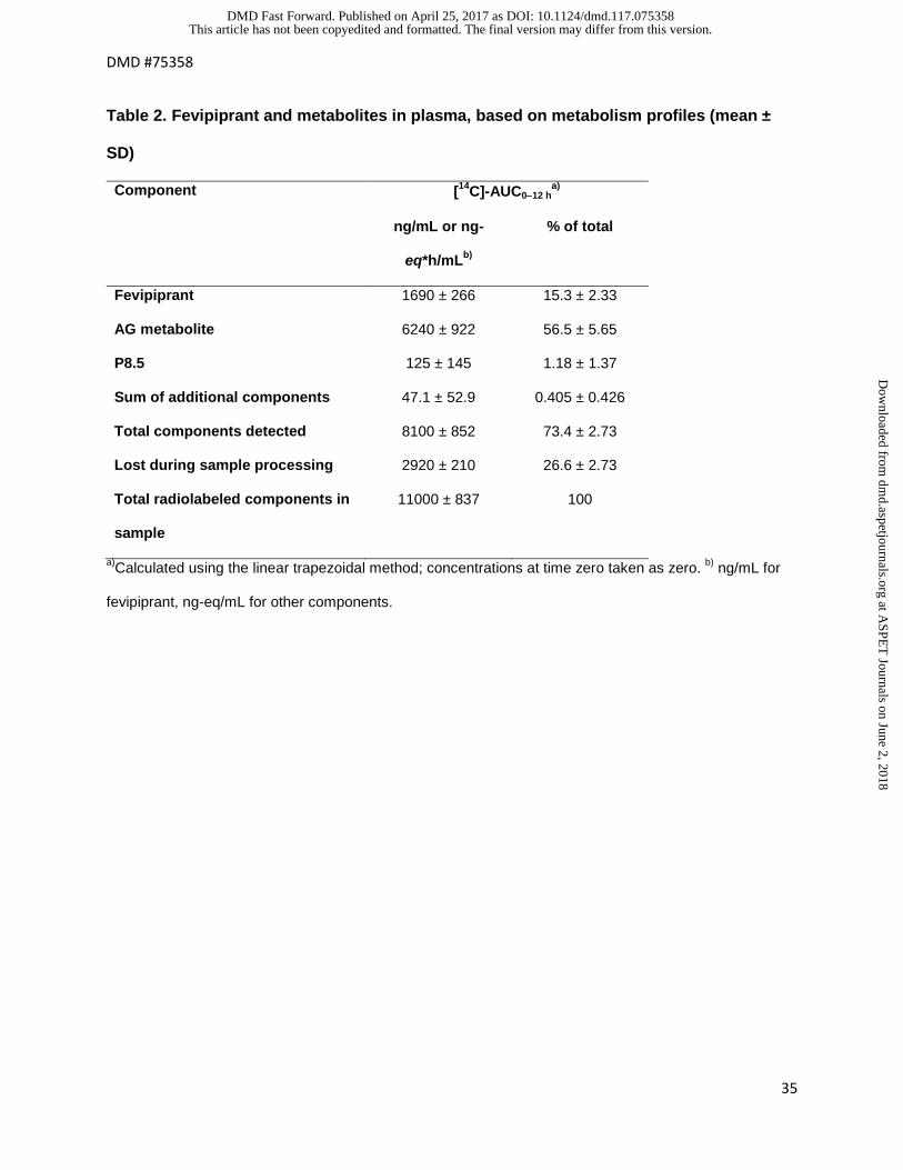
Metabolite profiles were determined after extraction of samples by solid phase extraction. The
extraction recoveries of total radioactivity were found to decrease with time post-dose for all
subjects. The average percentage of total radioactivity recovery at 2 hours was 92.6%, but at 48
hours was only 11.0%. Due to the low extractable radioactivity in plasma samples at later time
points, radiochromatograms were only analyzed up to 12 hours post-dose. Exposure (as
AUC0−12 h) of fevipiprant and its metabolites, based on the radiochromatogram profiles, is shown
in Table 2. A representative plasma radiochromatogram for one subject is shown in Figure 3.
The AG metabolite and fevipiprant represented the main proportion of radioactivity in plasma
(total 72% of [14C]-AUC0−12 h) based on radiochromatograms up to 12 hours. However,
fevipiprant and the AG metabolite accounted for a much lower proportion of the total plasma
[14C]-AUC0−240 h (around 12%) based on LC-MS/MS quantification up to 240 hours (Table 1).
Correspondingly, the loss of radioactivity observed during sample processing represented a
major proportion of radioactivity, amounting to 26.6 ± 2.7% of the plasma [14C]-AUC0−12 h and
estimated at 87.7% of the plasma [14C]-AUC0−240 h based on LC-MS/MS quantification of
fevipiprant and the AG metabolite. To further investigate the loss of radioactivity during sample
processing, additional plasma analysis and in vitro experiments were performed (Covalent
Binding and Acyl glucuronide Stability Investigations section).
Excretion of radiolabeled components
This article has not been copyedited and formatted. The final version may differ from this version.DMD Fast Forward. Published on April 25, 2017 as DOI: 10.1124/dmd.117.075358
at ASPE
T Journals on June 2, 2018
dmd.aspetjournals.org
Dow
nloaded from
DMD #75358
16
After oral administration of [14C]-fevipiprant, 42.1 ± 4.3% of the dose was recovered in urine and
51.9 ± 4.8% was recovered in feces over 240 hours post-dose (Table S2 (Supplemental Data);
Figure 4), indicating that radioactive drug-related material was primarily excreted via renal and
biliary/fecal excretion. The overall recovery of drug-related material was near complete (93.9 ±
2.6%). The majority of the radioactivity was recovered within 144 hours post-dose, with
subsequent samples up to 240 hours containing less than 1% of the dose.
Metabolite profiles were also determined in urine and feces pools (Figure 3). Extraction
recovery was high for all excreta samples (>88%). Urinary and feces excretion data for
fevipiprant and all metabolites based on metabolite profiles, and urinary excretion data for
fevipiprant and the AG metabolite based on LC-MS/MS quantification over 240 hours, are
presented in Table 3. In urine, fevipiprant and AG metabolite were the major components (total
approximately 40% of dose). Urinary excretion data for fevipiprant and the AG metabolite over
240 hours were similar to the 0−72 hours values (Table 3). In feces, fevipiprant was the major
component excreted. Overall, fevipiprant and AG metabolites represented approximately 85% of
the dose excreted in urine and feces, and other metabolites were only present in traces.
Based on urinary excretion data for the period 0−240 hours (Table 3), the renal clearance of
fevipiprant and AG metabolite were calculated to be 8.92 L/h and 7.99 L/h, respectively.
Absorption was estimated to be at least 43.5%, based on the total radioactive dose recovered
from urine (42.1%), and the metabolites excreted in feces (approximately 1.4%).
Covalent Binding and Acyl glucuronide Stability Investigations
Due to the observed long retention of radioactivity in plasma and low extractability of plasma
samples, we hypothesized that the AG metabolite could covalently bind to plasma proteins
(Regan et al., 2010), leading to long-lived radioactive metabolite-protein conjugates, as
This article has not been copyedited and formatted. The final version may differ from this version.DMD Fast Forward. Published on April 25, 2017 as DOI: 10.1124/dmd.117.075358
at ASPE
T Journals on June 2, 2018
dmd.aspetjournals.org
Dow
nloaded from
DMD #75358
17
observed for other compounds, such as ibuprofen (Castillo et al., 1995), tolmetin (Zia-
Amirhosseini et al., 1994), and bilirubin (Weiss et al., 1983). Additional in vitro experiments were
performed to investigate covalent binding. Initially, [14C]-fevipiprant was incubated with human
liver microsomes (HLM) and human hepatocytes to investigate possible covalent binding of
fevipiprant or metabolites to liver proteins mainly via oxidative processes. Additionally,
incubations of [14C]-fevipiprant and [14C]-AG metabolite in human plasma were performed,
followed by precipitation and LSC of proteins to identify whether radiolabeled protein conjugates
were formed.
When probing for covalent binding of fevipiprant or metabolites to human liver microsomes and
hepatocytes in the presence of various cofactors (Table 4) only trace levels of covalent binding
were observed in comparison to positive control compounds. Additionally, no increase in
covalent binding was observed with the addition of glucuronidation cofactor UDPGA, suggesting
that covalent binding to liver microsomes associated with the AG metabolite is insignificant.
In incubations with plasma, fevipiprant showed very low covalent binding to human plasma
proteins, while the AG metabolite showed 11-fold higher covalent binding after 24 hours
incubation (Figure S2 (Supplemental Data)), indicating that covalent binding in plasma occurs
via the AG metabolite rather than the parent compound.
Subsequently, to identify proteins involved in plasma covalent binding gel electrophoresis of the
AG metabolite plasma incubate was carried out. Radio-imaging of the resulting gel (Figure 5A,
lanes A1−3) showed that only one protein of around 68 kDa size was conjugated to the AG
metabolite. Peptide fingerprint analysis of the labeled protein band by LC-MS after gel excision
and trypsin/chymotrypsin digest identified that the abundant plasma protein human serum
albumin was present. Based on this result, gel electrophoresis of AG metabolite plasma
incubates after depletion of albumin was also performed. The intensity of the radio-signal
This article has not been copyedited and formatted. The final version may differ from this version.DMD Fast Forward. Published on April 25, 2017 as DOI: 10.1124/dmd.117.075358
at ASPE
T Journals on June 2, 2018
dmd.aspetjournals.org
Dow
nloaded from
DMD #75358
18
detected was significantly reduced by albumin depletion of the respective samples (Figure 5A,
lanes B1−3). These data show that the AG metabolite forms a conjugate with albumin in vitro.
To evaluate whether this in vitro finding corresponds to the in vivo observations, size exclusion
chromatography (SEC) of the AG metabolite plasma incubates and of the clinical plasma
samples was performed (gel electrophoresis could not be performed on the in vivo samples,
due to insufficient sensitivity). SEC of the AG metabolite in vitro incubation showed the
presence of fevipiprant, AG metabolite, and an earlier eluting peak assigned to a protein
conjugate (Figure 5B). Analysis of in vivo human plasma samples showed similar profiles
(representative samples are shown in Figure 5C), with an increase of the protein conjugate
peak over time post-dose. These data provide evidence that the in vivo plasma covalent binding
also involves albumin.
The recoveries of radioactivity for all plasma samples analyzed by SEC were between 90 and
110%, confirming that the low extraction recoveries for clinical plasma samples were explained
by loss of drug related material covalently bound to plasma protein. Correspondingly, the
amount of protein-conjugate detected by SEC at each time point was similar to the amount of
radioactivity lost on extraction.
Acyl glucuronides are often unstable at neutral pH (Bailey and Dickinson, 2003) but can be
stabilized by acidification of clinical samples (Ebner et al., 2010; Wang et al., 2011). As this
instability was observed for the AG metabolite in pre-clinical experiments, an aliquot of all
plasma and urine samples for analysis of fevipiprant and metabolites was acidified by addition
of 1% volume of a 70% lactic acid solution. Experiments in blank urine samples showed that a
pH in the range of 3.1−5.2 was obtained after acidification (compared with 5.3−7.3 before
acidification), and the AG metabolite was confirmed to be stable to hydrolysis for four weeks at
This article has not been copyedited and formatted. The final version may differ from this version.DMD Fast Forward. Published on April 25, 2017 as DOI: 10.1124/dmd.117.075358
at ASPE
T Journals on June 2, 2018
dmd.aspetjournals.org
Dow
nloaded from
DMD #75358
19
room temperature in acidified urine (less than 1% degradation to the parent compound
measured). However, isomerization of the acyl-glucuronide was not measured here, and may
have occurred during the incubation. All clinical data reported here were obtained using the
acidified aliquots, except for total radioactivity measurements.
To assess potential acyl-glucuronide isomerization, incubations were performed with a
synthesized standard of the 1-O-beta isomer of the AG metabolite to identify other acyl
glucuronide isomers formed. Several isomers of the AG metabolite were detected, and were
assigned to acyl glucuronide rearrangement products (Bailey and Dickinson, 2003). As these
isomers were considered to be degradation products of the AG metabolite that could not be
accurately quantified if formed during sample collection or processing, they were quantified
together with the AG metabolite for all analyses in this report.
Fevipiprant as a Substrate of Enzymes and Transporters
Based on the clinical excretion data, fevipiprant clearance pathways include glucuronidation and
direct excretion of the parent drug. To determine which enzymes and transporters are involved
in these processes, a number of in vitro investigations were carried out, as follows:
For the determination of the enzymes catalyzing the formation of the AG metabolite, in vitro
biotransformation of [14C]-fevipiprant, catalyzed by 12 recombinant uridine 5'-diphospho-
glucuronosyltransferase (UDP-glucuronosyltransferase; UGT) enzymes, was investigated.
Fevipiprant was metabolized by a number of the enzymes (Table S3 (Supplemental Data)), with
UDP-glucuronosyltransferase 1A3 (UGT1A3), UDP-glucuronosyltransferase 2B7 (UGT2B7),
and UDP-glucuronosyltransferase 2B17 (UGT2B17) showing the highest activities. Michaelis-
Menten enzyme kinetic parameters are shown in Table 5 and the kinetic profiles are presented
in Figures S3−S5 (Supplemental Data).
This article has not been copyedited and formatted. The final version may differ from this version.DMD Fast Forward. Published on April 25, 2017 as DOI: 10.1124/dmd.117.075358
at ASPE
T Journals on June 2, 2018
dmd.aspetjournals.org
Dow
nloaded from
DMD #75358
20
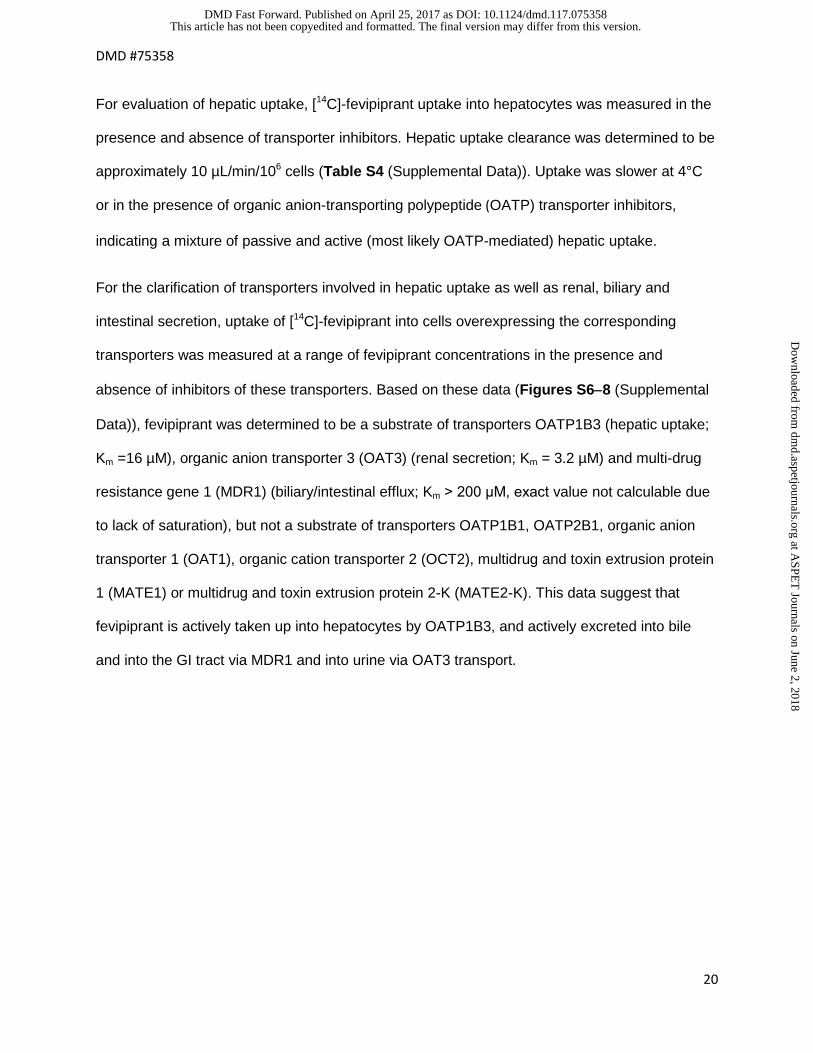
For evaluation of hepatic uptake, [14C]-fevipiprant uptake into hepatocytes was measured in the
presence and absence of transporter inhibitors. Hepatic uptake clearance was determined to be
approximately 10 µL/min/106 cells (Table S4 (Supplemental Data)). Uptake was slower at 4°C
or in the presence of organic anion-transporting polypeptide (OATP) transporter inhibitors,
indicating a mixture of passive and active (most likely OATP-mediated) hepatic uptake.
For the clarification of transporters involved in hepatic uptake as well as renal, biliary and
intestinal secretion, uptake of [14C]-fevipiprant into cells overexpressing the corresponding
transporters was measured at a range of fevipiprant concentrations in the presence and
absence of inhibitors of these transporters. Based on these data (Figures S6−8 (Supplemental
Data)), fevipiprant was determined to be a substrate of transporters OATP1B3 (hepatic uptake;
Km =16 µM), organic anion transporter 3 (OAT3) (renal secretion; Km = 3.2 µM) and multi-drug
resistance gene 1 (MDR1) (biliary/intestinal efflux; Km > 200 μM, exact value not calculable due
to lack of saturation), but not a substrate of transporters OATP1B1, OATP2B1, organic anion
transporter 1 (OAT1), organic cation transporter 2 (OCT2), multidrug and toxin extrusion protein
1 (MATE1) or multidrug and toxin extrusion protein 2-K (MATE2-K). This data suggest that
fevipiprant is actively taken up into hepatocytes by OATP1B3, and actively excreted into bile
and into the GI tract via MDR1 and into urine via OAT3 transport.
This article has not been copyedited and formatted. The final version may differ from this version.DMD Fast Forward. Published on April 25, 2017 as DOI: 10.1124/dmd.117.075358
at ASPE
T Journals on June 2, 2018
dmd.aspetjournals.org
Dow
nloaded from

DMD #75358
21
Discussion
In the present study, the absorption of fevipiprant was estimated to be at least 43.5%, as a
minimum estimate based on metabolite excretion and renal fevipiprant excretion. It is likely that
the actual absorption value is higher, as AG metabolites would likely hydrolyze to fevipiprant in
the gastrointestinal tract after biliary excretion, and in addition direct biliary excretion of
fevipiprant is possible. Preclinical data from rat support this hypothesis, with absorption of
around 60%, and bile-duct cannulated rat studies after intravenous dosing showing excretion of
both fevipiprant (25% of dose) and AG-metabolite (33% of dose) in bile.
The AG metabolite was the only major metabolite detected in plasma. In preclinical species, the
AG metabolite was also a major metabolite in plasma and/or excreta, but comprised a lower
proportion of total plasma AUC (<30%). However, in toxicity studies exposure to the AG
metabolite at the NOAEL was similar or higher than human exposure at the highest phase III
dose. The abundance of the AG metabolite was not well predicted from in vitro data. While the
AG metabolite was formed in hepatocytes of all species investigated (mouse, rat, dog, monkey,
human), the calculated intrinsic clearance was in all cases low (e.g. 0.35 µL/min/106 cells in
human). It is possible that the in vitro underestimation of metabolic clearance is due to reduced
activity of OATP1B3-mediated uptake in cryopreserved hepatocyte suspensions in vitro, in
comparison to the in vivo situation where transporters are fully active (Lundquist et al., 2014).
Aside from the AG metabolite, we detected a minor lactone metabolite. Due to its low
abundance, the formation of this metabolite was not investigated in more detail. However,
several possible formation pathways can be envisaged (see Figure S9 (Supplemental Data))
involving epoxidation, followed by either nucleophilic ring-closing or epoxide hydrolysis and
lactonization. It is unlikely that this possible epoxide intermediate is involved in the formation of
This article has not been copyedited and formatted. The final version may differ from this version.DMD Fast Forward. Published on April 25, 2017 as DOI: 10.1124/dmd.117.075358
at ASPE
T Journals on June 2, 2018
dmd.aspetjournals.org
Dow
nloaded from

DMD #75358
22
the observed covalent adduct in plasma, as oxidative covalent binding in microsomes and
hepatocytes was negligible (Table 4), and relevant reactive metabolites were not detected in
plasma.
Total radioactivity was found to decline slowly in blood and plasma after the elimination of the
majority of fevipiprant from plasma, likely due to the formation of protein conjugates by covalent
binding of the AG metabolite to human serum albumin. The observed terminal half-life of
radioactivity in plasma (254 h or 10.6 days) was in the range of, but shorter than, the half-life
reported for human albumin (19 days (Peters T., 1995)). Consequently, the terminal half-life of
total radioactivity, although calculated using data covering a relatively short time period, is likely
to be accurate or somewhat underestimated. Preclinical data in rat and dog show much shorter
terminal half-life values for total radioactivity, potentially due to a shorter half-life of albumin in
these species (e.g. 2.5 days in rat (Car et al., 2006)). The concentration of radioactivity
remaining in plasma after 240 h (500 pmol/mL) corresponds to around 1.5 µmol or 0.65 mg
fevipiprant equivalent, assuming 3 L human plasma volume. Assuming that approximately 60%
of albumin is distributed to extracellular space (Peters T., 1995), the amount of drug-albumin
conjugate remaining in the body at 240 hours post-dose is therefore approximately 1.6 mg
corresponding to 0.8% of dose (or 0.3% assuming no extra-vascular distribution of the protein
conjugate). Given total body albumin of around 360 g (Peters T., 1995), the extent of covalent
binding is then around 0.07% of total body albumin. Consequently, repeated dosing with
fevipiprant is unlikely to lead to the covalent modification of a large proportion of albumin in the
body.
The stability of acyl glucuronides has been previously investigated in detail (Regan et al., 2010).
Key factors proposed to be associated with AG stability are steric accessibility and electronic
properties of the carboxylic acid/ester functionality. Fevipiprant contains a sterically unhindered
carboxylic acid as part of an aryl-acetic acid moiety, similar to diclofenac and tolmetin, which
This article has not been copyedited and formatted. The final version may differ from this version.DMD Fast Forward. Published on April 25, 2017 as DOI: 10.1124/dmd.117.075358
at ASPE
T Journals on June 2, 2018
dmd.aspetjournals.org
Dow
nloaded from

DMD #75358
23
also form unstable AG-metabolites (Zia-Amirhosseini et al., 1994; Castillo et al., 1995). It is
therefore unsurprising that the AG-metabolite of fevipiprant is also unstable.
Acyl glucuronide-derived drug-protein conjugates have been observed for other drugs such as
ibuprofen and tolmetin, as well as bilirubin (Weiss et al., 1983; Zia-Amirhosseini et al., 1994;
Castillo et al., 1995), which has been reviewed extensively (Regan et al., 2010). A number of
AG-forming drugs have been associated with clinical AEs, such as hepatotoxicity. However, no
clear causal link of AEs to the reactivity or covalent protein binding of the AGs has been
established. In comparison with other covalent binding compounds (Usui et al., 2009) including
compounds that form AG metabolites (Darnell et al., 2015), the in vitro covalent binding of
fevipiprant to liver microsomes and hepatocytes (Table 4) is negligible. Additionally, no covalent
binding to liver microsomes associated with glucuronidation was observed, suggesting that the
AG-metabolite does not rapidly bind to liver proteins after its formation. From this it can be
inferred that covalent binding to albumin occurs in plasma rather than in the liver, likely due to
the high concentrations of both AG metabolite and albumin in plasma.
As of January 2017, over 1700 subjects have been exposed to fevipiprant in the clinical
program. Phase 3 trials in asthma are currently ongoing. There have been no AEs of idiopathic
drug reactions or liver toxicity in these clinical studies. Overall, the in vivo nonclinical
assessments and clinical safety data available to date indicate a low risk of idiosyncratic drug-
induced liver injury or other idiosyncratic drug reactions potentially associated with covalent
drug-protein binding, in line with the apparently negligible reactive metabolites formed by
oxidative pathways.
Excretion and metabolism data from this study indicate that fevipiprant is eliminated by several
pathways. Following oral administration, 57% of the dose was detected as unchanged
fevipiprant in urine and feces, indicating direct renal and possibly biliary excretion. In addition,
29% of the dose was excreted as metabolites. The exact amounts of dose eliminated by
This article has not been copyedited and formatted. The final version may differ from this version.DMD Fast Forward. Published on April 25, 2017 as DOI: 10.1124/dmd.117.075358
at ASPE
T Journals on June 2, 2018
dmd.aspetjournals.org
Dow
nloaded from
DMD #75358
24
metabolism and direct excretion are not clear, as back-conversion of the AG metabolite to
fevipiprant during/after excretion is possible. Data from preclinical species (mouse, rat and dog)
showed similar excretion routes, except with relatively higher fecal excretion. In mouse, rat and
dog, fecal/biliary excretion was around 80% of dose (after both intravenous and oral
administration) and renal excretion was 1% in mouse, 6-7% in rat and 11-13% in dog. Urinary
excretion in rat and dog consisted mainly of the parent drug, suggesting that the higher overall
renal excretion in human is due to a fast renal (rather than biliary) excretion of the AG
metabolite, or due to a more extensive formation of AG metabolite in human, in line with the
high plasma concentrations of the AG metabolite.
We demonstrated in vitro that fevipiprant was a substrate of several human UDP-
glucuronosyltransferases, as well as transporters involved in tubular secretion in the kidney
(OAT3), active hepatic uptake (OATP1B3), and biliary excretion (MDR1). The OAT3 data show
that the elimination of fevipiprant involves direct renal secretion. This is in line with the
measured renal clearance value (~9 L/h), which is higher than the expected glomerular filtration
clearance (GFR) (0.9 L/h, based on a GFR of 7.5 L/h (Davies and Morris, 1993) multiplied by
the fraction unbound (fu) of fevipiprant (0.118)), as expected in the case of active renal
secretion. As fevipiprant is a substrate of MDR1, biliary or intestinal secretion is also likely: this
would contribute to the large amount of unchanged fevipiprant excreted in feces. The UGT
phenotyping data show that at least three hepatic and extrahepatic (Tukey and Strassburg,
2000) UGT isoenzymes are involved in the metabolic clearance pathways of fevipiprant. Based
on the range of elimination pathways and isoenzymes involved in the clearance of fevipiprant, a
low risk of DDI or variability due to genetic differences is expected. However, in order to address
possible effects of the inhibition of these pathways on the PK of fevipiprant, clinical DDI studies
are planned or ongoing with inhibitors of OAT3, UGT enzymes, OATP1B3 and MDR1.
This article has not been copyedited and formatted. The final version may differ from this version.DMD Fast Forward. Published on April 25, 2017 as DOI: 10.1124/dmd.117.075358
at ASPE
T Journals on June 2, 2018
dmd.aspetjournals.org
Dow
nloaded from

DMD #75358
25
Additionally, the potential of fevipiprant and its AG-metabolite to inhibit these or other enzymes
and transporters involved in drug disposition has been assessed (Barve et al., 2016).
For the AG metabolite, the renal clearance was similar to fevipiprant (~8 L/h) and the expected
clearance by glomerular filtration is 1.76 L/h (0.234 [fu] × 125 mL/min, as described above for
fevipiprant). As the expected glomerular filtration clearance is five-fold lower than the observed
renal clearance, an active tubular secretion of the AG metabolite is also likely.
In conclusion, we demonstrated that fevipiprant is eliminated via various metabolic enzymes
and direct excretion and hence this novel compound is unlikely to be a victim of a strong drug
interaction or to display major variability or ethnic sensitivity in PK due to genetic polymorphism.
The major metabolite is an acyl glucuronide which forms covalent adducts to albumin in human
plasma.
This article has not been copyedited and formatted. The final version may differ from this version.DMD Fast Forward. Published on April 25, 2017 as DOI: 10.1124/dmd.117.075358
at ASPE
T Journals on June 2, 2018
dmd.aspetjournals.org
Dow
nloaded from

DMD #75358
26
Acknowledgements
We acknowledge Maxime Garnier, Stephan Utzinger, Claire Adcock, Luu Van Tong, Yves Metz,
Matthias Frommherz, Patrick Bross and Hubert Borell for technical assistance and Karine
Litherland, David Sandham, Matthias Kittelmann, Ines Rodriguez, Carsten Bauer, Matthew
Brown, Albrecht Glaenzel, Walid Elbast, Jagruti Desai, Paul Goldsmith, Ping Zhou and Piet
Swart for contribution to study design and execution. Delia Randall provided medical writing and
editing support.
This article has not been copyedited and formatted. The final version may differ from this version.DMD Fast Forward. Published on April 25, 2017 as DOI: 10.1124/dmd.117.075358
at ASPE
T Journals on June 2, 2018
dmd.aspetjournals.org
Dow
nloaded from

DMD #75358
27
Authorship Contributions
Participated in research design: Jin, van Lier, Erpenbeck, End, Glaenzel, Woessner
Conducted experiments: van Lier
Contributed new reagents or analytic tools: Eggimann
Performed data analysis: Pearson, Weiss, Jin, End, Glaenzel
Wrote or contributed to the writing of the manuscript: Pearson, Weiss, Jin, Erpenbeck, Glaenzel,
End, Eggimann, Camenisch
This article has not been copyedited and formatted. The final version may differ from this version.DMD Fast Forward. Published on April 25, 2017 as DOI: 10.1124/dmd.117.075358
at ASPE
T Journals on June 2, 2018
dmd.aspetjournals.org
Dow
nloaded from

DMD #75358
28
References
Bailey MJ and Dickinson RG (2003) Acyl glucuronide reactivity in perspective: biological consequences. Chem Biol Interact 145:117-137. Bala K, Leblanc C, Sandham DA, Turner KL, Watson SJ, Brown LN, and Cox B (2005) Pyrrolopyridines as CRTh2 receptor antagonists, their preparation, pharmaceutical compositions, and use in therapy, WO2005123731A2. Barve A, Tillmann H-C, Ilsley E, Vemula J, Nica A, Imbert G, Elbast W, Schiller H, Camenisch G, and Woessner R (2016) Impact of co-administration of fevipiprant (QAW039) and SLCO1B1 genotype on the PK of simvastatin and rosuvastatin. European Respiratory Journal 48:1108. Car BD, Eng VM, Everds NE, and Bounous DI (2006) Chapter 5 - Clinical Pathology of the Rat A2 - Suckow, Mark A, in: The Laboratory Rat (Second Edition) (Weisbroth SH and Franklin CL eds), pp 127-146, Academic Press, Burlington. Castillo M, Lam YW, Dooley MA, Stahl E, and Smith PC (1995) Disposition and covalent binding of ibuprofen and its acyl glucuronide in the elderly. Clin Pharmacol Ther 57:636-644. Darnell M, Breitholtz K, Isin EM, Jurva U, and Weidolf L (2015) Significantly Different Covalent Binding of Oxidative Metabolites, Acyl Glucuronides, and S-Acyl CoA Conjugates Formed from Xenobiotic Carboxylic Acids in Human Liver Microsomes. Chem Res Toxicol 28:886-896. Davies B and Morris T (1993) Physiological parameters in laboratory animals and humans. Pharm Res 10:1093-1095. Ebner T, Wagner K, and Wienen W (2010) Dabigatran acylglucuronide, the major human metabolite of dabigatran: in vitro formation, stability, and pharmacological activity. Drug Metab Dispos 38:1567-1575. EMA (2012) Guideline on the investigation of drug interactions. Erpenbeck VJ, Vets E, Gheyle L, Osuntokun W, Larbig M, Neelakantham S, Sandham D, Dubois G, Elbast W, Goldsmith P, and Weiss M (2016) Pharmacokinetics, Safety, and Tolerability of Fevipiprant (QAW039), a Novel CRTh2 Receptor Antagonist: Results From 2 Randomized, Phase 1, Placebo-Controlled Studies in Healthy Volunteers. Clin Pharmacol Drug Dev 5:306-313. Evans DC, Watt AP, Nicoll-Griffith DA, and Baillie TA (2004) Drug-protein adducts: an industry perspective on minimizing the potential for drug bioactivation in drug discovery and development. Chem Res Toxicol 17:3-16. FDA (2008) Guidance for industry: safety testing of drug metabolites. FDA (2012) Drug Interaction Studies — Study Design, Data Analysis, Implications for Dosing, and Labeling Recommendations.
This article has not been copyedited and formatted. The final version may differ from this version.DMD Fast Forward. Published on April 25, 2017 as DOI: 10.1124/dmd.117.075358
at ASPE
T Journals on June 2, 2018
dmd.aspetjournals.org
Dow
nloaded from

DMD #75358
29
Gao H, Jacobs A, White RE, Booth BP, and Obach RS (2013) Meeting report: metabolites in safety testing (MIST) symposium-safety assessment of human metabolites: what's REALLY necessary to ascertain exposure coverage in safety tests? AAPS J 15:970-973. ICH (2009) Guidance on Nonclinical Safety Studies for the Conduct of Human Clinical Trials and Marketing Authorization for Pharmaceuticals M3 (R2). ICRP (2007) The 2007 Recommendations of the International Commission on Radiological Protection, in: Ann ICRP Lundquist P, Loof J, Sohlenius-Sternbeck AK, Floby E, Johansson J, Bylund J, Hoogstraate J, Afzelius L, and Andersson TB (2014) The impact of solute carrier (SLC) drug uptake transporter loss in human and rat cryopreserved hepatocytes on clearance predictions. Drug Metab Dispos 42:469-480. Pawankar R (2014) Allergic diseases and asthma: a global public health concern and a call to action. World Allergy Organ J 7:12. Peters T. J (1995) Metabolism: Albumin in the Body, in: All about albumin, pp 188-250, Academic Press. Regan SL, Maggs JL, Hammond TG, Lambert C, Williams DP, and Park BK (2010) Acyl glucuronides: the good, the bad and the ugly. Biopharm Drug Dispos 31:367-395. Sykes DA, Bradley ME, Riddy DM, Willard E, Reilly J, Miah A, Bauer C, Watson SJ, Sandham DA, Dubois G, and Charlton SJ (2016) Fevipiprant (QAW039), a Slowly Dissociating CRTh2 Antagonist with the Potential for Improved Clinical Efficacy. Mol Pharmacol 89:593-605. Townley RG and Agrawal S (2012) CRTH2 antagonists in the treatment of allergic responses involving TH2 cells, basophils, and eosinophils. Ann Allergy Asthma Immunol 109:365-374. Tukey RH and Strassburg CP (2000) Human UDP-glucuronosyltransferases: metabolism, expression, and disease. Annu Rev Pharmacol Toxicol 40:581-616. Usui T, Mise M, Hashizume T, Yabuki M, and Komuro S (2009) Evaluation of the potential for drug-induced liver injury based on in vitro covalent binding to human liver proteins. Drug Metab Dispos 37:2383-2392. Wang L, Munsick C, Chen S, Bonacorsi S, Cheng PT, Humphreys WG, and Zhang D (2011) Metabolism and disposition of 14C-labeled peliglitazar in humans. Drug Metab Dispos 39:228-238. Weiss JS, Gautam A, Lauff JJ, Sundberg MW, Jatlow P, Boyer JL, and Seligson D (1983) The clinical importance of a protein-bound fraction of serum bilirubin in patients with hyperbilirubinemia. N Engl J Med 309:147-150. Xue L, Salimi M, Panse I, Mjosberg JM, McKenzie AN, Spits H, Klenerman P, and Ogg G (2014) Prostaglandin D2 activates group 2 innate lymphoid cells through chemoattractant receptor-homologous molecule expressed on TH2 cells. J Allergy Clin Immunol 133:1184-1194.
This article has not been copyedited and formatted. The final version may differ from this version.DMD Fast Forward. Published on April 25, 2017 as DOI: 10.1124/dmd.117.075358
at ASPE
T Journals on June 2, 2018
dmd.aspetjournals.org
Dow
nloaded from

DMD #75358
30
Zia-Amirhosseini P, Ojingwa JC, Spahn-Langguth H, McDonagh AF, and Benet LZ (1994) Enhanced covalent binding of tolmetin to proteins in humans after multiple dosing. Clin Pharmacol Ther 55:21-27. Zuberbier T, Lotvall J, Simoens S, Subramanian SV, and Church MK (2014) Economic burden of inadequate management of allergic diseases in the European Union: a GA(2) LEN review. Allergy 69:1275-1279.
This article has not been copyedited and formatted. The final version may differ from this version.DMD Fast Forward. Published on April 25, 2017 as DOI: 10.1124/dmd.117.075358
at ASPE
T Journals on June 2, 2018
dmd.aspetjournals.org
Dow
nloaded from
DMD #75358
31
Footnotes
Parts of this work were previously presented at the following conference: Pearson D, Jin Y,
Erpenbeck VE, Woessner R, Camenisch G, Weiss HM. Absorption, metabolism and excretion
of fevipiprant (QAW039) investigated in vivo and in vitro. European Respiratory Society
International Congress, 3–7 September, 2016, London, United Kingdom
Address correspondence to: Dr. David Pearson, PK Sciences, Novartis Institutes for Biomedical
Research, Postfach, CH-4002 Basel, Switzerland; Tel: +41799112654; Email:
DP, MW, YJ, VE, UG, PE, RW, FE and GC are employees of Novartis Pharma and may hold
shares in Novartis.
This article has not been copyedited and formatted. The final version may differ from this version.DMD Fast Forward. Published on April 25, 2017 as DOI: 10.1124/dmd.117.075358
at ASPE
T Journals on June 2, 2018
dmd.aspetjournals.org
Dow
nloaded from
DMD #75358
32
Legends for Figures
Figure 1. Structural formula of fevipiprant and proposed biotransformation pathways of
fevipiprant in humans. *Position of [14C]-radiolabel
Figure 2. Plasma concentrations of fevipiprant, AG-metabolite and total radioactivity after oral
administration of a 200 mg dose of [14C]-fevipiprant to four healthy subjects. Units for fevipiprant
and AG-metabolite concentrations are ng/mL, for total radioactivity concentrations ng-eq/mL.
Concentrations of fevipiprant an AG metabolite were determined by LC-MS/MS, concentrations
of total radioactivity were determined by LSC.
Figure 3. Representative radiochromatograms in plasma (3 h post dose), urine (time pooled 0-
72 h), and feces extracts (time pooled 0-96 h) for one subject after oral administration of a 200
mg dose of [14C]-fevipiprant. dpm: disintegrations per minute
Figure 4. Cumulative excretion of radioactivity in urine and feces after oral administration of a
200 mg dose of [14C]-fevipiprant to four healthy subjects. Radioactivity was determined by LSC.
Figure 5.
A) SDS-PAGE analysis of covalent protein binding after 24 h incubation of [14C]-AG-metabolite
(20 µM) with 1:1 human plasma/PBS at 37 °C. Lanes A1-3: untreated incubation sample; Lanes
B1-3: incubation sample after albumin depletion using a ProteoExtract Albumin Removal Kit
(Calbiochem). 5, 10, and 20 µg of protein was analysed in lanes A1/B1, A2/B2 and A3/B3,
respectively. Protein content was determined using a Protein DC Assay (BioRad). Relative
intensities of bands calculated using AIDA software: A1, 5878; A2, 9787; A3, 17839; B1, 2791;
B2, 6435; B3, 10635. The molecular weight markers on the left side of the gel were drawn with
3H-supplemented ink to mark the location of visible marker proteins (all blue standard, BioRad).
B) SEC analysis after 24 h incubation of [14C]-AG-metabolite (100 µM) with 1:1 human
plasma/PBS at 37 C.
This article has not been copyedited and formatted. The final version may differ from this version.DMD Fast Forward. Published on April 25, 2017 as DOI: 10.1124/dmd.117.075358
at ASPE
T Journals on June 2, 2018
dmd.aspetjournals.org
Dow
nloaded from

DMD #75358
33
C) Representative SEC analyses of plasma samples from one healthy subject 2, 6, and 120 h
after oral administration of a 200 mg dose of [14C]-fevipiprant
This article has not been copyedited and formatted. The final version may differ from this version.DMD Fast Forward. Published on April 25, 2017 as DOI: 10.1124/dmd.117.075358
at ASPE
T Journals on June 2, 2018
dmd.aspetjournals.org
Dow
nloaded from
DMD #75358
34
Tables
Table 1. PK parameters of total radioactivity in whole blood and plasma and of
fevipiprant and its main circulating metabolite (AG metabolite) in plasma (mean ± SD)
Total
radioactivity in
whole blood
Total
radioactivity in
plasma
Fevipiprant in
plasma
AG metabolite
in plasma
Tmax (h)a) 3.00 (3.00−6.00) 3.00 (3.00−6.00) 2.50 (2.00−6.00) 3.00 (3.00−6.00)
Cmax (ng/mL or
ng-eq/mL) b)
923 ± 216 1750 ± 396 348 ± 139 1450 ± 353
AUC0−240 h
(ng*h/mL or ng-
eq*h/mL) c)
36400 ± 6960 76500 ± 9740 2490 ± 312 9840 ± 1770
AUCinf (ng-
eq*h/mL)
69700 ± 10600 156000 ± 20700 - -
CL/F (L/h) - - 80.5 ± 9.42 -
Vz/F (L) - - 1370 ± 459 -
T1/2 (h) 230 ± 54.6 254 ± 24.8 12.3 ± 5.95 11.8 ± 5.19
a)Tmax is presented as median (range). b) ng/mL for fevipiprant and AG-metabolite, ng-eq/mL for total
radioactivity. c) ng*h/mL for fevipiprant and AG-metabolite, ng-eq*h/mL for total radioactivity.
This article has not been copyedited and formatted. The final version may differ from this version.DMD Fast Forward. Published on April 25, 2017 as DOI: 10.1124/dmd.117.075358
at ASPE
T Journals on June 2, 2018
dmd.aspetjournals.org
Dow
nloaded from
DMD #75358
35
Table 2. Fevipiprant and metabolites in plasma, based on metabolism profiles (mean ±
SD)
a)Calculated using the linear trapezoidal method; concentrations at time zero taken as zero. b) ng/mL for
fevipiprant, ng-eq/mL for other components.
Component [14C]-AUC0−12 ha)
ng/mL or ng-
eq*h/mLb)
% of total
Fevipiprant 1690 ± 266 15.3 ± 2.33
AG metabolite 6240 ± 922 56.5 ± 5.65
P8.5 125 ± 145 1.18 ± 1.37
Sum of additional components 47.1 ± 52.9 0.405 ± 0.426
Total components detected 8100 ± 852 73.4 ± 2.73
Lost during sample processing 2920 ± 210 26.6 ± 2.73
Total radiolabeled components in
sample
11000 ± 837 100
This article has not been copyedited and formatted. The final version may differ from this version.DMD Fast Forward. Published on April 25, 2017 as DOI: 10.1124/dmd.117.075358
at ASPE
T Journals on June 2, 2018
dmd.aspetjournals.org
Dow
nloaded from

DMD #75358
36
Table 3. Fevipiprant and metabolites in excreta (mean ± SD)
Component Excretion (% of dose)a) Excretion
(% of dose)b)
Urine
0−72 h
Feces
0−96 h
Total Urine
0−240 h
Fevipiprant 12.7 ± 2.21 44.5 ± 6.12 57.2 ± 6.29 11.0 ± 1.56
AG metabolite 26.9 ± 2.58 1.24 ± 0.244 28.1 ± 2.40 27.6 ± 3.53
Lactone metabolite 0.133 ± 0.0458 0.177 ± 0.355 0.310 ± 0.389 n/a
P5.3 0.0956 ± 0.0121 0.00 0.0956 ± 0.0121 n/a
Total components
detected
39.8 ± 3.35 46.0 ± 6.36 85.7 ± 5.16
n/a
Total excretion in
time periodc)
41.2 ± 3.78 50.5 ± 6.26 91.7 ± 4.97 n/a
a)Quantified from radioactivity profiles; b)Quantified by LC-MS/MS; c)The difference between total excretion
and total components detected corresponds to losses during sample preparation.
This article has not been copyedited and formatted. The final version may differ from this version.DMD Fast Forward. Published on April 25, 2017 as DOI: 10.1124/dmd.117.075358
at ASPE
T Journals on June 2, 2018
dmd.aspetjournals.org
Dow
nloaded from

DMD #75358
37
Table 4. Covalent binding of fevipiprant to microsomes and hepatocytes in vitro
Incubation conditions Amount of covalent drug protein adducts
HLM, GSH 1 ± 0.0 pmol/mg protein
HLM, NADPH 3 ± 0.4 pmol/mg protein
HLM, NADPH, UDPGA 3 ± 0.3 pmol/mg protein
HLM, NADPH, UDPGA, GSH 1 ± 0.5 pmol/mg protein
HH 8 ± 1.4 pmol/106 cells
HLM: human liver microsomes; HH: human hepatocytes; GSH: glutathione; NADPH: nicotine
adenine dinucleotide phosphate; UDPGA: uridine diphosphoglucuronic acid.
This article has not been copyedited and formatted. The final version may differ from this version.DMD Fast Forward. Published on April 25, 2017 as DOI: 10.1124/dmd.117.075358
at ASPE
T Journals on June 2, 2018
dmd.aspetjournals.org
Dow
nloaded from

DMD #75358
38
Table 5. Michaelis-Menten enzyme kinetic parameters for biotransformation of fevipiprant
by UGT enzymes.
UGT1A3 UGT2B7 UGT2B17
Vmax (mean ± SD,
pmol/min/mg)
5634 ± 1603 35.2 ± 6.8 43.3 ± 0.9
Km (mean ± SD, µM) 13352 ± 5854 320 ± 143 53.3 ± 4.5
Derived intrinsic clearance
(Vmax/Km, µL/mg/min)
0.422 0.11 0.812
This article has not been copyedited and formatted. The final version may differ from this version.DMD Fast Forward. Published on April 25, 2017 as DOI: 10.1124/dmd.117.075358
at ASPE
T Journals on June 2, 2018
dmd.aspetjournals.org
Dow
nloaded from

DMD #75358
39
Figures
This article has not been copyedited and formatted. The final version may differ from this version.DMD Fast Forward. Published on April 25, 2017 as DOI: 10.1124/dmd.117.075358
at ASPE
T Journals on June 2, 2018
dmd.aspetjournals.org
Dow
nloaded from
DMD #75358
40
This article has not been copyedited and formatted. The final version may differ from this version.DMD Fast Forward. Published on April 25, 2017 as DOI: 10.1124/dmd.117.075358
at ASPE
T Journals on June 2, 2018
dmd.aspetjournals.org
Dow
nloaded from

DMD #75358
41
This article has not been copyedited and formatted. The final version may differ from this version.DMD Fast Forward. Published on April 25, 2017 as DOI: 10.1124/dmd.117.075358
at ASPE
T Journals on June 2, 2018
dmd.aspetjournals.org
Dow
nloaded from

DMD #75358
42
This article has not been copyedited and formatted. The final version may differ from this version.DMD Fast Forward. Published on April 25, 2017 as DOI: 10.1124/dmd.117.075358
at ASPE
T Journals on June 2, 2018
dmd.aspetjournals.org
Dow
nloaded from

DMD #75358
43
This article has not been copyedited and formatted. The final version may differ from this version.DMD Fast Forward. Published on April 25, 2017 as DOI: 10.1124/dmd.117.075358
at ASPE
T Journals on June 2, 2018
dmd.aspetjournals.org
Dow
nloaded from