a unique case of late recurrent langerhans cell histiocytosis and
TRANSCRIPT
Central Journal of Hematology & Transfusion
Cite this article: Brammer JE, Leeborg N, Carneal E, Fan G, Gajewski J (2015) A Unique Case of Late Recurrent Langerhans Cell Histiocytosis and Postpartum Rheumatoid Arthritis Offers Insight into Possible Disease Triggers. J Hematol Transfus 3(1): 1034.
*Corresponding authorJonathan Brammer, Oregon Health & Science University, Mail Code L586 3181 SW Sam Jackson Park Road, Portland, OR 97239, Tel: 281-546-3730; Email:
Submitted: 23 December 2014
Accepted: 12 May 2015
Published: 15 May 2015
ISSN: 2333-6684
Copyright© 2014 Brammer et al.
OPEN ACCESS
Keywords•Langerhans cell histiocytosis•Rheumatoid arthritis•Postpartum•Treatment•Recurrence
Case Report
A Unique Case of Late Recurrent Langerhans Cell Histiocytosis and Postpartum Rheumatoid Arthritis Offers Insight into Possible Disease TriggersJonathan E Brammer*, Nicky Leeborg, Eugene Carneal, Guang Fan and James GajewskiOregon Health & Science University, Portland, USA
Abstract
We report a unique case of relapsed Langerhans cell histiocytosis (LCH) occurring 29 years after initial diagnosis. Recurrence was temporally related to discontinuation of methotrexate, originally used as a treatment for the patient’s poorly responsive rheumatoid arthritis, which also developed within a year postpartum. There are rare late relapses of LCH described in the literature and these have been largely limited to the orbit or bony structures. In contrast, our case showed widespread multisystem disease with lymph node, lung, and bone involvement at initial diagnosis and relapsed with only nodal disease and focal bone involvement. The association with new onset postpartum rheumatoid arthritis is also novel and offers possible insight into pathogenesis. We discuss the association between Langerhans cell histiocytosis, rheumatoid arthritis and pregnancy. We also review late recurrent LCH and potential treatment strategies for this unique population.
ABBREVIATIONSLCH: Langerhans Cell Histiocytosis: WHO: World Health
Organization: RA: Rheumatoid Arthritis: EBV: Epstein - Barr virus
INTRODUCTIONLangerhans cell histiocytosis (LCH) is a histiocytic disorder
defined by the current World Health Organization (WHO) classification of Tumors of Haematopoietic and Lymphoid tissue as a clonal, neoplastic proliferation of Langerhans cells [1]. LCH is a heterogeneous disease that can occur at any age, but classically presents in childhood. The clinical course is highly variable with some cases spontaneously remitting and others progressing to aggressive and destructive lesions. Outcome largely relates to the extent and distribution of tissue involvement. The Histiocyte Society recommends classifying LCH disease involvement as either single organ system or multisystem disease with specification of involvement of high-risk organs (i.e., lung, liver, -and spleen) or special sites such as the eye or CNS [2]. The optimal treatment regimen has not been determined given the
rarity of the disorder and lack of known pathogenesis. Although relapses are common within the first few years, late recurrence (≥ 10 years) is rare. The etiology and pathogenesis of LCH is unknown, but immune dysregulation within LCH cells expressing pro-inflammatory markers and langerhan cell clonality have been hypothesized as potential mechanisms. A recent study identified the oncogenic BRAF V600E mutation in 57% (35/61) of LCH cases suggests that LCH is a clonal neoplasm, [3]. However, a specific connection linking LCH to an autoimmune process has not been demonstrated to date.
We report the case of a patient with late relapsing LCH occurring 29 years after primary diagnosis, and document the first association with postpartum rheumatoid arthritis (RA). This case represents the first association of LCH with postpartum RA and may provide insights into the immunologic pathogenesis of this rare neoplasm.
CASE REPORTIn January 1981, a two and a half month old Caucasian female
Central
Brammer et al. (2015)Email:
J Hematol Transfus 3(1): 1034 (2015) 2/5
presented with diffuse lymphadenopathy. Her lymphadenopathy began as predominantly enlarged, matted, and non-tender cervical lymph nodes. However, the disease rapidly progressed to involve anterior and posterior cervical lymph node chains, bilateral axillae, occipital and inguinal lymph nodes. Mild hepatosplenomegaly and thymic enlargement were also noted. A chest x-ray demonstrated increased interstitial markings with several pulmonary nodules. Biopsy of the axillary lymph node was positive for LCH.
Additional thymic lymph node biopsies and biopsy of the right middle lobe of the lung confirmed the diagnosis of LCH in May of 1981. A liver biopsy and staging bone marrow biopsy were negative for disease. The disease initially appeared to be spontaneously remitting, but progressed in November of 1981 with increasing lymphadenopathy. Skeletal x-rays demonstrated multiple radiolucent lesions including abnormalities of the skull and 10th rib, with narrowing of the trachea due to external compression from neck and chest lymphadenopathy. In order to maintain the patient’s airway, 600 rads of radiation to the neck and a course of steroids were required with resolution of symptoms. The patient remained stable until February 1982, when she presented with impaired gait, and a partially collapsed L5 vertebra was detected. She was treated with 600 Rads of radiation over 3 days for LCH relapse. Subsequently, she developed worsening adenopathy, and received vinblastine and prednisone therapy and entered clinical remission.
The patient did well until 2009 when she developed Raynaud’s disease and then rheumatoid arthritis (RA) following pregnancy. Her initial RA treatment of prednisone and hydroxychloroquine was ineffective; however, methotrexate and etanercept were added. The patient received methotrexate for approximately 10 months and then elected to discontinue for fertility reasons. Within five months of stopping the medication, she developed rapidly progressing cervical and inguinal lymphadenopathy. A left axillary lymph node biopsy confirmed recurrent LCH, 29 years after the initial diagnosis. Further imaging was negative. A staging bone marrow biopsy was negative. Disease recurrence was also marked by weight loss, rigors, and hypoglycemia.
Soon after the axillary lymph node biopsy, both lymphadenopathy and rheumatoid arthritis resolved without any intervention. Periodically, the patient continues to develop large, localized lymph nodes, all of which are free of disease on biopsy. The patient’s second pregnancy was uncomplicated, resulting in an unremarkable delivery with no disease sequelae in the postpartum period.
PATHOLOGIC FINDINGSThe diagnostic axillary lymph node biopsy from 1981
demonstrated near complete effacement of nodal architecture with extensive proliferation of histiocytic mononuclear cells in the cortical, medullary, and sinus areas. There were focal areas of necrosis, and eosinophils, neutrophils and multinucleated giant cells were present throughout. The aggregate morphologic findings supported a diagnosis of LCH. Testing for langerin (CD207) by immunohistochemistry was also positive in the lesional cells. Epstein Barr virus (EBV) encoded mRNA using in situ hybridization analysis was negative. A subsequent lung
biopsy demonstrated similar findings as the axillary lymph node.
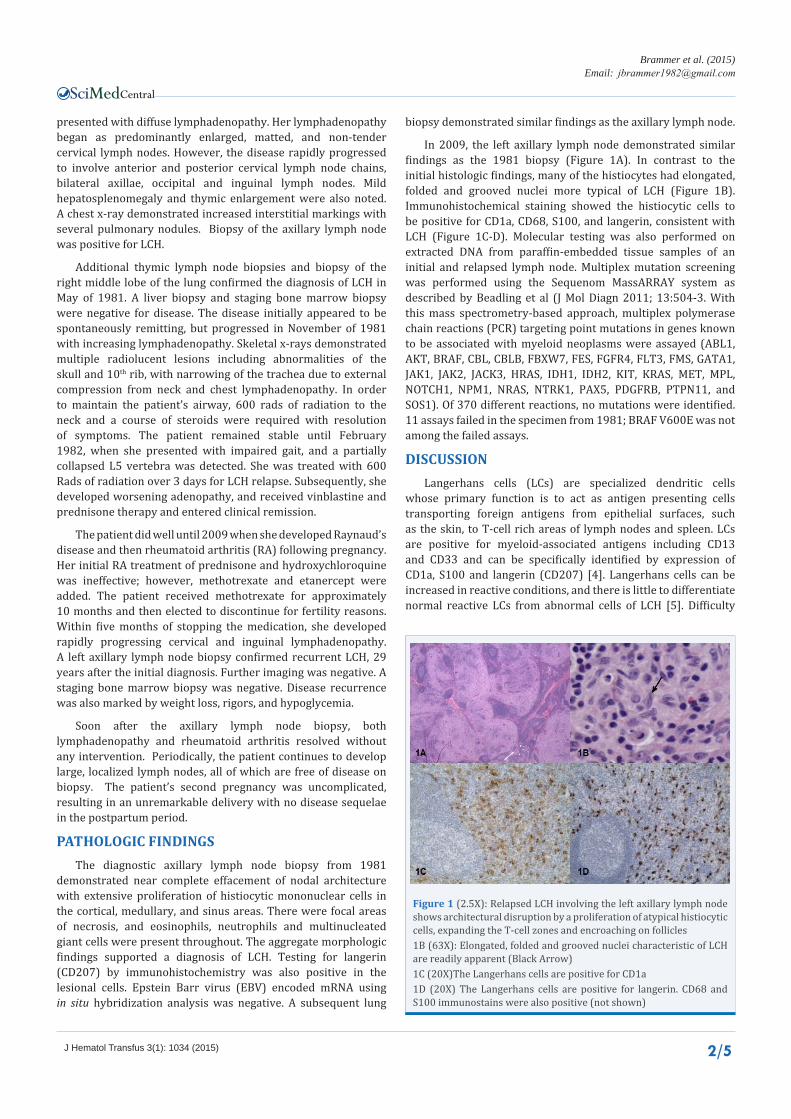
In 2009, the left axillary lymph node demonstrated similar findings as the 1981 biopsy (Figure 1A). In contrast to the initial histologic findings, many of the histiocytes had elongated, folded and grooved nuclei more typical of LCH (Figure 1B). Immunohistochemical staining showed the histiocytic cells to be positive for CD1a, CD68, S100, and langerin, consistent with LCH (Figure 1C-D). Molecular testing was also performed on extracted DNA from paraffin-embedded tissue samples of an initial and relapsed lymph node. Multiplex mutation screening was performed using the Sequenom MassARRAY system as described by Beadling et al (J Mol Diagn 2011; 13:504-3. With this mass spectrometry-based approach, multiplex polymerase chain reactions (PCR) targeting point mutations in genes known to be associated with myeloid neoplasms were assayed (ABL1, AKT, BRAF, CBL, CBLB, FBXW7, FES, FGFR4, FLT3, FMS, GATA1, JAK1, JAK2, JACK3, HRAS, IDH1, IDH2, KIT, KRAS, MET, MPL, NOTCH1, NPM1, NRAS, NTRK1, PAX5, PDGFRB, PTPN11, and SOS1). Of 370 different reactions, no mutations were identified. 11 assays failed in the specimen from 1981; BRAF V600E was not among the failed assays.
DISCUSSIONLangerhans cells (LCs) are specialized dendritic cells
whose primary function is to act as antigen presenting cells transporting foreign antigens from epithelial surfaces, such as the skin, to T-cell rich areas of lymph nodes and spleen. LCs are positive for myeloid-associated antigens including CD13 and CD33 and can be specifically identified by expression of CD1a, S100 and langerin (CD207) [4]. Langerhans cells can be increased in reactive conditions, and there is little to differentiate normal reactive LCs from abnormal cells of LCH [5]. Difficulty
Figure 1 (2.5X): Relapsed LCH involving the left axillary lymph node shows architectural disruption by a proliferation of atypical histiocytic cells, expanding the T-cell zones and encroaching on follicles1B (63X): Elongated, folded and grooved nuclei characteristic of LCH are readily apparent (Black Arrow)1C (20X)The Langerhans cells are positive for CD1a1D (20X) The Langerhans cells are positive for langerin. CD68 and S100 immunostains were also positive (not shown)
Central
Brammer et al. (2015)Email:
J Hematol Transfus 3(1): 1034 (2015) 3/5
in demonstrating clonality in LCH lesions has fueled dispute as to whether or not these lesions are truly neoplastic, or rather reactive/inflammatory, especially since some cases will remit without therapy [6]. A recent study identified the oncogenic BRAF V600E mutation in 57% (35/61) of LCH cases, which not only supports LCH is a clonal neoplasm, it offers a potential therapeutic target for RAF pathway inhibitors [3]. Due to the therapeutic implication, we performed multiplex mutational analysis to test for point mutations common to myeloid neoplasms, including BRAF V600E on both primary and relapsed specimens. No mutations were detected. Despite the fact that a majority of cases express BRAF V600E, the etiology and pathogenesis of LCH remains largely unknown, and it cannot be simply stated to be a clonal disorder. The development of LCH has been linked to immune dysregulation, although connection with a specific autoimmune disease, infection, or immunodeficiency has not been established [7]. Hypothesizing a role of immune dysregulation is not surprising as the CD1a and langerin of LCs are related to the major histocompatibility complex (MHC) class 1 and 2, respectively [8]. LCs express surface receptors for multiple inflammatory cytokines such as IL-1, IL-6, TNF-α, and IFN-γ. TNF-α aids in dendritic cell maturation and activation, and the effects of IL-1 and TNF-α can also result in down regulation of the cell adhesion molecule E-cadherin on cutaneous Langerhans cells, allowing migration from the skin with systemic spread [9]. Gene expression array data also show genes related to immune responses are upregulated in LCH, including IL-10, IFN-γ, TGF-β, TNF-α [4]. IL-10 and TGF-β limit the normal maturation of LCH
cells to more mature LC and are increased in LCH [5]. IL-17A is the main cytokine of T helper-17 cells, which are commonly increased in autoimmune diseases. Coury et al report increased levels of IL-17A in the serum, skin and bone lesions of LCH patients, suggesting a possible link between LCH and IL-17A [10]. Further suggesting immune dysregulation as the cause of LCH, T regulatory cells (T-regs) are commonly increased in both autoimmune disease and LCH [11]. Furthermore, the use of immunosuppression is the backbone of LCH therapy, further suggesting LCH is an immune-mediated disease caused by dysfunctional T-reg activation [4].
The temporal relationship between the postpartum onset of RA, requiring methotrexate therapy, and relapsed LCH makes our case unique, and may offer insights into this disease process. RA is an autoimmune disease of unknown etiology resulting in chronic inflammation, polyarthritis and joint destruction. Abnormal cytokine regulation is central to the pathogenesis of RA [12]. The coexistence of LCH with autoimmune disease is extremely rare [13]. However, it is possible the cytokine milieu of RA can potentiate the proliferation of pathogenic Langerhans cells. Similar to emerging evidence in LCH, T-cells are necessary for the development of RA. Cytokines produced by T helper 1 (TH1) and T helper 17 (TH17) cells likely contribute to disease [14]. TH17 cells produce IL-17, and animal models of inflammatory arthritis demonstrate that elevated IL-17 correlates with increased disease severity [15]. TH17 cells are induced by TGF-β [16]. TGF-β, along with IL-10 and other cytokines, is one of the upregulated genes identified in LCH cases [4]. As mentioned in the context of LCH, TGF-β and IL-10 can also limit the normal maturation of dendritic cells to fully mature LC [4]. IL-10 and TGF-β are a potential link between RA and LCH. Furthermore, these cytokines may contribute to the expansion of T regulatory cells common to both diseases [12].
During pregnancy, there are changes in cytokine secretion, such as increased TNF-α and IL-1 receptor antagonist, which limit inflammation in RA patients [17, 18]. There is also a cytokine shift towards a TH2 mediated response away from TH1 and TH17, and the majority of RA patients will have symptomatic improvement with pregnancy [19]. However, up to 98% of patients experience recurrence of RA within 4 months postpartum [20]. The risk of first developing RA is also increased in the first year after delivery, upon reconstitution of the TH1 and TH17 mediated immune response [21]. Our patient’s initial RA diagnosis was within the first postpartum year and accompanied by LCH relapse, suggesting postpartum environment can induce both entities. A model of possible pathogenesis is provided (Figure 2). We cannot definitively conclude from this case that LCH is primarily an immunologically mediated disease, and caution that further research into the causes of LCH needs to be performed to further elucidate the cause. However, given that the molecular PCR genotyping was identical in the recurrent disease, and temporally related to post-partum RA, this case provides evidence that LCH may indeed be an immunologically mediated disease and provides hypotheses for further investigation.
Primary LCH treatment depends on the extent and distribution of disease. Isolated cutaneous lesions may regress spontaneously [22], whereas multisystem involvement typically
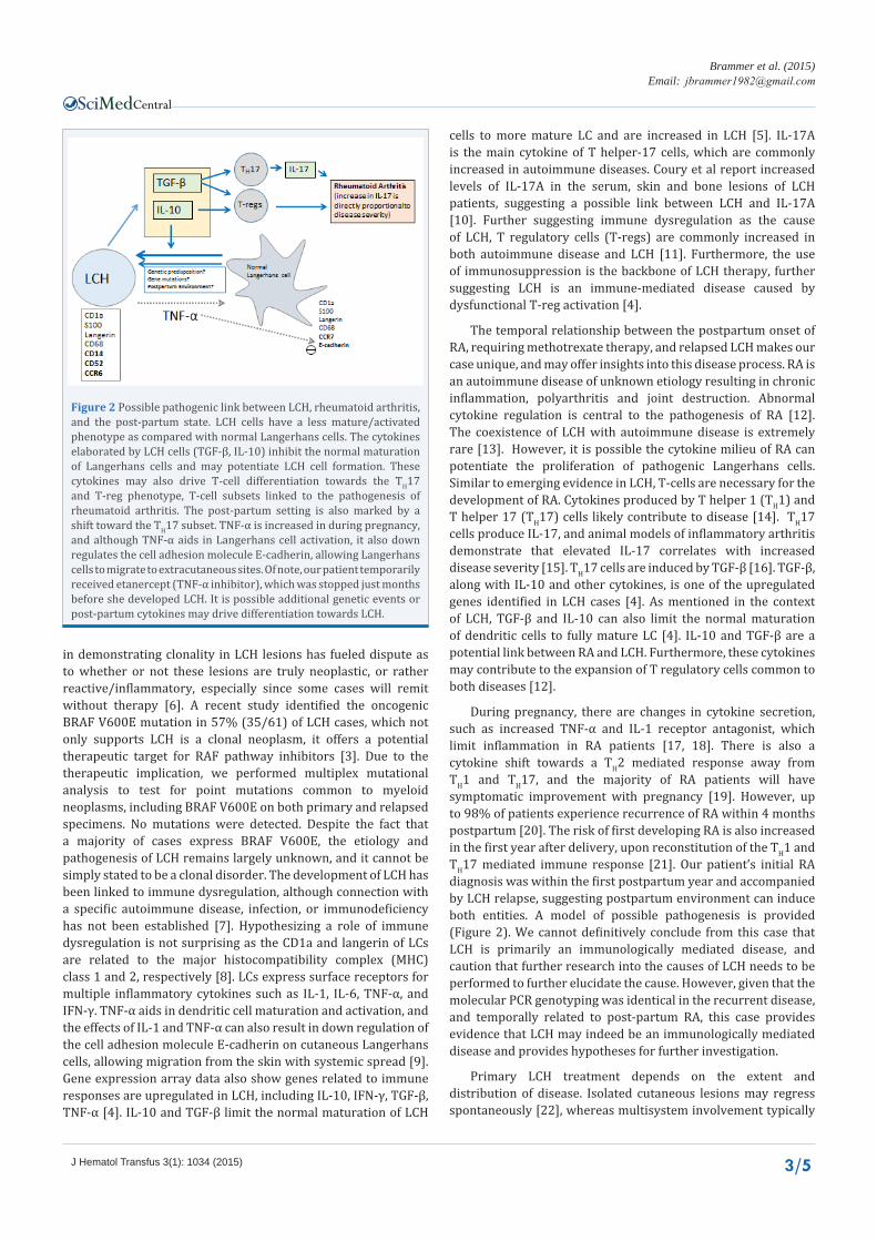
Figure 2 Possible pathogenic link between LCH, rheumatoid arthritis, and the post-partum state. LCH cells have a less mature/activated phenotype as compared with normal Langerhans cells. The cytokines elaborated by LCH cells (TGF-β, IL-10) inhibit the normal maturation of Langerhans cells and may potentiate LCH cell formation. These cytokines may also drive T-cell differentiation towards the TH17 and T-reg phenotype, T-cell subsets linked to the pathogenesis of rheumatoid arthritis. The post-partum setting is also marked by a shift toward the TH17 subset. TNF-α is increased in during pregnancy, and although TNF-α aids in Langerhans cell activation, it also down regulates the cell adhesion molecule E-cadherin, allowing Langerhans cells to migrate to extracutaneous sites. Of note, our patient temporarily received etanercept (TNF-α inhibitor), which was stopped just months before she developed LCH. It is possible additional genetic events or post-partum cytokines may drive differentiation towards LCH.

Central
Brammer et al. (2015)Email:
J Hematol Transfus 3(1): 1034 (2015) 4/5
requires a combination of prednisone and chemotherapy, such as vinblastine, cytarabine, or methotrexate [2, 8]. One of the most important predictors of a favorable outcome is the response within 6 weeks of induction therapy with 88-91% survival rates [23]. Relapse, or disease reactivation, is common and occurs in greater than 50% of patients with multisystem LCH in most studies [10, 22].
Most relapses occur within the first 1-2 years after diagnosis and are more frequent with multisystem or high-risk organ involvement and organ dysfunction [22, 23]. Although long-term follow-up is necessary to monitor for disease or complications secondary to treatment, late recurrences of LCH are rare and the etiology of relapse is not known. To our knowledge, there have been just four other very late recurrences (≥ 10 years) documented in the literature [24-26]. Unlike the extensive lymph node involvement in our patient, all previously reported patients were male and had limited bone lesions initially, followed by limited bone or orbit involvement at relapse (Table 1).
CONCLUSIONSWe report a case of late relapsing LCH occurring 29 years
after primary diagnosis and document the first association with postpartum rheumatoid arthritis (RA) The unique coexistence of relapsed LCH and postpartum RA in this case suggests an environmental factor in postpartum females is capable of triggering the cytokine dysregulation common to both diseases. The exact relationship between these two neoplasms separated by 29 years is not clear, but future studies aimed at defining the postpartum cytokine milieu may enhance our understanding of LCH pathogenesis and aid in directed therapies.
REFERENCES1. Swerdlow SH, Campo E, Harris NL, et al. WHO Classification of
Tumours of Haematopoietic and Lymphoid Tissues. 4th ed. Lyon, France: WHO Press. 2008: 358-360.
2. Langerhans Cell Histiocytosis: Histiocyte Society Evaluation and Treatment Guidelines, NJ: Histiocyte Society, 2009.
3. Badalian-Very G, Vergilio JA, Degar BA, MacConaill LE, Brandner B, Calicchio ML, et al. Recurrent BRAF mutations in Langerhans cell histiocytosis. Blood. 2010; 116: 1919-1923.
4. Allen CE, Li L, Peters TL, Leung HC, Yu A, Man TK, et al. Cell-specific gene expression in Langerhans cell histiocytosis lesions reveals a
Table 1: Late Recurrent LCH (>10 years).
Authors (year) Sex Age at first diagnosis Initial sites Relapse sites Time to relapse
(years) Comments
Kilpatrick (1995) M 36 years Scapula Scapula 13
Kilpatrick (1995) M 25 years Mandible Mastoid bone 33
Escardó-Paton (2004) M 7 years Sacroiliac bone Orbit 16 Mild ocular trauma about 2 weeks prior to relapse
Wladis (2008) M 3 years Mastoid bone Maxillary bone then orbit 5 and 15
Second relapse to orbit occurred 10 years after first relapse (15 years after initial diagnosis)
Present case F 2.5 months Lymph nodes, lung, bone
Lymph nodes, maxillary sinus 29 Association with postpartum
onset of RA Abbreviations: RA: Rheumatoid Arthritis
distinct profile compared with epidermal Langerhans cells. J Immunol. 2010; 184: 4557-4567.
5. Geissmann F, Lepelletier Y, Fraitag S, Valladeau J, Bodemer C, Debré M, et al. Differentiation of Langerhans cells in Langerhans cell histiocytosis. Blood. 2001; 97: 1241-1248.
6. Abla O, Egeler RM, Weitzman S. Langerhans cell histiocytosis: Current concepts and treatments. Cancer Treat Rev. 2010; 36: 354-359.
7. Garabedian L, Struyf S, Opdenakker G, Sozzani S, Van Damme J, Laureys G. Langerhans cell histiocytosis: a cytokine/chemokine-mediated disorder? Eur Cytokine Netw. 2011; 22: 148-153.
8. Ng-Cheng-Hin B, O’Hanlon-Brown C, Alifrangis C, Waxman J. Langerhans cell histiocytosis: old disease new treatment. QJM. 2011; 104: 89-96.
9. Abla O, Egeler RM, Weitzman S. Langerhans cell histiocytosis: Current concepts and treatments. Cancer Treat Rev. 2010; 36: 354-359.
10. Coury F, Annels N, Rivollier A, Olsson S, Santoro A, Speziani C, et al. Langerhans cell histiocytosis reveals a new IL-17A-dependent pathway of dendritic cell fusion. Nat Med. 2008; 14: 81-87.
11. Senechal B, Elain G, Jeziorski E, Grondin V, Patey-Mariaud de Serre N, Jaubert F, et al. Expansion of regulatory T cells in patients with Langerhans cell histiocytosis. PLoS Med. 2007; 4: e253.
12. McInnes IB, Schett G. Cytokines in the pathogenesis of rheumatoid arthritis. Nat Rev Immunol. 2007; 7: 429-442.
13. Robak T, Kordek R, Robak E, Bartkowiak J, Biernat W, Liberski P, et al. Langerhans cell histiocytosis in a patient with systemic lupus erythematosus: a clonal disease responding to treatment with cladribine, and cyclophosphamide. Leuk Lymphoma. 2002; 43: 2041-2046.
14. Schulze-Koops H, Kalden JR. The balance of Th1/Th2 cytokines in rheumatoid arthritis. Best Pract Res Clin Rheumatol. 2001; 15: 677-691.
15. Lubberts E, Koenders MI, Van den Berg WB. The role of T-cell interleukin-17 in conducting destructive arthritis: lessons from animal models. Arthritis Res Ther. 2005; 7: 29-37.
16. Schulze-Koops H, Kalden JR. The balance of Th1/Th2 cytokines in rheumatoid arthritis. Best Pract Res Clin Rheumatol. 2001; 15: 677-691.
17. Da Silva JA, Spector TD. The role of pregnancy in the course and aetiology of rheumatoid arthritis. Clin Rheumatol. 1992; 11: 189-194.
18. Østensen M, Förger F, Nelson JL, Schuhmacher A, Hebisch G, Villiger PM. Pregnancy in patients with rheumatic disease: anti-inflammatory cytokines increase in pregnancy and decrease post partum. Ann
Central
Brammer et al. (2015)Email:
J Hematol Transfus 3(1): 1034 (2015) 5/5
Brammer JE, Leeborg N, Carneal E, Fan G, Gajewski J (2015) A Unique Case of Late Recurrent Langerhans Cell Histiocytosis and Postpartum Rheumatoid Ar-thritis Offers Insight into Possible Disease Triggers. J Hematol Transfus 3(1): 1034.
Cite this article
Rheum Dis. 2005; 64: 839-844.
19. Straub RH, Buttgereit F, Cutolo M. Benefit of pregnancy in inflammatory arthritis. Ann Rheum Dis. 2005; 64: 801-803.
20. Nelson JL, Ostensen M. Pregnancy and rheumatoid arthritis. Rheum Dis Clin North Am. 1997; 23: 195-212.
21. OKA M. Effect of pregnancy on the onset and course of rheumatoid arthritis. Ann Rheum Dis. 1953; 12: 227-229.
22. Wilejto M, Abla O. Langerhans cell histiocytosis and Erdheim-Chester disease. Curr Opin Rheumatol. 2012; 24: 90-96.
23. Drutz JE. Histiocytosis. Pediatr Rev. 2011; 32: 218-219.
24. Wladis EJ, Tomaszewski JE, Gausas RE. Langerhans cell histiocytosis of the orbit 10 years after involvement at other sites. Ophthal Plast Reconstr Surg. 2008; 24: 142-143.
25. Kilpatrick SE, Wenger DE, Gilchrist GS, Shives TC, Wollan PC, Unni KK. Langerhans’ cell histiocytosis (histiocytosis X) of bone. A clinicopathologic analysis of 263 pediatric and adult cases. Cancer. 1995; 76: 2471-2484.
26. Escardó-Paton JA, Neal J, Lane CM. Late recurrence of Langerhans cell histiocytosis in the orbit. Br J Ophthalmol. 2004; 88: 838-839.