a specific inhibitor of calcium/calmodulin-dependent ... · a specific inhibitor of...
TRANSCRIPT
The Journal of Neuroscience, May 1995, 75(5): 4093-4101
A Specific Inhibitor of Calcium/Calmodulin-Dependent Protein Kinase-II Provides Neuroprotection against NMDA- and Hypoxia/ Hypoglycemia-Induced Cell Death
lradj Hajimohammadreza,2 Albert W. Prober-t,’ Linda L. Coughenour,’ Susan A. Borosky,’ Frank W. Marcoux,’ Peter A. Boxer,’ and Kevin K. W. Wang*
‘Department of Neuroscience Pharmacology and *Laboratory of Neuro-biochemistry, Parke-Davis Pharmaceutical Research, A Division of Warner Lambet-t Company, Ann Arbor, Michigan 48105
Calciumlcalmodulin-dependent protein kinase-II (CamK-II) is a major neuronal protein which plays a significant role in the cellular process of long-term potentiation (LTP), and vesicular release of neurotransmitters. Here, we show that KN-62, 1-[N,Obis(5-isoquinolinesulfonyl)Nmethyl-L-tyro- syl]-4-phenylpiperazine, a specific cell-permeable inhibitor of CamK-II substantially protected neurons from (1) acute NMDA toxicity and (2) hypoxia/hypoglycemia-induced neu- ronal injury in fetal rat cortical cultures. KN-62 did not di- rectly inhibit glutamate, kainate, cu-amino9-hydroxyd- methyl-6isoxazolepropionate (AMPA), glycine, or [piperi- dyl-3,4-(N)]-(N-[1-(2-thienyl)cyclohexyl]-3,4-piperidine) (TCP) binding to rat brain membranes. Finally, KN-62 sig- nificantly reduced cellular calcium accumulation following either NMDA challenge or hypoxia/hypoglycemia insult. Our results show that CamK-II plays a key role in mediating some of the biochemical events leading to cell death fol- lowing an acute excitotoxic insult.
[Key words: NMDA, hypoxia/hypoglycemia, calcium/cal- modulin-kinase-II, intraneuronal calcium, KN-62, spectrin]
Glutamate, the major excitatory amino acid (EAA) in the CNS has been implicated in neuronal injury associated with cerebral &hernia, epilepsy, and chronic neurodegenerative disorders (Meldrum and Garthwaite, 1990). Glutamate receptors have been classified as either ionotropic or metabotropic (Meldrum and Garthwaite, 1990). The recent molecular cloning of the sub- units for the ionotropic glutamate receptors has confirmed the original classification of AMPA, kainate and NMDA receptors. These receptors are composed of homo- or heteromeric combi- nations of subunits (GluRI-4 for AMPA/kainate receptors, GluR5-7 and KAI -2 for kainate receptors, and NMDARI and NMDAR2A-D for NMDA receptor) (Nakanishi, 1992; Seeburg, 1993). Activation of the NMDA subtype of the glutamate re- ceptor has been shown to be the major contributor to cell death in cortical cell cultures subjected to acute excitotoxicity or ox- ygen and glucose deprivation (Choi, 1987, 1988; Goldberg and Choi, 1993; Hartley et al., 1993). The early biochemical feature of NMDA-induced excitotoxicity in neurons is the disturbance in ionic balance triggered by calcium and sodium influx through
Received Sept. 15, 1994; revised Dec. 14, 1994; accepted Dec. 28, 1994.
Correspondence should be addressed Iradj Hajimohammadreza, Laboratory of Neuro-biochelnistry, Parke-Davis Pharmaceutical Research, A Division of Warner Lambert Company, 2800 Plymouth Road, Ann Arbor, MI 48 105.
Copyright 0 1995 Society for Neuroscience 0270~6474/95/154093-09$05.00/O
the NMDA receptor/channel complex (Choi, 1987, 1988; Mar- coux et al., 1990; Goldberg and Choi, 1993; Hartley et al., 1993; Weber et al., 1993). One consequence of such ionic imbalance is the activation/overactivation of many vital cellular enzymes (protein kinases and phosphatases, phospholipases and prote- ases), in particular those regulated by calcium followed by a cascade of both biochemical and physical changes (cytoskeletal breakdown) leading to neuronal death (Siman and Noszek, 1988; Saido et al., 1994).
One such enzyme, CamK-II, has an important role in decod- ing signals generated by neurotransmitters which raise intracel- lular free calcium ([Caz+],). CamK-II is highly enriched in neu- rons, both in the pre- and postsynaptic compartments where it is essential to neurotransmitter release and induction of LTP, respectively (Llinas et al., 198.5; Malenka et al., 1989; Green- gard, 1993). Recent studies have also linked CamK-II to regu- lation of both voltage-gated and ligand-gated ion channels, in particular those associated with glutaminergic neurotransmission (Greengard et al., 199 1; Wang et al., 199 I ; Keller et al., 1992; Raymond et al., 1993; McGlade-McCulloh et al., 1993). CamK- II is a serine-threonine kinase activated by an increase in [Ca?‘], (bound to calmodulin) following an appropriate agonist stimu- lation, such as NMDA (Fukunaga et al., 1990, 1992). Once ac- tivated by the Ca*+/calmodulin, 20-80s of the enzyme activity is maintained by its conversion to a Ca*‘-independent form via autophosphorylation. This posttranslational modification of CamK-II is a rapid process in neurons and could be a key factor in its sustained catalytic activity (Rich et al., 1990; Yamamoto et al., 1992).
The main feature of this multifunctional kinase is phosphor- ylation of a number of substrate proteins, which mediate many of the actions of cellular second messengers. Several investiga- tors have shown that phosphorylation of the AMPA/kainate re- ceptor by protein kinases such as CamK-II, protein kinase C (PKC) or CAMP-dependent protein kinase (PKA) regulates its function in neurons (Greengard et al., 1991; Wang et al., 1991; Keller et al., 1992; McGlade-McCulloh et al., 1993; Raymond et al., 1993). The NMDA subtype of glutamate receptor is also modulated by phosphorylation in neurons (Chen et al., 1992; Kitamura et al., 1993). In all cases, glutamate receptor phos- phorylation leads to a positive modulation on the receptor func- tion maintaining synaptic excitability. The importance of CamK- II in maintenance of synaptic excitability and also its extreme vulnerability in in viva models of ischemia (Churn et al., 1992; Hanson et al., 1994) and in vitro excitotoxicity (Churn et al.,

4094 Hajimohammadreza et al. * Calcium/Calmodulin-Dependent Protein Kinase-II and Excitotoxicity
1993) have led us to examine its potential contribution in EAA- mediated cell death.
Therefore, to examine the role of CamK-II in EAA-mediated neurotoxicity in fetal rat cerebrocortical cultures, we used the CamK-II specific inhibitor KN-62 with proven cell permeability (Tokumitsu et al., 1990; Hidaka and Hagiwara, 1992). KN-62 competitively inhibits CamK-II with respect to calcium/calmo- dulin and has no inhibitory effect on PKA, PKC, myosin light chain kinase, or casein kinase-I (Tokumitsu et al., 1990; Hidaka and Hagiwara, 1992). KN-62 is not a calmodulin antagonist and is only effective on inhibiting the activity of CamK-II before its autophosphorylation. KN-62 has also been used previously as a specific inhibitor for CamK-II in various neuronal systems (Fi- gurov et al., 1993; Hack et al., 1993).
Materials and Methods
Chemicals. KN-62 was purchased from Seikagaku America; K252a (Nocardiopsis sp.), genistein, calphostin C (Cludosporium cladospo- rioids), compound 5 [2-hydroxyl-5-(2,5-dihydroxy-benzyl) aminoben- zoic acid] and calmidozolium were purchased from LC laboratories. ‘Va*+ was from ICN Biomedicals. Tissue culture media and serum were from GIBCO-Bethesda Research Labs. All other reagents were of an- alytical grade and purchased from Sigma Chemical Company.
Neuronal cultures. Cortical hemispheres sectioned from fetal rat (Sprague-Dawley) in their 18th day of gestation and were trypsin di- gested and triturated into single cell suspension. cells were pipetted into individual wells of poly-L-lysine-coated plates, yielding a final cell con- centration of 200,000 cells/cm’ using GIBCO Minimum Essential Me- dium (MEM; containing 10% horse and 10% fetal bovine serum; heat inactivated). Non-neuronal cell division was halted three days into cul- ture by adding 25 kg/ml uridine and 10 kg/ml 5-fluoro-2’-deoxyuridine. Feeding were performed as necessary with MEM with 10% horse se- rum. Experiments were performed on 17-21 d (postisolation) old neurons. Cultures were triple washed with MEM (serum free) with or without the test agents (inhibitors). NMDA was added directly to the culture media. After 30 min incubation at 37”C, NMDA was washed by triple exchange with serum free MEM (+inhibitors). Final concen- tration of glucose in the MEM throughout the culture period was 30 rnM.
Cell viability assay. Neuronal injury was assessed qualitatively by light microscopic examination of the phase contrast appearance of cul- tures. Quantitative assessments of neuronal injury were made using lac- tate dehydrogenase (LDH) release as a marker for membrane breakage and cell death (Koh and Choi, 1987). One day post experiment initia- tion, 2.5 l.~l of culture medium was combined with 225 p,l of 0.1 mM potassium phosphate buffer containing 0.133 mg/ml P-NADH. Follow- ing a 20 min incubation at 37”C, wells received 30 p,l of sodium py- ruvate (2.39 mM in phosphate buffer, pH 7.5) and kinetic measurements (at 340 nm for 2 min) were immediately performed using a Titertek Multiscan plate reader.
Calcium microspectrojuorimetry. Experiments were performed using a PTI Deltascan system (Princeton, NJ). Neurons were loaded with 2- 4 FM FURA- AM (Molecular Probes, Eugene OR) in buffer (in mM: NaCl, 137; KCl, 2.6; Na,PO,, 8.1; MgCl,, 0.5; CaCl,, 0.9; HEPES- NaOH, 10; glucose, 11; with 150 mg/ml bovine serum albumin, pH = 7.4) at room temperature for 1 hr and rinsed in the same buffer without FURA- AM but with added glycine (10 PM) for at least 15 min. A single cell was observed (40X objective, n.a. 1.3) and alternatively illuminated at 340 and 380 nM. Photometric measurements at 510 nM were made continuously at a rate of 5 Hz. Drugs were bath applied by superfusion (1 ml/min). Tetrodotoxin (300 nM) was added in the super- fusion media to reduce spontaneous calcium oscillations. Results were quantified by measuring the area under the curve of the fluorescence ratio (F,,,IF,,,) versus time waveform; for each cell the second 1 min challenge with 25 JLM NMDA was used to normalize the results. Each neuron was challenged three times with 25 )LM NMDA (1 min), and waveforms from the second and third challenges were used for area quantification.
Recepfor binding assay. Well-washed rat brain membranes from a lysed buffy coat fraction were prepared according to the method of Kishimoto et al (198 1). ?H-glutamate and ZH-glycine binding were car- ried out in 50 mM Tris acetate buffer (pH 7.4). Brain membranes (200-
A
250
1 T ,,=18
125kM NMDA 250pM
6
110 A = 1 hour pretreatment only
B = pre-co-post treatment
B 100 C = co-treatment only
E 90 D = post-treatment only
2 80
z 7u
g 60
P 50
2 40 s ki 3
9 20 10
0 A B C D A B C D
-KN-62 - -CPP
NMDA 25OpM
Figure 1. Inhibition of NMDA-induced toxicity by KN-62 (5 PM),
CPP (100 FM) and MK801 (1 FM). A, Net LDH release 24 hr after a 30 min (at 37°C) challenge with NMDA. Data are means + SD. B, Effect of mode of administration of KN-62 (5 PM) and CPP (100 (1~)
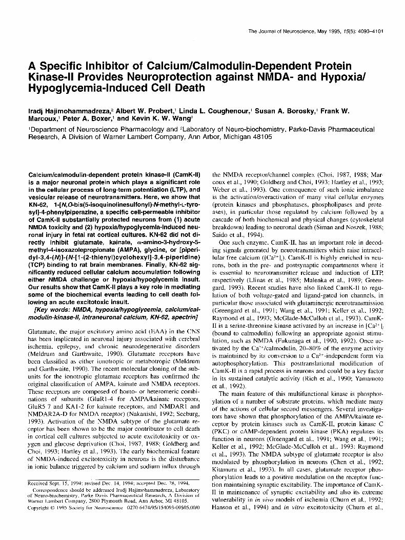
on the degree of protection against NMDA. These data are means ? SD of five separate experiments. C, Western blot analysis of proteins extracted from culture conditions shown in A with a monoclonal anti- spectrin antibody (Affiniti laboratories, U.K.). Solid arrow shows intact spectrin (240 kDa) and open arrowheads show proteolytic breakdown fragments of spectrin (150 and 145 kDa), molecular weight markers &Da) are shown on the left. The blot is a typical representative of at least four different analyses. D, A representative phase contrast photo- micrograph of cerebrocortical mixed cultures taken from a random field at the end of treatments shown. Photomicrograph of the KN-62 pre- treated neurons with NMDA challenge shows only the live neurons, which have normal morphology. There were also dead neurons in these cultures. NMDA, 250 PM; KN-62 (Km, 5 PM. **, p 5 0.0001; *, p 5 0.001, Student’s t test.
400 pg) were incubated with ‘H-glutamate (2 nM) and JH-glycine (20 nM) for 30 min on ice. Specific binding for ?H-glutamate (80%) was defined as that displaced in the presence of 0.1 mM NMDA and for ?H- glycine (65%) as that displaced by 0.1 mM 5,7-dichlorokynurenic acid. ‘H-TCP (2 nM) binding was carried out as described above except the incubations were for 1 hr at room temperature in 20 mM HEPES-KOH buffer (pH 7.4). ‘H-AMPA (10 nM) and ‘H-kainate (20 nM) binding was carried out in a 50 mM Tris-HCl buffer (pH 7.4) containing either IO mM potassium thiocyanate for ‘H-AMPA or 20 mM CaCI, for ‘H-kainate for 1 hr on ice. Specific binding was defined as that displayed in the
The Journal of Neuroscience, May 1995, 7~75) 4095
D
Control NMDA
presence of 1 mM glutamate. Filtration was done with a Brandell MB- either placed back in the normoxic conditions until 24 hr postexperi- 48R cell harvester through Whatman GF/B filters. Radioactivity was ment initiation for LDH measurement or used immediately for a5Ca*+ quantified by liquid scintillation counting. determinations.
Electrophoresis und Western blotting. Proteins were analyzed by SDS-PAGE according to the method of Laemmli (1970). After electro- phoresis, proteins were blotted onto PVDF membrane (Towbin et al., 1979) and blocked with 5% nonfat milk. Blots were incubated overnight in primary antisera and then one hour with biotinylated secondary an- tiserum. The immunoreactivity was visualized by alkaline phosphatase- conjugated streptavidin (Amersham). Anti-spectrin (cu-fodrin; monoclo- nal) was purchased from Affiniti Laboratories, U.K.
Results and Discussion
rrCaz+ accumulation studies. These were carried out according to the method of Birrell et al. (1993). Briefly, cells were washed three times with Mg?+-free HBSS containing 2.5 mM Ca’+, and incubated with the test drugs for one hour. 45CaZ+ (2 l&i/ml) was present 20 min prior to, during NMDA exoosure (30 min at 37°C). Ceils were washed three times-with saline and lysed with distilled water, and 45Ca2+ B emissions in the intracellular contents were counted by scintillation spectroscopy.
NMDA receptors appear to participate in the process of exci- totoxicity and neuronal death. The hallmark of NMDA-induced neuronal death is a sustained increase in the [Ca*+], and over- activation of vital Ca2+-dependent cellular enzymes such as calpain and CamK-II. Calpain overactivation with the presence of high [Ca*+], following NMDA receptor (NMDAR) overstimu- lation leads to breakdown of structural proteins (e.g., spectrin) and proteolysis of other cellular substrates (Siman and Noszek, 1988; Saido et al., 1994). Specific inhibitors of calpain have proved to be neuroprotectant in EAA-induced neurotoxicity (for review, see Saido et al., 1994; Wang and Yuen, 1994). On the other hand CamK-II is vital in maintaining synaptic excitability through its multifunctional property. Interestingly autophosphor- ylated CamK-II is a substrate for activated calpain, which pro- teolytically fragments and produces an active and calcium-in- dependent form of CamK-II (Kwiatkowski and King, 1989). To our knowledge, there have been no reports on examining specific inhibition of CamK-II in NMDA- and hypoxia/hypoglycemia- induced neurotoxicity in vitro.
Oxygen/glucose (hypoxia/hypoglycemiu) deprivation studies. Cere- brocortical cells were plated onto 96-well, PEI coated culture plates using DME/Fl2 medium containing 10% horse and 6% fetal bovine serum (heat inactivated). Non-neuronal cell division was halted 3 d into culture by adding 25 kg/ml uridine and 10 kg/ml 5-fluoro-2’-deoxyu- ridine. Oxygen/glucose deprivation in cultures were carried out accord- ing to Weber et al. (1993). Briefly, culture growth media was removed and replaced with defined media and then cultures were deprived of oxygen and glucose for O-330 min (exposure atmosphere: in triple gas incubator, I% O?, 8% CO,, 91% Nz; for exposure medium, 1.8 mM Ca2+, 0.8 mM Mg2+, 0.2 gm/liter D-ghCOSe; for normoxic condition, 21% O,, 8% CO,, 71% NJ. After the deprivation interval, cells were
NMDA+KN
Figure I. Continued.
To assess the degree of EAA-induced neuronal injury, we

4096 Hajimohammadreza et al. * Calcium/Calmodulin-Dependent Protein Kinase-II and Excitotoxicity
1 00
0.01 0.1 1 10 [KN-621 pM
Figure 2. Concentration-response relationship of KN-62 against 250 PM NMDA toxicity. The final concentration of DMF in cultures used either as the solvent control or in KN-62 solution is 0. I %, except for 10 JLM KN-62 where the solvent concentration was 0.2%. Data are means 2 SEM from three separate experiments. KN-62 was adminis- tered as pre-co-post treatment. **, p 5 0.0001; *, p 5 0.001, Student’s t test (compared to NMDA alone).
measured the activity of the cytoplasmic enzyme, lactate dehy- drogenase (LDH) released in the media (Koh and Choi, 1987); immunodetection of proteolytic fragments (I 50 and 145 kDa) of the cytoskeletal protein cr-fodrin (spectrin; 240 kDa) as produced by calpain (Siman and Noszek, 1988), and morphology by light microscopy. Previous work by others have shown a good cor- relation between neuronal death (by dye-exclusion method) and LDH measurements (Koh and Choi, 1987; Goldberg et al., 1987).
Pretreatment (I hr) with KN-62 (5 PM) offered a significant reduction (65%) in LDH release, 24 hr following application of 125 and 250 FM NMDA (30 min at 37°C) (Fig. 1A). Neuropro- tection achieved by KN-62 was robust when, administered 1 hr before NMDA application and included throughout the postin- cubation period (24 hr). However, we observed a significant but marginal protection (reduction in LDH release) against NMDA toxicity with KN-62 pretreatment only; neither simultaneous ad- ministration with NMDA nor post-NMDA application only of KN-62 was neuroprotective (Fig. 1B). In comparison, competi- tive NMDA receptor antagonist 3-[( + )-2-carboxypiperazine- 4-yllpropyl- 1 -phosphonic acid (CPP) at 100 FM was highly neu- roprotective in all treatment paradigms as expected (Fig. IB). Furthermore, both biochemical (LDH activity and spectrin im- munoblot) and morphological data show preservation of neurons by KN-62 when acutely treated with NMDA (Fig. l&D). In the same toxicity paradigm, prior exposure of cultures to the protein phosphatase inhibitor okadaic acid (1 OOnM) abolished the neuroprotective effects of KN-62 (data not shown), which agrees with, the binding characteristics of KN-62 to the non-
Table 1. Effect of various kinase inhibitors on NMDA-induced toxicity
LDH release (% NMDA max)
NMDA I25 FM NMDA 250 FM
38.4 k l5.0** 33.3 2 6.8** 49.9 ? 13.5** 80.1 ? lO.6*
109.1 + 7.6 136.1 k 5.0* 124.9 k 7.l*
109.4 + 31.6 119.4 k 26.3
105.0 + 21.1 157.6 t 38.4*
KN-62 (5 PM) (12)
Compound 5 (5 FM) (5)
Genistein (5 FM) (3) Calphostin C (0.25 FM) (3)
K252a (0.5 FM) (3)
Calmidazolium (5 PM) (3)
All inhibitors were present throughout the experiment and also as a 1 hr pre- treatment. LDH samples were taken 24 hr following the NMDA challenge (30 min at 37°C). The data are means & SEM from three or more separate exper- iments (number in parentheses). Concentrations of agents other than KN-62 used here were taken from published data in neuronal systems that provided effective inhibition. Compound 5 12.hydroxyl-5-(2.5.dihydroxy-benzyl)ami- nobenaoic acid; O’Dell et al., 199 I], K252a (Nocardiopis sp.), calphostin C (Clndosporium clndosporioids), calmidaTolium, and genistein (O’Dell et al., 199 1) were purchased from LC Laboratories, and KN-62 was purchased from Seikagaku America, Inc. *, P 5 0.05; **, P 5 0.0001, Student’s r test.
autophosphorylated CamK-II and the importance protein phos- phorylation in such a parameter.
Neuroprotection observed with KN-62 against 250 PM
NMDA-induced toxicity was concentration-dependent, being most effective at 0.5-10 FM (Fig. 2). This is in agreement with previous reports using KN-62 in cell-based systems (Tokumitsu et al., 1990; Hidaka and Hagiwara, 1992; Hack et al., 1993; Figurov et al., 1993). Effective concentrations of KN-62 (0.5- 10 PM) against NMDA-induced toxicity observed in this study have been shown to provide 75-80% inhibition of CamK-II ac- tivity in the exogenous substrate phosphorylation (Tokumitsu et al., 1990).
The effect of KN-62 appears to be selective since, calmida- zolium (a calmodulin antagonist), genistein (a tyrosine protein kinase inhibitor), K252a (a nonspecific protein kinase inhibitor) and calphostin C (a PKC inhibitor) were not neuroprotective against 125 and 250 PM NMDA challenge (Table 1). Calmida- zolium and calphostin C, at concentrations used here and mode of application (pre/co/post), exacerbated NMDA-induced toxic- ity (Table 1). However, compound 5 (a mixed tyrosine kinase and CamK-II inhibitor) produced a significant protection against both doses of NMDA tested (Table 1). The lower degree of protection observed by compound 5 could be due to its lack of binding specificity to CamK-II as compared to KN-62. It would be ideal to have highly specific inhibitors like KN-62 (which does not compete with ATP) for the major classes of protein kinases (e.g., PKA and PKG) to test in such excitotoxicity par- adigms. The availability of such compounds will allow one to determine the components involved, and also test combination strategies, in this model of neuronal death.
To complement these studies, the ability of KN-62 to protect neurons in an oxygen/glucose-deprivation (hypoxia/hypoglyce- mia) induced neurotoxicity model in rat cortical cultures was examined. This in vitro model of neurotoxicity has previously been shown to be primarily mediated through the NMDA re- ceptor and can be blocked effectively by NMDA antagonists (Choi, 1987, 1988; Marcoux et al., 1990; Goldberg and Choi, 1993; Hartley et al., 1993; Weber et al., 1993). Neuronal degen- eration in this type of insult is similar to those associated with partial focal cerebral ischemia in viva (e.g., middle cerebral ar- tery occlusion in rat). Oxygen/glucose deprivation interval of

The Journal of Neuroscience, May 1995, f5(5) 4097
A 400 -
E 300- E 2 8 2 200- @
E
fi loo-
0; 1,,,!,1(1,l,1,1
120 150 180 210 240 270 300 330
Deprivation time (min)
B 2 2 E ” k k -+KN-62 ,+
200
97.4 69.0 46.0
30.0
21.5
14.3
+ Hypoxia
-.- +CPP, 100~M
-c- +KN-62,0.3pM
--d- +KN-62,lj~M
-Lb- +KN-62, SpM
-+- +KN-62,lOpM
Figure 3. Inhibition of oxygen/glu- cose (hypoxia/hypoglycemia) depriva- tion-induced (labeled as hypoxia) neu- ronal death by KN-62 (0.3-10 JLM) and CPP (100 PM). A, LDH release with increasing duration of deprivation. Samples were taken 24 hr post experi- ment initiation. Data are means f SEM from six observations. All data points from 180-300 min were statis- tically significant compared to hypoxic control values with at least p 5 0.01 (Student’s t test). B, Western blot anal- ysis of proteins extracted from 240 min deprivation interval, probed with anti- spectrin antibody. molecular weight markers (kDa) are shown on the left. Solid arrow shows intact spectrin (240 kDa) and open arrowheads show pro- teolytic breakdown fragments of spec- trin (150 and 145 kDa).
240 min (4 hr; followed by 24 hr incubation in normoxic con- ditions) in our cultures produces maximum neuronal degenera- tion (Fig. 3). Pretreatment (1 hr) and continuous presence of KN- 62 dose dependently and significantly reduced neurotoxicity in this model, as evaluated by LDH release and calcium accumu- lation (Figs. 3, 4B). Immunoblot of proteins (labeled with anti- spectrin antibody) extracted from 240 min of hypoxia/hypogly- cemia condition show reduced proteolytic fragments of intact spectrin in KN-62 pretreated cultures compared to the appropri- ate control (Fig. 3B). However, immunoblots of spectrin and its proteolytic fragments, did not reflect the dose-response of KN- 62 observed with the LDH release assay in this toxicity para- digm (Fig. 3B). This may be due to the qualitative rather than quantitative nature of spectrin immunoblots in such a protocol. Neuroprotection seen by KN-62 (at 5 and 10 PM) was effective
up to 260 min of oxygen/glucose deprivation followed by 24 hr in normoxic condition. The competitive NMDA antagonist CPP (100 FM) produced almost complete protection against all de- privation intervals, as seen in LDH release (Fig. 3A) and calcium accumulation (Fig. 4B).
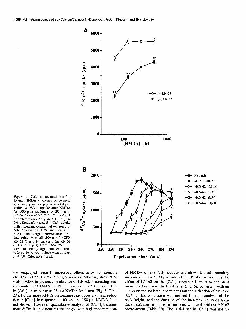
One common feature in NMDA or oxygen/glucose depriva- tion-induced toxicity is an increase in the level of intracellular free calcium ([Ca*+],), which has been shown to correlate well with consequent (delayed) neuronal death (Choi, 1987, 1988; Marcoux et al., 1990; Goldberg and Choi, 1993; Hartley et al., 1993; Weber et al., 1993). KN-62 reduced calcium (45Ca*+) ac- cumulation by 34-58% in NMDA toxicity and IO-100% in ox- ygen/glucose deprivation, depending on the degree of the insults (Fig. 4).
To further investigate the effect of KN-62 on calcium influx,
4098 Hajimohammadreza et al. * Calcium/Calmodulin-Dependent Protein Kinase-II and Excitotoxicity
Figure 4. Calcium accumulation fol- lowing NMDA challenge or oxygen/ glucose (hypoxia/hypoglycemia) depri- vation. A, ‘Va*+ uptake after NMDA (60-500 PM) challenge for 30 min in presence or absence of 5 FM KN-62 (1 hr pretreatment). **, p 5 0.001; *, p I 0.01, Student’s t test. B, Wa*+ uptake with increasing duration of oxygen/glu- cose deprivation. Data are means L SEM of six to eight determinations. All data points from 165-300 min for CPP, KN-62 (5 and 10 JLM) and for KN-62 (0.3 and 1 pM) from 165-225 min, were statistically significant compared to hypoxic control values with at least p 5 0.01 (Student’s t test).
A 6000-
2000
1
-o- (-)KN-62
-o- (+)KN-62
1000
1
1 00
100 1000 [NMDA] PM
B
1500-
1000-
soo-
00 120 150 180 210 240 270 300 330
Deprivation time (min)
we employed Fura- microspectrofluorimetry to measure of NMDA do not fully recover and show delayed secondary changes in free [Ca*+J in single neurons following stimulation increases in [Ca”], (Tymianski et al., 1994). Interestingly the with NMDA in presence or absence of KN-62. Pretreating neu- effect of KN-62 on the [Ca*+], response is most evident as a
rons with 5 pM KN-62 for 30 min resulted in a 50.3% reduction more rapid return to the basal level (Fig. 5), consistent with an in [Ca*+], in response to 25 FM NMDA for 1 min (Fig. 5, Table action on the maintenance rather than the induction of elevated 2A). Furthermore KN-62 pretreatment produces a similar reduc- [Ca2+],. This conclusion was derived from an analysis of the tion in [Ca”], in response to 100 pM and 250 pM NMDA (data peak height, and the duration of the half-maximal NMDA-in- not shown). However, quantitative analysis of [Ca*+], becomes duced calcium responses in neurons with and without KN-62 more difficult since neurons challenged with high concentrations pretreatment (Table 2B). The initial rise in [Ca”], was not re-
-B- Hypoxia
-.- +CPP, lOOj.tM
-0- +KN-62, 0.3pM
--fr +KN-62, lj.~M
-U- +KN-62, 5pM
-0- +KN-62, 10pM

The Journal of Neuroscience, May 1995, 15(5) 4099
/ / 0.1% DMF 1.4
1.2
1.0
.8
1.8 - 5 pM KN-62
s 1.6 -
1.4 -
1.2 -
1.0 -
.8 -
.6 -
.4- w_P// '- I I
0 5 10 45 50
TIME (min) Figure 5. [Ca*+], measurement in single neurons. A representative Fura- fluorescence ratio (F,,,IF,,,) versus time waveform in response to 25 FM NMDA with 30 min KN-62 (5 PM) or vehicle pretreatment. Second challenge with NMDA (25 PM) is indicated by zero (min) in the trace. For full analysis of data see Table 2. Small bars indicate 1 min NMDA (25 FM) application. First NMDA challenge not shown.
duced by KN-62 pretreatment, while there was a significant de- glutamate receptor subtypes at the level of the plasma mem- crease in the half recovery time (Table 2B). The reduction in brane, we examined KN-62 (1 and 5 PM) in radioligand binding [Ca”], could not be shown when KN-62 was only administered assays in rat brain membrane for glutamate, glycine, TCP (a with NMDA (Table 2A) ruling out a direct effect of this com- noncompetitive NMDA channel binding site), AMPA and kain- pound on the NMDA receptor or other modulatory/regulatory ate. At 1 FM, KN-62 had no effect in any of the ligand binding sites on the plasma membrane. sites. However at 5 FM, KN-62 slightly but significantly in-
To further eliminate the possibility of KN-62 interacting with creased the 3H-glutamate binding (Table 3). These observations
Table 2. Effect of KN-62 on NMDA-induced changes in [Caz+li in single neurons
DMF KN-62 (5 WM)
A. % Control (NMDA 25 FM)
V’dF,,, vs time)
Pretreatment (30 min) 99.2 + 4.3 (9) 51.0 2 2.9** (11) Coapplication 85.7 + 1.5 (10) 83.8 + 1.8 (10)
B. % Reduction (pretreatment) Peak height 14.4 + 1.4 (11) 18.1 * 1.7 (9)
Half-recovery time 1.6 + 1.9(11) 34.9 f 2.5** (9)
Data are means + SEM. Numbers of neurons examined for each condition are in parentheses. For section A results were quantified by measuring the area under the curve of the fluorescence ratio versus time waveform (see also Fig. 5). DMF, dimethyl formamide (used to prepare 5 rnivf stock solution of KN-62). Section B presents analysis of peak heights and duration of the half-maximal NMDA-induced calcium responses in neurons pretreated with DMF (0. I %) or KN-62 (5 (IM), as percentage reduction in these parameters between the second and first NMDA challenge (see Fig. 5). Half-recovery time is the time taken between 50% of the peak height on the rising and falling phase of the NMDA-induced increase in [Ca2+],. **, P -Z 0.0001, Student’s f test.
4100 Hajimohammadreza et al. * Calcium/Calmodulin-Dependent Protein Kinase-II and Excitotoxicity
Table 3. Effect of KN-62 on glutamate receptor-related ligand binding
% of control binding (?H)
TCP
Glutamate Glycine Basal GldGly AMPA Kainate
KN-62 1 fLM 105.9 +- 3.9 I 14.3 ? 30.4 98.4 k 8.8 97.5 t 5.8 90.7 2 10.6 90.6 k 3.1 KN-62 5 FM 118.6 k 4.8* 118.9 ? 15.2 91.1 2 8.0 90.4 t 10.2 99.9 + 10.3 94.2 2 5.2
Data are mean & SD of three experiments. *, P 5 0.01, Student’s t test.
strengthen our hypothesis that CamK-II is modulating NMDA channel activity directly or indirectly by a protein phosphory- lation pathway rather than a direct physical interaction with the receptor proper. The significance of the increase in the glutamate binding with 5 pM KN-62 is unclear.
The observation that pretreatment with KN-62 is necessary in reducing both the increase of [Ca2+], (Table 2) and NMDA-in- duced excitotoxicity (Fig. 1B) indicates that the neuroprotective effects of this compound are mediated by an intracellular mech- anism. Such a mechanism is most likely through the inhibition of CamK-II (due to the high specificity of KN-62 on CamK-II). Furthermore, with the assumption that the neuroprotective ef- fects of KN-62 can be attributed to the inhibition of CamK-II, it would mean that CamK-II normally has a positive modulatory effect on NMDA-mediated calcium accumulation. The NMDA receptor itself, voltage-sensitive calcium channels or other mech- anisms regulating calcium mobilization are potential targets of CamK-II.
In the event of excitotoxicity or ischemia, one can envision a positive feedback mechanism: overstimulation of NMDA recep- tor results in calcium influx and the activation of CamK-II which in turn potentiates cellular calcium overload by phosphorylating and enhancing NMDA receptor function and/or other factors in- volved. It is clear, however, that CamK-II inhibition does not provide a complete blockade of NMDA-mediated neurotoxicity (Figs. 1, 2), indicating that such an insult is multifactorial.
The subunit of NMDA receptor (NMDARl) essential for forming a functional receptor channel is a 938 amino acid pro- tein with putative phosphorylation sites for PKC in the C-ter- minal cytosolic region (Ishii et al., 1993). Upon phosphorylation by PKC calcium current through the NMDA receptor is en- hanced by removing the voltage-dependent Mg2+ block (Chen and Huang, 1992). According to the rat NMDARl sequence by Ishii et al. (1993), amino acid residues 880-883 (Arg-Ala-Ile- Thr) N-terminal from the PKC phosphorylation domain (902- 907), fits the consensus phosphorylation sites for CamK-II (Arg/ Lys-Xxx-Yyy-Ser/Thr) (Schulman and Hanson, 1993). Thus it is possible that, NMDA-mediated calcium influx is regulated by phosphorylation of this regulatory domain of NMDARI by CamK-II.
Besides the NMDA receptor, several other factors which may participate in NMDA neurotoxicity and be influenced by CamK- II are presynaptic release of glutamate, AMPA/kainate ionotrop- ic channel activity, voltage-gated calcium channels, and produc- tion of second messengers like nitric oxide. One can speculate that inhibition of CamK-II by KN-62 will also provide neuro- protection against non-NMDA (AMPA and kainate) receptor mediated neuronal injury, since there is direct evidence for pos- itive modulation of AMPA mediated responses by CamK-II (Tan et al., 1994). We are currently testing out these hypotheses in neuronal cultures. Further work is also needed to test KN-62
and/or related compounds in in vivo models of neurotoxicity (ischemia/stroke). The findings in this study provide clear evi- dence that inhibition of CamK-II can alter the course of NMDA-mediated excitotoxicity and this protein kinase may play a critical role in processes involved with neuronal pathophysi- ology.
References
Birrell GJ, Gordon MP, Marcoux FW (1993) (lS,3R)-l-aminocyclo- pentane-1,3-dicarboxylic acid attenuates N-methyl-o-aspartate-in- duced neuronal cell death in cortical cultures via a reduction in de- layed Ca2+ accumulation. Neuropharmacol 32: 135 I-1358.
Chen L, Huang LYM (1992) Protein kinase C reduces magnesium block of NMDA-receptor channels as a mechanism of modulation. Nature 356:521-523.
Choi DW (1987) Ionic dependence of glutamate neurotoxicity in cor- tical cell culture. J Neurosci 7:369-379.
Choi DW (1988) Glutamate neurotoxicity and diseases of the nervous system. Neuron 1:623-634.
Churn SB, Taft WC, Billingsley MS, Sankaran B, DeLorenzo RJ (1992) Global forebrain ischemia induces a posttranslational modification calcium- and calmodulin-dependent kinase 11. J Neurochem 59: 122l- 1232.
Churn SB, Sombati S, Taft WC, DeLorenzo RJ (1993) Excitotoxicity affects membrane potential and calmodulin kinase II activity in cul- tured rat cortical neurons. Stroke 24:27 l-278.
Figurov A, Boddeke H, Muller D (1993) Enhancement of AMPA- mediated synaptic transmission by the protein phosphatase inhibitor calyculin A in rat hippocampal slices. European. J Neurosci 5: lO35- 1041.
Fukunaga K, Soderling TR (1990) Activation of calciumlcalmodulin- dependent kinase II in cerebellar granule cells by N-methyl-o-aspar- tate receptor activation. Mol Cell Neurosci 1:133-l 38.
Fukunaga K, Soderling TR, Miyamoto E (1992) Activation of calcium/ calmodulin-dependent protein kinase II and protein kinase C by glu- tamate in cultured rat hippocampal neurons. J Biol Chem 267:22527- 22533.
Goldberg MP, Choi DW (1993) Combined oxygen and glucose depri- vation in cortical cell culture: calcium-dependent and calcium-inde- pendent mechanism of neuronal injury. J Neurosci 13:3510-3524.
Goldberg MP, Weiss JH, Pham PC, Choi DW (1987) N-Methyl-o-as- partate receptors mediate hypoxic neuronal injury in cortical cultures. J Pharmacol Exp Ther 243:784-791.
Greengard P, Jen J, Nairn AC, Stevens CF (I 99 1) Enhancement of the glutamate response by CAMP-dependent protein kinase in hippocam- pal neurons. Science 253: 1135-l 138.
Greengard F’, Valtorta F, Czernik AJ, Benfenati F (1993) Synaptic ves- icle phosphoproteins and regulation of synaptic function. Science 259:780-785.
Hack N, Hidaka H, Wakefield MJ, Balazs R (I 993) Promotion of gran- ule cell survival by high K+ or excitatory amino acid treatment and calcium/calmodulin-dependent protein kinase activity. Neuroscience 57:9-20.
Hanson SK, Grotta JC, Waxham MN, Aronowski J, Ostrow P (1994) Calcium/calmoduIin-dependent kinase II activity in focal ischemia with reperfusion in rats. Stroke 25:466-473.
Hartley DM, Kurth MC, Bjerkness L, Weiss JH, Choi DW (1993) Glu- tamate receptor-induced 45Ca*+ accumulation in cortical cell culture correlates with subsequent neuronal degeneration. J Neurosci 13: 1993-2000.

The Journal of Neuroscience, May 1995, 15(5) 4101
Hidaka H, Hagiwara M (1992) Advances in second messenger and phosphoprotein research, pp 241-248. New York: Raven. -
Ishii T. Morivoshi K. Sughihara H. Sakurada K. Kadotani H. Yokoi M. Akazawa C, Shigemo; R, Mizuno N, Masu M, Nakanishi S (1993) Molecular characterization of the family of the N-methyl-D-aspartate receptor subunits. J Biol Chem 268:2836-2843.
Keller BU, Hollmann M, Heinemann S, Konnerth A (1992) Calcium influx through subunits GluRI/GluR3 of kainate/AMPA receptor channels is regulated by CAMP dependent protein kinase. EMBO J 11:891-896.
Kishimoto H, Simon JR, Aprison MH (1981) Determination of the equilibrium dissociation constants and number of glycine binding sites in several areas of the rat central nervous system, using a so- dium-independent system. J Neurochem 37: 1015-I 024.
Kitamura Y, Miyazaki A, Yamanaka Y, Nomura Y (1993) Stimulatory effects of protein kinase C and calmodulin kinase II on N-methyl-o- aspartate receptor/channels in the postsynaptic density of rat brain. J Neurochem 61:100-109.
Koh JY, Choi DW (I 987) Quantitative determination of glutamate me- diated cortical neuronal injury in cell culture by lactate dehydroge- nase efflux assay. J Neurosci Methods 20:83-90.
Kwiatowski AP King MM (1989) Autophosphorylation of the type II calmodulin-dependent kinase is essential for formation of a proteo- lytic fragment with catalytic activity. Implications for long-term syn- aptic potentiation. Biochemistry 28:5380-5385.
Laemml; UK (1970) Cleavage of structural proteins during the assem- bly of the head of bacteriophage T4. Nature 227:680-685.
Lipton SA, Rosenberg PA (1994) Excitatory amino acids as final com- mon pathway for neurological disorders. New Engl J Med 330:613- 622.
Llinas R, McGuinness TL, Leonard CS, Sugimori M, Greengard P (1985) Intraterminal injection of synapsin I or calcium/calmodulin- dependent kinase II alters neurotransmitter release at the squid giant synapse. Proc Nat1 Acad Sci USA 82:3035-3039.
Malenka RC, Kauer JA, Perkel DJ, Mauk MD, Kelly PT, Waxham MD (1989) An essential role for postsynaptic calmodulin and protein ki- nase activity in LTI? Nature 340:554-557.
Marcoux FW, Probert AW, Weber ML (1990) Hypoxic neuronal injury in tissue culture is associated with delayed calcium accumulation. Stroke 21 [Suppl 111]:71-74.
McGlade-McCulloh E, Yamamoto H, Tan SE, Brickey DA, Soderling TR (1993) Phosphorylation and regulation of glutamate receptors by calcium/calmodulin-dependent kinase II. Nature 362:640-642.
Meldrum B, Garthwaite J (1990) Excitatory amino acid neurotoxicity and neurodegenerative disease. Trends Pharmacol Sci 11:379-387.
Nakanishi S (1992) Molecular diversity of glutamate receptors and implications for brain function. Science 258:597-603.
O’Dell TJ, Kandel ER, Grant SGN (1991) Long-term potentiation in the hippocampus is blocked by tyrosine kinase inhibitors. Nature 353: 558-560.
Raymond LA, Blackstone CD, Huganir RL (1993) Phosphorylation and modulation of recombinant GluR6 glutamate receptors by CAMP- dependent protein kinase. Nature 36 I :637-641.
Rich DP, Schwober CM, Colbran RJ, Soderling TR (1990) Proteolvtic activation of calcium/calmodulin-dependent-protein kinase II: puta- tive function in synaptic plasticitv. Mol Cell Neurosci I: 107-I 16.
Saido TC, Sorimachi H, Suzuki K- (1994) Calpain: new perspectives in molecular diversity and physiological-pathological involvement. FASEB J 8:814-822.
Schulman H, Hanson PI (1993) Multifunctional calciumlcalmodulin- dependent protein kinase. Neurochem Res 18:65-77.
Seeburg PH (1993) The molecular biology of mammalian glutamate receptor channels. Trends Neurosci 16:359-365.
Sirnan-R, Noszek JC (1988) Excitatory amino acids activate calpain I and induce structural protein breakdown in vivo. Neuron I :279-287.
Tan SE, Wenthold RJ, Soderling TR (1994) Phosphorylation of AMPA-type glutamate receptors by calcium/calmodulin-dependent protein kinase II and protein kinase C in cultured hippocampal neu- rons. J Neurosci 14:1123-l 129.
Tokumitsu H, Chijiwa T, Hagiwara M, Mizutani, Terasawa M, Hidaka H (1990) KN-62, l-[N,O-bis(5-isoquinolinesulfonyl)-N-methyI-~-ty- rosyl]+phenylpiperazine, a specific inhibitor of calcium/calmodulin- dependent kinase II. J Biol Chem 265:4315-4320.
Towbin H, Staehelin T, Gordon J (1979) Electrophoretic transfer of proteins from polvacrvlamide gels to nitrocellulose sheets: procedure and some appiicaiions. Proc i&l Acad Sci USA 76:43502354.
Tvmianski M, Charlton MP. Carlen. PL. Tator CH ( 1994) Pronerties of -neuroprotective cell-permeant calcium chelators: kffect’on [da?+], and glutamate neurotoxicity in vitro. J Neurophysiol 72: 1973-1992.
Wang KKW, Yuen P-W (1994) Calpain inhibition: an overview of its therapeutic potential. Trends Pharmacol Sci 15:4 124 19.
Wang L-Y, Slater MW, MacDonald JF (1991) Regulation of kainate receptors by CAMP-dependent protein kinase and phosphatases. Sci- ence 253:1132-1135.
Weber ML, Probert AW, Dominick MA, Marcoux FW (1993) Early ultrastructural injury in neuronal cell culture after hypoxia or com- bined oxygen and glucose deprivation: neuroprotection with 4-(3. phosphonopropyl)-2-piperazinecarboxylic acid (CPP). Neurodegener- ation 2:63-72.
Yamamoto H, Fukunaga K, Lee K, Soderling TR (1992) Ischemia- induced loss of brain calcium/calmodulin-dependent protein kinase II. J Neurochem 58: 11 IO-1 117.