a review of the weevil genus rhinoncomimus wagner ... · which is the best candidate to be the...
TRANSCRIPT
99Entomologische Abhandlungen 63 (1–2)
The Ceutorhynchinae is a weevil subfamily including 167 genera and 1301 extant species described by the end of 2003 (COLONNELLI 2004). These beetles are small, robust (except for a few mostly amphibiont or mesohygrophilous genera), 1.3–7 mm, mostly 2–3 mm long.Distribution of Ceutorhynchinae. Ceutorhynchinae are found worldwide except New Zealand, Oceania, Antarctic and Subantarctic regions and South America south of middle Argentina; no species is known from Chile. The greatest numbers of genera and species are known from the Palaearctic Region; next in species and genera numbers is the Oriental fauna, followed by the Nearctic, Afrotropical and Cape faunas. A lesser number of species has been described from Central and South America but many Neotropical species are awaiting description. The indigenous Australian fauna includes some 15 mostly undescribed species belonging to several genera with Oriental and Afrotropical affi nities.Systematics of Ceutorhynchinae. Ceutorhynchinae are currently subdivided into 11 tribes (COLONNELLI
2004), but at least 3 additional groups may also deserve tribal rank (Tapinotini, Oxyonychini and Coeliodini). Phylogenetic work on the group is still very limited, and hardly any characters are known that clearly suggest the monophyly of some subgroups above the generic level. Some tribes, mostly mono- and oligotypical ones like Amalini, Mononychini, Mecysmoderini, Hypohypurini, and Lioxyonychini, are clearly identifi ed and easily recognizable. Phytobiini include more genera but are still well defi ned except that some short-nosed genera of other tribes sometimes are erroneously attributed to it. The Scleropterini via the genera HomorosomaFrivaldszky, 1894 and Rhinoncomimus Wagner, 1940 closely approach the Phytobiini so that the two tribes may be merged. Vice versa, the Tapinotini seem rather isolated from the remaining Scleropterini and probably should be re-established as a distinct tribe; the globose body and catenulate elytra of Acallodes LeConte,
1876, relating them with Scleropterini, have been formed independently in Tapinotini, as evident from the absence of these characters in the defi nitely plesiotypic Tapeinotus Schoenherr, 1826. The saltaceous Hypurini are identifi ed by their swollen hind femora and seem to be monophyletic. The tribes with the greatest numbers of genera, the predominantly Old World Ceutorhynchini and the Cnemogonini subendemic of the New World, may probably deserve further splitting. Oxyonychini appear monophyletic but are very diffi cult to key from Ceutorhynchini because of the wide variation in most of the external characters and lack of a clear autapomorphy for Ceutorhynchini. Coeliodini are better identifi ed as a natural group than Oxyonychini but they also are not easily differentiated from Ceutorhynchini and amalgamated in the latter tribe together with Oxyonychini in COLONNELLI´s(2004) recent classifi cation. Lastly, the Isorhynchini, now rightfully in synonymy with Ceutorhynchini, are not always clearly differentiated from the separately standing Egriini.Morphology and phylogenetic position of Ceuto-rhynchinae. Ceutorhynchinae are usually easily recog-nized by their robust complexion and ability to place the rostrum between the coxae in repose. One of the main external structural characters are the dorsally visible apices of the mesepimera (except for the Palaeotropical genus Cyphosenus Schultze, 1899 and Oriental Ceutorhynchoides Colonnelli, 1979). This has been considered an important synapomorphy of the Ceutorhynchinae (including Orobitini), Baridinae, and Conoderinae (= Zygopinae) by ZHERIKHIN & EGOROV (1990) and ZHERIKHIN & GRATSHEV (1995), who combined these taxa together with Trigonocolini in a separate family Barididae based on this and several other (supposedly synapomorphic) characters, primarily wing venation, attenuate posterior angles of the 2nd–4th abdominal ventrites, and presence of a median carina on the inner surface of the metasternum. KOROTYAEV et al. (2000) summarized the morphological, bionomic, and
A review of the weevil genus Rhinoncomimus Wagner (Coleoptera: Curculionidae: Ceutorhynchinae)
BORIS A. KOROTYAEV
Laboratory of Insect Systematics, Zoological Institute, Russian Academy of Sciences, St. Petersburg 199034, Russia [[email protected]]
Abstract. The Eastern Asian genus Rhinoncomimus Wagner, 1940 with six species is revised and transferred from the tribe Phytobiini Gistel, 1856 to Scleropterini Schultze, 1902. A new subgenus Homorosomulus with the type species Rh.latipes Korotyaev, 1998 is established in Rhinoncomimus, and a new species Rh. rubripes sp.n. from China (Hupeh Prov.) is described in the nominotypical subgenus.
Key words. Rhinoncomimus, Homorosomulus subgen.n., Rhinoncomimus rubripes sp.n., Ceutorhynchinae, weevils, Polygonum perfoliatum, East Asia.
Introduction
© Museum für Tierkunde Dresden, ISSN 0373-8981, 23.06.2006: 99–122

100 KOROTYAEV: Review of Rhinoncomimus
distributional distinctions between the Ceutorhynchinae, Baridinae, Conoderinae, and Orobitidinae, and argued against the autapomorphic nature of the dorsally visible mesepimera, the posteriorly protruding posterior angles of the 2nd–4th ventrites, and the median metasternal carina because they occur in other subfamilies of Curculionidae. They also cast doubts on the wing venation characters as partly vaguely expressed and partly probably associated with body form and not refl ecting phylogenetic relation-ships. In my opinion there is no suffi cient basis for re-garding the four taxa as forming a monophyletic group. No detailed morphological description of the Ceuto rhyn-chinae is available; no single morphological character is known to be supposed an autapomorphy of the sub-family, but the set of morphological, locomotory, and distributional features of Ceutorhynchinae clearly distin-guishes them from Baridinae and other (supposedly) related groups. Larval characters have not been considered at all in the recent attempts of reclassifying the complex of the subfamilies in question, whereas, although very incomplete, they seem rather to support separation of the Baridinae, Ceutorhynchinae, and Conoderinae than to testify their close affi nity. There are some interesting biological and morphological peculiarities that occur in subgroups of Ceutorhynchinae. Noteworthy is the ability to leap, expressed to a varying degree in several tribes of Ceutorhynchinae, which is not developed to a comparable extent in other Curculionidae except for Rhamphini (Curculioninae) and has been newly acquired within the Ceutorhynchinae. A second peculiarity is the presence of aquatic Ceutorhynchinae with well-developed swimming abilities and morpho-logical adaptations comparable to those in the Bagoinae, the only other subfamily of the Curculionidae with specialized aquatic forms. Ceutorhynchinae of several tribes – both relatively advanced and primitive ones – do possess another apparently apomorphic character: the ability to fi rmly fi x the rostrum in repose so that it is very diffi cult to spread a collection specimen on card while mounting. No special structure is present on the short head capsule except for a fi ne transverse carina on the occiput, which probably provides perfect fi xation of the head when placed against the anterior margin of the pronotum. This character is well developed in the tribes Phytobiini, Scleropterini, Cnemogonini, Hypurini, and Mecysmoderini, but seems to be absent or ill defi ned in the Ceutorhynchini and Oxyonychini; I have not met it in any other weevil subfamily, not even in the Rhamphini with their completely opisthognathous head in RhamphusClairville, 1798. This feature is a potential autapomorphy of Ceutorhynchinae, with secondary reduction in some subgroups. The monophyly of Ceutorhynchinae has not been proved yet by molecular studies.Host plants of Ceutorhynchinae. The host plant range is wide in the Palaearctic and Nearctic faunas and poorly known for the tropics. The bulk of the Ceutorhynchinae are associated with herbs or lianas; the largest thamno- and dendrophilous taxa are the Palaearctic tribe Coeliodini with 49 species associated mostly with Fagaceae and Betulaceae, the Mediterranean tribe Oxyonychini com-
prising over 50 species developing on Ephedra Linnaeus, 1753 (Gnetales), and probably the Oriental and Eastern Palaearctic Mecysmoderini, of which all 10 species known from Japan develop on Rhododendron Linnaeus, 1753 (Ericaceae) (MORIMOTO 1994) and several species from India are associated with other woody plants (PAJNI
& KOHLI 1982). Most Ceutorhynchinae – except for Oxyonychini, which are unique in the subfamily in their association with Gnetales – develop on dicotyledons, but the Holarctic Prisistus Reitter, 1916 with 27 described species and western Palaearctic Oprohinus Reitter, 1916 with 9 species develop on Liliaceae and Alliaceae, respectively. Among the dicotyledons, the families with which the greatest numbers of Ceutorhynchinae species are associated are the Brassicaceae (375 species of Ceutorhynchus Germar, 1824 and 6 species of three other, small Palaearctic genera), Boraginaceae and Lamiaceae (over 70 Palaearctic species each), Polygonaceae (over 50 species worldwide), Asteraceae (over 40 Palaearctic species), Onagraceae (over 30 mostly New World species), Chenopodiaceae (about 20 mostly western and central Palaearctic species), and Scrophulariaceae (over 10 Nearctic species). A very characteristic feature of the trophic specialization of Ceutorhynchinae is their apparent preference for cenophobic plants (sensu RAZUMOVSKII 1981: 154; meaning weeds, ruderal species and other plants forming pioneer assemblages in newly arosen habitats and unable to exist in developed multi-species phytocenoses). An impressive example of the specialization on pioneer plants settling along water bodies are two species of unrelated subgroups of Ceutorhynchus living on the succulent sea-shore crucifers of the genus Cakile Linnaeus, 1753 on the Atlantic coast of Europe [Ceutorhynchus cakilis (Hansen, 1917) of the C. typhae (Herbst, 1795) group] and on the northeastern coast of North America [C. hamiltoni Dietz, 1896 of the C. querceti (Gyllenhal, 1813) group]. The preference for cenophobic host plants is usual of many weevils (KOROTYAEV 1992) and, probably, is shared by other higher herbivores like Chrysomelidae, whereas the association with edifi catory plant species, i.e., those playing an important role in forming phytocenotic linkages, is considered the dominant strategy in specialized herbivores in general and is best exemplifi ed by Homoptera Auchenorrhyncha (EMELJANOV 1967). In Ceutorhynchinae, this is supplemented with a tendency to exploit the chemically best protected plants such as Ephedra (Oxyonychini), Allium Linnaeus, 1753 (Oprohinus), Papaver Linnaeus, 1753 (NeoglocianusDieckmann, 1972, Stenocarus Thomson, 1859, EthelcusReitter, 1916), Cannabis Linnaeus, 1753 (CardipennisKorotyaev, 1980), Physalis Linnaeus, 1753 (AugustinusKorotyaev, 1981; see KOROTYAEV & HONG 2004), and is best manifested by an extraordinary diversifi cation of these weevils on Brassicaceae, which are used by few supraspecifi c taxa of other herbivores. Rubiaceae are extremely rarely used by herbivores (CROWSON 1981), but several genera of the New World tribe Cnemogonini are apparently associated with this family. Preference for cenophobic hosts may explain a considerable number

101Entomologische Abhandlungen 63 (1–2)
of Ceutorhynchinae species damaging cultivated plants which have poor phytocenotic properties (i.e., are cenophobes) and, on the other hand, their wide use in the biocontrol of weeds.Rhinoncomimus and its relatives: Scleropterini and Phytobiini. The Scleropterini, where the genus Rhinoncomimus belongs, is a comparatively small tribe including 10 genera with temperate and subtropical distribution in the Northern Hemisphere and with a rather narrow host range. This tribe probably can be combined (KOROTYAEV 1980) with the Phytobiini comprising 8 mostly northern-temperate genera with the largest, Rhinoncus Schoenherr, 1825, distributed almost worldwide. The distinctions between Scleropterini and Phytobiini are summarized in KOROTYAEV´s (1996) key to the Ceutorhynchinae genera of the Russian Far East. Phytobiini are identifi ed by an obviously apomorphic short, stout rostrum, a defi nitely derived state in many lineages of the Curculionidae Phanerognatha (= long-nosed weevils). Scleropterini have a longer and more slender rostrum except in Rhinoncomimus, which is very similar to Rhinoncus. On the other hand, Rhinoncomimushas dentate femora like other Scleropterini and unlike all Phytobiini including Rhinoncus. Homorosoma,which is the best candidate to be the closest relative of Rhinoncomimus, has a long rostrum (likely plesiomorphic) and dentate femora. Other characters distinguishing Scleropterini from Phytobiini are the more or less clearly depressed meso- and metasterna (for reception of rostrum) and the always coarse and uniform sculpture of the elytral intervals. The fi rst character is probably linked with the longer rostrum of Scleropterini (Phytobiini have nothing to hide even in the mesosternum), but the uniform granulation of the elytral intervals in Scleropterini is a potential autapomorphy of this tribe. In Phytobiini, only Rhinoncus has usually uniform elytral sculpture, while Neophytobius Wagner, 1936 and Pelenomus Thomson, 1859 have differently sculptured odd- and even-numbered intervals, an evidently derived character as compared to Scleropterini and other Phytobiini.The ability to leap, which is present in Homorosoma and, more strongly developed, in Rhinoncomimus but absent in other Scleropterini, may constitute a synapomorphy of these two genera. Rhinoncomimus and Homorosomafurthermore differ from other Scleropterini in the fl at or very shallowly depressed meso- and metasterna, the rounded-triangular body, and the 7-segmented antennal funicle (KOROTYAEV 1996). Except the body shape, these characters, and also the ability to leap and the host range (exclusively Polygonaceae), relate Homorosomaplus Rhinoncomimus with Rhinoncus against many other Phytobiini possessing a 6-segmented funicle, developing on non-polygonaceous hosts, and mostly (except Neophytobius) being non-leaping. If the larva of Homorosoma is less similar to (undescribed) that of Scleropterus Schoenherr, 1825, than to Rhinoncus,from which the larva of H. asper (Roelofs, 1875) seems not to sharply differ (LEE & MORIMOTO 1996), then the pair Homorosoma plus Rhinoncomimus is likely to be transferred from Scleropterini to Phytobiini.
Nonetheless, the confl icting distribution of (supposedly) apomorphic features across the various genera of Scleropterini and Phytobiini as well as the high degree of homoplasy in many of the relevant characters (as evident from an all-curculionid perspective) presently hardly allows clear conclusions on the monophyly of either of these two tribes. The hypothesis of Homorosomabeing the genus most closely related to Rhinoncomimus,however, appears suffi ciently supported. The two genera share most of the essential morphological characters and differ considerably only in the length of the rostrum and, less conspicuously, in the correlated characters of the thoracic venter. All known hosts of Homorosoma, Rhinoncomimus andRhinoncus are Polygonaceae. Host plants are known for only two species of Rhinoncomimus, Rh. rhytidosomoides and Rh. latipes; the fi rst is probably monophagous on Polygonum thunbergii Siebold & Zuccarini, 1846, thesecond is sup posedly associated with P. perfoliatumLinnaeus, 1762. Other hosts of Scleropterini are Rosa-ceae (in the comparatively primitive monotypical Eastern Palaearctic Scleropteroides Colonnelli, 1979 and Trans-Palaearctic Scleropterus Schoenherr, 1825 with six de-scribed species), Salicaceae (1–3 species of Rutidosoma),Saxifragaceae (for the monotypical Nearctic AsperosomaKorotyaev, 1999 and one species of the Holarcrtic Pelenomus), Oxalidaceae (two species of Rutidosoma),and Primulaceae (one species of Brachiodontus Schultze, 1897; COLONNELLI 2004). Host range of Phytobiini is similar to that of Scleropterini in the predominance of Polygonaceae, which are exclusive hosts of the largest, almost worldwide distributed genus Rhinoncus with36 species, the Holarctic Neophytobius with 10 species, and the monotypical Western Palaearcric MarmaropusSchoenherr, 1837. Polygonaceae are also hosts of several species of the second largest genus in the Phytobiini, the Holarctic Pelenomus. Other hosts of Phytobiini are Rosaceae (several species of Pelenomus), Lythraceae [the European Pelenomus olssoni (Israelson, 1972)], and Saxifragaceae (Pelenomus hygrophilus Hustache, 1932). Myriophyllum Linnaeus, 1753 (Haloragaceae) is the host for several species of the oligotypical genus PhytobiusSchoenherr, 1833 and monotypical genera EubrychiusThomson, 1859 (Palaearctic), Euhrychiopsis Dietz, 1896, and Parenthis Dietz, 1896 (both Nearctic) as well as for the Trans-Palaearctic Pelenomus canaliculatus (Fåhraeus, 1843) and the closely related Nearctic P. squamosusLeConte, 1876. The considerable bionomic similarity between Rhinoncus and Homorosoma + Rhinoncomimusmay suggest a closer phylogenetic affi nity of these taxa than refl ected by the recent classifi cation. On the other hand, the latter pair of genera is connected with the highly advanced Scleropterus via the intermediate Rutidosomaand Scleropteroides.Scope of the present study. A review of the Eastern Asian genus Rhinoncomimus Wagner, 1940 has been stimulated by Dr Judith Hough-Goldstein (Department of Entomology & Wildlife Ecology, University of Delaware, Newark) in connection with the planned introduction of Rhinoncomimus latipes Korotyaev, 1997, developing in
102 KOROTYAEV: Review of Rhinoncomimus
China on Polygonum perfoliatum, to the United States and Canada for biological control of this invasive plant. Rh. latipes has been released in the northeastern United States between the fi rst and the fi nal submission of the manuscript of this paper and has already successfully overwintered. This species is closely related to Rh.rhytidosomoides (Wagner, 1944), which was described in the genus Homorosoma Frivaldszky, 1894 but transferred (KOROTYAEV 1997) to Rhinoncomimus. This paper presents the results of the fi rst review of the genus Rhinoncomimus, providing a key to the six known species from eastern China, Korea, Japan, and the South of the Russian Far East. Distinctions between this genus and Homorosoma also are reported.
Material and methods
Body length was measured from apex of elytra to anterior eye margin; width of rostrum, at widest part near apex. Some morphometrical differences between sexes most likely are due to the small number of available specimens, yet most indices are given to support identifi cation. Material from the following collections and institutions has been examined in the course of this study: Zoological Institute, Russian Academy of Sciences, St. Petersburg; C.W. O’Brien private collection, Tallahassee, Florida; Bishop Museum, Honolulu, Hawaii; the Natural History Museum, London; California Academy of Sciences, San Francisco, California; Canadian Museum of Nature, Ottawa; Deutsches Entomologisches Institut, Eberswalde (now Müncheberg); Hungarian Natural History Museum, Budapest; Natural History Museum, Stockholm; Museum A. Koenig, Bonn; Museum National d’Histoire Naturelle, Paris; National Museum of Natural History, Washington, D.C.; University of Delaware, Newark; University of Suwon, Republic of Korea.
Abbreviations
ZIN Zoological Institute, Russian Academy of Sciences, St. Petersburg COBR C.W. O’Brien collection, Tallahassee, Florida NHML The Natural History Museum, London CAS California Academy of Sciences, San Francisco, California CMN Canadian Museum of Nature, Ottawa DEI Deutsches Entomologisches Institut, Eberswalde (now Müncheberg)HNHM Hungarian Natural History Museum, Budapest NHRM Natural History Museum, Stockholm ZFMK Museum A. Koenig, Bonn MNHN Museum National d’Histoire Naturelle, Paris USNM National Museum of Natural History, Washington, D.C. UDW University of Delaware, Newark
Descriptions of taxa
Genus Rhinoncomimus Wagner, 1940Type species Rhinoncomimus klapperichi Wagner, 1940,by original designation.
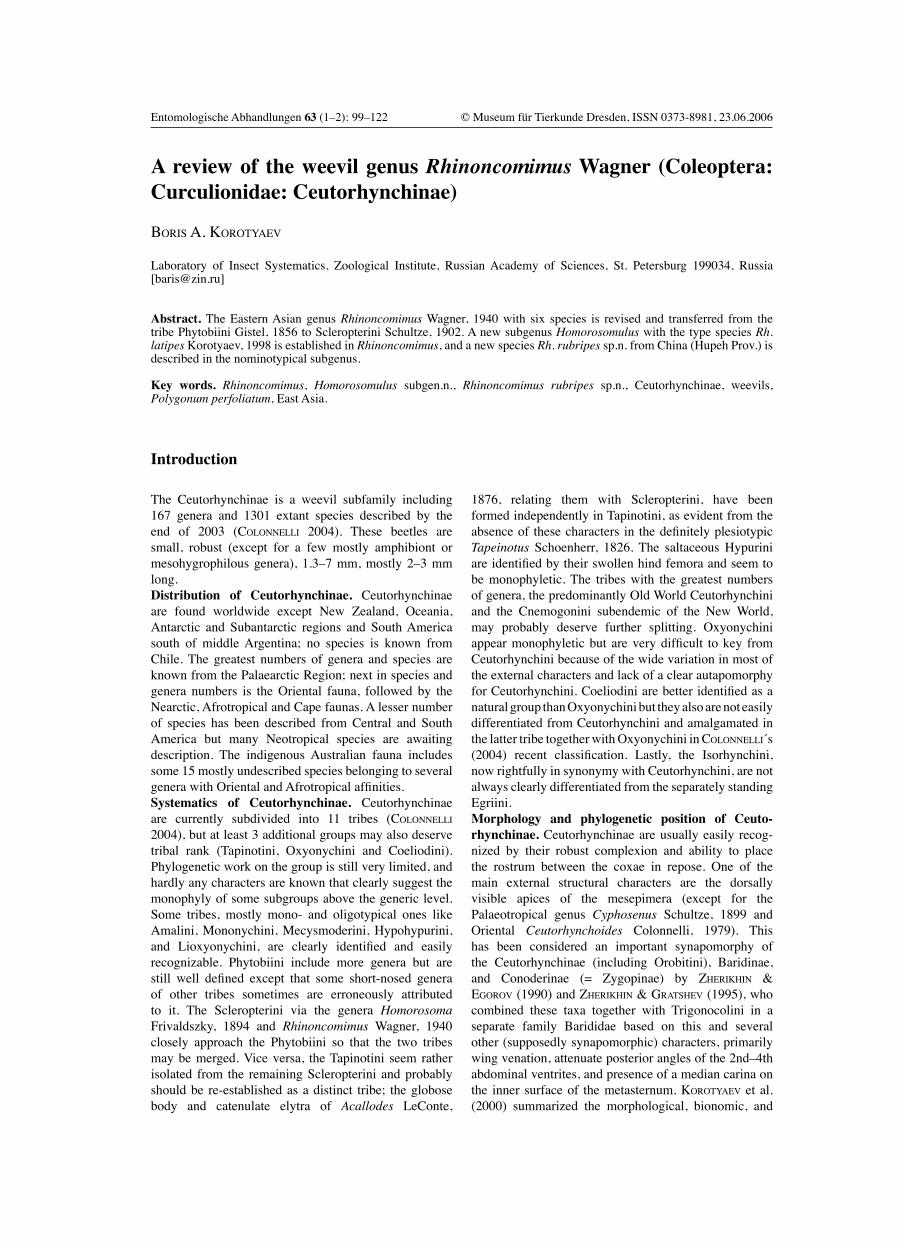
Description. 1.9–3.5 mm long, robust, with short rostrum and sparse dorsal vestiture of scales forming no distinct pattern other than more or less clearly defi ned scutellar spot and, in some species, ill-defi ned patches of narrow white scales at bases of 6th and 8th intervals of elytra (Fig. 1). Strial punctures with fi ne inconspicuous subrecumbent brown hairs. H e a d . Rostrum stout, shorter than pronotum, noti-ceably wider than forefemur (Figs. 2–7), weakly to rather strongly curved (Figs. 8–13), subcylindrical, with variably developed median carina; apical part of rostrum distinctly wider than basal part, parallel-sided or widening to the tip. Antennae inserted at about 1/3 length of rostrum from apex; scape moderately swollen and outcurved in apical 1/3, not reaching eye in repose, with 3 long setae at apex; funicle 7-segmented, fi ne, not fl attened, weakly widened apically; 2nd segment of funicle longest, 7th usually not wider than long. Club oblong-ovate, with 1st segment in some species separate (appearing like an 8th segment of funicle; Fig. 14). Eyes medium-sized, rounded-triangular to nearly round, moderately and evenly convex. Frons weakly depressed, slightly narrower than base of rostrum.P r o n o t u m . Weakly transverse, with base moderately protruding posteriorly in middle; basal margin smooth, not raised jointly with basal margin of elytra. Variably deep apical constriction separating a short to moderately long ring (Figs. 8–13); apical edge not raised and rarely weakly expanded collar-like, smoothly emarginate and slightly produced in middle, serrate laterally. Sides moderately or weakly convex. Lateral tubercles large and usually acute, conical or in the form of short transverse ridges. Disc moderately to strongly convex, often fl attened longitudinally in centre, with varyingly distinct oblique depressions before lateral tubercles. Median sulcus narrow, rather deep to obsolete. Punctation moderately coarse and usually regular, occasionally somewhat irregular and producing slightly uneven appearance of disc. Ocular lobes wanting; anterior margin of prothorax weakly concave on sides between ventral angle and dorsal part, bearing short scales pointed towards eyes. M e s o t h o r a x . Scutellum minute, deeply sunken. Mes-epimera visible dorsally, with outline weakly concave to almost straight, never convex. E l y t r a . Length subequal to width, with strongly convex although occasionally bevelled humeri, rounded-triangular or, more often, weakly narrowing in basal half and strongly narrowing in apical half, separately rounded at apex. Disc strongly and rather uniformly convex. Striae deep and wide, with distinct round punctures. Intervals usually uniform, variably strongly convex, occasionally almost costiform, with row of medium-sized sharp setiferous granules and smaller granules scattered along sides of intervals. 1st stria rather strongly incurved at
103Entomologische Abhandlungen 63 (1–2)
base, 2nd stria weakly incurved, 3rd and 4th striae almost straight. Preapical prominences obsolete to wanting, but 3rd+9th intervals often abruptly sloping toward 2nd+9th stria in sutural corner.L e g s a n d t h o r a c i c v e n t e r . Fore- and mid-coxae separated by not less than 1/2 basal width of rostrum; prosternum behind coxae shallowly depressed, with two oblique low folds, shallowly emarginate in middle. Mesosternal process wide, about as wide as apical part of rostrum, shallowly depressed, with rounded, rectangular, or slightly projecting posterior angles, forming one plane with metasternal process. The latter declivitous anteriorly or entirely deepened below mesocoxal level. Metasternum slightly depressed along midline, with medial part fl attened longitudinally; behind mid- and hind coxae moderately convex, somewhat shorter than mid-coxa. Femora long, moderately swollen, more strongly so in apical part but not distinctly clavate, all armed with small ventral spiniform tooth distally. Hind femur 1.15–1.30× as wide as mid-femur. Tibiae slender, weakly widening apically, usually parallel-sided in middle part, straight or weakly S-curved; apical combs short, on foretibia not or slightly extending on outer surface. Corbel on hind tibia short, with straight margin and angularly produced apex formed of dense and fi ne setae. Tarsi moderately long and narrow. 1st and 2nd tarsomeres in mid- and hind tarsi often weakly compressed, thicker than wide; 3rd noticeably less to somewhat more than twice as wide as 2nd (Figs. 18, 19), 4th minute but easily detectable, 5th in apical part moderately swollen and bearing dense long hairs ventrally, by more than half of length protruding from lobes of 3rd tarsomere. Claws large, appendiculate.A b d o m i n a l v e n t e r . Ventrites lying in one plane; 1st ventral suture more or less distinct along entire length, 2nd–4th sutures subequally deep. Punctation of venter more or less uniform and dense, moderately coarse. M a l e g e n i t a l i a . Aedeagus (Figs. 20–30) with ring-shaped tegmen lacking parameres; penis moderately to rather heavily sclerotized, weakly to strongly fl attened dorsoventrally, with membranous or weakly sclerotized medio-dorsal area and mostly membranous ventral sur-face. Apophyses of penis moderately long. S e x u a l d i m o r p h i s m . Moderate in external cha-racters. The rostrum slightly differs between sexes in length; the antennal insertion is shifted from the apex of the rostrum to approximately the same distance of about 1/3 the length of the rostrum in both sexes, similarly to Phytobiinae, whereas in most other Ceutorhynchinae except those with very short rostrum (Perigaster Dietz, 1896 and Perigasteromimus Colonnelli, 1998 of the Cnemogonini) the apical part of rostrum (the one distal to antennal insertion) in female is conspicuously longer than in male. In all species at least the mid- and the hind tibiae of the male are provided with a minute to rather long, acute mucro, present also on the foretibia in several species (Figs. 15–17). The female tibiae are always non-mucronate. The base of the venter in the female is more or less strongly convex, in the male it is fl at or somewhat depressed. The last visible ventrite is weakly evenly convex, fl at, or shallowly depressed near the
apex or along the midline in the female; fl at or weakly to moderately depressed in the male, lacking characteristic structures and erect setae at the base.Differential diagnosis. Rhinoncomimus clearly differs from Homorosoma in the very short rostrum (at least 3.45× as long as wide and always longer than pronotum in the latter) and the correlated characters of the thoracic anatomy: the forecoxae are separated by not less than 0.5 width of the rostrum (by much less than the width of the antennal club and less than 0.3 width of the rostrum in Homorosoma, except H. aterrimum (Hustache, 1916), in which this proportion comprises about 0.4); the prosternum is shallowly depressed, with oblique longitudinal folds behind the coxae and shallow median emargination (fl at or convex, without longitudinal folds, with straight or shallowly emarginate posterior edge in Homorosoma). The body in Rhinoncomimus is somewhat more compact, with the pronotum more transverse and convex, more strongly attenuate posteriorly in the middle at base. The elytral scaling in Rhinoncomimus is more conspicuous than in Homorosoma although the scutellar spot is often less defi ned. No erect setae typical of Homorosoma are present at the base of the last visible ventrite of the male in Rhinoncomimus.
Key to species of Rhinoncomimus
1 (8) Larger, body length 2.6–3.5 mm. Rostrum stout, 1.55–1.88× as long as wide, more strongly widening in apical part, at apex 1.13–1.28× as wide as at base (usually with obtuse, low median carina). Mandibles moderately to strongly projecting beyond clypeus. Disc of pronotum more strongly and not uniformly convex, often with well-developed oblique depressions before lateral tubercles. Elytra usually noticeably longer than wide, weakly nar-rowing or subparallel-sided in basal half. Granules on intervals of elytra small and not arranged in one regular row along interval. (In some species intervals of elytra with sparse lanceolate white scales, all tibiae of male with well-developed mucro.) .............................. (Subgenus Rhinoncomimus Wagner)2 (3) Frons shallowly to obsoletely depressed; rostrum weakly curved (Fig. 8). Disc of pronotum less strongly and almost evenly convex, (in lateral view) with apical constriction very shallow (Fig. 8), (in dorsal view) separated apical area shorter than width of antennal club. Elytra with fi ner granules on intervals, disc with more numerous lanceolate scales producing Rhinoncus-like appearance (Fig. 1). In male, mid- and hind tibiae with very long mucro (Figs. 16, 17); medial part of metasternum and 1st and 2nd ventrites covered with plumose scales. Aedeagus narrow, strongly fl attened in basal third and with complicated structure of the apex (Figs. 20–22). Body length 2.6–3.0 mm. [Continental China] ...................................... Rh. klapperichi Wagner3 (2) Frons distinctly depressed, rostrum more strongly and less regularly curved (Figs. 9–11). Disc of pronotum strongly convex, steeply sloping toward apical con-striction (Figs. 9–11); length of separated apical area

104 KOROTYAEV: Review of Rhinoncomimus
1.5× width of antennal club. Elytra with coarsely granulate intervals and less conspicuous scaling, thus not resembling Rhinoncus. In male, hind tibia fi nely mucronate or unarmed, vestiture of underside composed of uniform entire lanceolate scales, or only mesosternal process and medial part of metasternum with scales plumose apically. Aedeagus wider, less strongly (if at all) fl attened in basal part, with simple apex. [Continental China and Japan]4 (5) Disc of elytra with sparse lanceolate white scales more abundant on 2nd–4th intervals. Pronotum more strongly convex at base, more strongly declivitous anteriorly, with longer apical part (Fig. 9). All male tibiae mucronate. Last visible ventrite of male fl attened and densely, fi nely punctate but not foveate along midline. Last visible ventrite of female with obsolete round depression at apex. Aedeagus long, weakly curved and more strongly fl attened, shallowly constricted before apex, with narrow membranous medio-dorsal area and apex angularly bent (Figs. 23–25). [China].......................................................... Rh. robustus Voss5 (4) Lanceolate scales on elytra confi ned to scutellar spot, granules on intervals bearing only linear white and dark brown scales. Pronotum less strongly and more evenly convex, with shorter apical part (Figs. 10, 11). (Male unknown of one species included here). Only middle male tibia mucronate. Last visible ventrite of male with moderately deep depression, that of female with well-developed oblong depression. Aedeagus short, more strongly curved and not fl attened in basal 2/3, evenly convexly narrowing apically, with wide membranous medio-dorsal area and apex slightly bent (Figs. 26, 27). [Japan, Eastern China]6 (7) Larger, body length 3.0–3.5 mm. Antennae and entire legs black or very dark brown. Rostrum of female shorter and wider, 1.81× as long as wide, 0.76× as long as pronotum; at apex 1.28× as wide as at base. Pronotum slightly more transverse, 1.27–1.30× as wide as long, less strongly convex dorsally, distinctly fl attened longitudinally; base less protruding posteriorly in middle. Elytra slightly longer (1.10× as long as wide), less strongly narrowing apically (Fig. 32), more regularly convex in cross-section, with granules on intervals slightly smaller and less regularly arranged. Posterior margin of mesosternum concave, with posterior angles somewhat raised against mid-coxal cavities. Tarsi slightly wider, 1st tarsomere 2×, 2nd 1.25× as long as wide. Last visible ventrite of female with oblong depression, lacking long white hair-like scales. Only middle male tibia mucronate. Last visible ventrite of male with moderately deep depression, that of female with well-developed oblong depression. Aedeagus short, more strongly curved and not fl attened in basal 2/3, evenly convexly narrowing apically, with wide membranous medio-dorsal area and apex slightly bent (Figs. 26, 27). [Japan] .................................... Rh. niger Chûjô & Morimoto7 (6) Smaller, body length 2.75 mm. Antennae, tibiae, and tarsi bright reddish brown. Rostrum in female longer, 2.12× as long as wide, 0.85× as long as pronotum; at apex 1.13× as wide as at base. Pronotum slightly narrower,
1.26× as wide as long, more strongly convex, not fl attened longitudinally, with base more strongly angularly produced posteriorly. Elytra slightly shorter (1.05× as long as wide), more strongly narrowing apically (Fig. 33), somewhat fl attened along suture and along sides, with granules on intervals slightly larger and on 4–7th intervals arranged almost in regular row. Posterior margin of mesosternum almost straight, with posterior angles not raised against mid-coxal cavities. Tarsi slightly narrower, 1st tarsomere 3×, 2nd 1.25× as long as wide. Last visible ventrite of female with round depression bearing narrow white recumbent hair-like scales. (Male unknown.). [Eastern China] ............................ Rh. rubripes sp.n.8 (1) Smaller, body length 1.95–2.55 mm. Rostrum more slender, 2.40–2.87× as long as wide, 1.0–1.10× as wide at apex as at base (usually with sharp median carina). Mandibles not or slightly projecting beyond clypeus. Disc of pronotum less strongly and more regularly, often uniformly convex, occasionally in females with obsolete oblique depressions before lateral tubercles. Elytra about as wide as long, or slightly wider than long, rounded-triangular, with intervals bearing one row of larger granules along midline. Lanceolate white scales on elytral intervals confi ned to scutellar spot. Male tibiae unarmed or very fi nely mucronate. ....................... (Subgenus Homorosomulus subgen.n.)9 (10) 3rd tarsomere slightly less than twice as wide as 2nd (Fig. 19). 1st tarsomere in mid- and hind tarsi compressed, thicker than wide. Rostrum with high, sharp median carina along entire length; frons moderately deeply depressed (Fig. 12). Pronotum more transverse, less rounded at sides (Fig. 34); lateral tubercles transverse, with narrow shining ridge. Each interval of elytra with one regular row of large granules. Mucro on male fore- and hind tibiae minute but clearly visible, that on mid-tibia well developed. Aedeagus (Figs. 28, 30) with more narrowly attenuate apex. Last visible ventrite of female usually with round median depression in apical part, there occasionally fl attened and more coarsely punctate. Body length 2.0–2.55 mm. [SE continental China, Taiwan, Republic of Korea] ........................................ Rh. rhytidosomoides Wagner10 (9) 3rd tarsomere more than twice as wide as 2nd (Fig. 18). 1st tarsomere in mid- and hind tarsi not conspicuously compressed, not thicker than wide. Rostrum with lower, often obtuse median carina; frons very shallowly depressed (Fig. 13). Pronotum less transverse, more rounded at sides, with conical lateral tubercles (Fig. 35). Intervals of elytra with small and less regular granules along midline and with conspicuous smaller granules along sides. Male foretibia without mucro; that on mid-tibia minute; on hind tibia, barely visible under hairs. Aedeagus (Fig. 29) with more rapidly narrowing apex. Last visible ventrite of female evenly convex and fi nely punctate in apical part. Body length 1.95–2.50 mm. [S of the Russian Far East, Korea, SE continental China]..................................................... Rh. latipes Korotyaev
105Entomologische Abhandlungen 63 (1–2)
Subgenus Rhinoncomimus Wagner, 1940
Differential diagnosis. The nominotypical subgenus differs from Homorosomulus subgen.n. in the larger body size, wider and shorter rostrum with more strongly widened apical part and largely exposed mandibles (more than shown in Figs. 2–7), less regularly and more strongly convex pronotum with well-developed oblique depressions, and longer and less rounded elytra. The granules on the elytral intervals are somewhat smaller and less regularly arranged. (See identifi cation key for details.)Host plants. No data on the host plants are available, but these are most likely neither Polygonum perfoliatum nor P. thunbergii on which representatives of the subgenus Homorosomulus feed, as no sample examined from China contains representatives of both subgenera. It is noteworthy that the holotype of Rh. rubripes sp.n. has collection data identical with those of two specimens of Rh. klapperichi; it is not unlikely that species of the nominotypical subgenus, similar to species of the subgenus Homorosomulus, are associated with a narrow host group.
Rhinoncomimus klapperichi Wagner, 1940
WAGNER 1940: 79/179; VOSS 1958: 66; COLONNELLI 1986: 157; KOROTYAEV 1997: 288.
Description. Measu remen t s . Body length 2.6–3.0 mm.C o l o r a t i o n . Body black; mouthparts, antennae, tibiae, and tarsi reddish brown, femora very dark reddish brown; swollen apex of antennal scape and base of funicle paler brown.Ve s t i t u r e . Rather dense, similar to that in some species of Rhinoncus, e.g., Rh. sibiricus Faust, 1893, as noticed by WAGNER (1940). Rostrum with moderately dense narrow recumbent white scales widening apically to varying extent, wider and shorter scales bordering upper eye margin and arranged in diffuse median line on frons and vertex; temples with few oval scales. Pronotum with more or less extensive white scaling thinned on either side along median sulcus and occasionally reduced to narrow lines along margins and in median sulcus. Elytra with diffuse scutellar spot, diffuse patches of white lanceolate scales on bases of 4th and 6th intervals and on posterior slope of humeral prominence on 8th interval, and with sparse to moderately dense shorter lanceolate scales along most of 2nd, 3rd, 8th, and 9th intervals. Rest of elytra usually with scanty white scales, sutural interval dark in middle 2/3 of its length. Underside with moderately dense lanceolate white scales separated mostly by more than own width and not conspicuously condensed at sides except for white line along outer margin of metepimera. In male, medial part of metasternum and 1st ventrite densely covered with wide plumose scales; in female, underside with uniform lanceolate scales. Legs moderately densely clothed with linear white and yellow scales. Pygidium with fi ne yellow and white hairs.
H e a d . Rostrum in male 1.72×, in female 1.55–1.64× as long as wide, 0.72× and 0.69–0.72× as long as pronotum respectively, 1.3× as wide as forefemur; at apex in both sexes 1.26× as wide as at base. In lateral view, rostrum weakly curved, separated from vertex by very shallowly depressed frons, neither conspicuously tapering nor swollen apically. Ventral margin more deeply concave in basal half, and dorsal outline somewhat more steeply sloping in apical half. In dorsal view, rostrum moderately and gradually widening in apical half. Dorsal surface moderately convex in cross-section, obtusely raised along midline, weakly shining in basal part, with shallow elongate medium-sized punctures on either side, arranged in two confused rows partly separated by ill-defi ned wrinkles. Apical part of rostrum smooth, with sparse small punctures. Frons 0.74–0.84× as wide as rostrum at base, very shallowly depressed; frons and vertex weakly shining, uniformly covered with shallow medium-sized punctures. Antennae in male inserted at 0.40, in female at 0.37–0.43 length of rostrum from apex. Scape rather strongly bent and moderately swollen apically, with long and narrow apical translucent process. Funicle long, slender; 1st segment about twice as long as wide, 2nd segment about as long and half as wide as 1st, succeeding segments gradually shortening, 7th segment 1.5× as long as wide. Each segment with rosette of fi ne light semi-erect hairs longer on apical part of funicle. Basal segment separated from club to form obconical pseudo-8th segment of funicle (Fig. 14), noticeably wider than 7th segment and matte, similar to rest of club, but separated from it by a shallow constriction. Second segment of club short-pedunculate. Eyes medium-sized, almost round, moderately convex.P r o n o t u m . Width 1.38× length; base obtuse-angularly protruding backward along entire width, sides rather weakly rounded, weakly convexly converging to very shallow apical constriction separating short apical area. Apical margin not raised, its frontal section not dilated (see in front view), somewhat protruding above head and smoothly emarginate in middle. Lateral tubercles medium-sized, weakly transverse. Disc weakly to moderately and almost evenly convex, with slight depressions medio-anteriad to lateral tubercles. Surface moderately shining, evenly punctate; punctures medium-sized, shallow, separated by narrow but fl at, shining intervals. M e s o t h o r a x . Scutellum minute, almost punctiform. Mesepimera very narrowly visible dorsally, with clearly concave outline.E l y t r a . As long as wide, 1.61× as wide as pronotum, with strongly convex humeral prominences and weakly rounded sides, weakly narrowing from humeri to middle and then strongly narrowing to conjointly rounded apices. Disc moderately and rather evenly convex ex-cept for shallowly depressed postscutellar area. Striae deep, moderately wide, entire, weakly raised between punctures. First stria moderately, 2nd weakly bent toward scutellum at base, 3rd stria straight. Intervals about 1.5× as wide as striae, strongly convex, except for fl at sutural interval. All except sutural interval with two or three irregular rows of small rounded granules. Apical part of

106 KOROTYAEV: Review of Rhinoncomimus
elytra without distinct depression along 2nd+9th stria in sutural corners.L e g s a n d t h o r a c i c v e n t e r . Forecoxae separated by about 0.75× basal width of rostrum. Mid-coxae separated by apical width of rostrum. Posterior half of mesosternum slightly depressed, area between mid-coxae declivitous. Metasternum between mid- and hind coxae weakly convex, slightly shorter than mid-coxa. Posterior part of metasternum between hind coxae shallowly depressed. Legs moderately long. Femora weakly swollen along most of length, not clavate; hind femur 1.15× as wide as mid-femur. Tibiae long, slender; foretibia weakly S-curved, with rounded outer apical angle and well-developed mucro on inner angle. Mid- and hind tibiae weakly widening apically, almost straight; mucro on mid-tibia stout, pointed medially; that on hind tibia very long, straight, narrow, obliquely truncate at apex. Tarsi moderately long; 1st tarsomere about twice, 2nd almost 1.5× as long as wide, 3rd wide, twice as wide as 2nd; 5th strongly widened in apical half, by somewhat more than half of length extending from lobes of 3rd tarsomere. Claws large, with wide appendages in basal half. A b d o m i n a l v e n t e r . 1st ventrite slightly depressed between hind coxae; 1st ventral suture distinct along entire length, linear. All ventrites lying in one plane; 2nd–5th ventrites fl attened medially. Last visible ventrite in posterior half with shallow and narrow median depression covered with fi ne brown semi-erect reclinate hairs. Pygidium weakly transverse, moderately and evenly convex, slightly swollen before apex, with swelling produced in minute tubercle. Punctures on pygidium medium-sized, well defi ned, moderately dense; intervals between punctures slightly convex, shining, obsoletely microreticulate.M a l e g e n i t a l i a . Penis very long, length more than 1/3 length of body, heavily sclerotized, with fl attened basal part, sclerotized medio-ventral area, and two deep lateral excisions near apex (Figs. 20–22). The aedeagus of the holotype is fi gured in KOROTYAEV (1997: fi g. 14), but the complicated structure of the aedeagal apex is not shown there.Differential diagnosis. The species is easily distinguished from Rh. robustus by the slightly depressed frons, relatively weakly curved rostrum, long-pedunculate antennal club, weakly and evenly convex and rather fi nely punctate disc of pronotum with obsolete depressions anteromedial to the medium-sized lateral tubercles, fi ne granulation of the elytral intervals, and rather dense dorsal scaling which produces a Rhinoncus-like appearance. The males are unique among Rhinoncomimus in having very long mucrones on the mid- and hind tibiae, dense plumose scaling of the medial part of the metasternum and 1st and 2nd ventrites, and the complicated structure of the apex of the aedeagus (Figs. 20–22). VOSS (1958) misinterpreted this species in the key differentiating Rh.robustus from Rh. klapperichi.Distribution. Southeastern China.
Material. Holotype, P, CHINA, Fukien (ZFMK) and Para-types, 5O, see WAGNER (1940) for label data. – CHINA: 1P,Fukien, Pu Cheng (T.C. Maa, ex coll. L. Gressitt) (CAS); 1O,
Fukien, Tachuland, 05.v.1942 (T.C. Maa) (Bishop Museum); 1O, Fukien, Chungan City, 22.viii.1940 (T.C. Maa) (Bishop Museum); 1O, W Hupeh Prov., Lichuan Distr., Suisapa, 1000 m, 31.viii.1948 (Gressitt & Djou) (CAS); 2P, W Hupeh Prov., Leong-ho-kow, 09.ix.1948 (Gressitt & Djou) (CAS); 1O, as foregoing specimens but 10.ix.1948 (CAS).
Rhinoncomimus robustus Voss, 1958
VOSS 1958: 66; COLONNELLI 1986: 157 (Rh. klapperichisynonym); KOROTYAEV 1997: 288 (as a distinct species).
Description. Measu remen t s . Body length 3.0–3.5 mm.C o l o r a t i o n . Body black; mandibles, antennae (uni-formly) and tarsi (paler apically) dark brown; occasionally articulating part of femora and base of tibiae dark brown; antennae always paler than other parts. Ve s t i t u r e . Sparse; head with linear recumbent brown and white scales forming no conspicuous pattern, with short wider grey scales along punctate part of occiput. Pronotum with ill-defi ned lines of sparse narrow-lanceolate white scales along margins, in median sulcus, and in oblique depressions before lateral tubercles. Elytra with diffuse oval scutellar spot, patches of lanceolate white scales on base of 6th interval and on posterior slope of humeral prominence on 8th interval. Shorter semi-erect white scales scattered around midlength of 2nd–4th intervals, along most of 7th interval, and in sutural corners of elytra. Scales on disc often sitting in pairs across interval. Sutural interval between scutellar spot and apex dark, clothed only with narrow dark brown scales, but with few small white scales along inner margin near apex. Underside with lanceolate white and greyish scales in punctures, denser on thorax although not concealing any sclerite, and sparser, shorter, and narrower on abdomen clothed mostly with recumbent brown hairs. Scales on mesosternal process and, partly, on medial area of metasternum with plumose apices. Last visible ventrite covered mostly with hairs, augmented with few small scales in medio-basal part and at sides. Legs clothed with recumbent hairs and linear scales, with wider white scales dressing femoral teeth.H e a d . Rostrum stout; in male 1.88×, in female 1.73× as long as wide, 0.75× as long as pronotum, 1.45× (in male) and 1.65× (in female) as wide as forefemur; at apex 1.28× and 1.27× as wide as at base in male and female, respectively. In lateral view, rostrum strongly curved, with curvature of ventral surface in male almost regular or strongest at base, and that of dorsal surface strongest at antennal insertion. Distal to antennal insertion, rostrum moderately widening apically. Median carina narrow, obtuse, reaching anterior part of frons and widening to a short fl attened convexity at antennal insertion; occasionally the convexity prolonged apically gradually narrowing. Surface weakly shining, with dense, rather fi ne, shallow, elongate punctures more or less arranged in rows; no distinct lateral carinae present. Antennae inserted at 0.3 length of rostrum from apex; apical third of scape moderately swollen. Funicle slender; 6th segment about twice as long as wide, 7th noticeably longer than wide. Club spindle-shaped, with short basal segment

107Entomologische Abhandlungen 63 (1–2)
slightly attenuate and bearing rosette of long setae at apex, but forming no pronounced peduncle and clearly separated from funicle. Eyes medium-sized, moderately convex, and almost round. Frons slightly narrower than base of rostrum, shallowly depressed, weakly shining, with shallow, mostly polygonal punctures of varying size. Vertex with similar sculpture and well-developed median carina.P r o n o t u m . Width 1.25–1.30× length; base weakly protruding posteriorly in middle; sides weakly rounded, occasionally subparallel in basal half, moderately converging apically to rather deep apical constriction separating long apical part. Anterior edge shallowly emarginate in middle, serrate at sides. Disc strongly convex, with central part somewhat fl attened longitudinally (visible in lateral view), steeply sloping toward base, and somewhat more gently sloping toward deep apical constriction. Lateral tubercles large, acute; depressions separating them from convex central part of disc well developed, usually reaching deep and narrow median sulcus. Punctures medium-sized, not very deep, separated mostly by shining, slightly convex intervals. M e s o t h o r a x . Scutellum minute, deeply sunken.E l y t r a . In male 1.06–1.08×, in female 1.0–1.07× as long as wide, with strongly prominent humeri and sides subparallel or slightly converging in basal half, and strongly roundly converging in apical half; preapical pro minences obsolete. Disc strongly convex, except forshallowly depressed small area behind scutellum. Inter-vals somewhat wider than striae, strongly convex, shining; each, starting with 2nd, with more or less regular row of large granules along midline and with numerous fi ner granules along sides. Apices of 3rd–9th intervals abruptly sloping toward 2nd+9th stria in sutural corner, apical area separated by noticeable bend (visible in latero-posterior view).L e g s a n d t h o r a c i c v e n t e r . Forecoxae separated by 2/3 basal width of rostrum; posterior part of prosternum and mesosternum shallowly depressed medially, metasternal process not depressed. Entire metasternum convex longitudinally, more strongly so between mid- and hind coxae, and obsoletely depressed along linearly engraved midline. Legs long; femora moderately swollen in apical part; hind femur 4× as long as wide, 1.25× as wide as mid-femur. Tibiae straight, slender, slightly widening apically; foretibia 5.61–6.38× as long as wide. In male, all tibiae with well-developed mucro, on foretibia half as long as remainder. Tarsi narrow; 1st tarsomere twice as long as wide, 2nd 1.3–1.4× as long as wide, 3rd slightly shorter than, and almost twice as wide as 2nd, 5th tarsomere narrow at base and moderately widening apically extending from lobes of 3rd tarsomere by 2/3. Ventral surface of 5th tarsomere in basal half bare, in apical half with dense long semi-erect to erect hairs.A b d o m i n a l v e n t e r . All ventrites lying in one plane. In male, venter fl attened along midline, rather uniformly and fi nely punctate, nearly matte, with intervals between shallow punctures coarsely microreticulate. Last visible ventrite in male fl attened, without any trace of median depression, with punctation becoming fi ner and denser
toward apex of ventrite. In female, venter slightly convex, last visible ventrite slightly convex in cross-section; in apical part, also convex longitudinally and shallowly depressed near apex. Pygidium weakly transverse, almost matte, densely fi nely punctate, weakly convex in male and almost fl at in female, with moderately dense subrecumbent brown hairs longer and more strongly raised apically near midline and clearly visible dorsally.M a l e g e n i t a l i a . Penis long, length about 1/3 length of body, with sides heavily sclerotized along entire length, not conspicuously fl attened dorsally in any part, with entire apex bent ventrally at right angle (Figs. 23–25).Differential diagnosis. This is the largest species with the most strongly dorsally convex elytra and pronotum. It can be distinguished easily from Rh. klapperichi by the more deeply depressed frons and pronotal disc more abruptly sloping toward apical constriction (Fig. 9) and having coarser punctation and well-pronounced oblique depressions before lateral tubercles. The apical part of the pronotum separated by the constriction is longer than in the congeners, its length being about 1.5× width of the antennal club (Fig. 31). The scaling of the elytra may be sparser than in Rh. klapperichi but the scales are wider. Males of these two species can be distinguished easily by the length of mucro on the mid- and hind tibiae which is greater in Rh. klapperichi, and by the structure of scales at the base of the abdomen, entire in Rh. robustus and plumose in Rh. klapperichi.Distribution. Southeastern China.
Material. The holotype of this species from J. Klapperich’s collection is not in the A. Koenig Museum, Bonn; I could not fi nd it in any other European museum. Although the holotype has not been found and E. Voss misinterpreted Rh. klapperichi,which apparently was not known to him, it is likely that the holotype is conspecifi c with the four paratypes examined. – Paratypes 3P, 1O. 1P, CHINA (ZFMK); 1P, CHINA, Fukien, 20.vi.1946 (Tschung Sen) (Museum G. Frey; now in Basel); 1P, CHINA, Fukien (DEI); 1O, CHINA; “Kuatun, Fukien, China, 8.6.46 (Tschung Sen)”, red label “Type [printed] Rhinoncomimus robustus n.sp. det. Voss”, “= Rhinoncomimusklapperichi Wagner, det. Colonnelli [?]” (NHRM). – CHINA: 1P, 1O, Fukien, Schaowu, Tachuland, 01.vi. and 25.iv.1943 (T.C. Maa) (Bishop Museum); 1P, as foregoing specimens, but 1000 m, 01.v.1942 (Bishop Museum); 1P, as foregoing specimens, but 08.vii.1946 (Bishop Museum); 3O, Tachuland, 01. and 08.vii.1942 (T.C. Maa for L. Gressitt collection) (CAS).
Rhinoncomimus niger Chûjô & Morimoto, 1959
CHÛJÔ & MORIMOTO 1959: 154.
Description. M e a s u r e m e n t s . Body length 3.0–3.5 mm (according to original description; in four examined specimens, 3.0–3.1 mm).C o l o r a t i o n . Body black, antennae and tarsi dark brown.Ve s t i t u r e . Fine, sparse. Head with moderately dense white subrecumbent linear scales more conspicuous on sides of rostrum in apical part and along midline of head. Pronotum with sparser narrow-lanceolate white scales on sides, along lateral margins of disc, and in median sulcus. Elytra with diffuse scutellar spot of narrow-lanceolate white scales, with few greyish scales
108 KOROTYAEV: Review of Rhinoncomimus
at extremities of latter. Disc clothed mostly with brown subrecumbent linear scales, with few white linear scales scattered along margins of discal intervals and arranged in ill-defi ned patches on base of 6th interval, behind humeral prominences on 8th interval, and in sutural corners. Vestiture of underside as in Rh. robustus, but slightly denser, white scales somewhat coarser, almost concealing apices of mesepimera.H e a d . Rostrum in male 1.77×, in female 1.81× as long as wide, 0.80× and 0.76×, respectively, as long as pronotum, 1.5× as wide as forefemur, moderately and almost evenly curved; at apex 1.25× and 1.28× as wide as at base in male and female, respectively. Dorsal surface of rostrum matte, with fi ne, somewhat rugose punctation and fi ne linear median carina in basal part (in male), or weakly ridged along smooth median line (in female). Antennae inserted in male at 0.33, in female at 0.40 length of rostrum from apex, structure similar to Rh.robustus. Eyes medium-sized, round with straightened anterior and ventral margins, rather strongly convex. Frons scarcely narrower than base of rostrum, shallowly but conspicuously depressed.P r o n o t u m . Width 1.27–1.30× length; base moderately projecting posteriorly in middle; apex slightly raised and shallowly emarginate in middle, serrate at sides. Sides moderately rounded and converging to shallow apical constriction. Disc moderately convex, fl attened longitudinally around midlength, and equally sloping toward base and toward apical constriction. Lateral tubercles large, in the form of short transverse arcuate folds. Sides of disc obliquely depressed before tubercles. Median sulcus rather deep and narrow. Punctation moderately dense, somewhat irregular; shining intervals between medium-sized punctures in places wider and slightly swollen making surface uneven. E l y t r a . In male 0.99×, in female 1.10× as long as wide, with angular, strongly prominent humeri, weakly rounded and almost not narrowing in basal half and strongly narrowing in apical half. Preapical prominences not protruding from elytral outline but abruptly sloping toward 2nd+9th stria in sutural corners (visible in latero-posterior view). Disc strongly convex, somewhat fl attened along suture. Striae wide and very deep, intervals strongly convex, nearly costiform. 3rd, 5th, and 7th intervals more strongly convex and somewhat wider than even-numbered intervals. Each interval with row of medium-sized elongate pointed granules along midline and with smaller granules along sides.L e g s a n d t h o r a c i c v e n t e r . Forecoxae separated by 2/3 basal width of rostrum. Legs long. Hind femur 4.0× as long as wide, 1.2× as wide as mid-femur. All tibiae slender, parallel-sided in middle part, weakly widened and outcurved apically. Foretibia 6.25× as long as wide. In male, only mid-tibia fi nely mucronate. Tarsi narrow; 1st tarsomere noticeably compressed, in all tarsi thicker than, and about twice as long as wide; 2nd about 1.25× as long as wide, 3rd 1.75× as wide and 0.9× as long as 2nd, 5th slender at base and moderately widened in apical half, by slightly more than half of length extending beyond lobes of 3rd tarsomere.
A b d o m i n a l v e n t e r . Surface almost matte, with moderately dense punctures and fi nely wrinkled intervals between them. In male, 1st ventrite slightly depressed, 2nd–4th ventrites fl attened medially, last visible ventrite with weakly transverse shallow median depression along most of length, lacking erect setae in and along depression. In female, base of abdomen weakly convex, 3rd and 4th ventrites fl attened medially, last visible ventrite shallowly depressed along midline, evenly punctate. Pygidium as in Rh. robustus.M a l e g e n i t a l i a . Penis medium-long, length about 1/4 length of body, wide, moderately and evenly curved dorsoventrally, not fl attened, with rather narrow sclero-tized lateral areas and evenly narrowed apex slightly bent ventrally (Figs. 26, 27).Differential diagnosis. This species differs from Rh.klapperichi and Rh. robustus in the absence of lanceolate white sales on elytral disc, less strongly widened apically rostrum, more strongly convex eyes, less convex pronotal disc and in the non-mucronate fore- and hind tibiae in male. From Rh. rhytidosomoides and Rh. latipes, it differs in its larger body size, longer and less regularly rounded elytra and coarser punctation of the irregularly convex disc of pronotum.
Material. Types of this species have not been examined. In the original description, the holotype and a female paratype are stated to be in the collection of the Entomological Laboratory of the Kyushu University, and two male paratypes in the private collections of K. Morimoto and M. Chûjô. A few years ago, Professor Morimoto (pers. comm.) in an e-mail message answering my request for the types informed me that the type material was on a long-time loan to a colleague. – JAPAN: 1P,Honshu, Toyama Pref., Nei-gun, Yamada-mura, Numanomata, 05.v.1992 (D.G. Furth & K. Suzuki) (ZIN); 1P, Naka-Niikawagun, Kamiichimachi, Ooiwa, 10.v.1992 (D.G. Furth & K. Suzuki); 2O, Kurobe-shi, Higashi-Fuse, Kasayaburi, 23.iv.1992 (D.G. Furth & K. Suzuki) (ZIN). – In addition, a series in the COBR was examined.
Rhinoncomimus rubripes sp.n.
Description(O). Measurements . Body length 2.75 mm.C o l o r a t i o n . Body black; mandibles, antennae, apices of femora, tibiae, and tarsi reddish brown; femora very dark brown, almost black. Ve s t i t u r e . Very similar to that of Rh. niger, but slightly coarser and more conspicuous; in particular, brown linear scales along most of 1st interval of elytra clearly visible, and white linear scales present in middle part of 2nd and 3rd and in apical part of 7th and 8th intervals. Venter with sparse brown linear scales prevailing, white lanceolate scales present on base of 1st ventrite and at sides of all ventrites; 5th ventrite with narrow, almost hair-like scales at middle. Apices of mesepimera with condensed yellowish lanceolate scales.H e a d . Rostrum 2.12× as long as wide (Fig. 5), 0.85× as long as pronotum, 1.62× as wide as forefemur, moderately and almost evenly curved (Fig. 11); at apex 1.13× as wide as at base. Dorsal surface of rostrum almost matte, with fi ne, somewhat rugose punctation and fi ne linear median carina in middle of length; elongate punctures partly merging in striae. Antennae inserted at

109Entomologische Abhandlungen 63 (1–2)
0.38 length of rostrum from apex, their structure similar to that in Rh. niger. Eyes medium-sized, almost round with straightened ventral margin, rather strongly convex. Frons scarcely narrower than base of rostrum, shallowly but conspicuously depressed, with inner margins of eyes rather steeply raised; punctures on frons dense, round and shallow.P r o n o t u m . Width 1.26× length; base more strongly angularly projecting posteriad than in Rh. niger. Disc more strongly and evenly convex, not conspicuously fl attened longitudinally, equally sloping toward base and toward apical constriction. Middle part of disc more sharply separated from lateral tubercles by oblique depressions. Median sulcus deeper and narrower along entire length than in Rh. niger. Punctation somewhat denser than in Rh. niger, with narrower matte intervals and wider shining areas in places, mostly along median sulcus.E l y t r a . 1.05× as long as wide, with angular, strongly prominent humeri, weakly rounded behind humeri and strongly narrowing in apical 2/3 (Fig. 33). Preapical prominences not protruding from elytral outline but abruptly sloping toward 2nd+9th stria in sutural corners (visible in latero-posterior view). Disc moderately convex, somewhat fl attened along suture and along sides in apical half. Sculpture as in Rh. niger but granules on intervals slightly larger and more regularly arranged, on 4–6th intervals forming almost regular row.L e g s a n d t h o r a c i c v e n t e r . Forecoxae separated by 2/3 basal width of rostrum. Antero-ventral corner of mesepimera impunctate, shining. Posterior margin of mesosternum almost straight, with posterior angles not raised against mesocoxal cavities. Legs as in Rh. niger.Hind femur 4.2× as long as wide, 1.3× as wide as mid-femur. Foretibia 6.0× as long as wide. Tarsi narrow; 1st tarsomere noticeably compressed, in all tarsi thicker than, and about 3× as long as wide; 2nd tarsomere about 1.5× as long as wide, 3rd slightly longer than, and 1.8× as wide as 2nd, 5th slender at base and moderately widened in apical half, by about 2/3 of length extending beyond lobes of 3rd tarsomere.A b d o m i n a l v e n t e r . Surface shining, with sparse, small, superfi cial punctures. Base of abdomen weakly convex, 3rd and 4th ventrites fl attened medially, last visible ventrite with shallow round depression along midline, more densely and coarsely punctate than 4th ventrite. Pygidium weakly transverse, weakly and evenly convex, moderately densely fi nely punctate, punctation weakening along rounded posterior margin. Apices of elytra narrowly hanging over dorsal margin of pygidium.Differential diagnosis. This species is very similar to Rh.niger differing in the longer rostrum, which is narrower in its basal part and less strongly widened in its apical part; narrower pronotum with more strongly convex central part of disc limited by deeper depressions latero-basally; narrower, less rounded, subtriangular elytra with slightly larger and more regularly arranged granules on the intervals; straight posterior margin of the mesosternum; and bright reddish antennae, tibiae, and tarsi.
Material. Holotype O, “CHINA, W. Hupeh, Leong-ho-kow, Lichuan. ix.9.48”, “Gressitt & Djou Collectors” (CAS).
Subgenus Homorosomulus subgen.n.Type species Rhinoncomimus latipes Korotyaev, 1997.
Differential diagnosis. The new subgenus differs from the nominotypical one in the smaller body-size, narrower and longer rostrum with less widened apical part, and less developed mucro on the male tibiae. The mandibles are concealed by the clypeus or are only narrowly exposed. The structure of the aedeagus is similar to that in Rh.niger: the penis tube is short and only slightly fl attened dorsally, with moderately wide and poorly sclerotized medio-dorsal area, membranous ventral side, and simple apex. (See identifi cation key for details.)Host plants. Both species of this subgenus are apparently associated with two closely related species of the genus Polygonum Linnaeus, 1753: P. perfoliatum and P.thunbergii.
Rhinoncomimus rhytidosomoides (Wagner, 1944)
WAGNER 1944: 100 (282) (Homorosoma); VOSS 1958: 67 (?Rhinoncomimus); KOROTYAEV 1997: 287; HONG et al. 2000: 115.
Description. M e a s u r e m e n t s . Body length 1.95–2.55 mm, usually 2.2–2.4 mm.C o l o r a t i o n . Body black; antennal funicle, club and tarsi very dark brown. Ve s t i t u r e . Inconspicuous, fi ne and sparse. Sides ofrostrum, frons, and vertex with few white hair-like scales, otherwise with inconspicuous dark brown hairs and narrow scales. Pronotum with sparse very narrow parallel-sided white sales on sides and in median sulcus, with few narrow-lanceolate scales in prescutellar fovea. Scutellar spot ill-defi ned, strongly narrowing toward base, formed by long, very narrow, lanceolate white scales confi ned to inner half of sutural interval. Sutural interval behind spot without white scales and with few inconspicuous dark scales. Granules on elytral intervals bearing dark brown or white reclinate linear scales forming no distinct pattern. Femora and tibiae with sparse subrecumbent dark brown and white hairs and linear scales, tarsi with more strongly raised hairs and (on dorsal surface near apices of tarsomeres) linear scales. Underside with sparse white lanceolate scales separated by more than own width. Pygidium with sparse, short, subrecumbent brown hairs.H e a d . Rostrum in male 2.40–2.70×, in female 2.65–2.71× as long as wide, 0.80–0.90× and 0.88–0.96× as long as pronotum, respectively; 1.25–1.40× as wide as forefemur, weakly narrowing behind antennal insertion, slightly wider and parallel-sided apically; at apex 1.0–1.10× as wide as at base. Ventral margin of antennal scrobe narrowly visible dorsally at short distance around antennal base. In lateral view, rostrum obsoletely curved along most of length, with slightly stronger curvature at base; ventral margin nearly straight, dorsal margin evenly moderately

110 KOROTYAEV: Review of Rhinoncomimus
curved. Low but sharp median carina running from base of rostrum to epistome. Punctation dense and coarse; punctures oblong, with fl at, microreticulate bottoms, separated by narrow intervals merging longitudinally in places but forming no lateral carinae. Antennae in male attached at 0.33, in female at 0.38 length of rostrum from apex. Scape moderately swollen in apical part. Funicle medium-long, clearly 7-segmented; 7th segment about as long as wide. Club ovate, widely rounded at base and not pedunculate, with apical half bearing sparse long pale setae about half-length of 7th segment of funicle. Frons subparallel-sided in anterior third and moderately widening posteriorly, moderately depressed across entire width so that the inner eye orbits are steep. Punctures on frons large, superfi cial, with margins partly obliterated especially in posterior half of frons; punctures on vertex smaller, round, more distinct. Vertex with varyingly long and high median carina. Eyes medium-sized, moderately convex, rounded, with angular antero-ventral area.P r o n o t u m . Width 1.25–1.33× length; base weakly produced posteriad in middle; apical edge slightly raised and shallowly emarginate over head, lateral to emargination obsoletely undulate, lacking distinct serration. Sides weakly rounded, slightly diverging from base and moderately converging to well-pronounced apical constriction separating short ring. Disc strongly convex both longitudinally and in cross-section, more steeply sloping toward base than toward the shallow apical constriction. Median sulcus narrow, shallow in basal half and obsolete in apical half. Lateral tubercles large, conical, sides before tubercles obsoletely depressed. Punctation moderately coarse, uniform rounded dense punctures separated by slightly convex shining intervals ca. 1/4 as wide as punctures. M e s o t h o r a x . Scutellum punctiform, deeply sunken.E l y t r a . Both together 1.00–1.02× as wide as long, rounded-triangular, with obliquely rounded, moderately prominent humeri and smoothly rounded sides, lacking preapical prominences. Disc rather strongly evenly convex. Striae deep and wide, 1st stria weakly incurved at base, 2nd nearly straight. Intervals about as wide as striae, each with one usually regular row of pointed medium-sized granules occasionally supplemented with smaller granules along sides of interval. Granules on sutural interval smaller than on rest of disc but distinct along entire length. L e g s a n d t h o r a c i c v e n t e r . Forecoxae separated by approximately width of rostrum; prosternum behind coxae shallowly depressed. Mesosternum and metasternal process more deeply depressed. Hind femur 3.57–3.85× as long as wide, 1.25× as wide as mid-femur. Foretibia 4.80–5.40× as long as wide, almost straight, parallel-sided at middle and weakly widened at apex. Apical comb extending on outer surface for distance approximately equal to apical width of tibia. Hind tibia straight, spines of apical comb forming angular prominence closer to proximal end of comb. In male, mucro on foretibia concealed by hairs; on mid-tibia, clearly visible; on hind tibia, shorter but clearly visible. Tarsi rather narrow; 1st tarsomere almost twice as long as wide, in mid- and hind
tarsi compressed, with height slightly exceeding width; 2nd about 1.1× as long as wide; 3rd as long, and slightly less than twice as wide as 2nd; 5th slender at base and moderately widened apically, by 2/3 extending beyond lobes of 3rd tarsomere. A b d o m i n a l v e n t e r . 1st ventral suture obsolete, 2nd–4th sutures almost evenly deep. Punctation on venter uniform, moderately coarse; punctures on 1st and 2nd ventrites separated by half own width along midline of venter and by entire width at sides; 3rd and 4th ventrites with two confused rows of smaller punctures not obliterated in middle of ventrites. Venter in male fl attened along midline, with punctures separated by less than own diameter even in middle of 1st and 2nd ventrites; last visible ventrite with moderately deep rounded depression along most of length, covered with scales as on rest of ventrite. Female venter more convex, with punctures in medial part of basal ventrites separated by about own width; last visible ventrite densely and coarsely punctate, with somewhat fl attened small medio-posterior area. Pygidium weakly transverse, almost fl at, matte, shagreened, with obsolete punctation and convex shining intervals between punctures in places. M a l e g e n i t a l i a . Aedeagus weakly sclerotized, very similar to that of Rh. latipes, but with slightly more narrowly attenuate apex (Figs. 28, 29).Differential diagnosis. The species is very similar to Rh. latipes differing in the sharper median carina along the entire length of the rostrum, much more deeply depressed frons with steep inner eye orbits, more transverse pronotum with less rounded sides (especially in the female) and coarser punctation, shorter elytra with coarser sculpture and sparser vestiture, stouter legs with less developed mucro in the male, and the much narrower 3rd tarsomere.Biology. This species sometimes co-occurs with Rh.latipes in Korea (see labels recorded in HONG et al. 2000) but I have never taken specimens of the two species from one plant. At least three specimens have been swept in roadside ditches from small, non-creeping plants of Polygonum thunbergii or P. perfoliatum, whereas most samples of Rh. latipes were swept from plants creeping over bushes. It is likely that Rh. rhytidosomoides and Rh. latipes are associated with different species of Polygonum, one with P. thunbergii, and the other withP. perfoliatum.Distribution. Rh. rhytidosomoides has a more southern distribution than Rh. latipes; it is not recorded from Russia and North Korea, but occurs in Taiwan, where Rh. latipes is not found, and in SE China, where it is apparently more common than Rh. latipes.
Material. The holotype and a long series of paratypes in the A. Koenig Museum have been examined, all conspecifi c; see WAGNER (1944) for label data. – CHINA: 1P, 1O, Fukien, Nanping, 23.ix.1940 (T.C. Maa) (Bishop Museum); 1O, Fukien, Kienyang City, 14.viii.1940 (T.C. Maa) (Bishop Museum); 2P, Fukien, Yungan, 05. and 10.iii.1941 (T.C. Maa) (Bishop Museum); 1O, Fukien, Shaowu, Shupei-chieh, 24.v.1944 (T.C. Maa) (Bishop Museum); 1O, Fukien, Shaowu, Shupei-kai, 25.v.1944 (T.C. Maa) (Bishop Museum); 1P, 1O, as foregoing specimen, but 15.viii.1942 (T.C. Maa, for L. Gressitt collection) (CAS); 1P, Fukien, Sungki City, 01.vi.1945 (T.C. Maa, for
111Entomologische Abhandlungen 63 (1–2)
L. Gressitt collection) (CAS). TAIWAN: 7O, Kaohsiung Co., Shamping Forest Rec. Area, 01.vi.1997 (C.W. & L.B. O’Brien) (COBR, ZIN); 1O, Mt. Arisan, 2130 m (7300’), 22.viii.1947 (J.L. Gressitt) (Bishop Museum). KOREA: 1O,Chungcheongnam Prov., Mt. Kyeryong, Sansiri, along creek, 14.v.2000 (B. Korotyaev) (ZIN); 2O, Gyeongsangbuk Prov., Mt. Jiri, Sancheong City, Sicheonmyeon, Sincheonri, sweeping small erect plants of Polygonum thunbergii or P. perfoliatumin a roadside ditch, 17.v.2000 (B. Korotyaev) (ZIN). – Most of the 35 specimens listed in HONG et al. (2000) have also been examined.
Rhinoncomimus latipes Korotyaev, 1997
KOROTYAEV 1997: 287; HONG et al. 2000: 115.
Description. M e a s u r e m e n t s . Body length 2.0–2.5 mm, usually 2.1–2.3 mm.C o l o r a t i o n . Body black; antennae (occasionally only club), tarsi and bases of tibiae occasionally dark brown. Ve s t i t u r e . Rostrum with sparse linear white scales directed medially. Frons with sparse short linear scales, posterior part of vertex and temples with wider white scales. Disc of pronotum with inconspicuous yellowish narrow setae in the punctures and with lanceolate scales along base, midline, and sides; ventral half of pronotal sides with scales neither wider nor denser than on disc. Usually lateral stripes of sparse lanceolate scales on pronotum easily distinguishable. Elytra with well-defi ned white scutellar spot and white and dark brown linear or narrow parallel-sided scales on granules, white scales usually forming clearly detectable dotted lines on intervals. Occasionally 8th interval with ill-defi ned patch of white scales behind humeral prominence. Underside uniformly covered with white lanceolate scales, longer on thorax and shorter on abdomen; pygidium with long recumbent hairs or hair-like scales.H e a d . Rostrum in male 2.78–2.87×, in female 2.67–2.76× as long as wide, 0.83–0.87× (in male) and 0.88× (in female) as long as pronotum, 1.21–1.34× as wide as forefemur, 1.0–1.07× as wide at apex as at base; weakly curved, with ventral margin curved at base and almost straight in apical part in lateral view, and dorsal margin more evenly arcuate, more strongly convex over antennal insertion. Basal part of rostrum subcylindrical, sometimes obsoletely compressed at antennal insertion; apical part slightly fl attened dorsoventrally. Sides from base subparallel or slightly converging to near antennal insertion, moderately roundly widening around latter, then moderately convexly-roundly widening to apex. Dorsal surface of rostrum coarsely rugosely punctate except for apical part (occasionally back to antennal insertion), with sharp, narrow median carina extending from base to slightly beyond antennal insertion, and more or less pronounced longitudinal wrinkles, occasionally forming incomplete lateral carinae. Antennae in male inserted at 0.33–0.35, in female at 0.42 length of rostrum from apex, comparatively short. Scape moderately thickened in apical half, with three erect setae on apex. 1st segment of funicle less than twice as long as wide, 2nd–7th segments much narrower; funicle gradually widening to apex; 7th segment still noticeably longer than wide. Club
short spindle-shaped, with 1st segment produced into ill-defi ned peduncle, clearly separated from obconical 2nd segment. Apical half of club with dense erect pubescence longer than on basal half. 4th–7th segments of funicle and basal segment of club with rosettes of long erect hairs. Eyes moderately and evenly convex, large, with greater diameter slightly exceeding basal width of rostrum. Frons scarcely depressed, matte, with dense large very shallow punctures, merging in places; fl at bottoms of punctures and interspaces both densely microreticulate. Vertex with punctation similar to that on frons and with fi ne median carina in posterior part. P r o n o t u m . Width 1.27–1.33× length; base shallowly bisinuate or obtuse-angularly produced in middle; apical edge weakly raised, produced and shallowly emarginate over head, lateral to emargination obsoletely undulate, lacking distinct serration. Sides moderately convex, rounded, with slight, ill-defi ned apical constriction separating very short ring. Disc moderately or rather strongly and evenly convex, rather steeply sloping at base, with entire median sulcus shallow in middle and with dense medium-sized round punctures separated by fl at shining interspaces 1/4–1/3 as wide as punctures. Lateral tubercles large, sharp, conical.E l y t r a . Both together 1.54–1.55× as wide as pronotum, 0.98–1.01× as long as wide, with strongly convex humeral prominences; in basal half moderately, in apical half strongly narrowing toward widely separately rounded apices. Preapical prominences obsolete or wanting. Disc moderately convex, slightly depressed behind scutellum and obsoletely fl attened along suture. Striae large and very deep, punctures separated by about own diameter. Sutural interval fl at, others strongly convex; sutural and even-numbered intervals about 1.5× as wide as, odd-numbered intervals slightly wider than striae. Wide and nearly fl at sutural interval with two sparse rows of small granules, other intervals with one or two confused rows of medium-sized regularly spaced granules and with smaller granules along sides. 1st stria strongly, 2nd weakly curved medially at base, thus base of 2nd interval noticeably widened.L e g s a n d t h o r a c i c v e n t e r . Keels in front of forecoxae well developed, reaching postocular lobes at very obtuse angle. Forecoxae separated by slightly less than apical width of rostrum. Prosternum behind coxae with two very low, wrinkle-shaped carinae converging posteriorly. Mesosternum very shallowly depressed; mid-coxae separated by slightly more than apical width of rostrum. Metasternum fl at medially and moderately convex between mid- and hind coxae. Legs long, slender; femora weakly swollen slightly distal to middle, all provided with well-developed spiniform denticle. Hind femur 3.65–3.95× as long as wide, 1.3× as wide as mid-femur. Foretibia 4.80–5.29× as long as wide, parallel-sided around midlength, moderately outcurved and weakly widened apically, without mucro. Mid-tibia straight, weakly widening at apex, with small sharp mucro pointed almost inward. Hind tibia straight, with inconspicuous mucro and short apical comb of setae. Tarsi long; 1st tarsomere of foreleg almost twice as

112 KOROTYAEV: Review of Rhinoncomimus
long as wide, 2nd about 1.5× as long as wide, 3rd very wide, as long as, and slightly more than twice as wide as 2nd, with lobes as wide as 2nd tarsomere. 5th tarsomere moderately widening apically, by slightly more than half of own length extending beyond lobes of 3rd tarsomere. A b d o m i n a l v e n t e r . 1st ventrite fl attened medially, 2nd situated at obtuse angle to 1st, also fl attened. Third and 4th ventrites fl at or obsoletely depressed medially, with two confused rows of punctures. Last visible ventrite with small shallow median depression not reaching basal margin and not conspicuously differing in sculpture and scaling from rest of ventrite. Pygidium weakly transverse, weakly convex, matte, with fi ne, partly obliterated punctation. Underside densely, uniformly, rather fi nely punctate.M a l e g e n i t a l i a . Penis (Fig. 30) short, length about 1/4 length of body, moderately curved dorsoventrally, evenly convexly narrowing toward apex, with medial part of dorsal side membranous.Biology. The holotype is stated to have been collected from Polygonum thunbergii. In Korea, the species is common in the forests along roads in wet places and is swept from creeping plants of P. thunbergii or P. perfoliatum.Distribution. This species has the northernmost range in the genus, being commonest apparently throughout the entire Korean Peninsula and reaching the south of the Russian Far East, while material from China is poor.
Material. Holotype, P, and two paratypes from Primorskii Territory in the South of the Russian Far East (ZIN); a O and a P paratype from North Korea (ZIN, HNHM); see KOROTYAEV
(1997) for complete label data. – CHINA: 2P, 4O, from laboratory culture received from China and maintained in the Benefi cial Insects Research Laboratory, New Castle Co., Delaware, USA, from Polygonum perfoliatum, 20.vi.2003 (Keith Colpetzer) (ZIN); 3P, 5O, received from the same laboratory, Hunan Prov., Shaoshan City, Ruyi Vill., from P. perfoliatum,10.x.2003 (Fu Wei-Dong) (ZIN). RUSSIA: 1P, Primorskii Territory, “Kedrovaya Pad’” Nature Reserve, 05.vii.1981 (D.R. Kasparyan) (ZIN). KOREA: 3P, Gyeonggi Prov., Suwon City, Rural Development Administration, Mt. Yeogi, sweeping small non-creeping plants of P. perfoliatum or P. thunbergii,25. and 31.v.2000 (B. Korotyaev) (ZIN); 1P, 1O, Gyeonggi Prov., Yeoju, sweeping P. perfoliatum or P. thunbergii creeping on bushes along road in the forest, 01.vi.2000 (B. Korotyaev) (ZIN). – Most of the 72 specimens from Korea listed by HONG
et al. (2000) also have been examined.
Discussion
Six species are recognized in the genus Rhinoncomimus,forming two very distinct groups classifi ed here as the subgenera Rhinoncomimus and Homorosomulus subgen.n. Homorosomulus retains a longer and apically less widening rostrum and lack of scaly pattern on the elytra, but has acquired a more robust body shape; the smaller body size of species of Homorosomulus is probably derived. Rhinoncomimus s.str. has developed a thicker and shorter rostrum, dorsal scaling, and a peculiar vestiture of the male venter consisting partly of plumose scales; it has also considerably larger sexually-dimorphic structures, e.g., a very long mucro on all male tibiae and
a very long, heavily sclerotized aedeagus. Interestingly, the two subgenera of Rhinoncomimus otherwise share a very similar structure of the aedeagus, while well-defi ned species-groups of Homorosoma differ strongly in the anatomy of the aedeagus.Although likely closely related to the genus Homorosoma,Rhinoncomimus is separated from the latter by a clear gap in the adult anatomy and appears – considering the current absence of phylogenetic knowledge – worthy of generic rank. Nonetheless, generic rank has to be reconsidered if future results show that Rhinoncomimus is derived from within Homorosoma. A great external similarity of Rh. niger with Homorosoma asper combined with the apparently primitive structure of the aedeagus, lack of elytral scaly pattern, and presence of a medium-sized mucro only on the mid-tibia in male Rh. niger support the presumption that this species is morphologically closest to the ancestor of the genus Rhinoncomimus. Yet it has a much more advanced structure of the aedeagus manifested in the wide lateral sclerotization of the penis tube and a heavier sclerotization of its ventral surface. Thus the morphological gap between Homorosomaand Rhinoncomimus in the structure of the rostrum and associated characters of the venter (separation of the forecoxae) is supplemented by differences in the anatomy of the aedeagus.Unless the separation of the tribes Scleropterini Schultze, 1902 and Phytobiini Gistel, 1856 is rejected, Rhinoncomimus, likely being the closest relative of Homorosoma, should be placed in the former tribe and not in Phytobiini, where it has remained in the catalogue by ALONSO-ZARAZAGA & LYAL (1999) according to the initial placement by WAGNER (1940) followed by COLONNELLI
(1986). However, a well-supported placement of this genus inside the Ceutorhynchinae, the relationships between the tribes Phytobiini and Scleropterini, and indeed a convincing revised classifi cation of the entire Ceutorhynchinae must await comprehensive phylogenetic work on this taxon. Such work is presently in progress (H. Yoshitake, Kyushu University, Japan). The considerable difference between the two species of Homorosomulus subgen.n. in the width of the 3rd tarsomere is peculiar. The equal width of this tarsomere in the two sexes of Rh. latipes most probably means that the wide tarsi in this species initially were not an adaptation to the mating behaviour (but probably provides a better fi xation of the male on a female during copulation as the mucrones on all tibiae in the male Rh. latipes are smaller than in Rh. rhytidosomoides – which may mean that their function is partly given to the wider tarsi). The absence of evident differences in the pubescence of the actual (Polygonum perfoliatum) and supposed (P. thunbergii)hosts of Rh. latipes and Rh. rhytidosomoides gives no reason for relating the difference in the width of the tarsi between the two species with the characteristics of the substrate. The apparently apomorphic nature of this character in Rh. latipes, unique within Homorosoma andRhinoncomimus, may be associated with a transition from herbs to vines creeping on taller plants and thus be acquired in the course of transition to the new
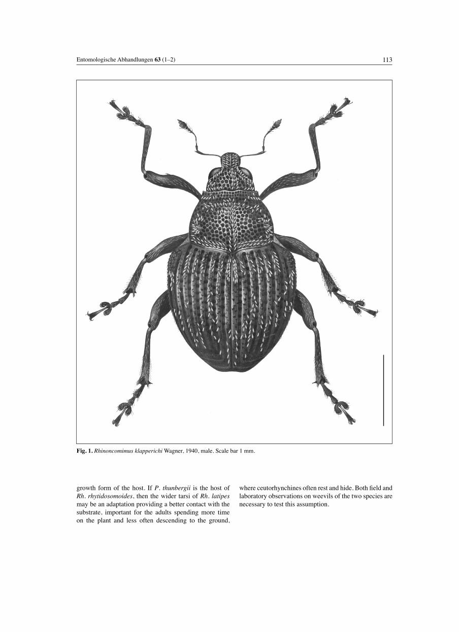
113Entomologische Abhandlungen 63 (1–2)
growth form of the host. If P. thunbergii is the host of Rh. rhytidosomoides, then the wider tarsi of Rh. latipesmay be an adaptation providing a better contact with the substrate, important for the adults spending more time on the plant and less often descending to the ground,
where ceutorhynchines often rest and hide. Both fi eld and laboratory observations on weevils of the two species are necessary to test this assumption.
Fig. 1. Rhinoncomimus klapperichi Wagner, 1940, male. Scale bar 1 mm.

114 KOROTYAEV: Review of Rhinoncomimus
Figs. 2–7. Rhinoncomimus Wagner, 1940, head of male (2–4, 7) and female (5, 6) dorsally. 2: Rh. klapperichi Wagner, 1940. 3: Rh. robustus Voss, 1957. 4: Rh. niger Chûjô & Morimoto, 1959. 5: Rh. rubripes sp.n. 6: Rh. rhytidosomoides (Wagner, 1944). 7: Rh. latipes Korotyaev, 1997. Scale bar 1 mm.
2 3 4
5 6 7
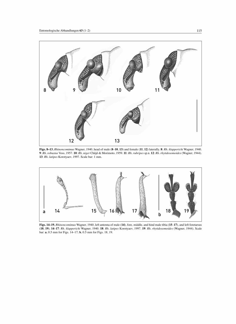
115Entomologische Abhandlungen 63 (1–2)
Figs. 8–13. Rhinoncomimus Wagner, 1940, head of male (8–10, 13) and female (11, 12) laterally. 8: Rh. klapperichi Wagner, 1940. 9: Rh. robustus Voss, 1957. 10: Rh. niger Chûjô & Morimoto, 1959. 11: Rh. rubripes sp.n. 12: Rh. rhytidosomoides (Wagner, 1944). 13: Rh. latipes Korotyaev, 1997. Scale bar: 1 mm.
Figs. 14–19. Rhinoncomimus Wagner, 1940, left antenna of male (14), fore, middle, and hind male tibia (15–17), and left foretarsus (18, 19). 14–17: Rh. klapperichi Wagner, 1940. 18: Rh. latipes Korotyaev, 1997. 19: Rh. rhytidosomoides (Wagner, 1944). Scale bar: a, 0.5 mm for Figs. 14–17; b, 0.5 mm for Figs. 18, 19.
8 9 10 11
12 13
191817161514ab
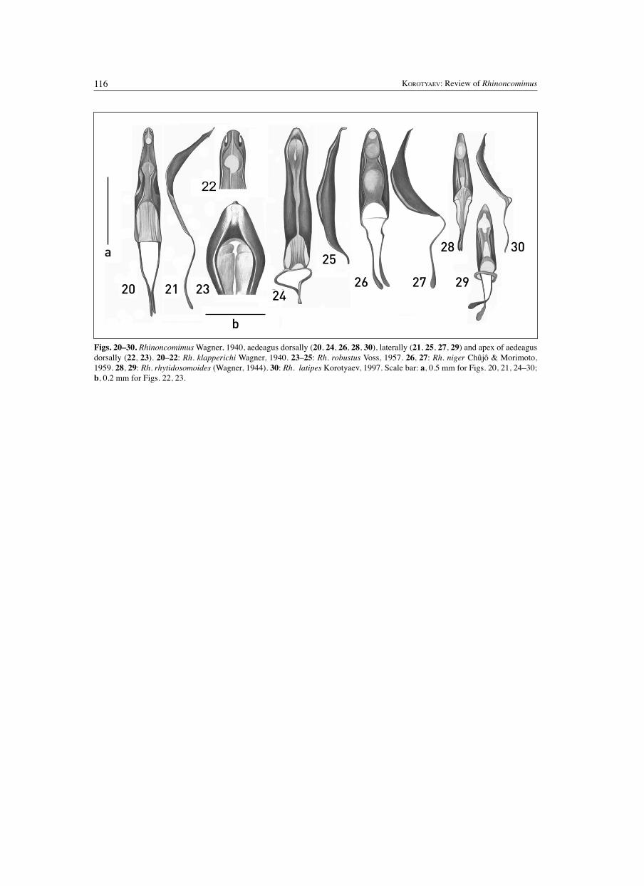
116 KOROTYAEV: Review of Rhinoncomimus
Figs. 20–30. Rhinoncomimus Wagner, 1940, aedeagus dorsally (20, 24, 26, 28, 30), laterally (21, 25, 27, 29) and apex of aedeagus dorsally (22, 23). 20–22: Rh. klapperichi Wagner, 1940. 23–25: Rh. robustus Voss, 1957. 26, 27: Rh. niger Chûjô & Morimoto, 1959. 28, 29: Rh. rhytidosomoides (Wagner, 1944). 30: Rh. latipes Korotyaev, 1997. Scale bar: a, 0.5 mm for Figs. 20, 21, 24–30; b, 0.2 mm for Figs. 22, 23.
20 21 23 24
25
26 27 29
28 30a
b

117Entomologische Abhandlungen 63 (1–2)
Fig 31. Rhinoncomimus robustus Voss, 1957, male. Scale bar: 1 mm.
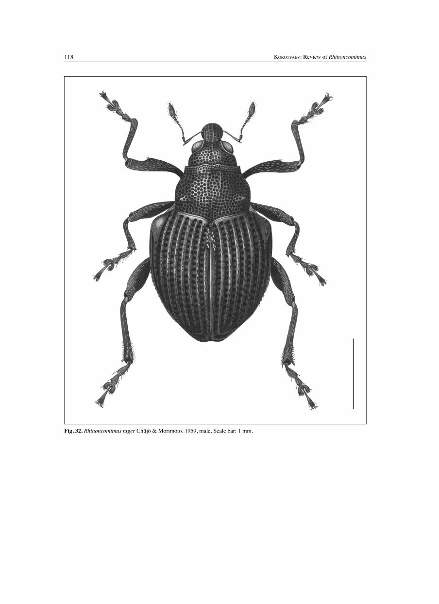
118 KOROTYAEV: Review of Rhinoncomimus
Fig. 32. Rhinoncomimus niger Chûjô & Morimoto, 1959, male. Scale bar: 1 mm.
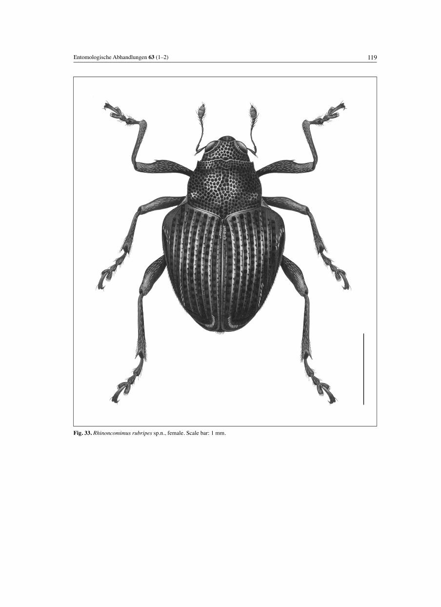
119Entomologische Abhandlungen 63 (1–2)
Fig. 33. Rhinoncomimus rubripes sp.n., female. Scale bar: 1 mm.

120 KOROTYAEV: Review of Rhinoncomimus
Fig. 34. Rhinoncomimus rhytidosomoides (Wagner, 1944), female. Scale bar: 1 mm.
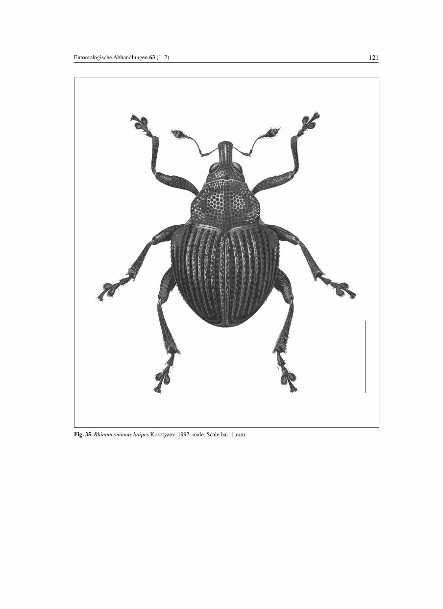
121Entomologische Abhandlungen 63 (1–2)
Fig. 35. Rhinoncomimus latipes Korotyaev, 1997, male. Scale bar: 1 mm.
122 KOROTYAEV: Review of Rhinoncomimus
References
ALONSO-ZARAZAGA, M.A. & C.H.C. LYAL 1999. A world catalogue of families and genera of Curculionoidea (Insecta: Coleoptera) (excepting Scolytidae and Platypodidae). – Entomopraxis, Barcelona. 315 pp.
CHÛJÔ, M. & K. MORIMOTO 1959. Curculionid-beetles of Niigata Prefecture collected by Dr K. Baba (2nd report). – Kontyu 27: 146–155.
COLONNELLI, E. 1986. Checklist of Phytobiini of the world, with a key to genera and description of a new species from South Africa (Coleoptera, Curculionidae, Ceutorhynchinae). – Fragmenta entomologica 19(1): 155–168.
COLONNELLI, E. 2004. Catalogue of Ceutorhynchinae of the world with a key to genera. – Argania, Barcelona. 124 pp.
CROWSON, R.A. 1981. The biology of the Coleoptera. – Academic Press, London, New York, etc. 802 pp.
EMELJANOV, A.F. 1967. Some characteristics of the distribution of oligophagous insects over host plants. – Doklady na XIX ezhegodnykh chteniyakh pamyati N.A. Kholodkovskogo 1 aprelya 1966 [Reports on the XIX annual readings in memory of N.A. Kholodkovskii, April 1, 1966]. Nauka, Leningrad: 28–65 [in Russian].
HONG, K.J., A.B. EGOROV & B.A. KOROTYAEV 2000. Illustrated catalogue of Curculionidae of Korea (Coleoptera). Insects of Korea, series 5. – Korea Research Institute of Bioscience and Biotechnology & Center for Insect Systematics, Korea. 337 pp.
KOROTYAEV, B.A. 1980. Material to the knowledge of Ceuto-rhynchinae (Coleoptera, Curculionidae) of Mongolia and the USSR. – Insects of Mongolia 7: 107–282. [in Russian]
KOROTYAEV, B.A. 1992. On trophic specialization of Palaearc-tic weevils of the subfamily Ceutorhynchinae (Coleoptera, Curculionidae). – Proceedings of the 4th ECE / 13th SIEEC, G d ll , 1991, Budapest, 2: 510–512.
KOROTYAEV, B.A. 1996. Key to genera of the tribe Ceutorhyn-chini. As part of: A.B. EGOROV, V.V. ZHERICHIN & B.A. KOROTYAEV, Sem. Curculionidae – Dolgonosiki. Key to the insects of the Russian Far East, vol. 3, Coleoptera, pt. 3, Supplement. Dal’nauka, Vladivostok: 455–468. [in Russian]
LEE, C.Y. & K. MORIMOTO 1996. Larvae of the weevil family Curculionidae of Japan, Part 4. Ceutorhynchinae to Molytinae (Insecta: Coleoptera). – Journal of the Faculty of Agriculture, Kyushu University, 40(3/4): 307–331.
KOROTYAEV, B.A. 1997. New and little known species of wee-vils from East Asia (Coleoptera: Apionidae, Curculionidae). – Zoosystematica Rossica 5(2) (1996): 285–288.
KOROTYAEV, B.A. & K.J. HONG 2004. A revised list of the wee-vil subfamily Ceutorhynchinae (Coleoptera; Curculionidae) of the Korean fauna, with contribution to the knowledge of the fauna of neighbouring countries. – Journal of Asia-Pacifi c Entomology 7(2): 143–169.
KOROTYAEV, B.A., A.S. KONSTANTINOV & C.W. O’BRIEN 2000. A new genus of the Orobitidinae and discussion of its relationships (Coleoptera: Curculionidae). – Proceedings of the Entomological Society of Washington 102(4):929–956.
References
MORIMOTO, K. 1994. Curculionidae. Pp. 269–345 in: M. HAYOSHI, K. MORIMOTO & S. KIMOTO (eds.), The Coleoptera of Japan in Color, vol. IV. (3rd ed.). – Hoikusha Publishing Co., Ltd.
PAJNI, H.R. & S.K. KOHLI 1982. Taxonomic studies on Indian Ceutorhynchinae (Coleoptera: Curculionidae). Tribe Mecys-moderini. – Oriental Insects 16(3): 337–372.
RAZUMOVSKII, S.M. 1981. Zakonomernosti dinamiki biotsenozov (Regularities of the biocenoses dynamics). – Nauka, Moscow. 231 pp. [in Russian]
VOSS, E. 1958. Ein Beitrag zur Kenntnis der Curculionidae im Grenzgebiet der Orientalischen zur Paläarktischen Region (Col., Curc.). Die von J. Klapperich und Tschung Sen in der Provinz Fukien gesammelten Rüsselkäfer. – Decheniana Beihefte 5: 1–139.
WAGNER, H. 1940. Monographie der paläarktischen Ceutho -rhyn chinae (Curcul). (Fortsetzung). – Entomologische Blätter 36(3): 65–81.
WAGNER, H. 1944. Monographie der paläarktischen Ceutho-rhynchinae. 3. Gen. Homorosoma Friv. – Entomologische Blätter 40(5/6): 279–294.
ZHERIKHIN, V.V. & A.B. EGOROV 1990. Zhuki-dolgonosiki (Coleoptera, Curculionidae) Dal’nego Vostoka SSSR (obzor podsemeistv s opisaniyem novykh taksonov) (Weevils (Coleoptera, Curculionidae) of the Far East of the USSR: Review of subfamilies with description of new taxa). – Vladivostok. 163 pp. [in Russian]
ZHERIKHIN, V.V. & V.G. GRATSHEV 1995. A comparative study of the hind wing venation of the superfamily Curculionoidea, with phylogenetic implications. Pp. 634–777 in: J. PAKALUK and S.A. LIPI SKI (eds.), Biology, phylogeny, and classifi cation of Coleoptera. Papers celebrating the 80th birthday of Roy A. Crowson. – Warszawa.
Acknowledgements
I greatly appreciate the invitation by Dr J. Hough-Goldstein to carry out this review and the fi nancial support from the USDA Forest Service, which enabled making elaborate illustrations by N.A. Florenskaya (St. Petersburg), whose effort and skills are equally acknowledged. I also thank Professor C.W. O’Brien (Tallahassee, Florida) for an exchange of specimens of Rhinoncomimus niger, loan of other material from his collection, and encouraging this study. I am greatly indebted to the curators of the collections and museums and all colleagues who supplied material for this study, helped me during the visits to the relevant institutions, and provided their kind hospitality: Dr R.S. Anderson (CMN), Dr M. Baehr (Zoologische Staatssammlung, München), Dr J. Balciunas (USDA, Agricultural Research Service, Albany, California),
Acknowledgements
M.V.L. Barclay and Dr C.H.C. Lyal (NHML), L. Behne and Dr L. Zerche (DEI), Dr M. Carl (Türkenfeld, Germany), Dr R. Gerstmeier, Dr G. Scherer (Wolfratshausen, Germany), Dr K.J. Hong (Suwon, Republic of Korea), O. Jäger and Dr K.-D. Klass (MTD), Dr D. Kavanaugh (CAS), Dr A.S. Konstantinov (USNM), Dr P. Lindskog (NHRM), Dr O. Merkl (HNHM), Dr H. Perrin (MNHN), Dr A.S. Samuelson (Bishop Museum, Honolulu), and Professor M. Schmitt (ZFMK). Identifi cation of the host plant from Korea by Dr V.I. Dorofeyev (V.L. Komarov Botanical Institute, Russian Academy of Sciences, St. Petersburg) is highly appreciated. Last, but not the least, I thank the editor, Dr K.-D. Klass, and the reviewers for their thorough and patient work on the manuscript which has greatly improved it.