neoteny and the plesiomorphic condition of the plesiosaur
TRANSCRIPT
Marshall UniversityMarshall Digital Scholar
Biological Sciences Faculty Research Biological Sciences
2006
Neoteny and the Plesiomorphic Condition of thePlesiosaur BasicraniumF. Robin O’KeefeMarshall University, [email protected]
Follow this and additional works at: http://mds.marshall.edu/bio_sciences_faculty
Part of the Animal Sciences Commons, and the Ecology and Evolutionary Biology Commons
This Article is brought to you for free and open access by the Biological Sciences at Marshall Digital Scholar. It has been accepted for inclusion inBiological Sciences Faculty Research by an authorized administrator of Marshall Digital Scholar. For more information, please [email protected], [email protected].
Recommended CitationO'Keefe, F. R. 2006. Neoteny and the plesiomorphic condition of the plesiosaur basicranium. In M. T. Carrano, T. J. Gaudin, R. W.Blob & J. R. Wible, (Eds.), Amniote Paleobiology. (pp. 391-409). Chicago, IL: University of Chicago Press.

12 Neoteny and the PlesiomorphicCondition of the PlesiosaurBasicranium
F. Robin O’Keefe
IntroductionHistorically, the systematics of the Plesiosauria (Reptilia, Sauroptery-
gia) were based largely on postcranial characters (Persson, 1963; Brown,
1981). Several factors account for this bias: plesiosaur skulls tend to be del-
icate and are often crushed even when preserved, postcranial elements are
relatively common and cranial elements are not, lack of knowledge about
the relationships of stem-group sauropterygians, and lack of knowledge of
plesiosaur cranial anatomy itself. However, recent detailed examinations
of plesiosaur cranial anatomy have identified many characters of use in
plesiosaur systematics (Brown, 1993; Cruickshank, 1994; Storrs & Taylor,
1996; Storrs, 1997; Carpenter, 1997; Evans, 1999; O’Keefe, 2001, 2004),
and the systematics of the group have changed markedly in response
(Carpenter, 1997; O’Keefe, 2001, 2004). The work of Rieppel and others
has clarified the anatomy and relationships of stem-group sauropterygians
(Storrs, 1991; see Rieppel, 2000, for review). This work has laid the
anatomic and phylogenetic foundations for a better understanding of ple-
siosaur cranial anatomy.
The purpose of this paper is to describe the condition of the braincase
in stratigraphically early and morphologically primitive plesiosaurs. In-
formation on the braincase of plesiomorphic taxa is important because
it establishes the polarity of characters occurring in more derived ple-
siosaurs. This paper begins with a short review of braincase anatomy in
stem-group sauropterygians. Data on braincase morphology of the ple-
siomorphic plesiosaur genera Thalassiodracon and Eurycleidus are then
presented and interpreted via comparison with other plesiosaurs, stem-
group sauropterygians, and stem diapsids (Araeoscelis). Early diapsids
are relevant because plesiosaur skulls more closely resemble early diap-
sids than stem-group sauropterygians in several key areas. Plesiosaurs
display a broad trend of delayed and reduced ossification compared with
stem-group sauropterygians (Storrs, 1991). This trend is linked to the
acquisition of a truly pelagic lifestyle in the Plesiosauria (Romer, 1956).
391

As pointed out by Rieppel (2000, 113), plesiosaurs show several rever-
sals to plesiomorphic character states, such as the reappearance of pos-
terior interpterygoid vacuities. This paper advances the hypothesis that
reversal in this and several other cranial characters is a consequence
of heterochrony, specifically neoteny in the ossification of the pterygoid
(Gould, 1977).
The Braincase in Stem-Group SauropterygiansOne barrier to understanding the plesiosaur braincase is the condition
of the basicranium in stem-group sauropterygians. All “nothosaur”-
grade taxa (i.e., all Sauropterygia exclusive of clade Pistosauria; see
fig. 12.7) lack interpterygoid vacuities in the posterior palate; the ptery-
goids meet in an unbroken median suture running from the vomers ante-
riorly and caudad to the occipital condyle and therefore hide the basicra-
nium in ventral view (Rieppel, 1994). In plesiosaurs, the palate is quite
different. Posterior interpterygoid vacuities are present, as in early diap-
sids, and an anterior interpterygoid vacuity sometimes allows a view of
the relations of the anterior end of the parasphenoid (O’Keefe, 2001).
The braincase in plesiosaurs is an open and poorly ossified struc-
ture, and different patterns of ossification are taxonomically informative
(O’Keefe, 2001). The occiput is closed and plate-like in many nothosaur-
grade taxa, further obscuring the more anterior relationships of brain-
case elements; however, Rieppel used well-preserved material to produce
detailed reconstructions of the braincase in Simosaurus, Nothosaurus(Rieppel, 1994), and Cymatosaurus (Rieppel & Werneburg, 1998).
Simosaurus and Nothosaurus are nothosauroids possessing a closed oc-
ciput, while Cymatosaurus is a pistosauroid possessing an open occiput
very similar to that found in all plesiosaurs (Rieppel, 2000). The closed
occiput of nothosauroids is characterized by large and plate-like exoc-
cipitals and opisthotics with a clear suture between the two elements,
whereas more laterally the opisthotic possesses a long dorsolateral suture
with the descending occipital flange of the squamosal (Rieppel, 1994).
The posttemporal fossa in both taxa is reduced to a small foramen. Below
the ventral margin of the exoccipital and opisthotic, the cranioquadrate
passage lies partially open in Simosaurus, whereas in Nothosaurus the
pterygoid meets the opisthotic in a long transverse suture, closing off the
cranioquadrate passage.
The closed nature of the occiput leaves several identifiable foramina
for the passage of structures into and out of the head; most prominent is
the foramen magnum, on either side of which rest the pillar-like bodies
392 F. R. O’Keefe

of the exoccipitals. Just lateral to these pillars and still within the exoc-
cipitals are the jugular foramina. Lateral and slightly ventral to this posi-
tion are foramina in the pterygoid for the passage of the internal carotid.
Lastly, two foramina in the ventromedial aspect of each exoccipital pillar
allow passage for the hypoglossal nerve (Rieppel, 1994); plesiosaurs usu-
ally have two foramina in the same position for the hypoglossal nerve
(Hopson, 1979; Storrs & Taylor, 1996; Carpenter, 1997; Evans, 1999). Ev-
ans (1999) illustrates a single foramen in Muraenosaurus.In contrast to the closed occiput in nothosauroids, the occiput is open
in the pistosauroids Corosaurus and Cymatosaurus as well as in all pis-
tosaurians including plesiosaurs. The occiput in all plesiosaurs is broadly
open, possessing a large posttemporal fenestra bounded dorsally by
the squamosal arch and ventrally by the slender paroccipital process
(Williston, 1903, 26; Storrs & Taylor, 1996; Carpenter, 1997). The paroc-
cipital process is formed by the opisthotic. The exoccipital is reduced to a
column forming the boundary of the foramen magnum. The paroccipital
process articulates laterally with the median surface of the squamosal in
most plesiosaur taxa (O’Keefe, 2001). Below the paroccipital process is a
large cranioquadrate passage bordered by the quadrate, squamosal, ptery-
goid, basioccipital, and exoccipital /opisthotic (Storrs and Taylor, 1996).
Rieppel described the unusual course of the internal carotid from the
occiput to the brain in both Nothosaurus (Rieppel, 1994) and Cymato-saurus (Rieppel & Werneburg, 1998). In both taxa, the internal carotid
travels anteriorly from the occiput within a prominent groove in the dor-
sal surface of the quadrate ramus of the pterygoid until reaching the ba-
sisphenoid and then courses ventrally between the lateral aspect of the
basisphenoid and prootic before dividing into the palatine artery and
the cerebral carotid (Rieppel, 1994). This course of the internal carotid—
characterized by a groove in the dorsal aspect of the pterygoid—is an
apomorphy of the Eusauropterygia, although the course of this vessel is
currently unknown in pachypleurosaurs and placodonts (Rieppel, 2000).
This character is reversed in most plesiosaurs (see below).
In nothosauroids, the cerebral carotid enters the base of the sella
turcica ventrolaterally through two prominent foramina separated by a
low ridge of bone (Hopson, 1979; Rieppel, 1994). This ridge of bone
runs anteriorly along the median dorsal aspect of the palate and is prob-
ably the root of a cartilaginous interorbital septum (Romer 1956, 57) ex-
tending back into the hypophyseal fossa and subdividing that structure,
as noted by Hopson (1979, 120). An identical condition exists in the
pistosauroid Cymatosaurus (Rieppel & Werneburg, 1998) and in the
393 Plesiosaur Basicranium
plesiosaur Thalassiodracon (see below). Last, the basal articulation be-
tween ossifications of the palatoquadrate cartilage and the lateral brain-
case wall is “obliterated” in all stem-group sauropterygians, yielding an
akinetic skull (Rieppel, 1994, 2000). The pterygoid encloses the basicra-
nium in these taxa, and a discreet basal articulation is obscured by the ex-
tensive contact between pterygoid and basicranium in general. In ple-
siosaurs, this character is also reversed; a discreet and well-developed
basal articulation is present in many taxa (O’Keefe, 2001), although it
does not always ossify in later pliosaurids.
The pattern of the semicircular canals is currently unknown in basal
sauropterygians. Because of the general reduction of ossification in the
endochondral elements of the plesiosaur braincase, however, the paths
of the three semicircular canals and the locations of the anterior and
posterior ampullae have been identified in several plesiosaur genera
(Peloneustes, Andrews, 1913; Cryptoclidus, Brown, 1981; Eurycleidus,Cruickshank, 1994; Libonectes and Dolichorhynchops, Carpenter, 1997;
Muraenosaurus, Evans, 1999). All these genera show a similar condition.
The posterior ampulla is located in the body of the fused exoccipital /
opisthotic, and this bone contains a deep fossa, open anteromedially, for
this structure. The horizontal semicircular canal leaves the posterior am-
pulla laterally and passes directly into the posterior edge of the prootic
through the suture between this bone and the exoccipital /opisthotic. The
anterior ampulla is located in the prootic, where it resides in a shallow,
medially open fossa. The anterior ampulla accepts the horizontal semi-
circular canal posteriorly, and the canal in the prootic for the anterior por-
tion of this canal is either open or roofed depending on the degree of
ossification of the prootic. The posterior vertical semicircular canal as-
cends through the body of the exoccipital and enters the supraoccipital,
where it communicates with the superior utriculus. The anterior vertical
semicircular canal leaves the superior utriculus and descends into the
prootic, passing through the suture between prootic and supraoccipital.
Again, the groove of the anterior vertical semicircular in the prootic can
be open or closed depending on the degree of ossification.
In summary, the internal ear in plesiosaurs is not derived and is similar
to the condition illustrated in Captorhinus by Price (1935). The posterior
ampulla resides in the fused exoccipital /opisthotic, and the suture between
these two bones is seldom apparent in plesiosaurs. However, Andrews
(1913) illustrated a specimen of Peloneustes that does preserve this suture.
On the medial wall of the foramen magnum, the suture between opisthotic
and exoccipital angles from anteroventral to posterodorsal; the jugular
394 F. R. O’Keefe
foramen is a gap within this suture as is expected given its developmental
origin within the metotic fissure (de Beer, 1937). The paroccipital process
is formed entirely of opisthotic, and the fossa for the posterior ampulla is
contained within the opisthotic, again like Captorhinus (Price, 1935). Be-
low I refer to the fused exoccipital /opisthotic as simply the “exoccipital”
except in cases in which the suture between the two elements is clear.
Materials and MethodsThis study utilizes material from the Permian diapsid Araeoscelis as
well as the Early Jurassic plesiosaur taxa Thalassiodracon, Eurycleidus,and Plesiopterys. Material examined is listed by taxon below.
Institutional abbreviations are as follows: FMNH, Field Museum of
Natural History, Chicago; OXFUM, Geological Collections, University
Museum, Oxford, UK; CAMSM, Sedgwick Museum, Cambridge, UK;
SMNS, Staatliches Museum für Naturkunde, Stuttgart, Germany.
Material
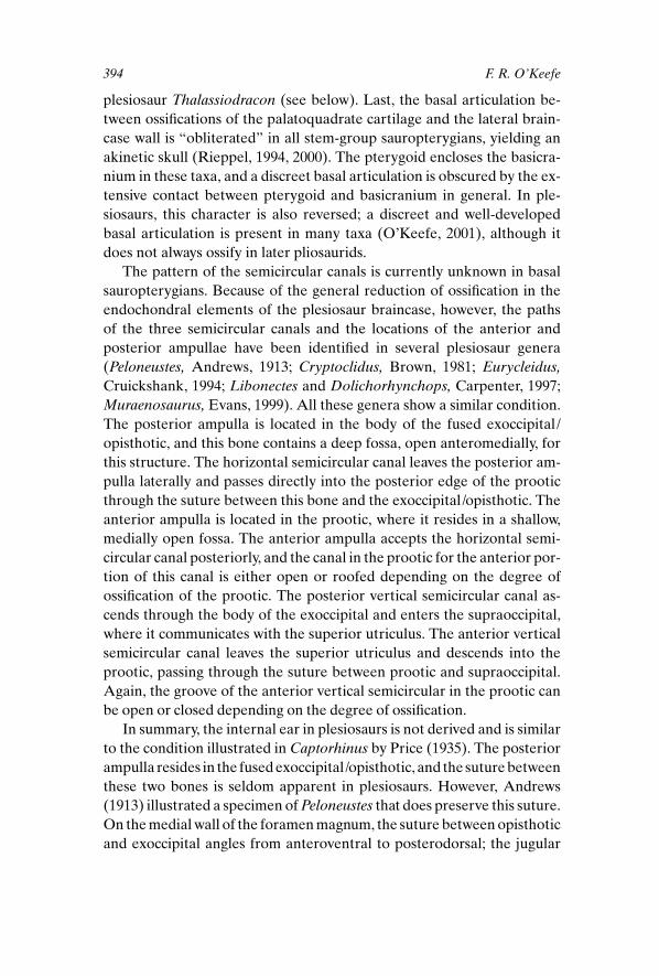
Araeoscelis casei. The partial skull figured here (fig. 12.1) is speci-
men number FMNH UR 2419. The skull is small and may represent a ju-
venile. At this time members of the genus Araeoscelis are assigned to two
395 Plesiosaur Basicranium
Figure 12.1. Basicranium of ajuvenile specimen of Araeosceliscasei, FMNH UR 2419.Abbreviations: art, articulation; ba,basal articulation; bot, basioccipitaltuber; bsf, basisphenoid fossa; icf, internal carotid foramen; oc,occipital condyle; ps, parasphenoid.

species based mainly on stratigraphic position, although the species are
very similar and the genus usually is treated as one form (Vaughn, 1955;
Reisz et al., 1984). Araeoscelis occurs in three localities in central Texas,
two of which are in the Wichita Group; the remaining locality is in the
Clear Forks Group (Lower Permian; see Reisz et al., 1984 for details). The
skull figured in this paper is poorly preserved except for the basicranium.
Thalassiodracon hawkinsi. This plesiosaur taxon is known from sev-
eral specimens originating from one locality, the lowermost Lias (Rhaet-
ian/Hettangian) of Street, Somerset, United Kingdom (see Storrs &
Taylor, 1996, for a thorough review of this taxon). This genus is one of the
stratigraphically earliest plesiosaurs for which adequate material is
known and is correspondingly plesiomorphic, sharing several symple-
siomorphies with pistosaurids that are lost in later plesiosaurs, including
low neural spine height, an angled humerus, presence of nasals, and the
presence of a posterior postorbital process (O’Keefe, 2001). The present
study deals with the skull figured and described in detail by Storrs and
Taylor (1996; CAMSM J.46986). Reference is also made to a skull in the
University Museum of Oxford, OXFUM J.10337.
Eurycleidus arcuatus. This taxon was redescribed by Cruickshank
(1994) following the extensive preparation of a historical specimen first
dealt with by Owen (1840). The specimen resides in the University
Museum of Oxford (OXFUM J.28585) and is a fragmentary but well-
preserved skull with accompanying postcranial material. The provenance
of the material is somewhat unclear, with the only sure information being
the source of the fossil, which is “Lower Lias, Lyme Regis.” Cruickshank
(1994) considered the most likely age to be Hettangian-Lower Sine-
murian, although the Pliensbachian is also possible.
Plesiopterys wildi. This taxon was named by O’Keefe (2004) and is a
morphologically primitive plesiosaur from the Posidonienschiefer near
Holzmaden, Germany. It is of Lower Toarcian age. The holotype of this
taxon is a complete skeleton with a crushed and disarticulated skull
(SMNS 16812); elements of the braincase are extremely well preserved
and are illustrated here. This material was originally referred to Eu-rycleidus by O’Keefe (2001), but further research proved this assignment
erroneous (see O’Keefe, 2004, for a discussion of the complex taxonomic
issues involved). The basicranium of this taxon is among the most primi-
tive of all known plesiosaurs and is therefore important for documenting
the condition from which later taxa are derived.
396 F. R. O’Keefe

DescriptionThe taxa Eurycleidus (sensu stricto) and Thalassiodracon are very sim-
ilar in most aspects of the braincase and are discussed together here;
Plesiopterys is discussed separately.
Palate and Context. In ventral view, the palate of Thalassiodraconis planar, lacking pterygoid flanges or other excrescences projecting
ventrally from the plane of the palate, and is similar to the palate in
nothosauroids. The palate is dominated by the pterygoids; these bones
have a long suture on the midline stretching from the vomers anteriorly
to the parasphenoid posteriorly (figs. 12.2, 12.3). This suture is broken by
a small anterior interpterygoid vacuity, but behind the vacuity the ptery-
goids meet again, obscuring the cultriform process of the parasphenoid.
This condition is again similar to the nothosauroid configuration and dif-
fers from the broadly open palate of Araeoscelis. (The genus Plesiosaurusis more derived compared with nothosauroids. In this genus, the palate is
397 Plesiosaur Basicranium
Figure 12.2. (A) Basicranium ofThalassiodracon hawkinsi,CAMSM J.46986. (B) Rightexoccipital-opisthotic of same inoblique medial view, modifiedfrom Storrs and Taylor (1996).Abbreviations as in figure 12.1,and XII, foramen for cranial nerveXII; avsc, anterior verticalsemicircular canal; hsc, horizontalsemicircular canal; jf, jugularforamen; pipv, posteriorinterpterygoid vacuity; ppr,paraoccipital process; pro art,prootic articulation; pt, pterygoid;soc art, supraoccipital articulation.

more open and similar to the condition in Araeoscelis; Storrs, 1997). Far-
ther posteriorly, the pterygoids separate and form the lateral borders of
the posterior interpterygoid vacuities, participate in a broad articulation
with the basioccipital tubera, and then continue laterally as the quadrate
flanges of the pterygoids (fig. 12.2). Just anterior to the posterior in-
terpterygoid vacuities, a triangular portion of the parasphenoid is ex-
posed on the palate surface, where it sutures with the pterygoids. The
posterior interpterygoid vacuities are large fenestrae affording an unob-
structed view of the basicranium similar to the condition in Araeoscelis.The pterygoids do not meet in a midline suture behind the posterior in-
terpterygoid vacuities in Thalassiodracon.
Basicranium. The basicrania of Thalassiodracon and Eurycleidus are
surprisingly plesiomorphic and are best understood by comparison with
the basicranium in Araeoscelis (fig. 12.1). In Araeoscelis (and Petrola-cosaurus; Reisz, 1981), the parasphenoid is a triangular structure with a
narrow cultriform process projecting anteriorly on the midline. Posteri-
orly, the parasphenoid broadens into a triangular sheet of bone covering
the basicranium, with the two lateral cristae ventrolaterales (Reisz, 1981)
398 F. R. O’Keefe
Figure 12.3. Skull ofThalassiodracon hawkinsi in dorsal view, OUM J. 10337.Abbreviations as in figure 12.2,and aipv, anterior interpterygoidvacuity; ccf, cerebral carotidforamen; exoc artic, exoccipitalarticulation; f, frontal; for,foramen; ios, interorbital septum;j, jugal; n, nasal; p, parietal; pm,premaxilla; po, postorbital; sq,squamosal; st, sella turcica. Skulllength 18 cm.
separated by a deep basisphenoid fossa on the midline. The parasphe-
noid has prominent articulations with the basioccipital tubera laterally
but does not reach to the margin of the occipital condyle on the midline.
Two small excavations in the posterior edge of the parasphenoid expose
a small portion of the body of the basioccipital near the midline, but the
suture between basioccipital and basisphenoid is covered by the paras-
phenoid. More anteriorly, the basipterygoid processes of the basisphe-
noid are visible on either side of the cultriform process of the parasphe-
noid. A groove runs around the anterior aspect of each basipterygoid
process and probably carried the internal carotid artery into the floor of
the hypophyseal fossa.
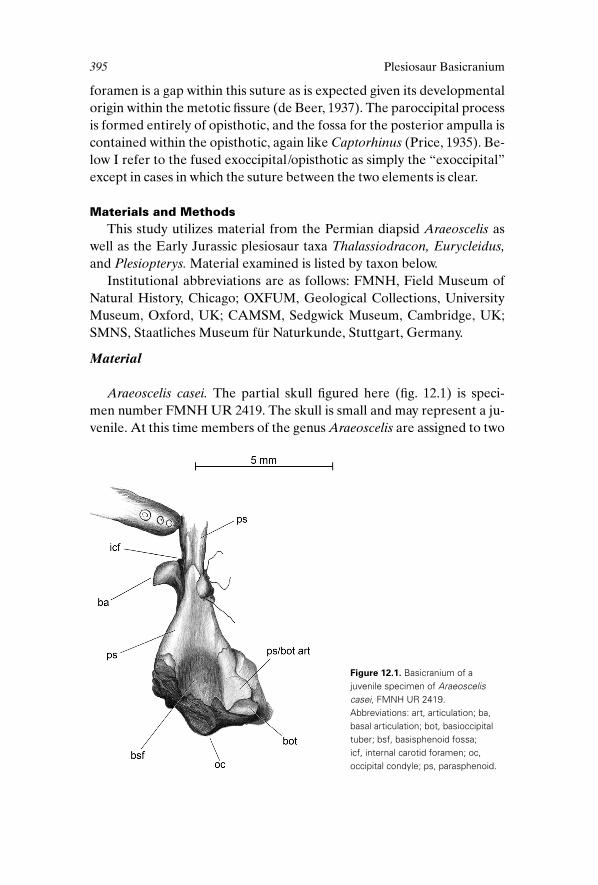
In Thalassiodracon and Eurycleidus, the parasphenoid is also triangu-
lar in outline (figs. 12.2, 12.4). The anterior cultriform process is obscured
by the midline suture of the pterygoids (but is present in Plesiosaurus;O’Keefe, 2001); posteriorly, the bone expands laterally into cristae ven-
trolaterales that articulate with the basioccipital tubera. The parasphe-
noid wraps around the basioccipital tubera and participates in the artic-
ulation with the pterygoid. The basisphenoid fossa is very shallow,
however, and the parasphenoid extends posteriorly for only a short dis-
tance on the midline, exposing the suture between basisphenoid and ba-
sioccipital. This suture is complex, with a slip of bone reaching posteri-
orly from the body of the basisphenoid and coursing ventral to the body
of the basioccipital. Dorsal to the parasphenoid, the body of the ba-
sisphenoid is visible, displaying a prominent basal articulation composed
of a shallow basipterygoid process and a deeper process of the pterygoid.
A large foramen for the internal carotid artery is present in the body of
the basisphenoid posterior to the basal articulation, in contrast to the lo-
cation of this structure in Araeoscelis.
399 Plesiosaur Basicranium
Figure 12.4. Braincase ofEurycleidus arcuatus, OXFUM J.28585. Abbreviations as in figures12.1–12.3, and frag, fragment.Fragment length 4.5 cm.

The lateral and dorsal aspects of the basicranium are illustrated best
in the isolated braincase of the Eurycleidus specimen (fig. 12.4). In lateral
view, the basioccipital tubera are prominent structures projecting ventro-
laterally from the body of the basioccipital, and their distal ends are
wrapped by the posterior margin of the parasphenoid. The articulations
for the exoccipitals are clearly delineated, rugose ovoids on the dorsal
surface of the basioccipital. The occipital condyle is rather poorly devel-
oped. It is a shallow dome with no groove between the condyle and the
body of the basioccipital (Thalassiodracon and Plesiosaurus display a
similar condition; O’Keefe, 2001). A notochordal pit is present in Eu-rycleidus and is often present in other plesiosaurs, although its presence
varies ontogenetically. The anterior face of the basioccipital carries a
deep fossa or notch; its position in the anterior edge of the basioccipital
presumably indicates it was overlain by the medulla oblongata (Romer,
1956), although there are no obvious soft tissue structures that might be
associated with this notch.
The basisphenoid does not articulate directly with the basioccipital in
either Eurycleidus (fig. 12.4) or Thalassiodracon (fig. 12.2). The only con-
tact between these two bones is formed by the ventroposterior process
of the basisphenoid exposed on the palate surface (described above);
hence there is a gap in dorsal view between the bodies of the basioccipi-
tal and the basisphenoid, floored by the ventrolateral basisphenoid pro-
cess on the midline and the dorsal surface of the parasphenoid laterally.
This condition contrasts that in nothosauroids, where the basisphenoid
and basioccipital bodies have a tight, broad connection (Rieppel, 1994).
Thalassiodracon and Eurycleidus also posses a deep notch in the back
of the clivus, a trait they share with the pistosauroid Cymatosaurus. Cy-matosaurus also might lack a tight connection between the bodies of the
basisphenoid and basioccipital, although the basioccipital is not pre-
served in the one skull of Cymatosaurus that visibly preserves this area
(Rieppel & Werneburg, 1998; see also Rieppel, 1997, 2000). If this char-
acter can be demonstrated, it will provide another link between the Ple-
siosauria and pistosauroids; the notch in the clivus mentioned above cer-
tainly does link these taxa.
The clivus terminates anteriorly in the dorsum sellae, a poorly devel-
oped structure in plesiosaurs consisting of the posterior wall of the sella
turcica, without an obvious raised ridge as seen in Captorhinus (Price,
1935). The foramina for the cerebral carotid arteries are not preserved in
Eurycleidus and probably were not ossified (they may be represented by
shallow grooves in the lateral edges of the body of the basisphenoid); the
foramina are large in Thalassiodracon (fig. 12.2). The cerebral carotids
400 F. R. O’Keefe
entered the floor of the hypophyseal fossa through paired posterolateral
foramina (fig. 12.3). The hypophyseal fossa itself is open laterally and an-
teriorly. A broad, low ridge extends forward from the back of the hy-
pophyseal fossa on the midline and seems to have been confluent with
the interorbital septum, similar to the condition seen in nothosauroids
(Rieppel, 1994) and Cymatosaurus (Rieppel & Werneburg, 1998). The
body of the basisphenoid lacks ossified clinoid processes in Eurycleidusand Thalassiodracon, although the elasmosaur Libonectes does have
stubby ossifications in this area (Carpenter, 1997). The basipterygoid pro-
cess is ossified but shallow in Thalassiodracon, whereas in Eurycleidus no
distinct process has ossified.
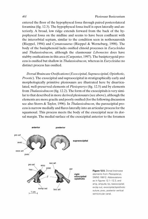
Dorsal Braincase Ossifications (Exoccipital, Supraoccipital, Opisthotic,Prootic). The exoccipital and supraoccipital in stratigraphically early and
morphologically primitive plesiosaurs are illustrated here by disarticu-
lated, well-preserved elements of Plesiopterys (fig. 12.5) and by elements
from Thalassiodracon (fig. 12.2). The form of the exoccipitals is very simi-
lar to that described in more derived plesiosaurs (see above), although the
elements are more gracile and poorly ossified (for the following discussion
see also Storrs & Taylor, 1996). In Thalassiodracon, the paroccipital pro-
cess is narrow medially and flares laterally into an articular process for the
squamosal. This process meets the body of the exoccipital near its dor-
sal margin. The medial surface of the exoccipital anterior to the foramen
401 Plesiosaur Basicranium
Figure 12.5. Dorsal braincaseelements from Plesiopterys,SMNS 16812. Abbreviations as in figures 12.1–12.3, and amp, ampulla; bo, basioccipital;ex /op sut, exoccipital /opisthoticsuture; pvsc, posterior verticalsemicircular canal.

magnum is pierced by a prominent jugular foramen, and passage of the hy-
poglossal nerve is restricted to a notch at the inferior margin of this bone.
The fossa for the posterior ampulla, generally open in plesiosaurs, seems
to be closed over except for a fissure above the jugular foramen in Thalas-siodracon. The suture between exoccipital and opisthotic ossifications is
not visible.
The exoccipital of Plesiopterys (fig. 12.5) is similar, except that the fossa
for the posterior ampulla is open. A foramen for the passage of the hori-
zontal semicircular canal is present on the articular surface for the prootic
in both Plesiopterys and Thalassiodracon, whereas in Plesiopterys a grove
for the posterior vertical semicircular canal enters into the articular sur-
face for the supraoccipital. Unlike Thalassiodracon and like Peloneustes(Andrews, 1913), the suture between the opisthotic and exoccipital
ossifications is visible in Plesiopterys, and this suture contains the jugular
foramen. The opisthotic is very similar to that in Peloneustes, forming the
paroccipital process as well as the anterodorsal portion of the “exoccipi-
tal pillar” lateral to the foramen magnum. Also like Peloneustes (and Cap-torhinus; Price, 1935), the opisthotic ossification contains a fossa for the
posterior ampulla and associated foramina for semicircular canals.
The supraoccipital of Plesiopterys is typical of those found in most
early Jurassic plesiosaurs (fig. 12.5; O’Keefe, 2001, for Plesiosaurus). The
bone is shallow anteroposteriorly, with a poorly developed articulation
for the parietals on its dorsal margin. Distinct articulations for the exoc-
cipitals and prootics are evident on the anterolateral edge. The prootic
articulation is perforated by a foramen for the anterior vertical semicir-
cular canal, and the exoccipital articulation carries a groove for the pos-
terior vertical semicircular canal. The superior edge of the foramen mag-
num is divided by a prominent, pointed process surmounted by a low
ridge on its posterior surface.
The prootic is a dish-shaped, quadrangular bone not exposed in the
Thalassiodracon material and poorly preserved in Plesiopterys (fig. 12.6).
In Plesiopterys, the prootics are crushed ventrally and anteriorly to their
articulations with the body of the basisphenoid; they now reside anterior
to the presumed position of the clinoid processes, and their articulations
are posterior to this position (fig. 12.6). Both prootics seem to be lying on
their medial faces, and little detailed morphology is visible. The ear re-
mains a poorly known region in plesiosaurs. Carpenter (1997) illustrates a
large fenestra ovalis bordered by the exoccipital column posteriorly, the
prootic anteriorly, and the basisphenoid ventrally in the elasmosaur Li-bonectes. Most plesiosaurs represented by adequate material do have this
402 F. R. O’Keefe

gap in the lateral braincase wall, although there is some doubt about its ho-
mology with the fenestra ovalis. Storrs and Taylor (1996) maintained that
the fenestra ovalis is absent in Thalassiodracon. The specimen discussed
by those authors and in this paper possesses a well-preserved stapes. Storrs
and Taylor believed that this element articulated tightly with the body of
the exoccipital; the present author could not determine whether the pres-
ent location of the stapes was in fact an articulation or an artifact of pres-
ervation. Considering the development of the otic capsule in squamates
(de Beer, 1937), the fenestra ovalis should pierce the lateral wall of the otic
capsule in the embryo and persist between the two ossifications derived
from the otic capsule in adults (prootic and opisthotic). The opisthotic is
reduced in plesiosaurs and has no suture with the prootic; however, the po-
sition of the gap in the lateral braincase wall behind the prootic and in
front of the opisthotic portion of the exoccipital pillar in both Plesiopterysand in Peloneustes is consistent with the position of the fenestra ovalis. The
gap in the lateral braincase wall of plesiosaurs is probably too large and ir-
regular to represent only the fenestra ovalis; the center of the otic capsule
may have failed to ossify completely in plesiosaurs and this may account
for some of the difficulty in assigning the relations of the footplate of the
stapes.
DiscussionIn general, the braincase in plesiosaurs is quite similar to those of more
basal diapsids such as Araeoscelis and Petrolacosaurus and is more similar
to those taxa than to the nothosaur-grade sauropterygians from which
403 Plesiosaur Basicranium
Figure 12.6. Dorsal view ofbasicranium from Plesiopterys,SMNS 16812. Abbreviations as infigures 12.1–12.3, and ds, dorsumsellae; ect, ectopterygoid; qfpt,quadrate flange of the pterygoid.
plesiosaurs arose. The possibility exists that plesiosaurs arose indepen-
dently from basal diapsids; however, there is excellent anatomic evidence
supporting the derivation of plesiosaurs from nothosaur-grade saurop-
terygians, and several parsimony analyses have yielded strong support for
this view (Storrs, 1991; O’Keefe, 2001; see above). Symplesiomorphy is
therefore a poor explanation for the many characters that show reversals
to more primitive states from the position of Nothosauroidea up to ple-
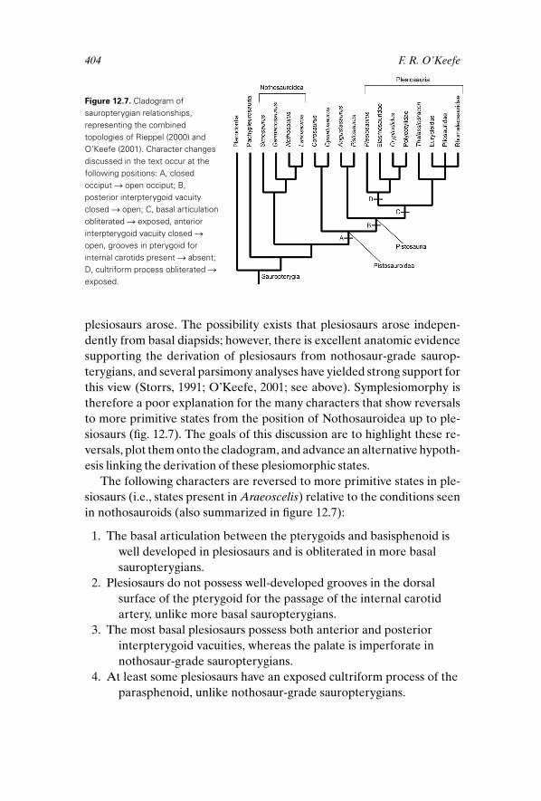
siosaurs (fig. 12.7). The goals of this discussion are to highlight these re-
versals, plot them onto the cladogram, and advance an alternative hypoth-
esis linking the derivation of these plesiomorphic states.
The following characters are reversed to more primitive states in ple-
siosaurs (i.e., states present in Araeoscelis) relative to the conditions seen
in nothosauroids (also summarized in figure 12.7):
1. The basal articulation between the pterygoids and basisphenoid is
well developed in plesiosaurs and is obliterated in more basal
sauropterygians.
2. Plesiosaurs do not possess well-developed grooves in the dorsal
surface of the pterygoid for the passage of the internal carotid
artery, unlike more basal sauropterygians.
3. The most basal plesiosaurs possess both anterior and posterior
interpterygoid vacuities, whereas the palate is imperforate in
nothosaur-grade sauropterygians.
4. At least some plesiosaurs have an exposed cultriform process of the
parasphenoid, unlike nothosaur-grade sauropterygians.
404 F. R. O’Keefe
Figure 12.7. Cladogram ofsauropterygian relationships,representing the combinedtopologies of Rieppel (2000) andO’Keefe (2001). Character changesdiscussed in the text occur at thefollowing positions: A, closedocciput S open occiput; B,posterior interpterygoid vacuityclosed S open; C, basal articulationobliterated S exposed, anteriorinterpterygoid vacuity closed Sopen, grooves in pterygoid forinternal carotids present S absent;D, cultriform process obliterated Sexposed.

5. The configuration of the basicranium in plesiosaurs is very similar
to that in Araeoscelis (i.e., presence of basisphenoid fossa, cristae
ventrolaterales, articulation of parasphenoid with basioccipital
tubera), whereas the configuration of this region is obscured by
the pterygoids in more basal sauropterygians.
6. Plesiosaurs and pistosauroids possess what Rieppel (2000) terms
an “open occiput,” meaning that the opisthotic is restricted to a
paroccipital process, leaving the cranioquadrate passage and the
posttemporal fenestra widely open. Most, but not all, nothosaur-
grade sauropterygians have a closed occiput.
Of the six changes listed above, the first five are associated with the
pterygoid, specifically with reduction in the extent of this bone. The sixth
change, opening of the occiput, concerns reduction of the pterygoid with
respect to the opening of the cranioquadrate passage but also concerns
reduction of the opisthotic as well as other bones surrounding the post-
temporal fenestra. Any alternative hypothesis meant to explain these six
character reversals therefore should account for reduction in the ptery-
goid bone.
Delayed and reduced ossification in all bones of the skeleton is a well-
known trend in aquatic tetrapods (Romer, 1956), and sauropterygians are
no exception. Plesiosaurs are thought to have been more pelagic than
more basal sauropterygians (Storrs, 1993) and show a general reduction
in ossification of the postcranial skeleton (Storrs, 1991). The reduction in
the pterygoid and other skull bones may be another manifestation of this
trend. The pterygoid is an intramembranous bone and as such is not
preformed in cartilage before ossification, as is the case in endochon-
dral elements (Romer, 1956). However, there is still an ontogeny to the
ossification of intramembranous bones; ossification begins at one or more
ossification centers and spreads from these locations. Knowledge about
the ontogeny of ossification of the pterygoid is available for Lacerta (Riep-
pel, 1992) and will be used as a model here.
The pterygoid is one of the first bones to ossify in the skull of Lacerta(and in Podarcis; Rieppel, 1987). Ossification begins in the middle por-
tion of the bone close to the processus ascendens of the palatoquadrate
cartilage, near the future basal articulation between basisphenoid pro-
cess, the processus ascendens ossified as the epipterygoid, and possibly
the pterygoid itself (Romer, 1956, 63– 64). The pterygoid then ossifies
quickly caudad to the quadrate portion of the palatoquadrate cartilage
and forms an anterior contact with the ossifying palatine (Rieppel, 1992).
405 Plesiosaur Basicranium

The major relations of the pterygoid therefore ossify very early in
Lacerta—namely, the contact with the basal articulation in the center of
the bone, with the quadrate via the quadrate flange and anterior and lat-
erally with the palate surface. These contacts are correspondingly ple-
siomorphic and present in a wide range of reptiles and are thought to be
the primitive conditions for amniotes (Romer, 1956).
Unfortunately, no information exists on skull ossification patterns in
nothosaur-grade sauropterygians. However, working from the supposi-
tion that ossification of the pterygoid proceeded in the same way in these
taxa as in other amniotes, the plesiomorphic contacts and relations of the
pterygoid would be expected to ossify first, followed later by the condi-
tions of the hypertrophied pterygoid that characterize nothosaur-grade
sauropterygians: the closing of anterior and posterior interpterygoid
vacuities and resulting obscuration of cultriform process and basicra-
nium, obliteration of the basal articulation, and closing of the cranio-
quadrate passage. The closing of the cranioquadrate passage requires the
presence of a foramen in the pterygoid for the passage of the internal
carotid artery (present in Simosaurus and Nothosaurus; Rieppel, 1994),
and the presence of bone between the occiput and basisphenoid requires
a groove for the passage of the same artery. All these features might be
expected to ossify later in ontogeny in accordance with von Baer’s first
and second laws of development (see discussion by Gould, 1977, 56). The
ossification of the pterygoid around the braincase in sauropterygians may
be compared with water filling a bathtub; the first water in the tub runs to
the drain, the lowest point in the tub and representing the plesiomorphic
contacts of the pterygoid in the analogy. As water continues to fill the tub,
fenestrae in the skull are closed and structures around the braincase are
enclosed: basal articulation, cultriform process, and internal carotid ar-
tery. A reduction of ossification in the plesiosaur pterygoid linked to an
aquatic lifestyle would simply reverse the above process. The more de-
rived conditions of the pterygoid would fail to ossify, leaving only the
more plesiomorphic conditions in the adult. The pterygoid of adult ple-
siosaurs therefore would be neotenous relative to more basal sauroptery-
gians (Gould, 1977).
The above interpretation is strongly supported by a phylogenetic
argument. The reversals in the characters discussed above are plotted
onto a cladogram of sauropterygian relationships in figure 12.7. If ple-
siosaurs had an independent derivation from basal diapsids, all these char-
acters should reverse on the branch directly below the Plesiosauria.
However, the changes are spread out over four branches, establishing a
pterygoid transformation series between plesiosaurs and nothosaur-grade
406 F. R. O’Keefe
sauropterygians. [Transformation series of this type also exist for several
aspects of the postcranial skeleton (reviewed by O’Keefe, 2001)]. The as-
signment of these reversals to symplesiomorphy rather than homoplasy is
therefore unlikely, and the conclusion that plesiosaurs are derived from
nothosaur-grade sauropterygians seems firm. There is no clear pattern in
the sequence of reversals and no ontogenetic series available for compar-
ison with the phylogeny.
The hypothesis that the basicranium in plesiosaurs reverts to a more
plesiomorphic condition due to neoteny in the ossification of the pterygoid
is testable in two ways. The first is the identification and study of ontoge-
netic series in stem-group sauropterygians in an attempt to gather data on
the ossification history of the pterygoid. Various pachypleurosaur taxa are
represented by good growth series (Rieppel, 1989; Sander, 1989; O’Keefe
et al., 1999), including at least one embryo. Given the early ossification of
the pterygoid in Lacerta, however, the available material may be too late in
ontogeny to offer much help. A second avenue of research would be to thin
section or computerized axial tomography (CAT) scan a well-preserved
nothosauroid skull. The presence and configuration of features such as the
basal articulation and parasphenoid might be discernible in this way. Last,
the six character state changes listed above can be accounted for by one
hypothesis—reduction in ossification of the pterygoid—and so might bear
consideration as a single character.
AcknowledgmentsThanks are due to R. Wild, R. Schoch, D. Norman, and P. Manning for
access to specimens in their care and institutional assistance of all kinds.
M. Maisch, O. Rieppel, and an anonymous reviewer read and improved
an earlier version of this manuscript. This study was funded in part by the
University of Chicago Hinds Fund. This paper is dedicated to Jim Hop-
son, teacher, mentor, and friend.
Literature Cited
Andrews, C. W. 1913. A Descriptive Catalogue of the Marine Reptiles of the Oxford Clay, Part II. London: British Museum (Natural History).
Brown, D. S. 1981. The English Upper Jurassic Plesiosauroidea (Reptilia) and a
review of the phylogeny and classification of the Plesiosauria. Bulletin of theBritish Museum of Natural History (Geology) 35(4):253–347.
Brown, D. S. 1993. A taxonomic reappraisal of the families Elasmosauridae and
Cryptoclididae (Reptilia: Plesiosauroidea). Revue de Paléobiologie, volumespéciale 7:9–16.
407 Plesiosaur Basicranium

Carpenter, K. 1997. Comparative cranial anatomy of two North American Cre-
taceous plesiosaurs; pp. 191–216 in J. M. Callaway and E. L. Nicholls (eds.),
Ancient Marine Reptiles. San Diego: Academic Press.
Cruickshank, A. R. I. 1994. A juvenile plesiosaur (Plesiosauria: Reptilia) from
the lower Lias (Hettangian: Lower Jurassic) of Lyme Regis, England: a
pliosauroid-plesiosauroid intermediate? Zoological Journal of the LinneanSociety 112:151–178.
de Beer, G. R. 1937. The Development of the Vertebrate Skull. Oxford: Oxford
University Press.
Evans, M. 1999. A new reconstruction of the skull of the Callovian elasmosaurid
plesiosaur Muraenosaurus leedsii Seeley. Mercian Geologist 14(4): 191–196.
Gould, S. J. 1977. Ontogeny and Phylogeny. Cambridge, MA: Harvard Univer-
sity Press.
Hopson, J. A. 1979. Paleoneurology; pp. 39–146 in C. Gans, R. G. Northcutt,
and P. Ulinski (eds.), Biology of the Reptilia. Vol. 9. Neurology. London:
Academic Press.
O’Keefe, F. R. 2001. A cladistic analysis and taxonomic revision of the Ple-
siosauria (Reptilia: Sauropterygia). Acta Zoologica Fennica 213:1– 63.
———. 2004. Preliminary description and phylogenetic position of a new
plesiosaur (Reptilia: Sauropterygia) from the Toarcian of Holzmaden,
Germany. Journal of Paleontology 78(5):973–988.
O’Keefe, F. R., O. Rieppel, and P. M. Sander. 1999. Shape disassociation and
inferred heterochrony in a clade of pachypleurosaurs (Reptilia, Sauroptery-
gia). Paleobiology 25(4):504 –517.
Owen, R. 1840. Report on British Fossil Reptiles, Part 1. Report of the ninth
meeting of the British Association for the Advancement of Science, Bir-
mingham, 1839, London; pp. 42–126.
Persson, P. O. 1963. A revision of the classification of the Plesiosauria with a
synopsis of the stratigraphical and geological distribution of the group.
Lunds Universites Årsskrift N. F. Ard. 2, 59(1):1–57.
Price, L. I. 1935. Notes on the braincase of Captorhinus. Proceedings of the Boston Society of Natural History 40(7):377–386.
Reisz, R. R. 1981. A diapsid reptile from the Pennsylvanian of Kansas. SpecialPublication of the Museum of Natural History, University of Kansas 7:1–74.
Reisz, R. R., D. S. Berman, and D. Scott. 1984. The anatomy and relationships
of the lower Permian reptile Araeoscelis. Journal of Vertebrate Paleontology4(1):57– 67.
Rieppel, O. 1987. The development of the trigeminal jaw adductor musculature
and associated skull elements in the lizard Podarcis sicula (Rafinesque).
Journal of Zoology, London 212:131–150.
———. 1989. A new pachypleurosaur (Reptilia: Sauropterygia) from the
Middle Triassic of Monte San Giorgio, Switzerland. Philosophical Transac-tions of the Royal Society of London B 323:1–73.
408 F. R. O’Keefe

———. 1992. Studies on skeleton formation in reptiles, III. Patterns of ossifi-
cation in the skeleton of Lacerta vivipara Jacquin (Reptilia, Squamata).
Fieldiana (Zoology) new series 68:1–25.
———. 1994. The braincases of Simosaurus and Nothosaurus: monophyly of the
Nothosauridae (Reptilia: Sauropterygia). Journal of Vertebrate Paleontology14(1):9–23.
———. 1997. Revision of the sauropterygian reptile genus Cymatosaurus v.
Fritsch, 1894, and the relationships of Germanosaurus Nopcsa 1928, from
the Middle Triassic of Europe. Fieldiana (Geology), new series 36:1–38.
———. 2000. Sauropterygia 1. Encyclopedia of Paleoherpetology, part 12A.Munich: Verlag Dr. Friedrich, Pfeil.
Rieppel, O. and R. Werneburg. 1998. A new species of the sauropterygian Cy-matosaurus from the lower Muschelkalk of Thuringia, Germany. Palaeontol-ogy 41(4):575–589.
Romer, A. S. 1956. Osteology of the Reptiles. Chicago: University of Chicago
Press.
Sander, P. M. 1989. The pachypleurosaurids (Reptilia: Nothosauria) from the
Middle Triassic of Monte San Giorgio (Switzerland), with the description of
a new species. Philosophical Transactions of the Royal Society of London B325:561– 670.
Storrs, G. W. 1991. Anatomy and relationships of Corosaurus alcovensis (Diap-
sida: Sauropterygia) and the Triassic Alcova Limestone of Wyoming. Bulle-tin of the Peabody Museum of Natural History 44:1–151.
———. 1993. Function and phylogeny in sauropterygian (Diapsida) evolution.
American Journal of Science 293A:63–90.
———. 1997. Clarification of the genus Plesiosaurus; pp. 145–190 in J. M. Call-
away and E. L. Nicholls (eds.), Ancient Marine Reptiles. San Diego: Aca-
demic Press.
Storrs, G. W. and M. A. Taylor. 1996. Cranial anatomy of a new plesiosaur
genus from the lowermost Lias (Rhaetian/Hettangian) of Street, Somerset,
England. Journal of Vertebrate Paleontology 16(3):403– 420.
Vaughn, P. P. 1955. The Permian reptile Araeoscelis restudied. Bulletin of theMuseum of Comparative Zoology 113(5):305– 467.
Williston, S. W. 1903. North American plesiosaurs, Part 1. Field Columbian Museum Publication (Geology) 73(2):1–77.
409 Plesiosaur Basicranium