a laboratory class exploring oral biofilms and the
TRANSCRIPT

A Laboratory Class Exploring Oral Biofilms and the Contamination ofToothbrushes
Resource Type: Curriculum: Laboratory
Publication Date: 4/7/2004
AuthorsJoanna VerranSchool of Biology, Chemistry and Health ScienceManchester Metropolitan UniversityManchester,United KingdomEmail: [email protected]
Abstract
The activity was designed (for seniors) to demonstrate the diversity of the oral flora and to enable discovery of some of thedifficulties associated with sampling, cultivation, isolation, and identification of biofilms. By comparing the yield and natureof the microorganisms isolated from plaque and toothbrushes under specified conditions, conclusions can be drawn as to thecontamination of an inert fomite (the toothbrush) with a dynamic ecological entity (dental plaque). Dental plaque is anexcellent example of a rich diverse biofilm, and this activity provides the student with an opportunity to uncover thisbiological complexity. The exercise may be carried out as an individual or group activity. Time management, report writing,data analysis and presentation are also important aspects of the exercise.
Activity
Invitation for User Feedback. If you have used the activity and would like to provide feedback, please send an e-mail [email protected]. Feedback can include ideas which complement the activity and new approaches forimplementing the activity. Your comments will be added to the activity under a separate section labeled "Feedback."Comments may be edited.
INTRODUCTION
Learning Objectives.On completion of this activity, students will be able to (i) recognize microbial diversity, especially in the biofilms found inthe mouth, (ii) interpret data, (iii) evaluate the relationship between the oral ecology and fomite (toothbrush)contamination, and (iv) apply lab skills such as sampling, cultivation, isolation, and quantification of microorganisms.
Background.This laboratory class has been carried out in a United Kingdom university. It was described in poster format at the 2001American Society for Microbiology Conference for Undergraduate Educators in Orlando, Florida, and received considerableinterest from other delegates. Two of these delegates, Hilda Merchant ([email protected]) and Jackie Reynolds([email protected]), have modified the exercise for their students, as have two United Kingdom colleagues, Jackie Parry([email protected]) and Rachel Sammons ([email protected]). Some differences in safety issues, suppliers,student course and level have arisen and are noted. Hence the information provided initially is that used for the class asoriginally carried out in Manchester.
Students are in the final (third) year of their honors degree, usually in Biological Sciences or Microbiology (or CombinedHonors, or other named routes within the departmental program where microbiology forms a significant contribution;http://www.mmu.ac.uk). The laboratory class forms part of the practical component of the Environmental Microbiologycourse. This course is one of six taken as part of the final year program, which also includes a laboratory-based project.Lectures and tutorials are the other components of the Environmental Microbiology course. Of the 22 lectures, six areconcerned with biofilms.
These lectures provide some of the background to the lab class and are given by the author, who also organizes,coordinates, and runs the laboratory exercise and assesses the reports. The lab class is introduced in a lecture the weekbefore it is conducted. The instruction sheet (see Procedure - Student Version) is issued and the first week’s activities areoutlined. Students are reminded strongly not to clean their teeth on the morning of the class and to remember to bring theirtoothbrushes to the class (in a clean bag rather than loose in their briefcases!). In addition, one tutorial is concerned withthe analysis of the results provided by the class. The lab class takes place in a morning, with two lectures and a tutorial inthe afternoon.
During the second laboratory exercise, students complete a form summarizing all of their results. This is given to theinstructor who then provides photocopies to all students in that afternoon’s tutorial, so that each student has a full set ofresults for analysis on the day that the class is completed. The tutorial enables the discussion of key points, unusual orinteresting observations, questions and answers, etc., to help the students to produce their report, which is to be handed in2 to 3 weeks later. There is also frequent one-to-one discussion in my office as students try to analyze data, drawconclusions, and make suggestions for future work which might help clarify observations.
Many principles of microbiology may be illustrated during this class. The class forms part of a course where lectures describe
MicrobeLibrary http://archive.microbelibrary.org/edzine/details_print.asp?id=1437&lang=
1 of 6 3/14/2012 9:40 AM

the oral flora and associated diseases, the properties of biofilms, and issues of nonculturability.
PROCEDURE
Materials.(per group)
Sterile plastic toothpick (from a catering supplies outlet)Sterile plastic bijou bottle (for plaque collection)Sterile 25-ml universal bottle (for toothbrush head)Ringers saline solution, ¼ strength, 100 ml1 ml pipettes (20)Sterile 5-ml (bijou) bottles or test tubes (for dilution series) (12)Replacement (new) toothbrush and toothpaste
Culture media – agar plates (per group)
Columbia blood agar (20)Columbia blood agar with streptococcus supplement (8)Mannitol salt agar (4)Pseudomonas selective agar (4)MacConkey agar (4)Sabouraud's agar (4)Wilkins-Chalgren agar (10)Tryptone yeast cystine medium with sucrose and bacitracin (4)
See Recipes section.
Class requirements
Balance (weighing to 3 decimal places)Sonicating water bath (or vortex mixer)Facility for anaerobe cultivationVortex mixer (to remove microorganisms from toothbrush)Spreaders
Demonstrations (second session)
Electron microscope, with representative images to issue to studentsNomarski microscope, attached to camera to provide images of unstained plaque and to visualize motilenonculturable speciesLight microscope with camera attached, to enable students to photograph their preparationsEquipment for Gram staining and wet mount preparation.
Student Version.Handouts for students:
Schedule and Practical HandoutDecayed, Missing, and Filled Teeth recording chart (DMFT)Results SheetEvaluation SheetMarking Scheme
Instructor Version.This class extends over two sessions, each of 3 to 4 hours duration. The first exercise may be completed in 2 hours, thesecond in 3 hours. A follow-up session, where class results are discussed, can take place at the end of the second session.Preparation of media requires approximately one day of a technician's time (class size around 16). The practical classhandout as provided to students is attached (Appendix 1). These sheets are provided to students the week before the classas a reminder not to clean teeth and to bring brushes to class. In addition to practical instructions, the following informationis provided:
a map of the mouth, enabling determination of decayed, missing, and filled teeth (DMFT),an example calculation of plaque counts per milligram and brush counts per brush head,newspaper articles (not attached) and references, andTWO copies of a results summary sheet, one for rough work and one to hand in for pooled class results.
Interpretation of findingsIn an open-ended exploration such as this, student results will vary from class to class. Suggestions to assist with
MicrobeLibrary http://archive.microbelibrary.org/edzine/details_print.asp?id=1437&lang=
2 of 6 3/14/2012 9:40 AM

interpretation are provided below, along with some general trends which may be observed. This information generally is notprovided to the class in advance; instead the instructor can look at each group's results and discuss findings with them,initially as a group and later as a class. Another option is to open up discussion of the results to the class and allow them todraw conclusions on their class data as it relates to the findings of past semesters and information in the publishedliterature.
Colony counts and selective mediaDuring the second lab class, for ease of reading results, ask each group to lay out plates across the bench in order ofdilution and consider findings. Work with each group on the following activities.
Note and discard any plates showing no growth. (This provides a psychological boost in that there are immediately fewerplates to read.) Inspect plates for accuracy of dilution. Errors are immediately apparent. Counts are therefore not valid andcannot be pooled with the class results.
Count colonies and calculate the number of CFU’s per milligram plaque and per toothbrush for each culture medium. Notedifferences between the microbiology of the mouth and the brush. "Total counts" on Columbia blood agar and counts onstreptococcal selective agar tend to be the highest for both brush and plaque. Sometimes the streptococcal counts arehigher than total count because the numerically predominant streptococci are overgrown by other organisms on thenonselective medium. Counts on Wilkins-Chalgren tend to be high for plaque but low for the toothbrush. Black pigmentedanaerobes (associated with mature plaque, gum disease) tend to be absent from the brushes because they cannot survive inan aerobic environment, whereas oral streptococci can. Media which are used to cultivate environmental organisms (e.g.,pseudomonads) will tend to demonstrate growth only from the toothbrushes. Media which are used to cultivate commensalflora (e.g., staphylococci, Candida, and bile-tolerant organisms) will indicate growth from both sites (although notnecessarily the same microorganisms).
Students may encounter difficulty in calculating plaque counts per milligram. An example calculation sheet is provided.Occasionally students obtain "virtual" plaque, that is an unweighable amount. Even in this scenario, plenty of counts areusually obtained, but they cannot be compared with counts obtained by other groups without the "per milligram"standardization; although the types of organisms and their presence or absence may still be noted.
Counts on brushes will vary considerably across student groups. Students often do not recognize that the organisms aremerely surviving on the brush; growth would be minimal. Thus contamination is inevitably varied, depending on day-to-daycontamination, usage, and storage. Repeated sampling of toothbrushes from the same individual might demonstrate more ofa pattern, for example carriage of mutans streptococci, or Candida spp.
Some brushes are apparently sterile (no growth on any plates); some are contaminated with one organism which has grownon a number of plates (e.g., pseudomonads will grow on several of the selective media, as will Candida spp.). Explain thatselective media are not as "selective" as first thought. Examination of colony morphology (see Appendix 3) and Gramstaining can help clarify these observations.
There will be some bizarre colony morphologies, for example the "molar tooth" colonies of actinomycetes. Attention mightneed to be drawn to the many, many very small streptococcal colonies and to the tiny, inhibited colonies present on severalof the selective media.
An interesting colony morphology is that of Streptococcus mutans on the tryptone yeast cystine medium with sucrose andbacitracin plates. Streptococci produce extracellular polysaccharides from sucrose, and some colonies will be EXTREMELYgummy (the material may seep out of the agar plate when inverted). However, S. mutans produces an insolublepolysaccharide; colonies are small, almost crystalline, and very hard. These colonies are difficult to dig out of the agar butmake a pleasant rattling sound when a loop is run over them.
Data presentationCollect individual data pool class results. Provide all students with results from every group to enable observation of generaltrends. Review key findings with the class and suggest ways of presenting results. A frequency distribution chart (i.e., thenumber of samples showing growth for a given medium for plaque and brush) summarizes results succinctly.
The student instruction sheet also suggests that students attempt to find correlations between certain variables and drawconclusions from these. Students occasionally try to relate high counts on brushes to low counts in plaque (i.e., that thebrush has removed the plaque). This is not valid.
Research has been carried out in our laboratories on the contamination of brushes and the effect of brush design, nature ofthe brush material, use of antimicrobial toothpastes, etc. (see references). These controlled studies, using large numbers ofsubjects and brushes, indicated that despite the enormous variability in counts observed between brushes (between 0 and
108 CFU per brush), there were some elements which correlated. For example, brush counts correlated with the amount oftime elapsed since the teeth were last cleaned and with the use of antimicrobial toothpaste.
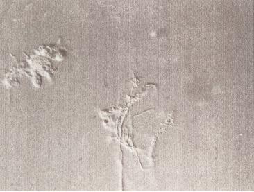
MicroscopyMicroscopy provides additional information. Spirochetes and their corkscrew motility are always very exciting to watch underNomarski illumination; they will not grow on routine culture media. Epithelial cells may be seen, occasionally harboringattached microorganisms. Clusters of microorganisms (microcolonies) of the same size and shape (if cocci, probablystreptococci; if rods, several possibilities) may be related to microcolonies and biofilm formation. Coaggregation of differentsizes and shapes of microorganisms is also observed; the corncob formation, cocci attached to a rod, may occasionally beapparent. The Gram stain, Nomarski examination, and the electron microscopy of plaque are very different from the Gramstain of the colonies derived from plaque after culture (see Appendix 3 for selected images).
BiofilmsFindings from this exercise may be used to illustrate fundamental principles of biofilms. Biofilms are accumulations ofmicroorganisms on surfaces, encased in a matrix of extracellular polymers of microbial origin. Dental plaque is one of the
MicrobeLibrary http://archive.microbelibrary.org/edzine/details_print.asp?id=1437&lang=
3 of 6 3/14/2012 9:40 AM

most well explored biofilms. Plaque formation begins when a salivary pellicle immediately coats newly cleaned teeth.Pioneer organisms, such as streptococci, attach to and colonize the surfaces, forming microcolonies. Secondary colonizersattach to the primary colonizers, and a complex biofilm results, where colonies of microorganisms grow vertically from thetooth surface, producing a columnar appearance typical of biofilms. As plaque matures, the microbial population becomesmore gram negative and anaerobic.
The isolation of a wide range of microorganisms, including obligate anaerobes, demonstrates the complexity of plaque.Microscopic examination of plaque reveals the microcolonies and coaggregates which are typical of biofilms. The productionof extracellular polysaccharide is clearly demonstrated on the sucrose-containing medium. Problems associated with thestudy of biofilms include the inability to culture all organisms present and the fact that sampling destroys the biofilmmorphology. The interactions occurring between microorganisms in biofilms—physical, metabolic, and via signalingmolecules—are best studied in situ.
Safety Issues. The students sample their own plaque using the sterile toothpick during the exercise before any other experimental work hastaken place. An alternative to this involves students collecting plaque at home and bringing it to the laboratory. Whenexamining plaque using the electron microscopy, the specimen was already prepared by the technician, and no otherspecimens were analyzed. Hence, photographs of typical plaque are provided to the students. For the Nomarski microscopy,students again sample their own plaque. For this aspect of the work, fresh plaque is essential because the spirochetes, withcharacteristic and impressive motility, are anaerobic and die very quickly.
If there is concern about using specimens taken from individuals, then the class could be modified to address only thetoothbrush contamination. However, there is always the possibility that pathogens will be present on the brushes.Anaerobes will not survive for long once removed from the anaerobic environment. If there is concern about students workingwith potentially pathogenic colonies, then plates could be sealed and the colonies pointed out by the instructor.
Control of substances hazardous to health analysis is performed prior to any laboratory exercise. Normal aseptic techniqueand appropriate handling of hazard group 2 microorganisms should be observed (1). Ethical approval should be sought fromthe appropriate body.
Microorganisms are removed from toothbrushes by immersing the brush in 10 ml of dilutent in a test tube which is vortexmixed. Attached microorganisms are removed into the suspending medium, which then is diluted and processed. Analternative, more destructive, method has also been used but is more hazardous: toothbrush heads are removed using anelectrically heated wire held between a Y-shaped piece of Perspex (produced in the University workshop). One student holdsthe brush and the Y-holder; the other holds the container into which the head drops. There is always a smell of meltedplastic. Care needs to be taken using the wire, which is only heated when the student switches on the current. Alternativemeans for removing the head include using bolt cutters or an equivalent cutter. If only bristles are removed, for exampleusing scissors, fewer organisms will be isolated because cells accumulate at the base of the bristles in the toothbrush head.
ML Safety Statement regarding Environmental Isolates
The Curriculum Resources Committee recognizes that isolated organisms can be a powerful learning tool as well as apotential biological hazard. We strongly recommend that:
· Environmental enrichment laboratories should only be performed in classes in which students have been trained towork at a BSL2.
· Direct environmental samples (eg. soil, water) which are known to contain infectious organisms should be handledaccording to the biosafety level of that infectious agent.
· Cultures of enriched microorganisms, derived from environmental samples, should be handled using Biosafety Level2 precautions.
· Mixed, enriched or pure cultures of microorganisms from environmental samples with a significant probability ofcontaining infectious agents should be manipulated in a biosafety cabinet if available.
· Where possible, media used for the enrichment of environmental isolates should contain an appropriate anti-fungalagent.
· Instructors should be aware if they are teaching in regions with endemic fungi capable of causing systemicinfections, and should avoid environmental isolations.
ASSESSMENT and OUTCOMES
Suggestions for Assessment.Information for students as to what is required in the report is provided on their instruction sheet. A marking schedule hasrecently been introduced and is provided to students.
Common weaknesses in reports are omission of toothbrush information in the introduction, no statement of aims, scruffyhistograms, graphs and/or figures presenting every piece of information without consideration of points to be made, lack ofcomments and poor organization of information in results, and failure to recognize that counts per milligram plaque andcounts per brush are not comparable.
Indicators of a good report are relevant data presentation and statistical analysis where possible; recognition of limitationsof cross-sectional, uncontrolled study and suggestions for improvement; and recognition of key findings irrespective oflimitations.
Field Testing.The exercise has been carried out over almost a 10-year period. Several modifications have taken place, usually involving areduction in the students workload and the cost of the exercise (LOTS of agar plates are required. Technician preparationtime would be 1 day for a typical class).
Microbiological analysis of saliva was initially included. Saliva is easier to sample than plaque and easier to disperse in
MicrobeLibrary http://archive.microbelibrary.org/edzine/details_print.asp?id=1437&lang=
4 of 6 3/14/2012 9:40 AM
dilution series. However, since saliva is acknowledged to be more of a transport medium than a habitat, anaerobes wereless common and a biofilm was not present; therefore analysis of saliva was discarded. Interestingly, counts on MacConkeyagar from saliva were extremely high (predominantly lactose negative); indeed, occasionally high counts from plaque arealso noted. It is difficult to explain this in terms of general use of MacConkey agar in coliform work other than to emphasizethat selective media are not always 100% effective, particularly on samples for which they were not designed. Otherwisestudents might think they have a mouthful of Escherichia coli! Further analysis of colonies might indicate the presence ofstreptococci (lactose positive)and occasionally staphylococci (also lactose positive); the large number of lactose-negativecolonies might also warrant investigation.
The transition from individual reports to group reports has been successful. It had often been apparent from previousmarking that students were working together to analyze the data. By offering students the choice of writing an individual ora group report, issues of plagiarism were obviated. The median mark increased from 55% to 65%.
In general, reports are produced with some pride. The open-ended nature of the work allows good students to explore thearea in depth, while less able students can present data and comment on general findings. The report contributes 10% tothe Environmental Microbiology course assessment overall, with two other coursework elements contributing a further 20%,and a 3-hour written exam 70%.
Student Data.From the worksheet provided at the beginning of the exercise and the tutorial on analysis of results, students are aware ofwhat is required in the report. The effort put into some of the reports demonstrates clearly the ownership and value of thework as recognized by the students.
The exercise enables students to see relatively unusual groups of organisms, particularly the spirochetes moving and theblack pigmented anaerobes on their Wilkins-Chalgren plates. Some students have been able to relate findings to their oralhealth status and have sought dental attention as a result, although I am not sure if this is necessarily a good outcome.
SUPPLEMENTARY MATERIALS
Possible Modifications.The study of toothbrush contamination provides a good exercise for final year student projects: retention of microorganismson brushes and their survival under different storage conditions, relation to brush head size and brush design, effect ofproprietary brush cleaning and disinfection products, commensal carriage in mouth and on brush, more in-depth investigationof findings of cross-sectional study, more controlled longitudinal studies, scanning electron microscopy of used toothbrushbristles, etc. (JV, JP, RS). The work can also be simplified by using fewer media. Inhibition of isolates by oral care productsis easily demonstrated.
References.
Advisory Committee on Dangerous Pathogens. 1995. Categorisation of biological agents according to hazard andcategories of containment, 4th ed. Her Majesty's Stationery Office (HMSO), London, England.
1.
General informationMarsh, P. D., and M. V. Martin. 1999. Oral microbiology, 4th ed. Wright, Oxford, England.
There are thousands of scientific papers on plaque! A web search will usually turn up something of general interest abouttoothbrushes.
Selected references on toothbrush contamination:
Glass, R. T., and H. G. Jensen. 1988. More on the contaminated toothbrush: the viral story. Quintessence Int.19:713–716.
1.
Glass, R. T., and M. M. Lare. 1986. Toothbrush contamination: a potential health risk? Quintessence Int. 17:39–42.2.Glaze, P. M., and A. B. Wade. 1986. Toothbrush age and wear as it relates to plaque control. J. Clin. Periodontol.13:52–56.
3.
Kozai, K., T. Iwai, and Muirak. 1989. Residual contamination of toothbrushes by microorganisms. J. Dent. Child.May-June:201–204.
4.
Malmberg, E., D. Birkhed, G. Norvenius, J. G. Noren, and C. T. Dahlen. 1994. Microorganisms on toothbrushes atday care centers. Acta Odont. Scand. 52:93–98.
5.
Marsh, P. D., and M. V. Martin. 1999. Oral microbiology, 4th ed. Wright, Oxford, England.6.Muller, H. P., D. E. Lange, and R. F. Muller. 1989. Actinobacillus actinomycetemcomitans contamination oftoothbrushes from patients harbouring the organism. J. Clin. Periodontol. 16:388–390.
7.
Taji, S. S., and A. H. Rogers. 1998. The microbial contamination of toothbrushes. A pilot study. Aust. Dent. J.43:128–130.
8.
Verran, J., and A. Leahy-Gilmartin. 1996. Investigations into the microbial contamination of toothbrushes. Microbios85:231–238.
9.
Verran et al., and Hammond et al. 1997. Abstracts in J. Dent. Res. 74:437.10.Warren, D. P., M. C. Goldschmidt, M. B. Thompson, K. Adler-Storthz, and H. J. Keene. 2001. The effects oftoothpastes on the residual microbial contamination of toothbrushes. J. Am. Dent. Assoc. 132:1241–1245.
11.
Appendices and Answer Keys.
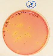
Appendix 1. Exploring Oral Biofilms: Schedule, Calculation sheet, DMFT, Results sheet, Evaluation sheet, and MarkingschemeAppendix 2. The Class in Action (images)Appendix 3. Colonies on selective media and microscopic images
i) Black pigmented colonies on Wilkins-Chalgren
MicrobeLibrary http://archive.microbelibrary.org/edzine/details_print.asp?id=1437&lang=
5 of 6 3/14/2012 9:40 AM
ii) Black pigmented, hemolytic colonies on Wilkins-Chalgreniii) Mannitol salt agariv) Pseudomonas agarv) MacConkey agarvi) Nomarski microscopy showing spirochetesvii) Nomarski microscopy showing microcolonies—rods, cocci, and epithelial cellsviii) Nomarski microscopy showing microcolonies: filamentous rods and cocciix) Nomarski microscopy showing corncobs, cocci attached to rodsx) Negative stain electron micrograph of range of plaque microorganismsxi) Gram stain of plaque showing gram-negative filamentous formsxii) Gram stain of plaque showing epithelial cell with attached gram-positive cells
Recipes.
(All plates were incubated at 37oC unless stated otherwise.)
Columbia blood agar (Oxoid). 5 to 6% sterile horse blood added. Incubated aerobically for 2 days (also anaerobicallyfor 3 days if desired; also in carbon dioxide—but few very obvious differences, dense population, and blood isexpensive).
1.
Streptococcus selective agar. Columbia blood agar, cooled to 50oC. 6% sterile horse blood. Colistin and oxolinic acid(COBA) supplement (Oxoid; code SR 126). Incubated in carbon dioxide environment for 2 to 3 days.
2.
Wilkins-Chalgren agar (Oxoid). Supplemented with 7% sterile horse blood and G-N supplement (Oxoid; code SR 108)for gram-negative anaerobes. Each G-N vial is sufficient for 500 ml and contains haemin (2.5 mg), menadione (0.25mg), sodium succinate (1,250 mg), nalidixic acid (5.0 mg), and vancomycin (1.25 mg). Plates were incubatedanaerobically for 7 days.
3.
MacConkey agar (Oxoid) incubated for 3 days.4.
Mannitol salt agar (Oxoid) incubated at 30oC for 2 days.5.Pseudomonas agar base (Oxoid) with 5 ml of glycerol per 500 ml medium added, plus cetrimide, fucidin, andcephaloridine (CFC) supplement (Oxoid; code SR 103).
6.
Sabouraud's dextrose agar (Oxoid) incubated for 2 days.7.Tryptone-yeast cystine medium (LabM or Difco) supplemented with sucrose and bacitracin. Sucrose 15%; bacitracin3.5 mg/L. Incubate for 3 days in carbon dioxide.
8.
MicrobeLibrary http://archive.microbelibrary.org/edzine/details_print.asp?id=1437&lang=
6 of 6 3/14/2012 9:40 AM

Student Handouts - Exploring Oral Biofilms
Overview - Oral MicrobiologyWeek 1Week 2Practical Report
Oral Microbiology
The oral microflora is very complex. Epithelial cells are continually being shed, thus they do notaccumulate a large microbial population. Saliva is essentially a transport medium containingmicroorganisms derived from other surfaces, primarily the tongue. Hard non-shedding surfaces such asteeth and dentures will accumulate a large mass of microorganisms, plaque, up to 1011 organisms per gram.The presence of plaque appears to be essential for the development of dental caries, periodontal diseases,and denture-induced stomatitis. Thus attempts are continually being made to characterise the plaquepopulation, the interactions between the organisms, their metabolic activity and pathogenic determinants,and to identify the aetiological agents of disease (to facilitate disease control).
The aim of this practical is primarily to demonstrate the diversity of the oral flora, and to note some of thedifficulties associated with sampling, cultivation, isolation and identification, and interpretation of results.We shall compare the yield and nature of the microorganisms isolated from plaque and toothbrush underspecified conditions.
Week 1
Cultivation of the oral flora
a) Plaque
Collect plaque carefully from all available surfaces using a sterile plastic (non-porous) toothpick.Weigh probe and plaque in a pre-weighed sterile 5 ml ( bijou) bottle.
Add 1.5 ml of Ringers solution (prepared for you) and sonicate for one minute to produce yourplaque suspension. Place bottle containing the plaque suspension and toothpick in a sonicatingwater bath. The vibrations dislodge the microorganisms from one another. A vortex mixer is alsoadequate. Remove clean toothpick, air dry and weigh. This will enable you to eventually calculatethe number of plaque microorganisms per mg plaque. Remove 0.2 of well-agitated plaque
suspension and dilute to 10-6 (0.2 : 1.8 ml).
Plate out (spread plate method) 0.1 ml, onto:
Columbia blood agar (CBA). This is an enriched medium, used to grow many of thefastidious oral microorganisms. This medium will give a ‘total count’. Incubate
aerobically, 37oC, 24 h. 10-2 - 10-6 in duplicate.
Streptococcus supplement agar (SS). This is selective for streptococci which form a
major part of the oral flora. CO2 incubation. 10-4, 10-5 in duplicate.
Mannitol salt agar (MSA), Pseudomonas agar, (Ps), MacConkeys agar (Mac),Saubourauds dextrose agar (Sab), selective for staphylococci, pseudomonads coliforms
Verran - Student Handouts http://archive.microbelibrary.org/edzine/Curriculum/Collection/VerranStu...
1 of 6 3/14/2012 9:41 AM

and yeast respectively Aerobic incubation (10-1) in duplicate.
Wilkins-Chalgren agar (WC) with anaerobe selective supplement for obligate anaerobes
(e.g. black pigmented Prevotella, Porphyromonas). Incubate 7 days, 10-2 - 10-5 induplicate.
Tryptone yeast cystine medium with sucrose and bacitracin (TSB) for S. mutans (10-1
in duplicate).
b) Cultivation of microorganisms from toothbrush
The toothbrush might be perceived as being an indirect indicator of the oral flora. However, the brush maybe exposed to contamination from other sources, and the brush environment might not be conducive to thesurvival of microorganisms.
Measure the length and width of the bristle mass on your brush to give an indication of splay (try not totouch the bristles).
Note make of toothbrush
Note the time since you last cleaned your teeth
Note the age of your brush
Note the condition of your brush
How far is your toilet from the sink?
How do you rinse your brush after use?
Do you use a mouthwash?
What toothpaste do you use?
How did you transport the brush?
Remove the head of your toothbrush using a hot wire. Allow the head to drop into a sterile 25 ml
(universal) bottle. Add 10 ml sterile Ringers, and sonicate. Dilute the resultant suspension to 10-5 and
spread onto the media used for plaque (you will then get counts per brush). CBA, 10-1-10-5; SS 10-2 -
10-3 ; MSA, Ps, WC, TSB, all 10-1. All plates in duplicate.
Total requirements
Plaque Brush Total
CBA 10 10 20
SS 4 4 8
MSA 2 2 4
Ps 2 2 4
MAC 2 2 4
Sab 2 2 4
Verran - Student Handouts http://archive.microbelibrary.org/edzine/Curriculum/Collection/VerranStu...
2 of 6 3/14/2012 9:41 AM

WC (Ano2) 8 2 10
TSB 2 2 4
Week 2
a) Microscopy of plaque
i) Electron microscopy
Sample micrographs will be provided.
This is useful for studying interactions between plaque microorganisms (for example by stainingextracellular material with ruthenium red); specific labeling (e.g. immunogold) can be used, or plaque insitu may be visualised by SEM techniques (e.g., corn cob formation).
ii) Dark ground illumination (Mickey Hoult)
Micro-organisms from the periodontal pocket or the gingival crevice are often impossible or extremelydifficult to cultivate. Spirochaetes are found in increasing numbers in gum disease in comparison tohealthy gums. Their slender forms (not Grammable) and characteristic corkscrew motility may bevisualised by dark ground illumination. Examine specimens of your plaque. Look for spirochaetes, andfusiform bacteria. Take pictures.
iii) Gram stain a smear of plaque, examine and photograph
iv) Gram stain a mass of colonies from aerobic and anaerobic plates (CBA, WC). Takepictures.
b) Cultivation of the oral flora
Calculate viable counts for plaque (per mg) and toothbrush (per brush) for all media.
Firstly, you need to check that your dilution series and duplicate plates have provided good results (i.e.,tenfold decrease in colony counts with dilution, and good agreement between duplicates). Plaque dilutionsmight not be too good if your plaque was particulate, because clumps of plaque are difficult to disperse.Select the dilution giving the highest countable number of colonies, and use this dilution for thecalculations given below.These calculations need to be done for each culture medium, and the appropriatedilution factor will vary between media.
In order to calculate plaque microorganisms per mg:
e.g. Average of 50 colonies on 10-3 dilution plates.
Therefore, 50 cells in 0.1 ml (plate inoculum) of 10-3
Therefore, 500 cells in 1 ml of 10-3
Therefore 500 x 103 (1000) cells in 1 ml of original plaque suspension (5.0 x 10⁵).
But the plaque suspension contained 1.5 ml.
Verran - Student Handouts http://archive.microbelibrary.org/edzine/Curriculum/Collection/VerranStu...
3 of 6 3/14/2012 9:41 AM

Therefore 5 x 10⁵ in 1 ml; plus 2.5 x 10⁵ in 0.5 ml, equals 7.5 x 10⁵ in 1.5 ml original suspension.
This 1.5 ml contained a known weight of plaque, for example 5 mg.
7.5 x 10⁵ cells in 5 mg plaque
Therefore 1.5 x 10⁵ cfu/mg plaque
In order to calculate microorganisms contaminating per brush:
e.g. Average of 30 colonies on 10-2 dilution
Therefore 30 cells in 0.1 ml of 10-2 dilution.
Therefore 300 cells in 1 ml of 10-2 dilution
Therefore 300 x 102 cells in 1 ml of brush suspension (3.0 x 10⁴)
But the brush suspension contained 10 ml, thus the total number extracted from the brush head is 3.0 x
104 x 10 = 3.0 x 10⁵.
c) Calculate your DMFT
The diagram below is a representation of the teeth in your mouth. To calculate your DMFT (decayed,missing and filled teeth), you need to examine your mouth and count the following. Filling in the teethchart might help you do this.
DT (Decayed teeth) = MT (Missing teeth) = FT (Filled teeth) = DMFT (total) =
d) Complete your results on the sheets provided
This will be copied so that you all have one another’s results.
Verran - Student Handouts http://archive.microbelibrary.org/edzine/Curriculum/Collection/VerranStu...
4 of 6 3/14/2012 9:41 AM

Practical report
The write-up of this practical is an intrinsic component of the unit and its assessment; the background isclearly complementary to lectures, so the write up provides an ideal opportunity to explore the literature.
Introduction. Couple of sides, relevant and succinct.1.
Aims. What is the aim of the investigation?2.
Methods. Insert schedule, but add any changes or comments.3.
Results. Present both raw data and manipulations of your results, and a summary of the classresults. Provide text indicating interesting results. Use statistical analyses where possible. Insertphotos and text indicating differences between appearances of organisms in plaque and from culturemedia (leave explanation of observations for the Discussion).
4.
How do plaque and brush counts and types of microorganisms relate to one another?
Are there any relationships between variables such as: 1) total brush count andtoothbrush wear (splay)? 2) total brush count and age? 3) DMFT and S. mutans plaquecount?
Discussion. Should be an explanation of points raised in the results section. Give suggestions forfuture work and explore the limitations of the work presented. Refer to background in introduction.
5.
What was the origin of the different microorganisms on the brushes?
Do any plaque microorganisms survive on the brush?
Does this pose a cross-infection hazard (e.g., S. mutans, C. albicans).
How was the dilution series?
Should we have had repeat experiments?
How variable are the data between individuals?
How good were the selective media at culturing the organisms required?
How different are the toothbrushes (age, use, etc.)?
How valid were the correlations investigated?
Have we got enough good data to enable interpretation of class results? If not, whynot?
How could we improve the work?
What are the limitations of the various microscopic, sampling, and cultivationtechniques used when attempting to describe the oral flora?
References. A really good report will require some extra reading.6.
Verran - Student Handouts http://archive.microbelibrary.org/edzine/Curriculum/Collection/VerranStu...
5 of 6 3/14/2012 9:41 AM

You can produce a group report or an individual report. Hand in date:
Verran - Student Handouts http://archive.microbelibrary.org/edzine/Curriculum/Collection/VerranStu...
6 of 6 3/14/2012 9:41 AM

DMFT Diagram
The diagram below is a representation of the teeth in your mouth. To calculate your DMFT (decayed, missingand filled teeth), you need to examine your mouth and count the following. Filling in the teeth chart mighthelp you do this.
DT (Decayed teeth) = MT (Missing teeth) = FT (Filled teeth) = DMFT (total) =
DMFT Diagram http://archive.microbelibrary.org/edzine/Curriculum/Collection/Verrandmf...
1 of 1 3/14/2012 10:05 AM

Plate Counts
Name:
Dilution
10-1 -2 -3 -4 -5 -6
Plaque
CBASSWC *PsMSAMacSabTSB ** Brush
CBASSWCPsMSAMacSabTSBViable Counts (after calculations)
CBA WC Ps MSA Mac Sab TSB SSPlaque
(per mg)
Brush
(per brush)
Toothbrush splay: % Normal storage procedure for brush:
Toothbrush condition: Transport of brush to lab:
Verran - Plate Counts http://archive.microbelibrary.org/edzine/Curriculum/Collection/VerranPla...
1 of 2 3/14/2012 9:42 AM

Toothbrush age: DMFT:
Make: Mouthwash?
Time since last cleaned teeth: Toothpaste?
Distance toilet from sink: Any comments:
Rinsing procedure for brush:
* Indicate % of black pigmented anaerobes for one dilution** Indicate % of presumptive S. mutans colonies for one dilution
back to activity
Verran - Plate Counts http://archive.microbelibrary.org/edzine/Curriculum/Collection/VerranPla...
2 of 2 3/14/2012 9:42 AM

Student Evaluation - Exploring Oral Biofilms
Evaluation of Oral Microbiology Practical (2003)
This class has been carried out for almost 10 years. It has been modified over time, and has also been usedin other Universities in the UK and US. We regularly review and alter our courses, and the next ‘newdegree’ intake will arrive in September. It seemed a good time to have a look at this practical. I havecertainly enjoyed running it, but I would like to know your views. Please complete this form as fully aspossible.
The aims of the practical are stated on the schedule.
Do you think these outcomes have been met?1.
If yes, to what extent.2.
If no, can you explain why, and indicate how the exercise might have been improved.3.
Is there anything particular you liked about the exercise?4.
Is there anything particular you disliked about the exercise?5.
Any other general comments about the practical?6.
Verran - Student Evaluation http://archive.microbelibrary.org/edzine/Curriculum/Collection/VerranEv...
1 of 1 3/14/2012 9:42 AM
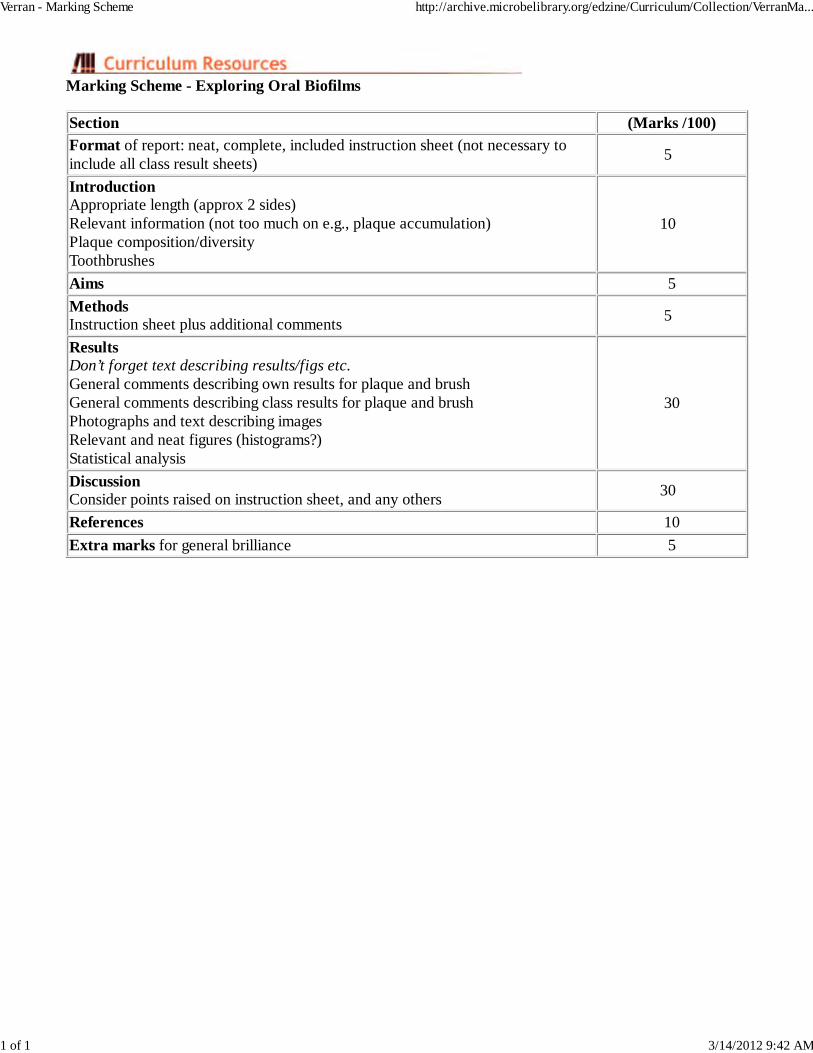
Marking Scheme - Exploring Oral Biofilms
Section (Marks /100)
Format of report: neat, complete, included instruction sheet (not necessary toinclude all class result sheets)
5
IntroductionAppropriate length (approx 2 sides) Relevant information (not too much on e.g., plaque accumulation) Plaque composition/diversity Toothbrushes
10
Aims 5
MethodsInstruction sheet plus additional comments
5
ResultsDon’t forget text describing results/figs etc. General comments describing own results for plaque and brush General comments describing class results for plaque and brush Photographs and text describing images Relevant and neat figures (histograms?) Statistical analysis
30
DiscussionConsider points raised on instruction sheet, and any others
30
References 10
Extra marks for general brilliance 5
Verran - Marking Scheme http://archive.microbelibrary.org/edzine/Curriculum/Collection/VerranMa...
1 of 1 3/14/2012 9:42 AM

Appendix 1: Exploring Oral Biofilms
Overview - Oral MicrobiologyWeek 1Week 2Practical ReportMarking SchemeClass Evaluation
Oral Microbiology
The oral microflora is very complex. Epithelial cells are continually being shed, thus they do notaccumulate a large microbial population. Saliva is essentially a transport medium containingmicroorganisms derived from other surfaces, primarily the tongue. Hard non-shedding surfaces such asteeth and dentures will accumulate a large mass of microorganisms, plaque, up to 1011 organisms per gram.The presence of plaque appears to be essential for the development of dental caries, periodontal diseases,and denture-induced stomatitis. Thus attempts are continually being made to characterise the plaquepopulation, the interactions between the organisms, their metabolic activity and pathogenic determinants,and to identify the aetiological agents of disease (to facilitate disease control).
The aim of this practical is primarily to demonstrate the diversity of the oral flora, and to note some of thedifficulties associated with sampling, cultivation, isolation and identification, and interpretation of results.We shall compare the yield and nature of the microorganisms isolated from plaque and toothbrush underspecified conditions.
Week 1
Cultivation of the oral flora
a) Plaque
Collect plaque carefully from all available surfaces using a sterile plastic (non-porous) toothpick.Weigh probe and plaque in a pre-weighed sterile 5 ml (bijou) bottle.
Add 1.5 ml of Ringers solution (prepared for you) and sonicate for one minute to produce yourplaque suspension. Place bottle containing the plaque suspension and toothpick in a sonicatingwater bath. The vibrations dislodge the microorganisms from one another. A vortex mixer is alsoadequate. Remove clean toothpick, air dry and weigh. This will enable you to eventually calculatethe number of plaque microorganisms per mg plaque. Remove 0.2 of well-agitated plaque
suspension and dilute to 10-6 (0.2:1.8 ml).
Plate out (spread plate method) 0.1 ml, onto:
Columbia blood agar (CBA). This is an enriched medium, used to grow many of thefastidious oral microorganisms. This medium will give a ‘total count’. Incubate
aerobically, 37oC, 24 h. 10-2 - 10-6 in duplicate.
Streptococcus supplement agar (SS). This is selective for streptococci which form a
major part of the oral flora. CO2 incubation. 10-4, 10-5 in duplicate.
Mannitol salt agar (MSA), Pseudomonas agar, (Ps), MacConkeys agar (Mac),
Verran - Appendix 1 http://archive.microbelibrary.org/edzine/Curriculum/Collection/VerranAp...
1 of 7 3/14/2012 9:42 AM

Saubourauds dextrose agar (Sab), selective for staphylococci, pseudomonads coliforms
and yeast respectively Aerobic incubation (10-1) in duplicate.
Wilkins-Chalgren agar (WC) with anaerobe selective supplement for obligate anaerobes
(e.g. black pigmented Prevotella, Porphyromonas). Incubate 7 days, 10-2 - 10-5 induplicate.
Tryptone yeast cystine medium with sucrose and bacitracin (TSB) for S. mutans (10-1
in duplicate).
b) Cultivation of microorganisms from toothbrush
The toothbrush might be perceived as being an indirect indicator of the oral flora. However, the brush maybe exposed to contamination from other sources, and the brush environment might not be conducive to thesurvival of microorganisms.
Measure the length and width of the bristle mass on your brush to give an indication of splay (try not totouch the bristles).
Note make of toothbrush
Note the time since you last cleaned your teeth
Note the age of your brush
Note the condition of your brush
How far is your toilet from the sink?
How do you rinse your brush after use?
Do you use a mouthwash?
What toothpaste do you use?
How did you transport the brush?
Remove the head of your toothbrush using a hot wire. Allow the head to drop into a sterile 25 ml
(universal) bottle. Add 10 ml sterile Ringers, and sonicate. Dilute the resultant suspension to 10-5 and
spread onto the media used for plaque (you will then get counts per brush). CBA, 10-1-10-5; SS 10-2 -
10-3 ; MSA, Ps, WC, TSB, all 10-1. All plates in duplicate.
Total requirements
Plaque Brush Total
CBA 10 10 20
SS 4 4 8
MSA 2 2 4
Ps 2 2 4
MAC 2 2 4
Sab 2 2 4
Verran - Appendix 1 http://archive.microbelibrary.org/edzine/Curriculum/Collection/VerranAp...
2 of 7 3/14/2012 9:42 AM

WC (Ano2) 8 2 10
TSB 2 2 4
Week 2
a) Microscopy of plaque
i) Electron microscopy
Sample micrographs will be provided.
This is useful for studying interactions between plaque microorganisms (for example by stainingextracellular material with ruthenium red); specific labeling (e.g. immunogold) can be used, or plaque insitu may be visualised by SEM techniques (e.g., corn cob formation).
ii) Dark ground illumination (Mickey Hoult)
Micro-organisms from the periodontal pocket or the gingival crevice are often impossible or extremelydifficult to cultivate. Spirochaetes are found in increasing numbers in gum disease in comparison tohealthy gums. Their slender forms (not Grammable) and characteristic corkscrew motility may bevisualised by dark ground illumination. Examine specimens of your plaque. Look for spirochaetes, andfusiform bacteria. Take pictures.
iii) Gram stain a smear of plaque, examine and photograph
iv) Gram stain a mass of colonies from aerobic and anaerobic plates (CBA, WC). Takepictures.
b) Cultivation of the oral flora
Calculate viable counts for plaque (per mg) and toothbrush (per brush) for all media.
Firstly, you need to check that your dilution series and duplicate plates have provided good results (i.e.,tenfold decrease in colony counts with dilution, and good agreement between duplicates). Plaque dilutionsmight not be too good if your plaque was particulate, because clumps of plaque are difficult to disperse.Select the dilution giving the highest countable number of colonies, and use this dilution for thecalculations given below. These calculations need to be done for each culture medium, and theappropriate dilution factor will vary between media.
In order to calculate plaque microorganisms per mg:
e.g. Average of 50 colonies on 10-3 dilution plates.
Therefore, 50 cells in 0.1 ml (plate inoculum) of 10-3
Therefore, 500 cells in 1 ml of 10-3
Therefore 500 x 103 (1000) cells in 1 ml of original plaque suspension (5.0 x 10⁵).
But the plaque suspension contained 1.5 ml.
Verran - Appendix 1 http://archive.microbelibrary.org/edzine/Curriculum/Collection/VerranAp...
3 of 7 3/14/2012 9:42 AM

Therefore 5 x 10⁵ in 1 ml; plus 2.5 x 10⁵ in 0.5 ml, equals 7.5 x 10⁵ in 1.5 ml original suspension.
This 1.5 ml contained a known weight of plaque, for example 5 mg.
7.5 x 10⁵ cells in 5 mg plaque
Therefore 1.5 x 10⁵ cfu/mg plaque
In order to calculate microorganisms contaminating per brush:
e.g. Average of 30 colonies on 10-2 dilution
Therefore 30 cells in 0.1 ml of 10-2 dilution.
Therefore 300 cells in 1 ml of 10-2 dilution
Therefore 300 x 102 cells in 1 ml of brush suspension (3.0 x 10⁴)
But the brush suspension contained 10 ml, thus the total number extracted from the brush head is 3.0 x
104 x 10 = 3.0 x 10⁵.
c) Calculate your DMFT
The diagram below is a representation of the teeth in your mouth. To calculate your DMFT (decayed,missing and filled teeth), you need to examine your mouth and count the following. Filling in the teethchart might help you do this.
DT (Decayed teeth) = MT (Missing teeth) = FT (Filled teeth) = DMFT (total) =
d) Complete your results on the sheets provided
This will be copied so that you all have one another’s results.
Verran - Appendix 1 http://archive.microbelibrary.org/edzine/Curriculum/Collection/VerranAp...
4 of 7 3/14/2012 9:42 AM

Practical report
The write-up of this practical is an intrinsic component of the unit and its assessment; the background isclearly complementary to lectures, so the write up provides an ideal opportunity to explore the literature.
Introduction. Couple of sides, relevant and succinct.1.
Aims. What is the aim of the investigation?2.
Methods. Insert schedule, but add any changes or comments.3.
Results. Present both raw data and manipulations of your results, and a summary of the classresults. Provide text indicating interesting results. Use statistical analyses where possible. Insertphotos and text indicating differences between appearances of organisms in plaque and from culturemedia (leave explanation of observations for the Discussion).
4.
How do plaque and brush counts and types of microorganisms relate to one another?
Are there any relationships between variables such as: 1) total brush count andtoothbrush wear (splay)? 2) total brush count and age? 3) DMFT and S. mutans plaquecount?
Discussion. Should be an explanation of points raised in the results section. Give suggestions forfuture work and explore the limitations of the work presented. Refer to background in introduction.
5.
What was the origin of the different microorganisms on the brushes?
Do any plaque microorganisms survive on the brush?
Does this pose a cross-infection hazard (e.g., S. mutans, C. albicans).
How was the dilution series?
Should we have had repeat experiments?
How variable are the data between individuals?
How good were the selective media at culturing the organisms required?
How different are the toothbrushes (age, use, etc.)?
How valid were the correlations investigated?
Have we got enough good data to enable interpretation of class results? If not, whynot?
How could we improve the work?
What are the limitations of the various microscopic, sampling, and cultivationtechniques used when attempting to describe the oral flora?
References. A really good report will require some extra reading.6.
Verran - Appendix 1 http://archive.microbelibrary.org/edzine/Curriculum/Collection/VerranAp...
5 of 7 3/14/2012 9:42 AM

You can produce a group report or an individual report. Hand in date:
Marking scheme
Section (Marks/100)
Format of report: neat, complete, included instruction sheet (not necessary toinclude all class result sheets)
5
IntroductionAppropriate length (approx 2 sides) Relevant information (not too much on e.g., plaque accumulation) Plaque composition/diversity Toothbrushes
10
Aims 5
MethodsInstruction sheet plus additional comments
5
ResultsDon’t forget text describing results/figs etc. General comments describing own results for plaque and brush General comments describing class results for plaque and brush Photographs and text describing images Relevant and neat figures (histograms?) Statistical analysis
30
DiscussionConsider points raised on instruction sheet, and any others
30
References 10
Extra marks for general brilliance 5
Evaluation of oral microbiology practical (2003)
This class has been carried out for almost 10 years. It has been modified over time, and has also been usedin other Universities in the UK and US. We regularly review and alter our courses, and the next ‘newdegree’ intake will arrive in September. It seemed a good time to have a look at this practical. I havecertainly enjoyed running it, but I would like to know your views. Please complete this form as fully aspossible.
The aims of the practical are stated on the schedule.
Do you think these outcomes have been met?1.
If yes, to what extent.2.
If no, can you explain why, and indicate how the exercise might have been improved.3.
Is there anything particular you liked about the exercise?4.
Is there anything particular you disliked about the exercise?5.
Any other general comments about the practical?6.
Verran - Appendix 1 http://archive.microbelibrary.org/edzine/Curriculum/Collection/VerranAp...
6 of 7 3/14/2012 9:42 AM

back to top
Verran - Appendix 1 http://archive.microbelibrary.org/edzine/Curriculum/Collection/VerranAp...
7 of 7 3/14/2012 9:42 AM